Review Diagnosis of inborn errors of metabolism using...
23
758 (2001) 3–25 Journal of Chromatography B, www.elsevier.com / locate / chromb Review Diagnosis of inborn errors of metabolism using filter paper urine, urease treatment, isotope dilution and gas chromatography–mass spectrometry * Tomiko Kuhara Division of Human Genetics, Medical Research Institute, Kanazawa Medical University,1-1 Daigaku, Uchinada-machi, Kahoku-gun, Ishikawa 920-0293, Japan Abstract This review will be concerned primarily with a practical yet comprehensive diagnostic procedure for the diagnosis or even mass screening of a variety of metabolic disorders. This rapid, highly sensitive procedure offers possibilities for clinical chemistry laboratories to extend their diagnostic capacity to new areas of metabolic disorders. The diagnostic procedure consists of the use of urine or filter paper urine, preincubation of urine with urease, stable isotope dilution, and gas chromatography–mass spectrometry. Sample preparation from urine or filter paper urine, creatinine determination, stable isotope-labeled compounds used, and GC–MS measurement conditions are described. Not only organic acids or polar ones but also amino acids, sugars, polyols, purines, pyrimidines and other compounds are simultaneously analyzed and quantified. In this review, a pilot study for screening of 22 target diseases in newborns we are conducting in Japan is described. A neonate with presymptomatic propionic acidemia was detected among 10,000 neonates in the pilot study. The metabolic profiles of patients with ornithine carbamoyl transferase deficiency, fructose-1,6-bisphosphatase deficiency or succinic semialdehyde dehydrogenase deficiency obtained by this method are presented as examples. They were compared to those obtained by the conventional solvent extraction methods or by the tandem mass spectrometric method currently done with dried filter blood spots. The highly sensitive, specific and comprehensive features of our procedure are also demonstrated by its use in establishing the chemical diagnosis of pyrimidine degradation defects in order to prevent side effects of pyrimidine analogs such as 5-flurouracil, and the differential diagnosis of three types of homocystinuria, orotic aciduria, uraciluria and other urea cycle disorders. Evaluation of the effects of liver transplantation or nutritional conditions such as folate deficiency in patients with inborn errors of metabolism is also described. 2001 Elsevier Science B.V. All rights reserved. Keywords: Reviews; Inborn errors of metabolism; Urease treatment Contents 1. Introduction ............................................................................................................................................................................ 4 2. Experimental .......................................................................................................................................................................... 5 2.1. Chemicals ...................................................................................................................................................................... 5 *Corresponding author. Tel.: 181-76-286-2464; fax: 181-76-286-3358. E-mail address: [email protected] (T. Kuhara). 0378-4347 / 01 / $ – see front matter 2001 Elsevier Science B.V. All rights reserved. PII: S0378-4347(01)00138-4
Transcript of Review Diagnosis of inborn errors of metabolism using...

758 (2001) 3–25Journal of Chromatography B,www.elsevier.com/ locate /chromb
Review
Diagnosis of inborn errors of metabolism using filter paper urine,urease treatment, isotope dilution and gas chromatography–mass
spectrometry
*Tomiko KuharaDivision of Human Genetics, Medical Research Institute, Kanazawa Medical University, 1-1 Daigaku, Uchinada-machi, Kahoku-gun,
Ishikawa 920-0293, Japan
Abstract
This review will be concerned primarily with a practical yet comprehensive diagnostic procedure for the diagnosis or evenmass screening of a variety of metabolic disorders. This rapid, highly sensitive procedure offers possibilities for clinicalchemistry laboratories to extend their diagnostic capacity to new areas of metabolic disorders. The diagnostic procedureconsists of the use of urine or filter paper urine, preincubation of urine with urease, stable isotope dilution, and gaschromatography–mass spectrometry. Sample preparation from urine or filter paper urine, creatinine determination, stableisotope-labeled compounds used, and GC–MS measurement conditions are described. Not only organic acids or polar onesbut also amino acids, sugars, polyols, purines, pyrimidines and other compounds are simultaneously analyzed and quantified.In this review, a pilot study for screening of 22 target diseases in newborns we are conducting in Japan is described. Aneonate with presymptomatic propionic acidemia was detected among 10,000 neonates in the pilot study. The metabolicprofiles of patients with ornithine carbamoyl transferase deficiency, fructose-1,6-bisphosphatase deficiency or succinicsemialdehyde dehydrogenase deficiency obtained by this method are presented as examples. They were compared to thoseobtained by the conventional solvent extraction methods or by the tandem mass spectrometric method currently done withdried filter blood spots. The highly sensitive, specific and comprehensive features of our procedure are also demonstrated byits use in establishing the chemical diagnosis of pyrimidine degradation defects in order to prevent side effects of pyrimidineanalogs such as 5-flurouracil, and the differential diagnosis of three types of homocystinuria, orotic aciduria, uraciluria andother urea cycle disorders. Evaluation of the effects of liver transplantation or nutritional conditions such as folate deficiencyin patients with inborn errors of metabolism is also described. 2001 Elsevier Science B.V. All rights reserved.
Keywords: Reviews; Inborn errors of metabolism; Urease treatment
Contents
1. Introduction ............................................................................................................................................................................ 42. Experimental .......................................................................................................................................................................... 5
2.1. Chemicals ...................................................................................................................................................................... 5
*Corresponding author. Tel.: 181-76-286-2464; fax: 181-76-286-3358.E-mail address: [email protected] (T. Kuhara).
0378-4347/01/$ – see front matter 2001 Elsevier Science B.V. All rights reserved.PI I : S0378-4347( 01 )00138-4

758 (2001) 3–254 T. Kuhara / J. Chromatogr. B
2.2. Filter paper urine and sample preparation ......................................................................................................................... 52.3. Creatinine determination ................................................................................................................................................. 52.4. GC–MS measurement ..................................................................................................................................................... 6
3. Pilot study for newborn screening of 22 target diseases .............................................................................................................. 64. Propionic acidemia .................................................................................................................................................................. 85. Ornithine carbamoyltransferase deficiency and other hyperammonemia....................................................................................... 146. Fructose-1,6-bisphosphatase deficiency ..................................................................................................................................... 157. Succinate semialdehyde dehydrogenase deficiency..................................................................................................................... 178. Deficiencies of pyrimidine degradation ..................................................................................................................................... 179. Differential diagnosis of homocystinuria ................................................................................................................................... 20Acknowledgements ...................................................................................................................................................................... 21References .................................................................................................................................................................................. 23
1. Introduction analyses of these compounds without the respectivestable isotope-labeled internal standards are difficult
Since the discovery of isovaleric acidemia in 1966 when using the solvent extraction method. Extraction[1], many inborn errors of metabolism (IEM) classi- with DEAE–Sephadex improves the recovery offied as organic acidemias, in which organic acids polar organic acids [5], but inorganic acids such asaccumulate in urine, have been discovered using gas phosphate are also well recovered thereby, which ischromatography–mass spectrometry (GC–MS). undesirable. Furthermore, the DEAE–Sephadex pro-Human urine gives evidence of the metabolism in the cedure takes several hours.body and contains numerous organic acids and other Shoemaker et al. (1991), reported that urinarychemical groups of metabolites at a variety of organic acids, amino acids and sugars can be ana-concentrations. GC–MS is indispensable for both lyzed simultaneously after excessive urea in urine isqualitative and quantitative analyses of urinary me- degraded with urease and removed [6]. Shoemaker’stabolites, termed ‘‘metabolic profiling’’. In the urine procedure, however, takes several hours, needs skil-of a patient with a deficiency of an enzyme or its led technicians, and is not very practical for thecofactor, the substrate of the enzyme reaction and/or purpose of multiple sample analysis. We drasticallythe metabolites formed secondarily via by-paths due modified and simplified Shoemaker’s proceduresto the accumulation of the substrate, markedly [7,8], based on our experiences with chemical diag-increase. In some cases, instead of the substrate or nosis of IEM using GC–MS during more than twothe secondary metabolites, a precursor of the sub- decades [9–18], and devised a procedure for multiplestrate increases due to the de-repression of end- sample analysis and for potential use in neonatalproduct inhibition. Chemical diagnoses have been screening. Our procedure takes 1 h for pretreatmentmade by comparing the urinary organic acid profiles of one sample or 3 h for a batch of 30 samples, plusof patients suspected of having organic acidemias 15 min (at intervals of 30 min) per sample forwith those of controls [2,3]. Many patients with GC–MS measurement. Human urine containsorganic acidemias have been identified by such numerous organic compounds at a variety of con-metabolic profiling [4]. centrations and gives evidence of the metabolism in
Urinary organic acids are extracted with ethyl the body. Our attempts to extract as much metabolicether and/or ethyl acetate under acidic conditions information as possible without fractionation into thewith or without adding sodium chloride, and are then organic acid fraction, amino acid fraction, polyoldehydrated with sodium sulfate and evaporated to fraction, etc., and to use our procedure for massdryness, and the residues are derivatized to increase screening have also been aided by the remarkabletheir volatility and therefore their suitability for GC– advances in GC–MS instrumentation and computerMS analyses [2,3]. Trimethylsilylation has been used software during the last two decades.by most laboratories for this purpose. Polar organic This review will be concerned with our simple yetacids such as orotate, methylcitrate and glycerol-3- highly specific procedure for the diagnosis or evenphosphate are poorly recovered, and quantitative mass screening of a variety of metabolic disorders.

758 (2001) 3–25 5T. Kuhara / J. Chromatogr. B
This diagnostic procedure consists of the use of urine A volume of 0.7 ml of eluate was recovered and thenor filter paper urine, urease-pretreatment, stable processed as described below for liquid urine. Thisisotope dilution and GC–MS. The method, we method of sample preparation is different from thebelieve, offers possibilities for clinical chemistry conventional solvent extraction. It includes no frac-laboratories to extend their diagnostic capacity to tionation, and requires urease pretreatment. A vol-new areas of metabolic disorders. ume of 100 ml of urine is used, but, depending on the
concentration of creatinine, urine volumes of 10 to50 ml are often preferred and volumes of 200 ml arevery rarely needed. Thus the size of an absorbent
2. Experimentalfilter paper and the scale of the following elution canbe reduced to half. The urine is incubated with 30
2.1. Chemicalsunits of urease at 378C for 10 min to decompose andremove excess urea present in the urine. For accurate2Methylcitrate and [ H ]methylcitrate were syn-3 quantification, the urine is spiked with a fixed
thesized at Cambridge Isotope Laboratory (Andover,amount of each stable isotope-labeled compound as2 15MA, USA). [ H ]Creatinine, [ N ]uracil and3 2 an internal standard: 100 nmol for creatinine, 4 nmol2[ H ]methionine were purchased from Isotec3 each for uracil, orotate, and methylmalonate, 5 nmol15(Miamisburg, OH, USA), [ N ]orotate,2 for methylcitrate, 10 nmol each for methionine,2 2 2[ H ]homocystine, [ H ]cystine, [ H ]glycine,8 4 5 homocystine, leucine, phenylalanine, tyrosine and2 2 2[ H ]leucine, [ H ]lysine, [ H ]phenylalanine and3 4 5 cystine, 50 nmol each for glycine and lysine. Five2[ H ]tyrosine from Cambridge Isotope Laboratory4 nanomoles each of 2,2-dimethylsuccinate and 2-hy-2(Andover, MA, USA), and [ H ]methylmalonate3 droxyundecanoate, as internal standards, and hepta-
from MSD Isotopes (Pointe-Claire-Dorval, Quebec, decanoate, as an external standard, are also added.Canada). Urease type C-3, thymine, 5,6- After deproteinization by addition of 0.9 ml ofdihydrothymine, 5,6-dihydrouracil and 5-fluorouracil ethanol, centrifugation to remove any precipitate, andwere obtained from Sigma (St Louis, MO, USA), evaporation to dryness, the residue is trimethyl-orotate and creatinine from Tokyo Kasei Kogyo silylated by adding 100 ml of a mixture of bis-(Tokyo, Japan) and uracil from Wako Pure Chemical (trimethylsilyl)trifluoroacetamide (BSTFA) and tri-Industries (Tokyo, Japan). Other reagents were from methylchlorosilane (TMCS; 10:1, v /v) and heatingcommercial sources. The purity of stable isotope- at 808C for 30 min, as described previously [7,8].containing compounds used as internal standards washigher than 99%, except for uracil (98%), as judged 2.3. Creatinine determinationby the lack of additional peaks on GC–MS. Theisotope enrichment of the stable isotopes was also With the above method of sample preparation,more than 99%. creatinine in urine is quantitatively recovered. How-
ever, creatine is almost completely converted tocreatinine during the procedure, as previously de-2.2. Filter paper urine and sample preparationscribed by Shoemaker et al., who therefore usedd -creatine as an internal standard [6]. By usingSample preparation of liquid urine samples or 3
either d -creatine or d -creatinine as an internalfilter paper urine was based on the method of 3 3
standard, we also confirmed that the value of endog-Matsumoto and Kuhara, which has been previouslyenous creatinine plus creatine is obtained [7,8]. Wedescribed [7,8]. Urine was poured onto a 3 cm38 cmseparately determined creatinine and creatine en-piece of absorbant filter paper (UA-5 from Toyozymatically by using an auto-analyzer, BeckmanRoshi, Tokyo, Japan), which was dried in room airCX5, and evaluated the urinary metabolite levelsand sent to our laboratory for chemical diagnosis orobtained by both methods, enzymatic and GC–MSscreening of IEM. The dried filter paper urine was(or total).placed in a disposable tube. The soluble urine
The evaluation of metabolite levels relative to totalcomponents were eluted with 1 ml of distilled water.

758 (2001) 3–256 T. Kuhara / J. Chromatogr. B
creatinine in urine has been reported to be especially quences can be prevented or significantly reduced byuseful during clinical episodes of patients with early treatment. Delayed treatment drastically re-metabolic disorders [19]. Trimethylsilylation of duces therapeutic effects. Therefore, early diagnosiscreatinine produces its tri-TMS derivative (major) is critical for achieving timely treatment [20]. Hence,and di-TMS derivative (minor), and the ratio of the practical, sufficiently specific, and cost-effectivetwo is not constant. Therefore, we used d -creatinine neonatal screening programs using filter paper blood3
as an internal standard to quantify endogenous spots, termed the Guthrie test [21], are currentlycreatine plus creatinine but did not use it as an conducted for six IEM in Japan. Since the 1970sinternal standard directly to quantify all the metabo- GC–MS techniques have become indispensable forlites, as Shoemaker did [7,8]. the chemical diagnosis of IEM, especially for or-
ganic acidurias, for high-risk individuals by selected2.4. GC–MS measurement institutions in various countries, and have been
proven to be the most efficient method for theirAliquots (0.5 or 1 ml) of derivatized extracts are diagnosis [3,4]. GC–MS is also used for the sec-
injected into a bench-top GC–MS apparatus using an ondary screening or scrutiny of positive cases de-automatic injection mode with a split ratio of 1:30 tected by current neonatal mass screening with the(1:10–1:50). Guthrie test. As for the diagnosis or for low-risk
A Hewlett-Packard GC–MSD (HP6890/ screening of large populations, such as in massMSD5973) and a Shimadzu QP5000 GC–MS are screening, however, only a limited number of pro-used for GC–MS measurement. Separation is carried jects have used GC–MS. Chamberlin et al. [22]out using a fused-silica DB-5 capillary column (30 m3 reported in 1987 that urine filter paper was generally0.25 mm I.D. with a 0.25 mm film thickness, J&W, more useful than blood spot filter paper, except forFolsom, CA, USA). The oven temperature is pro- diseases where very hydrophobic compounds ac-grammed to increase at the rate of 178C/min from cumulate. This is the reason why most laboratories608C to 3208C, with a final holding for 10 min. The throughout the world using GC–MS techniques havetemperatures of the injection port and the transfer- used and still use urine or filter paper urine ratherline are 2508C and 3008C, respectively, and a single than blood, filter paper blood spots or serum. We alsotapered deactivated liner is used. Electron impact confirmed this by comparing the results of GC–MSmass spectra are obtained by repetitive scanning at analysis using filter paper urine with those usingthe scan rate of 2.5 cycles / s from m /z 50 to m /z urine, serum or filter paper blood. Tuchman et al.650. After the set of analyses, the column oven adopted GC–MS for a mass screening program fortemperature is kept at 2908C for 1–2 h to clean the neuroblastoma at 3 weeks of age in Quebec, withcolumn. Helium gas is used as carrier at a flow-rate organic solvent extraction of filter paper urine underof 1.2 ml /min. All other conditions for GC–MS acidic conditions [23]. This program has been furthermeasurement are the same as described previously extended to the screening of acidic markers for 20 or[7,8]. more different metabolic conditions [24].
Dr I. Matsumoto, the pioneer in this field, hasestablished chemical diagnostic procedures using
3. Pilot study for newborn screening of 22 GC–MS techniques in Japan and has conductedtarget diseases chemical diagnosis for high-risk patients at the
request of outstanding medical institutions in JapanIEM, most of which cause severe pathological and even institutions in foreign countries [11,25–27].
consequences such as mental retardation, sudden The Japanese Society for Biomedical Mass Spec-infantile death or other irreversible conditions, can trometry was established more than 25 years agobe diagnosed based on the detection in blood, urine (1975) in order to stimulate the clinical applicationor other physiological fluids of abnormally increased of mass spectrometry in Japan. We have madelevels of compounds associated with each disorder. successful chemical diagnosis of most cases withIn many of these disorders, pathological conse- organic acidemias, ornithine transcarbamylase de-

758 (2001) 3–25 7T. Kuhara / J. Chromatogr. B
ficiency or other urea cycle disorders detected in published [28]. Through the end of October 2000,Japan during the past two decades. Based on these 40 000 newborns were examined at the four institu-experiences, a joint pilot study of neonatal screening tions. Kanazawa Medical University screened 11 045using a simplified procedure with urease-pretreat- newborns, and detected seven cases of metabolicment and GC–MS techniques was initiated in 1995 disorders: propionic acidemia (1 case), methyl-in Japan [7,8]. Specialists from four institutions malonic aciduria (2 case), Hartnup disease (1 case),(Kanazawa Medical University, Kurume University a-aminoadipic aciduria /a-ketoadipic aciduria (2Medical School, Shimane Medical University, and case) and cystinuria (1 case). The incidence of IEMChiba Prefecture Children’s Hospital) gathered and in Kanazawa Medical University was thus one perwere introduced to this simplified procedure in 1578. Kurume University (Prof. I. Yoshida) analyzedKanazawa Medical University in January 1995. 22 867 samples and detected 9 cases: citrullinemia (1
For the present pilot study, we examined the urine case), ornithine transcarbamylase deficiency (1 case),specimens taken from neonates with the informed methylmalonic aciduria (1 case), propionic acidemiaconsent of parents on day 5–7, when blood was also (2 cases), glyceroluria (2 cases), a-aminoadipictaken for the Guthrie test. The initially targeted 22 aciduria /a-ketoadipic aciduria (2 cases), cystinuriaIEM are listed in Table 1. These diseases were (2 cases) and neuroblastoma (2 cases). The estimatedtargeted for reasons such as severity of the illness incidence was thus one per 2540. Shimane Medicaland effectiveness of early treatment. Neuroblastoma, University (Prof. S. Yamaguchi) examined 4275which is not an IEM, was also examined although samples and found one case of mild phenylketonuria.there are still opposing opinions about whether this is Chiba Children’s Hospital (Dr. M. Takayanagi)appropriate. analyzed 2096 samples and did not encounter any
An early report about this pilot study has been abnormal cases. Of 40 283 newborns examined, 17
Table 1Twenty-two target diseases
1 Methylmalonic aciduria2 Propionic acidemia3 Isovaleric acidemia4 b-methylcrotonylglycinuria5 b-hydroxy-b-methylglutaric aciduria6 b-ketothiolase deficiency7 Multiple carboxylase deficiency8 Glutaric aciduria type I9 Tyrosinemia
a10 Urea cycle disorders and other hyperammonemia11 Dicarboxylic aciduria including glutaric aciduria type II12 Lactic aciduria including fructose-1,6-bisphosphatase deficiency13 Glyceroluria14 Alkaptonuria15 Pyroglutamic aciduria16 a-Aminoadipic /a-ketoadipic aciduria17 Dibasic amino aciduria (cystinuria, lysinuria)18 Nonketotic hyperglycinemia19 Maple syrup urine disease20 Phenylketonuria?hyperphenylalaninemia21 Galactosemia22 Hypermethioninemia?homocystinuria
b23 Neuroblastomaa Four urea cycle disorders, ornithine transcarbamylase deficiency, citrullinemia, arginase deficiency and argininosuccinic aciduria, can be
screened; for their differential diagnosis, the further analysis of arginine, argininosuccinate and citrulline is required by some other method.Deficiencies of carbamoylphosphate synthase and N-acetylglutamate synthase can not be screened.
b Not an inborn error of metabolism, but easy to detect.

758 (2001) 3–258 T. Kuhara / J. Chromatogr. B
cases were discovered; the incidence was one per treated, and the quality of analysis should be con-2370 at the four institutions. As two cases of sistently high. Therefore, a fully automatic pretreat-neuroblastoma were detected at Kurume University, ment system and GC–MS conditioning are desirable.19 cases of disease were found (1 /2120). For sample preparation, we have now almost com-
A method to analyze amino acids and acylcar- pleted development of automated sample preparationnitines in blood spots on filter paper by tandem mass using a Hewlett-Packard HP7686 Prep-Station; how-spectrometry has been developed by Millington et al. ever, as the system is mechanically linear and there[29], Chace et al. [30] and Rashed et al. [31]. The is no effective evaporation, this procedure still takesmethod has recently been used for neonatal screening too much time.and has been shown to be efficient with a reasonable Very recently, a GC–MS/MS method for screen-cost [32]. An incidence of one per 4300 babies was ing urine specimens for 10 organic acidurias wasobtained in a pilot study which targeted amino acids described [33]; in this method, 14 markers wereand acylcarnitines in filter paper blood spots by quantified after solid-phase extraction, oximation /tandem mass spectrometry conducted in Pennsyl- trimethylsilylation (90 min for derivatization) and avania and North Carolina [32]. Although this method very rapid GC–MS/MS measurement (10 min). Aspermits high-speed analyses, it might be more appro- those authors used a solid-phase extraction method,priate to refer it as a tool for screening rather than for analytes were restricted to organic acids. Further-chemical diagnosis. Therefore, the total time required more, polar organic acids, such as methylcitrate, theto make a diagnosis and start appropriate treatment most reliable index for propionic acidemia, were notfor neonates may be shorter with our method. This is targeted.because our method, in most cases, gives enoughinformation required for a conclusive diagnosis.
Our new procedure is a highly comprehensivediagnostic tool for a wide range of metabolic dis- 4. Propionic acidemiaorders. For screening or making a chemical diagnosisof 22 target diseases, more than 100 compounds are Propionic acidemia (PCCD, McKusick 232000,quantified in this method, which allows clear chemi- 232050), originally described as ketotic hyper-cal differentiation in a single analysis between, for glycinemia [34], is an autozomal recessive IEM ininstance, methylmalonic aciduria and propionic which the activity of biotin-dependent propionyl–acidemia, both of which are among the most fre- CoA carboxylase (propionil–CoA: carbon dioxidequently observed IEM. Methylmalonic aciduria is ligase; PCC, EC 6.4.1.3) is deficient or greatlycaused by abnormally reduced activity of reduced. Propionyl–CoA is normally metabolized tomethylmalonyl–CoA mutase either due to a mutase give D-methylmalonyl–CoA by PCC, and the latter isapoenzyme deficiency or cobalamine synthesis de- nonenzymatically racemized to L-methylmalonyl–fect. In blood analysis by tandem MS, propionylcar- CoA, which is then converted to succinyl–CoA bynitine is the target for both methylmalonic aciduria vitamin B -dependent L-methylmalonyl–CoA mut-12
and propionic acidemia. In our urine analysis using ase. The deficiency of L-methylmalonyl–CoA mutaseGC–MS, methylmalonate is the target for either due to mutase apoenzyme deficiency or vita-methylmalonic acidemia, and methylcitrate is the min B coenzyme deficiency causes methylmalonic12
target for propionic acidemia, as both methylmalo- acidemia (MMCMD). In patients with PCCD, prop-nate and methylcitrate are highly cleared in the ionyl–CoA, a catabolic intermediate of isoleucine,kidney. The incidence of IEM obtained by Prof. valine, methionine and threonine, undergoes alteredShigematsu, Fukui Medical University in Japan, by metabolism to give methylcitrate [35], 3-hydroxy-blood/ tandem mass spectrometry, was one per propionate, propionylglycine and other products [36].20 000 neonates, and four cases of propionic As of 1994, we had made successful chemicalacidemia were detected among 80 000 babies (per- diagnoses of more than 40 cases of PCCD bysonal communication). conventional solvent extraction and GC–MS. The
In mass screening, many specimens must be urinary metabolic profiles of PCCD patients vary

758 (2001) 3–25 9T. Kuhara / J. Chromatogr. B
significantly depending on the sampling time analysis of 3-hydroxyisovalerate, 3-methylcrotonyl-[12,14,37–40] as do those of methylmalonic glycine, glycine and creatinine is possible. Therefore,acidemia patients [10]. In the 1970s, with our GC– differential diagnosis between PCCD (Fig. 1)MS conditions using an OV-17 liquid-phase-packed and multiple carboxylase deficiency, which is causedcolumn, methylcitrate was often undetectable in the by holocarboxylase synthetase deficiency, biotinidaseurine of PCCD patients during ketotic episodes, but deficiency or biotin deficiency and results in theother metabolites associated with PCCD increased, simultaneous deficiency of four human carboxylaseswhich enabled us to make a correct diagnosis. At (Fig. 2), or isolated methylcrotonyl–CoA carboxy-present, using either solvent extraction or the sim- lase deficiency (3-methylcrotonyl–CoA: carbon di-plified urease procedure, an almost conclusive oxide ligase; EC 6.4.1.4) can be done simultan-chemical diagnosis can be made from a single urine eously (Fig. 3).specimen or filter paper urine sample. In the latter Treatment for PCCD is primarily based on dietaryprocedure, which enables high recovery and repro- protein restriction limiting propionyl–CoA precur-ducibility of quantification of polar compounds such sors while allowing for protein anabolism by supple-as methylcitrate and propionylglycine, simultaneous mentation of a combination of natural milk and
Fig. 1. Total ion current (TIC) and mass chromatograms of trimethylsilyl derivatives of metabolites from the urine of a patient withpropionic acidemia during severe metabolic crisis on the 7th day of life. The ions targeted were m /z 329 for creatinine, m /z 287 formethylcitrate (MC1 and MC2) and m /z 177 for 3-hydroxypropionate (bHP). Peak identifications are: 1. lactate-2; 2. alanine-2; 3. glycine-2;4. 3-hydroxypropionate-2; 5. phosphate-3; 6. proline-2; 7. 2, 2-dimethylsuccinate-2 (IS ); 8. serine-3; 9. propionylglycine-2; 10.1
4-hydroxyproline-3; 11. creatinine-3; 12. lysine-3; 13. 2-hydroxyundecanoate-2 (IS ); 14. citrate-4; 15. methylcitrate-4 (1); 16.2
methylcitrate-4 (2); 17. glucose-5 (1); 18. galactitol-6; 19. glucitol-6; 20. tyrosine-3 and d -tyrosine-3 (IS); 21. glucose-5 (2); 22.4
myo-inositol-5; 23. myo-inositol-6; 24. urate-4; 25. n-heptadecanoate-1 (IS ).3

758 (2001) 3–2510 T. Kuhara / J. Chromatogr. B
Fig. 2. TIC and mass chromatograms of trimethylsilyl derivatives of metabolites from the urine of a patient with multiple carboxylasedeficiency due to holocarboxylase synthetase deficiency. The ions targeted were m /z 219 for lactate(Lac), m /z 247 for 3-hydroxyisovalerate(bHIV), m /z 286 for 3-methylcrotonylglycine di-TMS (bMCG), m /z 287 for diastereomers of methylcitrate (MC1 and MC2) and m /z 233for 2-hydroxybutyrate (aHB) and 3-hydroxybutyrate (bHB).
artificial milk that is free from nutritionally essential patients. As neurological abnormalities in newbornsamino acids producing propionyl–CoA. Clinical are often difficult to notice, neonatal screening willprognosis and long-term outcome depend essentially meet the need for diagnosis. A different therapeuticon metabolic control. Nevertheless, recent reports approach, liver transplantation, was first attemptedsuggest that the treatment of PCCD patients still for PCCD in 1992 [44] and later in 1995 [45], butposes considerable problems; the current therapy for such transplants did not completely correct theneonatal onset PCCD improved survival and nutri- metabolic abnormality due to the significant contri-tional status, but it did not appear to significantly bution of PCC from extrahepatic tissues. Combinedalter the cognitive outcome, even in children with liver–kidney transplantation in MMCMD wasoptimal metabolic control [41]. Additional therapeu- attempted in 1998 [46]. Both liver and liver /kidneytic advances are therefore required to improve the transplantation improved the lifestyle of patients anddevelopmental and cognitive outcome. Neurological caused a marked reduction in urinary methylcitrateabnormalities occur in some patients even in the [45] or methylmalonate [46]. In Fig. 4, the urinaryabsence of metabolic acidosis. Therefore, the diag- metabolic profile of the PCCD patient after livernosis of PCCD should be considered in all newborn transplantation is shown; (the methylcitrate level wasinfants with unexplained neurological deterioration 334 mmol /mol creatinine, mean1200 SD): the sameeven in the absence of ketosis and metabolic acidosis patient before liver transplantation and at chemical[42,43]. Thus, presymptomatic diagnosis, as early as diagnosis made at Kanazawa Medical University at 7possible, is critical for the quality of life of such days of life; (the methylcitrate level was 1610 mmol /

758 (2001) 3–25 11T. Kuhara / J. Chromatogr. B
Fig. 3. TIC and mass chromatograms of trimethylsilyl derivatives of metabolites from the urine of a patient with isolated b-methylcrotonyl–CoA carboxylase deficiency. The ions targeted were m /z 329 for creatinine, m /z 286 for 3-methylcrotonylglycine (bMCG) di-TMS, m /z229 for 3-methylcrotonylglycine (bMCG) mono-TMS, m /z 247 for 3-hydroxyisovalerate (bHIV) and m /z 287 for methylcitrate.Methylcitrate was not increased at all.
mol creatinine, see also Fig. 1). This simple yet concentration of methylcitrate in this neonate wasspecific procedure was shown to be one of the most determined to be 35.3 mmol /mol creatinine (enzymeuseful methods for the evaluation of liver trans- creatinine), 9.4 times higher than that of the controlplantation as well as for other follow-up studies. and mean141 SD, and 18.0 mmol /mol creatinine
We discovered a presymptomatic patient with (total creatinine obtained with GC–MS), 6.5 timesPCCD in the pilot study of neonatal screening using higher and mean117.7 SD. We then asked Prof. Y.filter paper urine, thus confirming that this simplified Shigematsu, who is conducting a pilot study ofurease procedure is highly sensitive and specific. The neonatal screening by tandem mass spectrometrymethylcitrate concentration was 29 times higher than using blood spots in Japan, to examine the targetthat of age-matched controls when the two dia- propionylcarnithine level in blood spots of thisstereomers of methylcitrate were measured by patient taken on day 5 after birth (Guthrie blood).routinely using 2-hydroxyundecanoate as an internal The level of the target was determined to be only 2.1standard (Fig. 5). In Fig. 6, mass spectra of 2- times higher than that of the control, mean13.2 SD,hydroxyundecanoate and 2,2-dimethylsuccinate used and the ratio of propionylcarnitine to acetylcarnitine,as internal standards are shown. Methylcitrate is 0.287, was slightly more marked than the cut-offquantified by the stable isotope dilution method point (0.21) in that method. In our procedure usingusing d -methylcitrate in cases in which it is sig- urine and GC–MS, only methylcitrate was increased,3
nificantly increased in any urine specimen. The but the increase of this metabolite was far more

758 (2001) 3–2512 T. Kuhara / J. Chromatogr. B
Fig. 4. (a) TIC and mass chromatograms of trimethylsilyl derivatives of metabolites from the urine of a patient with propionic acidemia afterliving-related liver transplantation performed at Kyoto University [83]. The metabolic profile at chemical diagnosis made by us using theurine taken on day 7 of life is shown in Fig. 1. The ions targeted were m /z 329 for creatinine, m /z 287 for methylcitrate, MC1 and MC2,and m /z 177 for 3-hydroxypropionate (bHP). (b) Partly shown mass chromatograms of trimethylsilyl derivatives of diastereomers ofmethylcitrate from the urine of a patient with propionyl–CoA carboxylase deficiency (same patients as in Fig. 1). A urine specimen (33.3ml) spiked with 10 nmol of d -methylcitrate was prepared and analyzed for GC–MS. Ions at m /z 479 and 482 (M-15), m /z 377 and 3803
(M-COOTMS), and m /z 287 and 290 (M–COOTMS–TMSOH) for cold and labeled methylcitrate, respectively.

758 (2001) 3–25 13T. Kuhara / J. Chromatogr. B
Fig. 5. TIC and mass chromatograms of trimethylsilyl derivatives of metabolites from the filter paper urine of a 5-day-old neonate in whomthe chemical diagnosis of propionic acidemia was made based on this profile, during the pilot study, and later confirmed by measurement ofthe PCC activity by Assoc. Prof. N. Sakura, Hiroshima Univ. (1), from the same patient at 23 days of life (2), from the urine of a patientwith neonatal onset propionic acidemia on day 7 after birth (3) and from a 30-day-old healthy infant as a control (4). The ions targeted werem /z 329 for creatinine, and m /z 287 and m /z 479 for methylcitrate.
marked than the increase seen in the blood tandem using 2,2-dimethylsuccinate as an internal standard.method, that is, the methylcitrate level in urine was Methylmalonate is quantified by the stable isotope6.5 times higher than that of the control (Table 2). dilution method in cases in which it is significantly
Just before starting the pilot study, we compared increased in any urine specimen. The concentrationsthe levels of our targets for PCCD and confirmed of methylmalonate in the neonates were determinedthat methylcitrate and 3-hydroxypropionate were to be 0.6 and 0.7 mmol /mol creatinine (enzymedetected in large amounts in urine, but only the creatinine), respectively. Blood spots on filter paperformer in lower amounts in the serum of patients from the same patient did not show any abnormalwith PCCD [28]. In our pilot study, in neonates with signal on tandem mass spectrometry (MS/MS)benign methymalonic aciduria, there was a marked conducted either by Prof. C. Roe and Prof. L.increase of methylmalonate in the urine taken on day Sweetman, or by Prof. Y. Shigematsu, showing the5 and eluted from dried filter paper while there was difficulty of detecting propionylcarnitine, which isno such increase in the serum. The methylmalonate the target for MMCMD, with blood spots. This may,concentration was increased more than 100 fold however, have been partly due to weakness of thecompared with that of the age-matched controls block of the methylmalonate metabolism in ourwhen methylmalonate was measured by routinely patients.

758 (2001) 3–2514 T. Kuhara / J. Chromatogr. B
Fig. 6. Mass spectra of trimethylsilyl derivatives of two internal standards, 2,2-dimethylsuccinate (upper) and 2-hydroxyundecanoate(lower). The ion at m /z 231 of 2,2-dimethylsuccinate di-TMS and the ion at m /z 229 of 2-hydroxyundecanoate di-TMS were used toquantify the several metabolites that eluted earlier or later on the chromatogram, respectively.
5. Ornithine carbamoyltransferase deficiency of any enzyme involved in the urea cycle except forand other hyperammonemia carbamoylphosphate synthase and N-acetylglutamate
synthase. In these patients, carbamoylphosphate ac-Uracil and/or orotate are increased in the urine of cumulates in mitochondria. This elevated level of
patients with hyperammonemia due to the deficiency carbamoylphosphate of mitochondrial origin causes

758 (2001) 3–25 15T. Kuhara / J. Chromatogr. B
Table 2Comparison of abnormality magnitude of target in two methods
aMethod Target Magnituden in M1n SD Fold
Filter paper urine Methylcitrate 17.7 (41) 6.5(9.4)GC/MS (present procedure)
bFilter paper blood Propionylcarnitine 3.2 2.1MS/MS
a Compared with healthy newborns.b Determined by Prof. Y. Shigematsu, Fukui Medical College, Japan PC/AC: 0.287, cut-off ,0.21 in filter paper blood?MS/MS.
the increased de novo synthesis of pyrimidine in the extraction (ether twice without sodium chloridecytosol, resulting in the increase of orotate and/or under acidic conditions), the recovery of orotateuracil, which are therefore targeted for the screening relative to that of uracil was also very poor (Fig. 7of these disorders [47]. For patients with orotic lower), but in our simplified urease treatment, theaciduria and/or uraciluria, the levels of citrulline and recovery was as high as 83%. For patients witharginine are determined using a conventional amino ornithine carbamoyltransferase deficiency, liveracid autoanalyzer, or soft ionization MS, such as transplantation has been tried [50]. In male infantsFAB- or ESI (not GC–MS), and such patients are who received liver transplants in Japan, urinarydifferentially diagnosed for ornithine carbamoyltrans- uracil and orotate disappeared when determined byferase deficiency, citrullinemia, argininosuccinic our procedure, and it was found that this procedure isaciduria, arginase deficiency or HHH syndrom. Just also one of the most useful methods for the evalua-before starting our pilot study, we compared the tion of liver transplantation for patients with urealevels of our targets and confirmed that uracil and cycle disorders.orotate were detected in large amounts in urine, butonly the former in lower amounts in the serum ofpatients with deficiency of ornithine carbamoyl- 6. Fructose-1,6-bisphosphatase deficiencytransferase (carbamoylphosphate: L-ornithine car-bamoyltransferase; EC 2.1.3.3) [28]. Therefore, Fructose-1,6-bisphosphatase (D-fructose-1, 6-bis-analysis phosphate 1-phosphohydrolase; EC 3.1.3.11) is a keyof urine is far more sensitive than analysis of serum. enzyme of gluconeogenesis. Deficiency of fructose-
Stable isotope dilution GC–MS analysis is well 1,6-bisphosphatase (MIM 229700), originally de-suited for the quantitation of orotate even in dried scribed in 1970 [51], therefore causes severe lacticfilter paper urine specimens, as reported [48]. The acidemia and hypoglycemia during fasting conditionsextraction efficiency of orotate obtained using stable [52]. During remissions, the urinary metabolic pro-isotope-labeled orotate as an internal standard was files appear normal as compared with those of31% in the reported solvent extraction method (ethyl controls. Therefore, it becomes very difficult to makeacetate twice with sodium chloride under acidic a chemical diagnosis. During a hypoglycemic epi-conditions) [48], but was 83% in our simplified sode, however, the metabolic profile changesdiagnostic procedure. This procedure, therefore, ap- dramatically: lactate, glycerol and glycerol-3-phos-pears to be suitable also for the evaluation of the phate all markedly increase in urine [53]. Lactate andfemale carrier status [49]. In Fig. 7 (upper), TIC and glycerol are, however, not specific markers, as themass chromatograms of TMS derivatives of metabo- former increases under a variety of disease con-lites of the same urine specimen from a patient with ditions and the latter as a result of the glycerolornithine carbamoyltransferase deficiency obtained infusion treatment often administered. Glycerol-3-even after protein restriction are shown. Orotate was phosphate, a very polar organic acid, is poorlymarkedly increased. In our conventional solvent recovered, and quantitative analysis of its level

758 (2001) 3–2516 T. Kuhara / J. Chromatogr. B
Fig. 7. TIC and mass chromatograms of trimethylsilyl derivatives of metabolites from the urine of a patient with ornithine carbamoyltrans-ferase deficiency processed by simplified urease treatment (upper), and the solvent extraction method (lower). The ions targeted were m /z327 for IS , (external standard heptadecanoate; HDA), m /z 329 for creatinine, m /z 241and m /z 243 for uracil (cold and labeled) and m /z3
254 and m /z 256 for orotate (cold and labeled).
758 (2001) 3–25 17T. Kuhara / J. Chromatogr. B
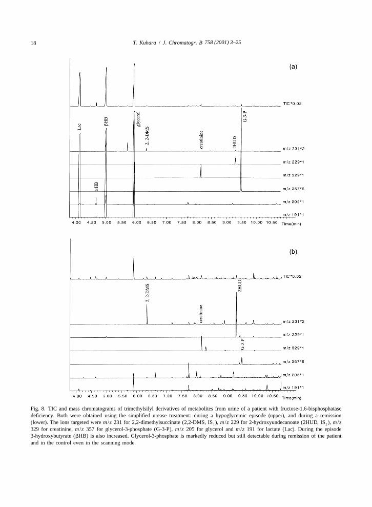
without the respective stable isotope-labeled internal cedure, in which organic acids are extracted fromstandard is difficult in the solvent extraction method. acidified urine using ethyl ether and ethyl acetate andExtraction with DEAE–Sephadex significantly im- converted to trimethylsilyl derivatives [58]. It isproves the recovery of glycerol-3-phosphate [5,54], known that, during evaporation of organic solvents,but inorganic acids such as phosphate or sulfate are lactone formation, which to some extent also occursalso well recovered, which is inconvenient for the in vivo [58], is enhanced, resulting in apparently lowsubsequent GC–MS analysis, and glycerol cannot be levels of these hydroxy acids [59], thereby makingrecovered. Furthermore the DEAE–Sephadex pro- the chemical diagnosis difficult. The TIC chromato-cedure takes several hours. In our simple urease grams of trimethylsilyl derivatives of urinary metab-treatment procedure, recovery of this polar acid is olites obtained by the urease treatment described invery high, as in the DEAE–Sephadex method, and the present study from the urine of a patient who wasthe diagnosis of this disease becomes remarkably chemically diagnosed by us as having succinicrapid, accurate and easy, as has been reported [8]. semialdehyde dehydrogenase deficiency are shown inVery recently, more sensitive quantification with the Fig. 9a. The profile of the same urine specimen wassingle ion monitoring (SIM) mode for glycerol-3- analyzed by ether extraction was compared (Fig. 9b,phosphate, glycerol and lactate with the simple upper and lower). The ratio of 3,4-dihydroxybutyrateurease pretreatment method was described [55], but relative to 4-hydroxybutyrate was much higher inusing our routine analytical conditions, all three are sample with the urease treatment than in that withadequately detectable in healthy controls, even in the ether extraction, suggesting that the recovery of polarscanning mode described in Experimental. Fig. 8 3,4-dihydroxybutyrate is better and that this newshows the TIC and mass chromatograms of TMS procedure would improve the sensitivity of diagnosisderivatives of metabolites from a patient diagnosed of patients with this disease and lower the rate ofby us as having fructose-1,6-bisphosphatase de- false negatives. Recently, the clinical heterogeneityficiency, obtained using the present procedure during and response to early treatment with vigabatrin inan episode (upper) and remission (lower). siblings with succinic semialdehyde dehydrogenase
deficiency was reported [60]. The author also foundsignificant differences of the urinary metabolite
7. Succinate semialdehyde dehydrogenase levels among siblings (unpublished observations).deficiency
Deficiency of succinate semialdehyde dehydro-1genase (succinate semialdehyde: NAD oxidoreduc- 8. Deficiencies of pyrimidine degradation
tase, SSADH; EC 1.2.1.24), also known as 4-hy-droxybutyric aciduria (McKusick 271980), is an Inborn errors of pyrimidine degradation are lessautosomal recessive inborn error in the metabolism rare than has generally been assumed, and clinicalof the neurotransmitter 4-aminobutyrate. Predomi- presentations of such patients are variable and non-nant oxidative conversion of succinate semialdehyde specific [61–63]. Pyrimidines are degraded in fourto succinate is impaired, and consequently succinic steps in humans, catalyzed by dihydropyrimidine
1semialdehyde is reduced to 4-hydroxybutyrate in a dehydrogenase (5,6-dihydropyrimidine: NADP oxi-reaction catalyzed by 4-hydroxybutyrate dehydro- doreductase; DHPDH, EC 1.3.1.2), dihydropyri-genase (EC 1.1.1.61). The first patient with this midinase (5,6-dihydropyrimidine amidohydrolase;disease was described by Jacob et al. in 1981 [56]. DHP, EC 3.5.2.2), b-ureidopropionase (UP, ECThe initial step in diagnosis is usually the recognition 3.5.1.6), and three aminotransferases. Many asymp-of excessive urinary excretion of 4-hydroxybutyrate tomatic cases with DHPDH deficiency have beenand 3,4-dihydroxybutyrate; the latter is thought to be reported, and for cases with symptoms, the clinicalformed by b-oxidation [57]. Other minor metabolites abnormalities are variable and nonspecific. Asympto-have also been reported [58]. These metabolites were matic infants and adults with DHP deficiency havedetected by the conventional solvent extraction pro- also been reported [63,64]. No treatment specific
758 (2001) 3–2518 T. Kuhara / J. Chromatogr. B
Fig. 8. TIC and mass chromatograms of trimethylsilyl derivatives of metabolites from urine of a patient with fructose-1,6-bisphosphatasedeficiency. Both were obtained using the simplified urease treatment: during a hypoglycemic episode (upper), and during a remission(lower). The ions targeted were m /z 231 for 2,2-dimethylsuccinate (2,2-DMS, IS ), m /z 229 for 2-hydroxyundecanoate (2HUD, IS ), m /z1 2
329 for creatinine, m /z 357 for glycerol-3-phosphate (G-3-P), m /z 205 for glycerol and m /z 191 for lactate (Lac). During the episode3-hydroxybutyrate (bHB) is also increased. Glycerol-3-phosphate is markedly reduced but still detectable during remission of the patientand in the control even in the scanning mode.

758 (2001) 3–25 19T. Kuhara / J. Chromatogr. B
Fig. 9. TIC and mass chromatograms of trimethylsilyl derivatives of metabolites from the urine of a patient with succinic semialdehydedehydrogenase deficiency. The present simplified urease treatment (upper and upper part of lower) and conventional solvent extraction(lower). The ions targeted were m /z 329 for creatinine, m /z 233 for 4-hydroxybutyrate, m /z 321 for 2-deoxytetronate and m /z 231 for2,2-dimethylsuccinate added as internal standard respectively. Ions at m /z 233(M-103) and 231(M-105) are also present in 2-deoxytetronate.Peak identifications are: 1. glycolate-2; 2. alanine-2; 3. glycine-2 and d -glycine (IS); 4. 3-hydroxypropionate-2; 5. 3-hydroxyisovalerate-22
15and 2-aminoisobutyrate-2; 6. 4-hydroxybutyrate-2; 7. phosphate-3; 8. 2,2-dimethylsuccinate-2 (IS ); 9. uracil-2 and N -uracil-2 (IS); 10.1 2
serine-3; 11. threonine-3; 12. 3-deoxytetronate-3; 13. 2-deoxytetronate-3; 14. xylitol-5; 15. 4-hydroxyproline-3 and 5-oxoproline-2; 16.threonate-4; 17. threo- and erythro-4,5-dihydroxyhexanoate; 18. erythronate-4; 19. d -creatinine-3 (IS) and creatinine-3; 20. 2-hydroxy-3
glutarate-3; 21. p-hydroxyphenylacetate-2; 22. d -lysine-3 (IS) and lysine-3; 23. 2-aminoadipate-3; 24. 2-hydroxyundecanoate-2 (IS ); 25.4 2
citrate-4; 26. galactose-5(1); 27. galactose-5 (2); 28. histidine-3; 29. glucose-5 (2); 30. galactonate-6; 31. myo-inositol-6; 32. urate-4; 33.n-heptadecanoate-1 (IS ); 34. unknown; 35. pseudouridine-5.3

758 (2001) 3–2520 T. Kuhara / J. Chromatogr. B
15 15for these enzyme defects has been described, but pyrimidines and orotate using N -uracil and N -2 2
withdrawal of 5-fluorouracil (5FU), a commonly orotate as their respective internal standards. Weused anticancer drug, from cancer chemotherapy prepared artificial urine specimens which simulatedregimens of patients with these defects is critical typical, moderate and heterozygotes of DHPDHbecause 5FU is degraded in vivo by these enzymes. deficiency and those of DHP deficiency, by spiking aPatients with these deficiencies suffer from severe urine specimen with different amounts of uracil,neurotoxicity, sometimes leading to death, following thymine and/or dihydrouracil and dihydrothymine,administration of 5FU, and even otherwise asympto- and confirmed that the chemical diagnosis can bematic homozygotes or heterozygotes may develop done. Recovery and CV were satisfactory, and thesevere clinical symptoms due to such medication. values of healthy controls were determined [74]. TheTherefore, rapid and specific identification of the method was established for rapid, highly sensitivecancer patients with these enzyme deficiencies is and specific determinations of thymine, uracil,critical prior to treatment with 5FU. DHPDH de- dihydrothymine, dihydrouracil, orotate and creatinineficiency is characterized by the presence of abnormal simultaneously in 0.1-ml liquid urine samples oramounts of uracil and thymine in urine [65] because filter paper urine.DHPDH is the initial and rate-limiting enzyme in thecatabolism of the pyrimidine base. DHP deficiencycan also be detected by the presence of large 9. Differential diagnosis of homocystinuriaamounts of dihydrouracil and dihydrothymine, andmoderate amounts of uracil and thymine, in urine The transsulfuration pathway converts the sulfur[66]. Several methods to screen for disorders of atom of methionine into the sulfur atom of cysteine,pyrimidine metabolism that use HPLC [67,68], two and reforms methionine by methylation of homo-dimensional thin-layer chromatography [69], amino cysteine. Homocystinuria types I, II and III areacid analysis of urine before and after acid hy- characterized by different etiologies, biochemicaldrolysis [70], gas chromatography, or GC–MS [71] abnormalities and therapeutic measures. In type I,have been reported. However, these methods have due to a deficiency of cystathionine b-synthase (L-been time-consuming or have lacked specificity or serine hydrolyase (adding homocysteine); ECsensitivity. Identification by HPLC of all the specific 4.2.1.22), homocysteine accumulated in this disordermetabolites excreted in DHP deficiency is difficult causes methionine overproduction. A simple treat-because the maximal UV-absorbance of the ment with pyridoxine for the pyridoxine-responsivedihydropyrimidines occurs below 230 nm [72]. GC– type, or a dietary restriction of methionine andMS analysis of trimethylsilyl derivatives of urinary supplementation with cystine for the pyridoxine-un-organic acid extracts could detect this deficiency [7] responsive type, greatly improve the outcome ofbut it has been pointed out that quantitation was not affected infants [75]. Homocystinuria type II ispossible because of variable extraction yields, and caused by defective remethylation due to the de-
5,10that methods involving use of two-dimensional TLC ficiency of N -methylenetetrahydrofolate reductaseor HPLC with or without prefractionation of urine (5-methyltetrahydrofolate: (acceptor) oxidoreductase;were more sensitive [62]. Very recently, a rapid and EC 1.1.99.15) (MTHFR, EC 1.1.1.68). Folate andspecific screening method for patients at risk of betaine may have the advantage of lowering homo-inherited disorders of pyrimidine and purine metabo- cysteine levels and increasing methionine levels [76].lism was described: it involves the use of urine and Recently, the importance of the role of folate and ofHPLC–ESI–MS/MS [73] where uracil, thymine, 5- early detection of type II patients has been stressedhydroxymethyluracil and orotate were targeted, but [77,78]. Homocystinuria type III is caused by the
5not dihydrothymine, dihydrouracil, creatinine or deficiency of N -methyltetrahydrofolate homocys-amino acids. teine methyltransferase (S-adenosyl-L-methionine: L-
The simplified procedure described here was homocysteine S-methyltransferase: EC 2.1.1.10) dueapplied to identify patients with these defects in the to the defective synthesis of methylcobalamin andpyrimidine degradative pathway by targeting these deoxyadenosylcobalamin. This condition or nutri-

758 (2001) 3–25 21T. Kuhara / J. Chromatogr. B
tional vitamin B deficiency is accompanied by silylation, and GC–MS conditioning thus becomes12
combined homocystinuria and methylmalonic more important for continuous GC–MS measure-aciduria [79]. As current neonatal screening for ment. It is hoped that a guard column can behomocystinuria type I targets methionine in filter successfully applied to address this problem. Forpaper blood spots, type II is not detected, since it amino acids, trimethylsilylation is not always quan-causes moderate homocystinuria with low or rela- titative. Therefore, in our procedure stable isotope-tively normal levels of plasma methionine. Instead, labeled internal standards are used for importantisolated hypermethioninemia due to a deficiency of amino acids and labeled omega-amino acid is used ashepatic methionine adenosyltransferase (S- the internal standard for omega-amino acids. In someadenosylmethionine synthetase, ATP: L-methionine reports this simplified urease?GC–MS method isS-adenosyltransferase; EC 2.5.1.6) is screened as termed the urease /direct method [81], and instead ofwell, and it is clinically free of symptoms, indicating trimethylsilylation, tert.-butyldimethylsilylation hasthat the accumulation of methionine in the body is been recommended [82], and reported to be usefulnot harmful. The simplified diagnostic procedure has for screening for organic acidemias [81]. However, ifproven to be able to differentiate the three types of sample preparation or GC–MS conditioning is in-homocystinuria by simultaneous quantification of adequate, measurements of not only amino acids butmethionine, homocystine, methylmalonate, uracil also organic acids such as methylcitrate become lessand creatinine in filter paper urine, when each quantitative, and the sensitivity of their detection isrespective stable isotope-labeled compound is used, lowered. As the tert.-butyldimethylsilyl moiety isas reported recently [80]. The mass chromatograms bulkier than the trimethylsilyl moiety, the tendencyof trimethylsilyl derivatives of metabolites from not to be fully silylated and to give several deriva-patients with type I and type II disease are shown in tives may be higher for tert.-butyldimethylsilylation,Fig. 10. especially for polyols and sugars. Under these con-
Urinary metabolite levels determined by using the ditions, data analysis may become more complicatedsimplified urease procedure were compared before after repeated GC–MS measurements.and after treatment with folate in a male patient who We believe that the present procedure will providetemporarily developed megaloblastic anemia (Fig. 11). valuable tool for screening of more than 80 targetAs his serum folate was below the normal range, he diseases. This procedure, technically practical yetwas treated with folate, after which megaloblastic comprehensive from the metabolic point of view,anemia disappeared and the level of orotate de- could become well established for screening of allcreased into the normal range. Thymidilate synthase, age groups ranging from neonates to the elderly.which catalyzes the conversion of dUMP to dTMP, isfolate-dependent, and pyrimidine biosynthesis isregulated by end-product inhibition. Folate de- Acknowledgementsficiency thus causes impaired DNA synthesis, en-hanced pyrimidine biosynthesis, megaloblastic This study was supported by a grant from theanemia and orotic aciduria. Folate supplementation JAOG Ogyaa Donation Foundation, a 1999–2000significantly reduced the level of homocystine and Grant-in-Aid for Scientific Research (11672312)dramatically increased that of methionine. This sim- from the Ministry of Education, Science and Cultureple diagnostic procedure has therefore proved useful of Japan, Health Sciences Research Grants forfor monitoring the biochemical and nutritional con- Research on Children and Families (H10-Kodomo-ditions of patients, especially for acquired deficiency 031) from the Ministry of Health and Welfare ofof folate and vitamin B , as well as for evaluating Japan, and by a grant for project research from the12
the efficacy of treatments. High-Technology Center of Kanazawa Medical Uni-The present method enables us to obtain numerous versity (H00-3). The author is grateful to Dr I.
kinds of information about the metabolism in the Matsumoto (Professor Emeritus, Kanazawa Medicalhuman body. However, a variety of compounds are University) and Dr S. Sakamoto (Professor Emeritus,recovered and yet derivatized only by trimethyl- The University of Tokyo) for their continuing inter-

758 (2001) 3–2522 T. Kuhara / J. Chromatogr. B
Fig. 10. Partly shown mass chromatograms of trimethylsilyl derivatives of metabolites from urine of patients with homocystinuria of type I(upper) and type II (lower). Methionine (Met) concentration in the patient with type I was significantly reduced on this occasion due tofolate deficiency. The ions targeted were m /z 329 and 331, m /z 176 and 179 and m /z 278 and 282 for creatinine and d -creatinine,3
methionine and d -methionine, and homocystine and d -homocystine, respectively.3 4

758 (2001) 3–25 23T. Kuhara / J. Chromatogr. B
Fig. 11. Metabolite levels in homocystinuria patient before and after treatment of megaloblastic anemia by administration of folate [80].
[10] T. Kuhara, I. Matsumoto, Biomed. Mass Spectrom. 7 (1980)est, encouragement and support. The author express424.thanks for Mrs T. Sakaida for her assistance in
[11] I. Matsumoto, in: A. Frigerio (Ed.), Recent Developments inpreparing the manuscript. Mass Spectrometry, Biochem. Med. Environmental Res.,Vol.
7, Elsevier, Amsterdam, 1981, p. 57.[12] T. Kuhara, T. Shinka, M. Matsuo, I. Matsumoto, Clin. Chim.
Acta 123 (1982) 101.References[13] T. Kuhara, T. Shinka, Y. Inoue, M. Matsumoto, M. Yoshino,
Y. Sakaguchi, I. Matsumoto, Clin. Chim. Acta 133 (1983)[1] K. Tanaka, M.A. Budd, M.L. Efron, K.J. Isselbacher, Proc. 133.
Natl. Acad. Sci. USA 56 (1966) 236. [14] I. Matsumoto, T. Kuhara, Mass Spectrom. Rev. 6 (1987) 77.[2] S.I. Goodman, S.P. Markey, Diagnosis of Organic Acidemias [15] I. Matsumoto, T. Kuhara, in: D.M. Desiderio (Ed.), Clinical
by Gas Chromatography–Mass Spectrometry, Alan R. Liss, and Biomedical Applications, Clinical Mass Spectrometry,New York, 1981. Vol. 1, Plenum Press, New York, 1993, p. 259.
[3] R.A. Chalmers, A.M. Lawson, Organic Acids in Man, [16] I. Matsumoto et al. (Ed.), Advances in Chemical DiagnosisChapman and Hall, London, 1982. and Treatment of Metabolic Disorders, Vol. 1, Wiley, Chi-
[4] Chapter 40 L. Sweetman, J.C. Williams, in: C.R. Scriver, chester, 1993.A.L. Beaudet, W.S. Sly, D. Valle (Eds.), The Metabolic and [17] I. Matsumoto, T. Kuhara, O.A. Mamer, L. Sweetman, R.G.Molecular Bases of Inherited Disease, 7th ed., McGraw– Calderhead (Eds.), Advances in Chemical Diagnosis andHill, New York, 1995, p. 1387. Treatment of Metabolic Disorders,Vol. 2, Kanazawa Medical
[5] R.A. Chalmer, R.W.E. Watts, Analyst 97 (1972) 958. University Press, 1994.[6] J.D. Shoemaker, W.H. Elliott, J. Chromatogr. 562 (1991) [18] I. Matsumoto, S. Sakamoto, T. Kuhara, M. Sudo, M. Yoshino
125. (Eds.), Practical Chemical Diagnosis, Soft Science, Tokyo,[7] T. Kuhara, I. Matsumoto, Proc. Jpn. Soc. Biomed. Mass 1995.
Spectrom. 20 (1995) 45. [19] S.E.C. Davies, R.A. Iles, T.E. Stacey, R.A. Chalmers, Clin.[8] I. Matsumoto, T. Kuhara, Mass Spectrom. Rev. 15 (1996) Chim. Acta 194 (1990) 203.
43. [20] R. Guthrie, in: T.P. Carter, A.M. Wiley (Eds.), Genetic[9] I. Matsumoto, T. Shinka, T. Kuhara, T. Ohura, H. Yama- Disease: Screening and Management, Alan R. Liss, New
moto, Y. Hase, H. Aoki, G. Issiki, K. Tada, in: A. Frigerio York, 1986, p. 319.(Ed.), Recent Developments in Mass Spectrometry, Bio- [21] R. Guthrie, A. Susi, Pediatrics 32 (1963) 338.chem. Med., Vol. 1, Plenum, New York, 1978, p. 203.

758 (2001) 3–2524 T. Kuhara / J. Chromatogr. B
[22] B.A. Chamberlin, C.C. Sweeley, Clin. Chem. 33/34 (1987) [44] M.S. Murphy, M.A. Preece, J. Collins, A. Green, D.A. Kelly,in: Soc. Study of Inborn Errors of Metab. Leuven, 1992, p.572.99.[23] M. Tuchman, B. Lemieux, C. Auray-Blais, L.L. Robinson,
[45] J.S. Schlenzig, F.P. Travert, J. Laurent, D. Rabier, D. Jan, U.R. Giguere, M.T. MacCann, W.G. Woods, Pediatrics 85Wendel, A.C. Sewell, Y. Revillon, P. Kamoun, J.M. Saudub-(1990) 765.ray, J. Inherit. Metab. Dis. 18 (1995) 448.[24] M. Tuchman, M.T. McCann, P.E. Johnson, B. Lemieux,
[46] W.G. van’t Hoff, M. Dixon, J. Taylor, P. Mistry, K. Rolles,Pediatr. Res. 30 (1991) 3.L. Rees, J.V. Leonard, J. Pediatr. 132 (1998) 1043.[25] I. Matsumoto, T. Kuhara, T. Shinka, Y. Inoue, A. Inoue, M.
[47] C. Bachmann, J.P. Colombo, Eur. J. Pediatr. 134 (1980) 109.Matsumoto, in: H. Naruse, M. Irie (Eds.), Intl. Congress[48] M.T. McCann, M.M. Thompson, I.C. Gueron, M. Tuchman,Series 606: Neonatal Screening, Excerpta Medica, Amster-
Clin. Chem. 41 (1995) 739.dam, 1983, p. 414.[49] K.J. van Acker, F.J. Eyskens, R.M. Verkerk, S.S. Scharpe,
[26] I. Matsumoto, T. Kuhara, T. Shinka, Y. Inoue, M. Mat-Pediatr. Res. 34 (1993) 762.
sumoto, H. Odani, in: H. Matsuda, T.-L. Chang (Eds.), Proc.[50] M. Kasahara, T. Kiuchi, K. Uryuhara, Y. Ogura, K. Taka-
First China–Japan Joint Symposium on Mass Spectrometry,kura, H. Egawa, K. Asonuma, S. Uemoto, Y. Inomata, K.
1984, p. 84.Tanaka, J. Pediatr. Surg. 33 (1998) 1753.
[27] I. Matsumoto, T. Kuhara, T. Shinka, Y. Inoue, M. Mat- [51] L. Baker, A.I. Winegrad, Lancet 7662 (1970) 13.sumoto, in: A.L. Burlingame, N. Castagnoli (Eds.), Mass [52] G.V. Berghe, J. Inherit. Metab. Dis. 19 (1996) 470.Spectrometry in the Health and Life Sciences, Elsevier, ¨[53] P.A. Dremsek, M. Sacher, W. Stogmann, R. Gitzelmann, C.Amsterdam, 1985, p. 425. Bachmann, Eur. J. Pediatr. 144 (1985) 203.
[28] T. Kuhara, T. Shinka, Y. Inoue, Z. Xia, M. Ohse, I. Yoshida, [54] A. Nakai, Y. Shigematsu, Y.Y. Lin, Y. Kikawa, M. Sudo, J.T. Inokuchi, S. Yamaguchi, M. Takayanagi, I. Matsumoto, J. Inherit. Metab. Dis. 13 (1990) 263.Chromatogr. B 731 (1999) 141. [55] M. Iga, M. Kimura, T. Ohura, Y. Kikawa, S. Yamaguchi, J.
[29] D.S. Millington, D.L. Norwood, N. Kodo et al., Anal. Chromatogr. B. 746 (2000) 75.Biochem. 180 (1989) 331. [56] C. Jakobs, M. Bojasch, E. Monch, D. Rating, H. Siemes, F.
[30] D.H. Chace, D.S. Millington, N. Terada et al., Clin. Chem. Hanefeld, Clin. Chim. Acta 111 (1981) 169.39 (1993) 66. [57] C.R. Lee, Biochem. Med. 17 (1977) 284.
[31] M.S. Rashed, P.T. Ozand, M.P. Bucknall, D. Little, Pediatr. [58] G.K. Brown, C.H. Cromby, N.J. Manning, R.J. Pollitt, J.Res. 38 (1995) 324. Inherit. Metab. Dis. 10 (1987) 367.
[32] E.W. Naylor, D.H. Chace, J. Child Neurol. 14 (1999) S4. [59] P. Divry, P. Baltassat, M.O. Rolland, J. Cotte, M. Hermier,[33] T. Hagen, M.S. Korson, M. Sakamoto, J.E. Envans, Clin. M. Duran, S.K. Wadaman, Clin. Chim. Acta 129 (1983) 303.
Chim. Acta 283 (1999) 77. [60] H. Peters, M. Cleary, A. Boneh, J. Inherit. Metab. Dis. 22[34] B. Childs, W.L. Nyhan, M. Borden, L. Bard, R.E. Cooke, (1999) 198.
Pediatrics 27 (1961) 522. [61] Chapter 55 D.M.O. D.R.Webster, D.P. Becroft, D.P. Sullte, in:[35] T. Ando, K. Rasmussen, J.M. Wright, W.L. Nyhan, J. Biol. C.R. Scriver, A.L. Beaudet, W.S. Sly, D. Valle (Eds.), The
Chem. 247 (1972) 2200. Metabolic and Molecular Bases of Inherited Disease, 7th ed,[36] Chapter 41 W.A. Fenton, L.E. Rosenberg, in: C.R. Scriver, McGraw-Hill, New York, 1995, p. 1799.
A.L. Beaudet, W.S. Sly, D. Valle (Eds.), The Metabolic and [62] A.H. van Gennip, N.G.G.M. Abeling, P. Vreken, A.B.P. vanMolecular Bases of Inherited Disease, 7th ed, McGraw–Hill, Kuilenburg, J. Inherit. Metab. Dis. 20 (1997) 203.New York, 1995, p. 1423. [63] S. Sumi, M. Imaeda, K. Kidouchi, S. Ohba, N. Hamajima, K.
[37] T. Kuhara, M. Matsuo, T. Shinka, Y. Inoue, M. Matsumoto, Kodama, H. Togari, Y. Wada, Am. J. Med. Genet. 78 (1998)I. Matsumoto, in: A. Frigerio (Ed.), Chromatography and 336.Mass Spectrometry in Biomedical Science 2, Elsevier, [64] S. Ohba, K. Kidouchi, S. Sumi, M. Imaeda, N. Takeda, H.Amsterdam, 1983, p. 449. Yoshizumi, A. Tatematsu, K. Kodama, K. Yamanaka, M.
[38] T. Kuhara, I. Matsumoto, T. Shinka, Int. J. Mass Spectrom. Kobayashi, Y. Wada, Adv. Exp. Med. Biol. 370 (1995) 383.Ion Physics 48 (1983) 97. [65] J.A.J.M. Bakkeren, R.A. de Abreu, R.C.A. Sengers, F.J.M.
[39] T. Kuhara, Y. Inoue, I. Matsumoto, J. Pediatr. 113 (1988) Gabreels, J.M. Maas, W.O. Renier, Clin. Chim. Acta 140787. (1984) 247.
[40] T. Kuhara, M. Matsumoto, Y. Inoue, T. Ohkura, T. Aoyama, [66] M. Duran, P. Rovers, P.K. de Bree, C.H. Schreuder, H.M. Matsuo, I. Matsumoto, J. Clin. Biochem. Nutr. 7 (1989) Beukenhorst, L. Dorlsnd, R. Berger, J. Inherit. Metab. Dis.1. 14 (1991) 367.
[41] K.N. North, M.S. Korson, Y.R. Gopal, F.J. Rohr, T.B. [67] A.H. van Gennip, N.G.G. M Abeling, L. Elzinga-Zoetekouw,Brazelton, S.E. Waisbren, M.L. Warman, J. Pediatr. 126 E.G. Scholten, A. van Cruchten, H.D. Bakker, Adv. Exp.(1995) 916. Med. Biol. 253 (1989) 111.
[42] J.H. Walter, J.E. Wraith, M.A. Cleary, Arch. Dis. Childhood [68] S. Ohba, K. Kidouchi, K. Katoh, M. Kobayshi, Y. Wada, J.72 (1995) F197. Chromatogr. 568 (1991) 325.
[43] W.L. Nyhan, C. Bay, E.W. Beyer, M. Mazi, Arch. Neurol. 56 [69] A.H. van Gennip, D.Y. van Noordenburg-Huistra, P.K. de(1999) 1143. Bree, S.K. Wadman, Clin. Chim. Acta 86 (1978) 7.

758 (2001) 3–25 25T. Kuhara / J. Chromatogr. B
[70] A.H. van Gennip, S. Busch, L. Elzinga, A.E. Stroomer, A. Bases of Inherited Disease, 7th ed., McGraw–Hill, Newvan Cruchten, E.G. Scholten, N.G.G.M. Abeling, Clin. York, 1995, p. 3111.Chem. 39 (1993) 380. [77] B.D. Guenther, C.A. Sheppard, P. Tran, R. Rozen, R.G.
Matthews, M.L. Ludwig, Nat. Struct. Biol. 6 (1999) 359.[71] S.K. Wadman, F.A. Beemer, P.K. de Bree, M. Duran, A.H.[78] A.F. Valevski, H. Bassan, S.H. Korman, T. Lerman-Sagie, A.van Gennip, D. Ketting, F.J. van Sprang, Adv. Exp. Med.
Gutman, S. Harel, J. Child Neurol. 15 (2000) 539.Biol. 165 (1984) 109.[79] Chapter 102 W.A. Fenton, L.E. Rosenberg, in: C.R. Scriver,[72] M.J. Henderson, K. Ward, H.A. Simmonds, J.A. Duley, P.M.
A.L. Beaudet, W.S. Sly, D. Valle (Eds.), The Metabolic andDavies, J. Inherit. Metab. Dis. 16 (1993) 574.Molecular Bases of Inherited Disease, 7th ed, McGraw–Hill,[73] T. Ito, A.B.P. van Kuilenburg, A.H. Bootsma, A.J. Haasnoot,New York, 1995, p. 3129.A. van Cruchten, Y. Wada, A.H. van Gennip, Clin. Chem. 46
[80] T. Kuhara, M. Ohse, C. Ohdoi, S. Ishida, J. Chromatogr. B.(2000) 445.742 (2000) 59.[74] T. Kuhara, C. Ohdoi, M. Ohse, J. Chromatogr. B. (2001) (in
[81] T. Ohie, X. Fu, M. Iga, M. Kimura, S. Yamaguchi, J.press).Chromatogr. B. 746 (2000) 63.[75] Chapter 35 S.H. Mudd, H.L. Levy, F. Skovby, in: C.R.
[82] X. Fu, M. Iga, M. Kimura, S. Yamaguchi, Early Hum. Dev.Scriver, A.L. Beaudet, W.S. Sly, D. Valle (Eds.), The58 (2000) 41.Metabolic and Molecular Bases of Inherited Disease, 7th ed,
[83] T. Yorifuji, J. Muroi, A. Uematsu, T. Nakahata, H. Egawa,McGraw–Hill, New York, 1995, p. 1279.K. Tanaka, J. Pediatr. 137 (2000) 572.[76] Chapter 101 D.S. Rosenblatt, in: C.R. Scriver, A.L. Beaudet,
W.S. Sly, D. Valle (Eds.), The Metabolic and Molecular

![Review Overview of the applications of liquid ...quimica.udea.edu.co/~carlopez/cromatohplc/review_hplc_ms_foods.pdfgiving positive identification of components of ... [29–31] mass](https://static.fdocuments.in/doc/165x107/5adb8ac87f8b9aee348e2d80/review-overview-of-the-applications-of-liquid-carlopezcromatohplcreviewhplcmsfoodspdfgiving.jpg)