Review article Role of basal ganglia–brainstem pathways … PDFs/basal... · Review article Role...
15
Review article Role of basal ganglia–brainstem pathways in the control of motor behaviors K. Takakusaki * , K. Saitoh, H. Harada, M. Kashiwayanagi Department of Physiology, Asahikawa Medical College, Midorigaoka-Higashi 2-1, Asahikawa 078-8510, Japan Received 25 February 2004; accepted 28 June 2004 Available online 13 August 2004 Abstract Here we review a role of a basal ganglia–brainstem (BG–BS) system throughout the mesopontine tegmentum in the control of various types of behavioral expression. First the basal ganglia–brainstem system may contribute to an automatic control of movements, such as rhythmic limb movements and adjustment of postural muscle tone during locomotion, which occurs in conjunction with voluntary control processes. Second, the basal ganglia–brainstem system can be involved in the regulation of awake–sleep states. We further propose the possibility that the basal ganglia–brainstem system is responsible for the integration of volitionally-guided and emotionally-triggered expression of motor behaviors. It can be proposed that dysfunction of the basal ganglia–brainstem system together with that of cortico-basal ganglia loop underlies the pathogenesis of behavioral disturbances expressed in basal ganglia dysfunction. # 2004 Elsevier Ireland Ltd and the Japan Neuroscience Society. All rights reserved. Keywords: GABAergic projection; The substantia nigra pars reticulata; The pedunculopontine tegmental nucleus; Locomotion; Postural muscle tone; REM sleep; Emotional behaviors; Parkinson disease www.elsevier.com/locate/neures Neuroscience Research 50 (2004) 137–151 Contents 1. Introduction ........................................................... 138 2. BG–BS systems and motor control ........................................... 138 2.1 General schema of the basal ganglia control of movements ...................... 138 2.2 Basic architectures of locomotor system and muscle tone control system ............ 138 2.3 BG–BS systems in the control of muscle tone and locomotion ................... 140 3. Concept for understanding BG–BS Systems’ involvement of motor control ............... 142 3.1 Concept for understanding BG–BS systems in the control of postural muscle tone and locomotion ....................................................... 142 3.2 Current concept for the basal ganglia control of saccadic eye movements ............ 144 3.3 Comparisons of two concepts .......................................... 145 4. BG–BS systems for brain function in general .................................... 145 4.1 REM sleep ....................................................... 145 4.2 Arousal, cognition and attention ......................................... 147 4.3 Emotional expression ................................................ 147 5. Concluding remarks ...................................................... 148 Acknowledgements ......................................................... 148 References ............................................................... 148 * Corresponding author. Tel.: +81 166 68 2331; fax: +81 166 68 2339. E-mail address: [email protected] (K. Takakusaki). 0168-0102/$ – see front matter # 2004 Elsevier Ireland Ltd and the Japan Neuroscience Society. All rights reserved. doi:10.1016/j.neures.2004.06.015
-
Upload
nguyenngoc -
Category
Documents
-
view
215 -
download
0
Transcript of Review article Role of basal ganglia–brainstem pathways … PDFs/basal... · Review article Role...

www.elsevier.com/locate/neures
Neuroscience Research 50 (2004) 137–151
Review article
Role of basal ganglia–brainstem pathways in the
control of motor behaviors
K. Takakusaki*, K. Saitoh, H. Harada, M. Kashiwayanagi
Department of Physiology, Asahikawa Medical College, Midorigaoka-Higashi 2-1,
Asahikawa 078-8510, Japan
Received 25 February 2004; accepted 28 June 2004
Available online 13 August 2004
Abstract
Here we review a role of a basal ganglia–brainstem (BG–BS) system throughout the mesopontine tegmentum in the control of various
types of behavioral expression. First the basal ganglia–brainstem system may contribute to an automatic control of movements, such as
rhythmic limb movements and adjustment of postural muscle tone during locomotion, which occurs in conjunction with voluntary control
processes. Second, the basal ganglia–brainstem system can be involved in the regulation of awake–sleep states. We further propose the
possibility that the basal ganglia–brainstem system is responsible for the integration of volitionally-guided and emotionally-triggered
expression of motor behaviors. It can be proposed that dysfunction of the basal ganglia–brainstem system together with that of cortico-basal
ganglia loop underlies the pathogenesis of behavioral disturbances expressed in basal ganglia dysfunction.
# 2004 Elsevier Ireland Ltd and the Japan Neuroscience Society. All rights reserved.
Keywords: GABAergic projection; The substantia nigra pars reticulata; The pedunculopontine tegmental nucleus; Locomotion; Postural muscle tone; REM
sleep; Emotional behaviors; Parkinson disease
Contents
1. Introduction . . . . . . . . . . . . . . . . . . . . . . . . . . . . . . . . . . . . . . . . . . . . . . . . . . . . . . . . . . . 138
2. BG–BS systems and motor control . . . . . . . . . . . . . . . . . . . . . . . . . . . . . . . . . . . . . . . . . . . 138
2.1 General schema of the basal ganglia control of movements . . . . . . . . . . . . . . . . . . . . . . 138
2.2 Basic architectures of locomotor system and muscle tone control system . . . . . . . . . . . . 138
2.3 BG–BS systems in the control of muscle tone and locomotion . . . . . . . . . . . . . . . . . . . 140
3. Concept for understanding BG–BS Systems’ involvement of motor control . . . . . . . . . . . . . . . 142
3.1 Concept for understanding BG–BS systems in the control of postural muscle tone and
locomotion . . . . . . . . . . . . . . . . . . . . . . . . . . . . . . . . . . . . . . . . . . . . . . . . . . . . . . . 142
3.2 Current concept for the basal ganglia control of saccadic eye movements . . . . . . . . . . . . 144
3.3 Comparisons of two concepts . . . . . . . . . . . . . . . . . . . . . . . . . . . . . . . . . . . . . . . . . . 145
4. BG–BS systems for brain function in general . . . . . . . . . . . . . . . . . . . . . . . . . . . . . . . . . . . . 145
4.1 REM sleep . . . . . . . . . . . . . . . . . . . . . . . . . . . . . . . . . . . . . . . . . . . . . . . . . . . . . . . 145
4.2 Arousal, cognition and attention. . . . . . . . . . . . . . . . . . . . . . . . . . . . . . . . . . . . . . . . . 147
4.3 Emotional expression . . . . . . . . . . . . . . . . . . . . . . . . . . . . . . . . . . . . . . . . . . . . . . . . 147
5. Concluding remarks . . . . . . . . . . . . . . . . . . . . . . . . . . . . . . . . . . . . . . . . . . . . . . . . . . . . . . 148
Acknowledgements . . . . . . . . . . . . . . . . . . . . . . . . . . . . . . . . . . . . . . . . . . . . . . . . . . . . . . . . . 148
References . . . . . . . . . . . . . . . . . . . . . . . . . . . . . . . . . . . . . . . . . . . . . . . . . . . . . . . . . . . . . . . 148
* Corresponding author. Tel.: +81 166 68 2331; fax: +81 166 68 2339.
E-mail address: [email protected] (K. Takakusaki).
0168-0102/$ – see front matter # 2004 Elsevier Ireland Ltd and the Japan Neuroscience Society. All rights reserved.
doi:10.1016/j.neures.2004.06.015
K. Takakusaki et al. / Neuroscience Research 50 (2004) 137–151138
1. Introduction
Basal ganglia disorders are manifested by an inability to
initiate and terminate voluntary movements in a certain
behavioral context, an inability to suppress involuntary
movements, an abnormality in the velocity and the amount
of movement, and an abnormal muscle tone (Obeso et al.,
1997; Saint-Cyr et al., 1995). Gait disturbances are also a
major impediment for Parkinsonian patients (Murray et al.,
1978; Morris et al., 1994). Marsden (1982) hypothesized, in
his lecture titled as ‘‘The mysterious motor function of the
basal ganglia’’, that ‘‘the basal ganglia are responsible for
the autonomic execution of learned motor plans’’. This
hypothesis was derived from his careful insight into the
consideration of the motor disturbances in basal ganglia
disorders. Specifically, primary clinical deficit in Parkinson
disease is slowness of movement, particularly when actions
are volitional (Marsden, 1989).
The current understanding is that the basal ganglia and
cerebellar loops with the motor areas of the cerebral cortex
are involved in the control of voluntary movements (Mid-
dleton and Strick, 2000). Additional evidence indicates that
the basal ganglia contribute to the planning and execution of
voluntary movements via a series of parallel basal ganglia
thalamocortical loops (Alexander and Crutcher, 1990;
Delong, 1990; Turner and Anderson, 1997). But how the
basal ganglia control muscle tone and gait performance is
still unclear. The basal ganglia outflow is also directed to
some of the motor networks in the brainstem (Inglis and
Winn, 1995; Hikosaka et al., 2000; Takakusaki et al., 2003a)
where fundamental neuronal networks for controlling mus-
cle tone and locomotor movements (Garcia-Rill, 1991;
Grillner, 1981; Mori, 1987; Rossignol, 1996) are located.
Therefore, it can be postulated that the basal ganglia projec-
tions to the brainstem networks contribute to the control of
postural muscle tone and locomotion.
Here, we propose that the basal ganglia outputs directly
toward to the brainstem, together with those via the thala-
mocortical loops, are involved in the integrative process of
postural muscle tone and locomotion. This article is roughly
divided into three parts. First, we introduce basic neural
substrates involved in the control of locomotion and muscle
tone, and their regulation by the GABAergic output from the
basal ganglia. In the second part, we propose a new concept
for understanding basal ganglia control of movements with
special reference to the role of the basal ganglia–brainstem
(BG–BS) systems in the integration of postural muscle tone
and locomotion. This proposition was largely based on our
recent experimental evidence which was obtained in decere-
brate animals (Takakusaki et al., 2003a, 2004b, 2004c). The
issues dealt within the third part are not limited to the role of
the BG–BS systems in the motor control but are of more
global importances for brain function in general. The idea,
which we provide here, may assist understanding the
mechanisms of disturbances of both motor and non-motor
functions in basal ganglia disorders.
2. BG–BS systems and motor control
2.1. General schema of the basal ganglia control of
movements
Voluntary movements are always associated with auto-
matic control processes that are performed unconsciously
(Grillner and Wallen, 2004). For example, initiation and
termination of locomotion and avoiding obstacles during
locomotion are volitional processes that require accurate
control (Georgopoulos and Grillner, 1989). Similarly, the
subject is largely unaware of the automatic control of rhythmic
limb movements, postural muscle tone, and the postural
reflexes that accompany locomotion (Takakusaki et al.,
2004a). The fact that each such aspect of locomotion is
seriously impaired in Parkinsonian patients (Morris et al.,
1994; Murray et al., 1978) indicates that the basal ganglia must
play a crucial role in integrating the volitional and automatic
aspects of the descending control of posture and movement.
Hikosaka et al. (2000) propose that the basal ganglia have two
ways to control movements using two kinds of output; one is
via the thalamocortical networks, and the other is a control
over brainstem motor networks. These outputs from the basal
ganglia are schematically illustrated in Fig. 1. The basal
ganglia output to the cerebral cortex can be responsible for
the volitional control processes of movements. Particular
patterns of movements such as saccade (Hikosaka et al.,
2000; Hikosaka and Wurtz, 1983a, 1983b, 1983c; Isa,
2002; Sparks, 2002), mastication (Scott et al., 2003), vocali-
zation (Dusterhoft et al., 2000), swallowing (Amirali et al.,
2001) and locomotion (Grillner, 2003; Rossignol, 1996) are
thought to be generated by specific neuronal networks in the
brainstem and spinal cord. Basal ganglia output to the net-
works for these movements in the brainstem and the spinal
cord could be involved in the achievements of automatic
control processes that accompany the voluntary movements.
In this article, we emphasize the importance of GABAer-
gic basal ganglia projections, via the SNr, toward to the
mesopontine tegmentum (Inglis and Winn, 1995; Hikosaka
et al., 2000; Takakusaki et al., 2003a) in the control of
postural muscle tone and locomotion, since muscle tone
inhibitory region in the pedunculopontine tegmental nucleus
(PPN; Lai and Siegel, 1990; Takakusaki et al., 2003a) and
the midbrain locomotor region (MLR; Garcia-Rill, 1991;
Grillner et al., 1997; Rossignol, 1996; Takakusaki et al.,
2003a) are located in the mesopontine tegmentum. There-
fore we first refer to the basic architectures of locomotor
system and muscle tone control system before considera-
tions of roles of BG–BS system in the control of postural
muscle tone and locomotion.
2.2. Basic architectures of locomotor system and muscle
tone control system
It is established that repetitive stimulation of the MLR
evokes controlled locomotion in decerebrate preparations

K. Takakusaki et al. / Neuroscience Research 50 (2004) 137–151 139
Fig. 1. Volitional and automatic control of locomotor movements. GABAergic basal ganglia output to the thalamocortical neurons and the brainstem neurons
integrate volitional and automatic control processes of movements. See text for further explanation.
(Fig. 2A). Decerebrate cats maintain reflex standing pos-
ture due to tonic contractions of postural muscles (decere-
brate rigidity). Stimulation of the MLR first increased
muscle tone and initiated alternating hindlimb stepping
movements. Then the stepping movements developed to
locomotor movements when treadmill was started to move
(Fig. 2B (a)). Moreover, microinjection of N-methyl-D-
aspartic acid (NMDA) into the MLR increased muscle
tone (Fig. 2C (a)), and initiated locomotion on the moving
treadmill belt (Fig. 2C (b)). These findings support pre-
vious notion that signals from cells in the MLR released the
activities of rhythm generating systems in addition to
muscle tone facilitatory systems (Mori et al., 1987). In
the PPN, both cholinergic and non-cholinergic neurons are
present (Spann and Grofova, 1992). Garcia-Rill and co-
workers (Garcia-Rill, 1991; Skinner et al., 1990) describe
that an activation of PPN cholinergic neurons is required to
initiate locomotion. However, most studies including our
study (Takakusaki et al., 2003a) demonstrate that the MLR
is mainly located in the area dorsomedial to the PPN but not
within the PPN (Fig. 2D). The area rather corresponds to
the cuneiform nucleus (CNF) where cholinergic neurons
are rarely distributed (Fig. 2E).
Fig. 3A illustrates our current perception of the locomo-
tion executing system which is based on our results in
addition to previous works (Grillner, 1981; Mori, 1987;
Rossignol, 1996). There at least two major pathways des-
cend from the MLR. One is via the medial medullary
reticulospinal tract and the other is via the pontomedullary
locomotor strip (PMLS). Both pathways activate the central
pattern generators (CPG) in spinal cord, whose output
generates locomotor rhythms. Signals from the MLR may
also activate muscle tone facilitatory systems such as the
raphespinal and coerulospinal tracts (Lai and Siegel, 1990;
Mori, 1987). A cortical input to the MLR is conceivably
mediated via polysynaptic connections through the subtha-
lamic locomotor region (SLR; Rossignol, 1996). A clinical
report shows a patient with a lesion in the dorsolateral
mesopontine tegmentum could not stand and walk (Masdeu
et al., 1994). Thus an MLR is also reality in the mesopontine
tegmentum of the human.
The neural architecture of the muscle tone inhibitory
system is perceived somewhat differently among researchers.
However there is a general agreement that cholinoceptive
pontomedullary reticular formation neurons excite reticulosp-
inal neurons in the medullary inhibitory region of Magoun and
Rhines (1946), which corresponds to the nucleus reticularis
gigantocellularis, the nucleus reticularis magnocellularis and
the nucleus reticularis paramedianus (Chase et al., 1986; Lai
and Siegel, 1988, 1991; Takakusaki et al., 2001, Habaguchi et
al., 2002). These provide postsynaptic inhibitory effects upon
motoneurons directly or via inhibitory interneurons (Chase
and Morales, 1990; Takakusaki et al., 2001, 2003b). A similar
action is induced from the ventrolateral part of the PPN
(Takakusaki et al., 2003a, 2004c). Either electrical or chemi-
cal stimulation applied to the ventrolateral PPN in decerebrate
cats suppressed postural muscle tone (Fig. 2B (b) and C (c)).
Cholinergic neurons were densely distributed in the optimal
stimulus sites (Fig. 2D and E), indicating that the inhibitory
effects are mediated by cholinergic PPN neurons. Fig. 3B
K. Takakusaki et al. / Neuroscience Research 50 (2004) 137–151140
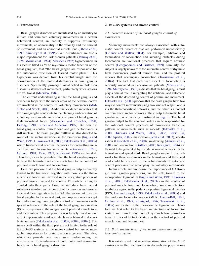
Fig. 2. Effects of electrical and chemical stimulations of the mesopontine tegmentum. (A) Experimental diagram. Either electrical or chemical stimulation was
delivered to the lateral part of the mesopontine tegmentum. (B) Effects on muscle activities following stimulation of the MLR (a) and the PPN (b). Each trace
was obtained from the left (L) and right (R) soleus (Sol) muscles. A downward filled arrowhead in (a) indicates the onset of the treadmill. An open triangle in (b)
indicates stimulation applied to the left pinna by pinching the scapha. (C) (a) An injection of NMDA into the left MLR increased the bilateral muscle tone. (b)
Commencement of the treadmill elicited locomotion. Downward and upward arrows indicate the onset and end of treadmill movements. (c) Two hours after the
first injection, NMDA was injected into the left PPN and inhibited the bilateral soleus muscle activity. Pinching the pinna after 5 min (indicated by an open
triangle) restored muscle activity. A dashed line above the recording indicates the period of the injection. (D) Effective sites on coronal (a) and parasagittal (b)
planes for evoking muscular atonia (filled circles) and locomotion (open circles). (A) Shaded area in both planes indicates the PPN. (E) Distribution of
cholinergic neurons stained by choline acetyltransferase (ChAT) immunohistochemistry. Light microscopic photographs of coronal (a and b) and parasagittal (c
and d) planes. Lower (a and c) and higher (b and d) magnification are shown in the right and left columns, respectively. Abbreviations: IC, inferior colliculus;
CNF, cuneiform nucleus; SCP, superior cerebellar peduncle; PPN, pedunculopontine tegmental nucleus; NRPo, nucleus reticularis pontis oralis; RD, raphe
dorsalis. Results in (B–E) are modified from Takakusaki et al. (2003a).
shows a possible architecture of the muscle tone inhibitory
system. PPN stimulation may activate cholinoceptive pontine
reticular formation (PRF) neurons (Lai et al., 1993; Mitani
et al., 1988), which, in turn, excite medullary reticulospinal
neurons and spinal interneurons to inhibit a-motoneurons.
Possibly suppressed in parallel are g-motoneurons and inter-
neurons intercalated in reflex pathways (Takakusaki et al.,
2001, 2003b). Monoaminergic systems such as the coeru-
lospinal (Fung and Barnes, 1981) and raphespinal (Sakai et
al., 2000) tracts are considered as muscle tone facilitatory
systems. There are serotonergic projections to the PPN
(Honda and Semba, 1994) and to the medial PRF (Semba,
1993). The former likely inhibits mesopontine cholinergic
neurons (Leonald and Llinas, 1994), and the latter reduces the
activity of the inhibitory system (Takakusaki et al., 1993,
1994). In contrast, the inhibitory system suppresses the
activity of the coerulospinal tract (Mileykovskiy et al.,
2000). Thus muscle tone can be regulated by a counterbalance
between the inhibitory and the facilitatory systems.
2.3. BG–BS systems in the control of muscle tone and
locomotion
In rats (Beckstead et al., 1979; Spann and Grofova, 1991)
and cats (Moriizumi et al., 1988) the mesopontine tegmen-
tum receives efferents of the basal ganglia particularly from
the SNr. The nigrotegmental efferents use GABA as a
neurotransmitter and have terminals preferentially on non-

K. Takakusaki et al. / Neuroscience Research 50 (2004) 137–151 141
Fig. 3. Neural architecture of locomotion executing system (A) and the muscle tone inhibitory system (B). See text for explanation. Abbreviations: ACh,
acetycholine; a,a-motoneuron; CPG, central pattern generator; E, extensor motoneurons, F, flexor motoneurons; FRA, flexion reflex afferents; GABA, g-
aminobutyric acid; LC, locus coeruleus; g,g-motoneuron; PMLS, pontomedullary locomotor strip; PRF, pontine reticular formation; RN, raphe nuclei; RSN,
reticulospinal neuron; SLR, subthalamic locomotor region; SNr, substantia nigra pars reticulata; NRGc, the nucleus reticularis gigangocellularis.
cholinergic neurons rather than cholinergic neurons (Gro-
fova and Zhou, 1998). Saitoh et al. (2003) demonstrated that
stimulation of the SNr induced monosynaptic IPSPs in PPN
neurons in vitro rat brainstem slice. Because the IPSP was
diminished by an application of bicuculline, one of GABAA
receptor antagonists, the IPSP was considered to be
mediated by GABAergic projections. A single-cell RT-
PCR amplification technique revealed that approximately
30% neurons were cholinergic in nature. These findings
suggest that not only non-cholinergic neurons but also
cholinergic neurons in the PPN receive GABAergic efferents
from the SNr.
How does the GABAergic nigrotegmental projection
control locomotion and muscle tone? This was examined
in decerebrate cats with the striatum, thalamus and cerebral
cortex removed, but the SNr preserved (Fig. 4A). An injec-
tion of bicuculline into the MLR also elicited locomotion on
a moving treadmill (Fig. 4B (a)). On the other hand,
microinjection of bicuculline into the ventrolateral PPN
inhibited the locomotor movements along with suppression
of postural muscle tone (Fig. 4B (b)). These findings suggest
that GABAergic efferents to the PPN and the MLR con-
ceivably suppress the activity of muscle tone inhibitory
system and locomotion executing system, respectively.
Next we examined how SNr stimulation altered the MLR/
PPN-induced movements. Stimulation of the SNr alone did
not alter muscular activity (Fig. 4C (a)). However, condi-
tioning stimuli applied to the lateral part of the SNr atte-
nuated and blocked the PPN-induced muscle tone
suppression (Fig. 4C (b)). In addition, stimuli applied to
the medial part of the SNr at a low strength reduced the
number of MLR-activated step cycles, increased the dura-
tion of the stance phase, and disrupted the rhythmic alter-
nation of limb movements (Fig. 4D). Stimulation of the SNr
at a higher strength eventually stopped MLR-activated
locomotion. Furthermore, the onset of the locomotion was
delayed by the SNr stimuli of progressively increasing
strength. Thus, the nigrotegmental projection affects both
the steady state (e.g., postural control and rhythmic limb
movements) and dynamic state (e.g., initiation and termina-
tion) of locomotion. Accordingly, our opinion is that the
basal ganglia control postural muscle tone and locomotion
by a combined inhibition/disinhibition of both the muscle
tone inhibitory system and the locomotion executing system
via the GABAergic nigrotegmental projections.
Moreover GABAergic nigrotegmental projections have a
partial functional topography: a lateral and medial SNr, for
regulation of postural muscle tone and locomotion, respec-
tively (Takakusaki et al., 2003a). Such a parallel organiza-
tion of the nigrotegmental projections may be capable of
controlling locomotion and muscle tone independently. It
follows that a variety of locomotor behaviors with various
step cycles and various levels of muscle tone could be
produced depending on the magnitude of inhibitory effects
K. Takakusaki et al. / Neuroscience Research 50 (2004) 137–151142
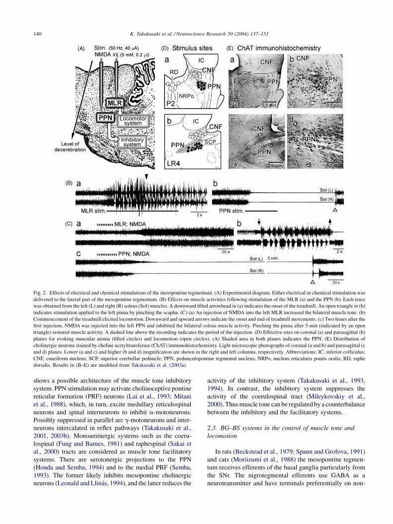
Fig. 4. GABAergic nigrotegmental projections control locomotion and muscle tone. (A) Experimental diagram. Either electrical or chemical stimulation was
delivered to the SNr, the MLR and the PPN. (B) (a) Quadrupedal locomotion observed at 15 min after injecting bicuculline into the MLR. (b) Another injection
of bicuculline into the PPN in the same cat suppressed the locomotion. A dashed line above the recording indicates the period of the injection. Muscle activities
were recorded from bilateral triceps brachial (TB) muscles and soleus muscles. (C) Nigral control of postural muscle tone. The effects induced by the SNr (a)
and PPN (b) on the postural muscle tone. PPN stimulation (20 mA) completely suppressed muscle tone. (c) When conditioning SNr stimuli of 50 mA were
delivered, the PPN-effect was abolished. (D) Nigral control of locomotion. (a) SNr stimulation did not change the level of muscle tone. (b) Locomotion on a
moving treadmill belt induced by the MLR. (c) Conditioning SNr stimuli of 30 mA reduced step cycles, delayed the onset (indicated by open arrowhead) and
disturbed rhythmic alteration of limb movements of MLR-activated locomotion. Results in (B–D) are modified from Takakusaki et al. (2003a).
from the functionally segregated nigrotegmental (medial
SNr-MLR and lateral SNr-PPN) projections.
3. Concept for understanding BG–BS Systems’
involvement of motor control
In this section, we first introduce our concept for under-
standing how the basal ganglia achieve an integration of the
volitional and automatic control of movement on the basis of
the results and viewpoints described above. On the other
hand, mechanisms of saccadic eye movements through
pathways from the basal ganglia to the superior colliculus
(SC) have been studied best among BG–BS systems, in
particular, by Hikosaka and his co-workers (Hikosaka, 1989;
Hikosaka and Wurtz, 1983a, 1983b, 1983b; Hikosaka et al.,
2000; Sato and Hikosaka, 2002). Thus, in the last part of this
section, we point out the similarities and differences
between these two concepts
3.1. Concept for understanding BG–BS systems in the
control of postural muscle tone and locomotion
There are multiple cortico-basal ganglia loops with var-
ious areas of the cerebral cortex which are concerned with
different aspects of motor behavior that requires volition,
cognition and attention (Brooks, 1995; Middleton and
Strick, 2000) (Fig. 5A). The majority of motor cortical
neurons significantly altered their discharge properties when
a walking subject has to overcome obstacles accurately
(Drew et al., 1996). This accuracy requires a precise visuo-
motor coordination (Georgopoulos and Grillner, 1989).
Thus, cortical processing is required for volitional aspects
of locomotor movements. Cortico-basal ganglia loop can
help serve this purpose. In contrast, a BG–BS system seems
required for the automatic regulation of postural muscle tone
and rhythmic limb movements during locomotion. The
motor cortices have projections to the PPN (Matsumura
et al., 2000) and to the pontomedullary reticular formation
(Matsuyama and Drew, 1997). Therefore the muscle tone
control system and the locomotor system can be controlled,
in parallel, by a combined input to the brainstem of net
inhibition from the basal ganglia, and net excitation from the
motor cortex (Fig. 5A).
Given the above consideration, the motor cortical neu-
rons that receive basal ganglia output may control the
velocity and the amount of voluntary movement (ordinate
on the left of the graph in Fig. 5B; Turner and Anderson,
1997). GABAergic inputs from the SNr to the MLR reduced
the drive from the MLR to the CPG in spinal cord, resulting
in disrupted the activity of locomotor pattern generator at the
level of spinal cord (Fig. 4D). Thus, a basal ganglia efferent
to the MLR may control the locomotor pattern (ordinate on
the right). In addition, a basal ganglia efferent to the PPN
may determine the level of muscle tone via the muscle tone
control systems (abscissa). Because the basal ganglia output

K. Takakusaki et al. / Neuroscience Research 50 (2004) 137–151 143
Fig. 5. A hypothetical model for the control of movements by the basal ganglia. (A) GABAergic basal ganglia projections to the thalamocortical neurons may
be involved in the volitional control aspects of movements, while those to the MLR and the PPN may be responsible for the automatic control processes of
locomotor movements and postural muscle tone. (B and C) See text for explanation. Abbreviations: BG, basal ganglia; HD, Huntington’s chorea; PD, Parkinson
disease; PMRF, pontomedullary reticular formation.
is variable in a normal condition, the degree of freedom for
the amount and the velocity of movement, the locomotor
velocity, and the muscle tone, can be large. Each parameter
can take any of the coordinates within the frame in Fig. 5B.
However, GABAergic basal ganglia output is thought to be
overactive in Parkinson disease (Wichmann and Delong,
1996, 2003). An excessive GABAergic inhibition upon
thalamocortical neurons may decrease the velocity and
amount of movement (bradykinesia and hypokinesia,
respectively). An increase in basal ganglia inhibition,
together with a decrease in cortical excitation of the PPN,
may increase the level of muscle tone (hypertonus). Simi-
larly, an excessive inhibition of the MLR and a decrease in
cortical excitation of the brainstem reticular formation may
elicit gait failure. Additionally, less activity of the premotor
cortex may disturb the motor programming required for
precise gait control (Hanakawa et al., 1999; Pahapill and
Lozano, 2000). As a result, the degree of freedom for each
movement would be restricted, and the frame will be smaller
and move to the upper right (Fig. 5C (a)).
Gait disturbances, including delays in gait onset (frozen
gait), an increase in the stance phase in locomotor cycles and
a decrease in locomotor velocity, are observed in Parkinso-
nian patients (Morris et al., 1994; Murray et al., 1978;
Pahapill and Lozano, 2000). Because these gait failure
resemble the locomotor pattern induced by SNr stimulation
(Takakusaki et al., 2003a), we consider that a dysfunction of
the BG–BS system is the primary basis for Parkinson
disease-induced gait impairments. Moreover, we propose
that muscular rigidity (hypertonus), which is one of the most
prominent symptoms of Parkinson disease, can be a result of
inhibition of the muscle tone inhibitory system (Fig. 2B).
Namely, muscular rigidity can be interpreted in terms of loss
of inhibition to a- and g-motoneurons.
In contrast, a reduction of output from the basal ganglia in
Huntington’s chorea may increase movement (hyperkine-
sias) and decrease muscle tone (hypotonus). The frame,
which indicates the degree of freedom for movement, would
be restricted and move to the lower left for this disease (Fig.
5C (b)). From these considerations, we suggest that a BG–
BS contributes to an automatic control of movement that
occurs in conjunction with voluntary control processes.
Moreover, the output of the basal ganglia would determine
the degree of freedom of each movement, and a restriction of
the degree of freedom could exist in the background of
Parkinson and Huntington diseases. We suggest that dys-
function of the BG–BS system together with that of cortico-
basal ganglia loop underlies the pathogenesis of motor
disturbances in these basal ganglia diseases.
Dystonia is a syndrome characterized by abnormal pos-
turing, muscle spasms, and tremor due to involuntary co-
contraction of muscle agonists and antagonists. Dystonia
can be task specific, patients only developing co-contraction
when performing skilled movements such as writing (Van
der Kamp et al., 1989). Using positron emission tomogra-
phy, inappropriate overactivity of the basal ganglia projec-
K. Takakusaki et al. / Neuroscience Research 50 (2004) 137–151144
tions to premotor and dorsal prefrontal cortex has been
observed in idiopathic and acquired dystonia (Brooks,
1995). However activities of primary sensorimotor and
caudal premotor cortices are rather attenuated (Hutchins
et al., 1988). Although alterations of noradrenaline and
dopamine levels in brainstem structures have been reported
in two cases (Hornykiewicz et al., 1986), most studies have
found no such abnormalities in the brainstem. These evi-
dences suggest that activity of the BG–BS system and that of
cortico-basal ganglia loop are controlled separately in this
disease.
3.2. Current concept for the basal ganglia control of
saccadic eye movements
The basal ganglia control saccadic eye movements (sac-
cades) through their connections to the superior colliculus
(SC) (Chevalier and Deniau, 1990; Chevalier et al., 1985;
Hikosaka and Wurtz, 1983a,b). Fig. 6A shows basic neural
connections involved in the generation of saccade. The SC
receives convergent inputs from the cerebral cortex (Hiko-
saka et al., 2000; Pierrot-Deseilligny et al., 2004) and the
basal ganglia (Anderson and Yoshida, 1980; Chevarier et al.,
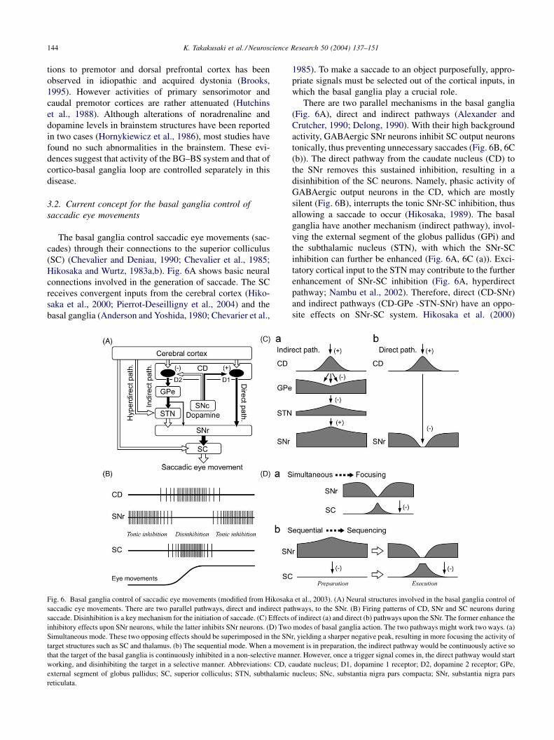
Fig. 6. Basal ganglia control of saccadic eye movements (modified from Hikosak
saccadic eye movements. There are two parallel pathways, direct and indirect pa
saccade. Disinhibition is a key mechanism for the initiation of saccade. (C) Effects
inhibitory effects upon SNr neurons, while the latter inhibits SNr neurons. (D) Two
Simultaneous mode. These two opposing effects should be superimposed in the SN
target structures such as SC and thalamus. (b) The sequential mode. When a movem
that the target of the basal ganglia is continuously inhibited in a non-selective man
working, and disinhibiting the target in a selective manner. Abbreviations: CD, c
external segment of globus pallidus; SC, superior colliculus; STN, subthalamic
reticulata.
1985). To make a saccade to an object purposefully, appro-
priate signals must be selected out of the cortical inputs, in
which the basal ganglia play a crucial role.
There are two parallel mechanisms in the basal ganglia
(Fig. 6A), direct and indirect pathways (Alexander and
Crutcher, 1990; Delong, 1990). With their high background
activity, GABAergic SNr neurons inhibit SC output neurons
tonically, thus preventing unnecessary saccades (Fig. 6B, 6C
(b)). The direct pathway from the caudate nucleus (CD) to
the SNr removes this sustained inhibition, resulting in a
disinhibition of the SC neurons. Namely, phasic activity of
GABAergic output neurons in the CD, which are mostly
silent (Fig. 6B), interrupts the tonic SNr-SC inhibition, thus
allowing a saccade to occur (Hikosaka, 1989). The basal
ganglia have another mechanism (indirect pathway), invol-
ving the external segment of the globus pallidus (GPi) and
the subthalamic nucleus (STN), with which the SNr-SC
inhibition can further be enhanced (Fig. 6A, 6C (a)). Exci-
tatory cortical input to the STN may contribute to the further
enhancement of SNr-SC inhibition (Fig. 6A, hyperdirect
pathway; Nambu et al., 2002). Therefore, direct (CD-SNr)
and indirect pathways (CD-GPe -STN-SNr) have an oppo-
site effects on SNr-SC system. Hikosaka et al. (2000)
a et al., 2003). (A) Neural structures involved in the basal ganglia control of
thways, to the SNr. (B) Firing patterns of CD, SNr and SC neurons during
of indirect (a) and direct (b) pathways upon the SNr. The former enhance the
modes of basal ganglia action. The two pathways might work two ways. (a)
r, yielding a sharper negative peak, resulting in more focusing the activity of
ent is in preparation, the indirect pathway would be continuously active so
ner. However, once a trigger signal comes in, the direct pathway would start
audate nucleus; D1, dopamine 1 receptor; D2, dopamine 2 receptor; GPe,
nucleus; SNc, substantia nigra pars compacta; SNr, substantia nigra pars

K. Takakusaki et al. / Neuroscience Research 50 (2004) 137–151 145
propose that two modes of basal ganglia action, ‘‘focusing’’
and ‘‘sequencing’’ of basal ganglia signals, are produced by
the interaction of the two opposing effects upon SNr neurons
(Fig. 6D). Simultaneous interaction of the two pathways
may produce more selective information and enhance the
spatial contrast of neural signals of the target systems
(focusing; Fig. 6D (a)). However, sequential interaction of
the pathways may produce switching of behavior from the
suppression of movement (when the indirect pathway is
dominant) to the initiation of movement (when the direct
pathway is dominant) (sequencing; Fig. 6Db). In this mode,
the effect would enhance the temporal contrast.
Accordingly, current concept for the basal ganglia control
of saccade can be summarized as follows. First, key mechan-
isms are an enhancement of tonic inhibition and a release
from the inhibition (disinhibition). The second mechanisms
are focusing and sequencing. These two modes can be
produced by an interaction of direct and indirect pathways.
The above mechanisms may act on brainstem networks in
addition to thalamocortical networks (Hikosaka et al., 2000).
3.3. Comparisons of two concepts
Similar to the basal ganglia control of saccade, disin-
hibition and enhancement of the inhibition can be also key
mechanisms for the basal ganglia control of postural muscle
tone and locomotion. Because muscle tone inhibitory region
in the PPN and the MLR, as well as the SC, receive
GABAergic input from the SNr, locomotor system and
muscle tone control system may be regulated by the balance
of direct and indirect pathways. When locomotor movement
is in preparation, tonic activity of SNr neurons would
continuously inhibit both systems. Once a trigger signal
comes in, the direct pathway would release the activity of
these systems, resulting in initiation of locomotion that is
followed by smooth reduction of the level of muscle tone.
Parallel organization from the SNr to the MLR/PPN would
be therefore beneficial to regulate the level of muscle tone
which accompanies with the initiation and termination of
locomotion.
However particular emphasis has not placed on the
importance of sustained inhibitory input from the SNr to
SC during the period of saccade. Here we emphasize a
crucial role of the sustained output from the basal ganglia to
the target motor systems (for example, PPN and MLR) for
controlling steady-state of ongoing movements such as
maintenance of postural muscle tone and rhythmic limb
movements during locomotion. As previously described the
sustained basal ganglia output signals may control the
degree of freedom of the excitability of the target systems
during movements. For example, when a subject needs to
adapt heavy load during walking, the subject may uncon-
sciously select an appropriate gait pattern which is asso-
ciated with higher level of muscle tone and slower walking
speed. Such a gait pattern could be realized by an increase in
sustained SNr outputs to the PPN and the MLR, resulting in
a decrease in the excitabilities of muscle tone inhibitory
system and locomotor rhythm generating system. The sus-
tained output from the basal ganglia may thus be necessary
to automatically optimize the excitabilities of plural target
motor systems so that the subject can unconsciously select
an appropriate motor pattern.
4. BG–BS systems for brain function in general
Cognitive and psychotic processes have been observed in
patients with degenerative disorders that involve primarily
the basal ganglia such as Parkinson disease (Graybiel, 1995;
Hikosaka et al., 2000; Mellers et al., 1995; Taylor et al.,
1986) and Huntington’s disease (McHugh and Folsten,
1975). In addition, awake–sleep states were also impaired
in Parkinsonian patients (Bliwise et al., 2000; Eisensehr et
al., 2001; Rye et al., 1999). In experimental studies in
primates, limited lesions of the striatum induce deficits in
rule acquisition (Divac, 1972), cognition (Taylor et al.,
1990), working memory performance (Goldman-Rakic,
1987) and selected attention (Battig et al., 1962). For
example, Laplane et al. (1984) reported a patient with
restricted bilateral pallidal lesions. He was appeared apa-
thetic and unconcerned or attention deficits, and his affect
was flattened and emotional responses were blunted in the
absence of any motor disorder or akinesia (pure psychic
akinesia). These symptoms were also described in progres-
sive supranuclear palsy (PSP) in which major lesions were
observed in the subcortical areas including the PPN (Zweig
et al., 1985). Because neuronal loss of cholinergic PPN
neurons were observed not only in PSP (75–80%) but also
Parkinson disease (43–57%) (Hirsch et al., 1987; Jellinger,
1988; Zweig et al., 1987, 1989), the loss of cholinergic PPN
neurons in both diseases could attribute to attentive and
cognitive impairments and sleep deficiencies in these dis-
eases (Scarnati and Florio, 1997). These clinical evidences
corroborate that the basal ganglia and their connections with
the brainstem are also involved in the expression of non-
motor function. In this section, we particularly discuss roles
of the BG–BS system in the regulation of REM sleep,
arousal state and an expression of emotional motor beha-
viors.
4.1. REM sleep
The pontomesencephalic reticular formation has been
known to comprise the ascending reticular activation system
(ARAS; Moruzzi and Magoun, 1949), and the PPN is
considered as a part of the ARAS (Garcia-Rill, 1997; Jones,
1991; Steriade, 1996). Cholinergic neurons in the PPN and
laterodorsal tegmental nucleus are involved in not only the
maintenance of arousal state but also generation of REM
sleep (Datta, 2002; Koyama and Sakai, 2000; Maloney et al.,
1999; Rye, 1997). Major ascending cholinergic projections
into the non-specific thalamic nuclei provide desynchroni-

K. Takakusaki et al. / Neuroscience Research 50 (2004) 137–151146
Fig. 7. Basal ganglia efferents to the PPN involved in the control of REM and muscular atonia. (A) Schematic model for the basal ganglia control of REM sleep.
See text for detailed explanation. Briefly, the pontomesencephalic reticular formation including the PPN comprises ascending reticular activation system
(ARAS). An interconnection between the mesopontine cholinergic nuclei and the caudoventral PRF could operate as a common generator of REM (Vanni-
Mercier and Debilly, 1998). Descending projections from the PPN may activate REM generator and muscle tone inhibitory system in the PMRF to induce REM
and muscular atonia. GABAergic basal ganglia efferents may affect REM sleep by modulating the activities of the PPN and the non-specific thalamic nuclei. (B)
Experimental diagram for examination of the involvement of a nigrotegmental projection in the control of REM and muscular atonia. (C) (a) Stimulation of the
PPN induced REM and muscular atonia. (b) Conditioning stimulation of the lateral part of the SNr diminished the PPN-effects. (c) Conditioning stimuli applied
to the mid part of the SNr did not block REM but blocked the muscular atonia (REM without atonia). Results in (C) are modified from Takakusaki et al. (2004b).
zation of electroencephalogram (EEG), i.e., EEG arousal
(Steriade, 1996). Moreover, cholinergic projections to the
lateral geniculate nucleus may provide ponto-geniculo-occi-
pital (PGO) waves (McCormick and Bal, 1997; Steriade,
2001). Descending projections to the pontomedullary reti-
cular formation (Lai et al., 1993; Shiromani et al., 1990) are
involved in muscular atonia. Projections to the caudoventral
pontine tegmentum are thought to be responsible for the
generation of both REM and PGO waves (Vanni-Mercier
and Debilly, 1998).
The SNr has a direct projection to the thalamic nuclei
(Hendry et al., 1979; Parent et al., 1983) in addition to the
PPN. Consequently, basal ganglia output may affect a REM
sleep state by a modulation of the ARAS through dual
systems (Fig. 7A). One is through a direct nigrothalamic
projection. The other is mediated via the PPN. We examined
how the latter projection (GABAergic SNr-PPN projection)
altered the activity of the REM generator and the muscle
tone inhibitory system (Takakusaki et al., 2004b; Fig. 7B).
Stimulation of inhibitory region of the PPN induced REM
which was associated with muscular atonia in decerebrate
cats (REM with atonia; Fig. 7C (a)). Conditioning stimulation
applied to the lateral part of the SNr completely abolished the
PPN-induced REM with atonia (Fig. 7C (b)). However,
stimuli applied to the mid part of the SNr did not block
REM but attenuated the muscular atonia, i.e., REM without
atonia, which is relevant to REM sleep behavioral disorder
(RBD; Culebras and Moore, 1989; Stanford et al., 1994), was
induced by stimulation of the SNr (Fig. 7C (c)). These
findings indicate that neuronal mechanisms for the induction
of REM and muscular atonia are under the regulation of a
GABAergic inhibition from the SNr. Patients with Parkin-
son’s disease experience a number of sleep disorders, includ-
ing reduction of REM sleep period and RBD (Bliwise et al.,
2000; Eisensehr et al., 2001). Accordingly, our results may
support the proposition that a decrease in dopaminergic
activities in the basal ganglia is involved in the reduction
of REM sleep and in RBD (Albin et al., 2001; Rye et al.,
1999), and provide a rational explanation for pathogenesis of
sleep disturbances in Parkinson disease.
However, the above idea does not agree with following
findings. First, a group of nigrotegmental neurons increased
their firing rate during REM sleep (Datta et al., 1991).
Second, a c-fos expression of GABAergic SNr neurons
during REM sleep was higher than during non-REM sleep
and wakefulness (Maloney et al., 2002). These findings
indicate that GABAergic SNr neurons do not necessarily
contribute to the induction of REM sleep in normal condi-
tion. We consider that cholinergic-monoaminergic recipro-
city (Hobson et al., 1986) in the brainstem may play a more
crucial role for the generation of REM sleep than the
GABAergic SNr-PPN projection in normal state. However,
the excessive GABAergic inhibition might affect the gen-
eration of REM sleep in Parkinsonian state.
K. Takakusaki et al. / Neuroscience Research 50 (2004) 137–151 147
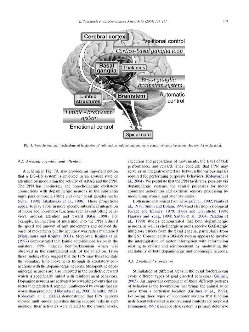
Fig. 8. Possible neuronal mechanisms of integration of volitional, emotional and automatic control of motor behaviors. See text for explanation.
4.2. Arousal, cognition and attention
A schema in Fig. 7A also provides an important notion
that a BG–BS system is involved in an arousal state or
attention by modulating the activity of ARAS and the PPN.
The PPN has cholinergic and non-cholinergic excitatory
connections with dopaminergic neurons in the substantia
nigra pars compacta (SNc) and other basal ganglia nuclei
(Kitai, 1998; Takakusaki et al., 1996). These projections
appear to play a role in more specific subcortical integration
of motor and non-motor functions such as controlling beha-
vioral arousal, attention and reward (Kitai, 1998). For
example, an injection of muscimol into the PPN reduced
the speed and amount of arm movements and delayed the
onset of movements but the accuracy was rather maintained
(Matsumura and Kojima, 2001). Moreover, Kojima et al.
(1997) demonstrated that kainic acid-induced lesion in the
unilateral PPN induced hemiparkisonism which was
observed in the contralateral side of the injection. From
these findings they suggest that the PPN may thus facilitate
the voluntary limb movements through its excitatory con-
nections with the dopaminergic neurons. Mesopontine dopa-
minergic neurons are also involved in the predictive reward
which is specifically linked with reinforcement behaviors.
Dopamine neurons are activated by rewarding events that are
better than predicted, remain uninfluenced by events that are
worse than predicted (Hikosaka et al., 2000; Schultz, 1998).
Kobayashi et al. (2002) demonstrated that PPN neurons
showed multi-modal activities during saccade tasks in alert
monkey; their activities were related to the arousal levels,
execution and preparation of movements, the level of task
performance, and reward. They conclude that PPN may
serve as an integrative interface between the various signals
required for performing purposive behaviors (Kobayashi et
al., 2004). We postulate that the PPN facilitates, possibly via
dopaminergic systems, the central processes for motor
command generation and extrinsic sensory processing by
modulating arousal and attentive states.
Both neuroanatomical (von Krosigk et al., 1992; Nauta et
al., 1978; Smith and Bolam, 1990) and electrophysiological
(Grace and Bunney, 1979; Hajos and Greenfield, 1994;
Hausser and Yung, 1994; Saitoh et al., 2004; Paladini et
al., 1999) studies demonstrated that both dopaminergic
neurons, as well as cholinergic neurons, receive GABAergic
inhibitory effects from the basal ganglia, particularly from
the SNr. Consequently a BG–BS system appears to involve
the interdigitation of motor information with information
relating to reward and reinforcement by modulating the
excitability of both dopaminergic and cholinergic neurons.
4.3. Emotional expression
Stimulation of different areas in the basal forebrain can
evoke different types of goal directed behaviors (Grillner,
2003). An important component of these different patterns
of behavior is the locomotion that brings the animal to or
away from a particular location (Grillner et al., 1997).
Following three types of locomotor systems that function
in different behavioral or motivational contexts are proposed
(Sinnamon, 1993); an appetitive system, a primary defensive

K. Takakusaki et al. / Neuroscience Research 50 (2004) 137–151148
system and an exploratory system. The nucleus accumbens
and the ventral pallidum, the older parts of the basal ganglia,
are considered to take part in locomotor control through the
MLR (Mogenson, 1991; Slawinska and Kasicki, 1995).
Projections from the limbic structures (hippocampus and
amygdala) to the nucleus accumbens are possibly involved
in the expression of emotional aspects of locomotor beha-
viors (Grillner et al., 1997). Therefore, as shown in Fig. 8,
the mesopontine tegmentum receives volitional signals from
the cerebral cortex (volitional control) and emotional signals
from the limbic structures (emotional control). Since the
basal ganglia receive afferents from these two structures, a
BG–BS system may play key roles for integration, selection
or switching of volitionally-guided and emotionally-trig-
gered motor behaviors (Fig. 8).
In narcoleptic patients and animals, emotional signals
elicit sudden loss of muscular tonus (cataplexy) (Nishino
and Mignot, 1997). Thus emotional signals may have a
capability of not only evoking locomotor behaviors but also
eliciting muscular atonia. It has been shown that the orex-
inergic system contributes to maintain awake state (Saper et
al., 2001; Taheri et al., 2002), and that deficiencies in the
orexinergic system result in narcolepsy (Chemelli et al.,
1999; Lin et al., 1999). Because the midbrain, including the
SNr, the PPN and the MLR, receive orexinergic efferents
from the perifornical lateral hypothalams (Nambu et al.,
1999; Peyron et al., 1998), we propose that orexinergic
projections to these midbrain areas must be critical for
the expression of different aspects of emotional motor
behaviors. Saper et al. (2001) have proposed that orexinergic
projections to the midbrain are involved in switching sleep–
awake states.
To test the above proposition, we examined effects of
injections of orexin-A (60 mM–1.0 mM, 0.20–0.25 ml) into
the MLR, PPN and the SNr upon motor behaviors in
decerebrate cats (Takakusaki et al., 2004d). We observed
that orexin injections into the MLR facilitated locomotion,
while those into either the PPN or the SNr suppressed PPN-
induced muscular atonia (cataplexic state). The latter
effects were reversed by subsequent injection of bicucul-
line into the PPN. These findings suggest that the excit-
ability seems to be higher in the locomotor system than in
the atonia system in the presence of orexin. On the other
hand, the excitability of the atonia system may be higher
than that of the locomotor system in the absence of orexin.
Thus emotional signals to the midbrain may induce loco-
motor behavior in the context of normal orexinergic system
function, but elicit cataplexy in narcolepsy when orexiner-
gic system is disturbed. Therefore orexin may be a deter-
minant of the selection of emotional motor behaviors
(Takakusaki et al., 2003c).
An integration of ‘‘the locomotor system’’ and ‘‘the
muscle tone control system’’ is essential to elicit a variety
of locomotor patterns. The mesopontine tegmentum receives
afferents from the cerebral cortex, the limbic systems, and
hypothalamus, in addition to the basal ganglia. Thus the
BG–BS system may contribute to the integrative process of
volitional and emotional signals from these forebrain struc-
tures so that an animal can elicit appropriate locomotor
behaviors depending on the behavioral context.
5. Concluding remarks
We proposed that following roles can be played by the
BG–BS system. First the system is involved in the automatic
or unconscious control of movements that accompany
voluntary movements. The basal ganglia outputs toward
the brainstem and the thalamocortical loop may determine
the degree of freedom of the automatic and volitional aspects
of movements, respectively. Second, BG–BS systems may
be involved in the maintenance of arousal and attentive
states and in the regulation of REM sleep. These global brain
function can be brought about by modulation of both
cholinergic and dopaminergic systems arising from the
brainstem. Third, the BG–BS systems may be involved in
the appropriate expression of locomotor behaviors by inte-
grating volitional and emotional signals from the forebrain
structures. In this article, we presented several schemas in
order to facilitate readers’ interpretation. Obviously, these
schemas are incomplete and overspecified. To test their
validity, it must be necessary to formulate computational
models based on the schemas and simulate the experimental
results.
Acknowledgements
This study was supported by the Japanese Grants-in-Aid
for Scientific Research (C) and Priority Areas (A), RISTEX
of JST (Japan Science and Technology Agency) and a grant
from the Uehara Memorial Foundation to KT.
References
Albin, R.L., Koeppe, R.A., Chervin, R.D., Consens, F.B., Wernette, K.,
Frey, K.A., Aldrich, M.S., 2001. Decreased striatal dopaminergic
innervation in REM sleep behavior disorder. Neurology 55,
1410–1412.
Alexander, G.E., Crutcher, M.D., 1990. Functional architecture of basal
ganglia circuits: neural substrates of parallel processing. Trends Neu-
rosci. 13, 267–271.
Amirali, A., Tsai, G., Schrader, N., Weisz, D., Sanders, I., 2001. Mapping of
brain stem neuronal circuitry active during swallowing. Ann. Otol.
Rhinol. Laryngol. 110, 502–513.
Anderson, M., Yoshida, M., 1980. Axonal branching patterns and location
of nigrothalamic and nigrocollicular neurons in the cat. J. Neurophysiol.
43, 883–895.
Battig, K., Rosvold, H.E., Mishkin, M., 1962. Comparison of the effects of
frontal and caudate lesions on discrimination learning in monkeys. J.
Comp. Physiol. Psychol. 55, 458–463.
Beckstead, R.M., Domesick, V.B., Nauta, W.J.H., 1979. Efferent connec-
tions of the substantia nigra and ventral tegmental area in the rat. Brain
Res. 175, 191–217.
K. Takakusaki et al. / Neuroscience Research 50 (2004) 137–151 149
Bliwise, D.L., Willians, M.L., Irbe, D., Ansari, F.P., Rye, D.B., 2000. Inter-
rater reliability for identification of REM sleep in Parkinson’s disease.
Sleep 23, 671–676.
Brooks, D.J., 1995. The role of the basal ganglia in motor control: con-
tributions from PET. J. Neurol. Sci. 128, 1–13.
Chase, M.H., Morales, F.R., 1990. The atonia and myoclonia of active
(REM) sleep. Ann. Rev. Psychol. 41, 557–584.
Chase, M.H., Morales, F.R., Boxer, P., Fung, S.J., Soja, P.J., 1986. Effect of
stimulation of the nucleus reticularis gigantocellularis on the membrane
potential of cat lumbar motoneurons during sleep and wakefulness.
Brain Res. 386, 237–244.
Chemelli, R.M., Willie, J.T., Sinton, C.M., Elmquist, J.K., Scammell, T.,
Lee, C., Richardson, J.A., Williams, S.C., Xiong, Y., Kisanuki, Y., Fitch,
T.E., Nakazato, M., Hammer, R.E., Saper, C.B., Yanagisawa, M., 1999.
Narcolepsy in orexin knockout mice: molecular genetics of sleep
regulation. Cell 98, 437–451.
Chevalier, G., Deniau, J.M., 1990. Disinhibition as a basic process in the
expression of striatal functions. Trends Neurosci. 13, 277–280.
Chevalier, G., Vacher, S., Deniau, J.M., Desban, M., 1985. Disinhibition as a
basic process in the expression of striatal functions. Part I. The striato-
nigral influence on tect-spino/tecto-diencephalic neurons. Brain Res.
334, 215–226.
Culebras, A., Moore, J.T., 1989. Magnetic resonance findings in REM sleep
behavior disorder. Neurology 39, 1519–1523.
Datta, S., 2002. Evidence that REM sleep is controlled by the activation of
brain stem pedunculopontine tegmental kainate receptor. J. Neurophy-
siol. 87 .
Datta, S., Curro Dossi, R., Pare, D., Oakson, G., Steriade, M., 1991.
Substantia nigra reticulata neurons during sleep-waking states: relation
with ponto-geniculo-occipital waves. Brain Res. 566, 344–347.
Delong, M.R., 1990. Primate models of movement disorders of basal
ganglia origin. Trends Neurosci. 13, 281–289.
Divac, I., 1972. Neostriatum and functions of the prefrontal cortex. Acta
Neurobiol. Exp. 32, 461–477.
Drew, T., Jiang, W., Kably, B., Lavoie, S., 1996. Role of the motor cortex in
the control of visually triggered gait modifications. Can. J. Physiol.
Pharmacol. 74, 426–442.
Dusterhoft, F., Hausler, U., Jurgens, U., 2000. On the search for the vocal
pattern generator. A single-unit recording study. Neuroreport 11, 231–234.
Eisensehr, I., Lindeiner, H., Jager, M., Noachtar, S., 2001. REM sleep
behavior disorder in sleep-disordered patients with versus without
Parkinson’s disease: is there a need for polysomnography? J. Neurol.
Sci. 186, 7–11.
Fung, S.J., Barnes, C.D., 1981. Evidence of facilitatory coerulospinal action
in lumbar motoneurons of cats. Brain Res. 261, 299–311.
Garcia-Rill, E., 1991. The pedunculopontine nucleus. Prog. Neurobiol. 36,
363–389.
Garcia-Rill, E., 1997. Disorders of the reticular activating system. Med.
Hypothesis. 49, 379–387.
Georgopoulos, A.P., Grillner, S., 1989. Visuomotor coordination in reaching
and locomotion. Science 245, 1209–1210.
Goldman-Rakic, P.S., 1987. Circuitry of primate prefrontal cortex and
regulation of behavior by representational memory. In: Plum, F., Cow-
man, M. (Eds.), Handbook of Physiology, The Nervous System, vol. V.
American Physiological Society, Bethesda, pp. 373–416.
Grace, A.A., Bunney, B.S., 1979. Paradoxical GABA excitation of nigral
dopaminergic cells: indirect mediation through reticulata inhibitory
neurons. Eur. J. Pharmacol. 59, 211–218.
Graybiel, A.M., 1995. Building action repertoires: memory and learning
functions of the basal ganglia. Curr. Opin. Neurobiol. 5, 733–741.
Grillner, S., 1981. Control of locomotion in bipeds, tetrapods, and fish. In:
Brooks, V.B. (Ed.), The Nervous System, vol. II. American Physiolo-
gical Society Press, Bethesda, pp. 1179–1236.
Grillner, S., 2003. The motor infrastructure: from ion channels to neuronal
networks. Nat. Rev. Neurosci. 4, 573–586.
Grillner, S., Georgopoulos, A.P., Jordan, L.M., 1997. Selection and initia-
tion of motor behavior, MIT Press, Boston.
Grillner, S., Wallen, P., 2004. Innate versus learned movements—a false
dichotomy? Prog. Brain Res. 143, 3–12.
Grofova, I., Zhou, M., 1998. Nigral innervation of cholinergic and gluta-
matergic cells in the rat mesopontine tegmentum: Light and electron
microscopic anterograde tracing and immunohistochemical studies. J.
Comp. Neurol. 395, 359–379.
Habaguchi, T., Takakusaki, K., Saitoh, K., Sugimoto, J., Sakamoto, T.,
2002. Medullary reticulospinal tract mediating the generalized motor
inhibition in cats: II. Functional organization within the medullary
reticular formation with respect to postsynaptic inhibition of forelimb
and hindlimb motoneurons. Neuroscience 113, 65–77.
Hajos, M., Greenfield, S.A., 1994. Synaptic connections between pars com-
pacta and pars reticulata neurones: electrophysiological evidence for
functional modules within the substantia nigra. Brain Res. 660, 216–
224.
Hanakawa, T., Katsumi, Y., Fukuyama, H., Honda, M., Hayashi, T., Kimura,
J., Shibasaki, H., 1999. Mechanisms underlying gait disturbance in
Parkinson’s disease: a single photon emission computed tomography
study. Brain 122, 1271–1282.
Hausser, M.A., Yung, W.H., 1994. Inhibitory synaptic potentials in guinea-
pig substantia nigra dopamine neurones in vitro. J. Physiol. 479, 401–422.
Hendry, S.H.C., Jones, E.G., Graham, J., 1979. Thalamic relay nuclei for
cerebellar and certain related fiber systems in the cat. J. Comp. Neurol.
185, 679–714.
Hikosaka, O., 1989. Role of basal ganglia in initiation of voluntary
movements, Springer-Verlag.
Hikosaka, O., Takikawa, Y., Kawgoe, R., 2000. Role of the basal ganglia in
the control of purposive saccadic eye movements. Physiol. Rev. 80,
954–978.
Hikosaka, O., Wurtz, R.H., 1983a. Visual and oculomotor functions of
monkey substantia nigra pars reticulata, vol. II. Visual responses related
to fixation of gaze. J. Neurophysiol. 49, 1254–1267.
Hikosaka, O., Wurtz, R.H., 1983b. Visual and oculomotor functions of
monkey substantia nigra pars reticulata, vol. III. Memory-contingent
visual and saccade responses. J. Neurophysiol. 49, 1268–1284.
Hikosaka, O., Wurtz, R.H., 1983c. Visual and oculomotor functions of
monkey substantia nigra pars reticulata. IV. Relation of substantia nigra
to superior colliculus. J. Neurophysiol. 49, 1285–1301.
Hirsch, E.C., Graybiel, A.M., Duyckaerts, C., Javoy-Agid, F., 1987. Neu-
ronal loss in the pedunculopontine tegmental nucleus in Parkinson
disease and in progressive supranuclear palsy. Proc. Natl. Acad. Sci.
USA 84, 5976–5980.
Hobson, J.A., Lydic, R., Baghdoyan, H.A., 1986. Evolving concepts of sleep
cycle generation: from brain centers to neuronal populations. Behav.
Brain Sci. 9, 371–448.
Honda, T., Semba, K., 1994. Serotoergic synaptic input to cholinergic
neurons in the rat mesopontine tegmentum. Brain Res. 647, 299–306.
Hornykiewicz, O., Kish, S.J., Becker, L.E., Farley, I., Shannak, K., 1986.
Brain neurotransmitters in dystonia musculorum deformans. N. Engl. J.
Med. 315, 347–353.
Hutchins, K.D., Martino, A.M., Strick, P.L., 1988. Corticospinal projections
from the medial wall of the hemisphere. Exp. Brain Res. 71, 667–672.
Inglis, W.L., Winn, P., 1995. The pedunculopontine tegmental nucleus:
where the striatum meets the reticular formation. Prog. Neurobiol. 47,
1–29.
Isa, T., 2002. Intrinsic processing in the mammalian superior colliculus.
Curr. Opin. Neurobiol. 12, 668–677.
Jellinger, K., 1988. The pedunculopontine nucleus in Parkinson’s disease,
progressive supranuclear palsy and Alzheimer’s disease. J. Neurol.
Neurosurg. Psychiatry. 51, 540–543.
Jones, B.E., 1991. Paradoxical sleep and its chemical/structural substrates in
the brain. Neuroscience 40, 637–656.
Kitai, S.T., 1998. Afferent control of substantia nigra compacta dopamine
neurons: anatomical perspective and role of glutamatergic and choli-
nergic inputs. Adv. Pharmacol. 42, 700–702.
Kobayashi, Y., Inoue, Y., Isa, T., 2004. Pedunculopontine control of visually
guided saccades. Brain Res. 143, 439–445.
K. Takakusaki et al. / Neuroscience Research 50 (2004) 137–151150
Kobayashi, Y., Inoue, Y., Yamamoto, M., Isa, T., Aizawa, H., 2002.
Contribution of pedunculopontine tegmental nucleus neurons to per-
formance of visually guided saccade tasks in monkeys. J. Neurophysiol.
88, 715–731.
Kojima, J., Yamaji, Y., Matsumura, M., Nambu, A., Inase, M., Tokuno, H.,
Takada, M., Imai, H., 1997. Excitotoxic lesions of the pedunculopontine
tegmental nucleus produce contralateral hemiparkinsonism in the mon-
key. Neurosci. Lett. 226, 111–114.
Koyama, Y., Sakai, K., 2000. Modulation of presumed cholinergic meso-
pontine tegmental neurons by acetylcholine and monoamines applied
iontophoretically in unanesthetized cats. Neuroscience 96, 723–733.
von Krosigk, M., Smith, Y., Bolam, J.P., Smith, A.D., 1992. Synaptic
organization of GABAergic inputs from the striatum and the globus
pallidus onto neurons in the substantia nigra and retrorubral field which
project to the medullary reticular formation. Neuroscience 50, 531–549.
Lai, Y.Y., Clements, J.R., Siegel, J.M., 1993. Glutamatergic and cholinergic
projections to the pontine inhibitory area identified with horseradish
peroxidase retrograde transport and immunohistochemistry. J. Comp.
Neurol. 336, 321–330.
Lai, Y.Y., Siegel, J.M., 1988. Medullary regions mediating atonia. J.
Neurosci. 8, 4790–4796.
Lai, Y.Y., Siegel, J.M., 1990. Muscle tone suppression and stepping
produced by stimulation of midbrain and rostral pontine reticular
formation. J. Neurosci. 10, 2727–2734.
Lai, Y.Y., Siegel, J.M., 1991. Pontomedullary glutamate receptors mediat-
ing locomotion and muscle tone suppression. J. Neurosci. 11, 2931–
2937.
Laplane, D., Baulac, M., Widlocher, D., Dubois, B., 1984. Pure psychic
akinesia with bilateral lesions of basal ganglia. J. Neurol. Neurosurg.
Psychiatry 47, 377–385.
Leonald, C.S., Llinas, R., 1994. Serotonergic and cholinergic inhibition of
mesopontine cholinergic neurons controlling REM sleep; an in vitro
electrophysiological study. Neuroscience 59, 309–330.
Lin, L., Faraco, J., Li, R., Kadotani, H., Rogers, W., Lin, X., Qiu, X., de
Jong, P.J., Nishino, S., Mignot, E., 1999. The sleep disorder canine
narcolepsy is caused by a mutation in the hypocretin (orexin) receptor 2
gene. Cell 98, 365–376.
Magoun, M.W., Rhines, R., 1946. An inhibitory mechanism in the bulbar
reticular formation. J. Neurophysiol. 9, 165–171.
Maloney, K.J., Mainville, L., Jones, B.E., 1999. Differential c-Fos expres-
sion cholinergic, monoaminergic and GABAergic cell groups of the
pontomesencephalic tegmentum after paradoxical sleep deprivation and
recovery. J. Neurosci. 19, 3057–3072.
Maloney, K.J., Mainville, L., Jones, B.E., 2002. c-Fos expression in
dopaminergic and GABAergic neurons of the ventral mesencephalic
tegmentum after paradoxical sleep deprivation and recovery. Eur. J.
Neurosci. 15, 1–6.
Marsden, C.D., 1982. The mysterious motor function of the basal ganglia:
The Robert Wartenberg Lecture. Neurology 32, 514–539.
Marsden, C.D., 1989. Slowness of movement in Parkinson’s disease. Mov.
Disord. 4 (Suppl. 1), 26–37.
Masdeu, J.C., Alampur, U., Cavaliere, R., Tavoulareas, G., 1994. Astasia
and gait failure with damage of the pontomesencephalic locomotor
region. Ann. Neurol. 35, 619–621.
Matsumura, M., Kojima, J., 2001. The role of the pedunculopontine
tegmental nucleus in experimental parkinsonism in primates. Stereotact.
Funct. Neurosurg. 77, 108–115.
Matsumura, M., Nambu, A., Yamaji, Y., Watanabe, K., Imai, H., Inase, M.,
Tokuno, H., Takada, M., 2000. Organization of somatic motor inputs
from the frontal lobe to the pedunculopontine tegmental nucleus in the
macaque monkey. Neuroscence 98, 97–110.
Matsuyama, K., Drew, T., 1997. The organization of the projection from the
pericruciate cortex to the pontomedullary brainstem of the cat: a study
using the anterograde tracer. Phaseolus vulgaris leucoagglutinin. J.
Comp. Neurol. 389, 617–641.
McCormick, D.A., Bal, T., 1997. Sleep and arousal: thalamocortical
mechanisms. Ann. Rev. Neurosci. 20, 185–215.
McHugh, P.R., Folsten, M.F., 1975. Psychiatric syndromes of Huntington’s
chorea: a clinical and phenomenologic study, Grune & Stratton, New
York.
Mellers, J.D., Quinn, N.P., Ron, M.A., 1995. Psychotic and depressive
symptoms in Parkinson’s disease. A study of the growth hormone
response to apomorphine. Br. J. Psychiatry. 167, 522–526.
Middleton, F.A., Strick, P.L., 2000. Basal ganglia and cerebellar loops:
motor and cognitive circuits. Brain Res. Rev. 31, 236–250.
Mileykovskiy, B.Y., Kiyashchenko, L.I., Kodama, T., Lai, Y.Y., Siegel, J.M.,
2000. Activation of pontine and medullary motor inhibitory regions
reduces discharge in neurons located in the locus coeruleus and the
anatomical equivalent of the midbrain locomotor region. J. Neurosci.
20, 8551–8558.
Mitani, A., Ito, K., Hallanger, A.E., Wainer, B.H., Kataoka, K., McCarley,
R.W., 1988. Cholinergic projections from the laterodorsal and pedun-
culopontine tegmental nuclei to the pontine giganotocellular tegmental
field in the cat. Brain Res. 451, 397–402.
Mogenson, G.I., 1991. The role of mesolimbic dopamine projections to the
ventral striatum in response initiation, Japan Scientific Press.
Mori, S., 1987. Integration of posture and locomotion in acute decere-
brate cats and in awake, free moving cats. Prog. Neurobiol. 28, 161–
196.
Moriizumi, T., Nakamura, Y., Tokuno, H., Kitao, Y., Kudo, M., 1988.
Topographic projections from the basal ganglia to the nucleus tegmenti
pedunculopontinus pars compacta of the cat with special reference to
pallidal projections. Expl. Brain Res. 71, 298–306.
Morris, M.E., Iansek, R., Matyas, T.A., Summers, J.J., 1994. The
pathogenesis of gait hypokinesia in Parkinson’s disease. Brain 117,
1169–1181.
Moruzzi, G., Magoun, H.W., 1949. Brain stem reticular formation and
activation of the EEG. Clin. Neurophysiol. 1, 455–473.
Murray, M.P., Sepic, S.B., Gardner, G.M., Downs, W.J., 1978. Walking
patterns of men with parkinsonism. Am. J. Phys. Med. 57, 278–294.
Nambu, T., Sakurai, T., Mizukami, K., Hosoya, Y., Yanagisawa, M., Goto,
K., 1999. Distribution of orexin neurons in the adult rat brain. Brain Res.
827, 243–260.
Nambu, A., Tokuno, H., Takada, M., 2002. Functional significance of the
cortico- subthalamo-pallidal ’hyperdirect’ pathway. Neurosci. Res. 43,
111–117.
Nauta, W.J., Smith, G.P., Faull, R.L., Domesick, V.B., 1978. Efferent
connections and nigral afferents of the nucleus accumbens septi in
the rat. Neuroscience 3, 385–401.
Nishino, S., Mignot, E., 1997. Pharmacological aspects of human and
canine narcolepsy. Prog. Neurobiol. 52, 27–78.
Obeso, J.A., Rodriguez, M.C., DeLong, M.R., 1997. Basal ganglia
pathophysiology, Lippincott-Raven Publishers.
Pahapill, P.A., Lozano, A.M., 2000. The pedunculopontine nucleus and
Parkinson’s disease. Brain 123, 1767–1783.
Paladini, C.A., Celada, P., Tepper, J.M., 1999. Striatal, pallidal, and pars
reticulata evoked inhibition of nigrostriatal dopaminergic neurons is
mediated by GABAA receptors in vivo. Neuroscience 89, 799–812.
Parent, A., Mackey, A., Smith, Y., Boucher, R., 1983. The output organiza-
tion of the substantia nigra in primate as revealed by a retrograde double
labeling method.. Brain Res. 10, 529–537.
Pierrot-Deseilligny, C., Milea, D., Muri, R.M., 2004. Eye movement control
by the cerebral cortex. Curr. Opin. Neurol. 17, 17–25.
Peyron, C., Tighe, D.K., van den Pol, A.N., de Lecea, L., Heller, H.C.,
Sutcliffe, J.G., Kilduff, T.S., 1998. Neurons containing hypocretin
(orexin) project to multiple neuronal systems. J. Neurosci. 18, 9996–
10015.
Rossignol, S., 1996. Neural control of stereotypic limb movements, New
York, Oxford University Press.
Rye, D.B., 1997. Contributions of the pedunculopontine region to normal
and altered REM sleep. Sleep 20, 757–788.
Rye, D.B., Johnston, L.H., Watts, R.L., Bliwise, D.L., 1999. Juvenile
Parkinson’s disease with REM sleep behavior disorder, sleepiness,
and daytime REM onset. Neurology 53, 1868–1870.

K. Takakusaki et al. / Neuroscience Research 50 (2004) 137–151 151
Saint-Cyr, J.A., Taylor, A.E., Nicholson, K., 1995. Behavior and the Basal
Ganglia, Raven Press, New York.
Saitoh, K., Hattori, S., Song, W.J., Isa, T., Takakusaki, K., 2003. Nigral
GABAergic inhibition upon cholinergic neurons in the rat pedunculo-
pontine tegmental nucleus. Eur. J. Neurosci. 18, 879–886.
Saitoh, K., Isa, T., Takakusaki, K., 2004. Nigral GABAergic inhibition upon
mesencephalic dopaminergic cell groups in rats. Eur. J. Neurosci. 19,
2399–2409.
Sakai, M., Matsunaga, M., Kubota, A., Yamanishi, Y., Nishizawa, Y., 2000.
Reduction in excessive muscle tone by selective depletion of serotonin
in intercollicularly decerebrated rats.. Brain Res. 860, 104–111.
Saper, C.B., Chou, T.C., Scammell, T.E., 2001. The sleep switch: hypo-
thalamic control of sleep and wakefulness. Trends Neurosci. 24, 726–
731.
Sato, M., Hikosaka, O., 2002. Role of primate substantia nigra pars
reticulata in reward-oriented saccadic eye movement. J. Neurosci.
22, 2363–2373.
Scarnati, E., Florio, T., 1997. The pedunculopontine nucleus and related
structures. Functional organization. Adv. Neurol. 74, 97–110.
Schultz, W., 1998. Predictive reward signals of dopamine neurons. J.
Neurophysiol. 80, 1–27.
Scott, G., Westberg, K.G., Vrentzos, N., Kolta, A., Lund, J.P., 2003. Effect
of lidocaine and NMDA injections into the medial pontobulbar reticular
formation on mastication evoked by cortical stimulation in anaesthe-
tized rabbits. Eur. J. Neurosci. 17, 2156–2162.
Semba, K., 1993. Aminergic and cholinergic afferents to REM sleep
induction regions of the pontine reticular formation in the rat. J. Comp.
Neurol. 330, 543–556.
Shiromani, P.J., Lai, Y.Y., Siegel, J.M., 1990. Descending projections from
the dorsolateral pontine tegmentum to the paramedian reticular nucleus
of the caudal medulla in the cat. Brain Res. 517, 224–228.
Sinnamon, H.M., 1993. Preoptic and hypothalamic neurons and initia-
tion of locomotion in the anesthetized rat. Prog. Neurobiol. 41, 323–
344.
Skinner, R.D., Kinjo, N., Ishikawa, Y., Biedermann, J.A., Garcia-Rill, E.,
1990. Locomotor projections from the pedunculopontine nucleus to the
medioventral medulla. Neuroreport 1, 207–210.
Slawinska, U., Kasicki, S., 1995. Theta-like rhythm in depth EEG activity of
hypothalamic area during spontaneous or electrically-induced locomo-
tion in the rat. Brain Res. 678, 117–126.
Smith, Y., Bolam, J.P., 1990. The output neurones and the dopaminergic
neurones of the substantia nigra receive a GABA-containing input from
the globus pallidus in the rat. J. Comp. Neurol. 296, 47–64.
Spann, B.M., Grofova, I., 1991. Nigro-pedunculopontine projection in the
rat: an anterograde tracing study with Phaseolus vulgaris-leucoagglu-
tinin (PHA-L). J. Comp. Neurol. 311, 375–388.
Spann, B.M., Grofova, I., 1992. Cholinergic and non-cholinergic neurons in
the rat pedunculopontine tegmental nucleus. Anat. Embryol. 186, 215–
227.
Sparks, D.L., 2002. The brainstem control of saccadic eye movements. Nat.
Rev. Neurosci. 3, 952–964.
Stanford, R.D., Morrison, A.D., Mann, G.L., Harris, J.S., Yoo, L., Ross, R.J.,
1994. Sleep pattern and behaviour in cats with pontine lesions creating
REM without atonia. J. Sleep Res. 3, 233–240.
Steriade, M., 1996. Arousal: revisiting the reticular activating system.
Science 272, 225–226.
Steriade, M., 2001. Impact of network activities on neuronal properties in
corticothalamic systems. J. Neurophysiol. 86, 1–39.
Taheri, S., Zeitzer, J.M., Mignot, E., 2002. The role of hypocretins
(orexins) in sleep regulation and narcolepsy. Ann. Rev. Neurosci. 25,
283–313.
Takakusaki, K., Habaguchi, T., Ohtinata-Sugimoto, J., Saitoh, K., Saka-
moto, T., 2003a. Basal ganglia efferents to the brainstem centers
controlling postural muscle tone and locomotion; A new concept for
understanding motor disorders in basal ganglia dysfunction. Neu-
roscience 119, 293–308.
Takakusaki, K., Habaguchi, T., Saitoh, K., Kohyama, J., 2004c. Changes in
the excitability of hindlimb motoneuros during muscular atonia induced
by stimulating the pedunculopontine tegmental nucleus in cats. Neu-
roscience 124, 467–480.
Takakusaki, K., Kohyama, J., Matsuyama, K., 2003b. Medullary reticu-
lospinal tract mediating a generalized motor inhibition in cats. Part III.
Functional organization of spinal interneurons in the lower lumbar
segments. Neuroscience 121, 731–746.
Takakusaki, K., Kohyama, J., Matsuyama, K., Mori, S., 1993. Synaptic
mechanisms acting on lumbar motoneurons during postural augmenta-
tion induced by serotonin injection into the rostral pontine reticular
formation in decerebrate cats. Exp. Brain Res. 93, 471–482.
Takakusaki, K., Kohyama, J., Matsuyama, K., Mori, S., 2001. Medullary
reticulospinal tract mediating the generalized motor inhibition in
cats: parallel inhibitory mechanisms acting on motoneurons and on
interneuronal transmission in reflex pathways. Neuroscience 103, 511–
527.
Takakusaki, K., Ohinata-Sugimoto, J., Saitoh, K., Habaguchi, T., 2004a.
Role of basal ganglia-brainstem systems in the control of postural
muscle tone and locomotion. Prog. Brain Res. 143, 231–237.
Takakusaki, K., Saitoh, K., Harada, H., Takahashi, K., Koyama, S., Okumura,
T., 2003c. Is orexin a determinant of the selection of motor behaviors
induced by emotional stimuli? Neurosci. Res. 46 (Suppl. 1), 19.
Takakusaki, K., Saitoh, K., Harada, H., Okumura, T., Takahashi, K.,
Koyama, S., 2004d. Orexinergic projections to the midbrain mediate
alternation of emotional behavioral states from locomotion to cataplexy.
Soc. Neurosci., Abstr.
Takakusaki, K., Saitoh, K., Harada, H., Okumura, T., Sakamoto, T., 2004b.
Evidence for a role of basal ganglia in the regulation of rapid eye
movement sleep by electrical and chemical stimulation for the pedun-
culopontine tegmental nucleus and the substantia nigra pars reticulata in
decerebrate cats. Neuroscience 124, 207–220.
Takakusaki, K., Shimoda, N., Matsuyama, K., Mori, S., 1994. Discharge
properties of medullary reticulospinal neurons during postural changes
induced by intrapontine injections of carbachol, atropine and serotonin,
and their functional linkages to hindlimb motoneurons in cats. Exp.
Brain Res. 99, 361–374.
Takakusaki, K., Shiroyama, T., Yamamoto, T., Kitai, S.T., 1996. Cholinergic
and non- cholinergic tegmental pedunculopontine projection neurons in
rats revealed by intracellular labeling. J. Comp. Neurol. 371, 345–361.
Taylor, J.R., Elsworth, J.D., Roth, R.H., Slodek, J.R., Redmond, D.E., 1990.
Cognitive and motor deficits in the acquisition of an object retrieval/
detour task in MPTP-treated monkeys. Brain 113, 617–637.
Taylor, J.R., Saint-Cyr, J.A., Lang, A.E., 1986. Frontal lobe dysfunction in
Parkinson’s disease. Brain 109, 845–883.
Turner, R.S., Anderson, M.E., 1997. Pallidal discharge related to the
kinematics of reaching movements in two dimensions. J. Neurophysiol.
77, 1051–1074.
Van der Kamp, W., Berardelli, A., Rothwell, J.C., Thompson, P.D., Day,
B.L., Marsden, C.D., 1989. Rapid elbow movements in patients with
torsion dystonia. J. Neurol. Neurosurg. Psychiatry. 52, 1043–1049.
Vanni-Mercier, G., Debilly, G., 1998. A key role for the caudoventral
pontine tegmentum in the simultaneous generation of eye saccades in
bursts and associated ponto-geniculo- occipital waves during paradox-
ical sleep in the cat. Neuroscience 86, 571–585.
Wichmann, T., Delong, M.R., 1996. Functional and pathological models of
the basal ganglia. Curr. Opin. Neurobiol. 6, 751–758.
Wichmann, T., Delong, M.R., 2003. Pathophysiology of Parkinson’s dis-
ease: the MPTP primate model of the human disorder. Ann. NY. Acad.
Sci. 991, 199–213.
Zweig, R.M., Jankel, W.R., Hedreen, J.C., Mayeux, R., Price, D.L., 1989.
The pedunculopontine nucleus in Parkinson’s disease. Ann. Neurol. 26,
41–46.
Zweig, R.M., Whitehouse, P.J., Casanova, M.F., Walker, L.C., Jankel, W.R.,
Price, D.L., 1987. Loss of pedunculopontine neurons in progressive
supranuclear palsy. Ann. Neurol. 22, 18–25.