Possible Contribution of the Basal Ganglia Brainstem ... · outflow directly toward to the midbrain...
27
20 Possible Contribution of the Basal Ganglia Brainstem System to the Pathogenesis of Parkinson’s Disease Kaoru Takakusaki 1 , Kazuhiro Obara 1 and Toshikatsu Okumura 2 1 Research Center for Brain Function and Medical Engineering, 2 Department of General Medicine, Asahikawa Medical University, School of Medicine, Japan 1. Introduction Insight into the organization of the motor and non-motor symptoms in Parkinson’s disease (PD) is critical for understanding the role of basal ganglia in the control of behavioral expression. Motor symptoms are generally characterized by hypokinesia-bradykinesia, resting tremor, muscular rigidity and posture-gait disabilities (Morris et al., 1994; Murrey et al., 1978). Sleep disturbances are major non-motor symptoms, which include insomnia, narcolepsy-like sleep attack and rapid eye movement (REM) sleep behavioral disorder (RBD) (Ferini-Strambi & Zucconi, 2000; Iranzo et al., 2006; Postuma et al., 2010; Schenck, 1996), in addition to disturbances of emotional expression and impairments of cognitive and executive functions (Aarsland et al., 2010). It has been well established that the cortico-basal ganglia loops (C-BG loop) contribute to the volitional and intentional control of movements (Delong & Wichmann, 2007). Basal ganglia outflow directly toward to the midbrain of the brainstem (basal ganglia-brainstem system; BG-BS system) has been recently recognized with respect to the regulation of muscle tone and posture-gait synergy (Takakusaki et al., 2003a, 2004c). It has been suggested that the BG-BS may also contribute to the modulation of vigilance states (Takakusaki et al., 2004c, 2005). Fundamental structures involved in the control of posture and locomotion and those in the muscle tone regulation during awake-sleep states exist in the brainstem and spinal cord (Chase & Morales 1990; Takakusaki et al., 1993, 1994, 2004a, 2006). The importance of the midbrain area including the pedunculopontine tegmental nucleus (PPN) has been particularly recognized in relation to these functions (Palphill & Lozano 2000; Datta, 2002; Rye 1997). The PPN and a vicinity of this nucleus (PPN area) receive excitatory projections from the cortical motor areas (Matsumura et al., 2000) and the limbic system via the hypothalamus. The PPN is also a major target of GABAergic projections from the basal ganglia output nuclei (Moriizumi et al., 1988; Rye et al., 1987; Span & Grofova, 1991; Lavoie & Parent 1994). The purpose of this review is to facilitate understanding the pathophysiological mechanism of motor and non-motor functions in PD. For this, we first refer general framework in the central nervous system for movement control in relation to volitional, emotional and www.intechopen.com
Transcript of Possible Contribution of the Basal Ganglia Brainstem ... · outflow directly toward to the midbrain...
20
Possible Contribution of the Basal Ganglia Brainstem System to the Pathogenesis of
Parkinson’s Disease
Kaoru Takakusaki1, Kazuhiro Obara1 and Toshikatsu Okumura2 1Research Center for Brain Function and Medical Engineering,
2Department of General Medicine, Asahikawa Medical University, School of Medicine,
Japan
1. Introduction
Insight into the organization of the motor and non-motor symptoms in Parkinson’s disease (PD) is critical for understanding the role of basal ganglia in the control of behavioral expression. Motor symptoms are generally characterized by hypokinesia-bradykinesia, resting tremor, muscular rigidity and posture-gait disabilities (Morris et al., 1994; Murrey et al., 1978). Sleep disturbances are major non-motor symptoms, which include insomnia, narcolepsy-like sleep attack and rapid eye movement (REM) sleep behavioral disorder (RBD) (Ferini-Strambi & Zucconi, 2000; Iranzo et al., 2006; Postuma et al., 2010; Schenck, 1996), in addition to disturbances of emotional expression and impairments of cognitive and executive functions (Aarsland et al., 2010). It has been well established that the cortico-basal ganglia loops (C-BG loop) contribute to the volitional and intentional control of movements (Delong & Wichmann, 2007). Basal ganglia outflow directly toward to the midbrain of the brainstem (basal ganglia-brainstem system; BG-BS system) has been recently recognized with respect to the regulation of muscle tone and posture-gait synergy (Takakusaki et al., 2003a, 2004c). It has been suggested that the BG-BS may also contribute to the modulation of vigilance states (Takakusaki et al., 2004c, 2005). Fundamental structures involved in the control of posture and locomotion and those in the muscle tone regulation during awake-sleep states exist in the brainstem and spinal cord (Chase & Morales 1990; Takakusaki et al., 1993, 1994, 2004a, 2006). The importance of the midbrain area including the pedunculopontine tegmental nucleus (PPN) has been particularly recognized in relation to these functions (Palphill & Lozano 2000; Datta, 2002; Rye 1997). The PPN and a vicinity of this nucleus (PPN area) receive excitatory projections from the cortical motor areas (Matsumura et al., 2000) and the limbic system via the hypothalamus. The PPN is also a major target of GABAergic projections from the basal ganglia output nuclei (Moriizumi et al., 1988; Rye et al., 1987; Span & Grofova, 1991; Lavoie & Parent 1994). The purpose of this review is to facilitate understanding the pathophysiological mechanism of motor and non-motor functions in PD. For this, we first refer general framework in the central nervous system for movement control in relation to volitional, emotional and
www.intechopen.com
Etiology and Pathophysiology of Parkinson's Disease
434
automatic aspects. In the second section we demonstrated recent findings obtained in animal experimentation how BG-BS system controlled postural muscle tone and locomotion. Then we propose hypothetical models that can provide rational explanations of motor disturbances in PD. In the third, final, section, we consider the role of the BG-BS systems in non-motor functions with special reference to the regulation of arousal state and awake-sleep states.
2. General framework of movement control
2.1 Fundamental mechanisms of gait control Activation of different areas in the forebrain evokes different types of goal directed
behaviors. On the basis of findings of our studies (Takakusaki et al., 2004b, 2006) as well as
those of previous works (Grillner 1981, Mori 1987, Rossignol 1996), current perception of the
neuronal pathways involved in locomotor control is illustrated in Fig.1A.
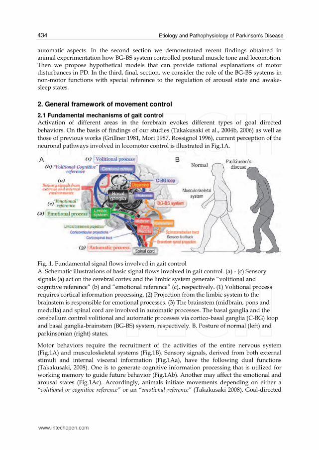
Fig. 1. Fundamental signal flows involved in gait control
A. Schematic illustrations of basic signal flows involved in gait control. (a) - (c) Sensory
signals (a) act on the cerebral cortex and the limbic system generate “volitional and
cognitive reference” (b) and “emotional reference” (c), respectively. (1) Volitional process
requires cortical information processing. (2) Projection from the limbic system to the
brainstem is responsible for emotional processes. (3) The brainstem (midbrain, pons and
medulla) and spinal cord are involved in automatic processes. The basal ganglia and the
cerebellum control volitional and automatic processes via cortico-basal ganglia (C-BG) loop
and basal ganglia-brainstem (BG-BS) system, respectively. B. Posture of normal (left) and
parkinsonian (right) states.
Motor behaviors require the recruitment of the activities of the entire nervous system (Fig.1A) and musculoskeletal systems (Fig.1B). Sensory signals, derived from both external stimuli and internal visceral information (Fig.1Aa), have the following dual functions (Takakusaki, 2008). One is to generate cognitive information processing that is utilized for working memory to guide future behavior (Fig.1Ab). Another may affect the emotional and arousal states (Fig.1Ac). Accordingly, animals initiate movements depending on either a “volitional or cognitive reference” or an “emotional reference” (Takakusaki 2008). Goal-directed
www.intechopen.com
Possible Contribution of the Basal Ganglia Brainstem System to the Pathogenesis of Parkinson’s Disease
435
behaviors therefore may require following the three processes; “volitional process” (Fig.1A(1)), “emotional process” (Fig.1A(2)) and “automatic processes” (Fig.1A(3)). The volitional process is derived from intentionally-elicited motor commands arising from the cerebral cortex based on volitional and cognitive references. This process requires cortical information processing and is executed by the corticoreticular and corticospinal projections. The emotional process is elicited by emotional reference via projections from the limbic-hypothalamus to the brainstem. This contributes to the emotional motor behaviors including fight or flight reactions. Regardless of whether the locomotion is volitional or emotional, it is accompanied by the automatic processes that are evoked by sequential activation of basic motor programs in the brainstem and spinal cord. The cerebellum regulates volitional and automatic processes by acting on the cerebral cortex and the brainstem, respectively. Sensory feedback via spinocerebellar tract plays an important role in this operation. The basal ganglia control these processes via loops with the cerebral cortex, brainstem and the limbic system. Because output of the basal ganglia is altered in basal ganglia disorders, all these movement processes can be disturbed.
2.2 Mechanisms of integrating posture and locomotion by subcortical structures In animal experiments, decerebrate cat preparation has been used to examine subcortical
mechanisms of controlling posture and locomotion. When the decerebration was made at
the precollicular-postmammillary level (x in Fig.2A), a cat maintained reflex standing
posture due to decerebrate rigidity (mesencephalic cat). Repetitive microelectrical
stimulation (50 Hz, 30 µA) applied to the cuneiform nucleus (CNF; a blue point in Fig.2B)
bilaterally increased the level of extensor (soleus) muscle tone, and then elicited stepping
movements which were developed to locomotion by moving a treadmill (an arrowhead in
Fig.2Ba). However the same type of stimuli applied to the ventral part of the PPN (red point
in Fig.2B) induced muscular atonia, which lasted even after termination of the stimulation
(Fig. 2Bc). Stimulation between these two sites (a green point in Fig.2C) evoked stepping
movements followed by muscular atonia (Fig.2Bb). Stimulation of the locus coeruleus (LC,
an orange point in Fig.2B) bilaterally increased extensor muscle tone (Fig.2Bd). Generally
the locomotion evoking sites (blue circles in Fig.2D), i.e. the midbrain locomotor region
(MLR), were located in the CNF, while the inhibitory region was located in the PPN (red
circles in in Fig.2C). Neurons between these regions may be involved in both locomotion
and muscular atonia. As show in Fig.2D, cholinergic neurons were abundantly distributed
in the area corresponding to the inhibitory region, indicating that an activation of
cholinergic neurons requires muscle tone suppression (Takakusaki et al., 2003a).
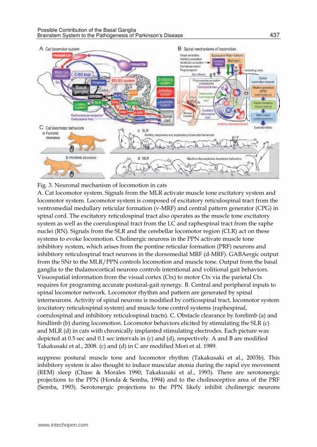
Our current perception of neuronal mechanisms of controlling postural muscle tone and locomotion is shown in Fig.3A on the basis of previous studies (Grillner 1981; Mori 1987; Rossignol 1996; Takakusaki et al., 2004b, 2006). Three locomotor regions are identified. They are the MLR, the subthalamic locomotor region (SLR) and the cerebellar locomotor region (CLR). Signals from the MLR may activate “muscle tone excitatory system” and “locomotor system or rhythm generating system”. The former is composed of monoaminergic descending pathways such as the coerulospinal and raphespinal tracts, and excitatory reticulospinal tract arising from the ventromedial medullary reticular formation (v-MRF) which approximately corresponds to the nucleus reticularis magnocellularis. The latter is composed of the excitatory reticulospinal tract and central pattern generators (CPG) in the spinal cord. Cortical projections to the MLR have not yet been identified. It is possibly
www.intechopen.com
Etiology and Pathophysiology of Parkinson's Disease
436
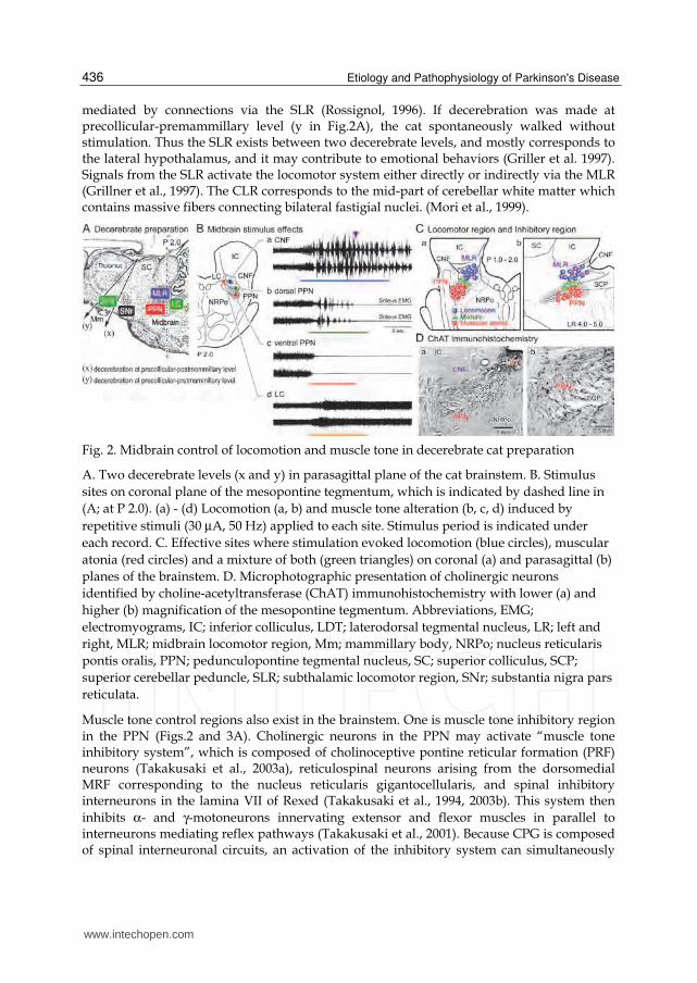
mediated by connections via the SLR (Rossignol, 1996). If decerebration was made at precollicular-premammillary level (y in Fig.2A), the cat spontaneously walked without stimulation. Thus the SLR exists between two decerebrate levels, and mostly corresponds to the lateral hypothalamus, and it may contribute to emotional behaviors (Griller et al. 1997). Signals from the SLR activate the locomotor system either directly or indirectly via the MLR (Grillner et al., 1997). The CLR corresponds to the mid-part of cerebellar white matter which contains massive fibers connecting bilateral fastigial nuclei. (Mori et al., 1999).
Fig. 2. Midbrain control of locomotion and muscle tone in decerebrate cat preparation
A. Two decerebrate levels (x and y) in parasagittal plane of the cat brainstem. B. Stimulus
sites on coronal plane of the mesopontine tegmentum, which is indicated by dashed line in
(A; at P 2.0). (a) - (d) Locomotion (a, b) and muscle tone alteration (b, c, d) induced by
repetitive stimuli (30 µA, 50 Hz) applied to each site. Stimulus period is indicated under
each record. C. Effective sites where stimulation evoked locomotion (blue circles), muscular
atonia (red circles) and a mixture of both (green triangles) on coronal (a) and parasagittal (b)
planes of the brainstem. D. Microphotographic presentation of cholinergic neurons
identified by choline-acetyltransferase (ChAT) immunohistochemistry with lower (a) and
higher (b) magnification of the mesopontine tegmentum. Abbreviations, EMG;
electromyograms, IC; inferior colliculus, LDT; laterodorsal tegmental nucleus, LR; left and
right, MLR; midbrain locomotor region, Mm; mammillary body, NRPo; nucleus reticularis
pontis oralis, PPN; pedunculopontine tegmental nucleus, SC; superior colliculus, SCP;
superior cerebellar peduncle, SLR; subthalamic locomotor region, SNr; substantia nigra pars
reticulata.
Muscle tone control regions also exist in the brainstem. One is muscle tone inhibitory region in the PPN (Figs.2 and 3A). Cholinergic neurons in the PPN may activate “muscle tone inhibitory system”, which is composed of cholinoceptive pontine reticular formation (PRF) neurons (Takakusaki et al., 2003a), reticulospinal neurons arising from the dorsomedial MRF corresponding to the nucleus reticularis gigantocellularis, and spinal inhibitory interneurons in the lamina VII of Rexed (Takakusaki et al., 1994, 2003b). This system then
inhibits α- and γ-motoneurons innervating extensor and flexor muscles in parallel to interneurons mediating reflex pathways (Takakusaki et al., 2001). Because CPG is composed of spinal interneuronal circuits, an activation of the inhibitory system can simultaneously
www.intechopen.com
Possible Contribution of the Basal Ganglia Brainstem System to the Pathogenesis of Parkinson’s Disease
437
Fig. 3. Neuronal mechanism of locomotion in cats
A. Cat locomotor system. Signals from the MLR activate muscle tone excitatory system and
locomotor system. Locomotor system is composed of excitatory reticulospinal tract from the
ventromedial medullary reticular formation (v-MRF) and central pattern generator (CPG) in
spinal cord. The excitatory reticulospinal tract also operates as the muscle tone excitatory
system as well as the coerulospinal tract from the LC and raphespinal tract from the raphe
nuclei (RN). Signals from the SLR and the cerebellar locomotor region (CLR) act on these
systems to evoke locomotion. Cholinergic neurons in the PPN activate muscle tone
inhibitory system, which arises from the pontine reticular formation (PRF) neurons and
inhibitory reticulospinal tract neurons in the dorsomedial MRF (d-MRF). GABAergic output
from the SNr to the MLR/PPN controls locomotion and muscle tone. Output from the basal
ganglia to the thalamocortical neurons controls intentional and volitional gait behaviors.
Visuospatial information from the visual cortex (Ctx) to motor Ctx via the parietal Ctx
requires for programing accurate postural-gait synergy. B. Central and peripheral inputs to
spinal locomotor network. Locomotor rhythm and pattern are generated by spinal
interneurons. Activity of spinal neurons is modified by corticospinal tract, locomotor system
(excitatory reticulospinal system) and muscle tone control systems (raphespinal,
coerulospinal and inhibitory reticulospinal tracts). C. Obstacle clearance by forelimb (a) and
hindlimb (b) during locomotion. Locomotor behaviors elicited by stimulating the SLR (c)
and MLR (d) in cats with chronically implanted stimulating electrodes. Each picture was
depicted at 0.5 sec and 0.1 sec intervals in (c) and (d), respectively. A and B are modified
Takakusaki et al., 2008. (c) and (d) in C are modified Mori et al. 1989.
suppress postural muscle tone and locomotor rhythm (Takakusaki et al., 2003b). This inhibitory system is also thought to induce muscular atonia during the rapid eye movement (REM) sleep (Chase & Morales 1990; Takakusaki et al., 1993). There are serotonergic projections to the PPN (Honda & Semba, 1994) and to the cholinoceptive area of the PRF (Semba, 1993). Serotonergic projections to the PPN likely inhibit cholinergic neurons
www.intechopen.com
Etiology and Pathophysiology of Parkinson's Disease
438
(Leonald & Llinás, 1994), and those to the PRF may reduce activity of the inhibitory system (Takakusaki et al., 1994). In contrast, the inhibitory system suppresses the activity of the coerulospinal tract (Mileykovskiy et al., 2000). Accordingly muscle tone can be regulated by a counterbalance between the inhibitory and the excitatory systems (Takakusaki et al., 2006). It was reported that a patient with a lesion in the dorsolateral mesopontine tegmentum did not lose muscle tone during REM sleep ("REM without atonia") (Boeve et al., 2007; Culebras & Moore 1989). Also, a patient with a lesion in the dorsal part of mesopontine tegmentum could not stand and walk (Masdeu et al., 1994). These clinical case reports suggest that both a muscle tone inhibitory region and a MLR are realities in the mesopontine tegmentum of the human. Spinal mechanisms of locomotor control are schematically illustrated in Fig.3B. Signals from
the cerebral cortex and the brainstem, and those from peripheral sensory afferents are
integrated at spinal cord to achieve appropriate locomotor control. Various combinations of
spinal reflexes operate during locomotion. Those mediating flexion reflex and crossed
extension reflex undertake major roles in the generation of locomotor rhythm (Rossignol
1996; Rossignol et al., 2006; Takakusaki et al., 2001, 2003b; McCrea & Rybak, 2008). Spinal
interneurons that constitute CPG generate detailed locomotor rhythm. The locomotor
rhythm is then translated to next order interneuronal groups which shape “locomotor
pattern”. Finally signals are sent to last-order interneurons, including reciprocal Ia
interneurons, Ib interneurons and Renshaw cells. They are located in lamina IV-VII of Rexed
and project to target motoneurons. Lamina VIII interneurons project to the contralateral side
of spinal cord and may control alternating limb movements (Matsuyama & Takakusaki,
2008). Signals generated by spinal locomotor network are then transmitted back to the
cerebral cortex, the brainstem and the cerebellum so that they monitor events in the spinal
cord (Fig.3B).
2.3 Initiation of movements by the forebrain structures 2.3.1 Cortical control of locomotor behaviors Drew et al. (1996) demonstrated, in cats with chronically implanted electrodes in the cerebral cortex, that a majority of motor cortical neurons exhibited simple rhythmic firing in relation to step cycles during steady-state locomotion. However their discharge rates considerably increased when the cats initiated to walk and had to accurately step over obstacles. Thus, commitment of cortical processing seems unnecessary during the automatic locomotor movements. On the other hand, stepping movements that accompany accurate foot placement resemble to the forelimb reaching of higher primates (Drew et al., 2004; Georgopoulos & Grillner, 1989). Such an accurate movement requires visuomotor cognitive processes (Fig.3A), which are controlled by neural circuits involving the cerebral cortex, basal ganglia, and cerebellum (Middleton & Strick, 2000). Subjects are aware of the locations of obstacles around them, and they are able to alter their stepping patterns even without available visual information of the location of the obstacles relative to the body (Fig.3C). McVea & Pearson (2007) reported that perturbing walking cats in a consistent manner evoked lasting changes to the walking pattern that were expressed only in the context in which walking was disturbed. Moreover, cats that had stepped over an obstacle by forelimb (Fig.3Ca) remembered the location of the obstacle and could use working memory to guide stepping for the hindlimb (Fig.3Cb). Therefore, sensory inputs that signal context –the surrounding visual and auditory
www.intechopen.com
Possible Contribution of the Basal Ganglia Brainstem System to the Pathogenesis of Parkinson’s Disease
439
environment– play an important role in shaping the basic pattern of locomotion. Lajoie & Drew (2007) observed, after unilateral lesion of area 5 of the posterior parietal cortex, that cats frequently hit the obstacle as they stepped over it. They also frequently hit the obstacle with their hindlimbs even when the forelimbs negotiated the obstacle successfully. These findings suggest an important role for the posterior parietal cortex in the coordination of the forelimbs and hindlimbs and in the planning and programming of visually-guided gait modification (Fig.3C). Neuroanatomical studies indicate that the posterior parietal cortex sends selected projections to the motor cortical areas from layer III, while those to the lateral cerebellum via the pontine nuclei arise from layer V (Andujar & Drew, 2007). Neurons in the primary motor cortex and those in the premotor/supplementary motor areas (PM/SMA) mainly project to the spinal cord and the reticular formation via corticospinal and corticoreticular projections, respectively (Matsuyama & Drew 1997).
2.3.2 Emotional locomotor behaviors The MLR was initially established as a functional region involved in the initiation of locomotion on the basis of its connections with limbic structures and the basal ganglia (Armstrong, 1986; Megensen et al., 1991). Regardless of the nature of emotional stimuli, they usually elicit alert responses that produce stereotyped movements such as increased postural muscle tone and/or locomotion that accompanies autonomic sympathetic responses. The limbic-hypothalamic systems play crucial roles in these processes. Sinnamon (1993) proposed the following three types of locomotor systems that function in different behavioral or motivational contexts; an appetitive system, a primary defensive system, and an exploratory system. In cats with chronically implanted electrodes, stimulation of the SLR elicited alerting responses followed by exploratory (searching) or defensive behaviors (Fig.3Cc; Mori et al., 1989). Signals from the SLR are mediated by dense fibers in the medial forebrain bundle projecting to the midbrain (Rossignol, 1996). On the other hand, stimulation of the MLR abruptly elicited machine-like explosive locomotion (Fig.3Cd). Neural circuits connecting the nucleus accumbens (the oldest part of the striatum), the hippocampus, and the amygdala, are involved in emotional memory, and projections from the nucleus accumbens to the MLR may contribute to the expression of exploratory behaviors (Mogenson, 1991). In addition, projections from the lateral and the medial hypothalamic areas to the MLR are thought to operate as defensive and appetitive systems, respectively (Grillner et al., 1997; Jordan, 1998). The orexin-containing neurons located in the prefornical lateral hypothalamic area are considered to control appetite, energy balance, and vigilance states via projections to various areas in the nervous system (Peyron et al., 1998; Sakurai, 2002; Siegel, 2004). The orexinergic projections to the MLR facilitated the activity of the locomotor system (Takakusaki et al., 2005), indicating that the hypothalamic orexinergic system contributes to appetitive behaviors.
3. Basal ganglia control of movements and motor disturbances by the basal ganglia dysfunction
It is established that the C-BG loop is required for volitional movement (Delong & Wichmann, 2007; Middleton & Strick, 2000). Neural circuits between the prefrontal cortex and the caudate nucleus (cognitive loop) are involved in the regulation of complex, visually-guided limb movements and the planning and programming those movements. Neural
www.intechopen.com
Etiology and Pathophysiology of Parkinson's Disease
440
circuits between motor cortical areas, including the primary motor cortex, PM and SMA, and the putamen (motor loop) contribute to the regulation of voluntary, discrete, ipsilateral limb movements. In addition the BG-BS system may control automatic and steady-state locomotor movements. This section first refers how the BG-BS system controls posture and locomotion and then considers how BG-BS contributes to pathophysiological mechanisms of motor disturbances in PD.
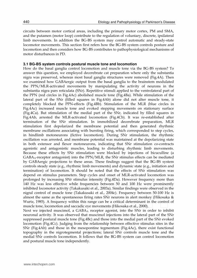
3.1 BG-BS system controls postural muscle tone and locomotion How do the basal ganglia control locomotion and muscle tone via the BG-BS system? To answer this question, we employed decerebrate cat preparation where only the substantia nigra was preserved, whereas most basal ganglia structures were removed (Fig.4A). Then we examined how GABAergic output from the basal ganglia to the brainstem modulated the PPN/MLR-activated movements by manipulating the activity of neurons in the substantia nigra pars reticulata (SNr). Repetitive stimuli applied to the ventrolateral part of the PPN (red circles in Fig.4Ac) abolished muscle tone (Fig.4Ba). While stimulation of the lateral part of the SNr (filled squares in Fig.4Ab) alone did not alter muscle tone, it completely blocked the PPN-effects (Fig.4Bb). Stimulation of the MLR (blue circles in Fig.4Ac) increased muscle tone and evoked stepping movements on stationary surface (Fig.4Ca). But stimulation of the medial part of the SNr, indicated by filled squares in Fig.4Ab, arrested the MLR-activated locomotion (Fig.4Cb). It was re-established after termination of the SNr stimulation. In immobilized decerebrate preparation, MLR stimulation first depolarized the membrane potential and then generated rhythmic membrane oscillations associating with bursting firing, which corresponded to step cycles, in hindlimb motoneurons (fictive locomotion). During SNr stimulation, the rhythmic oscillation was arrested, and membrane potential was maintained at the depolarizing state in both extensor and flexor motoneurons, indicating that SNr stimulation co-contracts agonistic and antagonistic muscles, leading to disturbing rhythmic limb movements. Because these effects by SNr stimulation were blocked by injections of bicuculline (a GABAA-receptor antagonist) into the PPN/MLR, the SNr stimulus effects can be mediated by GABAergic projections to these areas. These findings suggest that the BG-BS system controls steady state (e.g., rhythmic limb movements) and dynamic state (e.g., initiation and termination) of locomotion. It should be noted that the effects of SNr stimulation was depend on stimulus parameters. Step cycles and onset of MLR-activated locomotion was prolonged by increasing SNr stimulus intensity (Fig.4Da). However frequency more than 140 Hz was less effective while frequencies between 50 and 100 Hz were prominently inhibited locomotor activity (Takakusaki et al., 2003a). Similar findings were observed in the nigral control of muscle tone (Takakusaki et al., 2004c). Frequency between 50-100 Hz is almost the same as the spontaneous firing rates SNr neurons in alert monkey (Hikosaka & Wurtz, 1985). A frequency within this range can be a critical determinant in the control of muscle tone, locomotion and saccadic eye movements (Hikosaka et al., 2000). Next we injected muscimol, a GABAA receptor agonist, into the SNr in order to inhibit neuronal activity. It was observed that muscimol injections into the lateral part of the SNr suppressed postural muscle tone (Fig.4Bc) and those into the medial part of the SNr evoked locomotion (Fig.4Cc). Judging from the relationship between effective stimulus sites in the SNr (Fig.4Ab) and those in the mesopontine tegmentum (Fig.4Ac), there exist functional topography in the nigrotegmental projections; lateral SNr controls muscle tone and the medial SNr controls locomotion. It follows that the BG-BS system can control locomotion and postural muscle tone independently.
www.intechopen.com
Possible Contribution of the Basal Ganglia Brainstem System to the Pathogenesis of Parkinson’s Disease
441
Fig. 4. Nigral stimulus effects on PPN/MLR-induced muscle tone suppression and locomotion in decerebrate cats A. (a) Experimental design in decerebrate cat preparation. (b) Effective stimulus sites in the SNr for inhibition of the PPN (filled squares) and the MLR (open squares) effects. (c) Effective stimulus sites for evoking muscular atonia (PPN; red circles) and locomotion (MLR; blue circles) in the mesopontine tegmentum. B. (a) PPN-induced muscular atonia. (b) Inhibition of the PPN-induced atonia by SNr stimulation. (c) Muscular atonia induced by an injection of muscimol into the lateral part of the SNr. C. (a) MLR-activated stepping movements. (b) Inhibition of MLR-activated stepping by SNr stimulation. (c) Locomotion induced by an injection of muscimol into the medial part of the SNr. D. Changes in step cycles and gait onset of the MLR-activated locomotion following changes in stimulus intensity (a) and frequency (b) applied to the SNr. Abbreviations, III; oculomotor nerve, PAG; periaqueductal grey, RN; red nucleus.
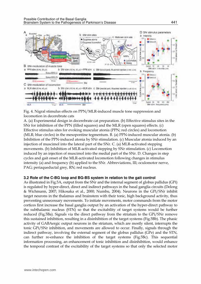
3.2 Role of the C-BG loop and BG-BS system in relation to the gait control As illustrated in Fig.5A, output from the SNr and the internal segment of globus pallidus (GPi) is regulated by hyper-direct, direct and indirect pathways in the basal ganglia circuits (Delong & Wichmann, 2007; Hikosaka et al., 2000; Numbu, 2004). Neurons in the GPi/SNr inhibit target neurons in the thalamus and brainstem with their tonic, high background activity, thus preventing unnecessary movements. To initiate movements, motor commands from the motor cortices first increase the basal ganglia output by an activation of the hyper-direct pathway to the subthalamic nucleus (STN) so that the excitability of target systems would be further reduced (Fig.5Ba). Signals via the direct pathway from the striatum to the GPi/SNr remove this sustained inhibition, resulting in a disinhibition of the target systems (Fig.5Bb). The phasic activity of GABAergic output neurons in the striatum, which are mostly silent, interrupts the tonic GPi/SNr inhibition, and movements are allowed to occur. Finally, signals through the indirect pathway, involving the external segment of the globus pallidus (GPe) and the STN, can further re-enhance the inhibition of the target systems (Fig.5Bc). This sequential information processing, an enhancement of tonic inhibition and disinhibition, would enhance the temporal contrast of the excitability of the target systems so that only the selected motor
www.intechopen.com
Etiology and Pathophysiology of Parkinson's Disease
442
program could be initiated, executed and terminated at the appropriate timing, whereas other competing programs can be cancelled (Hikosaka et al., 2000; Numbu, 2004). This is the “first key mechanism” of movement control by the basal ganglia. The above mechanisms may act on brainstem networks, including the locomotor system and muscle tone control systems (Fig.5A, lower right). Therefore the brainstem networks could be combined with basal ganglia motor circuits. In this “hybrid model”, output of the basal ganglia controls the MLR for locomotion and the PPN for muscle tone via GABAergic projection. When locomotor movement is being prepared, tonic activity of SNr neurons would continuously inhibit both systems. When a trigger signal occurred, the hyper-direct pathway would enhance the inhibition. Then the direct pathway would release the activity of these systems, resulting in an initiation of locomotion that would be followed by a smooth reduction of the level of muscle tone. To terminate the locomotion, the direct pathway would inhibit each system, resulting in a cessation of rhythmic locomotor movements and an accompanying increase in the level of muscle tone (muscle co-contraction). A parallel organization from the SNr to the MLR/PPN would be therefore assist regulation of the level of muscle tone which was appropriate for the initiation and termination of locomotion.
Fig. 5. Hybrid model of C-BG loop and BG-BS system A. Left; basal ganglia motor circuits. Lower right; BG-BS system for controlling locomotion and muscle tone. B and C. Changes in the basal ganglia output and in the excitability of target systems following sequential information processing of hyper-direct (a), direct (b) and indirect (c) pathways. When excitability of target systems goes beyond the threshold, movements occur. Excitability of direct and indirect pathways is modified by dopaminergic projections from the SNc to the striatum. B. Normal condition. C. Parkinson’s disease. Abbreviations, D1 and D2; D1 and D2-dopamine receptors, DA; dopamine, enk; enkephaline, Glu; glutamate, GPe; external segment of globus pallidus subP; substance P, STN; subthalamic nucleus
www.intechopen.com
Possible Contribution of the Basal Ganglia Brainstem System to the Pathogenesis of Parkinson’s Disease
443
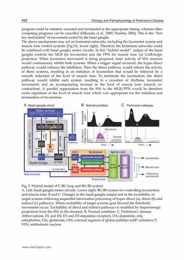
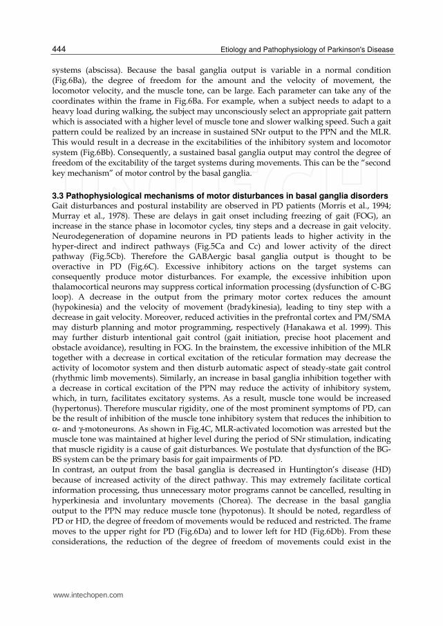
Fig. 6. Hypothetical models for movement control by the basal ganglia A. GABAergic basal ganglia projections to the thalamocortical neurons are involved in the volitional control of movements, while those to the MLR and the PPN may be responsible for the automatic control of locomotion and muscle tone. B. Normal operation of the basal ganglia control of voluntary movements, locomotion and muscle tone. C. Pathophysiological changes in the activities of the cortico-BG loop and BG-BS system in PD. Loss of dopamine results in an increase in the basal ganglia output to the cerebral cortex, the limbic system and the brainstem. Consequently, voluntary and cognitive activities of the cerebral cortex and emotional expression can be reduced. Reduced cortical output results in bradykinesia and hypokinesia. Reduced activity in the MLR-locomotor system may induce gait failure. Inhibition of the PPN-muscle tone inhibitory system may induce hypertonus. D. Motor disturbances in basal ganglia disorders. (a) Parkinson disease. (b) Huntington’s disease. Regardless of whether an increase or a decrease in the basal ganglia output, degree of freedom of movements may be restricted.
Given the above consideration, we propose a hypothetical model in Fig.6A for the basal ganglia control of movements. The motor cortical neurons that receive basal ganglia output may control the velocity and the amount of voluntary movement (Turner & Anderson, 1997), which is indicated in the ordinate on the left of the graph in Fig.6Ba. The GABAergic basal ganglia output to the MLR reduced the drive from the MLR, resulting in disruption of the activity of CPGs in the spinal cord. Basal ganglia efferents to the MLR may therefore control the locomotor pattern (ordinate on the right in Fig.6Ba). In addition, basal ganglia efferents to the PPN may determine the level of muscle tone via the muscle tone control
www.intechopen.com
Etiology and Pathophysiology of Parkinson's Disease
444
systems (abscissa). Because the basal ganglia output is variable in a normal condition (Fig.6Ba), the degree of freedom for the amount and the velocity of movement, the locomotor velocity, and the muscle tone, can be large. Each parameter can take any of the coordinates within the frame in Fig.6Ba. For example, when a subject needs to adapt to a heavy load during walking, the subject may unconsciously select an appropriate gait pattern which is associated with a higher level of muscle tone and slower walking speed. Such a gait pattern could be realized by an increase in sustained SNr output to the PPN and the MLR. This would result in a decrease in the excitabilities of the inhibitory system and locomotor system (Fig.6Bb). Consequently, a sustained basal ganglia output may control the degree of freedom of the excitability of the target systems during movements. This can be the “second key mechanism” of motor control by the basal ganglia.
3.3 Pathophysiological mechanisms of motor disturbances in basal ganglia disorders Gait disturbances and postural instability are observed in PD patients (Morris et al., 1994; Murray et al., 1978). These are delays in gait onset including freezing of gait (FOG), an increase in the stance phase in locomotor cycles, tiny steps and a decrease in gait velocity. Neurodegeneration of dopamine neurons in PD patients leads to higher activity in the hyper-direct and indirect pathways (Fig.5Ca and Cc) and lower activity of the direct pathway (Fig.5Cb). Therefore the GABAergic basal ganglia output is thought to be overactive in PD (Fig.6C). Excessive inhibitory actions on the target systems can consequently produce motor disturbances. For example, the excessive inhibition upon thalamocortical neurons may suppress cortical information processing (dysfunction of C-BG loop). A decrease in the output from the primary motor cortex reduces the amount (hypokinesia) and the velocity of movement (bradykinesia), leading to tiny step with a decrease in gait velocity. Moreover, reduced activities in the prefrontal cortex and PM/SMA may disturb planning and motor programming, respectively (Hanakawa et al. 1999). This may further disturb intentional gait control (gait initiation, precise hoot placement and obstacle avoidance), resulting in FOG. In the brainstem, the excessive inhibition of the MLR together with a decrease in cortical excitation of the reticular formation may decrease the activity of locomotor system and then disturb automatic aspect of steady-state gait control (rhythmic limb movements). Similarly, an increase in basal ganglia inhibition together with a decrease in cortical excitation of the PPN may reduce the activity of inhibitory system, which, in turn, facilitates excitatory systems. As a result, muscle tone would be increased (hypertonus). Therefore muscular rigidity, one of the most prominent symptoms of PD, can be the result of inhibition of the muscle tone inhibitory system that reduces the inhibition to
α- and γ-motoneurons. As shown in Fig.4C, MLR-activated locomotion was arrested but the muscle tone was maintained at higher level during the period of SNr stimulation, indicating that muscle rigidity is a cause of gait disturbances. We postulate that dysfunction of the BG-BS system can be the primary basis for gait impairments of PD. In contrast, an output from the basal ganglia is decreased in Huntington’s disease (HD) because of increased activity of the direct pathway. This may extremely facilitate cortical information processing, thus unnecessary motor programs cannot be cancelled, resulting in hyperkinesia and involuntary movements (Chorea). The decrease in the basal ganglia output to the PPN may reduce muscle tone (hypotonus). It should be noted, regardless of PD or HD, the degree of freedom of movements would be reduced and restricted. The frame moves to the upper right for PD (Fig.6Da) and to lower left for HD (Fig.6Db). From these considerations, the reduction of the degree of freedom of movements could exist in the
www.intechopen.com
Possible Contribution of the Basal Ganglia Brainstem System to the Pathogenesis of Parkinson’s Disease
445
background of PD and HD. Consequently, dysfunction of the BG-BS system together with that of the C-BG loop may underlie the pathogenesis of the motor disturbances in these basal ganglia diseases. Dystonia is a syndrome characterized by abnormal postures, muscle spasms and tremor, due to involuntary muscle co-contractions. Some dystonia are task specific, and patients only develop muscular co-contraction when performing skilled movements such as writing (Van der Kamp et al. 1989). By using positron emission tomography an inappropriate over-activity of the basal ganglia projections to the premotor and dorsal prefrontal cortex has been observed (Brooks 1995). However the activity of the primary sensorimotor and caudal premotor cortices is rather attenuated (Hutchins et al. 1988). Although alterations of noradrenaline and DA levels in brainstem structures have been reported in two cases (Hornykiewicz et al., 1986), most studies, by contrast, have not found abnormalities in the brainstem. This evidence suggests that the activity of the BG-BS system and that of the C-BG loop are controlled separately in dystonia. Recently PPN/MLR area became one of targets of deep brain stimulation (DBS) for neurosurgical therapy for PD (PPN-DBS) (Stefani et al., 2007; Pierantozzi et al., 2008; Alessandro et al., 2010). Low frequency stimulation (~25Hz) applied to the above area ameliorated postural disturbance and gait failure. On the other hand, DBS applied to the SNr (SNr-DBS) with high frequency (135-190 Hz), which possibly intervened to the output from the SNr, also ameliorated axial symptoms such as gait akinesia and postural disturbances (Chasetan et al. 2009). Although evidence of the PPN-DBS and the SNr-DBS is still limited, these clinical findings agree well with our results suggesting that the BG-BS system contributes to the postural and locomotor synergies in human.
4. Disturbances of non-motor functions in Parkinson’s disease
Disturbances in cognitive and psychotic processes have been observed in patients with degenerative disorders that involve primarily the basal ganglia such as PD (Mellers et al., 1995; Taylor et al., 1986) and HD (McHugh & Folsten, 1975). Awake-sleep states were also impaired in PD (Bliwise et al., 2000; Eisensehr et al., 2001). It is also reported that PD is preceded and accompanied by daytime sleep attacks, nocturnal insomnia, REM sleep behavior disorder, hallucinations and depression, symptoms which are frequently as troublesome as the motor symptoms of this disease. All these symptoms are present in narcolepsy (Thannical et al., 2007). These clinical evidences corroborate that the basal ganglia and their connections with the brainstem are also involved in the expression of non-motor function. In this section, we focus on the roles played by the BG-BS system in the regulation of vigilance states, arousal state, attention and cognition in relation to non-motor symptoms in PD.
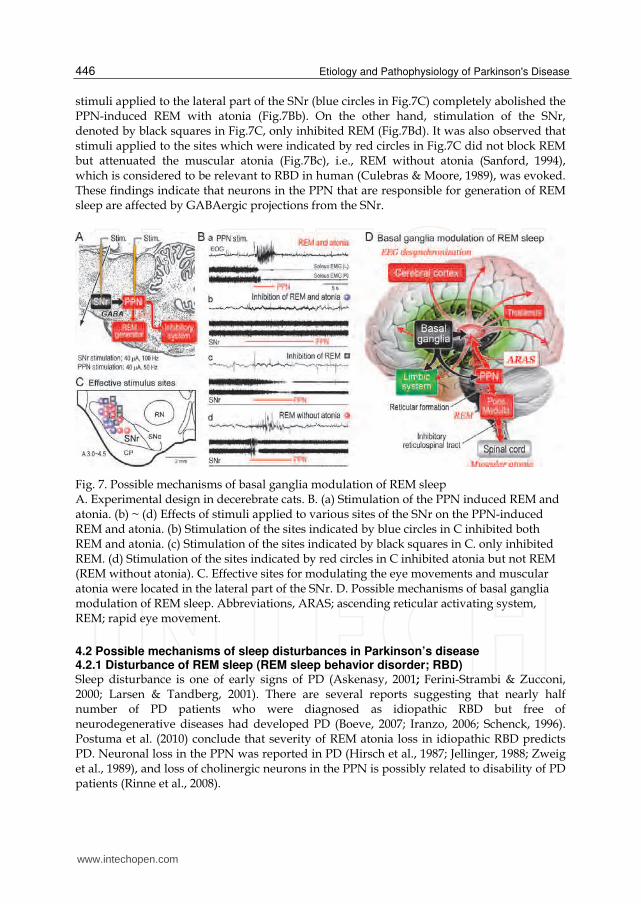
4.1 Does output of the basal ganglia modulate sleep? Cholinergic neurons in the PPN and laterodorsal tegmental nucleus are thought to be involved in not only the maintenance of arousal state but also generation of REM sleep (Datta and Siwek, 2002; Koyama & Sakai, 2000; Maloney et al., 1999). Therefore, we elucidated how GABAergic SNr-PPN projection altered the activities of the REM generator and the muscle tone inhibitory system (Takakusaki et al., 2004c). Summary of the results are shown in Fig.7. Stimulation of inhibitory region of the PPN induced REM which was associated with muscular atonia in decerebrate cats (REM and atonia; Fig.7Ba). Conditioning
www.intechopen.com
Etiology and Pathophysiology of Parkinson's Disease
446
stimuli applied to the lateral part of the SNr (blue circles in Fig.7C) completely abolished the PPN-induced REM with atonia (Fig.7Bb). On the other hand, stimulation of the SNr, denoted by black squares in Fig.7C, only inhibited REM (Fig.7Bd). It was also observed that stimuli applied to the sites which were indicated by red circles in Fig.7C did not block REM but attenuated the muscular atonia (Fig.7Bc), i.e., REM without atonia (Sanford, 1994), which is considered to be relevant to RBD in human (Culebras & Moore, 1989), was evoked. These findings indicate that neurons in the PPN that are responsible for generation of REM sleep are affected by GABAergic projections from the SNr.
Fig. 7. Possible mechanisms of basal ganglia modulation of REM sleep A. Experimental design in decerebrate cats. B. (a) Stimulation of the PPN induced REM and atonia. (b) ~ (d) Effects of stimuli applied to various sites of the SNr on the PPN-induced REM and atonia. (b) Stimulation of the sites indicated by blue circles in C inhibited both REM and atonia. (c) Stimulation of the sites indicated by black squares in C. only inhibited REM. (d) Stimulation of the sites indicated by red circles in C inhibited atonia but not REM (REM without atonia). C. Effective sites for modulating the eye movements and muscular atonia were located in the lateral part of the SNr. D. Possible mechanisms of basal ganglia modulation of REM sleep. Abbreviations, ARAS; ascending reticular activating system, REM; rapid eye movement.
4.2 Possible mechanisms of sleep disturbances in Parkinson’s disease 4.2.1 Disturbance of REM sleep (REM sleep behavior disorder; RBD) Sleep disturbance is one of early signs of PD (Askenasy, 2001; Ferini-Strambi & Zucconi, 2000; Larsen & Tandberg, 2001). There are several reports suggesting that nearly half number of PD patients who were diagnosed as idiopathic RBD but free of neurodegenerative diseases had developed PD (Boeve, 2007; Iranzo, 2006; Schenck, 1996). Postuma et al. (2010) conclude that severity of REM atonia loss in idiopathic RBD predicts PD. Neuronal loss in the PPN was reported in PD (Hirsch et al., 1987; Jellinger, 1988; Zweig et al., 1989), and loss of cholinergic neurons in the PPN is possibly related to disability of PD patients (Rinne et al., 2008).
www.intechopen.com
Possible Contribution of the Basal Ganglia Brainstem System to the Pathogenesis of Parkinson’s Disease
447
Several mechanisms are postulated in relation to the basal ganglia regulation of sleep. On one hand, recent brain imaging studies revealed that damage of brainstem, particularly the reticular formation, is critically involved in the pathogenesis of RBD (Unger et al., 2010). The brainstem damage could also explain some non-motor symptoms in this disease, which often precede diagnosis, such as autonomic dysfunction and sleep disorders. On the other hand, roles of dopaminergic influence on the basal ganglia in the control of sleep-wake behavior are suggested (Mena-Segovia J et al., 2008). It is also possible that basal ganglia efferents to the non-specific thalamic nuclei may affect awake-sleep states by modulating the activity of ascending reticular activating system (ARAS). Since the classical study of Moruzzi and Magoun (1949), the pontomesencephalic reticular formation has been known to comprise the ARAS. The PPN has been considered as a part of the ARAS (Garcia-Rill, 1991; Inglis & Winn, 1995; Jones, 1991). The SNr has a direct projection to the thalamic nuclei (Parent et al., 1983; Pare´ et al., 1990) in addition to the PPN (Fig.7D). Because PPN has dense projections to the midbrain dopaminergic neurons, activity of the PPN neurons may affect awake-sleep states by modulating dopaminergic systems projecting to the basal ganglia and extra-basal ganglia areas. Consequently, our idea is that basal ganglia output from the SNr may affect awake–sleep cycles by modulating the activity of the ARAS through dual mechanisms (Fig.7D). One is through direct nigro-thalamic projection, and the other, which is considered in this study, is though indirect connections via the PPN (Takakusaki et al. 2004c, 2006).
4.2.2 Narcolepsy-like symptoms The presence in PD patients of narcolepsy-like features, such as daytime REM sleep
intrusions associated with visual hallucinations, has led some authors to suggest that a
mechanism similar to that of narcolepsy might underlie excessive daytime sleepiness
(EDS) in PD (Arnulf et al., 2000). Thannickal et al. (2007) demonstrated that a massive loss
of orexin neurons was found in PD patients and suggested that it was a cause of the
narcolepsy-like symptoms. However, Compta et al. (2009) showed that orexin-A level was
normal in the cerebrospinal fluid and it was unrelated to severity of sleepiness or the
cognitive status of PD patients. Therefore alternative mechanisms other than dysfunction
of orexin neurons might be responsible for EDS and the disturbance of sleep architecture
in PD. In animal experiments, midbrain strucutes, including the SNr, the PPN and the
MLR, receive orexinergic efferents from the perifornical lateral hypothalams (Nambu et
al., 1999; Peyron et al., 1998). Therefore it is interesting to elucidate how orexinergic
projections to the midbrain are involved in alteration of sleep-awake states. Then we
examined effects of injections of orexin-A into the MLR, PPN and the SNr upon motor
behaviors in decerebrate cats (Takakusaki et al., 2005). Microinjections of orexin into the
MLR facilitated locomotion, while those into either the PPN or the SNr suppressed PPN-
induced muscular atonia. The latter effects were reversed by subsequent injection of
bicuculline into the PPN. Thus the excitability seems to be higher in the locomotor system
than in the atonia system in the presence of orexin. On the contrary the excitability of the
muscle tone inhibitory system may be higher than that of the locomotor system in the
absence of orexin. Accordingly GABAergic projection from the SNr to the PPN/MLR area
(BG-BS system) may underlie orexin-mediated vigilance state regulation and its
dysfunction may be one of pathophysiological mechanisms of narcolepsy-like features of
PD (Takakusaki 2008).
www.intechopen.com
Etiology and Pathophysiology of Parkinson's Disease
448
4.3 Disturbances of arousal state, attention and cognition Behavioral arousal requires an activation of dopaminergic projections arising from the SNc to the striatum and the ventral tegmental area (VTA) to the prefrontal cortex and the limbic system. The nigrostriatal projection is responsible for basal ganglia related motor functions. The mesocortical projection contributes to volitional expression and attention, and the mesolimbic projection is involved in emotional expression. On the other hand, ARAS plays a major role in the electroencephalographic arousal. An activation of the two arousal systems is required to maintain arousal state that enables alert, attention and cognition (Jones 1991). Because PPN has dense cholinergic and non-cholinergic excitatory connections with dopamine (DA) neurons in the SNc and other basal ganglia nuclei (Futami et al., 1994; Kitai, 1998; Takakusaki et al., 1996), these projections appear to play a role in more specific subcortical integration of motor and non-motor functions such as behavioral arousal, attention and reward (Kitai, 1998). For example, an injection of muscimol into the PPN reduced the speed and amount of arm movements and delayed the onset of movements but the accuracy was rather maintained (Matsumura and Kojima, 2001). Moreover, Kojima et al. (1997) demonstrated that kainic acid-induced lesion in the unilateral PPN induced hemiparkisonism which was observed in the contralateral side of the injection. From these findings they suggest that the PPN may thus facilitate the voluntary limb movements through its excitatory connections with the DA neurons. Midbrain DA neurons are also involved in the predictive reward which is specifically linked with reinforcement behaviors. DA neurons are activated by rewarding events that are better than predicted, remain uninfluenced by events that are worse than predicted (Hikosaka et al., 2000; Schultz, 1998). Kobayashi et al. (2002) demonstrated that PPN neurons showed multi-modal activities during saccade tasks in alert monkey; their activities were related to the arousal levels, execution and preparation of movements, the level of task performance, and reward. Therefore the PPN may serve as an integrative interface between the various signals required for performing purposive behaviors (Kobayashi et al., 2004). We postulate that the PPN facilitates, possibly via dopaminergic systems, the central processes for motor command generation and extrinsic sensory processing by modulating arousal and attentive states. In non-human primate, limited lesions of the striatum induce deficits in rule acquisition (Divac 1972), cognition (Taylor et al., 1990), working memory performance (Goldman-Rakic, 1987) and selected attention (Battig et al., 1962). Laplane et al. (1984) reported a patient with restricted bilateral pallidal lesions who was appeared apathetic and unconcerned or attention deficits, and his affect was flattened and emotional responses were blunted in the absence of any motor disorder or pure psychic akinesia. These symptoms were also described in progressive supranuclear palsy (PSP) in which major lesions were observed in the subcortical areas including the PPN. Because loss of cholinergic PPN neurons were observed not only in PSP (75-80%) but also PD (43-57%) (Hirsch et al., 1987; Jellinger, 1988; Zweig et al., 1987, 1989), the loss of cholinergic PPN neurons in both diseases could attribute to attentive and cognitive impairments and sleep deficiencies in these diseases (Scarnati & Florio, 1997). Both neuroanatomical (von Krosigk et al., 1992; Smith & Bolam, 1990) and electrophysiological (Häusser & Yung, 1994; Saitoh et al., 2004; Paladini et al., 1999) studies demonstrated that dopaminergic neurons, as well as cholinergic neurons, receive GABAergic inhibitory effects from the basal ganglia, particularly from the SNr. Consequently a BG-BS system appears to involve the interdigitation of motor information
www.intechopen.com
Possible Contribution of the Basal Ganglia Brainstem System to the Pathogenesis of Parkinson’s Disease
449
with information relating to reward and reinforcement by modulating the excitability of both dopaminergic and cholinergic systems.
5. Concluding thoughts
The basal ganglia controls various function by acting on thalamocortical loop (C-BG loop) and the brainstem (BG-BS system). There are two key mechanisms for the operation by the basal ganglia circuit. One is sequential information processing, which would enhance the temporal contrast of the excitability of the target systems so that only the selected motor program could be appropriately executed, whereas other competing programs can be cancelled. The other is sustained output from the basal ganglia, which may control the degree of freedom of the excitability of the target systems during movements. We suggest that following roles can be played by the BG-BS system. First this system is involved in the automatic or unconscious control of movements that accompany voluntary movements. Second, the BG-BS systems may be involved in the maintenance of arousal and attentive states and in the regulation of REM sleep. Because output from the basal ganglia is thought to be overactive in PD, dysfunction of the BG-BS system in addition to that of C-BG loop can be seriously involved in motor and non-motor functions in this disease.
6. Acknowledgment
This work is supported by Grants-in-Aid for Challenging Exploratory Research (Project # 23650202) and Priority Areas “Emergence of Adaptive Motor Function through Interaction between Body, Brain and Environment (Area #454)” from the Japanese Ministry of Education, Culture, Sports, Science and Technology to K.T. We express sincere appreciation to Ms. Mihoko Ebisawa for preparation of this manuscript.
7. References
Aarsland, D., Bronnick, K., Williams-Gray, C., Weintraub, D., Marder, K., Kulisevsky, J.,
Burn, D., Barone, P., Pagonabarraga, J., Allcock, L., Santangelo, G., Foltynie, T.,
Janvin, C., Larsen, JP., Barker, RA. & Emre, M. (2010) Mild cognitive impairment in
Parkinson disease: a multicenter pooled analysis. Neurology, Vol. 75,
No.12,(September 21), pp. 1062-1069, ISSN 0028-3878.
Arnulf, I., Bonnet, AM., Damier, P., Bejjani, BP., Seilhean, D., Derenne, JP. & Agid, Y. (2000)
Hallucinations, REM sleep, and Parkinson's disease: a medical hypothesis.
Neurology, Vol. 55, No.2, (July 25), pp. 281-288, ISSN 0028-3878 .
Alessandro, S., Ceravolo, R., Brusa, L., Pierantozzi, M., Costa, A., Galati, S., Placidi, F.,
Romigi, A., Iani, C., Marzetti, F. & Peppe, A. (2010) Non-motor functions in
parkinsonian patients implanted in the pedunculopontine nucleus: focus on sleep
and cognitive domains. J. Neurol. Sci., Vol. 289, No. 1-2, (Feb. 15) pp. 44-48, ISSN
0022-510X.
Andujar, J-E. & Drew, T. (2007) Organization of the projections from the posterior parietal
cortex to the rostral and caudal regions of the motor cortex of the cat. J. Comp.
Neurol. Vol. 504, No.1, (Sep. 1), pp. 17–41, ISSN 0092-7317.
www.intechopen.com
Etiology and Pathophysiology of Parkinson's Disease
450
Armstrong, DM. (1986) Supraspinal contribution to the initiation and control of locomotion
in the cat. Prog. Neurobiol., 26, (n.d.), pp. 273-361, ISSN 0301-0082.
Askenasy, JJ. (2001) Approaching disturbed sleep in late Parkinson’s disease: first step
toward a proposal for a revised UPDRS. Parkinsonism Relat. Disord., Vol.8, No.2,
(October) pp. 123–131, ISSN 1353-8020.
Battig, K., Rosvold, HE. & Mishkin, M. (1962) Comparison of the effects of frontal and
caudate lesions on discrimination learning in monkeys. J. Comp. Physiol. Psychol.,
Vol. 55, (August), pp. 458-463, ISSN 0021-9940.
Bliwise, DL., Willians, ML., Irbe, D., Ansari, FP. & Rye, DB. (2000) Inter-rater reliability for
identification of REM sleep in Parkinson's disease. Sleep, Vol. 23, No 1, (August 1)
pp. 671–676, ISSN 0161-8105.
Boeve, BF., Silber, MH., Saper, CB., Ferman, TJ., Dickson, DW., Parisi, JE., Benarroch, EE.,
Ahlskog, JE., Smith, GE., Caselli, RC., Tippman-Peikert, M., Olson, EJ., Lin S-C.,
Young, T., Wszolek, Z., Schenck, CH., Mahowald, MW., Castillo, PR., Del Tredici,
K. & Braak, H. (2007) Pathophysiology of REM sleep behaviour disorder and
relevance to neurodegenerative disease. Brain, Vol.130, Pt.11, (November), pp.
2770-2788, ISSN 0006-8950.
Brooks, DJ. (1995) The role of the basal ganglia in motor control: contributions from PET. J.
Neurol. Sci., Vol. 128, No. 1, (January), pp. 1-13, ISSN 0022-510X.
Chase, MH. & Morales, FR. (1990) The atonia and myoclonia of active (REM) sleep. Annu.
Rev. Psychol., Vol. 41, (n.d.), pp. 557–584, ISSN 0066-4308.
Chastan, N., Westby, GW., Yelnik, J., Bardinet, E., Do, MC., Agid, Y. & Welter, ML. (2009)
Effects of nigral stimulation on locomotion and postural stability in patients with
Parkinson's disease. Brain, 132, Pt.1, (January), pp. 172-184, ISSN 0006-8950.
Compta, Y., Santamaria, J., Ratti, L., Tolosa, E., Iranzo, A., Muñoz, E., Valldeoriola, F.,
Casamitjana, R., Ríos, J. & Marti, MJ. (2009) Cerebrospinal hypocretin, daytime
sleepiness and sleep architecture in Parkinson's disease dementia. Brain, Vol. 132,
Pt. 12, (December), pp. 3308-3317, ISSN 0006-8950.
Culebras, A, Moore, JT. (1989) Magnetic resonance findings in REM sleep behavior disorder.
Neurology, Vol. 39, No. 11, (November), pp. 1519–1523, ISSN 0028-3878.
Datta, S. (2002). Evidence that REM sleep is controlled by the activation of brain stem
pedunculopontine tegmental kainate receptor. J. Neurophysiol., Vol. 87, No. 4,
(April), pp. 1790–1978, ISSN 0022-3077.
Datta, S. & Siwek, DF. (2002) Single cell activity patterns of pedunculopontine tegmentum
neurons across the sleep-wake cycle in the freely moving rats. J. Neurosci. Res.,
Vol.70, No. 4, (November 15), pp. 611-621, ISSN 0168-0102.
DeLong, MR. & Wichmann, T. (2007) Circuits and circuit disorders of the basal ganglia.
Arch. Neurol., Vol. 64, No. 1, (January), pp. 20-24, ISSN 0003-9942.
Divac, I. (1972) Neostriatum and functions of the prefrontal cortex. Acta Neurobiol. Exp., Vol.
32, No. 2, (n.d.), pp. 461-477,ISSN 0065-1400.
Drew, T., Jiang, W., Kably, B. & Lavoie, S. (1996) Role of the motor cortex in the control of
visually triggered gait modifications. Can J. Physiol. Pharmacol., Vol. 74, No. 4,
(April), pp. 426-442, ISSN 0008-4212.
Drew, T., Prentice, S. & Schepens, B. (2004) Cortical and brainstem control of locomotion.
Prog. Brain Res., Vol. 143, (n.d.), pp. 251-261, ISSN 0079-6123.
www.intechopen.com
Possible Contribution of the Basal Ganglia Brainstem System to the Pathogenesis of Parkinson’s Disease
451
Eisensehr, I., Lindeiner, H., Jager, M. & Noachtar, S. (2001) REM sleep behavior disorder in
sleep-disordered patients with versus without Parkinson's disease: is there a need
for polysomnography? J. Neurol. Sci., Vol. 186, No. 1-2, (May 1), pp. 7–11, ISSN
0022-510X.
Ferini-Strambi, L. & Zucconi, M. (2000) REM sleep behavior disorder. Clin. Neurophysiol.,
Vol. 111, Suppl. 2, (September), S136-140, ISSN 1388-2457.
Futami, T., Takakusaki, K., Kitai, ST. (1995) Glutamatergic and cholinergic inputs from the
pedunculopontine tegmental nucleus to dopamine neurons in the substantia nigra
pars compacta. Neurosci. Res., Vol. 21, No. 4, (February), pp. 331-342, ISSN 0168-
0102.
Garcia-Rill, E. (1991) The pedunculopontine tegmental nucleus. Prog. Neurobiol., Vol. 36, No.
5, (n.d.), pp. 363-389, ISSN 0301-0082.
Georgopoulos, AP. & Grillner, S. (1989) Visuomotor coordination in reaching and
locomotion. Science, 245, pp.1209-1210, ISSN 0036-8075.
Goldman-Rakic, PS. (1987) Circuitry of primate prefrontal cortex and regulation of behavior
by representational memory. In: Handbook of Physiology, the Nervous System V, F.
Plum (Ed.), 273-416, ISBN 019-520662-2, American Physiological Society Press,
Bethesda.
Grillner, S. (1981) Control of locomotion in bipeds, tetrapods, and fish. In: Handbook of
Physiology, the Nervous System II, V.B. Brooks, (Ed.), 1179-1236, ISBN 10:
0195206592, ISBN 13: 9780195206593, (Published November 30, 1980), American
Physiological Society Press, Bethesda.
Grillner, S., Georgopoulos, A.P. & Jordan L.M. (1997) Selection and initiation of motor
behavior. In: Neurons, Networks, and Motor Behavior, P.S.G. Stein, S. Grillner, A.I.
Selverson, D.G. Stuart, (Eds.), 3-19, ISBN-10: 0-262-19390-6, MIT Press.
Hanakawa, T., Katsumi, Y., Fukuyama, H., Honda, M., Hayashi, T., Kimura, J. & Shibasaki,
H. (1999) Mechanisms underlying gait disturbance in Parkinson's disease: a single
photon emission computed tomography study. Brain, Vol. 122, Pt.7, (July), pp.
1271-1282, ISSN 0006-8950.
Häusser, MA. & Yung, WH. (1994) Inhibitory synaptic potentials in guinea-pig substantia
nigra dopamine neurones in vitro. J. Physiol., Vol. 479, Pt.3, (September 15), pp. 401-
422, ISSN 0022-3751.
Hikosaka, O., Takikawa, Y. & Kawgoe, R. (2000) Role of the basal ganglia in the control of
purposive saccadic eye movements. Physiol. Rev., Vol. 80, No. 3, (July), pp. 954–978,
ISSN 00319333.
Hikosaka, O. & Wurtz, RH. (1985) Modification of saccadic eye movements by GABA-
related substances: II. Effects of muscimol in monkey substantia nigra pars
reticulata. J. Neurophysiol., Vol. 53, No. 1, (January), pp. 292-308, ISSN 0022-3077.
Hirsch, EC., Graybiel, AM. ,Duyckaerts, C. & Javoy-Agid, F. (1987) Neuronal loss in the
pedunculopontine tegmental nucleus in Parkinson disease and in progressive
supranuclear palsy. PNAS, USA, Vol. 84, No. 16, (August), pp. 5976-5980, ISSN
0027-8424.
Honda, T. & Semba, K. (1994) Serotonergic synaptic input to cholinergic neurons in the rat
mesopontine tegmentum. Brain Res., Vol. 647, N0. 2, (Jun 6), pp. 299-306, ISSN 0006-
8993.
www.intechopen.com
Etiology and Pathophysiology of Parkinson's Disease
452
Hornykiewicz, O., Kish, S.J., Becker, L.E., Farley, I. & Shannak, K. (1986) Brain
neurotransmitters in dystonia musculorum deformans. New Engl. J. Med., Vol. 315,
No. 6. (August), pp. 347-353, ISSN 0028-4793.
Hutchins, KD., Martino, AM. & Strick, PL. (1988) Corticospinal projections from the medial
wall of the hemisphere. Exp. Brain Res., Vol. 71, No3, (n.d.), pp. 667-672, ISSN 0735-
7044.
Inglis, WL. & Winn, P. (1995). The pedunculopontine tegmental nucleus: where the striatum
meets the reticular formation. Prog. Neurobiol., Vol. 47, No.1, (September), pp. 1–29,
ISSN 0301-0082.
Iranzo, A., Molinuevo, JL., Santamaría, J., Serradell, M., Martí, MJ., Valldeoriola, F. & Tolosa,
E. (2006) Rapid-eye movement sleep behaviour disorder as an early marker for a
neurodegenerative disorder: a descriptive study. Lancet Neurol., Vol. 5, No. 7, (July),
pp. 572–577, ISSN 1474-4422.
Jellinger, K. (1988) The pedunculopontine nucleus in Parkinson's disease, progressive
supranuclear palsy and Alzheimer's disease. J. Neurol. Neurosurg. Psychiatry, Vo. 51,
No. 4, (April), pp. 540-543, ISSN 0022-3050.
Jones, BE. (1991) Paradoxical sleep and its chemical/structural substrates in the brain.
Neuroscience, Vo. 40, No. 3, (n.d.) pp. 637–656, ISSN 0306-4522.
Jordan, LM. (1998). Initiation of locomotion in mammals. Ann. NY. Acad. Sci., Vol. 860,
(November 16), pp. 83-93, ISSN 0077-8923.
Kitai, ST. (1998) Afferent control of substantia nigra compacta dopamine neurons:
anatomical perspective and role of glutamatergic and cholinergic inputs. Advances
in Pharmacology, Vol. 42, (n.d.), pp. 700-702.
Kobayashi, Y., Inoue, Y. & Isa, T. (2004) Pedunculopontine control of visually guided
saccades. Prog. Brain Res., Vol. 143, (n.d.), pp. 439-145, ISSN 0079-6123.
Kobayashi, Y., Inoue, Y., Yamamoto, M., Isa, T. & Aizawa, H. (2002) Contribution of
pedunculopontine tegmental nucleus neurons to performance of visually guided
saccade tasks in monkeys. J. Neurophysiol., Vol. 88, No. 2, (August), pp. 715-731,
ISSN 0022-3077.
Kojima. J., Yamaji. Y., Matsumura. M., Nambu. A., Inase. M., Tokuno. H., Takada. M. &
Imai. H. (1997) Excitotoxic lesions of the pedunculopontine tegmental nucleus
produce contralateral hemiparkinsonism in the monkey. Neurosci. Lett., Vol. 226,
No.2, (April 25) pp. 111- 114, ISSN 0304-3940.
Koyama, Y. & Sakai, K. (2000) Modulation of presumed cholinergic mesopontine tegmental
neurons by acetylcholine and monoamines applied iontophoretically in
unanesthetized cats. Neuroscience, Vol. 96, No. 4, (n.d.) pp. 723-733, ISSN 0306-4522.
Lajoie, K. & Drew T. (2007) Lesions of area 5 of the posterior parietal cortex in the cat
produce errors in the accuracy of paw placement during visually guided
locomotion. J. Neurophysiol., Vol. 97, No. 3, (March), pp. 2339-2354, ISSN 0022-3077.
Laplane, D., Baulac, M., Widlocher, D. & Dubois, B. (1984). Pure psychic akinesia with
bilateral lesions of basal ganglia. J. Neurol. Neurosurg. Psychiatry, Vol. 47, No. 4,
(April), pp. 377-385, ISSN 0022-3050.
Larsen, JP. & Tandberg, E. (2001) Sleep disorders in patients with Parkinson's disease:
epidemiology and management. CNS Drugs, Vol. 15, No. 4, (n.d.), pp. 267-75, ISSN
1172-7047.
www.intechopen.com
Possible Contribution of the Basal Ganglia Brainstem System to the Pathogenesis of Parkinson’s Disease
453
Lavoie, B. & Parent, A. (1994) Pedunculopontine nucleus in the squirrel monkey: projections
to the basal ganglia as revealed by anterograde tract-tracing methods. J. Comp.
Neurol., Vol. 344, No. 2, (Jun 8), pp. 210-231, ISSN 0021-9967.
Leonald, CS. & Llinás, R. (1994) Serotonergic and cholinergic inhibition of mesopontine
cholinergic neurons controlling REM sleep; an in vitro electrophysiological study.
Neuroscience Vol. 59, No. 2, (March), pp. 309-330, ISSN 0306-4522.
Maloney, KJ., Mainville, L. & Jones, BE. (1999) Differential c-Fos expression cholinergic,
monoaminergic and GABAergic cell groups of the pontomesencephalic tegmentum
after paradoxical sleep deprivation and recovery. J. Neurosci., Vol. 19, No. 8, (April
15), pp. 3057-3072, ISSN0306-4522.
Masdeu, JC., Alampur, U., Cavaliere, R. & Tavoulareas, G. (1994) Astasia and gait failure
with damage of the pontomesencephalic locomotor region. Ann. Neurol., Vo. 35,
No.5, (May), pp. 619-621, ISSN 0364-5134.
Matsumura, M. & Kojima, J. (2001) The role of the pedunculopontine tegmental nucleus in
experimental parkinsonism in primates. Stereotactic and Functional Neurosurgery,
Vol. 77, No. 1-4, (n.d.), pp.108- 115, ISSN 1011-6125.
Matsumura, M., Nambu, A., Yamaji, Y., Watanabe, K., Imai, H., Inase, M., Tokuno, H. &
Takada, M. (2000). Organization of somatic motor inputs from the frontal lobe to
the pedunculopontine tegmental nucleus in the macaque monkey. Neuroscience,
Vol. 98, No.1, (n.d.) pp. 97- 110, ISSN 0306-4522.
Matsuyama, K. & Drew, T. (1997) Organization of the projections from the pericruciate
cortex to the pontomedullary brainstem of the cat: a study using the anterograde
tracer Phaseolus vulgaris- leucoagglutinin. J.Comp. Neurol., Vol. 389, No. 4,
(December 29), pp. 617-641, ISSN 0021-9967.
Matsuyama, K. & Takakusaki, K. (2009) Organizing principles of axonal projections of the
long descending reticulospinal pathway and its target spinal lamina VIII
commissural neurons: with special reference to the locomotor function. In:
Handbook on White Matter: Structure, Function and Changes, Chapter XVIII, T.B.
Westland, R.N. Calton (eds). 335-356, (n.d.), Nova Science Publishing Co. New
York, USA, ISBN: 978-1-61668-975-9.
McCrea, DA. & Rybak, IA. (2008) Organization of mammalian locomotor rhythm and
pattern generation. Brain Res. Rev., Vol. 57, No. 1, (January), pp. 134-146, ISSN 0165-
0173.
McHugh, PR. & Folsten, MF. (1975) Psychiatric syndromes of Huntington’s chorea: a clinical
and phenomenologic study. In: Psychiatric aspects of neurological disease. vol. 13, F.
Beston, D. Blumer (Eds.), 267-286, (n.d.), Grune & Stratton, New York. ISBN13: 978-
0-19-530943-0, ISBN10: 0-19-530943-X
McVea, DA. & Pearson, KG. (2007) Long-lasting, context-dependent modification of
stepping in the cat after repeated stumbling-corrective responses. J. Neurophysiol.,
Vo. 97, No.1, (January), pp. 659-669, ISSN 0022-3077.
Mellers, JD., Quinn, NP. & Ron, MA. (1995) Psychotic and depressive symptoms in
Parkinson's disease. A study of the growth hormone response to apomorphine. Br.
J. Psychiatry, Vol. 167, No. 4, (October), pp. 522-526, ISSN 0007-1250.
www.intechopen.com
Etiology and Pathophysiology of Parkinson's Disease
454
Mena-Segovia, J., Winn, P. & Bolam, JP. (2008) Cholinergic modulation of midbrain
dopaminergic systems. Brain Res. Rev., Vol. 58, No. 2, (August), pp. 265–271, ISSN
0165-0173.
Middleton, FA. & Strick, PL (2000) Basal ganglia and cerebellar loops: motor and cognitive
circuits. Brain Res. Rev., Vol. 31, No. 2-3, (March), pp. 236–250, ISSN 0165-0173.
Mileykovskiy, BY., Kiyashchenko, LI., Kodama, T., Lai, YY. & Siegel, JM. (2000) Activation
of pontine and medullary motor inhibitory regions reduces discharge in neurons
located in the locus coeruleus and the anatomical equivalent of the midbrain
locomotor region. J. Neurosci., Vol. 20, No. 22, (November 15), pp. 8551-8558, ISSN
0270-6474.
Mogenson, GI. (1991) The role of mesolimbic dopamine projections to the ventral striatum in
response initiation. In: Neurobiological Basis of Human Locomotion, M. Shimamura, S.
Grillner, V.R. Edgarton (Eds.), 33-44, (October) Japan Scientific Press, Tokyo. ISBN
4762246468.
Mori, S. (1987) Integration of posture and locomotion in acute decerebrate cats and in
awake, free moving cats. Prog. Neurobiol., Vol. 28, No. 2, (n.d.), pp. 161-196, ISSN
0301-0082.
Mori, S., Matsui, T., Kuze, B., Asanome, M., Nakajima, K. & Matsuyama, K. (1999)
Stimulation of a restricted region in the midline cerebellar white matter evokes
coordinated quadrupedal locomotion in the decerebrate cat. J. Neurophysiol., Vol.
82, No. 1, (July), pp. 290-300, ISSN 0022-3077.
Mori, S., Sakamoto, T., Ohta, Y., Takakusaki, K. and Matsuyama, K (1989) Site-specific
postural and locomotor changes evoked in awake, freely moving intact cats by
stimulating the brainstem. Brain Res., Vol. 505, No. 1, (December 25), pp. 66-74,
ISSN 0006-8993.
Morris, ME, Iansek R, Matyas, TA & Summers, JJ. (1994) The pathogenesis of gait
hypokinesia in Parkinson’s disease. Brain, Vol. 117, Pt. 5, (October), pp. 1169-1181,
ISSN 0006-8950.
Moriizumi, T., Nakamura, Y., Tokuno, H., Kitao, Y. & Kudo, M. (1988) Topographic
projections from the basal ganglia to the nucleus tegmenti pedunculopontinus pars
compacta of the cat with special reference to pallidal projections. Exp. Brain Res.,
Vol. 71, No. 2, (n.d.), pp. 298–306, ISSN 0932-4011.
Moruzzi, G. & Magoun, HW (1949) Brain stem reticular formation and activation of the
EEG. Clin. Neurophysiol., Vol. 1, No. 4, (November), 455-473, ISSN 1388-2457.
Murray, MP, Sepic, SB, Gardner, GM & Downs, WJ. (1978) Walking patterns of men with
parkinsonism. Am. J. Phys. Med., Vol. 57, No. 6, (December), pp. 278-294, ISSN 0002-
9491.
Nambu A. (2004) A new dynamic model of the cortico-basal ganglia loop. Prog. Brain Res.,
143, (n.d.), pp. 461-466, ISSN 0079-6123.
Nambu, T., Sakurai, T., Mizukami, K., Hosoya, Y., Yanagisawa, M. & Goto, K. (1999)
Distribution of orexin neurons in the adult rat brain. Brain Res.,Vol. 827, No. 1-2,
(May 8), pp. 243-260, ISSN 0006-8993.
Pahapill, PA. & Lozano, AM. (2000) The pedunculopontine nucleus and Parkinson’s disease.
Brain, Vol. 123, Pt. 9, (September), pp. 1767-1783, ISSN 0006-8950.
www.intechopen.com
Possible Contribution of the Basal Ganglia Brainstem System to the Pathogenesis of Parkinson’s Disease
455
Paladini, CA., Iribe, Y. & Tepper, JM. (1999). GABAA receptor stimulation blocks NMDA-
induced bursting of dopaminergic neurons in vitro by decreasing input resistance.
Brain Res., Vol. 832, No. 1-2, (Jun 19), pp. 145-151, ISSN 0006-8993.
Paré, D., Curro-Dossi, R., Datta, S. & Steriade, M (1990) Brainstem genesis of reserpine -
induced ponto-geniculo-occipital waves: an electrophysiological and
morphological investigation. Exp. Brain Res., Vol. 81, No. 3, (n.d.), pp. 533-544, ISSN
0932-4011.
Parent, A., Mackey, A., Smith, Y. & Boucher, R. (1983) The output organization of the
substantia nigra in primate as revealed by a retrograde double labeling method.
Brain Res. Bull., Vol. 10, No. 4, (April), pp. 529-537, ISSN 0361-9230.
Peyron, C., Tighe, DK., van den Pol, AN., de Lecea, L., Heller, HC., Sutcliffe, JG. & Kilduff,
TS. (1998) Neurons containing hypocretin (orexin) project to multiple neuronal
systems. J. Neurosci., Vol. 18, No. 23, (December 1), pp. 9996-10015, ISSN 0270-6474.
Pierantozzi, M., Palmieri, MG., Galati, S., Stanzione, P., Peppe, A., Tropepi, D., Brusa, L.,
Pisani, A., Moschella, V., Marciani. MG., Mazzone, P. & Stefani A. (2008)
Pedunculopontine nucleus deep brain stimulation changes spinal cord excitability
in Parkinson's disease patients. J. Neural Transm., Vol. 115, No. 5, (May), pp. 731-
735, ISSN 0300-9564.
Postuma, RB., Gagnon, JF., Rompré, S. & Montplaisir, JY. (2010) Severity of REM atonia loss
in idiopathic REM sleep behavior disorder predicts Parkinson disease. Neurology,
Vol. 74, No. 3, (January 19) pp. 239-244, ISSN 0028-3878.
Rinne, JO., Ma, SY., Lee, MS., Collan, Y. & Röyttä, M. (2008) Loss of cholinergic neurons in
the pedunculopontine nucleus in Parkinson's disease is related to disability of the
patients. Parkinsonism Relat. Disord, Vol. 14, No. 7, (November), pp. 553-557, ISSN
1353-8020.
Rossignol, S. (1996) Neural control of stereotypic limb movements. In: Handbook of
Physiology, sec. 12, L.B. Rowell, J.T. Shepherd (Eds.), 173-216, (n.d.), New York:
Oxford University Press, ISBN 019-509174-4
Rossignol, S , Dubuc, R. & Gossard, JP. (2006) Dynamic sensorimotor interactions in
locomotion. Physiological Reviews, Vol. 86, No. 1, (January), pp. 89-154, ISSN 0031-
9333.
Rye, DB (1997) Contributions of the pedunculopontine region to normal and altered REM
sleep. Sleep, Vol. 20, No. 9, (September), pp. 757–788, ISSN 0161-8105.
Rye DB., Saper CB., Lee HJ. & Wainer, BH. (1987) Pedunculopontine tegmental nucleus of
the rat: cytoarchitecture, cytochemistry, and some extrapyramidal connections of
the mesopontine tegmentum. J.Comp. Neurol., Vol. 259, No. 4, (May 22), pp. 483–
528, ISSN 0028-3878.
Saitoh, K., Isa, T. & Takakusaki, K. (2004) Nigral GABAergic inhibition upon mesencephalic
dopaminergic cell groups in rats. Eur. J. Neurosci., Vol. 19, No. 9, (May), pp. 2399-
2409, ISSN 0953-816X.
Sakurai, T. (2002) Roles of orexins in regulation of feeding and wakefulness. Neuroreport,
Vol. 13, No. 8, (Jun 12), pp. 987–995, ISSN 0959-4965.
Sanford, RD., Morrison, AD., Mann, GL., Harris, JS., Yoo, L. & Ross, RJ. (1994) Sleep pattern
and behaviour in cats with pontine lesions creating REM without atonia. J. Sleep
Res., Vol. 3, No. 4, (December), pp. 233–240, ISSN 0962-1105.
www.intechopen.com
Etiology and Pathophysiology of Parkinson's Disease
456
Scarnati, E. & Florio, T. (1997) The pedunculopontine nucleus and related structures.
Functional organization. Adv. Neurol., 74, (n.d.), pp. 97-110, ISSN 0091-3952.
Schenck, CH., Bundlie, SR. & Mahowald, MW. (1996) Delayed emergence of a parkinsonian
disorder in 38% of 29 older men initially diagnosed with idiopathic rapid eye
movement sleep behaviour disorder. Neurology, Vol. 46, No. 2, (February), pp.388 –
393, ISSN 0028-3878.
Schultz, W. (1998) Predictive reward signals of dopamine neurons. J. Neurophysiol., Vol. 80,
No. 1, (July), pp. 1-27, ISSN 0022-3077.
Semba, K. (1993) Aminergic and cholinergic afferents to REM sleep induction regions of the
pontine reticular formation in the rat. J. Comp. Neurol., Vol. 330, No. 4, (April 22),
pp. 543–556, ISSN 0021-9967.
Siegel, JM. (2004) Hypocretin (orexn): Role in normal behavior and neuropathology. Annu.
Rev. Psychol., 55, (n.d.), pp. 125-148, ISSN 0066-4308.
Sinnamon, HM. (1993) Preoptic and hypothalamic neurons and initiation of locomotion in
the anesthetized rat. Prog. Neurobiol., Vol. 41, No. 3, (September), pp. 323-344, ISSN
0301-0082.
Smith, Y. & Bolam, JP. (1990) The output neurones and the dopaminergic neurones of the
substantia nigra receive a GABA-containing input from the globus pallidus in the
rat. J. Comp. Neurol., Vol. 296, No. 1, (Jun 1), pp. 47-64, ISSN 0021-9967.
Spann, BM. & Grofova, I. (1991) Nigro-pedunculopontine projection in the rat: an
anterograde tracing study with Phaseolus Vulgaris-Leucoagglutinin (PHA-L). J.
Comp. Neurol., Vol. 311, No.3, (September 15), pp. 375–388, ISSN 0021-9967.
Stefani, A., Lozano, AM., Peppe, A., Stanzione, P., Galati, S., Tropepi, D., Pierantozzi, M.,
Brusa, L., Scarnati, E. & Mazzone, P. (2007) Bilateral deep brain stimulation of the
pedunculopontine and subthalamic nuclei in severe Parkinson's disease. Brain, Vol.
130, Pt. 6, (Jun), pp. 1596-1607, ISSN 0006-8950.
Takakusaki, K. (2008) Forebrain control of locomotor behaviors. Brain Res. Rev., Vol. 57, No.
1, (January), pp. 192-198, ISSN 0165-0173.
Takakusaki, K., Habaguchi, T., Ohtinata-Sugimoto, J., Saitoh, K. & Sakamoto, T. (2003a)
Basal ganglia efferents to the brainstem centers controlling postural muscle tone
and locomotion; A new concept for understanding motor disorders in basal ganglia
dysfunction. Neuroscience, Vol. 119, No. 1, (n.d.),pp. 293-308, ISSN 0306-4522.
Takakusaki, K., Habaguchi, T., Saitoh, K. & Kohyama, J. (2004a) Changes in the excitability
of hindlimb motoneurons during muscular atonia induced by stimulating the
pedunculopontine tegmental nucleus in cats. Neuroscience, Vol. 124, No. 2, (n.d.),
pp. 467-480, ISSN 0306-4522.
Takakusaki, K., Kohyama, J. & Matsuyama, K. (2003b) Medullary reticulospinal tract
mediating a generalized motor inhibition in cats: III. Functional organization of
spinal interneurons in the lower lumbar segments. Neuroscience, Vol. 121, No. 3,
(n.d.), pp. 731-746, ISSN 0306-4522.
Takakusaki, K., Kohyama, J., Matsuyama, K. & Mori, S. (1993) Synaptic mechanisms acting
on lumbar motoneurons during postural augmentation induced by serotonin
injection into the rostral pontine reticular formation in decerebrate cats. Exp. Brain
Res., Vol. 93, No. 3, (n.d.), pp. 471-82, ISSN 0932-4011.
www.intechopen.com
Possible Contribution of the Basal Ganglia Brainstem System to the Pathogenesis of Parkinson’s Disease
457
Takakusaki, K., Kohyama, J., Matsuyama, K., Mori, S. (2001) Medullary reticulospinal tract
mediating the generalized motor inhibition in cats: parallel inhibitory mechanisms
acting on motoneurons and on interneuronal transmission in reflex pathways.
Neuroscience, Vol. 103, No. 2, (n.d.), pp. 511–527, ISSN 0306-4522.
Takakusaki, K., Saitoh, K., Harada H. & Kashiwayanagi, M (2004b) Role of basal ganglia –
brainstem pathways in the control of motor behaviors Neurosci. Res., Vol. 50, No. 2,
(October), pp. 137-151, ISSN 0168-0102.
Takakusaki, K., Saitoh, K., Harada, H., Okumura, T. & Sakamoto T. (2004c) Evidence for a
role of basal ganglia in the regulation of rapid eye movement sleep by electrical and
chemical stimulation for the pedunculopontine tegmental nucleus and the
substantia nigra pars reticulata in decerebrate cats. Neuroscience, Vol. 124, No. 1,
(n.d.), pp. 207-220, ISSN 0306-4522.
Takakusaki, K., Saitoh, K., Nonaka, S., Okumura, T., Miyokawa, N. & Koyama, Y. (2006)
Neurobiological basis of state-dependent control of motor behavior. Sleep and
Biological Rhythms, 4, (n.d.), pp. 87-104, ISSN 1446-9235.
Takakusaki, K., Shimoda, N., Matsuyama, K. & Mori, S. (1994) Discharge properties of
medullary reticulospinal neurons during postural changes induced by intrapontine
injections of carbachol, atropine and serotonin, and their functional linkages to
hindlimb motoneurons in cats. Exp. Brain Res., Vol. 99, No. 3, (n.d.), pp. 361–374,
ISSN 0932-4011.
Takakusaki, K., Shiroyama, T., Yamamoto, T., Kitai, ST. (1996) Cholinergic and
noncholinergic tegmental pedunculopontine projection neurons in rats revealed by
intracellular labeling. J. Comp. Neurol., Vol. 371, No. 3, (July 29), pp. 345-361, ISSN
0021-9967.
Takakusaki, K., Takahashi, K., Saitoh, K., Harada, H., Okumura, T., Kayama, Y. & Koyama,
Y. (2005) Orexinergic projections to the cat midbrain mediate alternation of
emotional behavioural states from locomotion to cataplexy. J. Physiol., Vol. 568, Pt.
3, (November 1), pp. 1003–1020, ISSN 0022-3751.
Taylor, JR., Elsworth, JD., Roth, RH., Slodek, JR. & Redmond, DE. (1990) Cognitive and
motor deficits in the acquisition of an object retrieval/detour task in MPTP-treated
monkeys. Brain, Vol. 113, Pt. 3, (Jun), pp. 617-637, ISSN 0006-8950..
Taylor, JR., Saint-Cyr, JA. & Lang, AE. (1986) Frontal lobe dysfunction in Parkinson’s
disease. Brain, Vol. 109, Pt.5, (October), pp. 845-883, ISSN 0006-8950..
Thannickal, TC., Lai, YY. & Siegel, JM. (2007) Hypocretin (orexin) cell loss in Parkinson's
disease. Brain, Vol. 130, Pt. 6, (Jun), pp. 1586-1595, ISSN 0006-8950.
Turner, RS. & Anderson, ME. (1997) Pallidal discharge related to the kinematics of reaching
movements in two dimensions. J. Neurophysiol., Vol. 77, No. 3, (March), pp. 1051-
1074, ISSN 0022-3077.
Unger, MM., Belke, M., Menzler, K., Heverhagen, JT., Keil, B., Stiasny-Kolster, K., Rosenow,
F., Diederich, NJ., Mayer, G., Möller, JC., Oertel, WH. & Knake S. (2010) Diffusion
tensor imaging in idiopathic REM sleep behavior disorder reveals microstructural
changes in the brainstem, substantia nigra, olfactory region, and other brain
regions. Sleep, Vol. 33, No. 6, (Jun 1)pp. 767-773, ISSN 0161-8105.
www.intechopen.com
Etiology and Pathophysiology of Parkinson's Disease
458
Van der Kamp, W., Berardelli, A., Rothwell, JC., Thompson, PD., Day, BL. & Marsden, CD.
(1989) Rapid elbow movements in patients with torsion dystonia. J. Neurol.,
Neurosurg. Psychiatry, Vol. 52, No. 9, (September), 1043-1049, ISSN 0022-3050.
von Krosigk, M., Smith, Y., Bolam, JP. & Smith, AD. (1992) Synaptic organization of
GABAergic inputs from the striatum and the globus pallidus onto neurons in the
substantia nigra and retrorubral field which project to the medullary reticular
formation. Neuroscience, Vol. 50, No. 3, (October), pp. 531-549, ISSN 0306-4522.
Zweig, RM., Jankel, WR., Hedreen, JC., Mayeux, R. & Price, DL. (1989) The
pedunculopontine nucleus in Parkinson's disease. Ann. Neurol., Vol. 26, No. 1,
(July)pp. 41-46, ISSN 0364-5134.
Zweig, RM., Whitehouse, PJ., Casanova, MF., Walker, LC., Jankel, WR. & Price, DL. (1987)
Loss of pedunculopontine neurons in progressive supranuclear palsy. Ann. Neurol.,
Vol. 22, No. 1SN 0364-5134.
www.intechopen.com
Etiology and Pathophysiology of Parkinson's DiseaseEdited by Prof. Abdul Qayyum Rana
ISBN 978-953-307-462-7Hard cover, 542 pagesPublisher InTechPublished online 12, October, 2011Published in print edition October, 2011
InTech EuropeUniversity Campus STeP Ri Slavka Krautzeka 83/A 51000 Rijeka, Croatia Phone: +385 (51) 770 447 Fax: +385 (51) 686 166www.intechopen.com
InTech ChinaUnit 405, Office Block, Hotel Equatorial Shanghai No.65, Yan An Road (West), Shanghai, 200040, China
Phone: +86-21-62489820 Fax: +86-21-62489821
This book about Parkinson’s disease provides a detailed account of etiology and pathophysiology ofParkinson’s disease, a complicated neurological condition. Environmental and genetic factors involved inthe causation of Parkinson’s disease have been discussed in detail. This book can be used by basicscientists as well as researchers. Neuroscience fellows and life science readers can also obtain sufficientinformation. Beside genetic factors, other pathophysiological aspects of Parkinson’s disease have beendiscussed in detail. Up to date information about the changes in various neurotransmitters, inflammatoryresponses, oxidative pathways and biomarkers has been described at length. Each section has been writtenby one or more faculty members of well known academic institutions. Thus, this book brings forth both clinicaland basic science aspects of Parkinson’s disease.
How to referenceIn order to correctly reference this scholarly work, feel free to copy and paste the following:
Kaoru Takakusaki, Kazuhiro Obara and Toshikatsu Okumura (2011). Possible Contribution of the BasalGanglia Brainstem System to the Pathogenesis of Parkinson’s Disease, Etiology and Pathophysiology ofParkinson's Disease, Prof. Abdul Qayyum Rana (Ed.), ISBN: 978-953-307-462-7, InTech, Available from:http://www.intechopen.com/books/etiology-and-pathophysiology-of-parkinson-s-disease/possible-contribution-of-the-basal-ganglia-brainstem-system-to-the-pathogenesis-of-parkinson-s-disea