Review Article - JGTPS · 2013-01-28 · Review Article ISSN:2230-7346 Journal of Global Trends in...
15
Saravana Kumar et al. /JGTPS July-September - 2011, Vol.2 (3)-249-263. 249 Review Article ISSN:2230-7346 Journal of Global Trends in Pharmaceutical Sciences Vol.2, Issue 3, pp- 249-263-July–Septamber 2011 POLYMERS IN MUCOADHESIVE MICROSPHERE DRUG DELIVERY SYSTEM- A REVIEW Saravana Kumar.K* 1 , Jayachandra Reddy.P 2 , Chandra Sekhar.K.B 3 1. Department of Pharmaceutics, Seshachala College of Pharmacy, Puttur, Chittoor (District)-517 583, Andhra Pradesh, India. 2. Department of Pharmaceutical Analysis, Krishna Teja Pharmacy College, Tirupati, Chittoor (District)-517 506, Andhra Pradesh, India. 3. Department of Chemistry, Jawaharlal Nehru Technological University Anantapur, Anantapur-515 002, Andhra Pradesh, India. *Corresponding author E-mail. [email protected] ABSTRACT In the development of drug delivery systems, mucoadhesion of the device is a key element. The term ‘mucoadhesive’ is commonly used for materials that bind to the mucin layer of a biological membrane. Mucoadhesive polymers have been utilised in many different dosage forms in efforts to achieve systemic delivery of drugs through the different mucosa. These dosage forms include tablets, patches, tapes, films, semisolids and powders. To serve as mucoadhesive polymers, the polymers should possess some general physiochemical features such as predominantly anionic hydrophilicity with numerous hydrogen bond-forming groups, suitable surface property for wetting mucus/mucosal tissue surfaces and sufficient flexibility to penetrate the mucus network. Key Words: Mucoadhesive, Biodegradable, Drug delivery systems, Target site.
Transcript of Review Article - JGTPS · 2013-01-28 · Review Article ISSN:2230-7346 Journal of Global Trends in...
Saravana Kumar et al. /JGTPS July-September - 2011, Vol.2 (3)-249-263.
249
Review Article
ISSN:2230-7346
Journal of Global Trends in Pharmaceutical Sciences
Vol.2, Issue 3, pp- 249-263-July–Septamber 2011
POLYMERS IN MUCOADHESIVE MICROSPHERE DRUG DELIVERY SYSTEM-A REVIEW
Saravana Kumar.K*1, Jayachandra Reddy.P2, Chandra Sekhar.K.B3
1. Department of Pharmaceutics, Seshachala College of Pharmacy, Puttur,
Chittoor (District)-517 583, Andhra Pradesh, India.
2. Department of Pharmaceutical Analysis, Krishna Teja Pharmacy College, Tirupati,
Chittoor (District)-517 506, Andhra Pradesh, India.
3. Department of Chemistry, Jawaharlal Nehru Technological University Anantapur,
Anantapur-515 002, Andhra Pradesh, India.
*Corresponding author E-mail. [email protected] ABSTRACT
In the development of drug delivery systems, mucoadhesion of the device is a key
element. The term ‘mucoadhesive’ is commonly used for materials that bind to the mucin layer
of a biological membrane. Mucoadhesive polymers have been utilised in many different dosage
forms in efforts to achieve systemic delivery of drugs through the different mucosa. These
dosage forms include tablets, patches, tapes, films, semisolids and powders. To serve as
mucoadhesive polymers, the polymers should possess some general physiochemical features
such as predominantly anionic hydrophilicity with numerous hydrogen bond-forming groups,
suitable surface property for wetting mucus/mucosal tissue surfaces and sufficient flexibility to
penetrate the mucus network.
Key Words: Mucoadhesive, Biodegradable, Drug delivery systems, Target site.

Saravana Kumar et al. /JGTPS July-September - 2011, Vol.2 (3)-249-263.
250
INTRODUCTION
In pharmaceutical research, the focus
is steadily shifted from the development of
new chemical entities to the development of
novel drug delivery system of existing drug
molecule to maximize their effectiveness in
terms of therapeutic action, patient
compliance and reduced adverse effects. In
the recent years the interest is growing to
develop a drug delivery system with the use
of a mucoadhesive polymer that will attach
to related tissue or to the surface coating of
the tissue for targeting various absorptive
mucosa such as ocular, nasal, pulmonary,
buccal, vaginal, etc. This system of drug
delivery is called mucoadhesive drug
delivery system1.
Of the many polymeric drug delivery
systems, biodegradable polymers have been
used widely as drug delivery systems
because of their biocompatibility and
biodegradability. The majority of
biodegradable polymers have been used in
the form of microparticles, from which the
incorporated drug is released to the
environment in a controlled manner. The
factors responsible for controlling the drug
release rate are physicochemical properties
of drugs, degradation rate of polymers, and
the morphology and size of microparticles2.
Bioadhesion can be defined as the process
by which a natural or a synthetic polymer
can adhere to a biological substrate. When
the biological substrate is a mucosal layer
then the phenomena is known as
mucoadhesion3. According to potential site
of application the mucoadhesive drug
delivery system can be classified as
follows4,
Buccal delivery system
Vaginal delivery system
Rectal delivery system
Nasal delivery system
Ocular delivery system
Controlled and modified release
formulations are widely used in the modern
era for the delivery of various ingredient
including pharmaceutical and
biopharmaceuticals. Release of ingredients
may be controlled by several mechanisms
for the delivery of pharmaceuticals and
biopharmaceuticals5.
In the early 1980s, the concepts of
mucoadhesives are introduced into the
controlled drug delivery area.
Mucoadhesives are synthetic or
natural polymers that interact with the
mucus layer covering the mucosal epithelial
surface and main molecules constituting a
major part of mucus. The concept of
mucoadhesives has alerted many

Saravana Kumar et al. /JGTPS July-September - 2011, Vol.2 (3)-249-263.
251
investigators to the possibility that these
polymers can be used to overcome
physiological barriers in long-term drug
delivery. Extensive research efforts
throughout the world have resulted in
significant advances in understanding the
various aspects of mucoadhesion. The
research on mucoadhesives, however, is still
in its early stage, and further advances need
to be made for the successful translation of
the concept into practical application in
controlled drug delivery6,7.
CHARACTERISTICS OF AN IDEAL
MUCOADHESIVE POLYMER8, 9
An ideal mucoadhesive polymer has the
following characteristics,
It should be nonirritant to the
mucous membrane.
It should allow daily incorporation to
the drug and offer no hindrance to its
release.
The polymer and its degradation
products should be nontoxic and
should be nonabsorable from the
gastrointestinal tract.
The cost of polymer should not be
high so that the prepared dosage
form remains competitive.
It should preferably form a strong
noncovalent bond with the mucin-
epithelial cell surfaces.
It should adhere quickly to most tissue
and should possess some site-
specificity.
The polymer must not decompose on
storage or during the shelf life of the
dosage form.
POLYMERS IN MUCOADHESIVE
DRUG DELIVERY
Mucoadhesive delivery systems are
being explored for the localization of the
active agents to a particular site.
Polymers10,11,12 have played an significant
role in designing such systems so as to
enhance the residence time of the active
agent at the desired location. Polymers used
in mucosal delivery system may be of
natural or synthetic origin. In this section we
will briefly discuss some of the common
types of mucoadhesive polymers.
Synthetic polymers
Poly (acrylic acid) polymers
(carbomers,polycarbophil).
Cellulose derivatives (MC,EC,
HPMC, Sodium CMC).
Poly (hydroxyethyl methylacrylate).
Poly (ethylene oxide).
Poly (vinyl pyrrolidone).
Poly (vinyl alcohol).
Natural polymers
Guar gum

Saravana Kumar et al. /JGTPS July-September - 2011, Vol.2 (3)-249-263.
252
Xanthan gum
Lectin
Soluble starch
Tragacanth
Sodium alginate
Karaya gum
Gelatin
Pectin
Chitosan
Mucodhesive polymers that adhere to
the mucin-epithelial surface can be
conveniently divided into three broad
classes13,14.
Polymers that adhere through
nonspecific, noncovalent interactions
that is primarily electrostatic in
nature.
Polymers that become sticky when
placed in water and owe their
mucoadhesion to stickiness.
Polymers that combine to specific
receptor site on tile self surface.
THE MUCUS LAYER15
Mucus is a translucent and viscid
secretion, which forms a thin, continuous gel
blanket adherent to mucosal epithelial
surface. The mean thickness of this layer
differ from about 50-450 μm in humans. It is
secreted by the goblet cells lining the
epithelia or by special exocrine glands with
mucus cells. The exact composition of the
mucus layer varies substantially, depending
on the species, the anatomical location and
pathological states. However, it has general
composition as shown in table 1.
Table No.1: Composition of mucusSr. No.
Components % Amount
1 Water 95
2 Glycoprotein and lipids 0.5-5.0
3 Minerals salts 1
4 Free proteins 0.5-1.0
FUNCTIONS OF MUCUS LAYER
The primary functions of the mucus
layer are protective, barrier, adhesion and
lubrication.
Protective: Resulting particularly from its
hydrophobic.
Barrier: The role mucus layer as barrier in
tissue absorption of drugs and other

Saravana Kumar et al. /JGTPS July-September - 2011, Vol.2 (3)-249-263.
253
substances is well known as it influences the
bioavailibity of the drugs.
Adhesion: Mucus has strong cohesional
properties and firmly binds to the epithelial
cells surface as continuous gel layer.
Lubrication: An important role of the
mucus layer is to keep the mucosal
membrane moist. Continuous secretion of
mucus from the goblet cells is necessary to
compensate for the removal of mucus layer
due to digestion, bacterial degradation and
solubilization of mucin molecules. At
physiological pH, the mucus network may
carry a significant negative charge due to the
presence of salicylic acid and sulphate
residues and this high charge density due to
negative charge contributes significantly to
the bioadhesion.
MECHANISMS OF MUCOADHESION
The mechanism of adhesion of
certain macromolecules to the surface of a
mucous tissue is not well understood yet.
The mucoadhesive must spread over the
substrate to initiate close contact and
increase surface contact, promoting the
diffusion of its chains within the mucus.
Attraction and repulsion forces arise
and, for a mucoadhesive to be successful,
the attraction forces must dominate. Each
step can be facilitated by the nature of the
dosage form and how it is administered. For
example, a partially hydrated polymer can
be adsorbed by the substrate because of the
attraction by the surface water16.
Thus, the mechanism of mucoadhesion
is generally divided in two steps, the contact
stage and the consolidation stage (Fig.1).
The first stage is characterized by the
contact between the mucoadhesive and the
mucous membrane, with spreading and
swelling of the formulation, initiating its
deep contact with the mucus layer.
In the consolidation step (Fig.1), the
mucoadhesive materials are activated by the
presence of moisture. Moisture plasticizes
the system, allowing the mucoadhesive
molecules to break free and to link up by
weak Van der Waals and hydrogen bonds.
Essentially, there are two theories explaining
the consolidation steps such as the diffusion
theory and the dehydration theory17.

Saravana Kumar et al
Fig. 1 –
According to dehydration theory,
materials that are able to readily gelify in an
aqueous environment, when placed in
contact with the mucus can cause its
dehydration due to the difference of osmotic
pressure18. The difference in concentration
gradient draws the water into the formu
lation until the osmotic balance is reached.
This process leads to the mixture of
Fig. 2 –
al. /JGTPS July-September - 2011, Vol.2 (3)-249
254
The two steps of the mucoadhesion process
According to dehydration theory,
materials that are able to readily gelify in an
aqueous environment, when placed in
contact with the mucus can cause its
dehydration due to the difference of osmotic
. The difference in concentration
gradient draws the water into the formu-
lation until the osmotic balance is reached.
This process leads to the mixture of
formulation, mucus and
contact time with the mucous membrane.
Therefore, it is the water motion that leads
to the consolidation of the adhesive bond,
and not the interpenetration of
macromolecular chains. However, the
dehydration theory is not applicable for solid
formulations or highly hydrated form
Dehydration theory of mucoadhesion
249-263.
mucus and thus enhance
contact time with the mucous membrane.
s the water motion that leads
dation of the adhesive bond,
and not the interpenetration of
macromolecular chains. However, the
dehydration theory is not applicable for solid
formulations or highly hydrated form19.
Saravana Kumar et al. /JGTPS July-September - 2011, Vol.2 (3)-249-263.
255
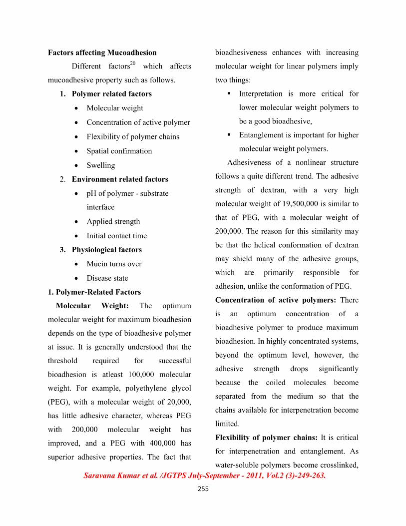
Factors affecting Mucoadhesion
Different factors20 which affects
mucoadhesive property such as follows.
1. Polymer related factors
Molecular weight
Concentration of active polymer
Flexibility of polymer chains
Spatial confirmation
Swelling
2. Environment related factors
pH of polymer - substrate
interface
Applied strength
Initial contact time
3. Physiological factors
Mucin turns over
Disease state
1. Polymer-Related Factors
Molecular Weight: The optimum
molecular weight for maximum bioadhesion
depends on the type of bioadhesive polymer
at issue. It is generally understood that the
threshold required for successful
bioadhesion is atleast 100,000 molecular
weight. For example, polyethylene glycol
(PEG), with a molecular weight of 20,000,
has little adhesive character, whereas PEG
with 200,000 molecular weight has
improved, and a PEG with 400,000 has
superior adhesive properties. The fact that
bioadhesiveness enhances with increasing
molecular weight for linear polymers imply
two things:
Interpretation is more critical for
lower molecular weight polymers to
be a good bioadhesive,
Entanglement is important for higher
molecular weight polymers.
Adhesiveness of a nonlinear structure
follows a quite different trend. The adhesive
strength of dextran, with a very high
molecular weight of 19,500,000 is similar to
that of PEG, with a molecular weight of
200,000. The reason for this similarity may
be that the helical conformation of dextran
may shield many of the adhesive groups,
which are primarily responsible for
adhesion, unlike the conformation of PEG.
Concentration of active polymers: There
is an optimum concentration of a
bioadhesive polymer to produce maximum
bioadhesion. In highly concentrated systems,
beyond the optimum level, however, the
adhesive strength drops significantly
because the coiled molecules become
separated from the medium so that the
chains available for interpenetration become
limited.
Flexibility of polymer chains: It is critical
for interpenetration and entanglement. As
water-soluble polymers become crosslinked,
Saravana Kumar et al. /JGTPS July-September - 2011, Vol.2 (3)-249-263.
256
mobility of individual polymer chains
decrease and thus the effective length of the
chain that can penetrate into the mucus layer
decreases, which reduces bioadhesive
strength.
Spatial conformation: Besides molecular
weight or chain length, spatial conformation
of a molecule is also important. Despite a
high molecular weight of 19,500,000 for
dextrans, they have similar adhesive strength
to the polyethylene glycol with a molecular
weight of 200,000. The helical conformation
of dextran may shield many adhesively
active groups, primarily responsible for
adhesion, unlike PEG polymers which have
a linear conformation.
Swelling: It depends on the polymer
concentration, ionic concentration, as well
as the presence of water. Over hydration
results in the formation of a slippery
mucilage without adhesion.
2. Environment Related Factors
pH of polymer - substrate interface:
It can influence the formal charge on the
surface of mucus as well as certain ionisable
bioadhesive polymers. Mucus will have a
different charge density depending on pH
due to difference in dissociation of
functional groups on the carbohydrate
moiety and the amino acids of the
polypeptide backbone. pH of the medium is
important for the degree of hydration of
crosslinked polyacrylic acid, showing
consistently increased hydration from pH 4
to 7 and then a decrease as alkalinity and
ionic strength increases.
Applied strength: To place a solid
bioadhesive system, it is necessary to apply
a defined strength. Whatever the polymer,
poly(acrylic acid / vinyl benzene poly
(HEMA) or carbopol 934, the adhesion
strength increases with the applied strength
or with the duration of its application, upto
an optimum. The pressure initially applied
to the mucoadhesive tissue contact site can
affect the depth of interpenetration. If high
pressure is applied for a sufficiently long
period of time, polymers become
mucoadhesive even though they do not have
attractive interaction with mucin.
Initial Contact Time: Contact time
between the bioadhesive and mucus layer
determines the extent of swelling and
interpenetration of the bioadhesive polymer
chains. Moreover, bioadhesive strength
increases as the initial contact time
increases.
3. Physiological Variables
Mucin Turnover: The natural turnover
of mucin molecules is important for at least
two reasons. First, the mucin turnover is
expected to limit the residence time of the
Saravana Kumar et al. /JGTPS July-September - 2011, Vol.2 (3)-249-263.
257
mucoadhesive on the mucus layer. Second,
mucin turnover results in substantial
amounts of soluble mucin molecules. These
molecules interact with the mucoadhesive
before they have a chance to interact with
the mucus layer. Mucin turnover may
depend on other factors such as presence of
food.
Disease States: The physiochemical
properties of mucus are known to change
during disease conditions such as common
cold, gastric ulcers, ulcerative colitis, cystic
fibrosis, bacterial and fungal infections of
the female reproductive tract.
SITES FOR MUCOADHESIVE DRUG
DELIVERY SYSTEMS 21-23
Buccal cavity: At this site, first-pass
metabolism is avoided, and the non-
keratinized epithelium is relatively
permeable to drugs. Due to flow of saliva
and swallowing, materials in the buccal
cavity have a short residence time and so it
is one of the most suitable areas for the
development of bioadhesive devices that
adhere to the buccal mucosa and remain in
place for a considerable period of time.
Gastrointestinal tract: The gastrointestinal
tract has been the subject of intense study
for the use of bioadhesive formulations to
improve drug bioavailability. The problem
associated is that the polymeric bioadhesive
formulations bind the intestinal mucus,
which is constantly turning over and are
transported down the gut by peristalsis.
Another problem is that with conventional
formulations such as tablets, the active
ingredient may diffuse relatively rapidly
away from the bioadhesive.
Nasal cavity: Ease of access, avoidance of
first-pass metabolism and a relatively
permeable and well-vascularised membrane,
contribute to make the nasal cavity an
attractive site for drug delivery. Although
the surface area is not large (between 150-
200 cm2), one major disadvantage of nasal
mucosa is the rapid removal of substances
by mucociliary action (with a residence time
half-life of 15-30 min). This makes it a
prime target for bioadhesive formulations to
prolong the residence time to allow drug
release and absorption
Eye: One major problem for drug
administration to the eye is rapid loss of the
drug and or vehicle as a result of tear flow,
and so it is a target for prolonging the
residence time by bioadhesion. The
bioadhesive polymers are finding increasing
use in ophthalmic formulations, but often as
viscosity enhancers rather than as
bioadhesives.
Vagina: The vagina is a highly suitable site
for bioadhesive formulations and it is here

Saravana Kumar et al. /JGTPS July-September - 2011, Vol.2 (3)-249-263.
258
that the success of the concept can be seen
convincingly. The bioadhesion increases the
retention time (up to 72 h) and a smaller
amount of the active ingredient can be used,
reducing any adverse effects.
Oesophagus: Tablets or capsules lodging in
the oesophagus leads to delayed absorption
and therefore delayed onset of action, as the
oesophageal epithelial layer is impermeable
to most drugs. Development of a DDS that
adheres to the oesophagus has implications
in both the protection of the epithelial
surface from damage caused by reflux and
as a vehicle to deliver drugs for local action
within the oesophagus. Bioadhesive dosage
forms that adhere to the oesophageal mucosa
and prolong contact have been investigated
to improve the efficacy of locally acting
agents.
MUCOADHESION THEORIES24, 25
Although the chemical and physical
bases of mucoadhesion are not yet well
understood, varies theories adapted from
studies on the performance of several
materials and polymer-polymer adhesion
which explain the phenomenon.
Electronic theory
It defined as the electron transfer
from contact of an adhesive polymer with a
glycoprotein network, they form an
electrical interface at adhesive polymer and
glycoprotein network. Adhesion can
produce by attractive forces across the
double layer.
Absorption theory
Absorption theory are defined as
they cause after initial contact between two
surfaces that is material surface because a
force formed between two surfaces, the
force is two types of chemical bond that is,
Primary chemical bond of covalent
bond: they are high strength so they
cause permanent bonds.
Secondary chemical bond has types
of force of attraction like
electrostatic force, Vander Waals
forces, hydrogen and hydrophobic
bonds.
Wetting theory
The wetting theory applies to liquid
systems which present affinity to the surface
in order to spread over it. This affinity can
be found by using measuring techniques
such as the contact angle. The general rule
states that the lower the contact angle then
the greater the affinity (Fig.2). The contact
angle should be equal or close to zero to
provide adequate spreadability.

Saravana Kumar et al
Fig. 3 – Schematic diagram
Diffusion theory
Diffusion theory describes the
interpenetration of both polymer and mucin
chains to a sufficient depth to create a semi
permanent adhesive bond (Fig. 4). It is
believed that the adhesion force increases
Fig. 4 – Secondary interactions resulting from inter diffusion of polymer chains
Fracture Theory:
This theory related for difficulty of
separation of two surfaces after adhesion,
The equation,
al. /JGTPS July-September - 2011, Vol.2 (3)-249
259
Schematic diagram showing influence of contact angle between device and
Mucous membrane on bioadhesion
Diffusion theory describes the
interpenetration of both polymer and mucin
chains to a sufficient depth to create a semi-
(Fig. 4). It is
believed that the adhesion force increases
with the degree of penetration of the
polymer chains. This penetration rate
depends on the diffusion coefficient,
flexibility and nature of the mucoadhesive
chains, mobility and contact time.
Secondary interactions resulting from inter diffusion of polymer chains
of bioadhesive device and of mucus
This theory related for difficulty of
adhesion,
G = (E e/L) 1/2
E = Young’s formula of elasticity
e = Fracture energy
L= Critical crack length
249-263.
showing influence of contact angle between device and
gree of penetration of the
polymer chains. This penetration rate
depends on the diffusion coefficient,
flexibility and nature of the mucoadhesive
chains, mobility and contact time.
Secondary interactions resulting from inter diffusion of polymer chains
E = Young’s formula of elasticity

Saravana Kumar et al. /JGTPS July-September - 2011, Vol.2 (3)-249-263.
260
EVALUATION OF MUCOADHESIVE
PROPERTIES26-28
Various in vivo and in vitro methods
are used for testing the efficacy of the
mucoadhesive nature of a polymer matrix.
Commonly used in vitro/ex vivo methods
include tensile strength measurement, shear
strength measurement and chip based
systems whereas various imaging techniques
are used for the evaluation of the delivery
systems under in vivo conditions. This
section will describe various methods used
to study the mucoadhesive properties.
In vitro tensile strength measurement
is done by dipping a filter paper in 8%
mucin dispersion. There after, the mucin
coated filter paper is placed in contact with
the hydrated polymeric samples (in
physiological solutions) for a definite period
of time, followed by the determination of the
maximum force required to detach the filter-
paper and polymer surfaces after the
mucoadhesive bonding. Similarly, ex vivo
experimentations are also done with the
exception that the mucin coated filter-paper
is replaced with excised mucosal tissues
(e.g. buccal mucosa, intestinal mucosa,
vaginal mucosa.
The mucoadhesive properties can
also be determined by incubating the
hydrated polymer matrix surface kept in
contact with a viscoelastic 30 % (w/w)
mucin solution in water with the subsequent
determination of the maximum detachment
force required to separate the polymer
matrix and mucin solution surfaces after the
adhesion. Wash-off test may also be used to
determine the mucoadhesive property of
delivery systems. In the test, the mucosal
tissue is attached onto a glass slide with the
help of a double-sided cyanoacrylate tape.
Thereafter, the delivery system is put on the
surface of the tissue (exposed mucosal
surface) with the subsequent vertical
attachment of the system into the USP tablet
disintegrator apparatus, which contains 1 L
of physiological solution maintained at
37oC. The operation of the equipment gives
an up-and-down movement to the tissue-
delivery matrix system.
In this study, the time for the
complete detachment of the delivery system
from the mucosal layer is determined. For
the relative measurement of mucoadhesive
nature of powder polymer samples modified
Du Noiy’s tensiometer may be used, while
in the shear strength determination method
the force required to slide the polymer
matrix over the mucus layer is determined.
Recently mucoadhesion studies have been
reported by using BIACORE® integrated
chip (IC) systems. The method involves

Saravana Kumar et al. /JGTPS July-September - 2011, Vol.2 (3)-249-263.
261
immobilization of the polymer (powder) on
to the surface of the IC with the subsequent
passage of the mucin solution over the same.
This results in the interaction of the mucin
with that of the polymer surface.
CONCLUSION
Mucoadhesive microsphere drug
delivery system have a high potential of
being useful means of releasing drugs to the
body, perhaps particularly for local
administration where the mechanical trauma
experienced by the dosage form may be
decreased. Current use of mucoadhesive
polymers to enhance resident time for a
wide variety of drugs and routes of
administration has shown dramatic
improvement in both specific therapies and
more general patient compliance. The
general properties of these polymers for
purpose of sustained release of chemicals
are marginal in being able to accommodate a
wide range of physicochemical drug
properties. Mucoadhesive polymers may
provide an important tool to enhance the
bioavailability of the active agent by
improving the residence time at the target site.
REFERENCES:
1. Prasanth, V.V., Sirisha Mudiyala, Sam T Mathew, Rinku Mathapan, Buccal tablet-As
mucoadhesive drug delivery: An over view, Journal of Pharmacy Research, 2011, 4(3),
706-709.
2. Jae Hyung Park, Mingli Ye and Kinam Park, Biodegradable Polymers for
Microencapsulation of Drugs, Molecules, 2005, 10, 146-161.
3. Roy, S., Pal, K., Anis, A., Pramanik, K. and Prabhakar, B., Polymers in Mucoadhesive
Drug Delivery System: A Brief Note, Designed monomers and polymers, 2009, 12, 483-
495.
4. Jimenez-Castellanous, M.R., Zia, H., Rhodes, C.T., Mucosal drug delivery system, Drug
Dev.Ind.Pharm., 1993, 19, 143-194.
5. Manish Kumar Singh, Pramod Kumar Sharma, Nitin Sharma, Gastroretentive Drug
Delivery System Based On Natural Mucoadhesive Polymers: A Brief Review, Journal of
Pharmacy Research, 2011, 4(2), 519-521.
6. Patil, S.B., Murthy, R.S.R., Mahajan, H.S., Wagh, R.D., Gattani, S.G., Mucoadhesive
polymers: Means of improving drug delivery, Pharma Times, 2006, 38(4), 25-28.

Saravana Kumar et al. /JGTPS July-September - 2011, Vol.2 (3)-249-263.
262
7. Cicek, H., Tuncel, A., Tuncel, M., Piskin, E., Degradation and drug release
characteristics of monosize polyethylcyanoacrylate microspheres, J. Biomater. Sci.
Polym. Ed., 1995, 6, 845-856.
8. Jimenez - Castellannos M.R., Zia. H., Rhodes C.T., Mucoadhesive drug delivery system,
Drug Dev. Ind Phar., 1993, 19(142), 143.
9. Leon Lachman, Herbert, A., Lieberman, Joseph, L., Kangi., The Theory and Practice of
Industrial Pharmacy, 1991, 296- 302.
10. Andrew, G.P., Laverty, T.P. and Jones D.S., Mucoadhesive polymeric for controlled drug
delivery, European Journal of Pharmaceutics and Biopharmaceutics, 2009, 71(3), 505-
518.
11. Ludwig, A., The use of mucoadhesive polymers in ocular drug delivery, Advanced Drug
Delivery Reviews, 2005, 57(11), 1595-1639.
12. Lee, J.W., Park, J.H., Robinson, J.R., Bioadhesive-based dosage forms: The next
generation, Journal of Pharmaceutical Sciences, 2000, 89(7), 850 – 866.
13. Flory, P.J., Principle of Polymer Chemistry, Cornell University Press, Ithaca, New York,
1953, 541-556.
14. Schnurch, A.B., Mucoadhesive systems in oral drug delivery, Drug Discovery Today:
Technologies, 2005, 2(1), 83-87.
15. Park, H. and Robinson, J.R., Mechanisms of mucoadhesion of polyacrylic acid and
hydrogels, Pharm. Res., 1987, 4, 457-464.
16. Flavia Chiva Carvalho, Marcos Luciano Bruschi, Raul Cesar Evangelista, Maria Palmira
Daflon Gremiao, Mucoadhesive drug delivery systems, Brazilian Journal of
Pharmaceutical Sciences, 2010, 46(1), 1-18.
17. Hagerstrom, H., Edsman, K., Stromme, M., Low- Frequency Dielectric Spectroscopy as a
Tool for Studying the Compatibility between Pharmaceutical Gels and Mucus Tissue, J.
Pharm. Sci., 2003, 92(9), 1869-1881.
18. Smart, J.D., The basics and underlying mechanisms of mucoadhesion, Adv.Drug Del.
Rev., 2005, 57(11), 1556- 1568.

Saravana Kumar et al. /JGTPS July-September - 2011, Vol.2 (3)-249-263.
263
19. Mathiowitz, E., Chickering, D.E., Lehr, C.M., (Eds.)., Bioadhesive drug delivery
systems: fundamentals, novel approaches, and development, Drugs and the
Pharmaceutical Sciences. New York: Marcel Dekker, 1999, 696.
20. Jain, N.K., Controlled release and Novel Drug Delivery, 1st edition.CBS publishers and
Distributors New Delhi.1997, 353-370.
21. Lee, J.W., Park, J.H., Robinson, J.R., Bioadhesive-based dosage forms: The next
generation, J. Pharm. Sci., 2000, 89(7), 850-866.
22. Woodley, J., Bioadhesion: New Possibilities for Drug Administration, Clin
Pharmacokinet, 2001, 40(2), 77-84.
23. O’Neill, J.L., Remington, T.L., Drug-induced esophageal injuries and dysphagia, Ann
Pharmacother, 2003, 37, 1675–1683.
24. Huang, Y., Leobandung, W., Foss, A., Peppas, N.A., Molecular aspects of muco- and
bioadhesion: Tetheres structures and site-specific surfaces, J. Control. Release, 2000,
65(1), 63-71.
25. Peppas, N.A., Sahlin, J.J., Hydrogels as mucoadhesive and bioadhesive materials: a
review, Biomaterials, 1996, 17(11), 1553-1561.
26. Perumal, V.A., Lutchman, D., Mackraj, I., Govender, T., Polymers of Opposing
Solubilities, Int. J. Pharm., 2008, 35, 184–191.
27. Takeuchi, H, Thongborisute, J., Matsui, Y., Sugihara, H., Yamamoto, H., Kawashima,
Y., Novel mucoadhesion tests for polymers and polymer-coated particles to design
optimal mucoadhesive drug delivery systems, Advanced Drug Delivery Reviews, 2005,
57(11), 1583-1594.
28. Kreuter, J., Muller, U., Munz, K., Quantitative and microautoradiographic study on
mouse intestinal distribution of polycyanoacrylate nanoparticles, International Journal of
Pharmaceutics, 1989, 55(1), 39-45.