Review Article DiagnosisofParasiticDiseases...
16
Hindawi Publishing Corporation Interdisciplinary Perspectives on Infectious Diseases Volume 2009, Article ID 278246, 15 pages doi:10.1155/2009/278246 Review Article Diagnosis of Parasitic Diseases: Old and New Approaches Momar Ndao National Reference Centre for Parasitology, McGill University Centre for Tropical Diseases, Montreal General Hospital, 1650 Cedar Avenue R3-137, Montreal, QC, Canada H3G 1A4 Correspondence should be addressed to Momar Ndao, [email protected] Received 31 May 2009; Accepted 29 August 2009 Recommended by Herbert B. Tanowitz Methods for the diagnosis of infectious diseases have stagnated in the last 20–30 years. Few major advances in clinical diagnostic testing have been made since the introduction of PCR, although new technologies are being investigated. Many tests that form the backbone of the “modern” microbiology laboratory are based on very old and labour-intensive technologies such as microscopy for malaria. Pressing needs include more rapid tests without sacrificing sensitivity, value-added tests, and point-of-care tests for both high- and low-resource settings. In recent years, research has been focused on alternative methods to improve the diagnosis of parasitic diseases. These include immunoassays, molecular-based approaches, and proteomics using mass spectrometry platforms technology. This review summarizes the progress in new approaches in parasite diagnosis and discusses some of the merits and disadvantages of these tests. Copyright © 2009 Momar Ndao. This is an open access article distributed under the Creative Commons Attribution License, which permits unrestricted use, distribution, and reproduction in any medium, provided the original work is properly cited. 1. Introduction Currently, the detection and diagnosis of parasite infections rely on several laboratory methods in addition to clinical symptoms, clinical history, travel history, and geographic location of patient. The primary tests currently used to diagnose many parasitic diseases have changed little since the development of the microscope in the 15th century by Antonie van Leeuwenhoek. Furthermore, most of the current tests cannot distinguish between past, latent, acute, and reactivated infections and are not useful for following response to therapy or for prognosis. Recent developments in new diagnostic tools, however, have opened new avenues for a vast improvement in parasite detection. Firstly, a number of newer serology-based assays that are highly specific and sensitive have emerged, such as the Falcon assay screening test ELISA (FAST-ELISA) [1], Dot-ELISA [2, 3], rapid antigen detection system (RDTS) [4], and luciferase immunoprecipitation system (LIPS) [5]. Secondly, molecular-based approaches such as loop- mediated isothermal amplification (LAMP) [6], real-time polymerase chain reaction [7], and Luminex [8] have shown a high potential for use in parasite diagnosis with increased specificity and sensitivity. Thirdly, proteomic technology has also been introduced for the discovery of biomarkers using tissues or biological fluids from the infected host. The aim of this review is to highlight the poten- tial for these new technologies in parasite diagnosis. For convenience, old and new parasitic diagnostic tools are summarized in Tables 1 and 2. The diagnostic tools offered by the CDC (Centre for Disease Control, Atlanta, USA) and the NRCP (National Reference Centre for Parasitology, Montr´ eal, Canada) are also highlighted in Tables 3 and 4. 2. Microscopy For many years, microscopy has been the only tool available for the detection of parasites through inspection of blood smears [10–14], tissue specimens [15–17], feces, lymph node aspirates [18, 19], bone marrow [20], and even cere- brospinal fluid [21]. However, sample preparation for direct observation is time-consuming, labour intensive, and proper diagnosis depends on qualified laboratory technicians. In the case of slide reading, a second independent reading is preferable, but not always required for accurate diagnosis. If need be, divided readings are resolved by a third reader. In endemic regions, where resources are limited, this proves to
-
Upload
nguyentuyen -
Category
Documents
-
view
214 -
download
0
Transcript of Review Article DiagnosisofParasiticDiseases...
Hindawi Publishing CorporationInterdisciplinary Perspectives on Infectious DiseasesVolume 2009, Article ID 278246, 15 pagesdoi:10.1155/2009/278246
Review Article
Diagnosis of Parasitic Diseases: Old and New Approaches
Momar Ndao
National Reference Centre for Parasitology, McGill University Centre for Tropical Diseases, Montreal General Hospital,1650 Cedar Avenue R3-137, Montreal, QC, Canada H3G 1A4
Correspondence should be addressed to Momar Ndao, [email protected]
Received 31 May 2009; Accepted 29 August 2009
Recommended by Herbert B. Tanowitz
Methods for the diagnosis of infectious diseases have stagnated in the last 20–30 years. Few major advances in clinical diagnostictesting have been made since the introduction of PCR, although new technologies are being investigated. Many tests that form thebackbone of the “modern” microbiology laboratory are based on very old and labour-intensive technologies such as microscopyfor malaria. Pressing needs include more rapid tests without sacrificing sensitivity, value-added tests, and point-of-care tests forboth high- and low-resource settings. In recent years, research has been focused on alternative methods to improve the diagnosis ofparasitic diseases. These include immunoassays, molecular-based approaches, and proteomics using mass spectrometry platformstechnology. This review summarizes the progress in new approaches in parasite diagnosis and discusses some of the merits anddisadvantages of these tests.
Copyright © 2009 Momar Ndao. This is an open access article distributed under the Creative Commons Attribution License,which permits unrestricted use, distribution, and reproduction in any medium, provided the original work is properly cited.
1. Introduction
Currently, the detection and diagnosis of parasite infectionsrely on several laboratory methods in addition to clinicalsymptoms, clinical history, travel history, and geographiclocation of patient. The primary tests currently used todiagnose many parasitic diseases have changed little sincethe development of the microscope in the 15th centuryby Antonie van Leeuwenhoek. Furthermore, most of thecurrent tests cannot distinguish between past, latent, acute,and reactivated infections and are not useful for followingresponse to therapy or for prognosis.
Recent developments in new diagnostic tools, however,have opened new avenues for a vast improvement in parasitedetection. Firstly, a number of newer serology-based assaysthat are highly specific and sensitive have emerged, suchas the Falcon assay screening test ELISA (FAST-ELISA) [1],Dot-ELISA [2, 3], rapid antigen detection system (RDTS)[4], and luciferase immunoprecipitation system (LIPS)[5]. Secondly, molecular-based approaches such as loop-mediated isothermal amplification (LAMP) [6], real-timepolymerase chain reaction [7], and Luminex [8] have showna high potential for use in parasite diagnosis with increasedspecificity and sensitivity. Thirdly, proteomic technology has
also been introduced for the discovery of biomarkers usingtissues or biological fluids from the infected host.
The aim of this review is to highlight the poten-tial for these new technologies in parasite diagnosis. Forconvenience, old and new parasitic diagnostic tools aresummarized in Tables 1 and 2. The diagnostic tools offeredby the CDC (Centre for Disease Control, Atlanta, USA)and the NRCP (National Reference Centre for Parasitology,Montreal, Canada) are also highlighted in Tables 3 and 4.
2. Microscopy
For many years, microscopy has been the only tool availablefor the detection of parasites through inspection of bloodsmears [10–14], tissue specimens [15–17], feces, lymphnode aspirates [18, 19], bone marrow [20], and even cere-brospinal fluid [21]. However, sample preparation for directobservation is time-consuming, labour intensive, and properdiagnosis depends on qualified laboratory technicians. Inthe case of slide reading, a second independent reading ispreferable, but not always required for accurate diagnosis. Ifneed be, divided readings are resolved by a third reader. Inendemic regions, where resources are limited, this proves to

2 Interdisciplinary Perspectives on Infectious Diseases
Table 1: Diagnostic tools for the detection of specific blood-borne parasitic diseases.
Africantrypanosomiasis
BabesiosisChagasdisease
Leishmaniasis Malaria Toxoplasmosis
Trypanosomabrucei
Babesiamicroti
Trypanosomacruzi
Leishmaniaspecies
Plasmodiumspecies
Toxoplasmagondii
MICROSCOPY [23] [10] [11] [12] [13] —
SEROLOGY-BASEDASSAYS
— — — — — —
ELISA [24, 25] [26] [27–30] — — [31]
FAST-ELISA — — — — [32, 33] —
Dot-ELISA orDipstick
[34] — [35] [2, 18] — —
RIPA-ELISA — — [36, 37] — — —
DHA or IHA [38] — — [18] — —
DFA or IFA [39] [40, 41] — — [42] [43]
Immunoblot — — [44, 45] — — [46]
PRISM — — [47] — — —
RDT — — [48] — [49] —
MOLECULAR-BASEDASSAYS
— — — — — —
PCR [23] [50, 51] [52–54] [55] [56] [57, 58]
RT-PCR [59] — — [60–62] [4, 56] [63]
QT-NASBA — — — [64] [65, 66] —
RT-QB-NASBA — — — — [67] —
LAMP [68] — [69] — [70–74] —
Luminex — — — — [75] —
PCR-ELISA — — — [62, 76] [77–79] —
OC-PCR [80] — — [81] — —
PROTEOMICS — — — — — —
MassSpectrometry(LDMS,MALDI-TOF,SELDI-TOF)
[82, 83] — — — [84–86] —
FAST-ELISA: Falcon assay screening test; RIPA-ELISA: radioimmunoprecipitation assay; DHA or IHA: direct or indirect hemagglutination assay; DFA orIFA: direct or indirect immunofluorescence assay; RDT: rapid diagnostic test; LIPS: luciferase immunoprecipitation system; CATT: Card Agglutination testfor Trypanosomiasis; PCR: polymerase chain reaction; RT-PCR: real-time polymerase chain reaction; QT-NASBA: quantitative nucleic acid sequenced-basedamplification; RT-QT-NASBA: real-time quantitative nucleic acid sequenced-based amplification; LAMP: loop-mediated isothernal amplification; OC-PCR:oligochromatography Polymerase chain reaction; LDMS: laser desorption mass spectrometry; MALDI-ToF: matrix-assisted laser desorption/ionization timeof flight; SELDI-Tof: surface-enhanced laser desorption/ionization time of flight, IFA: immunofluorescent assay, EIA: Enzyme immunoassay, RT-PCR: Realtime PCR, IB: immunoblot.
be difficult and misdiagnosis can significantly impact patientcare. In reality, all major intestinal helminth infectionsare still solely dependent on microscopy for diagnosis. Asfor other parasite infections, many are confirmed by theuse of microscopy in conjunction to other methods ofdiagnosis including serology-based assays and more recentlymolecular-based assays.
3. Serology-Based Assays
In situations where biologic samples or tissue specimens areunavailable, serology alone is the gold standard for diagnosis.
Serology-based diagnosis tools can be divided into twocategories: antigen-detection assays and antibody-detectionassays. These include the enzyme-linked immunosorbentassay (ELISA), also called enzyme immunoassay (EIA), andall its derived tests such as the Falcon assay screeningtest ELISA (FAST-ELISA) and the dot-ELISA. Other assaysinclude the hemagglutination (HA) test, indirect or directimmunofluorescent antibody (IFA or DFA) tests, comple-ment fixation (CF) test, and immunoblotting and rapiddiagnostic tests (RDTs).
Although the ease of use and turnaround times forserologic assays are similar to microscopy, serology-based
Interdisciplinary Perspectives on Infectious Diseases 3
Table 2: Diagnostic tools for the detection of specific intestinal parasitic diseases.
PROTOZOA TREMATODES CESTODES NEMATODES
Cryptosporidiosis Fasciolosis SchistosomiasisTaeniasis/
CysticercosisHydatidosis Filariasis Strongylodiasis
Cryptosporidiumparvum, C. hominis
Fasciolahepatica, F.gigantica
Schistosomamansoni
Taenia soliumEchinococcusgranulosus, E.multilocularis
Wuchereriabancrofti,
Brugiamalayi, B.timori, Loa
loa
Strongyloidesstercoralis
MICROSCOPY [87] [88] [89] — [90] [91] [92]
SEROLOGYBASED ASSAYS
— — — — — — —
ELISA [93, 94] [88, 95] [96, 97] [98–101] [102–106] [14] [107–111]
FAST-ELISA — [112] [1] — — — —
Dot-ELISA orDipstick
— [113, 114] [115] — [116, 117] [118–120] —
DHA or IHA — — [121] — [122] [123] [124]
DFA or IFA [93, 125, 126] — — — — — [127, 128]
Immunoblot — [112] [129] [130, 131] [122] — [132]
LIPS — — — — — [133] [134]
MOLECULAR-BASEDASSAYS
— — — — — — —
PCR [135, 136] — [137, 138] [139, 140] — [141–143] [144]
RT-PCR [145–147] — [148, 149] — — — [150]
LAMP [151, 152] — — [153] — — —
Luminex [154] — — — — — —
PCR-ELISA — — — — — [155] —
OC-PCR — — [156] — — — —
PROTEOMICS — — — — — — —
MassSpectrometry(LDMS,MALDI-TOF,SELDI-TOF)
— [157] — [158] — — —
Abbreviations: see Table 1.
Table 3: Diagnostic tools for the detection of specific blood-borne parasitic diseases offered by the CDC and the NRCP.
Africantrypanosomiasis
Babesiosis Chagas disease Leishmaniasis Malaria Toxoplasmosis
Trypanosomabrucei species
Babesia microti Trypanosoma cruziLeishmania
speciesPlasmodium
speciesToxoplasma
gondii
CDCDIAGNOSTIC
TOOLSMicroscopy
Microscopy IFA,PCR
Microscopyculture, IFA, EIA
Microscopy,Culture, IFA
MicroscopyPCR, IFA
Microscopy IFA,EIA
NRCPDIAGNOSTIC
TOOLS
Microscopy,culture, CATT,
PCRMicroscopy, IFA
Microscopy,culture, EIA,
RT-PCR
Microscopy,Culture, IFA,
RT-PCR
Microscopy,IFA, IB, PCR
RT-PCR
CDC: Centre for Disease Control, Atlanta, Georgia, USA. NRCP: National Reference Centre for Parasitology, Montreal General Hospital, Montreal, Quebec,Canada. Abbreviations: see Table 1.

4 Interdisciplinary Perspectives on Infectious Diseases
(a)
(b)
(c)
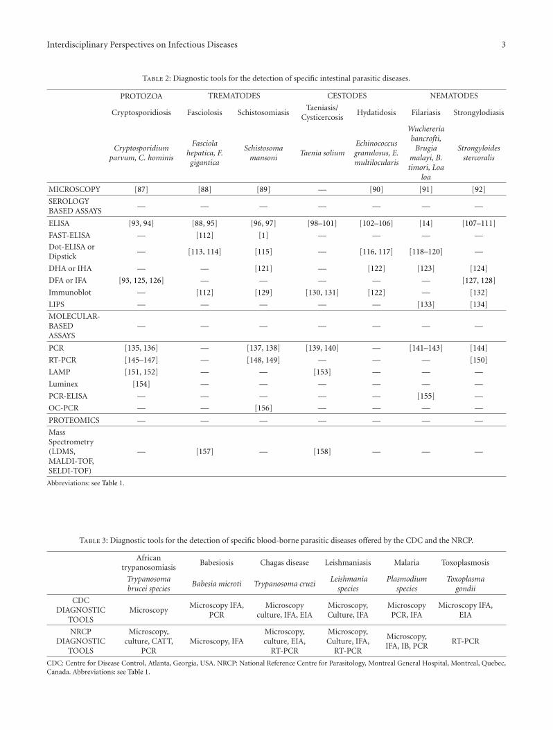
Figure 1: Microscopy. Comparison of Trypanosoma cruzi trypo-mastogote (a) with Plasmodium malariae schizont (b) and withmicrofilaria (c: Mansonella perstans) in squirrel monkey bloodsmear. Giemsa stain: 70x oil-immersion objective (a) and (b)and 27.2x objective (c), adapted with permission of ComparativeMedicine from [9].
00.20.40.60.8
11.21.41.6
Opt
ical
den
sity
(492.6
nm
)
Negativecontrol (kit)
ELISAnegative
ELISA +/− ELISApositive
Positivecontrol (kit)
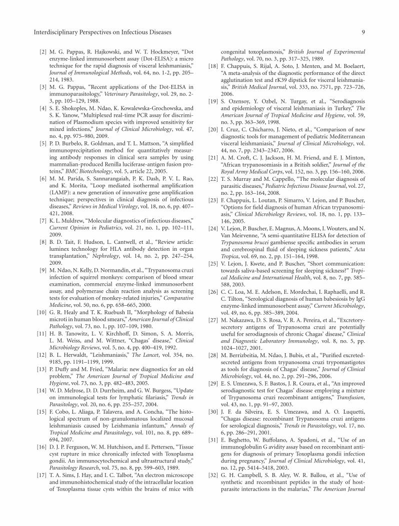
Figure 2: Serology-based assays: ELISA. Enzyme-linkedimmunosorbent assay (ELISA) absorbance values for antibodiesto T. cruzi in monkey samples. Median values are indicated byhorizontal lines within the boxes; the 25th and 75th percentilesare enclosed by the boxes; the 5th and 95th percentiles areenclosed by the bars outside the boxes. Adapted with permission ofComparative Medicine from [9].
M 1 2 3 4 5 6 7 8
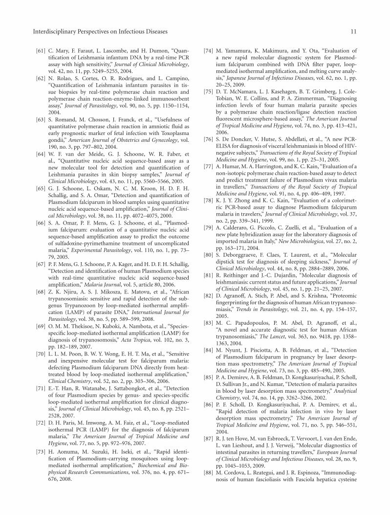
Figure 3: Molecular-based assay: PCR. Example of PCR resultsobtained for seven monkey samples, using the TCRUZ primers.Blood samples were processed as described in Materials andMethods. The PCR products were electrophoresed in a 2% agarosegel and stained with ethidium bromide. The 168 bp band (arrow)is the expected T. cruzi-specific product. The 360 and 550 bp arealso specific products resulting from amplification of two or threeof the 195 bp repeats found in tandem arrays in the T. cruzi genome.Lanes 1 to 6 contain the amplification products of DNA fromT. cruzi-infected monkeys; 7, blood from noninfected monkey; 8,negative control (distilled water); and M, 100 bp ladder, adapted,with permission of Comparative Medicine from [9].
assays are more sensitive and specific. It becomes importantfor individuals whose blood smears do not permit identifi-cation of the parasite (e.g., differentiating between Babesiaand Plasmodium) [159] or for patients exhibiting low-parasitemia and/or who are asymptomatic (e.g., Chagasicpatients) [54]. Classifying an infected asymptomatic patientas negative could lead to transmission of the parasite duringblood transfusions or organ transplants. In the case ofFasciola infection, serology tests have also been shown to beuseful in the confirmation of chronic fascioliasis when eggproduction is low or sporadic [112]. Finally, having thesetests readily available allows for the monitoring of parasiteclearance following therapy.
3.1. Falcon Assay Screening Test ELISA (FAST-ELISA). TheFalcon assay screening test ELISA (FAST-ELISA) consistsof using synthetic and recombinant peptides to evaluateantibody responses to an antigen [1]. In the past, the methodhas been applied to the study of malaria [32], fasciolosis[112], schistosomiasis (reviewed in [160]), and taeniasis[161]. However, this technique is subjected to the samedrawbacks as most serology-based tests. Antibodies raisedagainst a peptide from one parasite protein may cross-reactwith proteins from other species. Moreover, antibodies raisedagainst a peptide may react in some assays but not in othersand some regions of a peptide may be more immunogenicthan others. No recent studies have been published on the useof the FAST-ELISA for the diagnosis of parasitic infections.

Interdisciplinary Perspectives on Infectious Diseases 5
Table 4: Diagnostic tools for the detection of specific intestinal parasitic diseases offered by the CDC and the NRCP.
PROTOZOA TREMATODES CESTODES NEMATODES
Cryptosporidiosis Fasciolosis SchistosomiasisTaeniasis
CysticercosisHydatidosis Filariasis Strongylodiasis
Cryptosporidiumparvum, C. hominis
Fasciolahepatica, F.gigantica
Schistosomamansoni
Taenia soliumEchinococcusgranulosus, E.multilocularis
Wuchereriabancrofti,
Brugiamalayi, B.timori, Loa
loa
Strongyloidesstercoralis
CDC DIAG-NOSTICTOOLS
Microscopy, EIA,PCR, RT-PCR
—Microsocopy,
FAST-ELISA, IBImmunoblot, IB
Microscopy,EIA
Microscopy, EIA
NRCP DIAG-NOSTICTOOLS
Microscopy, EIAMicroscopy,
EIAMicroscopy, EIA IB EIA
Microscopy,EIA
Microscopy,culture, EIA, IB
CDC: Centre for Disease Control, Atlanta, Georgia, USA. NRCP: National Reference Centre for Parasitology, Montreal General Hospital, Montreal, Quebec,Canada.
3.2. Dot-ELISA. The main difference between the regularELISA and the dot-ELISA lies in the surface used to bindthe antigen of choice. In the dot-ELISA, the plastic plate isreplaced by a nitrocellulose or other paper membrane ontowhich a small amount of sample volume is applied. Thechoice of binding matrix greatly improved the specificity andsensitivity of the assay by reducing the binding of nonspecificproteins usually observed when plastic binding matrixesare used. The principle is similar to the immunoblot. Thedotted membrane is incubated first with an antigen-specificantibody followed by an enzyme-conjugated anti-antibody.The addition of a precipitable, chromogenic substrate causesthe formation of a colored dot on the membrane which canbe visually read [2]. The benefits of this technique include itsease of use, its rapidity, and the ease of result interpretation.It is fast, and cost-effective and more importantly can be usedin the field (e.g., as a dipstick). For all these reasons, the Dot-ELISA has been and still is extensively used in the detectionof human and animal parasitic diseases, including amebiasis,babesiosis, fascioliasis, cutaneous and visceral leishmania-sis, cysticercosis, echinococcosis, malaria, schistosomiasis,toxocariasis, toxoplasmosis, trichinosis, and trypanosomiasis(all reviewed in [3]). In the last few years, publishedstudies have demonstrated the use of the dot-ELISA for thedetection of Fasciola gigantica [113], Haemonchus contortus[162], Theileria equi [163], Trypanosoma cruzi [164], andTrypanosoma brucei [34]. In the latter study the researcherswere able to demonstrate that the dot-ELISA had bettersensitivity and specificity than the ELISA in the detectionof antineurofilament and antigalactocerebrosides antibodiesin cerebrospinal fluid of subjects infected with Africantrypanosomes. They attributed the greater sensitivity andspecificity of the dot-ELISA to the use of the nitrocellulosemembrane and showed that their assay was successfullyreproducible in the field.
3.3. Rapid Antigen Detection System (RDTS). Rapid antigendetection tests (RDTs) based on immunochromatographicantigen detection have been implemented in many diagnos-tic laboratories as an adjunct to microscopy for the diagnosisof malaria. RDTs consist of capturing soluble proteins bycomplexing them with capture antibodies embedded on anitrocellulose strip. A drop of blood sample is applied to thestrip and eluted from the nitrocellulose strip by the additionof a few drops of buffer containing a labeled antibody. Theantigen-antibody complex can then be visualized directlyfrom the membrane [4].
Since the appearance of the first RDTs in the 990s, majorimprovements have been made to the technique, making theuse of RDTs in rural endemic regions feasible. RDTs arenow rapid, stable at temperatures up to 40◦C, easy to use,and cost-effective thereby providing many advantages overtraditional microscopic methods [165]. RDTs are useful inthe identification of P. falciparum and P. vivax infections butcannot be used to identify P. malariae and P. ovale infections[4]. In addition, they are useless at detecting very low-densityinfections. PCR-based approaches remain the tool of choicein that situation. More than 80 RDTs exist for the detection ofeither histidine-rich protein (HRP) specific to P. falciparumor species-specific isotypes of lactate dehydrogenase (LDH)[49]. However, as reported by Murray et al. [165] only 23have met the WHO’s criteria for international marketing.
Malaria RDTs have recently been introduced in Africancountries to help prevent misdiagnosis of malaria infectionsand to subsequently reduce the practice of presumptivetreatment [49]. In fact, the tendency to treat slide-negativesamples with antimalarials is still a common phenomenon.This practice causes concern not only for the patient’shealth care but also to the costs it generates in prescribingthe more expensive antimalarial sulfadoxine/pyrimethamineand artemisinin-based combinations [165]. Finally, misuseof antimalarials could lead to the appearance of drug-resistant strains.

6 Interdisciplinary Perspectives on Infectious Diseases
3.4. Luciferase Immunoprecipitation System (LIPS). Theluciferase immunoprecipitation system (LIPS) is a modifiedELISA-based assay in which serum containing antigen-specific antibodies can be identified by measuring lightproduction. Basically, an antigen of choice is fused to theenzyme reporter Renilla luciferase (Ruc) and expressed asa Ruc-fusion in mammalian cells to allow for mammalian-specific posttranslational modifications. The crude proteinextract is then incubated with the test serum and proteinA/G beads. During the incubation, the Ruc-antigen fusionbecomes immobilized on the A/G beads, which allows theantigen-specific antibody to be quantitated by washing thebeads and adding coelenterazine substrate and measuringlight production [5].
In recent years, LIPS has been successfully applied forthe identification of sera samples infected with Strongyloidesstercoralis (using a Ruc-NIE fusion) [134] and Loa loa (usinga Ruc-LlSXP-1 fusion) [133]. Some of the advantages ofthe LIPS technology include its rapidity and accuracy indetecting infected patients. Sensitivity is improved in part bythe use of mammalian cells which produce fusion antigensfree of contaminating bacterial proteins. In addition, lowbackgrounds are produced compared to the ELISA. Thisgreatly facilitates the separation between negative and pos-itive samples. In addition, the Strongyloides LIPS based onthe NIE antigen showed greater specificity than the ELISAas no cross-reaction was observed with serum from filarial-infected subjects [134].
A LIPS assay can be performed in 2.5 hours. Burbeloet al. 2008 [133] were able to obtain 100% specificity andsensitivity when performing an LIPS assay based on theLoa loa SXP-1 antigen with only a small-degree of cross-reactivity with a few Onchocerca volvulus- and Wuchereriabancrofti-infected patient sera. By decreasing the incubationtimes of a normal LIPS assay, they were able to minimizecross-reaction. Many of the O. volvulus sera samples testedas positive with the LIPS assay were negative using this 15-minute LIPS assay also called QLIPS. Of interest for theapplication of this technique in the field is the observationthat blood obtained by finger-prick (contaminated with redblood cells and other components) did not interfere with theLIPS assay. Further studies will be useful in exploring andvalidating the accuracy and potential usefulness of the LIPSand QLIPS assays in the field.
As discussed, immunodiagnostics tests have someserious limitations. Parasitic diseases such as amebiasis,cryptosporidiosis, filariasis, giardiasis, malaria, cysticercosis,schistosomiasis, and African trypanosomiasis do not havecommercially or FDA approved antibody detection testsfor their diagnosis. Experimental results have been toovariable due to the type of antigen preparations used (e.g.,crude, recombinant purified, adult worm, egg) and alsobecause of the use of nonstandardized test procedures.Cross-reaction leading to false-positives and misdiagnosis isalso a problem, especially in regions where more than oneparasite is endemic. Despite the fact that some parasites inSouth America share common epitopes, it is common to seecoinfection with Trypanosoma cruzi and Leishmania species[166]. It is also a problem in Africa, where cross-reactivity
exists between filarial and other helminth antigens [133]. Toa lesser extent but nonetheless important is the inability ofantibody-detection tests to differentiate between past andcurrently active infections [167]. Furthermore, antibody-detection tests cannot be used in parasitic infectionsthat do not develop a significant antibody response.This has been observed in some individuals carryingEchinococcus cysts [168] or during cutaneous leishmaniasis(http://www.dpd.cdc.gov/dpdx/HTML/Leishmaniasis.htm).Similarly, in the case of African trypanosomiasis diagnosis,such tests are of limited use because seroconversion occursonly after the onset of clinical symptoms [83].
For all these reasons, there is still a need to improve onthe current diagnosis approaches available. Since the adventof the polymerase chain reaction (PCR), parasitologists haveturned to molecular-based approaches in the hopes to betterthe existing diagnosis tools.
4. Molecular-Based Approaches
4.1. Nucleic Acid-Based Approaches. The many limitationsof microscopy and serology-based assays have influencedparasitologists towards the use of gene amplification meth-ods made possible with the advent of the polymerasechain reaction (PCR). Besides the traditional PCR, includingnested and multiplexed PCR, we have seen the implemen-tation of the real-time PCR (RT-PCR) for the detectionof several parasitic infections. Newer technologies such asloop-mediated isothermal amplification and Luminex-basedassays have also emerged as possible new approaches for thediagnosis of parasitic diseases.
Molecular-based approaches based on nucleic acids offergreater sensitivity and specificity over the existing diagnostictests. They permit the detection of infections from verylow parasitized samples including those from asymptomaticpatient’s samples [169]. Moreover, multiplexed PCR allowsfor the detection of multiple sequences in the same reactiontube proving useful in the diagnosis of several parasiticinfections simultaneously [170].
4.2. Real-Time Polymerase Chain Reaction (RT-PCR). RT-PCR system unlike conventional PCR, allow for the quantifi-cation of the original template’s concentration through theuse of various fluorescent chemistries, such as Sybergreen,Taqman probes, fluorescence resonance energy transfer(FRET), and Scorpion primers [7]. The concentration ismeasured through comparison to standard curves. Thiseliminates the need to visualize the amplicons by gelelectrophoresis thereby greatly reducing the risk of contam-ination and the introduction of false-positives. When mul-tiplexed, RT-PCR allows for the high-throughput analysis ofdifferent sequences in one single-closed tube reaction [171].Using multiplexed RT-PCR, Shokoples et al. [4] were ableto identify the four human Plasmodium species (falciparum,vivax, malariae, and ovale) in a single reaction tube even invery low parasitized samples. Running the multiplex assaynot only reduced the cost per test but also allowed for arapid turnaround time, the assay taking only three hours to

Interdisciplinary Perspectives on Infectious Diseases 7
complete. It is a clear advantage over microscopy which islabour intensive and time-consuming with slow turnaroundtimes especially during high-throughput settings. Similarly,multiplexed RT-PCR proved useful in differentiating drug-sensitive strains of malaria [172]. This is important forproper antimalarial prescription. In another example, Diezet al. [54] were able to detect the presence of T. cruzi infec-tion following heart transplants using PCR. This allowedimmediate treatment of the patients before reactivation ofChagas disease could occur. These examples demonstratethat efficient and early diagnosis can directly impact patientscare and that PCR-based approaches have the potential tohelp in making the right choice for treatment.
Although DNA-based methods have shown excellentsensitivity and specificity, the introduction of these methodsin daily laboratory practice is still uncommon especially inrural endemic regions. In addition, as observed with manyserology-based assays, PCR-based methods also suffer bythe lack of standardization [22]. DNA extraction, choice ofprimer sets, and use of various amplification protocols areall factors that may cause this diversification in results [173].Adding an automated DNA extraction step would certainlyimprove PCR assays for use in the diagnosis of parasiticdiseases.
4.3. Loop-Mediated Isothermal Amplification (LAMP). Loop-mediated isothermal amplification (LAMP) is a uniqueamplification method with extremely high specificity andsensitivity able to discriminate between a single nucleotidedifference [6]. It is characterised by the use of six differentprimers specifically designed to recognise eight distinctregions on a target gene, with amplification only occurring ifall primers bind and form a product [174]. In the past, LAMPhas been successfully applied for the rapid detection of bothDNA and RNA viruses such as the West Nile [175] andSARS viruses [176]. Recently, parasitologists have adaptedthe LAMP approach for the detection of several parasiticdiseases including the human parasites Entamoeba [177],Trypanosoma [68], Taenia [153], Plasmodium [70], andCryptosporidium [152], the animal parasites Theilera [178]and Babesia [178, 179], and even to the identification ofvector mosquitoes carrying Plasmodium [73] and Dirofilariaimmitis [180] parasites. Most of these studies have brought tolight the many advantages of this method over the commonPCR technique.
Unlike a regular PCR reaction, LAMP is carried out ata constant temperature (usually in the range of 60–65◦C).This unique feature not only results in higher yields, but alsoeliminates the need to buy a thermal cycler and shortensthe reaction time by eliminating time lost during thermalchanges. In addition, the reaction can be carried out withoutextracting the DNA from the collected samples as shown inthe case of RIME, a nonautonomous retroelement found inTrypanosoma brucei rhodesiense and T. b. gambiense [68]. In35 minutes, using a simple water bath, RIME LAMP was ableto detect both T. b. gambiense and T. b. rhodesiense directlyfrom blood, serum, and CSF samples. More importantly, thestudy has shown reproducibility in the field. In addition to
the above advantages, LAMP reactions are easy to set up,and results can readily be assessed. The sample of interestis mixed with primers, substrates, and a DNA polymerasecapable of strand displacement in a microcentrifuge tube.During the reaction, large amounts of pyrophosphate ionsare produced, leading to the formation of a white precipitate[181]. This turbidity is proportional with the amount ofDNA synthesized therefore one can assess the reaction byreal-time measurement of turbidity or more importantly,simply through the naked-eye.
For all these reasons, the future adoption of LAMP asa diagnostic tool for parasite infections in rural endemicregions shows promise. Furthermore, as more groups applyLAMP to the field of parasitology, we will see the appear-ance of LAMP-modified assays that meet specific detectionneeds. For example, in a recent study on bovine Babesia[182], a multiplex-LAMP (mLAMP) assay was developed tosimultaneously detect B. bovis and B. bigemina from DNAextracted from blood spotted on filter paper. Similarly, Hanet al. [71] implemented a LAMP assay based on the 18S rRNAgene for the detection of the four human Plasmodium species(falciparum, vivax, malariae, and ovale). LAMP had a similarsensitivity and a greater specificity than nested PCR, yieldingsimilar results but at a faster turnaround time. Their resultsare consistent with other studies demonstrating the rapidityand the improved specificity and sensitivity obtained usingthe LAMP assay.
4.4. Luminex xMAP Technology. Luminex technologyis a bead-based flow-cytometric assay that allowsthe detection of various targets simultaneously(http://www.luminexcorp.com/). The microsphere beadscan be covalently bound to antigens, antibodies, or oligonu-cleotides that will serve as probes in the assay. Up to 100microspheres are available each emitting unique fluorescentsignals when excited by laser therefore allowing the identifi-cation of different targets [183]. Adapted to the study of par-asites, the Luminex assay could identify multiple organismsor different genotypes of one particular organism during thesame reaction utilizing very low volume. The approach couldprove useful in the study of antigenic diversity and drug-resistance alleles and for the diagnosis of parasitic diseases.
Luminex was applied to the study of Cryptosporidium[154]. C. hominis and C. parvum cannot be distinguishedusing antigen detection or serology assays. Only DNA-basedapproaches have been successful in doing so by exploitingthe single nucleotide difference in the microsatellite-2 region(ML-2) of both species [154]. Ultimately DNA sequencing isthe diagnosis tool of choice but it is costly, labour-intensiveand time-consuming. In a recent study, Bandyopadhyayet al. [154] successfully detected and distinguished C.hominis and C. parvum in 143 DNA extracts using Luminextechnology by using oligonucleotide probes specific to theML-2 regions of each species. Turnaround time was about5 hours making this assay not only much faster but alsoless expensive than PCR followed by DNA sequencing. Italso proved to be 100% specific and more sensitive than adirect fluorescent antibody (DFA) test, a method routinely

8 Interdisciplinary Perspectives on Infectious Diseases
used to identify Cryptosporidium and Giardia species. Notethat DFA cannot differentiate between C. hominis and C.parvum.
Similarly in other research, Luminex technology wasable to detect all-blood stage parasite levels of the fourhuman Plasmodium species (falciparum, vivax, malariae, andovale) simultaneously [75]. This study demonstrated thatLuminex technology can improve the speed, the accuracy,and the reliability of other PCR methods. For example,the need for gel electrophoresis to differentiate the LDRproducts representing the four human Plasmodium speciesis eliminated. Second, all samples are handled simultaneouslyand continuously through a 96-well plate format from DNAextraction all through data analysis. The process is auto-mated and therefore uniformity can be achieved. Finally, thehigh-throughput capability of the Luminex system confers ita clear advantage over the use of labour-intensive microscopyfor large scale studies.
4.5. Proteomics. Since proteins are the main catalysts,structural elements, signalling messengers, and molecularmachines of biological tissues, proteomic studies are ableto provide substantial clinical relevance. Proteins can beutilized as biomarkers for tissues, cell types, developmentalstages, and disease states as well as potential targets fordrug discovery and interventional approaches. The nextgeneration of diagnostic tests for infectious diseases willemerge from proteomic studies of serum and other bodyfluids. Recent advances in this area are attributable largelyto the introduction of mass spectrometry platforms capa-ble of screening complex biological fluids for individualprotein and peptide “biomarkers.” Proteomic strategy canidentify proteins in two ways: bottom-up and top-downapproaches. In the former, the proteins in a biological fluidare proteolytically shattered into small fragments that can beeasily sequenced and the resultant spectra are compared withthose in established peptide databases. This is the proteinequivalent of “shotgun” genomics. Bottom-up strategiesare difficult to quantitate and cannot identify modifiedmolecules (e.g., alternately spliced, glycosylated). Since eachopen reading frame in the human genome is thought togenerate at least 10 modified proteins, this issue is a majorlimitation.
The classic top-down strategy is 2-dimensional gelelectrophoresis. Top-down strategies seek to identify proteinsand peptides (and their natural variants) in complex biologi-cal fluids. Two-dimensional (2D) gel electrophoresis was firstdescribed in 1975. With this method, proteins are resolved inthe first dimension based on pH (a process called isoelectricfocusing) and in the second dimension by their molecularweight. This technique is labor intensive, and low throughputand requires large amounts of sample. Such limitationshave encouraged the search for improved approaches. Othertechniques used for the expression analysis of proteins arematrix-assisted laser desorption ionization time-of-flightmass spectrometry (MALDI-TOF MS), surface-enhancedlaser desorption ionization time of flight mass spectrom-etry (SELDI-TOF MS), liquid chromatography combined
with MS (LC–MS–MS), isotope-coded affinity tags (ICAT),and isotope tags for relative and absolute quantification(iTRAQ).
The development of automated, high-throughput pro-teomic technologies such as MALDI-TOF and SELDI-TOFMS has enabled large numbers of clinical samples to beanalyzed simultaneously in a short time. These platformshave made “population-based proteomics” feasible for thefirst time (reviewed in [184]). All proteomics-based diag-nostic efforts seek to identify biomarkers that, alone or incombination, can distinguish between “case” and “control”groups.
The main limitation of SELDI compared to MALDIresides in the fact that SELDI has lower resolution and lowermass accuracy. In addition, SELDI is unsuitable for highmolecular weight proteins (>100 kDa) and is limited to thedetection of bound proteins on to the ProteinChip Array.
Most studies published about parasitic diseases havefocused on SELDI. The SELDI, a derivation of MALDI,allows sample binding to chemically active ProteinChip sur-faces. Several types of ProteinChip arrays are available withdiffering abilities to bind proteins with different chemical(anionic, cationic, hydrophobic, metallic, and normal phase)or biological (antibody, enzymes, receptors) properties,thereby allowing the direct analysis of proteins from complexbiological samples without the need for prior separationby 2D gel electrophoresis. The output of the SELDI is aspectrum of mass-to-charge ratios (m:z values) with theircorresponding relative intensities (approximating to relativeabundance).
SELDI analyses were initially applied to the discoveryof early diagnostic or prognostic biomarkers of cancer(reviewed in [185]). Recently, this technique has been appliedto the study of serum biomarkers of infectious diseasessuch as Severe Acute Respiratory Syndrome [186], Africantrypanosomiasis [83], fascioliasis [157], cysticercosis [158],and Chagas diseases (Ndao et al., submitted). Such studieshave focused on identifying a distinctive configuration ofcirculating serum proteins that are indicative of a specificpathophysiological state, a so-called “proteomic fingerprint.”
The real potential of proteomic fingerprinting is inits use as a discovery tool for novel biomarkers that canthen be incorporated into simple bedside diagnostics basedon affordable technologies such as immunologically basedantigen-detection tests that could be implemented in dipstickor cassette formats.
Acknowledgments
The author would like to thank the Public Health Agencyof Canada and the National Microbiology Laboratory forsupporting the National Reference Centre for Parasitology.
References
[1] K. Hancock and V. C. W. Tsang, “Development and opti-mization of the FAST-ELISA for detecting antibodies toSchistosoma mansoni,” Journal of Immunological Methods,vol. 92, no. 2, pp. 167–176, 1986.
Interdisciplinary Perspectives on Infectious Diseases 9
[2] M. G. Pappas, R. Hajkowski, and W. T. Hockmeyer, “Dotenzyme-linked immunosorbent assay (Dot-ELISA): a microtechnique for the rapid diagnosis of visceral leishmaniasis,”Journal of Immunological Methods, vol. 64, no. 1-2, pp. 205–214, 1983.
[3] M. G. Pappas, “Recent applications of the Dot-ELISA inimmunoparasitology,” Veterinary Parasitology, vol. 29, no. 2-3, pp. 105–129, 1988.
[4] S. E. Shokoples, M. Ndao, K. Kowalewska-Grochowska, andS. K. Yanow, “Multiplexed real-time PCR assay for discrimi-nation of Plasmodium species with improved sensitivity formixed infections,” Journal of Clinical Microbiology, vol. 47,no. 4, pp. 975–980, 2009.
[5] P. D. Burbelo, R. Goldman, and T. L. Mattson, “A simplifiedimmunoprecipitation method for quantitatively measur-ing antibody responses in clinical sera samples by usingmammalian-produced Renilla luciferase-antigen fusion pro-teins,” BMC Biotechnology, vol. 5, article 22, 2005.
[6] M. M. Parida, S. Sannarangaiah, P. K. Dash, P. V. L. Rao,and K. Morita, “Loop mediated isothermal amplification(LAMP): a new generation of innovative gene amplificationtechnique; perspectives in clinical diagnosis of infectiousdiseases,” Reviews in Medical Virology, vol. 18, no. 6, pp. 407–421, 2008.
[7] K. L. Muldrew, “Molecular diagnostics of infectious diseases,”Current Opinion in Pediatrics, vol. 21, no. 1, pp. 102–111,2009.
[8] B. D. Tait, F. Hudson, L. Cantwell, et al., “Review article:luminex technology for HLA antibody detection in organtransplantation,” Nephrology, vol. 14, no. 2, pp. 247–254,2009.
[9] M. Ndao, N. Kelly, D. Normandin, et al., “Trypanosoma cruziinfection of squirrel monkeys: comparison of blood smearexamination, commercial enzyme-linked immunosorbentassay, and polymerase chain reaction analysis as screeningtests for evaluation of monkey-related injuries,” ComparativeMedicine, vol. 50, no. 6, pp. 658–665, 2000.
[10] G. R. Healy and T. K. Ruebush II, “Morphology of Babesiamicroti in human blood smears,” American Journal of ClinicalPathology, vol. 73, no. 1, pp. 107–109, 1980.
[11] H. B. Tanowitz, L. V. Kirchhoff, D. Simon, S. A. Morris,L. M. Weiss, and M. Wittner, “Chagas’ disease,” ClinicalMicrobiology Reviews, vol. 5, no. 4, pp. 400–419, 1992.
[12] B. L. Herwaldt, “Leishmaniasis,” The Lancet, vol. 354, no.9185, pp. 1191–1199, 1999.
[13] P. Duffy and M. Fried, “Malaria: new diagnostics for an oldproblem,” The American Journal of Tropical Medicine andHygiene, vol. 73, no. 3, pp. 482–483, 2005.
[14] W. D. Melrose, D. D. Durrheim, and G. W. Burgess, “Updateon immunological tests for lymphatic filariasis,” Trends inParasitology, vol. 20, no. 6, pp. 255–257, 2004.
[15] F. Cobo, L. Aliaga, P. Talavera, and A. Concha, “The histo-logical spectrum of non-granulomatous localized mucosalleishmaniasis caused by Leishmania infantum,” Annals ofTropical Medicine and Parasitology, vol. 101, no. 8, pp. 689–694, 2007.
[16] D. J. P. Ferguson, W. M. Hutchison, and E. Pettersen, “Tissuecyst rupture in mice chronically infected with Toxoplasmagondii. An immunocytochemical and ultrastructural study,”Parasitology Research, vol. 75, no. 8, pp. 599–603, 1989.
[17] T. A. Sims, J. Hay, and I. C. Talbot, “An electron microscopeand immunohistochemical study of the intracellular locationof Toxoplasma tissue cysts within the brains of mice with
congenital toxoplasmosis,” British Journal of ExperimentalPathology, vol. 70, no. 3, pp. 317–325, 1989.
[18] F. Chappuis, S. Rijal, A. Soto, J. Menten, and M. Boelaert,“A meta-analysis of the diagnostic performance of the directagglutination test and rK39 dipstick for visceral leishmania-sis,” British Medical Journal, vol. 333, no. 7571, pp. 723–726,2006.
[19] S. Ozensoy, Y. Ozbel, N. Turgay, et al., “Serodiagnosisand epidemiology of visceral leishmaniasis in Turkey,” TheAmerican Journal of Tropical Medicine and Hygiene, vol. 59,no. 3, pp. 363–369, 1998.
[20] I. Cruz, C. Chicharro, J. Nieto, et al., “Comparison of newdiagnostic tools for management of pediatric Mediterraneanvisceral leishmaniasis,” Journal of Clinical Microbiology, vol.44, no. 7, pp. 2343–2347, 2006.
[21] A. M. Croft, C. J. Jackson, H. M. Friend, and E. J. Minton,“African trypanosomiasis in a British soldier,” Journal of theRoyal Army Medical Corps, vol. 152, no. 3, pp. 156–160, 2006.
[22] T. S. Murray and M. Cappello, “The molecular diagnosis ofparasitic diseases,” Pediatric Infectious Disease Journal, vol. 27,no. 2, pp. 163–164, 2008.
[23] F. Chappuis, L. Loutan, P. Simarro, V. Lejon, and P. Buscher,“Options for field diagnosis of human African trypanosomi-asis,” Clinical Microbiology Reviews, vol. 18, no. 1, pp. 133–146, 2005.
[24] V. Lejon, P. Buscher, E. Magnus, A. Moons, I. Wouters, and N.Van Meirvenne, “A semi-quantitative ELISA for detection ofTrypanosoma brucei gambiense specific antibodies in serumand cerebrospinal fluid of sleeping sickness patients,” ActaTropica, vol. 69, no. 2, pp. 151–164, 1998.
[25] V. Lejon, J. Kwete, and P. Buscher, “Short communication:towards saliva-based screening for sleeping sickness?” Tropi-cal Medicine and International Health, vol. 8, no. 7, pp. 585–588, 2003.
[26] C. C. Loa, M. E. Adelson, E. Mordechai, I. Raphaelli, and R.C. Tilton, “Serological diagnosis of human babesiosis by IgGenzyme-linked immunosorbent assay,” Current Microbiology,vol. 49, no. 6, pp. 385–389, 2004.
[27] M. Nakazawa, D. S. Rosa, V. R. A. Pereira, et al., “Excretory-secretory antigens of Trypanosoma cruzi are potentiallyuseful for serodiagnosis of chronic Chagas’ disease,” Clinicaland Diagnostic Laboratory Immunology, vol. 8, no. 5, pp.1024–1027, 2001.
[28] M. Berrizbeitia, M. Ndao, J. Bubis, et al., “Purified excreted-secreted antigens from trypanosoma cruzi trypomastigotesas tools for diagnosis of Chagas’ disease,” Journal of ClinicalMicrobiology, vol. 44, no. 2, pp. 291–296, 2006.
[29] E. S. Umezawa, S. F. Bastos, J. R. Coura, et al., “An improvedserodiagnostic test for Chagas’ disease employing a mixtureof Trypanosoma cruzi recombinant antigens,” Transfusion,vol. 43, no. 1, pp. 91–97, 2003.
[30] J. F. da Silveira, E. S. Umezawa, and A. O. Luquetti,“Chagas disease: recombinant Trypanosoma cruzi antigensfor serological diagnosis,” Trends in Parasitology, vol. 17, no.6, pp. 286–291, 2001.
[31] E. Beghetto, W. Buffolano, A. Spadoni, et al., “Use of animmunoglobulin G avidity assay based on recombinant anti-gens for diagnosis of primary Toxoplasma gondii infectionduring pregnancy,” Journal of Clinical Microbiology, vol. 41,no. 12, pp. 5414–5418, 2003.
[32] G. H. Campbell, S. B. Aley, W. R. Ballou, et al., “Use ofsynthetic and recombinant peptides in the study of host-parasite interactions in the malarias,” The American Journal

10 Interdisciplinary Perspectives on Infectious Diseases
of Tropical Medicine and Hygiene, vol. 37, no. 3, pp. 428–444,1987.
[33] M. Salcedo, L. Barreto, M. Rojas, R. Moya, J. Cote, and M.E. Patarroyo, “Studies on the humoral immune response to asynthetic vaccine against Plasmodium falciparum malaria,”Clinical and Experimental Immunology, vol. 84, no. 1, pp.122–128, 1991.
[34] B. Courtioux, S. Bisser, P. M’Belesso, et al., “Dot enzyme-linked immunosorbent assay for more reliable staging ofpatients with human African trypanosomiasis,” Journal ofClinical Microbiology, vol. 43, no. 9, pp. 4789–4795, 2005.
[35] F. G. Araujo, “A method for demonstration of antibodiesto Trypanosoma cruzi by using antigen-coated nitrocellulosepaper strips,” The American Journal of Tropical Medicine andHygiene, vol. 34, no. 2, pp. 242–245, 1985.
[36] L. V. Kirchhoff, A. A. Gam, R. A. Gusmao, R. S. Goldsmith,J. M. Rezende, and A. Rassi, “Increased specificity ofserodiagnosis of Chagas’ disease by detection of antibodyto the 72- and 90-kilodalton glycoproteins of Trypanosomacruzi,” Journal of Infectious Diseases, vol. 155, no. 3, pp. 561–564, 1987.
[37] D. A. Leiby, S. Wendel, D. T. Takaoka, R. M. Fachini, L.C. Oliveira, and M. A. Tibbals, “Serologic testing for Try-panosoma cruzi: comparison of radioimmunoprecipitationassay with commercially available indirect immunofluores-cence assay, indirect hemagglutination assay, and enzyme-linked immunosorbent assay kits,” Journal of Clinical Micro-biology, vol. 38, no. 2, pp. 639–642, 2000.
[38] E. Magnus, T. Vervoort, and N. Van Meirvenne, “A card-agglutination test with stained trypanosomes (C.A.T.T.) forthe serological diagnosis of T. B. gambiense trypanosomia-sis,” Annales de la Societe Belge de Medecine Tropicale, vol. 58,no. 3, pp. 169–176, 1978.
[39] F. Noireau, J. L. Lemesre, M. Y. Nzoukoudi, M. T. Louembet,J. P. Gouteux, and J. L. Frezil, “Serodiagnosis of sleepingsickness in the Republic of the Congo: comparison of indirectimmunofluorescent antibody test and card agglutinationtest,” Transactions of the Royal Society of Tropical Medicine andHygiene, vol. 82, no. 2, pp. 237–240, 1988.
[40] P. J. Krause, R. Ryan, S. Telford III, D. Persing, and A.Spielman, “Efficacy of immunoglobulin M serodiagnostictest for rapid diagnosis of acute babesiosis,” Journal of ClinicalMicrobiology, vol. 34, no. 8, pp. 2014–2016, 1996.
[41] P. J. Krause, S. R. Telford III, R. Ryan, et al., “Diagnosis ofbabesiosis: evaluation of a serologic test for the detection ofBabesia microti antibody,” Journal of Infectious Diseases, vol.169, no. 4, pp. 923–926, 1994.
[42] A. J. Sulzer and M. Wilson, “The fluorescent antibody testfor malaria,” CRC Critical Reviews in Clinical LaboratorySciences, vol. 2, no. 4, pp. 601–619, 1971.
[43] M. E. Camargo, “Improved technique of indirectimmunofluorescence for serological diagnosis oftoxoplasmosis,” Revista do Instituto de Medicina Tropical deSao Paulo, vol. 12, pp. 117–118, 1964.
[44] E. S. Umezawa, M. S. Nascimento, and A. M. S. Stolf,“Enzyme-linked immunosorbent assay with Trypanosomacruzi excreted-secreted antigens (TESA-ELISA) for serodi-agnosis of acute and chronic Chagas’ disease,” DiagnosticMicrobiology and Infectious Disease, vol. 39, no. 3, pp. 169–176, 2001.
[45] K. Y. Cheng, C.-D. Chang, V. A. Salbilla, et al., “Immunoblotassay using recombinant antigens as a supplemental test toconfirm the presence of antibodies to Trypanosoma cruzi,”
Clinical and Vaccine Immunology, vol. 14, no. 4, pp. 355–361,2007.
[46] V. Rilling, K. Dietz, D. Krczal, F. Knotek, and G. Enders,“Evaluation of a commercial IgG/IgM Western blot assayfor early postnatal diagnosis of congenital toxoplasmosis,”European Journal of Clinical Microbiology and InfectiousDiseases, vol. 22, no. 3, pp. 174–180, 2003.
[47] C.-D. Chang, K. Y. Cheng, L. X. Jiang, et al., “Evaluation ofa prototype Trypanosoma cruzi antibody assay with recom-binant antigens on a fully automated chemiluminescenceanalyzer for blood donor screening,” Transfusion, vol. 46, no.10, pp. 1737–1744, 2006.
[48] A. O. Luquetti, C. Ponce, E. Ponce, et al., “Chagas’ diseasediagnosis: a multicentric evaluation of Chagas Stat-Pak,a rapid immunochromatographic assay with recombinantproteins of Trypanosoma cruzi,” Diagnostic Microbiology andInfectious Disease, vol. 46, no. 4, pp. 265–271, 2003.
[49] C. Drakeley and H. Reyburn, “Out with the old, in withthe new: the utility of rapid diagnostic tests for malariadiagnosis in Africa,” Transactions of the Royal Society ofTropical Medicine and Hygiene, vol. 103, no. 4, pp. 333–337,2009.
[50] P. J. Krause, S. Telford III, A. Spielman, et al., “Comparison ofPCR with blood smear and inoculation of small animals fordiagnosis of Babesia microti parasitemia,” Journal of ClinicalMicrobiology, vol. 34, no. 11, pp. 2791–2794, 1996.
[51] D. H. Persing, D. Mathiesen, W. F. Marshall, et al., “Detectionof Babesia microti by polymerase chain reaction,” Journal ofClinical Microbiology, vol. 30, no. 8, pp. 2097–2103, 1992.
[52] D. R. Moser, L. V. Kirchhoff, and J. E. Donelson, “Detectionof Trypanosoma cruzi by DNA amplification using thepolymerase chain reaction,” Journal of Clinical Microbiology,vol. 27, no. 7, pp. 1477–1482, 1989.
[53] R. Gutierrez, V. M. Angulo, Z. Tarazona, C. Britto, and O.Fernandes, “Comparison of four serological tests for thediagnosis of Chagas disease in a Colombian endemic area,”Parasitology, vol. 129, no. 4, pp. 439–444, 2004.
[54] M. Diez, L. Favaloro, A. Bertolotti, et al., “Usefulness of PCRstrategies for early diagnosis of Chagas’ disease reactivationand treatment follow-up in heart transplantation,” AmericanJournal of Transplantation, vol. 7, no. 6, pp. 1633–1640, 2007.
[55] S. Singh, A. Dey, and R. Sivakumar, “Applications ofmolecular methods for Leishmania control,” Expert Reviewof Molecular Diagnostics, vol. 5, no. 2, pp. 251–265, 2005.
[56] L. K. Erdman and K. C. Kain, “Molecular diagnostic andsurveillance tools for global malaria control,” Travel Medicineand Infectious Disease, vol. 6, no. 1-2, pp. 82–99, 2008.
[57] P. V. Vidigal, D. V. Santos, F. C. Castro, J. C. Couto, R.W. Vitor, and G. Brasileiro Filho, “Prenatal toxoplasmosisdiagnosis from amniotic fluid by PCR,” Revista da SociedadeBrasileira de Medicina Tropical, vol. 35, no. 1, pp. 1–6, 2002.
[58] M. H. Bessieres, A. Berrebi, S. Cassaing, et al., “Diagnosis ofcongenital toxoplasmosis: prenatal and neonatal evaluationof methods used in Toulouse University Hospital and inci-dence of congenital toxoplasmosis,” Memorias do InstitutoOswaldo Cruz, vol. 104, no. 2, pp. 389–392, 2009.
[59] S. Becker, J. R. Franco, P. P. Simarro, A. Stich, P. M. Abel, andD. Steverding, “Real-time PCR for detection of Trypanosomabrucei in human blood samples,” Diagnostic Microbiology andInfectious Disease, vol. 50, no. 3, pp. 193–199, 2004.
[60] S. Bretagne, R. Durand, M. Olivi, et al., “Real-time PCRas a new tool for quantifying Leishmania infantum inliver in infected mice,” Clinical and Diagnostic LaboratoryImmunology, vol. 8, no. 4, pp. 828–831, 2001.
Interdisciplinary Perspectives on Infectious Diseases 11
[61] C. Mary, F. Faraut, L. Lascombe, and H. Dumon, “Quan-tification of Leishmania infantum DNA by a real-time PCRassay with high sensitivity,” Journal of Clinical Microbiology,vol. 42, no. 11, pp. 5249–5255, 2004.
[62] N. Rolao, S. Cortes, O. R. Rodrigues, and L. Campino,“Quantification of Leishmania infantum parasites in tis-sue biopsies by real-time polymerase chain reaction andpolymerase chain reaction-enzyme-linked immunosorbentassay,” Journal of Parasitology, vol. 90, no. 5, pp. 1150–1154,2004.
[63] S. Romand, M. Chosson, J. Franck, et al., “Usefulness ofquantitative polymerase chain reaction in amniotic fluid asearly prognostic marker of fetal infection with Toxoplasmagondii,” American Journal of Obstetrics and Gynecology, vol.190, no. 3, pp. 797–802, 2004.
[64] W. F. van der Meide, G. J. Schoone, W. R. Faber, etal., “Quantitative nucleic acid sequence-based assay as anew molecular tool for detection and quantification ofLeishmania parasites in skin biopsy samples,” Journal ofClinical Microbiology, vol. 43, no. 11, pp. 5560–5566, 2005.
[65] G. J. Schoone, L. Oskam, N. C. M. Kroon, H. D. F. H.Schallig, and S. A. Omar, “Detection and quantification ofPlasmodium falciparum in blood samples using quantitativenucleic acid sequence-based amplification,” Journal of Clini-cal Microbiology, vol. 38, no. 11, pp. 4072–4075, 2000.
[66] S. A. Omar, P. F. Mens, G. J. Schoone, et al., “Plasmod-ium falciparum: evaluation of a quantitative nucleic acidsequence-based amplification assay to predict the outcomeof sulfadoxine-pyrimethamine treatment of uncomplicatedmalaria,” Experimental Parasitology, vol. 110, no. 1, pp. 73–79, 2005.
[67] P. F. Mens, G. J. Schoone, P. A. Kager, and H. D. F. H. Schallig,“Detection and identification of human Plasmodium specieswith real-time quantitative nucleic acid sequence-basedamplification,” Malaria Journal, vol. 5, article 80, 2006.
[68] Z. K. Njiru, A. S. J. Mikosza, E. Matovu, et al., “Africantrypanosomiasis: sensitive and rapid detection of the sub-genus Trypanozoon by loop-mediated isothermal amplifi-cation (LAMP) of parasite DNA,” International Journal forParasitology, vol. 38, no. 5, pp. 589–599, 2008.
[69] O. M. M. Thekisoe, N. Kuboki, A. Nambota, et al., “Species-specific loop-mediated isothermal amplification (LAMP) fordiagnosis of trypanosomosis,” Acta Tropica, vol. 102, no. 3,pp. 182–189, 2007.
[70] L. L. M. Poon, B. W. Y. Wong, E. H. T. Ma, et al., “Sensitiveand inexpensive molecular test for falciparum malaria:defecting Plasmodium falciparum DNA directly from heat-treated blood by loop-mediated isothermal amplification,”Clinical Chemistry, vol. 52, no. 2, pp. 303–306, 2006.
[71] E.-T. Han, R. Watanabe, J. Sattabongkot, et al., “Detectionof four Plasmodium species by genus- and species-specificloop-mediated isothermal amplification for clinical diagno-sis,” Journal of Clinical Microbiology, vol. 45, no. 8, pp. 2521–2528, 2007.
[72] D. H. Paris, M. Imwong, A. M. Faiz, et al., “Loop-mediatedisothermal PCR (LAMP) for the diagnosis of falciparummalaria,” The American Journal of Tropical Medicine andHygiene, vol. 77, no. 5, pp. 972–976, 2007.
[73] H. Aonuma, M. Suzuki, H. Iseki, et al., “Rapid identi-fication of Plasmodium-carrying mosquitoes using loop-mediated isothermal amplification,” Biochemical and Bio-physical Research Communications, vol. 376, no. 4, pp. 671–676, 2008.
[74] M. Yamamura, K. Makimura, and Y. Ota, “Evaluation ofa new rapid molecular diagnostic system for Plasmod-ium falciparum combined with DNA filter paper, loop-mediated isothermal amplification, and melting curve analy-sis,” Japanese Journal of Infectious Diseases, vol. 62, no. 1, pp.20–25, 2009.
[75] D. T. McNamara, L. J. Kasehagen, B. T. Grimberg, J. Cole-Tobian, W. E. Collins, and P. A. Zimmerman, “Diagnosinginfection levels of four human malaria parasite speciesby a polymerase chain reaction/ligase detection reactionfluorescent microsphere-based assay,” The American Journalof Tropical Medicine and Hygiene, vol. 74, no. 3, pp. 413–421,2006.
[76] S. De Doncker, V. Hutse, S. Abdellati, et al., “A new PCR-ELISA for diagnosis of visceral leishmaniasis in blood of HIV-negative subjects,” Transactions of the Royal Society of TropicalMedicine and Hygiene, vol. 99, no. 1, pp. 25–31, 2005.
[77] A. Humar, M. A. Harrington, and K. C. Kain, “Evaluation of anon-isotopic polymerase chain reaction-based assay to detectand predict treatment failure of Plasmodium vivax malariain travellers,” Transactions of the Royal Society of TropicalMedicine and Hygiene, vol. 91, no. 4, pp. 406–409, 1997.
[78] K. J. Y. Zhong and K. C. Kain, “Evaluation of a colorimet-ric PCR-based assay to diagnose Plasmodium falciparummalaria in travelers,” Journal of Clinical Microbiology, vol. 37,no. 2, pp. 339–341, 1999.
[79] A. Calderaro, G. Piccolo, C. Zuelli, et al., “Evaluation of anew plate hybridization assay for the laboratory diagnosis ofimported malaria in Italy,” New Microbiologica, vol. 27, no. 2,pp. 163–171, 2004.
[80] S. Deborggraeve, F. Claes, T. Laurent, et al., “Moleculardipstick test for diagnosis of sleeping sickness,” Journal ofClinical Microbiology, vol. 44, no. 8, pp. 2884–2889, 2006.
[81] R. Reithinger and J.-C. Dujardin, “Molecular diagnosis ofleishmaniasis: current status and future applications,” Journalof Clinical Microbiology, vol. 45, no. 1, pp. 21–25, 2007.
[82] D. Agranoff, A. Stich, P. Abel, and S. Krishna, “Proteomicfingerprinting for the diagnosis of human African trypanoso-miasis,” Trends in Parasitology, vol. 21, no. 4, pp. 154–157,2005.
[83] M. C. Papadopoulos, P. M. Abel, D. Agranoff, et al.,“A novel and accurate diagnostic test for human Africantrypanosomiasis,” The Lancet, vol. 363, no. 9418, pp. 1358–1363, 2004.
[84] M. Nyunt, J. Pisciotta, A. B. Feldman, et al., “Detectionof Plasmodium falciparum in pregnancy by laser desorp-tion mass spectrometry,” The American Journal of TropicalMedicine and Hygiene, vol. 73, no. 3, pp. 485–490, 2005.
[85] P. A. Demirev, A. B. Feldman, D. Kongkasuriyachai, P. Scholl,D. Sullivan Jr., and N. Kumar, “Detection of malaria parasitesin blood by laser desorption mass spectrometry,” AnalyticalChemistry, vol. 74, no. 14, pp. 3262–3266, 2002.
[86] P. F. Scholl, D. Kongkasuriyachai, P. A. Demirev, et al.,“Rapid detection of malaria infection in vivo by laserdesorption mass spectrometry,” The American Journal ofTropical Medicine and Hygiene, vol. 71, no. 5, pp. 546–551,2004.
[87] R. J. ten Hove, M. van Esbroeck, T. Vervoort, J. van den Ende,L. van Lieshout, and J. J. Verweij, “Molecular diagnostics ofintestinal parasites in returning travellers,” European Journalof Clinical Microbiology and Infectious Diseases, vol. 28, no. 9,pp. 1045–1053, 2009.
[88] M. Cordova, L. Reategui, and J. R. Espinoza, “Immunodiag-nosis of human fascioliasis with Fasciola hepatica cysteine
12 Interdisciplinary Perspectives on Infectious Diseases
proteinases,” Transactions of the Royal Society of TropicalMedicine and Hygiene, vol. 93, no. 1, pp. 54–57, 1999.
[89] N. Katz, A. Chaves, and J. Pellegrino, “A simple device forquantitative stool thick-smear technique in Schistosomiasismansoni,” Revista do Instituto de Medicina Tropical de SaoPaulo, vol. 14, no. 6, pp. 397–400, 1972.
[90] W. Zhang and D. P. McManus, “Recent advances inthe immunology and diagnosis of echinococcosis,” FEMSImmunology and Medical Microbiology, vol. 47, no. 1, pp. 24–41, 2006.
[91] R. Chandrashekar, K. C. Curtis, R. M. Ramzy, F. Liftis, B.-W. Li, and G. J. Weil, “Molecular cloning of Brugia malayiantigens for diagnosis of lymphatic filariasis,” Molecular andBiochemical Parasitology, vol. 64, no. 2, pp. 261–271, 1994.
[92] A. A. Siddiqui and S. L. Berk, “Diagnosis of Strongyloidesstercoralis infection,” Clinical Infectious Diseases, vol. 33, no.7, pp. 1040–1047, 2001.
[93] T. Weitzel, S. Dittrich, I. Mohl, E. Adusu, and T. Jelinek,“Evaluation of seven commercial antigen detection tests forGiardia and Cryptosporidium in stool samples,” ClinicalMicrobiology and Infection, vol. 12, no. 7, pp. 656–659, 2006.
[94] M. T. Katanik, S. K. Schneider, J. E. Rosenblatt, G.S. Hall, and G. W. Procop, “Evaluation of ColorPACGiardia/Cryptosporidium rapid assay and ProSpect Gia-rdia/Cryptosporidium microplate assay for detection ofGiardia and Cryptosporidium in fecal specimens,” Journal ofClinical Microbiology, vol. 39, no. 12, pp. 4523–4525, 2001.
[95] J. R. Espinoza, V. Maco, L. Marcos, et al., “Evaluation ofFAS2-ELISA for the serological detection of Fasciola hepaticainfection in humans,” The American Journal of TropicalMedicine and Hygiene, vol. 76, no. 5, pp. 977–982, 2007.
[96] A. Rabello, “Diagnosing schistosomiasis,” Memorias do Insti-tuto Oswaldo Cruz, vol. 92, no. 5, pp. 669–676, 1997.
[97] J. Pardo, J. L. P. Arellano, R. Lopez-Velez, C. Carranza, M.Cordero, and A. Muro, “Application of an ELISA test usingSchistosoma bovis adult worm antigens in travellers andimmigrants from a schistosomiasis endemic area and itscorrelation with clinical findings,” Scandinavian Journal ofInfectious Diseases, vol. 39, no. 5, pp. 435–440, 2007.
[98] N. Rosas, J. Sotelo, and D. Nieto, “ELISA in the diagnosis ofneurocysticercosis,” Archives of Neurology, vol. 43, no. 4, pp.353–356, 1986.
[99] J. C. Allan, G. Avila, J. Garcia Noval, A. Flisser, and P. S. Craig,“Immunodiagnosis of taeniasis by coproantigen detection,”Parasitology, vol. 101, no. 3, pp. 473–477, 1990.
[100] H. H. Garcia, R. M. E. Parkhouse, R. H. Gilman, et al.,“Serum antigen detection in the diagnosis, treatment, andfollow-up of neurocysticercosis patients,” Transactions of theRoyal Society of Tropical Medicine and Hygiene, vol. 94, no. 6,pp. 673–676, 2000.
[101] E. C. Bueno, C. M. Scheel, A. J. Vaz, et al., “Applica-tion of synthetic 8-kD and recombinant GP50 antigensin the diagnosis of neurocysticercosis by enzyme-linkedimmunosorbent assay,” The American Journal of TropicalMedicine and Hygiene, vol. 72, no. 3, pp. 278–283, 2005.
[102] B. Gottstein, “Molecular and immunological diagnosis ofechinococcosis,” Clinical Microbiology Reviews, vol. 5, no. 3,pp. 248–261, 1992.
[103] V. G. Virginio, A. Hernandez, M. B. Rott, et al., “A set ofrecombinant antigens from Echinococcus granulosus withpotential for use in the immunodiagnosis of human cystichydatid disease,” Clinical and Experimental Immunology, vol.132, no. 2, pp. 309–315, 2003.
[104] A. M. Qaqish, M. A. Nasrieh, K. M. Al-Qaoud, P. S.Craig, and S. K. Abdel-Hafez, “The seroprevalences of cysticechinococcosis, and the associated risk factors, in rural-agricultural, bedouin and semi-bedouin communities inJordan,” Annals of Tropical Medicine and Parasitology, vol. 97,no. 5, pp. 511–520, 2003.
[105] A. Hernandez, G. Cardozo, S. Dematteis, et al., “Cysticechinococcosis: analysis of the serological profile relatedto the risk factors in individuals without ultrasound liverchanges living in an endemic area of Tacuarembo, Uruguay,”Parasitology, vol. 130, no. 4, pp. 455–460, 2005.
[106] D. Carmena, A. Benito, and E. Eraso, “The immunodiagnosisof Echinococcus multilocularis infection,” Clinical Microbiol-ogy and Infection, vol. 13, no. 5, pp. 460–475, 2007.
[107] R. M. Genta, “Predictive value of an enzyme-linkedimmunosorbent assay (ELISA) for the serodiagnosis ofstrongyloidiasis,” American Journal of Clinical Pathology, vol.89, no. 3, pp. 391–394, 1988.
[108] D. J. Conway, N. S. Atkins, J. E. Lillywhite, et al., “Immunodi-agnosis of Strongyloides stercoralis infection: a method forincreasing the specificity of the indirect ELISA,” Transactionsof the Royal Society of Tropical Medicine and Hygiene, vol. 87,no. 2, pp. 173–176, 1993.
[109] F. M. de Paula, E. de Castro, M. Goncalves-Pires, M. Marcal,D. M. Campos, and J. M. Costa-Cruz, “Parasitological andimmunological diagnoses of strongyloidiasis in immuno-compromised and non-immunocompromised children atUberlandia, State of Minas Gerais, Brazil,” Revista do Institutode Medicina Tropical de Sao Paulo, vol. 42, no. 1, pp. 51–55,2000.
[110] M. R. Loutfy, M. Wilson, J. S. Keystone, and K. C. Kain,“Serology and eosinophil count in the diagnosis and man-agement of strongyloidiasis in a non-endemic area,” TheAmerican Journal of Tropical Medicine and Hygiene, vol. 66,no. 6, pp. 749–752, 2002.
[111] S. Lim, K. Katz, S. Krajden, M. Fuksa, J. S. Keystone, andK. C. Kain, “Complicated and fatal Strongyloides infectionin Canadians: risk factors, diagnosis and management,”Canadian Medical Association Journal, vol. 171, no. 5, pp.479–484, 2004.
[112] G. V. Hillyer, M. S. de Galanes, J. Rodriguez-Perez, etal., “Use of the FalconTM assay screening test-enzyme-linked immunosorbent assay (FAST-ELISA) and the enzyme-linked immunoelectrotransfer blot (EITB) to determine theprevalence of human fascioliasis in the Bolivian Altiplano,”The American Journal of Tropical Medicine and Hygiene, vol.46, no. 5, pp. 603–609, 1992.
[113] N. Kumar, S. Ghosh, and S. C. Gupta, “Early detection ofFasciola gigantica infection in buffaloes by enzyme-linkedimmunosorbent assay and dot enzyme-linked immunosor-bent assay,” Parasitology Research, vol. 103, no. 1, pp. 141–150, 2008.
[114] G. L. Zimmerman, M. J. Nelson, and C. R. B. Clark,“Diagnosis of ovine fascioliasis by a dot enzyme-linkedimmunosorbent assay: a rapid microdiagnostic technique,”American Journal of Veterinary Research, vol. 46, no. 7, pp.1513–1515, 1985.
[115] A. L. T. Rabbello, M. M. A. Garcia, E. Dias Neto, R.S. Rocha, and N. Katz, “Dot-dye-immunoassay and dot-ELlSA for the serological differentiation of acute and chronicschistosomiasis mansoni using keyhole limpet haemocyaninas antigen,” Transactions of the Royal Society of TropicalMedicine and Hygiene, vol. 87, no. 3, pp. 279–281, 1993.

Interdisciplinary Perspectives on Infectious Diseases 13
[116] M. G. Pappas, P. M. Schantz, L. T. Cannon Sr., and S. P.Wahlquist, “Dot-ELISA for the rapid serodiagnosis of humanhydatid disease,” Diagnostic Immunology, vol. 4, no. 6, pp.271–276, 1986.
[117] M. M. Al-Sherbiny, A.-A. M. M. K. Farrag, M. H. Fayad, M.K. Makled, G. M. Tawfeek, and N. M. S. Ali, “Application andassessment of a dipstick assay in the diagnosis of hydatidosisand trichinosis,” Parasitology Research, vol. 93, no. 2, pp. 87–95, 2004.
[118] P. J. Lammie, G. Weil, R. Noordin, et al., “Recombinantantigen-based antibody assays for the diagnosis and surveil-lance of lymphatic filariasis—a multicenter trial,” FilariaJournal, vol. 3, no. 1, article 9, 2004.
[119] M. Jamail, K. Andrew, D. Junaidi, A. K. Krishnan, M.Faizal, and N. Rahmah, “Field validation of sensitivity andspecificity of rapid test for detection of Brugia malayiinfection,” Tropical Medicine and International Health, vol.10, no. 1, pp. 99–104, 2005.
[120] N. Rahmah, S. Taniawati, R. K. Shenoy, et al., “Specificityand sensitivity of a rapid dipstick test (Brugia Rapid) inthe detection of Brugia malayi infection,” Transactions of theRoyal Society of Tropical Medicine and Hygiene, vol. 95, no. 6,pp. 601–604, 2001.
[121] T. van Gool, H. Vetter, T. Vervoort, M. J. Doenhoff, J.Wetsteyn, and D. Overbosch, “Serodiagnosis of importedschistosomiasis by a combination of a commercial indi-rect hemagglutination test with Schistosoma mansoni adultworm antigens and an enzyme-linked immunosorbent assaywith S. mansoni egg antigens,” Journal of Clinical Microbiol-ogy, vol. 40, no. 9, pp. 3432–3437, 2002.
[122] M. A. Nasrieh and S. K. Abdel-Hafez, “Echinococcus granu-losus in Jordan: assessment of various antigenic preparationsfor use in the serodiagnosis of surgically confirmed casesusing enzyme immuno assays and the indirect haemagglu-tination test,” Diagnostic Microbiology and Infectious Disease,vol. 48, no. 2, pp. 117–123, 2004.
[123] G. J. Weil, P. J. Lammie, and N. Weiss, “The ICT filariasistest: a rapid-format antigen test for diagnosis of bancroftianfilariasis,” Parasitology Today, vol. 13, no. 10, pp. 401–404,1997.
[124] Y. Sato, H. Toma, S. Kiyuna, and Y. Shiroma, “Gelatinparticle indirect agglutination test for mass examination forstrongyloidiasis,” Transactions of the Royal Society of TropicalMedicine and Hygiene, vol. 85, no. 4, pp. 515–518, 1991.
[125] L. S. Garcia and R. Y. Shimizu, “Evaluation of nineimmunoassay kits (enzyme immunoassay and direct fluores-cence) for detection of Giardia lamblia and Cryptosporidiumparvum in human fecal specimens,” Journal of ClinicalMicrobiology, vol. 35, no. 6, pp. 1526–1529, 1997.
[126] L. S. Garcia, A. C. Shum, and D. A. Bruckner, “Evaluation ofa new monoclonal antibody combination reagent for directfluorescence detection of Giardia cysts and Cryptosporidiumoocysts in human fecal specimens,” Journal of ClinicalMicrobiology, vol. 30, no. 12, pp. 3255–3257, 1992.
[127] J. M. Costa-Cruz, C. B. Hullamah, M. R. Goncalves-Pires,D. M. B. Campos, and M. A. Vieira, “Cryo-microtomesections of coproculture larvae of strongyloides stercoralisand strongyloides raft/as antigen sources for the immunodi-agnosis of human strongyloidiasis,” Revista do Instituto deMedicina Tropical de Sao Paulo, vol. 39, no. 6, pp. 313–317,1997.
[128] E. R. Machado, M. T. Ueta, M. R. Goncalves-Pires, J.B. Alves de Oliveira, L. H. Faccioli, and J. M. Costa-Cruz, “Diagnosis of human strongyloidiasis using particulate
antigen of two strains of Strongyloides venezuelensis inindirect immunofluorescence antibody test,” ExperimentalParasitology, vol. 99, no. 1, pp. 52–55, 2001.
[129] M. M. Al-Sherbiny, A. M. Osman, K. Hancock, A. M.Deelder, and V. C. W. Tsang, “Application of immun-odiagnostic assays: detection of antibodies and circulatingantigens in human schistosomiasis and correlation withclinical findings,” The American Journal of Tropical Medicineand Hygiene, vol. 60, no. 6, pp. 960–966, 1999.
[130] V. C. W. Tsang, J. A. Brand, and A. E. Boyer, “Anenzyme-linked immunoelectrotransfer blot assay and glyco-protein antigens for diagnosing human cysticercosis (Taeniasolium),” Journal of Infectious Diseases, vol. 159, no. 1, pp. 50–59, 1989.
[131] P. P. Wilkins, J. C. Allan, M. Verastegui, et al., “Developmentof a serologic assay to detect Taenia solium taeniasis,” TheAmerican Journal of Tropical Medicine and Hygiene, vol. 60,no. 2, pp. 199–204, 1999.
[132] L. P. Silva, I. S. Da Costa Barcelos, A. B. Passos-Lima, F. S.Espindola, D. M. Barbosa Campos, and J. M. Costa-Cruz,“Western blotting using Strongyloides ratti antigen for thedetection of IgG antibodies as confirmatory test in humanstrongyloidiasis,” Memorias do Instituto Oswaldo Cruz, vol.98, no. 5, pp. 687–691, 2003.
[133] P. D. Burbelo, R. Ramanathan, A. D. Klion, M. J. Iadarola,and T. B. Nutman, “Rapid, novel, specific, high-throughputassay for diagnosis of Loa loa infection,” Journal of ClinicalMicrobiology, vol. 46, no. 7, pp. 2298–2304, 2008.
[134] R. Ramanathan, P. D. Burbelo, S. Groot, M. J. Iadarola, F. A.Neva, and T. B. Nutman, “A luciferase immunoprecipitationsystems assay enhances the sensitivity and specificity ofdiagnosis of Strongyloides stercoralis infection,” Journal ofInfectious Diseases, vol. 198, no. 3, pp. 444–451, 2008.
[135] D. W. Johnson, N. J. Pieniazek, D. W. Griffin, L. Misener,and J. B. Rose, “Development of a PCR protocol for sensitivedetection of Cryptosporidium oocysts in water samples,”Applied and Environmental Microbiology, vol. 61, no. 11, pp.3849–3855, 1995.
[136] U. M. Morgan, C. C. Constantine, D. A. Forbes, and R. C.A. Thompson, “Differentiation between human and animalisolates of Cryptosporidium parvum using rDNA sequencingand direct PCR analysis,” Journal of Parasitology, vol. 83, no.5, pp. 825–830, 1997.
[137] F. G. C. Abath, A. L. D. V. Gomes, F. L. Melo, C. S. Barbosa,and R. P. Werkhauser, “Molecular approaches for the detec-tion of Schistosoma mansoni: possible applications in thedetection of snail infection, monitoring of transmission sites,and diagnosis of human infection,” Memorias do InstitutoOswaldo Cruz, vol. 101, supplement 1, pp. 145–148, 2006.
[138] L. A. Pontes, E. Dias-Neto, and A. Rabello, “Detection bypolymerase chain reaction of Schistosoma mansoni DNA inhuman serum and feces,” The American Journal of TropicalMedicine and Hygiene, vol. 66, no. 2, pp. 157–162, 2002.
[139] L. M. Gonzalez, E. Montero, S. Puente, et al., “PCR toolsfor the differential diagnosis of Taenia saginata and Taeniasolium taeniasis/cysticercosis from different geographicallocations,” Diagnostic Microbiology and Infectious Disease,vol. 42, no. 4, pp. 243–249, 2002.
[140] H. Yamasaki, M. Nakao, Y. Sako, K. Nakaya, M. O. Sato,and A. Ito, “Mitochondrial DNA diagnosis for taeniasis andcysticercosis,” Parasitology International, vol. 55, supplement1, pp. S81–S85, 2006.
[141] R. M. Ramzy, “Field application of PCR-based assays formonitoring Wuchereria bancrofti infection in Africa,” Annals

14 Interdisciplinary Perspectives on Infectious Diseases
of Tropical Medicine and Parasitology, vol. 96, supplement 2,pp. S55–S59, 2002.
[142] M. Zhong, J. McCarthy, L. Bierwert, et al., “A polymerasechain reaction assay for detection of the parasite Wuchereriabancrofti in human blood samples,” The American Journal ofTropical Medicine and Hygiene, vol. 54, no. 4, pp. 357–363,1996.
[143] S. A. Williams, L. Nicolas, M. Lizotte-Waniewski, et al.,“A polymerase chain reaction assay for the detection ofWuchereria bancrofti in blood samples from French Polyne-sia,” Transactions of the Royal Society of Tropical Medicine andHygiene, vol. 90, no. 4, pp. 384–387, 1996.
[144] S. Ramachandran, A. A. Gam, and F. A. Neva, “Moleculardifferences between several species of Strongyloides andcomparison of selected isolates of S. stercoralis using apolymerase chain reaction-linked restriction fragment lengthpolymorphism approach,” The American Journal of TropicalMedicine and Hygiene, vol. 56, no. 1, pp. 61–65, 1997.
[145] J. R. Limor, A. A. Lal, and L. Xiao, “Detection and differen-tiation of Cryptosporidium parasites that are pathogenic forhumans by real-time PCR,” Journal of Clinical Microbiology,vol. 40, no. 7, pp. 2335–2338, 2002.
[146] R. ten Hove, T. Schuurman, M. Kooistra, L. Moller, L. vanLieshout, and J. J. Verweij, “Detection of diarrhoea-causingprotozoa in general practice patients in The Netherlandsby multiplex real-time PCR,” Clinical Microbiology andInfection, vol. 13, no. 10, pp. 1001–1007, 2007.
[147] N. Jothikumar, A. J. da Silva, I. Moura, Y. Qvarnstrom, and V.R. Hill, “Detection and differentiation of Cryptosporidiumhominis and Cryptosporidium parvum by dual TaqManassays,” Journal of Medical Microbiology, vol. 57, no. 9, pp.1099–1105, 2008.
[148] R. J. ten Hove, J. J. Verweij, K. Vereecken, K. Polman, L.Dieye, and L. van Lieshout, “Multiplex real-time PCR forthe detection and quantification of Schistosoma mansoniand S. haematobium infection in stool samples collected innorthern Senegal,” Transactions of the Royal Society of TropicalMedicine and Hygiene, vol. 102, no. 2, pp. 179–185, 2008.
[149] A. L. D. V. Gomes, F. L. Melo, R. P. Werkhauser, and F. G.C. Abath, “Development of a real time polymerase chainreaction for quantitation of Schistosoma mansoni DNA,”Memorias do Instituto Oswaldo Cruz, vol. 101, supplement 1,pp. 133–136, 2006.
[150] J. J. Verweij, M. Canales, K. Polman, et al., “Moleculardiagnosis of Strongyloides stercoralis in faecal samples usingreal-time PCR,” Transactions of the Royal Society of TropicalMedicine and Hygiene, vol. 103, no. 4, pp. 342–346, 2009.
[151] P. Karanis, O. Thekisoe, K. Kiouptsi, J. Ongerth, I. Igarashi,and N. Inoue, “Development and preliminary evaluationof a loop-mediated isothermal amplification procedure forsensitive detection of Cryptosporidium oocysts in fecal andwater samples,” Applied and Environmental Microbiology, vol.73, no. 17, pp. 5660–5662, 2007.
[152] M. A. Bakheit, D. Torra, L. A. Palomino, et al., “Sensitiveand specific detection of Cryptosporidium species in PCR-negative samples by loop-mediated isothermal DNA ampli-fication and confirmation of generated LAMP products bysequencing,” Veterinary Parasitology, vol. 158, no. 1-2, pp. 11–22, 2008.
[153] A. Nkouawa, Y. Sako, M. Nakao, K. Nakaya, and A. Ito,“Loop-mediated isothermal amplification method for differ-entiation and rapid detection of Taenia species,” Journal ofClinical Microbiology, vol. 47, no. 1, pp. 168–174, 2009.
[154] K. Bandyopadhyay, K. L. Kellar, I. Moura, et al., “Rapidmicrosphere assay for identification of Cryptosporidiumhominis and Cryptosporidium parvum in stool and environ-mental samples,” Journal of Clinical Microbiology, vol. 45, no.9, pp. 2835–2840, 2007.
[155] P. Fischer, X. Liu, M. Lizotte-Waniewski, I. H. Kamal, R.M. R. Ramzy, and S. A. Williams, “Development of a quan-titative, competitive polymerase chain reaction-enzyme-linked immunosorbent assay for the detection of Wuchereriabancrofti DNA,” Parasitology Research, vol. 85, no. 3, pp. 176–183, 1999.
[156] O. P. Akinwale, T. Laurent, P. Mertens, et al., “Detection ofschistosomes polymerase chain reaction amplified DNA byoligochromatographic dipstick,” Molecular and BiochemicalParasitology, vol. 160, no. 2, pp. 167–170, 2008.
[157] M.-C. Rioux, C. Carmona, D. Acosta, et al., “Discoveryand validation of serum biomarkers expressed over thefirst twelve weeks of Fasciola hepatica infection in sheep,”International Journal for Parasitology, vol. 38, no. 1, pp. 123–136, 2008.
[158] N. Deckers, P. Dorny, K. Kanobana, et al., “Use of Pro-teinChip technology for identifying biomarkers of para-sitic diseases: the example of porcine cysticercosis (Taeniasolium),” Experimental Parasitology, vol. 120, no. 4, pp. 320–329, 2008.
[159] W. J. Carter, Z. Yan, N. D. Cassai, and G. S. Sidhu, “Detectionof extracellular forms of babesia in the blood by electronmicroscopy: a diagnostic method for differentiation fromPlasmodium falciparum,” Ultrastructural Pathology, vol. 27,no. 4, pp. 211–216, 2003.
[160] S. E. Maddison, “The present status of serodiagnosis andseroepidemiology of schistosomiasis,” Diagnostic Microbiol-ogy and Infectious Disease, vol. 7, no. 2, pp. 93–105, 1987.
[161] R. C. Ko and T. F. Ng, “Evaluation of excretory/secretoryproducts of larval Taenia solium as diagnostic antigens forporcine and human cysticercosis,” Journal of Helminthology,vol. 72, no. 2, pp. 147–154, 1998.
[162] A. Prasad, A. Nasir, and N. Singh, “Detection of anti-Haemonchus contortus antibodies in sheep by dot-ELISAwith immunoaffinity purified fraction of ES antigen duringprepatency,” Indian Journal of Experimental Biology, vol. 46,no. 2, pp. 94–99, 2008.
[163] S. Kumar, R. Kumar, A. K. Gupta, and S. K. Dwivedi, “Passivetransfer of Theileria equi antibodies to neonate foals ofimmune tolerant mares,” Veterinary Parasitology, vol. 151, no.1, pp. 80–85, 2008.
[164] H. J. Carrasco, A. Torrellas, C. Garcia, M. Segovia, and M. D.Feliciangeli, “Risk of Trypanosoma cruzi I (Kinetoplastida:Trypanosomatidae) transmission by Panstrongylus genic-ulatus (Hemiptera: Reduviidae) in Caracas (MetropolitanDistrict) and neighboring States, Venezuela,” InternationalJournal for Parasitology, vol. 35, no. 13, pp. 1379–1384, 2005.
[165] C. K. Murray, R. A. Gasser Jr., A. J. Magill, and R. S. Miller,“Update on rapid diagnostic testing for malaria,” ClinicalMicrobiology Reviews, vol. 21, no. 1, pp. 97–110, 2008.
[166] A. D. C. Vexenat, J. M. Santana, and A. R. L. Teixeira,“Cross-reactivity of antibodies in human infections bythe kinetoplastid protozoa Trypanosoma cruzi, Leishmaniachagasi and Leishmania (Viannia) braziliensis,” Revista doInstituto de Medicina Tropical de Sao Paulo, vol. 38, no. 3, pp.177–185, 1996.
[167] B. Singh, “Molecular methods for diagnosis and epidemio-logical studies of parasitic infections,” International Journalfor Parasitology, vol. 27, no. 10, pp. 1135–1145, 1997.

Interdisciplinary Perspectives on Infectious Diseases 15
[168] P. Moro and P. M. Schantz, “Echinococcosis: a review,”International Journal of Infectious Diseases, vol. 13, no. 2, pp.125–133, 2009.
[169] P. Mens, N. Spieker, S. Omar, M. Heijnen, H. Schallig,and P. A. Kager, “Is molecular biology the best alternativefor diagnosis of malaria to microscopy? A comparisonbetween microscopy, antigen detection and molecular testsin rural Kenya and urban Tanzania,” Tropical Medicine andInternational Health, vol. 12, no. 2, pp. 238–244, 2007.
[170] D. S. Zarlenga and J. Higgins, “PCR as a diagnostic andquantitative technique in veterinary parasitology,” VeterinaryParasitology, vol. 101, no. 3-4, pp. 215–230, 2001.
[171] R. B. Gasser, “Molecular tools—advances, opportunities andprospects,” Veterinary Parasitology, vol. 136, no. 2, pp. 69–89,2006.
[172] G. A. Farcas, R. Soeller, K. Zhong, A. Zahirieh, and K. C.Kain, “Real-time polymerase chain reaction assay for therapid detection and characterization of chloroquine-resistantPlasmodium falciparum malaria in returned travelers,” Clin-ical Infectious Diseases, vol. 42, no. 5, pp. 622–627, 2006.
[173] S. Bretagne and J.-M. Costa, “Towards a nucleic acid-baseddiagnosis in clinical parasitology and mycology,” ClinicaChimica Acta, vol. 363, no. 1-2, pp. 221–228, 2006.
[174] T. Notomi, H. Okayama, H. Masubuchi, et al., “Loop-mediated isothermal amplification of DNA,” Nucleic AcidsResearch, vol. 28, no. 12, p. E63, 2000.
[175] M. Parida, G. Posadas, S. Inoue, F. Hasebe, and K. Morita,“Real-time reverse transcription loop-mediated isothermalamplification for rapid detection of West Nile virus,” Journalof Clinical Microbiology, vol. 42, no. 1, pp. 257–263, 2004.
[176] L. L. M. Poon, B. W. Y. Wong, K. H. Chan, et al., “Evaluationof real-time reverse transcriptase PCR and real-time loop-mediated amplification assays for severe acute respiratorysyndrome coronavirus detection,” Journal of Clinical Micro-biology, vol. 43, no. 7, pp. 3457–3459, 2005.
[177] S. Y. Liang, Y. H. Chan, K. T. Hsia, et al., “Development ofloop-mediated isothermal amplification assay for detectionof Entamoeba histolytica,” Journal of Clinical Microbiology,vol. 47, no. 6, pp. 1892–1895, 2009.
[178] A. Alhassan, O. M. M. Thekisoe, N. Yokoyama, et al.,“Development of loop-mediated isothermal amplification(LAMP) method for diagnosis of equine piroplasmosis,”Veterinary Parasitology, vol. 143, no. 2, pp. 155–160, 2007.
[179] G. Guan, A. Chauvin, J. Luo, et al., “The developmentand evaluation of a loop-mediated isothermal amplification(LAMP) method for detection of Babesia spp. infective tosheep and goats in China,” Experimental Parasitology, vol.120, no. 1, pp. 39–44, 2008.
[180] H. Aonuma, A. Yoshimura, N. Perera, et al., “Loop-mediatedisothermal amplification applied to filarial parasites detec-tion in the mosquito vectors: Dirofilaria immitis as a studymodel,” Parasites and Vectors, vol. 2, no. 1, article 15, 2009.
[181] Y. Mori, K. Nagamine, N. Tomita, and T. Notomi, “Detectionof loop-mediated isothermal amplification reaction by tur-bidity derived from magnesium pyrophosphate formation,”Biochemical and Biophysical Research Communications, vol.289, no. 1, pp. 150–154, 2001.
[182] H. Iseki, A. Alhassan, N. Ohta, et al., “Development of a mul-tiplex loop-mediated isothermal amplification (mLAMP)method for the simultaneous detection of bovine Babesiaparasites,” Journal of Microbiological Methods, vol. 71, no. 3,pp. 281–287, 2007.
[183] S. A. Dunbar, “Applications of Luminex xMAP technologyfor rapid, high-throughput multiplexed nucleic acid detec-tion,” Clinica Chimica Acta, vol. 363, no. 1-2, pp. 71–82, 2006.
[184] N. Tang, P. Tornatore, and S. R. Weinberger, “Current devel-opments in SELDI affinity technology,” Mass SpectrometryReviews, vol. 23, no. 1, pp. 34–44, 2004.
[185] Z. Xiao, D. Prieto, T. P. Conrads, T. D. Veenstra, and H.J. Issaq, “Proteomic patterns: their potential for diseasediagnosis,” Molecular and Cellular Endocrinology, vol. 230,no. 1-2, pp. 95–106, 2005.
[186] R. T. K. Pang, T. C. W. Poon, K. C. A. Chan, et al., “Serumproteomic fingerprints of adult patients with severe acuterespiratory syndrome,” Clinical Chemistry, vol. 52, no. 3, pp.421–429, 2006.

Submit your manuscripts athttp://www.hindawi.com
Stem CellsInternational
Hindawi Publishing Corporationhttp://www.hindawi.com Volume 2014
Hindawi Publishing Corporationhttp://www.hindawi.com Volume 2014
MEDIATORSINFLAMMATION
of
Hindawi Publishing Corporationhttp://www.hindawi.com Volume 2014
Behavioural Neurology
EndocrinologyInternational Journal of
Hindawi Publishing Corporationhttp://www.hindawi.com Volume 2014
Hindawi Publishing Corporationhttp://www.hindawi.com Volume 2014
Disease Markers
Hindawi Publishing Corporationhttp://www.hindawi.com Volume 2014
BioMed Research International
OncologyJournal of
Hindawi Publishing Corporationhttp://www.hindawi.com Volume 2014
Hindawi Publishing Corporationhttp://www.hindawi.com Volume 2014
Oxidative Medicine and Cellular Longevity
Hindawi Publishing Corporationhttp://www.hindawi.com Volume 2014
PPAR Research
The Scientific World JournalHindawi Publishing Corporation http://www.hindawi.com Volume 2014
Immunology ResearchHindawi Publishing Corporationhttp://www.hindawi.com Volume 2014
Journal of
ObesityJournal of
Hindawi Publishing Corporationhttp://www.hindawi.com Volume 2014
Hindawi Publishing Corporationhttp://www.hindawi.com Volume 2014
Computational and Mathematical Methods in Medicine
OphthalmologyJournal of
Hindawi Publishing Corporationhttp://www.hindawi.com Volume 2014
Diabetes ResearchJournal of
Hindawi Publishing Corporationhttp://www.hindawi.com Volume 2014
Hindawi Publishing Corporationhttp://www.hindawi.com Volume 2014
Research and TreatmentAIDS
Hindawi Publishing Corporationhttp://www.hindawi.com Volume 2014
Gastroenterology Research and Practice
Hindawi Publishing Corporationhttp://www.hindawi.com Volume 2014
Parkinson’s Disease
Evidence-Based Complementary and Alternative Medicine
Volume 2014Hindawi Publishing Corporationhttp://www.hindawi.com