Antifungal activity of sodium chloride on Saprolegnia diclina and Aphanomyces sp.
ORIGINAL RESEARCHpublished: 03 April 2017
doi: 10.3389/fmicb.2017.00550
Frontiers in Microbiology | www.frontiersin.org 1 April 2017 | Volume 8 | Article 550
Edited by:
Elizabeth M. H. Wellington,
University of Warwick, UK
Reviewed by:
Polpass Arul Jose,
Central Salt and Marine Chemicals
Research Institute (CSIR), India
Mehdi Razzaghi-Abyaneh,
Pasteur Institute of Iran, Iran
*Correspondence:
Guoqing Li
Specialty section:
This article was submitted to
Antimicrobials, Resistance and
Chemotherapy,
a section of the journal
Frontiers in Microbiology
Received: 03 November 2016
Accepted: 16 March 2017
Published: 03 April 2017
Citation:
Lyu A, Liu H, Che H, Yang L, Zhang J,
Wu M, Chen W and Li G (2017)
Reveromycins A and B from
Streptomyces sp. 3–10: Antifungal
Activity against Plant Pathogenic
Fungi In vitro and in a Strawberry
Food Model System.
Front. Microbiol. 8:550.
doi: 10.3389/fmicb.2017.00550
Reveromycins A and B fromStreptomyces sp. 3–10: AntifungalActivity against Plant PathogenicFungi In vitro and in a StrawberryFood Model SystemAng Lyu 1, Hao Liu 1, Hongjie Che 1, Long Yang 1, Jing Zhang 1, Mingde Wu 1,
Weidong Chen 2 and Guoqing Li 1*
1 State Key Laboratory of Agricultural Microbiology, Key Laboratory of Plant Pathology of Hubei Province, Huazhong
Agricultural University, Wuhan, China, 2United States Department of Agriculture, Agricultural Research Service, Washington
State University, Pullman, WA, USA
This study was conducted to determine the antifungal activity of the metabolites from
Streptomyces sp. 3–10, and to purify and identify the metabolites. Meanwhile, the
taxonomic status of strain 3–10 was re-evaluated. The cultural filtrates of strain 3–10
in potato dextrose broth were extracted with ethyl acetate. The resulting crude extract
at 1 and 5µg/ml inhibited growth of 22 species in 18 genera of plant pathogenic
fungi and Oomycetes, accounting for 92% of the total 24 tested species, suggesting
that it has a wide antifungal spectrum. Two compounds were purified from the crude
extract and were identified as reveromycins A and B, which demonstrated high antifungal
activity against Botrytis cinerea, Mucor hiemails, Rhizopus stolonifer, and Sclerotinia
sclerotiorum under acidic pH conditions. Both the crude extract and reveromycin A from
strain 3–10 at 10, 50, and 100µg/ml showed high efficacy in suppression of strawberry
fruit rot caused by the above-mentioned four pathogens. The efficacy was comparable
to that of corresponding commercial fungicides (pyrimethanil, captan, dimetachlone)
used inmanagement of these pathogens. Morphological, physiological, and phylogenetic
characterization showed that strain 3–10 is closely related to Streptomyces yanglinensis
1307T, representing a novel phylotype in that species. This study reported a new strain
with reveromycins-producing capability. The finding is important for further exploitation
of reveromycins for agricultural use.
Keywords: Streptomyces sp. 3–10, reveromycins, plant pathogenic fungi, strawberry, antifungal activity, biological
control
INTRODUCTION
Streptomyces species (Order: Streptomycetales, family: Streptomycetaceae) are Gram-positive,filamentous, and spore-producing actinobacteria with high G + C content in their genomes. Theyare generally regarded as pharmaceutically important biological resources, because many speciesof Streptomyces can produce various physiologically active metabolites, including antibiotics,
Lyu et al. Antifungal Metabolites from Streptomyces sp. 3–10
antimicrobial enzymes, antioxidants, as well asanti-inflammatory and anti-tumor compounds. It is estimatedthat up to 80% bioactive metabolites are produced by species ofStreptomyces in Streptomycetales (Bérdy, 2012).
Streptomyces as well as other actinobacteria widely live ordwell in various terrestrial environments, including soil, plant,and air dust (Kettleson et al., 2013; Guo et al., 2015; Supong et al.,2016). While a few species of Streptomyces are plant pathogens(e.g., S. scabies caused potato scabies), majority of Streptomycesspecies live as saprophytes in soil or live as endophytes inplant tissues. The non-pathogenic Streptomyces usually producebeneficial effects on plant growth either through providingplants with nutrients from degradation of complex biologicalpolymers like cellulose and chitin in soil (Brzezinska et al.,2013), or through secretion of plant hormones like indole-3-acetic acid, gibberellic acid, and zeatine (Solans et al., 2011).Meanwhile, many species of Streptomyces have been found tobe capable of suppressing growth, development, and/or survivalof plant pathogens (fungi, bacteria, nematodes, viruses) throughdiverse mechanisms, including production of antibiotics andantimicrobial enzymes (Anitha and Rabeeth, 2010; Zacky andTing, 2013; Mander et al., 2016), hyperparasitism (Chen et al.,2016), and induction of plant resistance response (Conn et al.,2008; Lehr et al., 2008; Kurth et al., 2014). Therefore, Streptomycesspecies are important resources of biofungicides or biofertilizersfor agricultural use.
Attempts to use Streptomyces species to control plant diseasescan be traced back to 1927, whenMillard and Taylor (1927) foundthat Streptomyces praecox (an obligate saprophyte, formerlyknown as Actinomyces praecox) effectively suppressed potatoscabies when it was applied in soil either alone or together withgreen manures. Streptomycin produced by S. griseus was the firstantibiotic successfully used for control of Erwinia amylovora, thecausal agent of pear fire blight (Beer et al., 1984). Since then,extensive studies have been carried out to exploit Streptomycesas biofungicides and/or biofertilizers. Many Streptomyces specieshave been successfully developed into commercial biofungicideseither based on their live spores such as Mycostop R© (Minutoet al., 2006) and Rhizovit R© (Berg et al., 2010), or based ontheir bioactive metabolites such as blasticidin S, kasugamycin,polyoxins, and validamycins (Kim and Hwang, 2007). Dueto these successful applications, discovering new Streptomycesstrains and identifying novel antibiotics from Streptomycesspecies continue to be a hot and attractive research area. Manynovel antibiotics such as bafilomycin K produced by Streptomycesflavotricini Y12-26 (Zhang et al., 2011), elaiomycins B producedby Streptomyces sp. BK190 (Kim et al., 2011) and novonestmycinsproduced by S. phytohabitans (Wan et al., 2015) are beingevaluated as new biofungicides.
Reveromycins are the antibiotics first isolated from thecultures of Streptomyces reveromyceticus SN-593 in the early1990s (Osada et al., 1991). Reveromycin A showed inhibitoryeffect on growth and proliferation of the human pathogenCandida albicans and the human cancer cell lines KB andK562 (Osada et al., 1991). Osada et al. (1991) indicated thatreveromycin A had antifungal activity against plant pathogenicfungi. However, they did not provide the detailed information
about the plant pathogenic fungi inhibited by reveromycin Ain that report (Osada et al., 1991). Therefore, whether or notreveromycin A and other reveromycins can be used to controlplant fungal diseases remains unknown.
Wan et al. (2008) isolated an antagonistic strain (F-1) ofS. platensis from a healthy leaf of rice (Oryza sativa L.). Theyfound that S. platensis F-1 is an effective biocontrol agent forsuppression of many plant pathogenic fungi, including Botrytiscinerea, Rhizoctonia solani, and Sclerotinia sclerotiorum, thecausal agents of tomato gray mold, rice sheath blight, andSclerotinia stem rot of oilseed rape, respectively (Wan et al.,2008). In order to improve the antifungal activity of S. platensisF-1, Che (2011) conducted a study to mutate the wild type strainF-1 by combined treatments of the spores of strain F-1 with UV-C and LiCl. A putative mutant named strain 3–10 was foundto have enhanced antifungal activity by approximately 100 timescompared to that of the original strain F-1 (Che, 2011). However,the chemical identity of the antifungal metabolites produced bythe putative mutant strain 3–10 remains unknown. Moreover,strain 3–10 differed greatly from the original strain F-1 in colonymorphology on potato dextrose agar, implying that it may notbe derived from S. platensis F-1 (Che, 2011). Therefore, thetaxonomic status of strain 3–10 needs to be clarified.
The objectives of this study are: (i) to determine the antifungalspectrum of the crude extract from the cultures of Streptomycessp. 3–10; (ii) to purify and identify the antifungal metabolitesproduced by Streptomyces sp. 3–10; (iii) to evaluate the efficacyof the metabolites from strain 3–10 in suppression of strawberryfruit rot caused by B. cinerea,Mucor hiemails, Rhizopus stolonifer,and S. sclerotiorum; and (iv) to clarify the taxonomic status ofStreptomyces sp. 3–10.
MATERIALS AND METHODS
Microbial Species and Cultural MediaFive species of bacteria and 24 species of fungi and Oomyceteswere used in this study and their origins were listed in TableS1. Among these microbial species, Streptomyces sp. 3–10 wasused for production of antifungal metabolites. It was isolatedfrom a culture containing spores of S. platensis F-1 jointly treatedby UV-irradiation and LiCl (Che, 2011). Aspergillus niger A-1(a soil saprophyte) was used as indicator in bioassays to detectthe antifungal activity of the metabolites of strain 3–10 (Shakeelet al., 2016). The remaining two species of Oomycetes, fourspecies of bacteria, and 21 species of fungi were used to testthe antimicrobial spectrum of the metabolites of Streptomyces sp.3–10.
A total of 22 cultural media (BM, CDM, GA, GS-1, ISM, ISP-1to ISP7, ISP-9, KB, NA, NB, OCD, PDA, PDB, SDM, TDM, andTYM) were used in this study. The full names of these media andtheir ingredient compositions were listed in Table S2.
Preparation of the Crude ExtractSpore suspension (1 × 10⁸ spores/ml) of Streptomyces sp. 3–10was inoculated in 250-ml Erlenmeyer flasks each containing 100ml potato dextrose broth (PDB), 1 ml spore suspension perflask. The flasks were incubated on a rotary shaker (150 rpm)
Frontiers in Microbiology | www.frontiersin.org 2 April 2017 | Volume 8 | Article 550
Lyu et al. Antifungal Metabolites from Streptomyces sp. 3–10
at 28◦C for 3 days for production of antifungal metabolites. Theculture supernatant was collected by centrifugation at 6,000 ×
g for 10 min, and then extracted with ethyl acetate. The ethylacetate phase was then dried by vacuum using R-250 Rotavapor R©
(BUCHI Corporation, New Castle, USA) at 37◦C and, a kind ofbrownish colloidal substance was obtained. It was treated as thecrude extract of the antifungal metabolites of Streptomyces sp.3–10.
Determination of the AntimicrobialSpectrum of the Crude ExtractInhibition of fungi and Oomycetes was done using the PDA-amendment method. The crude extract (CE) dissolved inmethanol at 12.5 mg/ml (w/v) was used as stock solution, whichwas then amended to PDA in Petri dishes (9 cm diameter,20 ml per dish) to the final concentrations of 1 or 5µg/ml.PDA amended with methanol alone (0.2%, v/v) was treated ascontrol. Mycelial agar plugs (5mmdiameter) were removed fromthe margin area of the actively-growing colonies of the targetorganisms, and inoculated on the PDA amended with either thecrude extract or methanol alone, one mycelial plug per dish, andthree dishes (as three replicates) for each treatment. The resultingcultures were incubated at 20◦C, 25◦C, or 28◦C for 1–7 daysdepending on the mycelial growth rates of the target organisms(Table 1). Diameter of the colony in each dish was measured.Inhibition of mycelial growth (IMG) was calculated using theformula: IMG (%) = (DCK − DCE)/DCK × 100, where the DCK
and DCE represent the colony diameters in the treatments ofcontrol (CK) and CE, respectively.
Inhibition of the bacteria by the CE of Streptomyces sp. 3–10was done using the agar diffusion method (Bonev et al., 2008).The four investigated bacteria (Table S1) were separately shake-incubated at 28◦C (150 rpm) in nutrient broth for 48 h. Then,aliquots (100µl) of the liquid culture of each bacterium wereevenly spread on the nutrient agar in Petri dishes (9 cm diameter,20 ml medium per dish). Sterilized stainless-steel Oxford cups(10 × 6 × 8mm, height × inner diameter × outer diameter)were placed in the dishes, three cups per dish and three dishes foreach bacterium. The three concentrations (10, 50, and 100µg/ml)of the CE of Streptomyces sp. 3–10 were pipetted into the threeOxford cups in each dish, respectively, 200µl per cup. For thecontrol treatment, three Oxford cups in a bacteria-inoculatedPetri dish were loaded with 1% methanol (v/v), 200µl per cup.The cultures were incubated at 28◦C for 48 h and formationof the clear zones around the cups was used as an indicator ofantibacterial activity.
Suppression of Fungal Spore Germinationby the Crude ExtractRhizopus stolonifer and Botrytis cinerea were incubated on PDAat 20◦C for 3 and 7 days, respectively. Spores of R. stolonifer(sporangiospores) and B. cinerea (conidia) were harvested fromthe respective cultures by washing with sterile distilled water andthe spore suspensions (1 × 10⁶ spores/ml) were then prepared.Aliquots (100µl) of the spore suspensions were pipetted to andevenly spread on the D-glucose agar (GA) medium amended
TABLE 1 | Antifungal spectrum of the metabolites of Streptomyces sp.
3–10 (PDA, pH 5.3).
Fungus/omycetes % Inhibition of growth
(Means ± S.D.)
Cultural conditions
(temperature, duration)
1µg/ml 5µg/ml
OOMYCOTA
Pythium apanidermatum 40.5 ± 2.8 73.2 ± 0.9 20◦C at 4 dpia
Pythium ultimum 20.1 ± 1.7 48.2 ± 0.5 20◦C at 1 dpi
ZYGOMYCOTA
Mucor hiemails 80.8 ± 2.5 100.0 20◦C at 2 dpi
Rhizopus stolonifer 91.4 ± 2.8 100.0 20◦C at 1 dpi
ASCOMYCOTA
Amphobotrys ricini 69.3 ± 0.9 83.7 ± 1.4 20◦C at 7 dpi
Alteraria alternata 38.7 ± 0.6 74.5 ± 7.5 25◦C at 5 dpi
Aspergillus flavus 84.1 ± 2.7 95.2 ± 0.6 28◦C at 3 dpi
Aspergillus niger 76.6 ± 1.3 97.5 ± 0.7 28◦C at 3 dpi
Aspergillus parasiticus 23.6 ± 7.2 45.8 ± 1.4 28◦C at 3 dpi
Bipolaris maydis 27.7 ± 8.1 52.7 ± 0.6 28◦C at 5 dpi
Botrytis cinerea 86.0 ± 0.2 97.0 ± 0.6 20◦C at 3 dpi
Colletotrichum siamense 79.8 ± 0.6 87.1 ± 1.5 28◦C at 3 dpi
Curvularia lunata 17.2 ± 2.7 85.3 ± 0.7 25◦C at 3 dpi
Drechslera graminea 82.7 ± 0.9 100.0 25◦C at 3 dpi
Fusarium graminearum 0.2 ± 0.1 1.5 ± 0.1 25◦C at 3 dpi
Fusarium moniliforme 28.9 ± 5.3 57.2 ± 4.7 25◦C at 3 dpi
Fusarium oxysporum 83.6 ± 1.0 100.0 20◦C at 5 dpi
Monilia fructigena 75.5 ± 3.3 94.3 ± 0.8 25◦C at 3 dpi
Pestalotia theae 57.5 ± 5.0 87.7 ± 2.9 25◦C at 3 dpi
Pyricularia oryzae 3.6 ± 2.5 9.4 ± 5.6 28◦C at 3 dpi
Sclerotinia minor 92.5 ± 0.3 100.0 20◦C at 2 dpi
Sclerotinia sclerotiorum 97.2 ± 0.3 100.0 20◦C at 2 dpi
BASIDIOMYCOTA
Rhizoctonia solani 16.6 ± 4.5 84.7 ± 2.4 28◦C at 3 dpi
Sclerotium rolfsii 57.1 ± 1.8 91.8 ± 0.4 28◦C at 3 dpi
adpi, days post-incubation.
with the crude extract of Streptomyces sp. 3–10 at 0 (control), 1,5, 10, 50, and 100µg/ml, six dishes (as six replicates) for eachtreatment. The cultures were incubated at 20◦C in the dark for 6,9, and 12 h. At each time point, the number of germinated sporesamong randomly-selected at least 100 spores in each culturewere counted under a compound light microscope. Additionally,length of the germ tubes of randomly-selected 20 germinatedspores was measured in each replicate culture at 12 h post-incubation.
Isolation of the Antifungal Metabolitesfrom the Crude ExtractThe crude extract (10 g) of Streptomyces sp. 3–10 was dissolvedin 15 ml methanol and the solution was mixed with 15 g60-mesh silica-gel granules (Qingdao Haiyang Chemical Co.,Ltd., China). The mixture was then loaded as the top layer ina chromatography column (4.5 × 80 cm, inner diameter ×
length) containing 400 g silica gel (200-mesh, Qingdao HaiyangChemical Co., Ltd.). The column was eluded with gradient
Frontiers in Microbiology | www.frontiersin.org 3 April 2017 | Volume 8 | Article 550

Lyu et al. Antifungal Metabolites from Streptomyces sp. 3–10
chloroform/methanol solutions (from 99/1 to 0/100, v/v). Theresulting fractions were individually assayed for antifungalactivity against A. niger using the Oxford cup-agar diffusionmethod described by Shakeel et al. (2016). The fractions thathad antifungal activity were combined, concentrated, and loadedin a Sephadex LH-20 chromatography column (GE Healthcare),which was eluded with methanol to remove impurities. Thefractions that had antifungal activity were again combined,dried, and subjected to semi-preparative high performanceliquid chromatography (HPLC). Finally, two pure compounds,designated as Compound Nos. 1 and 2, were obtained.
Identification of the Purified CompoundsElectrospray ionization mass spectrometric (ESI-MS) analysisand nuclear magnetic resonance spectroscopy (NMR) were usedto determine the chemical structure of the two pure compoundsfrom Streptomyces sp. 3–10. The ESI-MS analysis was done on aWaters ACQUITY UPLC H-Class system coupled to the XEVOTQ-S tandem quadrupole (Waters Cooperation, Milford, MA,USA). The compounds were separately dissolved in methanolto the final concentration of 1µg/ml. An aliquot (1µl) ofeach solution was injected into the instrument. The operatingparameters were set as: capillary voltage at 1.0 kV, cone voltageat 30 kV, Z-spray source temperature at 100◦C, desolvationtemperature (N2) at 400◦C, desolvation gas flow at 800 L/h,mass range of m/z from 50 to 1,200. The mass spectra werecollected both in the positive mode and in the negative mode,and compared with those in Chapman Combined ChemicalDictionary on CD-ROM version 6.1 (Chapman and Hall, 2003)for determination of the chemical identity of the compounds.Moreover, synthetic reveromycin A (purity > 98%) purchasedfrom Abcam Trading (Shanghai) Co. Ltd. (Shanghai, China) wasused as a reference chemical in the UV-Vis spectrum analysis onWaters ACQUITY UPLC H-Class system.
In the NMR analysis, the two pure compounds weredissolved in methanol-d4 (CD3OD, Sigma-Aldrich R©), and werethen determined in a 400 MR DD2 spectrometer (AgilentTechnologies, USA) for spectra of 1H-NMR and 13C-NMR.Tetramethylsilane (TMS) was used as the internal standard in theNMR analysis.
Determination of the Content ofReveromycin A in the Crude ExtractSynthetic reveromycin A (purity > 98%) purchased from AbcamTrading (Shanghai) Co. Ltd. was used as standard in quantitativedetermination of the content of reveromycin A in the CE fromstrain 3–10. Five solutions of the synthetic reveromycin A withthe concentrations of 0.08, 0.16, 0.31, 0.63 and 1.25µg/ml inmethanol were prepared. An aliquot (1µl) of each solution ofthe synthetic reveromycin A or the solution of the CE of strain3–10 at 3.125µg/ml was injected into Waters ACQUITY UPLCH-Class system. The peak area for reveromycin A in each solutionsample was measured. A standard curve was plotted based onthe peak areas for the solutions of the synthetic reveromycinA and the concentrations of those solutions. The content ofreveromycin A in the CE of strain 3–10 was then calculated
based on that standard curve. The quantitative determinationwas repeated for three times.
Antifungal Activity of the PurifiedCompoundsThe purified compounds were separately dissolved in methanolto 10 mg/ml (w/v) as stock solutions, which were used inthe following two trials, a mycelial growth trial and a sporegermination trial. In the mycelial growth trial, the stock solutionof each compound was amended in PDA, which was adjustedto pH 4.5, 5.5, or 7.0 with 1 mol/L HCl or 1 mol/L NaOH. Thefinal concentrations in PDA ranged from 0.3125 to 50.0µg/ml forCompound No. 1, and from 0.3125 to 100µg/ml for CompoundNo. 2 (Table S3). Meanwhile, methanol solution (0.2%, v/v) wasadded to PDA at pH 4.5, 5.5, or 7.0 as controls. Mycelial agarplugs of B. cinerea, M. hiemails, R. stolonifer, and S. sclerotiorum(5 mm diameter) were individually inoculated on PDA, threedishes for each treatment (fungus× compound× concentration× pH). The cultures were incubated at 20◦C in the dark for1 day for R. stolonifer, and for 3 days for the other threefungi. The colony diameter was measured and the percentagevalue of inhibition of mycelial growth (IMG) under each pHwas calculated using the formula mentioned above. EC50-values(effective concentrations that gave 50% inhibition) for eachcompound under each pHwere estimated based on the IMG dataand the concentrations of that compound applied in PDA (VanEwijk and Hoekstra, 1993).
In the spore germination trial, the stock solution of eachcompound was amended in GA with Ph-values of 4.5, 5.5, or7.0. The final concentrations in GA ranged from 0.0625 to50.0µg/ml for Compound No. 1, and from 0.3125 to 100µg/mlfor Compound No. 2 (Table S3). Meanwhile, methanol solution(0.2%, v/v) was added to GA with pH 4.5, 5.5, and 7.0 as controls.Aliquots (100µl) of the spore suspension (1 × 10⁶ spores/ml)of B. cinerea, M. hiemails, or R. stolonifer were pipetted on GAalone or on GA amended with a purified compound, three dishesfor each treatment (fungus× compound× concentration× pH).The cultures were incubated at 20◦C for 12 h. Spore germinationin each culture was observed under light microscope. Percentinhibition of spore germination (ISG) by each compound undereach pH was calculated using the formula: ISG (%) = (PCK–PC)/PCK × 100, where the PCK and PC represent the percentagesof germinated spores in the treatments of control (CK) and a purecompound, respectively. Finally, EC50-values for each compoundunder each pH were estimated based on the ISG data and theconcentrations of that compound applied in GA (Van Ewijk andHoekstra, 1993).
Control of Strawberry Fruit Rot byAntifungal Metabolites from Strain 3–10Mature strawberries (Fragaria × ananassa cultivar “Jing Yu”)of similar size (3.0–3.5 × 2.0–2.5 cm, length × width, 16–18 g per berry) were collected from strawberry plants grown ina plastic tunnel. They were surface sterilized in 70% ethanol(v/v) for 1 min, followed by washing in sterile distilled waterand blotting on sterilized paper towels to remove the excess
Frontiers in Microbiology | www.frontiersin.org 4 April 2017 | Volume 8 | Article 550

Lyu et al. Antifungal Metabolites from Streptomyces sp. 3–10
water on the fruit surface. Finally, they were placed in glassPetri dishes (9 cm diameter), six berries per dish. For eachpathogen, there were 10 treatments, including one negativecontrol treatment with 1% methanol (v/v), three CE treatmentswith the concentrations of 10, 50, and 100µg/ml (pHs at 5.4, 4.3,and 4.2, respectively), three treatments of reveromycin A (fromstrain 3–10) with the concentrations at 10, 50, and 100µg/ml (pHat 4.5), and three fungicide treatments (positive controls) withthe concentrations of an appropriate fungicide (either captan,pyrimethanil, or dimetachlone) at 10, 50, and 100µg a.i./ml.Captan (3a, 4, 7, 7a-tetrahydro-2-[(trichloromethyl) thio]-1 H-isoindole-1,3(2H)-dione) was used as the positive control for thepathogens M. hiemails and R. stolonifer, whereas pyrimethanil(2-anilino-4,6-dimethylpyrimidine) and dimetachlone [N-(3,5-dichlorophenyl) succinimide] were used as positive controlsfor the pathogens B. cinerea and S. sclerotiorum, respectively.Both captan (50% wettable powder) and pyrimethanil (80%water dispersible granule) were purchased from Hebei GuanLong Agrichemical Co. Ltd. (Hengshui, China). Dimetachlone(40% wettable powder) was purchased from Zhejiang SPACEAgrichemical Co. Ltd. (Wenzhou, China).
The six strawberries in a Petri dish were immerged for 1 mineither in the 1% methanol solution, or in a solution containingeither the CE, reveromycin A or a fungicide. Then, they weremaintained in a laminar flow hood for 15–30 min, re-placedin the dish, and inoculated with one of the four pathogens. B.cinerera,M. hiemails, andR. stoloniferwere spray-inoculated withthe spore suspensions (1 × 10⁶ spores/ml) amended with 0.5%D-glucose (w/v), approximately 10 ml spore suspension on thesix strawberries in a Petri dish. S. sclerotiorum was inoculatedwith mycelial agar plugs (5 mm diameter), one mycelial agarplug on each berry. The dishes with the treated strawberrieswere placed in plastic boxes (80 × 60 × 50 cm, length × width× height), which were individually covered with a transparentplastic film (0.1 mm thick, Gold Mine Plastic Industry Ltd.,Jiangmen, China) to maintain the high humidity condition. Theboxes were placed in a growth room at 20◦C under the lightregime of 12-h light/12-h dark. After incubation for 3 and 5 daysfor the inoculation treatments with R. stolonifer andM. hiemails,respectively, and for 7 days for the inoculation treatments with B.cinerea, and S. sclerotiorum, disease severity on the strawberrieswas individually rated using a numeric scale from 0 (completelyhealthy) to 8 (completely rotten) according to the descriptionby Huang et al. (2011). Disease severity index (DSI) was thencalculated using the following formula:
DSI = 100 ×
n∑
i = 0
(Sn × n)
/
8 ×
n∑
i = 0
(Sn)
Where n represents the rating scale (0–8) and Sn represents thenumber of strawberries corresponding to the rating scale n. Thisexperiment was repeated two more times.
Determination of PhytotoxicityThe strawberry seedlings (Fragaria × ananassa cultivar “JingYu”) were trans-planted in a plastic tunnel (Length × Width× Height, 29 × 6.5 × 2 m) in September 28 of 2016. The
plants were carefully managed (watering, weeding) as required.The toxicity experiment was performed from December 11 of2016 to December 25 of 2016, when most strawberry plantsbecame bloomed. There were four treatments in this experiment:(i) control (CK); (ii), (iii), and (iv) the crude extract of strain3–10 (CE) at 10, 50, and 100 mg/ml, respectively. Twentystrawberry plants for each treatment were randomly selected inthe field and the leaves on each selected plants were individuallyimmerged for 1 min either in 1% methanol (v/v) (CK) or ineach aqueous solution of CE. At 0, 1, 3, 7, and 14 days posttreatment (dpt), the treated plants were observed for the toxicosissymptoms on the leaves (yellowing, necrosis and malformationon leaves and flowers). Three plants for each treatment wererandomly selected and the three leaves on each treated plant weredetached as a leaf sample, one leaf from each plant. The leaveswere immediately taken to laboratory for determination of thechlorophyll content (a, b and total). For chlorophyll extraction,the three-leaf sample for each treatment was homogenized in95% ethanol. The homogenate was centrifuged at 10,000× gand the supernatant was transferred out for determination ofthe absorbance values in DU730 Beckman spectrophotometer at663 and 645 nm, respectively (Rout et al., 1998). The resultingabsorbance values A663 and A645 were used to calculate thecontent of chlorophyll a (Chl a), chlorophyll b (Chl b) and thetotal chlorophyll (Chl T) using the following formula:
Chl a (mg/g. F.W.)= [12.72× A663 − 2.59× A645]× V/WChl b (mg/g. F.W.)= [22.88× A645 − 4.67× A663]× V/WChl T (mg/g. F.W.)= [20.29× A645 + 8.05× A663]× V/W
where V represents volume of 95% ethanol used for chlorophyllextraction in homogenization; W represents weight of the leafsample; F. W. represent fresh weight of the leaf sample.
Additionally, at 0 or 14 dpt, five other plants for eachtreatment were randomly selected, uprooted and again taken tolaboratory. They were washed under the running tap water anddried in an oven at 50◦C. The upper part of that plant (the rootswere trimmed) was then weighed.
Morphological and PhysiologicalCharacterizationStreptomyces sp. 3–10 was streak-inoculated on various agarmedia (Tables S2, S5). The cultures were incubated at 28◦C inthe dark for 14 days for observation of the colony morphology(shape, size, color of the substrate mycelium and the aerialmycelium, soluble pigments in media). For morphologicalobservation of the substrate mycelium, a sterilized glass slide(7.5 × 2.5 × 0.1 cm, length × width × thickness) was placedon a PDA culture of strain 3–10 at 28◦C for 14 days. Then,the slide was removed from the culture, and the hyphae on theslide were stained with 1% methyl green and observed undera compound light microscope (Ruan and Huang, 2011). Forobservation of the spore morphology, a sterilized cellophanefilm was placed on PDA. Strain 3–10 was inoculated on thecellophane film and the culture was incubated at 28◦C for 14days. Then, the cellophane film was removed, and cut to smallpieces (approximately 3 × 3 mm, length × width). The resulting
Frontiers in Microbiology | www.frontiersin.org 5 April 2017 | Volume 8 | Article 550
Lyu et al. Antifungal Metabolites from Streptomyces sp. 3–10
film pieces with the colonies of strain 3–10 were immediatelyfixed in the glutaraldehyde fixative, followed by dehydration withethanol, drying in a Critical Point Dryer (Model: 13200E-AB,SPI SUPPLIES, West Chester, PA, USA), and gold-coating ina sputter coater (Model: JFC-1600, NTC, Tokyo, Japan) usingthe conventional procedures. Finally, the specimens on the filmpieces were observed under a scanning electron microscope(Model: JSM-6390/LV, NTC, Tokyo, Japan). To characterizephysiological features, strain 3–10 was inoculated on specificmedia (Tables S2, S6, S7) using related procedures described byRuan and Huang (2011).
Phylogenetic AnalysisStreptomyces sp. 3–10 was shake-incubated at 28◦C for 2 daysin the liquid ISP-2 medium (Table S2). The mycelia in thecultures were harvested by centrifugation at 7,000 × g for 5min.Genomic DNA (gDNA) was extracted from the mycelium usingthe reagents in the Wizard R© Genomic DNA Purification Kit(Promega, Madison, WI, USA). It was used as template inPCR for amplification of the 16S rDNA sequence using theuniversal primers 27f (5′-AGAGTTTGATCCTGGCTCAG-3′)and 1492r (5′-TACGGCTACCTTGACGACTT-3′; Zhang et al.,2016). The 25-µl PCR reaction system contained 1µl of gDNA(approximately 50 ng), 2.5 U Taq DNA polymerase (TaKaRaBiotechnol. Co., Ltd., Dalian, China), 2.5µl 10× PCR buffer,1µl of each primer (20µmol) and 0.5µl of dNTPs mixture (10mmol/L). The PCR was performed in a S1000TM thermal cycler(Bio-Rad, USA) with the following thermal program: initialdenaturation at 95◦C for 5 min, followed by 35 cycles (95◦C for30 s, 56◦C for 30 s, 72◦C for 1.5 min), and final extension at72◦C for 5 min. The resulting PCR product was purified fromthe agarose gel after electrophoresis and cloned into the pMD18-T vector (TaKaRa Biotechnol Co. Ltd., Dalian, China), whichwas subsequently transformed into E. coli DH5α. A positive E.coli clone with the correct DNA insert size was sequenced atSangon Biotechnol. Co. Ltd. (Shanghai, China). The sequencewas submitted to GenBank at NCBI and was assigned with theaccession number KX811537.
For phylogenetic analysis, a dataset was established based onthe 16S rDNA sequences of Streptomyces sp. 3–10, 39 other taxaof Streptomyces species, and strain DSM44928 of Catenulisporaacidiphila (Table S4). They were aligned using the Clustal Wprogram in the MEGA 7.0 software and phylogenetic analysiswas done based on the alignment using the maximum likelihood(ML) methods. All the nucleotides in the DNA sequences weretreated as un-ordered and un-weighted, and the gaps were treatedas the missing data. The bootstrap consensus trees were inferredfrom 1,000 replicates.
Statistical AnalysisData on spore germination rate, length of germ tubes, diseaseincidence and disease severity index in related experimentswere separately analyzed for ANOVA (analysis of variance)using the PROC ANOVA procedure (SAS Institute, Cary, NC,USA, version 8.0, 1999). Treatment means in each experimentwere separated using Least significance Different (LSD) testat α = 0.05. Before ANOVA, the percentage data on spore
germination rate and disease incidence were transformed tonumerical data by multiplication with 100. After analysis, thedata were back-transformed to the percentage values. Data oncontent of chlorophyll a, chlorophyll b, the total chlorophyll anddry weight of strawberry plant between the treatments of controland the crude extract of Streptomyces sp. 3–10 was compared atα = 0.05 using the PROC TTEST in the SAS software.
RESULTS
Antifungal Spectrum of the Crude ExtractThe crude extract from Streptomyces sp. 3–10 showed a wideantifungal spectrum (Table 1). It inhibited mycelial growth oftwo species of Pythium and 20 species of fungi. The percentagesof inhibition of mycelial growth varied greatly among the targetspecies, ranging from 16.6 to 97.2% at 1µg/ml, and from 45.8to 100% at 5µg/ml. Among the 20 fungal species, M. hiemails,B. cinerea, R. stolonifer, and S. sclerotiorum are the causal agentsof strawberry fruit rot. They were inhibited by 80.8, 86.0, 91.4,and 97.2%, respectively, at 1µg/ml, and by the rates higher than97% at 5µg/ml (Table 1). In contrast, the crude extract weaklyinhibited (<10%) mycelial growth of Fusarium graminearum(the causal agent of wheat head blight) and Pyricularia oryzae(the causal agent of rice blast) at two concentrations (Table 1).
Results from the antibacterial experiment showed that thecrude extract of Streptomyces sp. 3–10 had no antibacterialactivity (Figure S1). It failed even at 100µg/ml to inhibit growthand proliferation of all the four investigated bacteria, includingAcidovorax citrulli (the causal agent of watermelon bacterial fruitblotch), Curtobacterium flaccumfaciens pv. flaccumfaciens (thecausal agent of bacterial wilt of beans), and Erwinia carotovora(the causal agent of potato soft rot), and Bacillus subtilis (asaprophyte).
Suppression of Fungal Spore Germinationby the Crude ExtractThe crude extract effectively inhibited spore germinationof B. cinerea and R. stolonifer in GA at 20◦C (Table 2).For B. cinerea, the inhibitory efficacy depended greatly onconcentration of the crude extract. In the treatments of the crudeextract at 1 and 5µg/ml, the conidia germinated by 93–99% at 6–12 h post-incubation (hpi), not significantly different (P > 0.05)from the germination rate of 99% in the control treatment. Theaverage values of germ-tube length reached 146.6 and 106.5µmfor the two concentrations of the crude extract, respectively. Inthe treatments of the crude extract at 10, 50, and 100µg/ml, thepercentages of germinated conidia at 12 hpi were reduced by12, 90, and 99%, respectively, and the average germ tube lengthwas reduced by 51, 89, and 92%, respectively, compared to thecontrol treatment (Table 2). For R. stolonifer, while the controltreatment had percentages of germinated sporangiospores of 0,8.3 and 63.1% at 6, 9, and 12 hpi, respectively, and the averagegerm-tube length reached 67.4µm at 12 hpi, the treatmentsof the crude extract at 1, 5, 10, 50, and 100µg/ml completelyinhibited sporangiospore germination at 6, 9, and 12 hpi, just asthe fungicide treatment with captan at 100µg a.i./ml (Table 2).
Frontiers in Microbiology | www.frontiersin.org 6 April 2017 | Volume 8 | Article 550
Lyu et al. Antifungal Metabolites from Streptomyces sp. 3–10
TABLE 2 | Effect of the crude extract (CE) of the antifungal substances
produced by Streptomyces sp. 3–10 on spore germination and germ tube
extension of conidia of B. cinerea and sporangiospores of R. stolonifer
(20◦C, GA).
Fungus Treatmenta Spore germination (%) Length of germ
tubes (µm, 12 hpi)
6 hpib 9 hpi 12 hpi
B. cinerea Control 95.1 ac 96.6 a 98.7 a 147.3 a
CE 1µg/ml 94.5 a 95.7 a 98.8 a 146.6 a
CE 5µg/ml 93.5 a 96.5 a 98.2 a 106.5 b
CE 10µg/ml 39.3 b 81.5 b 86.7 b 72.3 c
CE 50µg/ml 7.5 c 10.9 c 9.9 c 17.0 d
CE 100µg/ml 0.8 d 0.9 d 1.0 d 11.4 d
Pyrimethanil
100µg a.i./ml
0.8 d 0.9 d 1.1 d 9.3 d
LSD (0.05) 2.2 1.7 2.4 7.9
R. stolonifer Control 0.0 a 8.3 a 63.1 a 67.4 a
CE 1µg/ml 0.0 a 0.0 b 0.0 b 0.0 b
CE 5µg/ml 0.0 a 0.0 b 0.0 b 0.0 b
CE 10µg/ml 0.0 a 0.0 b 0.0 b 0.0 b
CE 50µg/ml 0.0 a 0.0 b 0.0 b 0.0 b
CE 100µg/ml 0.0 a 0.0 b 0.0 b 0.0 b
Captan 100µg
a.i./ml
0.0 a 0.0 b 0.0 b 0.0 b
LSD (0.05) 0.0 0.3 4.6 3.2
apH-values in GA alone, and in GA amended with the crude extract of Streptomyces sp.
3–10 at 1, 5, 10, 50, and 100µg/ml were 5.8, 5.9, 5.8, 5.4, 4.8, and 4.6, respectively.bhpi, hours post-incubation.cMeans within the same column for each fungus followed by the same letter are not
significantly different (P > 0.05) according to least significance test.
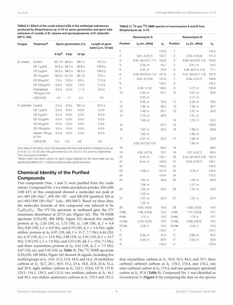
Chemical Identity of the PurifiedCompoundsTwo compounds (Nos. 1 and 2) were purified from the crudeextract. CompoundNo. 1 is a white amorphous powder. ESI±MS(100 kV) of this compound showed a molecular ion peak atm/z 683 (M+Na)+, 659 (M−H)− and HR-ESI (positive) MS atm/z 683.3394 (M+Na)+ (calc., 683.3047). Based on these data,the molecular formula of this compound was inferred to beC36H52O11. The UV-Vis spectrum in methanol gave the UVmaximum absorbance at 237.9 nm (Figure S2). The 1H-NMRspectrum (CD3OD, 400 MHz; Figure S3) showed five methylprotons at δH 2.26 (3H, s), 1.75 (3H, s), 1.08 (3H, d, J = 6.8Hz), 0.85 (3H, t, J = 6.9 Hz), and 0.79 (3H, d, J = 5.6 Hz), eightolefinic protons at δH 6.97 (1H, dd, J = 15.7, 7.7 Hz), 6.44 (2H,m), 6.25 (1H, d, J = 15.6 Hz), 5.88 (1H, s), 5.81 (1H, d, J = 15.7Hz), 5.59 (1H, t, J = 7.0 Hz), and 5.53 (1H, dd, J = 15.6, 7.3 Hz),and three oxymethine protons at δH 4.62 (1H, d, J = 7.3 Hz),4.07 (1H, m), and 3.45 (1H, m; Table 3). The 13C NMR spectrum(CD3OD, 100 MHz; Figure S4) showed 36 signals, including fivemethyl groups at δC 14.6, 15.3, 13.0, 18.0, and 14.2, 10 methylenecarbons at δC 32.7, 28.7, 36.9, 35.2, 25.4, 34.8, 23.8, 23.4, 31.2,and 29.9, eight olefinic carbons at δC 122.7, 152.6, 127.9, 137.8,129.5, 134.1, 139.3, and 121.9, two methine carbons at δC 44.1and 36.3, two olefinic quaternary carbons at δC 135.5 and 152.2,
TABLE 3 | 1H and 13C NMR spectra of reveromycins A and B from
Streptomyces sp. 3–10.
Reveromycin A Reveromycin B
Position δH [m, J(Hz)] δC Position δH [m, J(Hz)] δC
1 170.3 1 170.4
2 5.81, d (15.7) 122.7 2 5.79, d (15.6) 121.2
3 6.97, dd (15.7, 7.7) 152.6 3 6.99, dd (15.8, 7.5) 152.9
4 2.52, m 44.1 4 2.51, m 44.0
5 4.07, m 76.9 5 4.08, dd (7.4, 5.4) 77.1
6 5.53, dd (15.6, 7.3) 127.9 6 5.47, dd (15.7, 7.6) 127.2
7 6.25, d (15.6) 137.8 7 6.39, d (15.7) 138.6
8 135.5 8 135.2
9 5.59, t (7.0) 129.5 9 5.77, m 130.9
10 2.40, m 32.7 10 2.57, m 32.6
2.33, m 2.17, m
11 3.45, m 76.3 11 3.45, m 78.5
12 1.39, m 36.3 12 1.38, m 35.7
13 1.46, m 28.7 13 1.61, m 30.3
14 1.72, m 36.9 1.51, m
1.46, m 14 1.72, m 35.3
15 97.0 15 108.6
16 1.84, m 35.2 16 1.99, m 39.8
1.60, m 1.80, m
17 2.31, m 25.4 17 1.99, m 32.9
2.02, td (13.6, 4.0) 1.84, m
18 84.2 18 88.8
19 4.62, d (7.3) 79.7 19 5.57, d (3.7) 80.4
20 6.44, m 134.1 20 6.24, dd (16.0, 3.8) 132.6
21 6.44, m 139.3 21 6.28, d (16.1) 136.1
22 152.2 22 152.5
23 5.88, s 121.9 23 5.78, s 122.5
24 170.4 24 170.2
25 1.84, m 34.8 25 1.59, m 35.6
1.68, m 1.47, m
26 1.25, m 23.8 26 1.31, m 26.6
1.23, m
27 1.27, m 23.4 27 1.31, m 24.4
1.22, m
28 0.85, t (6.9) 14.6 28 0.92, t (6.9) 14.5
4-Me 1.08, d (6.8) 15.3 4-Me 1.01, d (6.9) 15.1
8-Me 1.75, s 13.0 8-Me 1.74, s 12.7
12-Me 0.79, t (5.6) 18.0 12-Me 0.89, d (6.5) 18.2
22-Me 2.26, s 14.2 22-Me 2.24, s 14.0
1′ 173.4 1′ 173.1
2′ 2.59, m 31.2 2′ 2.60, m 30.4
3′ 2.59, m 29.9 3′ 2.52, m 29.8
4′ 176.2 4′ 175.9
four oxymethine carbons at δC 76.9, 76.3, 84.2, and 79.7, threecarboxyl carbonyl carbons at δC 170.3, 170.4, and 176.2, oneester carbonyl carbon at δC 173.4, and one quaternary spiroketalcarbon at δC 97.0 (Table 3). Compound No. 1 was identified asreveromycin A (Figure 1) by comparing the data on the spectra
Frontiers in Microbiology | www.frontiersin.org 7 April 2017 | Volume 8 | Article 550
Lyu et al. Antifungal Metabolites from Streptomyces sp. 3–10
of 1H NMR, 13C NMR, MS and UV-Vis of this compound withthe related spectra of reveromycin A reported by Fremlin et al.(2011). The average content of reveromycin A in the crude extractof strain 3–10 was 37.7% (Figure S5).
Compound No. 2 was also a white amorphous powder.ESI±MS (100 kV) of this compound had a molecular ion peakat m/z 683 (M+Na)+, 659 (M−H)−, HR-ESI (positive) MS atm/z 683.3041 (M+Na)+ (calc., 683.3047). Based on these data,the molecular formula of this compound was inferred to beC36H52O11. The UV spectrum in methanol gave a UVmaximumabsorbance at 238.9 nm (Figure S6). The 1H-NMR spectrum(CD3OD, 400 MHz; Figure S7) showed five methyl protons at δH2.24 (3H, s), 1.74 (3H, s), 1.01 (3H, d, J = 6.9 Hz), 0.92 (3H, t,J = 6.9 Hz) and 0.89 (3H, d, J = 6.5 Hz), eight olefinic protons atδH 6.99 (1H, dd, J = 15.8, 7.5 Hz), 6.39 (1H, d, J = 15.7 Hz), 6.28(1H, d, J = 16.1 Hz), 6.24 (1H, dd, J = 16.0, 3.8 Hz), 5.79 (1H, d, J= 15.6 Hz), 5.78, (1H, s), 5.77 (1H, m), 5.47 (1H, dd, J = 15.7, 7.6Hz), and three oxymethine protons at δH 5.57 (1H, d, J = 3.7 Hz),4.08 (1H, dd, J = 7.4, 5.4 Hz), 3.45, (1H, m; Table 3). The 13CNMR spectrum (CD3OD, 100 MHz; Figure S8) also showed 36signals, including five methyl groups at δC 14.5, 15.1, 12.7, 18.2,and 14.0, 10 methylene carbons at δC 32.6, 30.3, 35.3, 39.8, 32.9,35.6, 26.6, 24.4, 30.4, and 29.8, eight olefinic carbons at δC 121.2,152.9, 127.2, 138.6, 130.9, 132.6, 136.1, and 122.5, two methinecarbons at δC 44.0 and 35.7, two olefinic quaternary carbons at δC135.2 and 152.5, four oxymethine carbons at δC 77.1, 78.5, 88.8,and 80.4, three carboxyl carbonyl carbons at δC 170.4, 170.2, and175.9, one ester carbonyl carbon at δC 173.1 and one quaternaryspiroketal carbon at δC 108.6 (Table 3). Compound No. 2 wasidentified as reveromycin B (Figure 1) by comparing the dataon the spectra of 1H NMR, 13C NMR, MS, and UV-Vis of thiscompound with related spectra of reveromycin B reported byFremlin et al. (2011).
Antifungal Activity of the PurifiedCompoundsReveromycins A and B from Streptomyces sp. 3–10 effectivelysuppressed mycelial growth of B. cinerea, M. hiemails,
FIGURE 1 | Chemical structures of reveromycins A and B produced by
Streptomyces sp. 3–10.
R. stolonifer, and S. sclerotiorum, and spore germinationof B. cinerea, M. hiemails, and R. stolonifer (Table 4). Thesuppressive efficacy was greatly affected by ambient pH. Forreveromycin A, the EC50-values ranged from 0.10 to 0.88µg/mlat pH 4.5, from 0.42 to 1.76µg/ml at pH 5.5, and from 14.04to 53.35µg/ml at pH 7.0. For reveromycin B, the EC50-valuesranged from 1.15 to 5.49µg/ml at pH 4.5, from 6.12 to35.46µg/ml at pH 5.5, and >100µg/ml at pH 7.0.
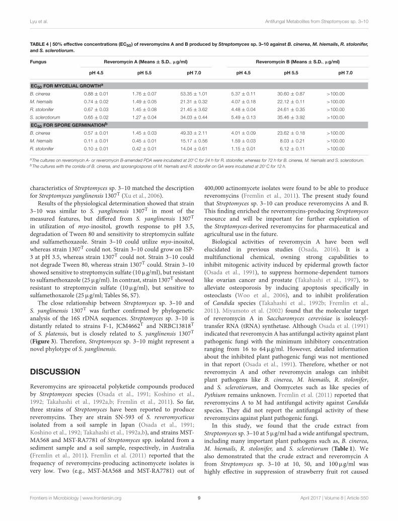
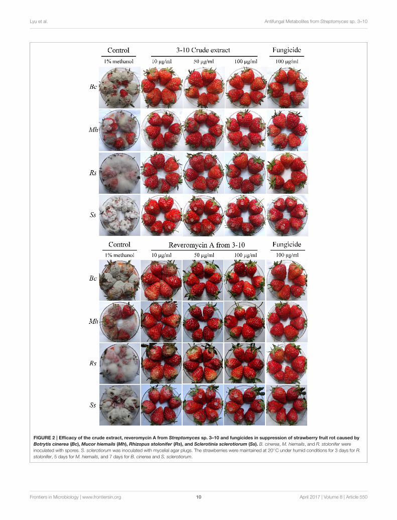
Control Efficacy against Strawberry FruitRotAt 3–7 days post inoculation (20◦C), the strawberries in thecontrol treatment were severely diseased (Figure 2, Table 5).The percentages of diseased strawberries reached 100, 88.9,100, and 94.4% for the control inoculations with B. cinerea,M. hiemails, R. stolonifer, and S. sclerotiorum, respectively. Thedisease severity index values reached 78.5, 62.5, 88.1, and 82.6in these treatments, respectively. In contrast, most strawberriesin the treatments of the crude extract and reveromycin A fromstrain 3–10 at 10, 50, and 100µg/ml, as well as in the treatmentsof the fungicides at 10, 50, and 100µg a.i./ml appeared healthy(Figure 2, Table 5). The percentages of diseased strawberrieswere lower than 40% and the disease severity index values werelower than 35 in these treatments. Reveromycin A at 50 and100µg/ml completely suppressed strawberry fruit rot caused byall the four fungi. Statistical analysis indicated that for eachpathogen, the treatments of the CE, reveromycin A and fungicidediffered significantly (P < 0.05) from the control treatment bothin disease incidence and in disease severity index. Under thesame concentration, the crude extract and reveromycin A fromstrain 3–10 did not significantly (P > 0.05) differ from thecorresponding fungicide.
PhytotoxicityThe treatments of the strawberry leaves with the crude extractof Streptomyces sp. 3–10 at 10, 50 and 100µg/ml did notproduce any visible toxic symptoms (yellowing, necrosis andmalformation) on leaves and flowers (Figure S9). They grewnormally and did not significantly differ (P > 0.05) from thecontrol treatment (water) both in leaf chlorophyll content andin dry weight of the upper part of strawberry plants.
Taxonomic Identity of Streptomyces sp.3–10Streptomyces sp. 3–10 grew and sporulated on nine agar media(BM, GS-1, ISP-1, ISP-2, ISP-3, ISP-5, ISP-6, ISP-7, PDA) afterincubation at 28◦C for 14 days with formation of whitish tograyish colonies. They hardly grew on the ISP-4 medium (FigureS10, Table S5). In the PDA cultures, the single colonies ofStreptomyces sp. 3–10 were averagely sized by 1.6 ± 0.3 mmin diameter, dome-shaped in the colony center, and the sawtooth-shaped at the colony margin (Figure S11). The substratemycelium was pale yellowish to orange in color and the aerialmycelium was whitish to grayish in color (Figure S11). Thespore chains were rectiflexible in shape with the spores beingshort rod in shape, 0.9 × 0.6µm (length × width) in size, andsmooth surfaced (Figure S11). The cultural and morphological
Frontiers in Microbiology | www.frontiersin.org 8 April 2017 | Volume 8 | Article 550
Lyu et al. Antifungal Metabolites from Streptomyces sp. 3–10
TABLE 4 | 50% effective concentrations (EC50) of reveromycins A and B produced by Streptomyces sp. 3–10 against B. cinerea, M. hiemails, R. stolonifer,
and S. sclerotiorum.
Fungus Reveromycin A (Means ± S.D., µg/ml) Reveromycin B (Means ± S.D., µg/ml)
pH 4.5 pH 5.5 pH 7.0 pH 4.5 pH 5.5 pH 7.0
EC50 FOR MYCELIAL GROWTHa
B. cinerea 0.88 ± 0.01 1.76 ± 0.07 53.35 ± 1.01 5.37 ± 0.11 30.60 ± 0.87 >100.00
M. hiemails 0.74 ± 0.02 1.49 ± 0.05 21.31 ± 0.32 4.07 ± 0.18 22.12 ± 0.11 >100.00
R. stolonifer 0.67 ± 0.03 1.45 ± 0.08 21.45 ± 3.62 4.48 ± 0.04 24.61 ± 0.35 >100.00
S. sclerotiorum 0.65 ± 0.02 1.27 ± 0.04 34.03 ± 0.44 5.49 ± 0.13 35.46 ± 3.92 >100.00
EC50 FOR SPORE GERMINATIONb
B. cinerea 0.57 ± 0.01 1.45 ± 0.03 49.33 ± 2.11 4.01 ± 0.09 23.62 ± 0.18 >100.00
M. hiemails 0.11 ± 0.01 0.45 ± 0.01 15.17 ± 0.56 1.59 ± 0.03 8.03 ± 0.21 >100.00
R. stolonifer 0.10 ± 0.01 0.42 ± 0.01 14.04 ± 0.61 1.15 ± 0.01 6.12 ± 0.11 >100.00
aThe cultures on reveromycin A- or reveromycin B-amended PDA were incubated at 20◦C for 24 h for R. stolonifer, whereas for 72 h for B. cinerea, M. hiemails and S. sclerotiorum.bThe cultures with the conidia of B. cinerea, and sporangiospores of M. hiemails and R. stolonifer on GA were incubated at 20◦C for 12 h.
characteristics of Streptomyces sp. 3–10 matched the descriptionfor Streptomyces yanglinensis 1307T (Xu et al., 2006).
Results of the physiological determination showed that strain3–10 was similar to S. yanglinensis 1307T in most of themeasured features, but differed from S. yanglinensis 1307T
in utilization of myo-inositol, growth response to pH 3.5,degradation of Tween 80 and sensitivity to streptomycin sulfateand sulfamethoxazole. Strain 3–10 could utilize myo-inositol,whereas strain 1307T could not. Strain 3–10 could grow on ISP-3 at pH 3.5, whereas strain 1307T could not. Strain 3–10 couldnot degrade Tween 80, whereas strain 1307T could. Strain 3–10showed sensitive to streptomycin sulfate (10µg/ml), but resistantto sulfamethoxazole (25µg/ml). In contrast, strain 1307T showedresistant to streptomycin sulfate (10µg/ml), but sensitive tosulfamethoxazole (25µg/ml; Tables S6, S7).
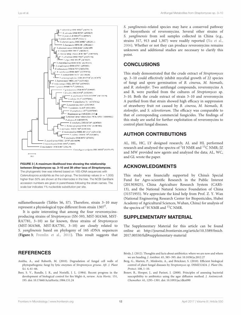
The close relationship between Streptomyces sp. 3–10 andS. yanglinensis 1307T was further confirmed by phylogeneticanalysis of the 16S rDNA sequences. Streptomyces sp. 3–10 isdistantly related to strains F-1, JCM4662T and NRBC13818T
of S. platensis, but is closely related to S. yanglinensis 1307T
(Figure 3). Therefore, Streptomyces sp. 3–10 might represent anovel phylotype of S. yanglinensis.
DISCUSSION
Reveromycins are spiroacetal polyketide compounds producedby Streptomyces species (Osada et al., 1991; Koshino et al.,1992; Takahashi et al., 1992a,b; Fremlin et al., 2011). So far,three strains of Streptomyces have been reported to producereveromycins. They are strain SN-593 of S. reveromyceticusisolated from a soil sample in Japan (Osada et al., 1991;Koshino et al., 1992; Takahashi et al., 1992a,b), and strains MST-MA568 and MST-RA7781 of Streptomyces spp. isolated from asediment sample and a soil sample, respectively, in Australia(Fremlin et al., 2011). Fremlin et al. (2011) reported that thefrequency of reveromycins-producing actinomycete isolates isvery low. Two (e.g., MST-MA568 and MST-RA7781) out of
400,000 actinomycete isolates were found to be able to producereveromycins (Fremlin et al., 2011). The present study foundthat Streptomyces sp. 3–10 can produce reveromycins A and B.This finding enriched the reveromycins-producing Streptomycesresource and will be important for further exploitation ofthe Streptomyces-derived reveromycins for pharmaceutical andagricultural use in the future.
Biological activities of reveromycin A have been wellelucidated in previous studies (Osada, 2016). It is amultifunctional chemical, owning strong capabilities toinhibit mitogenic activity induced by epidermal growth factor(Osada et al., 1991), to suppress hormone-dependent tumorslike ovarian cancer and prostate (Takahashi et al., 1997), toalleviate osteoporosis by inducing apoptosis specifically inosteoclasts (Woo et al., 2006), and to inhibit proliferationof Candida species (Takahashi et al., 1992b; Fremlin et al.,2011). Miyamoto et al. (2002) found that the molecular targetof reveromycin A in Saccharomyces cerevisiae is isoleucyl-transfer RNA (tRNA) synthetase. Although Osada et al. (1991)indicated that reveromycin A has antifungal activity against plantpathogenic fungi with the minimum inhibitory concentrationranging from 16 to 64µg/ml. However, detailed informationabout the inhibited plant pathogenic fungi was not mentionedin that report (Osada et al., 1991). Therefore, whether or notreveromycin A and other reveromycin analogs can inhibitplant pathogens like B. cinerea, M. hiemails, R. stolonifer,and S. sclerotiorum, and Oomycetes such as like species ofPythium remains unknown. Fremlin et al. (2011) reported thatreveromycins A to M had antifungal activity against Candidaspecies. They did not report the antifungal activity of thesereveromycins against plant pathogenic fungi.
In this study, we found that the crude extract fromStreptomyces sp. 3–10 at 5µg/ml had a wide antifungal spectrum,including many important plant pathogens such as, B. cinerea,M. hiemails, R. stolonifer, and S. sclerotiorum (Table 1). Wealso demonstrated that the crude extract and reveromycin Afrom Streptomyces sp. 3–10 at 10, 50, and 100µg/ml washighly effective in suppression of strawberry fruit rot caused
Frontiers in Microbiology | www.frontiersin.org 9 April 2017 | Volume 8 | Article 550
Lyu et al. Antifungal Metabolites from Streptomyces sp. 3–10
FIGURE 2 | Efficacy of the crude extract, reveromycin A from Streptomyces sp. 3–10 and fungicides in suppression of strawberry fruit rot caused by
Botrytis cinerea (Bc), Mucor hiemails (Mh), Rhizopus stolonifer (Rs), and Sclerotinia sclerotiorum (Ss). B. cinerea, M. hiemails, and R. stolonifer were
inoculated with spores. S. sclerotiorum was inoculated with mycelial agar plugs. The strawberries were maintained at 20◦C under humid conditions for 3 days for R.
stolonifer, 5 days for M. hiemails, and 7 days for B. cinerea and S. sclerotiorum.
Frontiers in Microbiology | www.frontiersin.org 10 April 2017 | Volume 8 | Article 550

Lyu et al. Antifungal Metabolites from Streptomyces sp. 3–10
TABLE 5 | Efficacy of the crude extract (CE) and reveromycin A (RA) from Streptomyces sp. 3–10 in suppression of strawberry fruit rot caused by
B. cinerea, M. hiemails, R. stolonifer, and S. sclerotiorum in comparison with respective fungicides.
Treatment a B. cinerea M. hiemails R. stolonifer S. sclerotiorum
DIb DSIb DI DSI DI DSI DI DSI
Control 100.0 ac 78.5 a 88.9 a 62.5 a 100.0 a 88.1a 94.4 a 82.6 a
CE 10µg/ml 22.2 b 4.9 b 11.1 c 8.3 cd 27.7 b 7.6 c 5.6 b 1.4 b
CE 50µg/ml 11.1 bc 4.2 b 5.6 c 2.1 de 11.1 c 2.8 d 0.0 b 0.0 b
CE 100µg/ml 0.0 c 0.0 c 0.0 c 0.0 e 0.0 c 0.0 d 0.0 b 0.0 b
RA 10µg/ml 22.2 b 4.2 b 11.1 c 2.1 e 11.1 c 2.7 d 0.0 b 0.0 b
RA 50µg/ml 0.0 c 0.0 c 0.0 c 0.0 e 0.0 c 0.0 d 0.0 b 0.0 b
RA 100µg/ml 0.0 c 0.0 c 0.0 c 0.0 e 0.0 c 0.0 d 0.0 b 0.0 b
FU 10µg a.i./ml 11.1 bc 3.5 bc 27.7 b 34.7 b 38.8 b 22.8 b 5.6 b 2.1 b
FU 50µg a.i./ml 5.6 c 2.1 bc 11.1 c 10.4 c 11.1 c 3.5 cd 0.0 b 0.0 b
FU 100µg a.i./ml 0.0 c 0.0 c 0.0 c 0.0 e 0.0 c 0.0 d 0.0 b 0.0 b
LSD (0.05) 11.6 3.9 12.7 6.3 11.6 4.6 9.0 2.4
aFungicides (FU): Pyrimethanil for B. cinerea, captan for M. hiemails and R. stolonifer, and dimetachlone for S. sclerotiorum.bDI, Disease incidence (%); DSI, Disease severity index (0–100).cMeans within the same column followed by the same letter are not significantly different (P > 0.05) according to least significance test.
by these pathogens and the efficacy was comparable to thatof the corresponding commercial fungicides (Table 5). It iswell recognized that B. cinerea, M. hiemails, R. stolonifer, andS. sclerotiorum are the necrotrophic plant pathogens. Theycan aggressively infect mature strawberry fruit under cool andhumid conditions, thereby causing severe economic losses forstrawberry production. Nowadays, control of B. cinerea as wellas three other fungi depends largely on repeated application offungicides. In most cases, the fungicide application can suppressinfection by these fungi. However, frequent application of thefungicides may cause some undesirable side effects, such asfungicide residues in strawberry fruit, pollution to environment,and development of fungicide-resistant fungal strains. The resultsabout the wide antifungal spectrum and high antifungal activityfor themetabolites of Streptomyces sp. 3–10 suggest that they havea promising potential to be exploited for control of strawberryfruit rot caused by these four fungi. This study found that thecrude extract of Streptomyces sp. 3–10 at 10, 50 and 100µg/mldid not produce any visible toxic symptoms on leaves ofstrawberry (Figure S9). This result suggests that application of themetabolites of Streptomyces sp. 3–10 on strawberry plants at theflowering stage might be a safe measure for control of strawberryfruit rot.
Among the fungi and Oomycetes inhibited by the metabolitesfrom Streptomyces sp. 3–10, Fusarium oxysporum, Pythiumapanidermatum, P. ultimum, Rhizoctonia solani, Sclerotiumrolfsii, Sclerotinia minor, and S. sclerotiorum are soilborne plantpathogens. Antifungal activity against these organisms suggeststhat inoculation of Streptomyces sp. 3–10 into soil may yieldsuppressive effect on these organisms. This study found thatStreptomyces sp. 3–10 can grow at pH 3.5 and 4.5, but failedto grow at pH 7.5 (Table S6). This acidophilic characteristic ofStreptomyces sp. 3–10 may help it to adapt to the acidic soilconditions in south of China. Moreover, this study found that theantifungal activity of reveromycins A and B from Streptomyces
sp. 3–10 is high at pH 4.5 and 5.5, whereas is decreased at pH 7.0(Table 4), implying that application of Streptomyces sp. 3–10 inacidic soil may achieve high antifungal efficacy in suppression ofthese soil-dwelling fungi.
Previous studies showed that many plant pathogenic fungisuch as B. cinerea and S. sclerotiorum can secrete oxalic acidto facilitate their infection and colonization of plant tissues(Choquer et al., 2007; Williams et al., 2011). Oxalic acid canacidify the surrounding environment. In this study, we found thatreveromycins A and B from Streptomyces sp. 3–10 showed higherantifungal activity at pH 4.5 than at pH 5.5 and 7.0 (Table 4).Thus, the acidic environment created by oxalic acid producedby B. cinerea and S. sclerotiorum may enhance the antifungalactivity of Streptomyces sp. 3–10 against the two pathogens. Thismay be one of the reasons responsible for the high antifungalactivity of reveromycins A and B in inhibition of B. cinerea andS. sclerotiorum.
Streptomyces sp. 3–10 was previously thought to be a mutantS. platensis F-1, as it was isolated from a PDA culture ofstrain F-1 treated with UV-C and LiCl (Che, 2011). Due tothe dramatic difference between strains 3–10 and F-1 in colonymorphology, this study tried to clarify the taxonomic status ofStreptomyces sp. 3–10. The results showed that strain 3–10 isclosely related to S. yanglinensis 1307T (possibly representing anovel phylotype of S. yanglinensis), but is distantly related to S.platensis F-1 (Figure 3). This result suggests that Streptomyces sp.3–10 is probably a contaminant, rather than a derivative fromS. platensis F-1.
Streptomyces yanglinensis was established by Xu andcolleagues in 2006 with strain 1307T as the type strain (Xu et al.,2006). Results of this study showed that strain 3–10 was similarto strain 1307T in the majority of the measured physiologicalcharacteristics, but is different from strain 1307T in utilizationof myo-inositol, in growth response to pH 3.5, in degradation ofTween 80, as well as in sensitivity to streptomycin sulfate and
Frontiers in Microbiology | www.frontiersin.org 11 April 2017 | Volume 8 | Article 550
Lyu et al. Antifungal Metabolites from Streptomyces sp. 3–10
FIGURE 3 | A maximum-likelihood tree showing the relationship
between Streptomyces sp. 3–10 and 39 other taxa of Streptomyces.
The phylogenetic tree was inferred based on 16S rDNA sequences with
Catenuliospora acidiphila as the out-group. The bootstrap values (n = 1,000)
higher than 50% are shown at the internodes in the tree. The NCBI GenBank
accession numbers are given in parentheses following the strain names. The
scale bar indicates 1% nucleotide substitution per site.
sulfamethoxazole (Tables S6, S7). Therefore, strain 3–10 mayrepresent a physiological type different from strain 1307T.
It is quite interesting that among the four reveromycins-producing strains of Streptomyces (SN-593, MST-MA568, MST-RA7781, 3–10) so far known, three strains of Streptomyces(MST-MA568, MST-RA7781, 3–10) are closely related toS. yanglinensis based on phylogeny of 16S rDNA sequences(Figure 3; Fremlin et al., 2011). This result suggests that
S. yanglinensis-related species may have a conserved pathwayfor biosynthesis of reveromycins. Several other strains ofS. yanglinensis from soil samples collected in China (e.g.,strains 317, 913 and 1,307) were readily reported (Xu et al.,2006). Whether or not they can produce reveromycins remainsunknown and additional studies are necessary to clarify thispoint.
CONCLUSIONS
This study demonstrated that the crude extract of Streptomycessp. 3–10 could effectively inhibit mycelial growth of 22 speciesof fungi and spore germination of B. cinerea, M. hiemails,and R. stolonifer. Two antifungal compounds, reveromycins Aand B, were purified from the cultures of Streptomyces sp.3–10. Both the crude extract of strain 3–10 and reveromycinA purified from that strain showed high efficacy in suppressionof strawberry fruit rot caused by B. cinerea, M. hiemails, R.stolonifer, and S. sclerotiorum. The efficacy was comparable tothat of corresponding commercial fungicides. The findings ofthis study are useful for further exploitation of reveromycins tocontrol plant fungal diseases.
AUTHOR CONTRIBUTIONS
AL, HL, HC, LY designed research; AL and HL performedresearch and analyzed the spectra of 1H NMR and 13C NMR; JZand MW provided new agents and analyzed the data; AL, WC,and GL wrote the paper.
ACKNOWLEDGMENTS
This study was financially supported by China’s SpecialFund for Agro-scientific Research in the Public Interest(201303025), China Agriculture Research System (CARS-13), and the National Natural Science Foundation of China(31571955). We appreciate the kind help from Prof. Z. Y. Wan(National Engineering Research Center for Biopesticides, HubeiAcademy of Agricultural Sciences, Wuhan, China) for analysis ofthe spectra of 1H NMR and 13C NMR.
SUPPLEMENTARY MATERIAL
The Supplementary Material for this article can be foundonline at: http://journal.frontiersin.org/article/10.3389/fmicb.2017.00550/full#supplementary-material
REFERENCES
Anitha, A., and Rabeeth, M. (2010). Degradation of fungal cell walls of
phytopathogenic fungi by lytic enzymes of Streptomyces griseus. Afr. J. Plant
Sci. 4, 61–66.
Beer, S. V., Rundle, J. R., and Norielli, J. L. (1984). Recent progress in the
development of biological control for fire blight-A, review. Acta Hortic. 151,
195. doi: 10.17660/ActaHortic.1984.151.24
Bérdy, J. (2012). Thoughts and facts about antibiotics: where we are now and where
we are heading. J. Antibiot. 65, 385–395. doi: 10.1038/ja.2012.27
Berg, G., Marten, P., Minkwitz, A., and Brückner, S. (2010). Efficient biological
control of plant fungal diseases by Streptomyces sp. DSMZ12424. J. Plant Dis.
Protect. 108, 1–10.
Bonev, B., Hooper, J., and Parisot, J. (2008). Principles of assessing bacterial
susceptibility to antibiotics using the agar diffusion method. J. Antimicrob.
Chemother. 61, 1295–1301. doi: 10.1093/jac/dkn090
Frontiers in Microbiology | www.frontiersin.org 12 April 2017 | Volume 8 | Article 550
Lyu et al. Antifungal Metabolites from Streptomyces sp. 3–10
Brzezinska, M. S., Jankiewicz, U., and Walczak, M. (2013). Biodegradation
of chitinous substances and chitinase production by the soil
actinomycete Streptomyces rimosus. Int. Biodeter. Biodegr. 84, 104–110.
doi: 10.1016/j.ibiod.2012.05.038
Chapman and Hall (2003). The Combined Chemical Dictionary on CD-ROM v 6.1.
Head FAST CD software.
Che, H. J. (2011). Mutation Improvement of Streptomyces platensis F-1 and the
Biocontrol Potential Assessment of Mutant Strains. [Master thesis]. Wuhan:
Huazhong Agricultural University.
Chen, Y. Y., Chen, P. C., and Tsay, T. T. (2016). The biocontrol efficacy
and antibiotic activity of Streptomyces plicatus on the oomycete
Phytophthora capsici. Biol. Control 98, 34–42. doi: 10.1016/j.biocontrol.2016.
02.011
Choquer, M., Fournier, E., Kunz, C., Levis, C., Pradier, J. M., Simon,
A., et al. (2007). Botrytis cinerea virulence factors: new insights into a
necrotrophic and polyphageous pathogen. FEMS Microbiol. Lett. 277, 1–10.
doi: 10.1111/j.1574-6968.2007.00930.x
Conn, V. M., Walker, A. R., and Franco, C. M. M. (2008). Endophytic
actinobacteria induce defense pathways in Arabidopsis thaliana. Mol. Plant
Microbe Interact. 21, 208–218. doi: 10.1094/MPMI-21-2-0208
Fremlin, L., Farrugia, M., Piggott, A. M., Khalil, Z., Lacey, E., and Capon,
R. J. (2011). Reveromycins revealed: new polyketide spiroketals from
Australian marine-derived and terrestrial Streptomyces spp. A case of natural
products vs. artifacts. Organ. Biomol. Chem. 9, 1201–1211. doi: 10.1039/C0OB
00654H
Guo, X. X., Liu, N., Li, X. M., Ding, Y., Shang, F., Gao, Y. S., et al. (2015).
Red soils harbor diverse culturable actinomycetes that are promising sources
of novel secondary metabolites. Appl. Environ. Microbiol. 81, 3086–3103.
doi: 10.1128/AEM.03859-14
Huang, R., Li, G. Q., Zhang, J., Yang, L., Che, H. J., Jiang, D. H., et al.
(2011). Control of postharvest Botrytis fruit rot of strawberry by volatile
organic compounds of Candida intermedia. Phytopathology 101, 859–869.
doi: 10.1094/PHYTO-09-10-0255
Kettleson, E., Kumar, S., Reponen, T., Vesper, S., Meheust, D., Grinshpun,
S. A., et al. (2013). Stenotrophomonas, Mycobacterium, and Streptomyces
in home dust and air: associations with moldiness and other
home/family characteristics. Indoor Air. 23, 387–396. doi: 10.1111/ina.
12035
Kim, B. S., and Hwang, B. K. (2007). Microbial fungicides in the control of
plant diseases. J. Phytopathol. 155, 641–653. doi: 10.1111/j.1439-0434.2007.
01314.x
Kim, B. Y., Willbold, S., Kulik, A., Helaly, S. E., Zinecker, H., and Wiese, J. (2011).
Elaiomycins, B., and C, novel alkylhydrazides produced by Streptomyces sp. BK
190. J. Antibiot. 64, 595–597. doi: 10.1038/ja.2011.53
Koshino, H., Takahashi, H., Osada, H., and Isono, K. (1992). Reveromycins, new
inhibitors of eukaryotic cell growth. III. Structures. J. Antibiot. 45, 1420–1427.
doi: 10.7164/antibiotics.45.1420
Kurth, F., Mailander, S., Bonn, M., Feldhahn, L., Herrmann, S., Große,
I., et al. (2014). Streptomyces-induced resistance against oak powdery
mildew involves host plant responses in defense, photosynthesis, and
secondary metabolism pathways. Mol. Plant Microbe Interact. 27, 891–900.
doi: 10.1094/MPMI-10-13-0296-R
Lehr, N. A., Schrey, S. D., Hampp, R., and Tarkka, M. T. (2008). Root
inoculation with a forest soil streptomycete leads to locally and systemically
increased resistance against phytopathogens in Norway spruce. New Phytol.
177, 965–976. doi: 10.1111/j.1469-8137.2007.02322.x
Mander, P., Cho, S. S., Choi, Y. H., Panthi, S., Choi, Y. S., Kim, H. M., et al.
(2016). Purification and characterization of chitinase showing antifungal and
biodegradation properties obtained from Streptomyces anulatus CS242. Arch.
Pharm. Res. 39, 878–886. doi: 10.1007/s12272-016-0747-3
Millard, W. A., and Taylor, C. B. (1927). Antagonism of microorganisms as the
controlling factor in the inhibition of scab by green-manuring. Ann. Appl. Biol.
14, 202–216. doi: 10.1111/j.1744-7348.1927.tb07076.x
Minuto, A., Spadaro, D., Garibaldi, A., and Gullino, M. L. (2006). Control
of soilborne pathogens of tomato using a commercial formulation of
Streptomyces griseoviridis and solarization. Crop Protect. 25, 468–475.
doi: 10.1016/j.cropro.2005.08.001
Miyamoto, Y., Machida, K., Mizunuma,M., Emoto, Y., Sato, N., Miyahara, K., et al.
(2002). Identification of Saccharomyces cerevisiae isoleucyl-tRNA synthetase as
target of G1-specific inhibitor reveromycin A. J. Biol. Chem. 277, 28810–28814.
doi: 10.1074/jbc.M203827200
Osada, H. (2016). Chemical and biological studies of reveromycin A. J. Antibiot.
69, 723–730. doi: 10.1038/ja.2016.57
Osada, O., Koshino, O., and Isono, K. (1991). Reveromycin A, a new antibiotic
which inhibits the mitogenic activity of epidermal growth factor. J. Antibiot.
44, 259–261. doi: 10.7164/antibiotics.44.259
Rout, N. P., Tripathi, S. B., and Shaw, P.W. (1998). Effect of salinity on chlorophyll
and proline contents in three aquatic macrophytes. Biol. Plant. 40, 453–458.
doi: 10.1023/A:1001186502386
Ruan, J. S., and Huang, Y. (2011). Rapid Identification and Systematics of
Actinobacteria. Beijing: Science Press.
Shakeel, Q., Lyu, A., Zhang, J., Wu, M. D., Chen, S. W., Chen, W. D., et al.
(2016). Optimization of the cultural medium and conditions for production
of antifungal metabolites by Streptomyces platensis 3–10 and evaluation of
its efficacy in suppression of clubroot disease (Plasmodiophora brassicae)
of oilseed rape. Biol. Control 101, 59–68. doi: 10.1016/j.biocontrol.2016.
06.007
Solans, M., Vobis, G., Cassan, F., Luna, V., and Wall, L. G. (2011). Production
of phytohormones by root-associated saprophytic actinomycetes isolated from
the actinorhizal plant Ochetophila trinervis. World J. Microbiol. Biotechnol. 27,
2195–2202. doi: 10.1007/s11274-011-0685-7
Supong, K., Thawaia, C., Choowong, W., Kittiwongwattana, C. C., Thanaboripat,
D., Laosinwattana, C., et al. (2016). Antimicrobial compounds from endophytic
Streptomyces sp. BCC72023 isolated from rice (Oryza sativa L.). Res. Microbiol.
167, 290–298. doi: 10.1016/j.resmic.2016.01.004
Takahashi, H., Osada, H., Koshino, H., Kudo, T., Amano, S., Shimizu, S., et al.
(1992a). Reveromycins, new inhibitors of eukaryotic cell growth I. Producing
organism, fermentation, isolation and physiochemical properties. J. Antibiot.
45, 1409–1413. doi: 10.7164/antibiotics.45.1409
Takahashi, H., Osada, H., Koshino, H., Sasaki, M., Onose, R., Nakakoshi,
M., et al. (1992b). Reveromycins, new inhibitors of eukaryotic cell growth
II. Biological activities. J. Antibiot. 45, 1414–1419. doi: 10.7164/antibiotics.
45.1414
Takahashi, H., Yamashita, Y., Takaoka, H., Nakamura, J., Yoshihama, M.,
and Osada, H. (1997). Inhibitory action of reveromycin A on TGF-alpha-
dependent growth of ovarian carcinoma BG-1 in vitro and in vivo. Oncol. Res.
9, 7–11.
Van Ewijk, P. H., and Hoekstra, J. A. (1993). Calculation of the EC50 and its
confidence interval when subtoxic stimulus is present. Ecotoxicol. Environ. Saf.
25, 25–32. doi: 10.1006/eesa.1993.1003
Wan, M. G., Li, G. Q., Zhang, J. B., Jiang, D. H., and Huang, H. C. (2008).
Effect of volatile substance of Streptomyces platensis F-1 on control of plant
fungal diseases. Biol. Control 46, 552–559. doi: 10.1016/j.biocontrol.2008.
05.015
Wan, Z. Y., Fang, W., Shi, L. Q., Wang, K. M., Zhang, Y. N., Zhang, Z. G.,
et al. (2015). Nononestmycins A and B, two new 32-membered bioactive
macrolides from Streptomyces phytohabitans HBERC-20821. J. Antibiot. 68,
186–190. doi: 10.1038/ja.2014.123
Williams, B., Kabbage, M., Min, J. Y., Britt, R., and Dickman, M. B. (2011).
Tipping the balance: Sclerotinia sclerotiorum secreted oxalic acid suppresses
host defenses by manipulating the host redox environment. PLoS Pathog.
7:e1002107. doi: 10.1371/journal.ppat.1002107
Woo, J. T., Kawatani, M., Kato, M., Shinki, T., Yonezawa, T., Kanoh, N., et al.
(2006). Reveromycin A, an agent for osteoporosis, inhibits bone resorption by
inducing apoptosis specifically in osteoclasts. Proc. Natl. Acad. Sci. U.S.A. 103,
4729–4734. doi: 10.1073/pnas.0505663103
Xu, C. G., Wang, L. M., Cui, Q. F., Huang, Y., Liu, Z. H., Zheng, G.
Y., et al. (2006). Neutrotolerant acidophilic Streptomyces species isolated
from acidic soils in China: Streptomyces guanduensis sp. nov., Streptomyces
paucisporeus sp. nov., Streptomyces rubidus sp. nov. and Streptomyces
yanglinensis sp. nov. Int. J. Syst. Evol. Microbiol. 56, 1109–1115. doi: 10.1099/ijs.
0.63959-0
Zacky, F. A., and Ting, A. S. Y. (2013). Investigating the bioactivity of cells
and cell-free extracts of Streptomyces griseus towards Fusarium oxysporum f.
Frontiers in Microbiology | www.frontiersin.org 13 April 2017 | Volume 8 | Article 550
Lyu et al. Antifungal Metabolites from Streptomyces sp. 3–10
sp. cubense race 4. Biol. Control 66, 204–208. doi: 10.1016/j.biocontrol.2013.
06.001
Zhang, B. H., Ding, Z. G., Li, H. Q., Mou, X. Z., Zhang, Y. Q., Yang,
J. Y., et al. (2016). Algicidal activity of Streptomyces eurocidicus
JXJ-0089 metabolites and their effects on Microcystis physiology.
Appl. Environ. Microbiol. 82, 5132–5143. doi: 10.1128/AEM.
01198-16
Zhang, D. J., Wei, G., Wang, Y., Si, C. C., Tian, L., Tao, L. M.,
et al. (2011). Bafilomycin K, a new antifungal macrolide from
Streptomyces flavotricini Y12-26. J. Antibiot. 64, 391–393. doi: 10.1038/ja.
2011.12
Conflict of Interest Statement: The authors declare that the research was
conducted in the absence of any commercial or financial relationships that could
be construed as a potential conflict of interest.
Copyright © 2017 Lyu, Liu, Che, Yang, Zhang, Wu, Chen and Li. This is an open-
access article distributed under the terms of the Creative Commons Attribution
License (CC BY). The use, distribution or reproduction in other forums is permitted,
provided the original author(s) or licensor are credited and that the original
publication in this journal is cited, in accordance with accepted academic practice.
No use, distribution or reproduction is permitted which does not comply with these
terms.
Frontiers in Microbiology | www.frontiersin.org 14 April 2017 | Volume 8 | Article 550