Response of Osteosarcoma to Chemotherapy in Scotland · 2 Summary Introduction Osteosarcomas are...
64
1 Response of Osteosarcoma to Chemotherapy in Scotland Ewan Semple, 5 th Year Medical Student, University of Aberdeen
Transcript of Response of Osteosarcoma to Chemotherapy in Scotland · 2 Summary Introduction Osteosarcomas are...
1
Response of Osteosarcoma to Chemotherapy in Scotland
Ewan Semple, 5th Year Medical Student, University of Aberdeen
2
Summary
Introduction
Osteosarcomas are the most common primary bone tumour and affect mainly young people (1,2). 5
year survival is between 34% and 66% (3-5).
The aim of this study was to investigate the response of osteosarcomas in the Scottish population
toneoadjuvant chemotherapy which were diagnosed between 1st January 2000 and 1st April 2013.
Methods
The Scottish Bone Tumour Registry files of all patients diagnosed with an osteosarcoma between 1st
January 2000 and 1st April 2013 were manually searched to identify demographic information and
information on potential prognostic factors. Survival was calculated using univariate analysis and
multivariate analysis in SPSS version 20.0 (SPSS Inc., Chicago Ill).
Results
Relapse occurred after a mean of 6 years and 10 months and patients had a mean overall survival of
7 years and 10 months. 5 year relapse free survival (RFS) was 48.2% and the 5 year overall survival
was 56.4%.
Univariate analysis showed that RFS and overall survival were significantly correlated with location
of tumour, type of osteosarcoma, presence of metastases at presentation, stage at diagnosis,
presenting symptoms, timing of chemotherapy,having neo-adjuvant chemotherapy and completing
planned number of cycles of chemotherapy andtype of surgery. Other factors associated with overall
survival but not RFS were time from symptom onset to presentation, percentage necrosis following
neo-adjuvant chemotherapy anddisease relapse.
Factors significantly associated with RFS and overall survival in the multivariate analysiswere location
of tumour, presence of metastases at presentation and completing planned number of cycles of

3
chemotherapy. RFS also remained significant for timing of chemotherapy and having neo-adjuvant
chemotherapy. Overall survival was also affected by whether the patient had disease relapse and
the presenting symptoms. RFS neared significance for the patients presenting symptoms.
Conclusion
Good pathological response (≥90% tumour necrosis) to chemotherapy improved overall survival in
patients with osteosarcoma in univariate analysis but this did not occur in multivariate analysis. A
number of factors were found to be associated with RFS and overall survival which have also been
found to be good prognostic factors by other researchers.

4
Acknowledgements
I would like to thank Dr Jeff White who supervised my project work and clinical work during my
elective and Mr Ashish Mahendra who supervised my clinical work. I would also like to thank Mrs
Jean Campbell and Dr Elaine MacDuff for their help with the project and Ms Dawn Currie, Mr Mike
Jane, Miss Kim Ferguson, Mrs Helen Finlay and Dr Fiona Cowie for all of the teaching and help during
the clinical part of my elective. Thank you to all of the above for making me feel so welcome and for
your constant enthusiasm throughout my time working with you.
Also, thank you to all of the patients who spoke to me and allowed me to be present during
consultations for helping to give me a better understanding of their experience of sarcoma.
Thank you, too, to Mr Kevin Baird and Mr Piers Renshaw for their help in organising my elective.
5
Contents
Summary ............................................................................................................................................. 2
Acknowledgements ............................................................................................................................. 4
Contents .............................................................................................................................................. 5
Introduction ........................................................................................................................................ 6
Methods ............................................................................................................................................ 12
Results ............................................................................................................................................... 14
Patient Characteristics .................................................................................................................. 14
Tumour Characteristics ................................................................................................................. 18
Treatment Details ......................................................................................................................... 22
Relapse-free Survival and Overall Survival.................................................................................... 27
Discussion.......................................................................................................................................... 53
Conclusion ..................................................................................................................................... 56
References ........................................................................................................................................ 58
Appendix 1: Data Collection Sheet ................................................................................................... 60

6
Introduction
Osteosarcomas are the most common primary bone tumour and affect mainly young people (1,2).
They commonly occur in the metaphysis of long bones, most often the distal femur and proximal
tibia (2,6,7). They have an incidence of 0.2-0.3 per 100,000 per year (2). 5 year survival is between
34% and 66% (3-5). There are several types and subtypes of osteosarcoma. The commonest is
conventional osteosarcoma which is high grade and makes up 75% of high grade osteosarcomas (2).
Low-grade central and paraosteal osteosarcoma are low-grade malignant tumours and periosteal
osteosarcomas are intermediate-grade chondroblastic osteosarcomas (2). Approximately 20% of
patients presenting with osteosarcoma have clinically detectable metastases (6). The most common
site of metastasis in osteosarcoma is to the lungs (7) and a study in 2009 found that one in seven
patients have metastases at presentation (7).
Presenting symptoms of malignant bone tumours commonly include pain and a lump or area of
swelling (2,8) and diagnosis can be difficult because symptoms are often non-specific, especially in
areas such as the pelvis, abdomen and chest (8). Investigation should include blood tests, including
alkaline phosphatase and lactate dehydrogenase (LDH); and radiological investigations (2)(8).
Commonly, the first radiological investigations performed are plain x-rays. These allow a differential
diagnosis to be established, assessment of tumour aggressiveness and detection of pathological
fractures (9). Typical x-ray features of a bone tumour are a lesion in the metadiaphysis with a
permeative destruction pattern, cortical destruction and malignant periosteal reaction (Figure 1) (9).
Further, more detailed radiological assessment is usually with a combination of MRI, and
occasionally CT scan of the affected area along with a CT scan of the chest, abdomen and pelvis and
a bone scan to look for metastases. MRI is especially useful in determining the relationship of the
tumour to the adjacent joint, muscles and neurovascular structures (Figure 2) (9). Definitive
diagnosis is made on pathological examination of a biopsy specimen (Figure 3) as it is difficult to
differentiate malignant bone tumours from benign disease clinically and radiologically (9). After

7
diagnosis, imaging investigations are also used in surgical planning and assessing the response of the
tumour to chemotherapy (10). Staging of bone tumours is performed using TNM staging as shown in
Table 1 and Table 2. In Scotland, bone tumours are managed in three regional centres in Glasgow,
Edinburgh and Aberdeen which all collaborate and liaise with other hospitals in Scotland to form the
Scottish Sarcoma Network. Management of bone tumours is usually by a combination of
chemotherapy, surgery and/or radiotherapy dependant on the likely response of the tumour (2).
Figure 1: Plain film x-rays of an osteosarcoma affecting the proximal tibia with arrows indicating the area of abnormality

8
Figure 2: MRI appearance of an osteosarcoma affecting the proximal tibia with arrows indicating the area of abnormality
Figure 3: (a) low and (b) high power microscopy images of an osteosarcoma on pathological examination
(a) (b)

9
Table 1: TNM staging of bone tumours
Staging Description
T stage
• T0
• T1
• T2
• T3
No evidence of primary tumour Tumour 8cm or less in greatest dimension Tumour more than 8cm in greatest dimension Discontinuous tumours in the primary bone site
N stage
• N0
• N1
No regional lymph nodes Regional lymph node metastases
M stage
• M0
• M1a
• M1b
No distant metastases Distant metastases in the lungs Distant metastases at sites other than the lungs
Table 2: Tumour stage calculation from T, N and M stages and tumour grade
Stage grouping T stage N stage M stage Tumour grade
Stage IA T1 N0 M0 Low grade Stage IB T2 N0 M0 Low grade Stage IIA T1 N0 M0 High grade Stage IIB T2 N0 M0 High grade Stage III T3 N0 M0 Any grade Stage IVA Any T N0 M1a Any grade Stage IVB Any T
Any T N1 Any N
Any M M1b
Any grade Any grade
The European Society for Medical Oncology (ESMO) have produced guidelines for the diagnosis,
treatment and follow-up of patients with bone tumours (2). These state that patients with high
grade osteosarcoma should generally be treated with pre-operative and post-operative
chemotherapy, while low grade osteosarcoma may be treated with surgery alone. Chemotherapy
often consists of doxorubicin, cisplatin and high dose methotrexate, but ifosfamide and etoposide
also have activity in osteosarcoma and can be included. Mifamurtide is a recent addition to the
chemotherapy drugs available (4,6). It is a biological agent which induces an immune response to
help to control any micrometastatic tumour cells. Its use is currently restricted to patients under the
10
age of 30 years with no clinically detectable metastatic disease (4,6). Management of recurrent
disease and metastases usually comprises of surgery and/or chemotherapy often including
ifosfamide, etoposide and/or carboplatin. Response to chemotherapy is considered to be good if
90% or greater necrosis is achieved and poor if less than 90% necrosis is achieved on pathological
examination of the tumour (3,10-14).
Predicting response of osteosarcomas to chemotherapy is an area which many researchers have
investigated, but few predictive factors have so far been reliably identified. The most reliable
predictive factor of response to neo-adjuvant chemotherapy is the percentage necrosis of the
tumour on pathological examination with patients with 90% or more tumour necrosis predicted to
have a better outcome than those with less than 90% tumour necrosis (3,10-14). This is the most
commonly used predictor of response by researchers (3,10-14) and in clinical practice. If researchers
are able to identify more factors influencing response of osteosarcomas to chemotherapy, then
clinicians will be better able to target treatment of sarcomas based on these factors to maximise the
benefit of treatment for each patient. Risk factors which have been found to be associated with
prognosis in osteosarcoma include age, gender, ethnicity, tumour size, tumour location, histological
subtype of tumour, presence of metastases, tumour stage, presence of pathological fracture,
alkaline phosphatase level, lactate dehydrogenase (LDH) level, local recurrence, treatment received
and surgical margins (3,4,7,11-13). However, evidence on the influence of these is conflicting, with
different authors finding different combinations of these factors to influence prognosis in
osteosarcoma (3,4,7,11-13). Suggestions have also been made thatP-glycoprotein, Ki-67 and HER2
are associated with better survival in osteosarcoma patients, but the evidence relating to these is
not currently clear (13).
The Scottish Bone Tumour Registry was established in 1962 in Glasgow. Since then, all cases of bone
tumours in Scotland have been recorded using it and data on all patients with bone tumours have
been preserved in it. Data saved in the bone tumour registry includes copies of radiological

11
investigations, pathological specimens and details of patients’ diagnosis, treatment and follow-up.
This registry is therefore able to provide a unique insight into the management of bone tumours
throughout the past 50 years.
The aim of this study was to retrospectively review the response of osteosarcoma to neo-adjuvant
chemotherapy using the Scottish Bone Tumour Registry. The secondary outcome was to identify
other factors which may affect survival.
12
Methods
Potential influences on response of osteosarcomas to chemotherapy were identified in the literature
in order to conduct a retrospective review of all patients with a diagnosis of osteosarcoma in the
Scottish Bone Tumour Registry from the 1st January 2000 to 6th March 2013. The files of these
patients in the Scottish Bone Tumour Registry were manually searched to obtain information on
patient demographics, tumour characteristics and treatment details.Patient information searched
for included patient community health index (CHI) number, patient sex, age at diagnosis, alkaline
phosphatase level at diagnosis, lactate dehydrogenase at diagnosis,postcode, Scottish index of
multiple deprivation (15), current status,date at diagnosis, date of death, death of disease, date last
seen, survival, relapse,time from symptom onset to primary presentation and presenting symptoms.
The tumour characteristics which were recorded were tumour location and site, tumour subtype,
presence of metastases, tumour grade, tumour stage at diagnosis, tumour volume, histology of
primary site, main site of metastatic disease and TNM stage. Treatment details searched for were
timing of chemotherapy, first line treatment, type of chemotherapy, use of methotrexate, use of
Mifamurtide, era of treatment, date of first treatment, percentage necrosis with neo-adjuvant
chemotherapy, date of end of treatment, pathological response to treatment, MRI response, CT
response, second line treatment, date of relapse, clinical trial, neo-adjuvant chemotherapy use,
completed planned number of chemotherapy cycles, date of relapse after first line therapy, local or
distant relapse, relapse free survival, primary surgery, date of primary surgery, histological response,
radiotherapy, radiotherapy location and radiotherapy dose. The information relevant to
osteosarcoma was collected using a standardised data collection sheet used for all types of bone
tumours (Appendix 1).
Where information was missing from the SBTR,electronically stored clinic letters, results and reports
were accessed to complete this. Electronic storage of this information only began in 2007 and each
region of Scotland uses a different system of electronic storage which is not accessible in other

13
health boards. Therefore, only information about more recent cases was able to be accessed in this
way, and only on patients who had been treated in the Glasgow centre.
Statistical analysis was performed by entering the data collectedinto IBM SPSS Version 20(SPSS Inc.,
Chicago Ill) for analysis. Kaplan-Meier survival curves were generated for relapse-free survival (RFS)
and overall survival using univariate analysis for each potential prognostic factor. Multivariate
analysis was also conducted in SPSS Version 20 using Cox Regression to analyse factors which were
found to be significant in the univariate analysis.Relapse free survival (RFS) and overall survival were
defined as time from diagnosis of osteosarcoma to diagnosis of relapse and time from diagnosis to
death, respectively.
14
Results
Patient Characteristics
The bone tumour registry contained 114 patients with diagnoses of osteosarcoma from 1st of
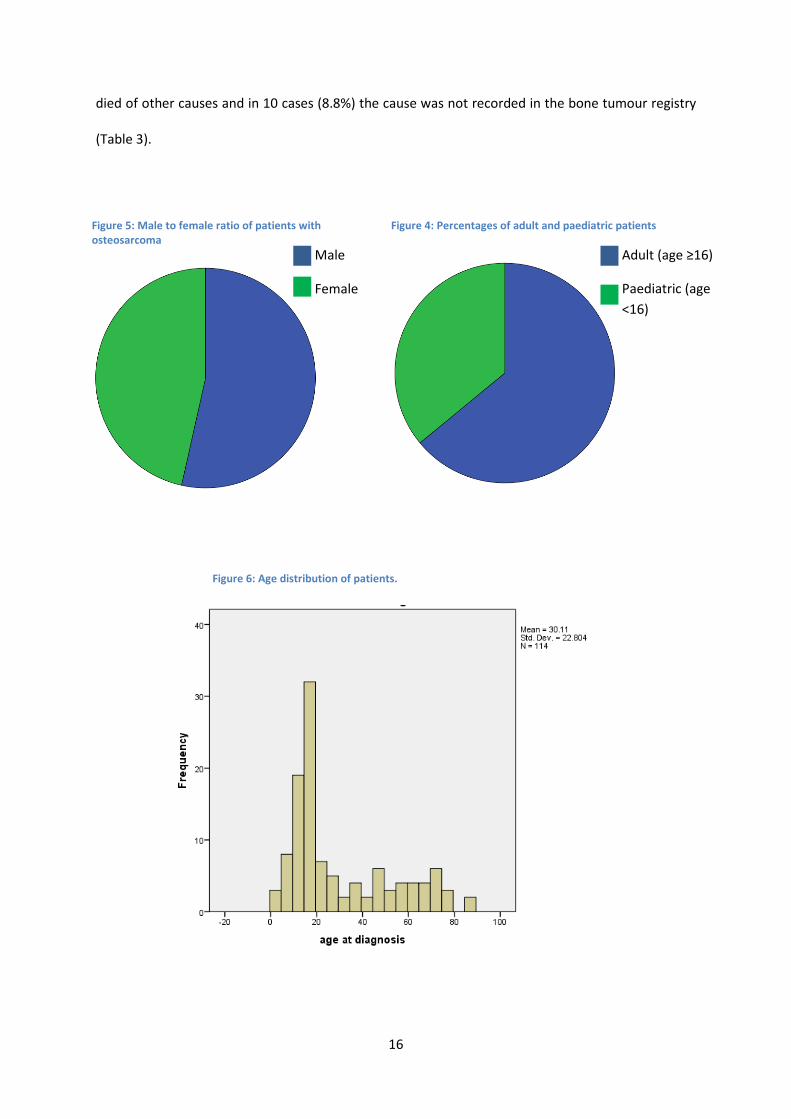
January 2000 to 1st of April 2013, all of whom were included in this study. 61 (53.5%) were male and
53 (46.5%) were female (Table 3and Figure 5). The median age of the patients was 18 years old
(range 2 to 86)(Table 3and Figure 6) with 73 (64%) of the patients being older than 16 years old and
41 (36%) under 16 years old (Table 3and Figure 4).
Scottish index of multiple deprivation (SIMD) was calculated for the patients, except in 10 cases
where this was not possible as the postcode was not available or the postcode was not recognised as
valid. These values were then separated into quintiles(15). The number of patients in each quintile is
shown in Table 3and Figure 9.
The most common presenting symptom was pain with 49 patients (43.0%) presenting with this
symptom, followed by a combination of lump and pain (36 patients, 31.6%). Other presenting
symptoms included lump alone (16 patients, 14.0%) and pathological fracture (3 patients, 2.6%)
while some tumours were incidental findings (2 patients, 1.8%) (Table 3). The length of time from
symptom onset to first presentation was quite evenly divided between 3 months or more (48
patients, 42.1%) and less than 3 months (53 patients, 46.5 %) (Table 3and Figure 7).
Alkaline phosphatase level was recorded for 22 patients. The median level was 311.50 U/l (range 85
to 2787). Because of the low number of patients for which this was recorded it was excluded from
the main analysis.
At their last follow-up 56 patients (49.1%) were alive and free of disease, 7 (6.1%) were alive with
active tumour and 49 (43.0%) had died. The status of 2 patients (1.8%) was unknown (Table 3and
Figure 8). Of those who had died, the death of 37 (32.5%) was caused by osteosarcoma, 2 (1.8%)
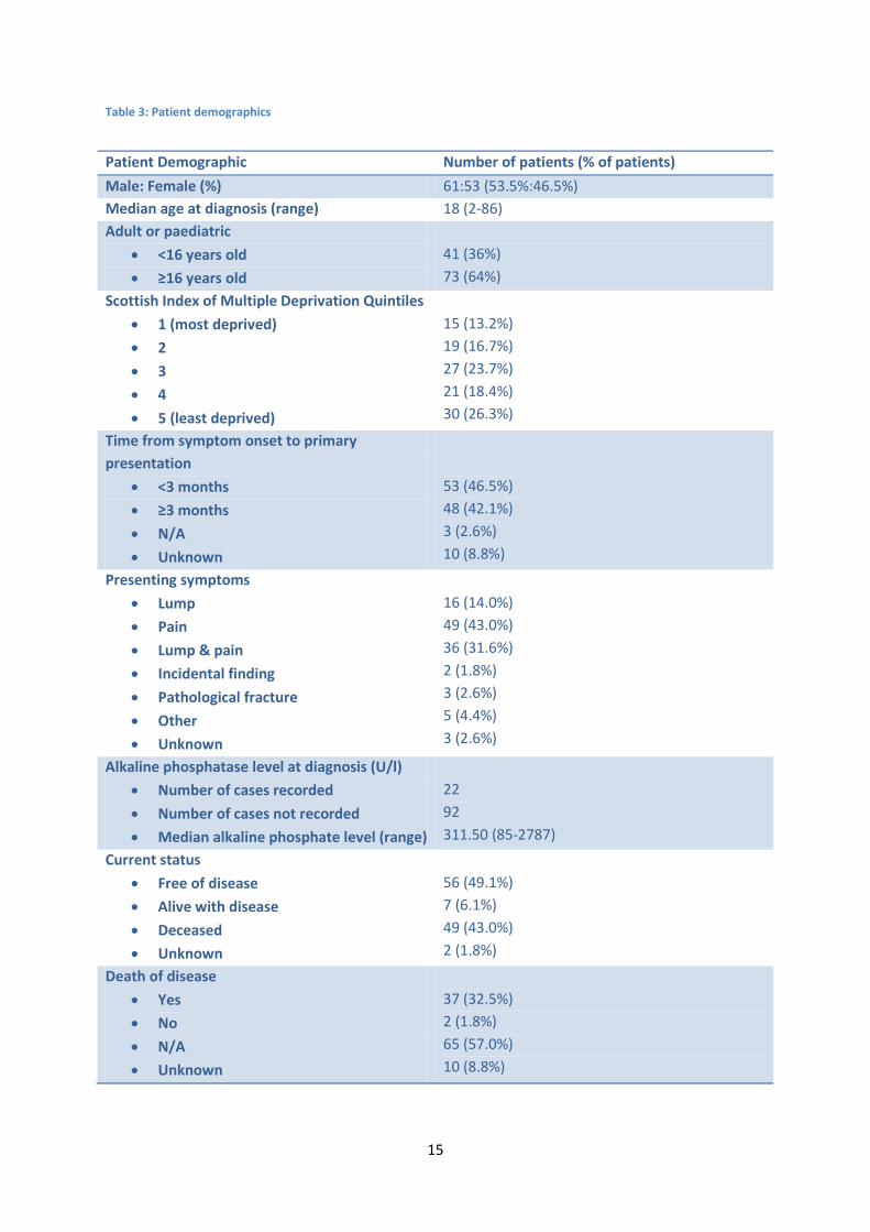
15
Table 3: Patient demographics
Patient Demographic Number of patients (% of patients)
Male: Female (%) 61:53 (53.5%:46.5%) Median age at diagnosis (range) 18 (2-86) Adult or paediatric
• <16 years old
• ≥16 years old
41 (36%) 73 (64%)
Scottish Index of Multiple Deprivation Quintiles
• 1 (most deprived)
• 2
• 3
• 4
• 5 (least deprived)
15 (13.2%) 19 (16.7%) 27 (23.7%) 21 (18.4%) 30 (26.3%)
Time from symptom onset to primary presentation
• <3 months
• ≥3 months
• N/A
• Unknown
53 (46.5%) 48 (42.1%) 3 (2.6%) 10 (8.8%)
Presenting symptoms
• Lump
• Pain
• Lump & pain
• Incidental finding
• Pathological fracture
• Other
• Unknown
16 (14.0%) 49 (43.0%) 36 (31.6%) 2 (1.8%) 3 (2.6%) 5 (4.4%) 3 (2.6%)
Alkaline phosphatase level at diagnosis (U/l)
• Number of cases recorded
• Number of cases not recorded
• Median alkaline phosphate level (range)
22 92 311.50 (85-2787)
Current status
• Free of disease
• Alive with disease
• Deceased
• Unknown
56 (49.1%) 7 (6.1%) 49 (43.0%) 2 (1.8%)
Death of disease
• Yes
• No
• N/A
• Unknown
37 (32.5%) 2 (1.8%) 65 (57.0%) 10 (8.8%)
16
died of other causes and in 10 cases (8.8%) the cause was not recorded in the bone tumour registry
(Table 3).
Figure 6: Age distribution of patients.
Figure 5: Male to female ratio of patients with osteosarcoma
Male
Female
Figure 4: Percentages of adult and paediatric patients
Adult (age ≥16)
Paediatric (age <16)

17
Figure 9: Distribution of Scottish Index of Multiple Deprivation quintiles
1 4 5 Deprivation Quintile
3 2
Figure 8: Percentage of patients currently alive
Unknown
Free of disease
Alive with disease
Deceased
Figure 7: Time from symptom onset to primary presentation
<3 months
≥3 months
N/A
Unknown

18
Tumour Characteristics
By categorising the location of osteosarcomas in several ways, it was possible to analyse different
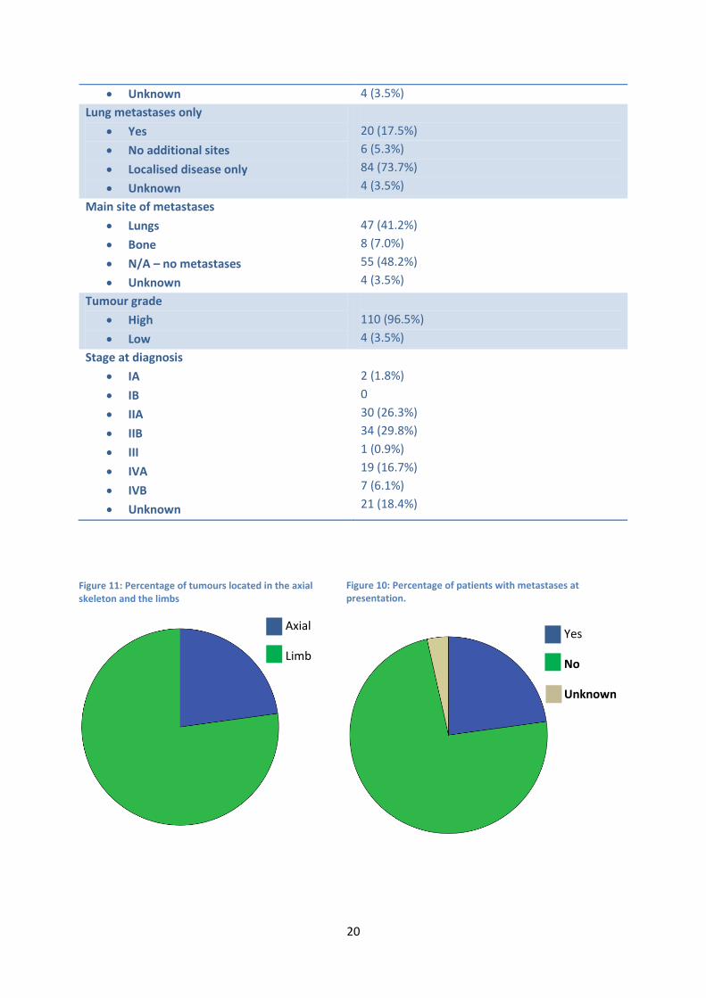
aspects of this. Osteosarcoma occurred in the limbs (88 patients, 77.2%) more commonly than in the
axial skeleton (26 patients, 22.8%) (Table 4and Figure 11).Most of them were in the lower limb (78
patients, 68.4%)(Table 4) with the femur being a more common location than any other bone (46
patients, 40.4%)(Table 4) and of those tumours located in long bones, most were located distally (48
patients, 42.1%)(Table 4). The tumours were mainly conventional type (104 patients, 91.2%)(Table 4)
osteoblastic osteosarcomas (71 patients, 62.3%)(Table 4) with small numbers of other types and
subtypes of osteosarcoma (Table 4) and almost all of the tumours were high grade (110 patients,
96.5%) (Table 4and Figure 13).
The size of the tumour in its maximum dimension was available in 80 cases from MRI, CT, x-ray or
pathological measurements. The median tumour size was 90mm (range 20mm to 300mm,Table 4)
with the distribution of sizes shown inFigure 12.
At presentation 26 (22.8%) of the tumours were metastatic (Table 4and Figure 10) with 20 of these
having metastasised to the lungs only and 6 having spread to other sites (Table 4). 29 further
tumours metastasised at a later date. In total 47 tumours metastasised to the lungs and 8 to bone
(Table 4).
In 93 of the cases it was possible to calculate the stage of the tumour at diagnosis. In the cases
where this wasn’t possible this was because the presence of metastases or the maximum dimension
of the tumour was not recorded. Stage IIA and IIB were the most common tumour stages (30, 26.3%
and 34, 29.8%, respectively) (Table 4 and Figure 14).

19
Table 4: Tumour characteristics
Tumour Characteristic Number of patients (% of patients)
Axial or limb tumour location
• Axial
• Limb
26 (22.8%) 88 (77.2%)
Tumour location
• Upper limb
• Lower limb
• Pelvis
• Skull
• Chest wall
• Head & neck
• Vertebrae
10 (8.8%) 78 (68.4%) 9 (7.9%) 8 (7.0%) 5 (4.4%) 1 (0.9%) 3 (2.6%)
Bone location
• Femur
• Tibia
• Humerus
• Other
46 (40.4%) 26 (22.8%) 5 (4.4%) 37 (32.5%)
Site in long bones
• Distal
• Proximal
• Diaphysis
• Whole bone
• Not applicable
48 (42.1%) 24 (21.1%) 11 (9.6%) 1 (0.9%) 30 (26.3%)
Type of osteosarcoma
• Conventional
• Paraosteal
• Periosteal
• Other
104 (91.2%) 4 (3.5%) 4 (3.5%) 2 (1.8%)
Subtype of osteosarcoma
• Osteoblastic
• Osteoclastic
• Telangectatic
• Small cell
• Chondroblastic
• Fibroblastic
• Epithelioid
71 (62.3%) 4 (3.5%) 4 (3.5%) 0 24 (21.1%) 9 (7.9%) 2 (1.8%)
Median tumour maximum dimension, mm (minimum, maximum)
90 (20, 300)
Metastases at presentation
• Yes
• No
26 (22.8%) 84 (73.7%)
20
• Unknown 4 (3.5%)
Lung metastases only
• Yes
• No additional sites
• Localised disease only
• Unknown
20 (17.5%) 6 (5.3%) 84 (73.7%) 4 (3.5%)
Main site of metastases
• Lungs
• Bone
• N/A – no metastases
• Unknown
47 (41.2%) 8 (7.0%) 55 (48.2%) 4 (3.5%)
Tumour grade
• High
• Low
110 (96.5%) 4 (3.5%)
Stage at diagnosis
• IA
• IB
• IIA
• IIB
• III
• IVA
• IVB
• Unknown
2 (1.8%) 0 30 (26.3%) 34 (29.8%) 1 (0.9%) 19 (16.7%) 7 (6.1%) 21 (18.4%)
Figure 11: Percentage of tumours located in the axial skeleton and the limbs
Axial
Limb
Figure 10: Percentage of patients with metastases at presentation.
Yes
No
Unknown

21
Figure 14: Stage of tumours at diagnosis.
Figure 12: Distribution of size of tumours
Figure 13: Percentage of high and low grade tumours.
High
Low
22
Treatment Details
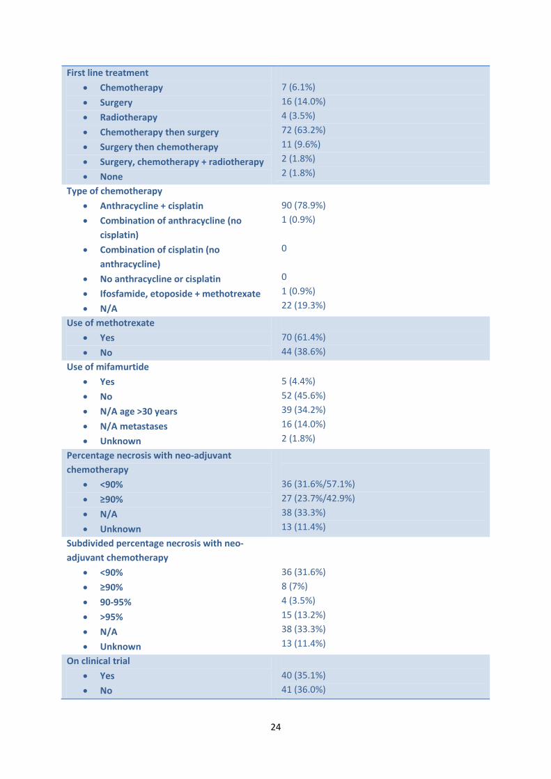
The time periods in which patients received treatment were divided into three ‘eras’: 2000 to 2005,
2006 to 2010 and 2011 to 2013. 51 patients, 49 patients and 14 patients were treated in each of
these eras, respectively (Table 5 and Figure 15). The patients were treated using combinations of
chemotherapy, surgery and radiotherapy. 7 patients (6.1%) received chemotherapy only; 16 (14%)
surgery only; 4 (3.5%) radiotherapy only; 72 (63.2%) chemotherapy then surgery; 11 (9.6%) surgery
then chemotherapy; 2 (1.8%) had surgery, chemotherapy and radiotherapy; and 2 (1.8%) had no
treatment (Table 5). Of those who received chemotherapy, 63 (55.3%) had this pre-operatively and
post-operatively, 11 (9.6%) had it only pre-operatively, 11 (9.6%) only post-operatively and 6 (5.3%)
had chemotherapy without surgery (Table 5).
Almost all of the patients who received chemotherapy were treated with a combination including
anthracycline and cisplatin (90, 78.9%) (Table 5 and Figure 16). Methotrexate was used in 70 cases
(61.4%) (Table 5) and 5 patients (4.4%) received Mifamurtide (Table 5). Whether patients were
treated on a clinical trial was also recorded with 40 patients (35.1%) treated on a clinical trial, 41
(36.0%) not treated on a trial and in 33 patients (28.9%) this was not applicable because of the
treatment they received, for example those treated with surgery alone (Table 5). However, it was
difficult to interpret from the information recorded in the bone tumour registry whether patients
had been treated on a trial or using the trial protocol, but not included in the trial. This information
was therefore excluded from the main analysis.
Tumour necrosis following neo-adjuvant chemotherapy was greater than or equal to 90% in
27patients (23.7%), while 36 (31.6%) patients had less than 90% tumour necrosis with neo-adjuvant
chemotherapy. 38 (33.3%) patients did not have neo-adjuvant chemotherapy or did not have their
tumour excised, so it had no pathological response to record and in 13 cases the percentage
response was not recorded (Table 5 and Figure 17). Therefore of the cases in which percentage
necrosis was available, 57.1% had less than 90% necrosis and 42.9% had 90% or more necrosis (Table

23
5). In some cases it was possible to subdivide those who had greater than 90% response into those
having 90 to 95% necrosis and those having greater than 95% necrosis (Table 5). 4 patients (3.5%)
had 90 to 95% necrosis and 15 had greater than 95% necrosis. This subdivision was not possible in 8
cases (7.0%) because the pathology report only recorded whether the necrosis was greater than or
less than 90%.
One hundred and one patients had surgery as part of the management of their osteosarcoma, while
12 did not (10.5%). Of the patients who had surgery, the majority had local resection or limb sparing
procedures (81 patients, 71.1%), while twenty patients had amputations (17.5%). In one case the
type of surgery is unknown (Table 5).
A small number of patients received radiotherapy (22 patients, 19.3%), but this was done
palliatively, not with curative intent, so further analysis was not performed on this data. Information
on the second line treatment which patients received was also not analysed because the line of
chemotherapy was not reliably designated in the bone tumour registry.
Forty two patients (36.8%) are known to have suffered a relapse of their osteosarcoma (Table 5 and
Figure 19). Of them, 9 (7.9%) had local relapse, 30 (26.3%) had distant relapse, 2 (1.8%) had both
local and distant relapse and in 1 case (0.9%) the location was unknown (Table 5).
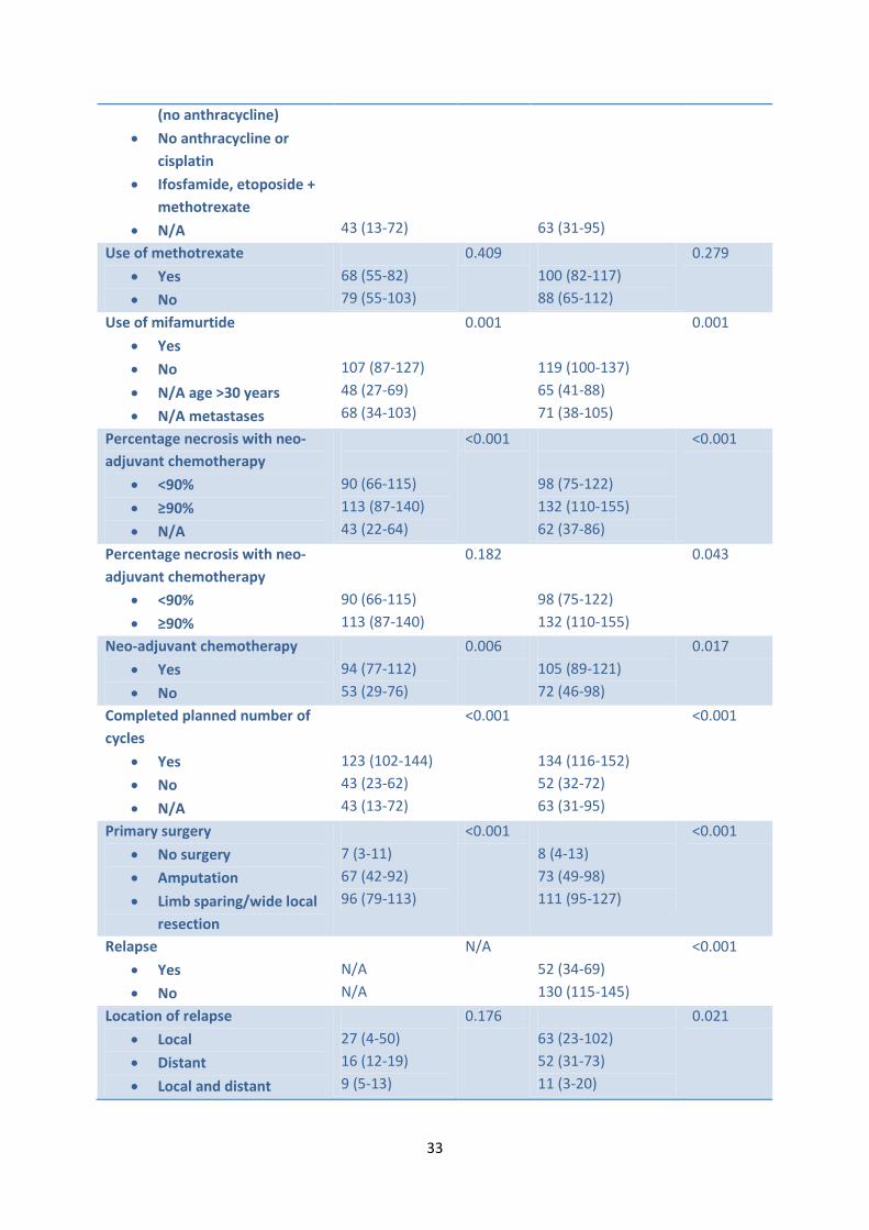
Table 5: Treatment details
Treatment detail Number of patients (% of patients)
Era of treatment
• 2000-2005
• 2006-2010
• 2011-present
51 (44.7%) 49 (43.0%) 14 (12.3%)
Timing of chemotherapy
• Pre- & post-operatively
• Pre-operatively
• Post-operatively
• No chemotherapy
• Chemotherapy, but no surgery
• Unknown
63 (55.3%) 11 (9.6%) 11 (9.6%) 22 (19.3%) 6 (5.3%) 1 (0.9%)
24
First line treatment
• Chemotherapy
• Surgery
• Radiotherapy
• Chemotherapy then surgery
• Surgery then chemotherapy
• Surgery, chemotherapy + radiotherapy
• None
7 (6.1%) 16 (14.0%) 4 (3.5%) 72 (63.2%) 11 (9.6%) 2 (1.8%) 2 (1.8%)
Type of chemotherapy
• Anthracycline + cisplatin
• Combination of anthracycline (no cisplatin)
• Combination of cisplatin (no anthracycline)
• No anthracycline or cisplatin
• Ifosfamide, etoposide + methotrexate
• N/A
90 (78.9%) 1 (0.9%) 0 0 1 (0.9%) 22 (19.3%)
Use of methotrexate
• Yes
• No
70 (61.4%) 44 (38.6%)
Use of mifamurtide
• Yes
• No
• N/A age >30 years
• N/A metastases
• Unknown
5 (4.4%) 52 (45.6%) 39 (34.2%) 16 (14.0%) 2 (1.8%)
Percentage necrosis with neo-adjuvant chemotherapy
• <90%
• ≥90%
• N/A
• Unknown
36 (31.6%/57.1%) 27 (23.7%/42.9%) 38 (33.3%) 13 (11.4%)
Subdivided percentage necrosis with neo-adjuvant chemotherapy
• <90%
• ≥90%
• 90-95%
• >95%
• N/A
• Unknown
36 (31.6%) 8 (7%) 4 (3.5%) 15 (13.2%) 38 (33.3%) 13 (11.4%)
On clinical trial
• Yes
• No
40 (35.1%) 41 (36.0%)

25
• Unknown 33 (28.9%)
Neo-adjuvant chemotherapy
• Yes
• No
77 (67.5%) 37 (32.5%)
Chemotherapy regimen
• MAP
• Cisplatin/adriamycin
• Adriamycin/ifosfamide
• Ifosfamide/etoposide/methotrexate
• Cisplatin/adriamycin/ifosfamide
• MAPIE
• N/A
• Unknown
52 (45.6%) 29 (25.4%) 1 (0.9%) 1 (0.9%) 6 (5.3%) 1 (0.9%) 22 (19.3%) 2 (1.8%)
Completed planned number of cycles
• Yes
• No
• N/A
• On-going
• Unknown
37 (32.5%) 26 (22.8%) 22 (19.3%) 1 (0.9%) 28 (24.6%)
Primary surgery
• No surgery
• Amputation
• Limb sparing/wide local resection
• Unknown
12 (10.5%) 20 (17.5%) 81 (71.1%) 1 (0.9%)
Relapse
• Yes
• No
• Unknown
42 (36.8%) 69 (60.5%) 3 (2.6%)
Location of relapse
• Local
• Distant
• Local and distant
• N/A
• Unknown
9 (7.9%) 30 (26.3%) 2 (1.8%) 69 (60.5%) 4 (3.5%)

26
Figure 15: Era of treatment
Figure 16: Type of chemotherapy received
Anthracycline & cisplatin
Anthracycline, no cisplatin
N/A
Ifosfamide, etoposide & methotrexate
Figure 17: Pathological response to neo-adjuvant chemotherapy
N/A
≥90%
<90%
Unknown

27
Univariate Analysis of Relapse-free Survival and Overall Survival
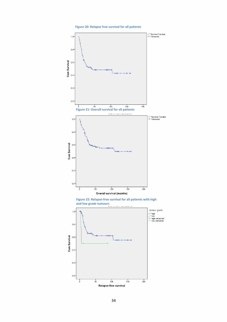
Relapse free survival (RFS) was able to be calculated for 111 patients, 56 of whom had relapsed or
died andoverall survival was able to be calculated for 112 patients, of whom 49 had died. The mean
RFS for all patients was 82 months (95% confidence interval 67-96) (Figure 20) and the mean overall
survival for all patients was 94 months (95% CI 80-108 months) (Figure 21). The percentage of
patients who had not relapsed after 5 years was 48.2% and the percentage of patients alive after 5
years was 56.4%.When all patients with high grade tumours were compared with all patients with
low grade tumours, the RFS and overall survival did not significantly differ for either (P = 0.129 and P
= 0.121, respectively) (Figure 22). Patients with low grade tumours would be expected to have better
survival than patients with high grade tumours. The finding that there is no significant difference in
Figure 18: Type of primary surgery Unknown
No surgery
Amputation
Limb sparing or local resection
Figure 19: Percentage of patients who suffered disease relapse
Yes
No
Unknown

28
survival between high and low grade tumours is likely to be caused by the very low number of low
grade tumours and that these occurred in older patients with other comorbidities.
Univariate analysis was performed to analyse RFS and overall survival for each of the demographic
factors. Thiswas performed only for high grade tumours because the number of low grade
tumourswas insufficient to allow accurate survival curves to be generated. RFS and overall survival
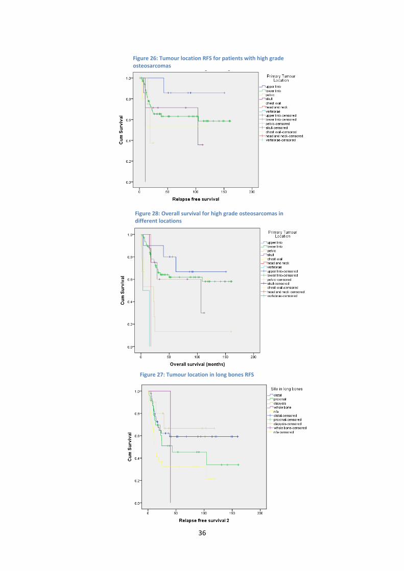
for high grade osteosarcomas in the population are shown in Table 6 and Figure 23toFigure 60.
Relapse free survival and overall survival were shorter in patients with tumours affecting the pelvis,
head and neck and vertebrae than those affecting the limbs, chest wall and skull. This pattern also
emerges in axial tumours having a poorer RFS and overall survival than limb tumours. Axial tumours
are more difficult to treat surgically than limb tumours which may explain a poorer RFS and overall
survival than for limb tumours. However, very few tumours occurred in some locations and
therefore one tumour could result in a large effect on survival in these groups. Tumours located in
the femur did not differ significantly in their RFS or overall survival than those in the tibia, humerus
or other bones. The part of the bone affected altered RFS and overall survival, though overall
survival did not reach significance (P=0.051).Patients with the whole bone affected had worse
prognosis than patients with proximal, distal or diaphyseal tumours or those whose tumours were
not in long bones. This result is not likely to be reliable however, because only one patient had the
whole bone affected.
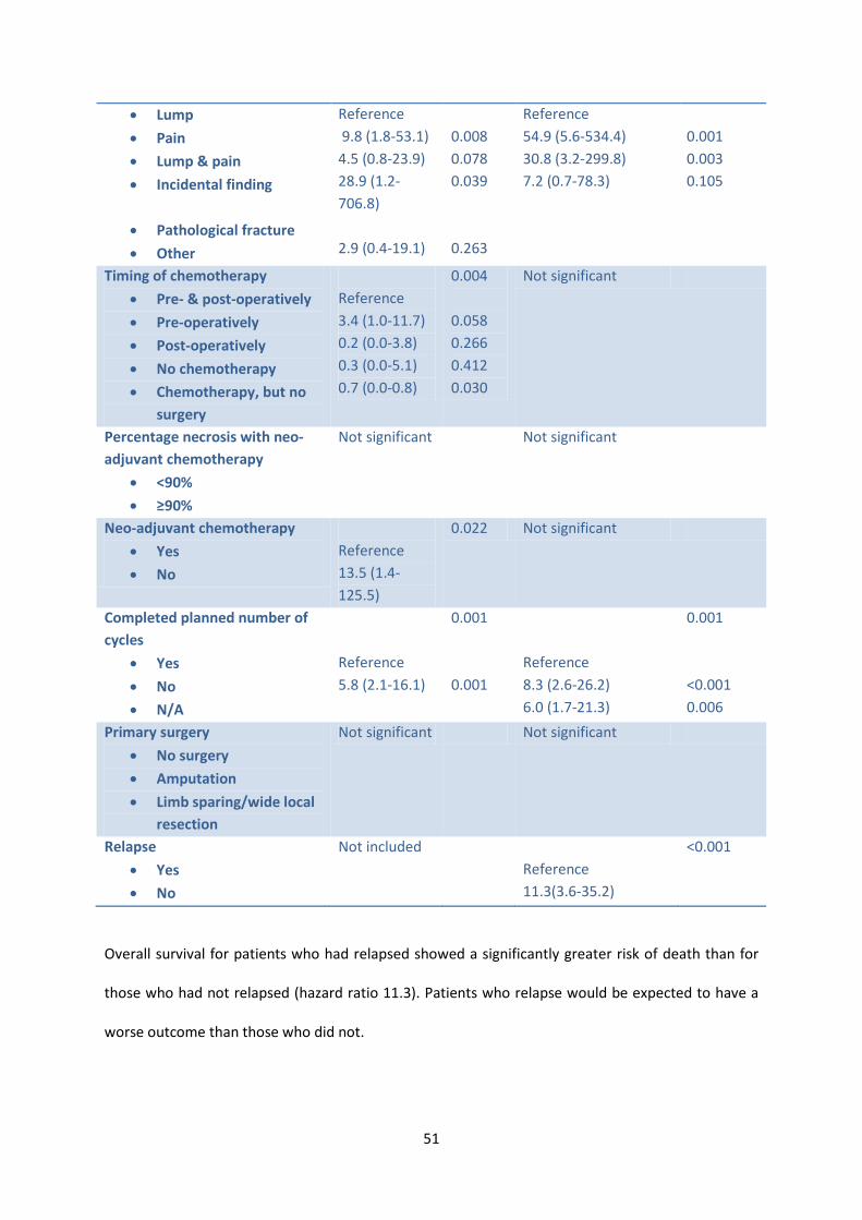
Type of osteosarcoma was associated with an improved relapse free survival and overall survival for
patients with conventional and paraosteal osteosarcomas over those with other types. This result is
not likely to be representative of a true association as 91.2% of the tumours were of conventional
type.
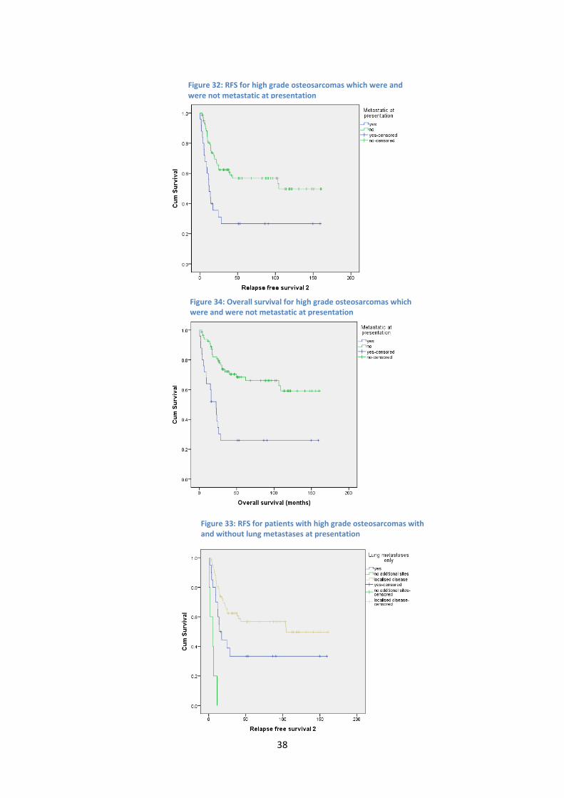
Presence of metastases significantly decreased relapse free survival and overall survival, with
tumours with metastases in locations other than lung suffering greater relapses and surviving a
29
much shorter time than those with lung metastases only or those with no metastases. This effect is
also shown by stage IVA and IVB having a worse prognosis than those of IIA and IIB.
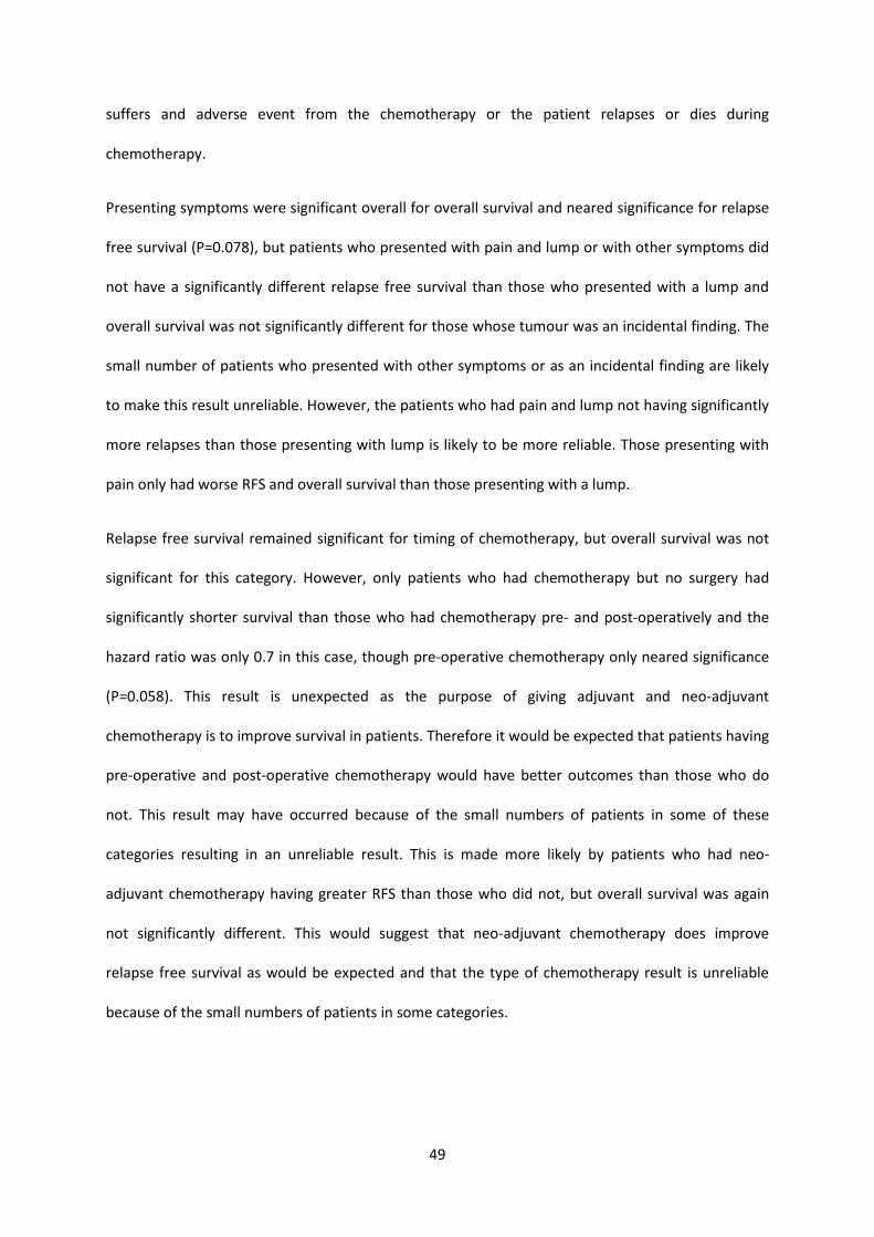
Overall survival was worse in patients who presented 3 months or more after symptom onset than in
those who presented less than 3 months after symptom onset, but this did not affect time to
relapse. The type of symptom which the patient presented with affected relapse free and overall
survival, with patients with lump, pain or lump and pain having a better outcome than those with
other symptoms. This may be an effect of the small number of patients who presented with
symptoms other than a lump, pain or both lump and pain.
Several aspects of the treatment received affected the relapse free and overall survival for patients.
Patients who did not have surgery or did not have further chemotherapy after surgery did worse
than those who had other timings of chemotherapy and patients who had neo-adjuvant
chemotherapy had better relapse free survival and overall survival than patients who did not. Type
of chemotherapy showed better relapse free survival and overall survival for those who had cisplatin
and anthracycline than for those who had no chemotherapy.It was not possible to identify whether
the use of Mifamurtide was associated with improved relapse free survival or overall survival
because it has only recently been added into the treatment of osteosarcoma, only a small number of
patients have received it, and none of them have yet suffered relapse or died. There is likely to be a
selection bias in the use of Mifamurtide because it is only licenced for use in patients under the age
of 30 without metastases. Therefore, patients who receive Mifamurtide have been selected out
because of their likelihood to have a better prognosis than other patients. It is already possible to
see this selection bias as those who were eligible, but did not receive Mifamurtide had significantly
better relapse free survival and overall survival than those who were ineligible because of age or
metastases. Patients with a 90% or greater response to chemotherapy had better overall survival
than patients with less than 90% response to chemotherapy, which confirms that this is a reliable

30
predictive factor of survival following neo-adjuvant chemotherapy. Patients who completed the
planned number of cycles of chemotherapy did better than those who did not.
Patients who relapsed had a significantly shorter overall survival than those who did not and
patients who had local relapse or distant relapse survived longer than those who had both local and
distant relapse.
Several of the categories which were recorded were not included in the results. In several cases this
was because we initially planned to look at Ewing’s sarcomas as well as osteosarcomas, but decided
to concentrate only on osteosarcomas because of the quantity of data collected. For this reason,
Ewing’s pelvic location, glycogen on biopsy, bone marrow metastases on aspirates, circulating
tumour cells, CD99 positivity, LDH, Ewing’s translocation, use of radiotherapy, dose of radiotherapy
and radiotherapy location were excluded from the analysis. Other criteria were excluded because of
insufficient recording in the bone tumour registry. Alkaline phosphatase level at diagnosis, tumour
volume, MRI, CT and PET response and response were all excluded because of insufficient recording.
Clinical trial name was removed because of the difficulty interpreting whether a patient was on a
clinical trial which therefore made this category likely to be inaccurate. Second line treatment and
main site of metastases were not recorded because it was not relevant to response to first line
treatment.
Table 6: Mean relapse-free survival and overall survival for high and low grade tumours
Subset of Tumours Mean RFS (95% CI)
P-value Mean overall survival (95% CI)
P-value
Patient gender
• Male
• Female
77 (58-97) 89 (67-110)
0.408 89 (70-108) 103 (82-123)
0.270
Adult or paediatric Patient
• <16 years old
• ≥16 years old
97 (73-121) 74 (56-93)
0.140 105 (83-127) 89 (71-107)
0.290
Axial or limb tumour location
• Axial
• Limb
38 (19-58) 94 (78-111)
<0.001 60 (32-87) 106 (91-122)
0.001

31
Location of tumour
• Upper limb
• Lower limb
• Pelvis
• Skull
• Chest wall
• Head & neck
• Vertebrae
110 (72-148) 93 (75-110) 11 (4-17) 71 (39-104) 51 (3-99) 11 (11-11) 7 (0-13)
<0.001 113 (77-149) 105 (88-122) 32 (0-68) 74 (43-105) 73 (24-121) 17 (17-17) 9 (0-20)
<0.001
Bone location
• Femur
• Tibia
• Humerus
• Other
86 (63-109) 103 (75-131) 79 (52-105) 59 (36-82)
0.103 99 (77-121) 115 (89-141) 82 (59-106) 76 (51-101)
0.172
Site in long bones
• Distal
• Proximal
• Diaphysis
• Whole bone
• Not applicable
100 (78-122) 76 (46-105) 84 (53-116) 40 (40-40) 43 (24-62)
0.021 112 (92-133) 91 (62-120) 84 (53-115) 40 (40-40) 68 (41-94)
0.051
Type of osteosarcoma
• Conventional
• Paraosteal
• Periosteal
• Other
82 (67-97) 115 (43-187) 78 (23-133) 2 (1-4)
<0.001 94 (79-109) 117 (47-186) 2 (1-4)
<0.001
Subtype of osteosarcoma
• Osteoblastic
• Osteoclastic
• Telangectatic
• Small cell
• Chondroblastic
• Fibroblastic
• Epithelioid
89 (70-107) 34 (7-60) 38 (8-68) 64 (34-94) 67 (29-105) 27 (0-60)
0.807 97 (80-114) 44 (31-57) 38 (8-68) 86 (55-117) 97 (67-127) 27 (0-60)
0.837
Metastases at presentation
• Yes
• No
50 (24-77) 94 (78-111)
0.001 51 (26-77) 111 (95-126)
<0.001
Lung metastases only
• Yes
• No, additional sites
• Localised disease only
61 (30-93) 5 (1-9) 94 (78-111)
<0.001 63 (32-93) 6 (1-11) 111 (95-126)
<0.001
Stage at diagnosis
• IA
• IB
• IIA
100 (76-123)
<0.001 111 (89-132)
<0.001

32
• IIB
• III
• IVA
• IVB
84 (58-111) 64 (31-96) 9 (1-17)
109 (85-133) 65 (33-97) 10 (1-18)
Tumour maximum dimension
• <90 mm
• ≥90 mm
91 (67-116) 83 (59-107)
0.573 106 (83-129) 99 (76-121)
0.482
Scottish Index of Multiple Deprivation quintiles
• 1
• 2
• 3
• 4
• 5
65 (25-105) 54 (22-87) 92 (62-122) 58 (37-79) 97 (72-121)
0.329 70 (31-109) 87 (53-120) 96 (66-127) 80 (50-111) 114 (89-139)
0.564
Time from symptom onset to primary presentation
• <3 months
• ≥3 months
• N/A
94 (72-115) 77 (55-98) 9 (9-9)
0.119 104 (84-124) 87 (66-108)
0.041
Presenting symptoms
• Lump
• Pain
• Lump & pain
• Incidental finding
• Pathological fracture
• Other
128 (96-160) 74 (52-96) 95 (70-120) 9 (9-9) 10 (2-19) 31 (0-66)
0.018 139 (112-166) 87 (65-108) 106 (82-129) 9 (9-9) 10 (2-19) 36 (1-70)
0.001
Era of treatment
• 2000-2005
• 2006-2010
• 2011-present
92 (71-112) 44 (33-54) 22 (17-27)
0.259 103 (83-122) 54 (43-64) 22 (17-27)
0.562
Timing of chemotherapy
• Pre- & post-operatively
• Pre-operatively
• Post-operatively
• No chemotherapy
• Chemotherapy, but no surgery
110 (92-128) 26 (3-49) 76 (46-106) 43 (13-72) 9 (5-12)
<0.001 120 (104-136) 38 (12-64) 112 (70-155) 63 (31-95) 11 (5-16)
<0.001
Type of chemotherapy
• Anthracycline + cisplatin
• Combination of anthracycline (no cisplatin)
• Combination of cisplatin
90 (74-106)
0.007 101 (86-116)
0.044
33
(no anthracycline)
• No anthracycline or cisplatin
• Ifosfamide, etoposide + methotrexate
• N/A
43 (13-72)
63 (31-95)
Use of methotrexate
• Yes
• No
68 (55-82) 79 (55-103)
0.409 100 (82-117) 88 (65-112)
0.279
Use of mifamurtide
• Yes
• No
• N/A age >30 years
• N/A metastases
107 (87-127) 48 (27-69) 68 (34-103)
0.001 119 (100-137) 65 (41-88) 71 (38-105)
0.001
Percentage necrosis with neo-adjuvant chemotherapy
• <90%
• ≥90%
• N/A
90 (66-115) 113 (87-140) 43 (22-64)
<0.001 98 (75-122) 132 (110-155) 62 (37-86)
<0.001
Percentage necrosis with neo-adjuvant chemotherapy
• <90%
• ≥90%
90 (66-115) 113 (87-140)
0.182 98 (75-122) 132 (110-155)
0.043
Neo-adjuvant chemotherapy
• Yes
• No
94 (77-112) 53 (29-76)
0.006 105 (89-121) 72 (46-98)
0.017
Completed planned number of cycles
• Yes
• No
• N/A
123 (102-144) 43 (23-62) 43 (13-72)
<0.001 134 (116-152) 52 (32-72) 63 (31-95)
<0.001
Primary surgery
• No surgery
• Amputation
• Limb sparing/wide local resection
7 (3-11) 67 (42-92) 96 (79-113)
<0.001 8 (4-13) 73 (49-98) 111 (95-127)
<0.001
Relapse
• Yes
• No
N/A N/A
N/A 52 (34-69) 130 (115-145)
<0.001
Location of relapse
• Local
• Distant
• Local and distant
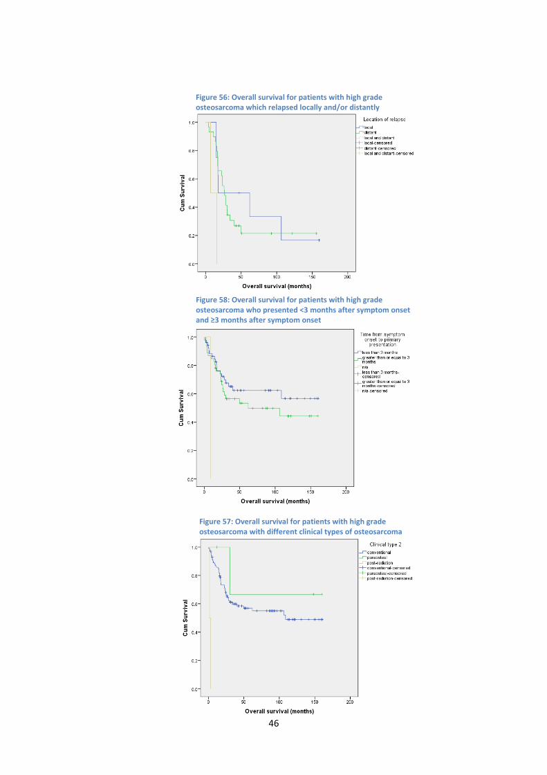
27 (4-50) 16 (12-19) 9 (5-13)
0.176 63 (23-102) 52 (31-73) 11 (3-20)
0.021
34
Figure 22: Relapse-free survival for all patients with high and low grade tumours
Figure 20: Relapse free survival for all patients
Figure 21: Overall survival for all patients

35
Figure 25: Overall survival for high grade osteosarcomas located in limbs and in the axial skeleton
Figure 24: Overall survival for patients with high and low grade osteosarcomas
Figure 23: Axial and limb high grade osteosarcoma RFS
36
Figure 26: Tumour location RFS for patients with high grade osteosarcomas
Figure 28: Overall survival for high grade osteosarcomas in different locations
Figure 27: Tumour location in long bones RFS

37
Figure 31: Overall survival for different types of high grade osteosarcoma
Figure 29: Overall survival for patients with high grade osteosarcoma located in different parts of long bones
Figure 30: RFS for different types of high grade osteosarcoma
38
Figure 34: Overall survival for high grade osteosarcomas which were and were not metastatic at presentation
Figure 32: RFS for high grade osteosarcomas which were and were not metastatic at presentation
Figure 33: RFS for patients with high grade osteosarcomas with and without lung metastases at presentation

39
Figure 35: Overall survival for patients with high grade osteosarcoma with and without lung metastases at presentation
Figure 37: Overall survival for patients with high grade osteosarcoma of different stages at diagnosis
Figure 36: RFS for patients with high grade osteosarcoma of different stages at diagnosis
40
Figure 40: Overall survival for patients with high grade osteosarcoma with different presenting symptoms
Figure 38: Presenting symptoms RFS for high grade osteosarcomas
Figure 39: RFS for timing of chemotherapy in patients with high grade osteosarcoma
41
Figure 42: Overall survival for patients with high grade osteosarcoma with different timings of chemotherapy
Figure 43: Overall survival for patients with high grade osteosarcoma who had different types of chemotherapy
Figure 41: RFS for patients with high grade osteosarcoma who had different types of chemotherapy
42
Figure 45: Overall survival for patients with high grade osteosarcoma who were and were not treated with mifamurtide
Figure 44: Use of mifamurtide RFS for patients with high grade osteosarcomas
Figure 46: Pathological response to treatment RFS for high grade osteosarcomas

43
Figure 49: Overall survival for patients with high grade osteosarcoma who had <90% or ≥90% response to neo-adjuvant chemotherapy with those for whom this was not applicable removed
Figure 48: RFS for patients with high grade osteosarcoma who did and did not have neo-adjuvant chemotherapy
Figure 47: Overall survival for patients with high grad osteosarcoma who had <90% or ≥90% response to neo-adjuvant chemotherapy
44
Figure 50: Overall survival for patients with high grade osteosarcoma who did and did not have neo-adjuvant chemotherapy
Figure 52: Overall survival for patients with high grade osteosarcoma who did and did not complete the planned number of cycles of chemotherapy
Figure 51: RFS for patients with high grade osteosarcoma who completed or did not complete their planned number of cycles of chemotherapy

45
Figure 55: Overall survival for patients with high grade osteosarcoma who did and did not relapse
Figure 54: Overall survival for patients with high grade osteosarcoma who had different surgical management
Figure 53: RFS for patients with high grade osteosarcoma who had different surgical management
46
Figure 56: Overall survival for patients with high grade osteosarcoma which relapsed locally and/or distantly
Figure 58: Overall survival for patients with high grade osteosarcoma who presented <3 months after symptom onset and ≥3 months after symptom onset
Figure 57: Overall survival for patients with high grade osteosarcoma with different clinical types of osteosarcoma

47
Figure 59: Overall survival for patients with high grade osteosarcoma who received different types of chemotherapy
Figure 60: Overall survival for patients with high grade osteosarcoma having different types of surgical management

48
Multivariate analysis
Multivariate analysis was conducted for variables which were found to be significant in the
univariate analysis. However, type of osteosarcoma, type of chemotherapy, use of mifamurtide and
location of relapse were not analysed because the small numbers of participants in some of the
categories in each of these variables made the data unlikely to be reliable. Variables which were
related to other variables were also excluded from the multivariate analysis to prevent confounding
because of this. This included site in long bones, metastases at presentation and stage at diagnosis.
The hazard ratios for each factor are shown inTable 7.
Relapse free survival and overall survival remained significant for axial or limb location of tumour
with limb tumours having a 5.7 and 35.8 times greater relapse free survival and overall survival,
respectively. Tumours affecting the axial skeleton can be more difficult to treat because of the
proximity to organs. This often limits the surgical options available for treatment and would explain
the reduced survival in patients with axial tumours.
Site of metastases also remained significant predictors of outcome with those with lung metastases
having worse survival than those with no metastases (hazard ratio 3.7 and 10.3 for RFS and overall
survival, respectively) and those with metastases elsewhere doing worse still (hazard ratio 245.0 and
239.6 for RFS and overall survival, respectively). Metastatic disease would be expected to reduce
survival and forms part of the staging of osteosarcomas for this reason. Lung metastases may have
better survival than metastases in other locations because they are easier to detect and to treat with
chemotherapy and/or excision than metastases at other sites.
Relapse free survival and overall survival remained significant for whether or not the patient had
completed the planned number of cycles of chemotherapy. Risk for relapse free survival and overall
survival was 5.8 and 8.3 times greater, respectively, in those who had not completed the planned
number of cycles of chemotherapy than in those who had. This result is logical as chemotherapy
common reasons for chemotherapy stopped include the tumour is not responding, the patient
49
suffers and adverse event from the chemotherapy or the patient relapses or dies during
chemotherapy.
Presenting symptoms were significant overall for overall survival and neared significance for relapse
free survival (P=0.078), but patients who presented with pain and lump or with other symptoms did
not have a significantly different relapse free survival than those who presented with a lump and
overall survival was not significantly different for those whose tumour was an incidental finding. The
small number of patients who presented with other symptoms or as an incidental finding are likely
to make this result unreliable. However, the patients who had pain and lump not having significantly
more relapses than those presenting with lump is likely to be more reliable. Those presenting with
pain only had worse RFS and overall survival than those presenting with a lump.
Relapse free survival remained significant for timing of chemotherapy, but overall survival was not
significant for this category. However, only patients who had chemotherapy but no surgery had
significantly shorter survival than those who had chemotherapy pre- and post-operatively and the
hazard ratio was only 0.7 in this case, though pre-operative chemotherapy only neared significance
(P=0.058). This result is unexpected as the purpose of giving adjuvant and neo-adjuvant
chemotherapy is to improve survival in patients. Therefore it would be expected that patients having
pre-operative and post-operative chemotherapy would have better outcomes than those who do
not. This result may have occurred because of the small numbers of patients in some of these
categories resulting in an unreliable result. This is made more likely by patients who had neo-
adjuvant chemotherapy having greater RFS than those who did not, but overall survival was again
not significantly different. This would suggest that neo-adjuvant chemotherapy does improve
relapse free survival as would be expected and that the type of chemotherapy result is unreliable
because of the small numbers of patients in some categories.

50
Table 7: Hazard ratios for each factor from the multivariate analysis
Subset of Tumours RFS hazard ratio (95% CI)
P-value Overall survival hazard ratio (95% CI)
P-value
Axial or limb tumour location
• Axial
• Limb
5.7 (2.0-16.5) Reference
0.001 35.8 (10.0-128.2) Reference
<0.001
Location of tumour
• Upper limb
• Lower limb
• Pelvis
• Skull
• Chest wall
• Head & neck
• Vertebrae
Not included Not included
Site in long bones
• Distal
• Proximal
• Diaphysis
• Whole bone
• Not applicable
Not included Not included
Metastases at presentation
• Yes
• No
Not included Not included
Lung metastases only
• Yes
• No, additional sites
• Localised disease only
3.7 (1.1-12.1) 245.0 (19.2-3123.2) Reference
<0.001 0.032 <0.001
10.3 (3.3-31.4) 239.6 (30.7-1868.7) Reference
<0.001 <0.001 <0.001
Stage at diagnosis
• IA
• IB
• IIA
• IIB
• III
• IVA
• IVB
Not included Not included
Time from symptom onset to primary presentation
• <3 months
• ≥3 months
• N/A
Not included Not significant
Presenting symptoms 0.078 0.002
51
• Lump
• Pain
• Lump & pain
• Incidental finding
• Pathological fracture
• Other
Reference 9.8 (1.8-53.1) 4.5 (0.8-23.9) 28.9 (1.2-706.8) 2.9 (0.4-19.1)
0.008 0.078 0.039 0.263
Reference 54.9 (5.6-534.4) 30.8 (3.2-299.8) 7.2 (0.7-78.3)
0.001 0.003 0.105
Timing of chemotherapy
• Pre- & post-operatively
• Pre-operatively
• Post-operatively
• No chemotherapy
• Chemotherapy, but no surgery
Reference 3.4 (1.0-11.7) 0.2 (0.0-3.8) 0.3 (0.0-5.1) 0.7 (0.0-0.8)
0.004 0.058 0.266 0.412 0.030
Not significant
Percentage necrosis with neo-adjuvant chemotherapy
• <90%
• ≥90%
Not significant Not significant
Neo-adjuvant chemotherapy
• Yes
• No
Reference 13.5 (1.4-125.5)
0.022 Not significant
Completed planned number of cycles
• Yes
• No
• N/A
Reference 5.8 (2.1-16.1)
0.001 0.001
Reference 8.3 (2.6-26.2) 6.0 (1.7-21.3)
0.001 <0.001 0.006
Primary surgery
• No surgery
• Amputation
• Limb sparing/wide local resection
Not significant Not significant
Relapse
• Yes
• No
Not included Reference 11.3(3.6-35.2)
<0.001
Overall survival for patients who had relapsed showed a significantly greater risk of death than for
those who had not relapsed (hazard ratio 11.3). Patients who relapse would be expected to have a
worse outcome than those who did not.
52
Time from symptom onset to presentation was included in the multivariate analysis of overall
survival, but was found to no longer be a significant predictor of outcome. It could be that this is
because there truly is no relationship between time from symptom onset to presentation or perhaps
by using different time intervals a difference may be found which was not possible with the data
recorded in this study.
Percentage tumour necrosis of less than 90% was found to no longer have a significantly worse
outcome than 90% or greater necrosis. As percentage necrosis is used as the main predictor of
response to chemotherapy in practice this result was very unexpected. This may have occurred
because of the relationship of necrosis to some of the other factors being analysed. Particularly neo-
adjuvant chemotherapy as only in patients who had neo-adjuvant chemotherapy is it possible to
calculate percentage necrosis.
Type of primary surgery also ceased to be a significant predictor of outcome in the multivariate
analysis. This would suggest that patients are receiving the appropriate type of surgery for their
tumour in this patient group. That there is also no difference in the patients who had no surgery may
be a spurious result because of the small number of patients in this category.
53
Discussion
The results from the univariate analysis demonstrate an association between several factors and RFS
and overall survival.Longer RFS was seen in patients with tumours affecting the limbs than those
affecting the axial skeleton; in tumours which affected the limbs, chest wall and skull than those
affecting the pelvis, head and neck and vertebrae; tumours affecting distal, proximal or diaphyseal
parts of bones than tumours which affected the whole bone; conventional, paraosteal and periosteal
osteosarcomas compared with other types of osteosarcoma; patients with no metastases at
diagnosis than those with metastases and those with lung metastases than those with metastases at
other sites; tumours of stage IIA and IIB compared with stage IVA and IVB; patients presenting with
pain, lump or pain and lump than those with other symptoms; those who had pre-operative and
post-operative chemotherapy, post-operative chemotherapy or no chemotherapy than those who
had pre-operative chemotherapy or chemotherapy but no surgery; patients who had neo-adjuvant
chemotherapy than those who did not; patients who completed the planned number of cycles of
chemotherapy than in those who did not; and patients who had amputation, limb sparing surgery or
wide local resection than those who had no surgery. These factors were also associated with
increased length of overall survival. Other factors which increased overall survival, but were not
associated with improved RFS were presenting less than 3 months after symptom onset; 90% or
greater necrosis of tumour following neo-adjuvant chemotherapy; and not suffering disease relapse.
Site in long bones neared, but did not reach significance (P = 0.051) for overall survival.
In the multivariate analysis, associations were still present between several factors and RFS and
overall survival. Limb tumours had better RFS and overall survival than axial tumours;patients with
lung metastases did better than those with metastases elsewhere and those with no metastases did
better still; and patients who completed the planned number of cycles of chemotherapy had better
survival than those who did not. Presenting symptoms and whether the patient had relapsed

54
significantly influenced overall survival and timing of chemotherapy and whether the patient had
neo-adjuvant chemotherapy affected relapse free survival.
While these factors were significantly associated with greater RFS and/or overall survival in the
analysis, in some cases this may have occurred because of the small number of patients in some of
the subgroups.
Pakos et al. conducted similar research across several centres internationally (7). The demographics
of their population are compared with that of the population in this study inTable 8. The number of
patients included in the study by Pakos et al. was much greater than that of this study, but the
demographic details of the patients, tumours and treatment did not differ greatly from those in this
study. Pakos et al. found presence of metastases at presentation to be associated with decreased
overall survival in their study. This study replicated this result and we can add to this that the
location of the metastases is also important with those with bone metastases had a shorter survival
than those with lung metastases or no metastases. This study found improved overall survival in
patients with 90% or more tumour necrosis following neo-adjuvant chemotherapy in the univariate
analysis, but not in the multivariate analysis. Pakos et al. did find an association between survival
and percentage necrosis in their multivariate analysis. This may be a result of the larger number of
patients in Pakos et al.’s study enabling them to have greater sensitivity to detect this effect.
Patients who had amputation suffered worse overall survival and metastatic RFS in Pakos et al.’s
study which was not seen in this study. Use of anthracycline and platinum was found to improve
survival in Pakos et al.’s patients, but the data unfortunately did not allow analysis for this
association in this study. The study by Pakos et al. also found increasing age to be associated with
worse overall survival and increased local recurrence; tibial tumours to have better metastatic
relapse free survival and overall survival than femoral tumours; and patients who received
methotrexate had worse overall survival than those who were not treated with methotrexate, which
are in disagreement with this study. Another prognostic factor found by Pakos et al. which we did
55
Table 8: Comparison of demographics of this study with Pakos et al. 2009
SBTR Pakos et al.
Number of patients 114 2680 Male: Female 53.5%:46.5% 57%:43% Median tumour size 90mm 100mm Tumour location
• Femur
• Tibia
• Humerus
• Others
46 (40.4%) 26 (22.8%) 5 (4.4%) 37 (32.5%)
1400 (52%) 638 (24%) 266 (10%) 376 (14%)
Site in long bones
• Distal
• Proximal
• Diaphysis
• Whole bone
48 (42.1%) 24 (21.1%) 11 (9.6%) 1 (0.9%)
946 (56%) 652 (38%) 72 (4%) 26 (2%)
Clinical type
• Conventional
• Paraosteal
• Periosteal
• Others
104 (91.2%) 4 (3.5%) 4 (3.5%) 2 (1.8%)
2284 (96%) 59 (2%) 25 (1%) 9 (1%)
Histological type
• Osteoblastic
• Chondroblastic
• Fibroblastic
• Telangiectatic
• Small Cell
• Others
71 (62.3%) 24 (21.1%) 9 (7.9%) 4 (3.5%) 0 6 (5.3%)
1041 (49%) 309 (15%) 175 (8%) 95 (4%) 15 (1%) 479 (23%)
Metastases at presentation 26 (22.8%) 362 (13%) Initial treatment
• Surgery + chemotherapy
• Surgery
• Chemotherapy
• Surgery + chemotherapy + radiotherapy
• Others
83 (72.8%) 16 (14.0%) 7 (6.1%) 7 (6.1%) 6 (5.3%)
2091(78%) 385 (14%) 73 (3%) 86 (3%) 43 (2%)
Type of surgery
• Limb salvage/wide resection
• Amputation
81 (71.1%) 20 (17.5%)
1891 (77%) 572 (23%)
Timing of chemotherapy
• Pre- & post-operatively
63 (55.3%) 11 (9.6%)
1982 (91%) 35 (2%)
56
not analyse were receiving radiotherapy, chemotherapy and surgery which had a worse overall
survival and metastatic RFS than receiving chemotherapy and surgery with no radiotherapy.
One of the problems encountered in this study was that recording of some pieces of information in
the bone tumour registry was inconsistent, which resulted in not all information being available for
all patients. This problem was also encountered by Pakos et al. in their study (7). Therefore it may be
useful to review the information which is collected by the Scottish Bone Tumour Registry and to
create a more standardised set of information which is collected on each patient in order that
information in the future is more complete. If this were put in place it might also be useful to review
the information collected on a regular basis, such as annually or 5 yearly in order to ensure that the
information being collected is as relevant as possible to current practice and areas of research.
Conclusion
Patients in Scotland, from the year 2000 to 2013 relapsed after a mean of 6 years and 10 months
and had a mean overall survival of 7 years and 10 months. 5 year relapse free survival was 48.2% and
• Pre-operatively
• Post-operatively
11 (9.6%) 153 (7%)
Type of chemotherapy
• Anthracycline & platinum included
• Combination with anthracycline, but not platinum
• Combination with platinum, but not anthracycline
• Others – not specified
90 (78.9%) 1 (0.9%) 0 23 (20.2%)
1665 (74%) 52 (2%) 14 (1%) 530 (23%)
Use of methotrexate
• No
• Yes
• Not specified
44 (38.6%) 70 (61.4%)
601 (27%) 1278 (55%) 412 (18%)
Histological response
• ≥90%
• <90%
27 (42.9%) 36 (57.1%)
509 (29%) 1225 (71%)

57
the 5 year overall survival was 56.4%. Several factors were identified which alter RFS and overall
survival. Percentage necrosis following neo-adjuvant chemotherapy was found to be a strong
predictor of outcome in univariate analysis, but not in multivariate analysis. Further work with large
numbers of patients is needed to more definitively identify factors which predict outcome in
patients with osteosarcoma.
58
References
(1) Kirkwood JM, Tarhini A, Sparano JA, Patel P, Schiller JH, Vergo MT, et al. Comparative clinical benefits of systemic adjuvant therapy for paradigm solid tumors. Cancer Treat Rev 2013;39(1):27-43.
(2) Bone sarcomas: ESMO clinical practice guidelines for diagnosis, treatment and follow-up. Annals of Oncology 2012;23(SUPPL. 7):vii100-vii109.
(3) Bacci G, Ferrari S, Longhi A, Perin S, Forni C, Fabbri N, et al. Pattern of relapse in patients with osteosarcoma of the extremities treated with neoadjuvant chemotherapy. Eur J Cancer 2001;37(1):32-38.
(4) Chou AJ, Kleinerman ES, Krailo MD, Chen Z, Betcher DL, Healey JH, et al. Addition of muramyl tripeptide to chemotherapy for patients with newly diagnosed metastatic osteosarcoma: A report from the Children's Oncology Group. Cancer 2009;115(22):5339-5348.
(5) Spina A, Sorvillo L, Di Maiolo F, Esposito A, D'Auria R, Di Gesto D, et al. Inorganic phosphate enhances sensitivity of human osteosarcoma U2OS cells to doxorubicin via a p53-dependent pathway. J Cell Physiol 2013;228(1):198-206.
(6) Meyers PA, Schwartz CL, Krailo MD, Healey JH, Bernstein ML, Betcher D, et al. Osteosarcoma: The addition of muramyl tripeptide to chemotherapy improves overall survival - A report from the children's oncology group. Journal of Clinical Oncology 2008;26(4):633-638.
(7) Pakos EE, Nearchou AD, Grimer RJ, Koumoullis HD, Abudu A, Bramer JAM, et al. Prognostic factors and outcomes for osteosarcoma: An international collaboration. Eur J Cancer 2009;45(13):2367-2375.
(8) Bölling T, Hardes J, Dirksen U. Management of Bone Tumours in Paediatric Oncology. Clin Oncol 2013;25(1):19-26.
(9) Van Der Woude H-, Bloem JL, Hogendoorn PCW. Preoperative evaluation and monitoring chemotherapy in patients with high-grade osteogenic and Ewing's sarcoma: Review of current imaging modalities. Skeletal Radiol 1998;27(2):57-71.
(10) Brisse H, Ollivier L, Edeline V, Pacquement H, Michon J, Glorion C, et al. Imaging of malignant tumours of the long bones in children: Monitoring response to neoadjuvant chemotherapy and preoperative assessment. Pediatr Radiol 2004;34(8):595-605.
(11) Bacci G, Ferrari S, Ruggieri P, Biagini R, Fabbri N, Campanacci L, et al. Telangiectatic osteosarcoma of the extremity: Neoadjuvant chemotherapy in 24 cases. Acta Orthop Scand 2001;72(2):167-172.
(12) Bacci G, Ferrari S, Tienghi A, Bertoni F, Mercuri M, Longhi A, et al. A comparison of methods of loco-regional chemotherapy combined with systemic chemotherapy as neo-adjuvant treatment of osteosarcoma of the extremity. European Journal of Surgical Oncology 2001;27(1):98-104.
(13) Borys D, Canter RJ, Hoch B, Martinez SR, Tamurian RM, Murphy B, et al. P16 expression predicts necrotic response among patients with osteosarcoma receiving neoadjuvant chemotherapy. Hum Pathol 2012;43(11):1948-1954.

59
(14) Li H, Zhao H, Wang B, Wang X, Wang Z, Zheng S, et al. 18F-FDG positron emission tomography for the assessment of histological response to neoadjuvant chemotherapy in osteosarcomas: A meta-analysis. Surg Oncol 2012;21(4):e165-e170.
(15) The Scottish Government. Scottish Index of Multiple Deprivation. 2012; Available at: http://www.scotland.gov.uk/Topics/Statistics/SIMD. Accessed April/15, 2013.

60
Appendix 1: Data Collection Sheet
Data Collection Sheet
Patient CHI number: ……………………….
Sex: Male Female
Age at diagnosis: ……….. Adult or Paediatric: ≥16 <16
Tumour location 1: Upper limb Lower Limb Pelvis
Skull Chest Wall Head & Neck
Vertebrae
Tumour location 2: Femur Tibia Humerus Other
Site in long bones: Distal Proximal Diaphysis
Whole Bone
Ewing’s pelvic location: Pelvic Non-pelvic N/A osteosarcoma
Type of Osteosarcoma: Conventional Paraosteal Periosteal
Other N/A Ewing’s
Osteosarcoma subtype: Osteoblastic Osteoclastic Small Cell
Telangectatic Others N/A Ewing’s
Metastases at presentation: Yes No Unknown
Lung metastases only: Yes No Localised disease
Tumour grade: High Low
Stage at diagnosis: IA IB IIA IIB III
IVA IVB Unknown
Tumour volume: …….
Bone marrow metastases on aspirates: Yes No Unknown

61
N/A for this histology
Circulating tumour cells: Yes No Unknown
N/A for this histology
Glycogen on biopsy: Yes No Unknown
N/A for this histology
CD99 positive: Yes No Unknown
N/A for this histology
Ewing’s translocation: Yes No Unknown
N/A for this histology
Alkaline phosphatase at diagnosis: ……..
Lactate dehydrogenase at diagnosis: ……..
Timing of chemotherapy: Pre-op Post-op Pre-op & Post-op
No chemotherapy
First line treatment: Surgery Chemotherapy Radiotherapy
Chemo then Surgery Surgery, Chemo & XRT
Type of chemotherapy: Anthracyclin & Cisplatin
Combination of anthracyclin (no cisplatin)
Combination of cisplatin (no anthracyclin)
VIDE
VID
VIDE + VAI
Use of methotrexate: Yes No N/A Ewing’s
62
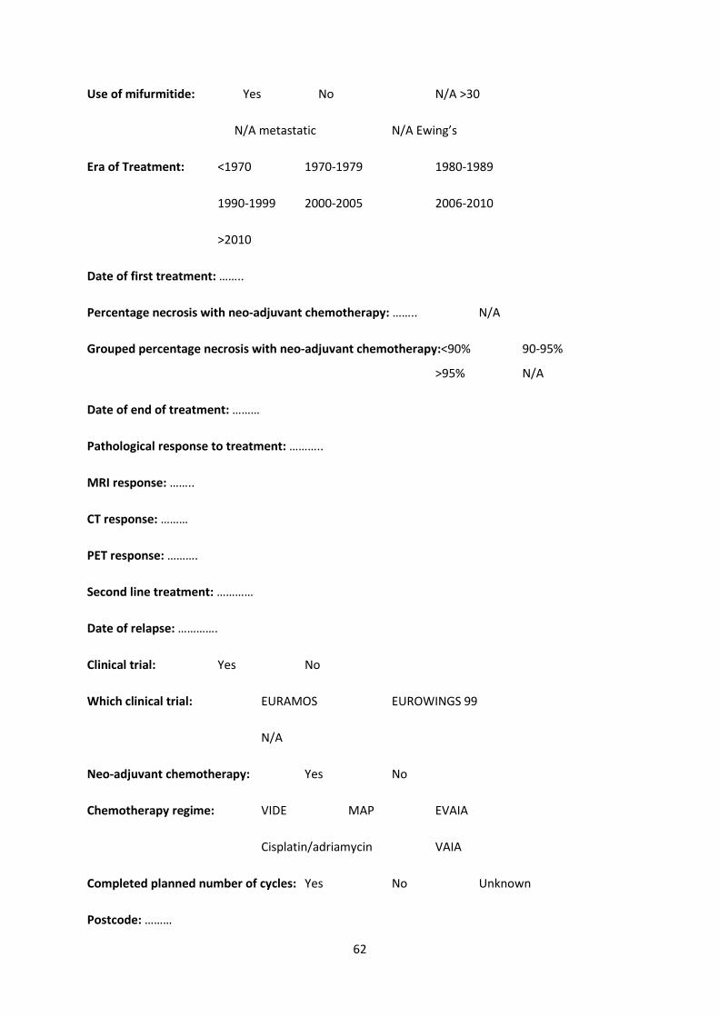
Use of mifurmitide: Yes No N/A >30
N/A metastatic N/A Ewing’s
Era of Treatment: <1970 1970-1979 1980-1989
1990-1999 2000-2005 2006-2010
>2010
Date of first treatment: ……..
Percentage necrosis with neo-adjuvant chemotherapy: …….. N/A
Grouped percentage necrosis with neo-adjuvant chemotherapy:<90% 90-95%
>95% N/A
Date of end of treatment: ………
Pathological response to treatment: ………..
MRI response: ……..
CT response: ………
PET response: ……….
Second line treatment: …………
Date of relapse: ………….
Clinical trial: Yes No
Which clinical trial: EURAMOS EUROWINGS 99
N/A
Neo-adjuvant chemotherapy: Yes No
Chemotherapy regime: VIDE MAP EVAIA
Cisplatin/adriamycin VAIA
Completed planned number of cycles: Yes No Unknown
Postcode: ………

63
Scottish index of multiple deprivation: ………..
Current status: Free of disease Alive with disease Deceased
Date of diagnosis: …….
Date of death: ………….
Death of disease: Yes No
Date last seen: ………
Survival (months): ……….
Relapse: Yes No N/A
Date of relapse after first line therapy: ……….
Local or distant relapse: Local Distant Local & Distant N/A
Relapse free survival: ……….
Histology of primary site: Ewing’s PNET Osteosarcoma
Chondrosarcoma Other
Main site of metastatic disease: Liver Lungs Bone Brain
T stage: T0 T1 T2 T3 TX N/A
N stage: N0 N1 NX
M stage: M0 M1a M1b MX N/A stage 1
Primary surgery: No surgery Amputation Limb sparing
Date of primary surgery: ……………
Histological Response: ≥90% <90% N/A
N/A no surgery
Response: CR PRm- PRm+ SD PR
N/A stage 1
Radiotherapy: Yes No Unknown N/A stage 1
64
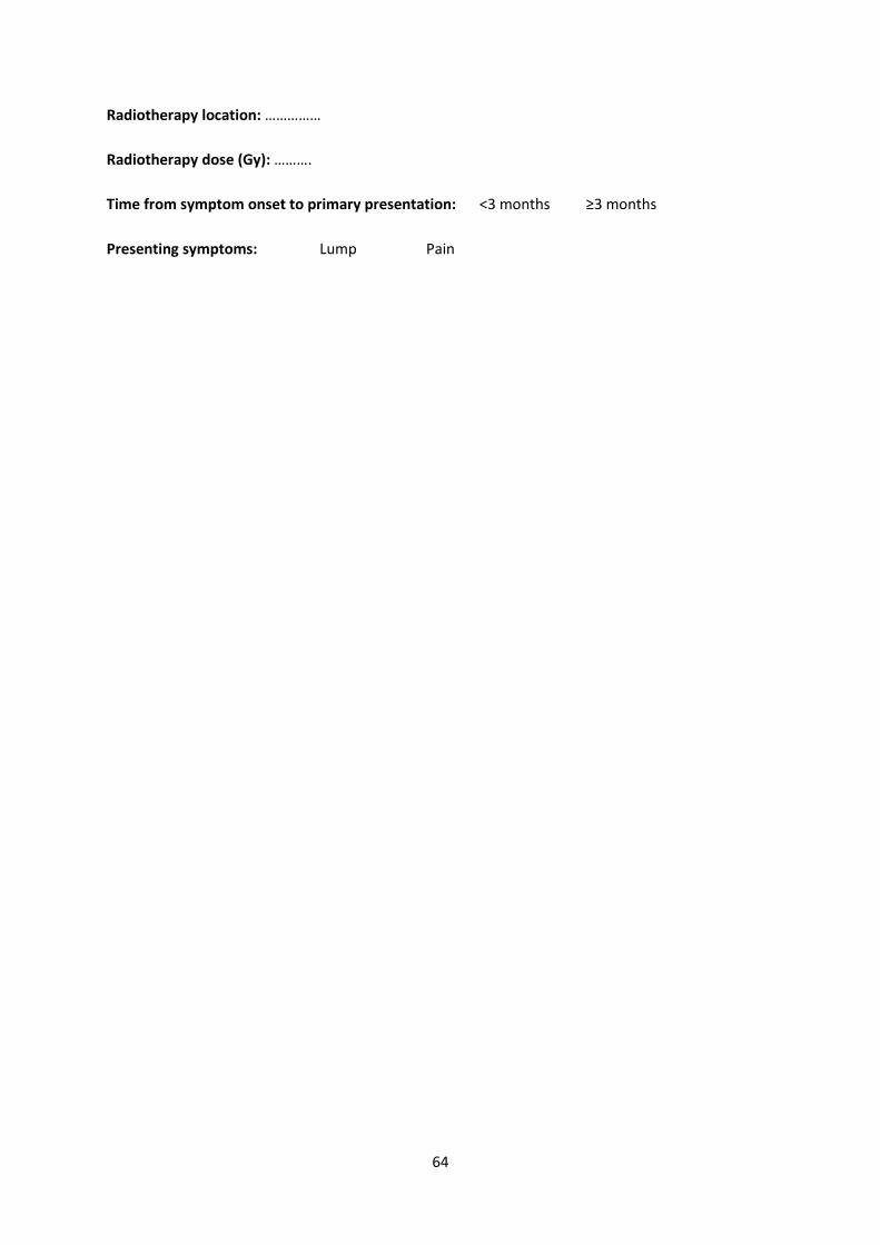
Radiotherapy location: ……………
Radiotherapy dose (Gy): ……….
Time from symptom onset to primary presentation: <3 months ≥3 months
Presenting symptoms: Lump Pain