Radiationtherapy. Radiation therapy Radiotherapy Radiation oncology.
Upload
dr-aaditya-prakashCategory
view
140download
0
OSTEOSARCOMA
Dr. AADITYA PRAKASH
DNB Resident, Radiation Oncology
BMCHRC, Jaipur

INTRODUCTION
• Osteosarcoma is most common primary bone cancer (35%).
• Osteosarcoma is most common radiation induced sarcoma.
• Osteosarcoma has a bimodal distribution as a function of age, withcases arising during the teenage years as well as cases associated withother conditions (Paget Disease, fibrous dysplasia) that arise in anolder (age >65 yrs) population.
• Osteosarcoma is more common in boys (> girls) and in blacks (>whites).

INTRODUCTION
• Osteosarcoma arises most frequently in the appendicular skeleton (80% of
cases) at the metaphyseal portions of the distal femur, tibia, and humerus.
• Osteosarcoma broadly classified in 3 histologic subtypes:-intramedullary,
surface and extra-skeletal.
• High grade intramedullary osteosarcoma (conventional /classic) refers to
the most common (80% of all cases) variant of osteosarcoma, which
typically presents within areas of rapidly proliferating skeletal bone.
• Osteosarcoma is associated with Li-Fraumeni syndrome (germline
inactivation of p53) as well as retinoblastoma.

INTRODUCTION
• Other types of less common osteosarcomas include telangiectatic,small cell, juxtacortical, periosteal, and high-grade surface sarcomas.
• Juxtacortical osteosarcoma refers to a set of more rare osteosarcomavariants that arise adjacent to the outer surface of cortical bone.
• Osteosarcoma spreads hematogenously, with the lung being mostcommon metastatic site.

CLINICAL PRESENTATION
• Pts with osteosarcoma typicallypresent with localized bone pain(often associated with an injury) ofseveral months duration and a softtissue mass/swelling.
• Osteosarcoma is derived frombone-forming mesenchyme, and isdescribed as a malignantsarcomatous stroma associatedwith the production of osteoidbone.

DISTRIBUTION OF PRIMARY & SECONDARIES
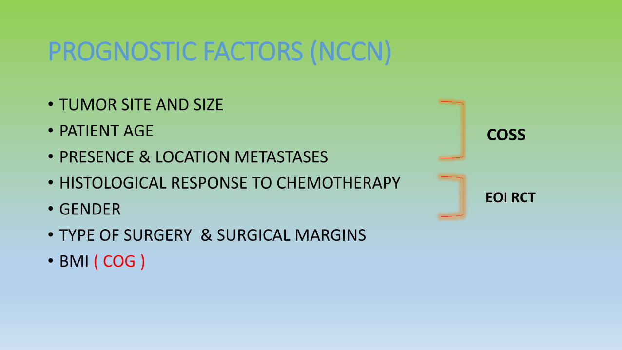
PROGNOSTIC FACTORS (NCCN)
• TUMOR SITE AND SIZE
• PATIENT AGE
• PRESENCE & LOCATION METASTASES
• HISTOLOGICAL RESPONSE TO CHEMOTHERAPY
• GENDER
• TYPE OF SURGERY & SURGICAL MARGINS
• BMI ( COG )
COSS
EOI RCT

WORKUP/STAGING• HISTORY AND PHYSICAL
EXAMINATION
• MRI +/- CT OF PRIMARY SITE
• CHEST CT SCAN
• PET SCAN AND /OR BONE SCAN
• MRI or CT OF SKELETAL METASTATIC SITES
• LDH
• ALP
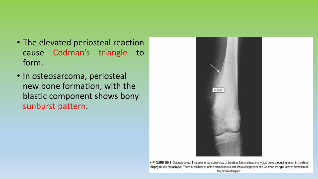
• The elevated periosteal reactioncause Codman’s triangle toform.
• In osteosarcoma, periosteal new bone formation, with the blastic component shows bony sunburst pattern.

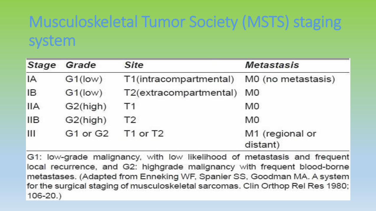
Musculoskeletal Tumor Society (MSTS) staging system
TREATMENT OF OSTEOSARCOMA
SURGERY• Complete en bloc resection of tumor is the mainstay of local
treatment of osteosarcoma.
• Surgical resection is performed either by amputation or a limb-sparing approach and has a 5% local failure rate.
• For extremity lesions, limb preservation is preferred and can beaccomplished in the majority of cases.

SURGERY
• Pelvic tumors require a hemipelvectomy for en bloc resection.
• Adjuvant radiation has been used to improve outcomes in patients withincomplete resections of pelvic tumors.
• Spinal tumors are difficult to resect with negative margins. Typically, an enbloc resection with vertebrectomy is performed, combined with mechanicalstabilization.
• Postoperative radiation therapy used when negative margins cannot beobtained, particularly when there is microscopic dural involvement.

CHEMOTHERAPY
• Chemotherapy plays an important role for all patients withintermediate- and high-grade tumors.
• Eilber et al.:-
1. 59 patients with nonmetastatic osteosarcoma randomized to surgery followed by observation versus adjuvant chemotherapy.
2. DFS at 2 years was 55% with chemotherapy and 20% with observation (p < .01).
3. OS was also superior at 2 years: 80% versus 48% with and without chemotherapy, respectively (p < .01).

CHEMOTHERAPY
• Link et al.:-
1. 36 patients with nonmetastatic, high-grade osteosarcoma randomized to observation versus adjuvant chemotherapy after primary surgery.
2. DFS at 2 years was 66% with chemotherapy and 17% with observation (p < .001).

CHEMOTHERAPY
• POG 8651 randomized patients with nonmetastatic, high-gradeosteosarcoma to neoadjuvant chemotherapy followed by surgery orsurgery followed by the same chemotherapy.
• 5-year relapse-free survival was not statistically different between thetwo groups (65% vs. 61%, respectively), nor was the rate of limbsalvage (55% vs. 50%, respectively).
• This trial did not show improved outcomes with neoadjuvantchemotherapy, it did show equivalence and established a benchmarkfor comparison with future trials.

Memorial Sloan-Kettering Cancer Center T10 regimen is frequentlyused for nonprotocol patients :- high-dose methotrexate, doxorubicin,bleomycin, cyclophosphamide, and actinomycin D.
EURAMOS I (AOST 0331):- This ongoing trial is evaluating the benefit ofadditional chemotherapy after preoperative and postoperativechemotherapy consisting of methotrexate, doxorubicin, and cisplatin.Patients with a poor response to preoperative chemotherapy arerandomized to the addition of ifosfamide and etoposide, whereas thosewith a good response to preoperative chemotherapy are randomized tothe addition of interferon.

RADIATION THERAPY
INDICATION
• Unresectable primary tumors
• Incompletely resected tumors with positive margins
• Patients who refuse surgery
• For palliation of symptomatic metastases

Radiation Therapy Techniques
• 3-D treatment planning with the aid of presurgical and postsurgical imaging isused to define gross tumor volumes and areas of subclinical disease.
• Typically, a 2-cm margin is used for axial tumors, which can be extended to 4to 5 cm for extremity tumors.
• These margins can be restricted at natural tissue and fascial boundaries.
• The radiation technique used, either 3D-CRT or IMRT, should be tailored tothe individual patient.
• Dose to uninvolved organs should be minimized to prevent late organdysfunction, as should the integral dose to minimize risk of secondarymalignancy.

DOSE
• 60 Gy in 2-Gy fractions used for microscopically involvedmargins
• 66 Gy is used for macroscopic residual disease and
• 70 Gy is used for inoperable tumors.
• Chemotherapy should not be interrupted to deliver localradiation therapy.
• Radiation can be given concurrently but is usually deliveredafter chemotherapy due to increased acute toxicity withconcurrent administration.

Cooperative Osteosarcoma Study Group (COSS)• Total of 175 pts with histologically proven osteosarcoma irradiated over the period of
1980−2007. 100 pts were eligible for analysis.
• The median age was 18 (3–66) years.
• Indication for RT was :- a primary tumor in 66, a local recurrence in 11, and
metastases in 23 pts. 94 pts got external photon therapy; 2 pts, proton therapy; 2
pts, neutron therapy; and 2 pts, intraoperative RT.
• In addition, a group of 17 pts received bone-targeted radionuclide therapy by
samarium-153-EDTMP-therapy alone or in combination with external RT.
• The median dose for external RT was 55.8 Gy (30–120). All the pts received
chemotherapy in accordance with different COSS-protocols.
• The median follow-up :- 1.5 (0.2–23) years.

Cooperative Osteosarcoma Study Group (COSS)• Survival and local control rates at 5 years were calculated.
• The overall survival rate after biopsy was 41% at 5 years, while the overallsurvival rates after RT for the whole group, for treatment of primary tumors,local recurrence, and metastases were 36%, 55%, 15%, and 0% respectively.
• Local control for the whole group was 30%. Local control rates for combinedsurgery and RT were significantly better than those for RT alone (48% vs. 22%, p= 0.002).
• Local control for treatment of primary tumors, local recurrence, and metastaseswere 40%, 17%, and 0% respectively.
• Local control for pts given an addition of samarium-153-EDTMP was poor,though not statistically significant . A dose of over 60 Gy had no significanteffect on local control.

Schwarz et al.
• Reported on an analysis of 100 patients treated with radiationtherapy in the COSS registry.
• Local control and overall survival for the whole group were 30% and36%, respectively, at 5 years.
• Local control was significantly better when surgery was combinedwith radiation compared to radiation alone: 48% vs. 22%, respectively(p = .002)
Machak et al.
• Reported on a series of 187 patients with nonmetastaticosteosarcoma treated with induction chemotherapy.
• 31 patients refused surgery and were treated with radiation to amean dose of 60 Gy.
• Local control was related to response to induction chemotherapy.There were no local recurrences in 11 patients who had a goodresponse to chemotherapy.
• However, local progression-free survival was 31% at 3 years and 0% at5 years for nonresponders.

De Laney et al.
• Reported on 41 pts with osteosarcoma who were either not resectedor were excised with close or positive margins and who underwent RTwith external beam photons (median dose of 66 Gy (10–80 Gy))and/or protons at the Massachusetts General Hospital (MGH).
• Local control rates, according to the extent of resection, were 78.4%for gross total resection, 77.8% for subtotal resection, and 40% forbiopsy only.

OTHER MODALITIES
• Intraoperative radiation therapy has been used to deliver dose directly to close or involved surgical margins.
• Radionuclide therapy with rhenium, strontium and samarium has been used for palliation of extensive bone metastases with good effects.
• Radium-223 dichloride is bone seeking radiopharmaceutical ,under investigation for treatment of metastatic or recurrent osteosarcoma. This agent is approved in US for treating bone mets assoc. with castration resistant prostate cancer.
EXTRACORPOREAL IRRADIATION (ECI)• It consists of en-bloc removal of the tumor bearing bone segment, removal of the tumor
from the bone ,irradiation, and re-implantation back in the body.
• First reported by Spira et al in 1968, there are limited reports available in the literature.
• ECI has several potential advantages.:-
1. The affected bone segment is removed from the body and irradiated and therefore,avoidance of radiation injury to the un-irradiated bone, muscles, joint, and otherhealthy tissues of the body.
2. The delivery of very high doses of radiation to tumor bearing bone by ECI, which isotherwise not possible in the intact bone. These higher doses in the range of 50-300 Gy,are lethal to the remaining tumor cells and therefore, reduce the risk of recurrence.
3. It provides an anatomically size-matched graft for biological reconstruction.
4. It is cost effective as compared to the prosthetic devices.
5. It has psychological advantage as patients feel that their own bone is being used asprosthesis.

Davidson et al (2005)
• Reported a series of 50 patients with different malignant bone tumormainly ESFT (21 patients) and OS (16 patients) using en bloc resectionand ECI (50 Gy).
• The mean time of ECI process was 35 min. With a mean follow-up of38 months (range 12-92), 84% patients were alive without anydisease and only 8% developed LR. The mean MSTS score was 77.

Poffyn et al (2011)
• Recently published a retrospective analysis of 107 patients with 108malignant or locally aggressive bone tumors treated by ECI with 300Gy, and re-implantation of the bone as an orthotopic autograft.
• At 5 year follow-up, there was no LR and 64% of patients had wellhealed graft. The 0% LR rate could be due to relatively very high doseof ECI (300 Gy) used in their study.

• radiological images and operativephotographs of a 10 year old boy diagnosedwith osteosarcoma of lower end of rightfemur that underwent extracorporealirradiation (ECI).
• (a) (plain radiograph) and
• (b) (reconstructed computed tomographyscan) showing the tumor at the lower end offemur.
• (c) the resected bone segment from whichthe tumor has been removed. The same waspackaged and sent for ECI.
• (d) the operated limb after the resection ofthe bone segment


THANK YOU