Research Article Protective Effect of Huoxiang Zhengqi...
11
Research Article Protective Effect of Huoxiang Zhengqi Oral Liquid on Intestinal Mucosal Mechanical Barrier of Rats with Postinfectious Irritable Bowel Syndrome Induced by Acetic Acid Yao Liu, 1,2 Wei Liu, 1 Qiu-Xian Peng, 1,3 Jiang-Li Peng, 4 Lin-Zhong Yu, 1 and Jian-Lan Hu 1 1 School of Traditional Chinese Medicine, Southern Medical University, Guangzhou 510515, China 2 New Drug Research and Development Center, Guangdong Institute of Traditional Chinese Medicine, Guangzhou 510520, China 3 Department of Biology, Hong Kong Baptist University, Kowloon Tong, Hong Kong 4 College of Pharmacy, Hunan University of Traditional Chinese Medicine, Changsha 410028, China Correspondence should be addressed to Lin-Zhong Yu; [email protected] Received 4 June 2014; Revised 22 July 2014; Accepted 6 August 2014; Published 28 August 2014 Academic Editor: Roja Rahimi Copyright © 2014 Yao Liu et al. is is an open access article distributed under the Creative Commons Attribution License, which permits unrestricted use, distribution, and reproduction in any medium, provided the original work is properly cited. In this study, a rat model with acetic acid-induced PI-IBS was used to study the role of HXZQ oral liquid in repairing the colonic epithelial barrier and reducing intestinal permeability. Pathomorphism of colonic tissue, epithelial ultrastructure, DAO activity in serum, and the protein expression of ZO-1 and occludin were examined to investigate protective effect mechanisms of HXZQ on intestinal mucosa barrier and then present experimental support for its use for prevention and cure of PI-IBS. 1. Introduction Postinfectious irritable bowel syndrome (PI-IBS) is a com- mon disorder wherein symptoms of IBS begin aſter an episode of acute gastroenteritis. Published studies have reported incidence of PI-IBS that ranged between 5% and 32% [1]. Its high prevalence and the considerable effects on quality of life make PI-IBS a disease with high social cost. e majority of cases of PI-IBS meet the Rome II criteria for diarrhea-predominant irritable bowel syndrome (IBS-D), with predominantly loose stool passed more frequently than normal and with urgency [2]. e causes and underlying mechanisms of PI-IBS are still not fully understood, although it is believed that altered gut flora, changed intestinal perme- ability, activated gut immunity, and functional or structural changes in enteric nervous system are important factors [3– 7]. Recent studies have suggested that increased intestinal permeability could be an important factor in the sequence of events leading to low-grade intestinal inflammation and disturbed bowel function [8, 9]. Intestinal epithelial perme- ability is regulated by a complex protein system comprising tight junction (TJ) and adherens junction proteins [10]. is increased permeability could be due to alterations of tight junction proteins. In addition, the changes of the ultrastructure of intestinal mucosal epithelial cells may be associated with the pathogenesis of PI-IBS [11]. To date, there are no successful treatments for PI-IBS, and management of PI-IBS primarily aims at relieving the symptoms [12, 13]. Natural medicine is generally considered safe and with few adverse drug reactions. As a representative natural medicine, traditional Chinese medicine (TCM) is more and more widely used nowadays. HXZQ oral liquid is a Chinese patent medicine composed of 10 kinds of Chinese medical medicines. It was modified based on a traditional formula named Huoxiang Zhengqi powder, which originated from the ancient Chinese pharmacy book “Formularies of the Bureau of the People’s Welfare Pharmacies” (Taiping Huimin Hejiju Fang) in the Song Dynasty of China; it has been mainly used to treat gastrointestinal diseases for more than 1000 years. Clinical researches have proved that HXZQ has definite clin- ical effect for treating IBS-D [14]. Pharmacological research has shown that HXZQ can regulate gastrointestinal motility Hindawi Publishing Corporation Evidence-Based Complementary and Alternative Medicine Volume 2014, Article ID 218383, 10 pages http://dx.doi.org/10.1155/2014/218383
Transcript of Research Article Protective Effect of Huoxiang Zhengqi...
Research ArticleProtective Effect of Huoxiang Zhengqi Oral Liquid on IntestinalMucosal Mechanical Barrier of Rats with Postinfectious IrritableBowel Syndrome Induced by Acetic Acid
Yao Liu,1,2 Wei Liu,1 Qiu-Xian Peng,1,3 Jiang-Li Peng,4 Lin-Zhong Yu,1 and Jian-Lan Hu1
1 School of Traditional Chinese Medicine, Southern Medical University, Guangzhou 510515, China2New Drug Research and Development Center, Guangdong Institute of Traditional Chinese Medicine, Guangzhou 510520, China3Department of Biology, Hong Kong Baptist University, Kowloon Tong, Hong Kong4College of Pharmacy, Hunan University of Traditional Chinese Medicine, Changsha 410028, China
Correspondence should be addressed to Lin-Zhong Yu; [email protected]
Received 4 June 2014; Revised 22 July 2014; Accepted 6 August 2014; Published 28 August 2014
Academic Editor: Roja Rahimi
Copyright © 2014 Yao Liu et al. This is an open access article distributed under the Creative Commons Attribution License, whichpermits unrestricted use, distribution, and reproduction in any medium, provided the original work is properly cited.
In this study, a rat model with acetic acid-induced PI-IBS was used to study the role of HXZQ oral liquid in repairing the colonicepithelial barrier and reducing intestinal permeability. Pathomorphism of colonic tissue, epithelial ultrastructure, DAO activity inserum, and the protein expression of ZO-1 and occludin were examined to investigate protective effect mechanisms of HXZQ onintestinal mucosa barrier and then present experimental support for its use for prevention and cure of PI-IBS.
1. Introduction
Postinfectious irritable bowel syndrome (PI-IBS) is a com-mon disorder wherein symptoms of IBS begin after anepisode of acute gastroenteritis. Published studies havereported incidence of PI-IBS that ranged between 5% and32% [1]. Its high prevalence and the considerable effects onquality of life make PI-IBS a disease with high social cost.The majority of cases of PI-IBS meet the Rome II criteriafor diarrhea-predominant irritable bowel syndrome (IBS-D),with predominantly loose stool passed more frequently thannormal and with urgency [2]. The causes and underlyingmechanisms of PI-IBS are still not fully understood, althoughit is believed that altered gut flora, changed intestinal perme-ability, activated gut immunity, and functional or structuralchanges in enteric nervous system are important factors [3–7]. Recent studies have suggested that increased intestinalpermeability could be an important factor in the sequenceof events leading to low-grade intestinal inflammation anddisturbed bowel function [8, 9]. Intestinal epithelial perme-ability is regulated by a complex protein system comprising
tight junction (TJ) and adherens junction proteins [10].This increased permeability could be due to alterations oftight junction proteins. In addition, the changes of theultrastructure of intestinal mucosal epithelial cells may beassociated with the pathogenesis of PI-IBS [11]. To date, thereare no successful treatments for PI-IBS, and management ofPI-IBS primarily aims at relieving the symptoms [12, 13].
Natural medicine is generally considered safe and withfew adverse drug reactions. As a representative naturalmedicine, traditional Chinese medicine (TCM) is more andmore widely used nowadays. HXZQ oral liquid is a Chinesepatent medicine composed of 10 kinds of Chinese medicalmedicines. It was modified based on a traditional formulanamedHuoxiangZhengqi powder, which originated from theancient Chinese pharmacy book “Formularies of the Bureauof the People’s Welfare Pharmacies” (Taiping Huimin HejijuFang) in the Song Dynasty of China; it has been mainly usedto treat gastrointestinal diseases for more than 1000 years.Clinical researches have proved that HXZQ has definite clin-ical effect for treating IBS-D [14]. Pharmacological researchhas shown that HXZQ can regulate gastrointestinal motility
Hindawi Publishing CorporationEvidence-Based Complementary and Alternative MedicineVolume 2014, Article ID 218383, 10 pageshttp://dx.doi.org/10.1155/2014/218383
2 Evidence-Based Complementary and Alternative Medicine
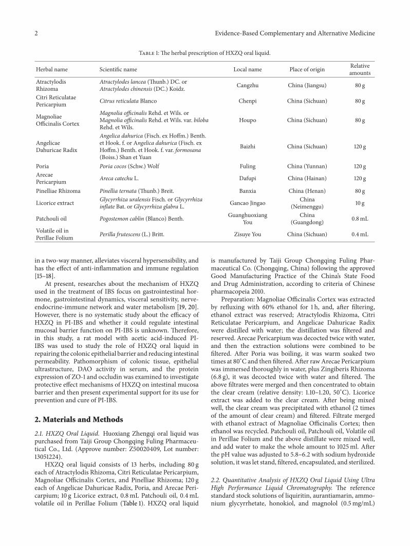
Table 1: The herbal prescription of HXZQ oral liquid.
Herbal name Scientific name Local name Place of origin Relativeamounts
AtractylodisRhizoma
Atractylodes lancea (Thunb.) DC. orAtractylodes chinensis (DC.) Koidz. Cangzhu China (Jiangsu) 80 g
Citri ReticulataePericarpium Citrus reticulata Blanco Chenpi China (Sichuan) 80 g
MagnoliaeOfficinalis Cortex
Magnolia officinalis Rehd. et Wils. orMagnolia officinalis Rehd. et Wils. var. bilobaRehd. et Wils.
Houpo China (Sichuan) 80 g
AngelicaeDahuricae Radix
Angelica dahurica (Fisch. ex Hoffm.) Benth.et Hook. f. or Angelica dahurica (Fisch. exHoffm.) Benth. et Hook. f. var. formosana(Boiss.) Shan et Yuan
Baizhi China (Sichuan) 120 g
Poria Poria cocos (Schw.) Wolf Fuling China (Yunnan) 120 gArecaePericarpium Areca catechu L. Dafupi China (Hainan) 120 g
Pinelliae Rhizoma Pinellia ternata (Thunb.) Breit. Banxia China (Henan) 80 g
Licorice extract Glycyrrhiza uralensis Fisch. or Glycyrrhizainflate Bat. or Glycyrrhiza glabra L. Gancao Jingao China
(Neimenggu) 10 g
Patchouli oil Pogostemon cablin (Blanco) Benth. GuanghuoxiangYou
China(Guangdong) 0.8mL
Volatile oil inPerillae Folium Perilla frutescens (L.) Britt. Zisuye You China (Sichuan) 0.4mL
in a two-way manner, alleviates visceral hypersensibility, andhas the effect of anti-inflammation and immune regulation[15–18].
At present, researches about the mechanism of HXZQused in the treatment of IBS focus on gastrointestinal hor-mone, gastrointestinal dynamics, visceral sensitivity, nerve-endocrine-immune network and water metabolism [19, 20].However, there is no systematic study about the efficacy ofHXZQ in PI-IBS and whether it could regulate intestinalmucosal barrier function on PI-IBS is unknown. Therefore,in this study, a rat model with acetic acid-induced PI-IBS was used to study the role of HXZQ oral liquid inrepairing the colonic epithelial barrier and reducing intestinalpermeability. Pathomorphism of colonic tissue, epithelialultrastructure, DAO activity in serum, and the proteinexpression of ZO-1 and occludin was examined to investigateprotective effect mechanisms of HXZQ on intestinal mucosabarrier and then present experimental support for its use forprevention and cure of PI-IBS.
2. Materials and Methods
2.1. HXZQ Oral Liquid. Huoxiang Zhengqi oral liquid waspurchased from Taiji Group Chongqing Fuling Pharmaceu-tical Co., Ltd. (Approve number: Z50020409, Lot number:13051224).
HXZQ oral liquid consists of 13 herbs, including 80 geach of Atractylodis Rhizoma, Citri Reticulatae Pericarpium,Magnoliae Officinalis Cortex, and Pinelliae Rhizoma; 120 geach of Angelicae Dahuricae Radix, Poria, and Arecae Peri-carpium; 10 g Licorice extract, 0.8mL Patchouli oil, 0.4mLvolatile oil in Perillae Folium (Table 1). HXZQ oral liquid
is manufactured by Taiji Group Chongqing Fuling Phar-maceutical Co. (Chongqing, China) following the approvedGood Manufacturing Practice of the China’s State Foodand Drug Administration, according to criteria of Chinesepharmacopeia 2010.
Preparation: Magnoliae Officinalis Cortex was extractedby refluxing with 60% ethanol for 1 h, and, after filtering,ethanol extract was reserved; Atractylodis Rhizoma, CitriReticulatae Pericarpium, and Angelicae Dahuricae Radixwere distilled with water; the distillation was filtered andreserved. Arecae Pericarpium was decocted twice with water,and then the extraction solutions were combined to befiltered. After Poria was boiling, it was warm soaked twotimes at 80∘C and then filtered. After rawArecae Pericarpiumwas immersed thoroughly in water, plus Zingiberis Rhizoma(6.8 g), it was decocted twice with water and filtered. Theabove filtrates were merged and then concentrated to obtainthe clear cream (relative density: 1.10–1.20, 50∘C). Licoriceextract was added to the clear cream. After being mixedwell, the clear cream was precipitated with ethanol (2 timesof the amount of clear cream) and filtered. Filtrate mergedwith ethanol extract of Magnoliae Officinalis Cortex; thenethanol was recycled. Patchouli oil, Patchouli oil, Volatile oilin Perillae Folium and the above distillate were mixed well,and add water to make the whole amount to 1025ml. Afterthe pH value was adjusted to 5.8–6.2 with sodium hydroxidesolution, it was let stand, filtered, encapsulated, and sterilized.
2.2. Quantitative Analysis of HXZQ Oral Liquid Using UltraHigh Performance Liquid Chromatography. The referencestandard stock solutions of liquiritin, aurantiamarin, ammo-nium glycyrrhetate, honokiol, and magnolol (0.5mg/mL)

Evidence-Based Complementary and Alternative Medicine 3
were prepared in methanol and stored at <4∘C.The standardsolutions were prepared using five concentrations of dilutedsolutions (methanol). All the calibration curves were gener-ated by assessing the peak areas for the five concentrationsin the range of 0.0017∼0.128𝜇g for all standard samples. Thelinearity of the peak area (𝑦) versus concentration (𝑥, 𝜇g/mL)curve for each component was used to calculate the contentsof the main components in HXZQ oral liquid. Quantitativeanalysis was performed under concurrent conditions usingan 1290 infinity high-performance liquid chromatography(UPLC; Agilent Technologies) equipped with an autosampler(G1367E), column oven (G1316C), binary pump (G4220A),and DAD (G4212A).The analytical column comprised a Zor-bax RRHD Eclipse C18 (2.1 × 100mm; particle size, 1.8𝜇m)and was maintained at 30∘C. The data were acquired andprocessed by chemstation software (Agilent Technologies).The mobile phase comprised acetonitrile (A) with 0.05%phosphate acid (B).The gradient flowwas as follows: 0–5min,17–20% A; 5–12min, 20–50% A; 12–18min, 50–70% A; 18-18.5min, 70–95% A; 18.5-23.5min, 95% A. The analysis wascarried out at a flow rate of 0.6mL/min, and detection wasperformed at a wavelength of 220 nm. The injection volumewas 1 𝜇L.
2.3. Positive Control Drug. Trimebutine maleate tablets werepurchased from Kaikai Yuansheng Medicine Co., Ltd. (Lotnumber: 13032817).
2.4. Animals. Sprague-Dawley (SD) rats (250–270 g, 2months old) were purchased from the Laboratory AnimalCentre of Southern Medical University (License: SCXKGuangdong 2006-0015) and were acclimated in the facilityfor 1 week before being used in the experiments. The animalswere housed at 25 ± 1∘C with a 12:12 h light/dark cycle(lights on at 7:00 a.m.) and were allowed to access waterand food. All procedures in this study were conducted inaccordance with the NIH Guide for the Care and Use ofLaboratory Animals, and the experiments were approvedby the Laboratory Animal Ethics Committee of SouthernMedical University.
2.5. Experimental Design
2.5.1. Grouping and Administration. The rats were randomlydivided into six groups including normal control group, PI-IBS model group, trimebutine maleate tablet group (TMTgroup), Huoxiang Zhengqi low-dose group (low-dose ofHXZQ), middle-dose group (middle-dose of HXZQ), andhigh dose group (high-dose of HXZQ) with 10 rats in eachgroup and treated with water (control group and PI-IBSmodel group), trimebutine maleate tablets (0.1 g/kg/d, TMTgroup), and HXZQ oral liquid (1.7, 3.3, and 6.6mL/kg/d, low-dose, middle-dose, and high dose of HXZQ). Administrationdose of HXZQ oral liquid was established according tothe usage and dosage criteria of Chinese pharmacopeia2010. At the seventh day after acetic acid administration,agent was administered to each group through intragastricadministration for 5 days.
2.5.2. PI-IBS Animal Model Establishment. The PI-IBS ratmodel was developed according to previous report [21]. Afteran overnight fast, the rats were anesthetized with ether, and aplastic catheter (external diameter = 0.96mm) was insertedinto the descending colon at the depth of 8 cm from anus;then acetic acid (4%, 1mL/rat) aqueous solution was instilledslowly for 30 s. Then, 1mL phosphate buffered saline (PBS)was instilled to dilute the acetic acid and flush the colon.The normal control group animals were handled identicallyexcept that 1mL saline was instilled instead of 4% acetic acid.
2.5.3. Sample Collection. On the day after the last treat-ment, the rats were sacrificed. A 6 cm long proximal colon(1-2 cm from caecum) was harvested and divided into 3parts. The proximal was fixed in 4% paraformaldehyde andembedded in paraffin for histopathological observation; thenimmunohistochemical detection was conducted to observethe expression of ZO-1 and occludin; the middle part wascollected for microscopic evaluation by TEM; the distal partwas proceeded for Western blotting analysis. Abdominalaortic blood was collected for DAO assay in serum.
2.5.4. Histological Examination of Colon Tissue. A 1 cm sec-tion of fresh colon tissues were fixed in 4% paraform for12 h, then dehydrated in 50%, 60%, 70%, 80%, 90%, and100% alcohol separately, and embedded in paraffin at 56–60∘C. The samples were cut into sections (4 𝜇m for each),deparaffinized in dimethyl benzene, gradiently dehydratedin alcohol and stained with hematoxylin, eosin (H&E),dehydrated in 70%, 90%, and 95% ethanol, and finally clearedin xylene. Pathological histology slice of colonic tissue wasobserved under a light microscope (original magnification×400).
2.5.5. Ultrastructural Observation by TEM. The colon seg-ments were cut into 1mm3 strips, fixed for 4 h at 4∘C in 3%glutaraldehyde, fully washed for 3 times in 0.1mol/L PBS,postfixed for 2 h at 4∘C in 2% osmium tetroxide, dehydratedin a graded series of ethanols, embedded in Epon 812, cutinto ultrathin sections (75 nm), and then stained with uranylacetate and lead citrate. Sections were viewed in a HITACHIH-600 electron microscope at 60 kV (HITACHI, Tokyo,Japan).
2.5.6. ELISAAssay of DAO. Serumof abdominal aortic bloodwas separated and stored at −80∘C until use. DAO level wasdetermined using ELISA kits according to the protocol asdescribed previously [22].
2.5.7. Immunohistochemistry for ZO-1 and Occludin Expres-sion. The tissue sections were rinsed in PBS (pH.7.4) andplaced in polycarbonate staining jars (Kartell, Italy) filledwith10mM sodium citrate buffer solution (pH.6.0). After beingexposed to microwave for 15min to retrieve antigen, thenthe tissues were incubated in a solution of 3% bovine serumalbumin, 0.5% Triton X-100 in PBS. Sections were incubatedwith rabbit anti-zo-1 antibody (1 : 200, Millipore) and rabbitanti-occludin antibody (1 : 200, abcam), respectively, in PBS

4 Evidence-Based Complementary and Alternative Medicine
Table 2: Quantitative analysis of HXZQ oral liquid and its reference compounds.
Compound Retention time (min) Mean (g/L) SDLiquiritin 2.52 0.045 3.53Aurantiamarin 5.02 0.18 5.52Ammonium glycyrrhetate 10.85 0.66 1.05Honokiol 14.02 0.19 3.33Magnolol 15.32 0.20 2.09
containing 0.5% Triton X-100 overnight at 4∘C. After rinsedin PBS, secondary antibody (polyperoxidase rabbit IgG,Zhongshan Biotechnology Co. Ltd.) was incubated for 2 hat room temperature, and then rinsed and cover-slipped.Sections were observed with a microscope (Nikon 80, Japan),captured with 400x, and analyzed using Image-Pro Plus 6.0software. The optical densities in colonic mucosa from 8random fields per section were calculated.
2.5.8. Western Blotting for ZO-1 and Occludin Expression.The colonic tissues were homogenized and sonicated onice for protein extraction. After the protein was quantified,lysates were denatured at 100∘C for 5min. An equal amountof protein (30mg/well) was electrophoresed on 10% SDS-PAGE gel. The proteins were then electroblotted to PVDFmembranes (Bio-Rad) and blockedwith 5%nonfat skimmilkfor 1 h. Subsequently, the blot was incubated in rabbit anti-ZO-1 and rabbit anti-occludin antibody (1 : 1000, Santa Cruz,CA) supplemented with 2% nonfat skim milk for 2 h at roomtemperature. After washing, the membrane was incubated inhorseradish peroxidase- (HRP-) conjugated secondary goatanti- rabbit antibodies (1 : 5000, Zymed Laboratories) in 2%nonfat skim milk for an hour at room temperature. Bandson the membranes were visualized using ECL (Amersham,UK). Distinct immunoreaction products were revealed andimages of the bands were developed on film (Biomax X-rayfilm, Kodak). To check the amount of protein loaded, theimmunoblots were treated with stripping solution (62.5mMTris buffer, pH 6.7, containing 2% SDS and 100mM b-mercaptoethanol) for 30min at 50∘C and incubated withmouse monoclonal anti-𝛽-actin antibody (Sigma) followedby horseradish peroxidase-coupled goat anti-mouse IgG(Pierce). Optical densities of the individual bands werescanned and quantified using NIH Image J. All detectedprotein bands were normalized to 𝛽-action levels.
2.6. Statistical Analysis. Statistical analysis was performedusing SPSS 13.0 software. Data were expressed as the mean ±standard error (SE). One-way ANOVA tests were performedto assess the differences in the cytokine expression levels. Apost-hoc Bonferroni test was used to assess the differencesbetween the individual groups. 𝑃 < 0.05 was consideredsignificant.
3. Results
3.1. Fingerprint Analysis of HXZQ Oral Liquid. The standardcurves for the five components containing liquiritin, auran-tiamarin, ammonium glycyrrhetate, honokiol, and magnolol
were 𝑦 = 36251𝑥 − 15.336 (𝑟 = 0.9999), 𝑦 = 26551𝑥 − 11.451(𝑟 = 1.000), 𝑦 = 2488𝑥 − 11.552 (𝑟 = 0.9999), 𝑦 =81440𝑥 − 51.221 (𝑟 = 0.9999), and 𝑦 = 91221𝑥 − 11.635(𝑟 = 0.9993), respectively. The components obtained fromUPLC analysis of HXZQ oral liquid and standard mixtureswere detected at 220 nm. The retention times of each com-ponent were 2.52min (liquiritin), 5.02min (aurantiamarin),10.85min (ammonium glycyrrhetate), 14.02min (honokiol),and 15.32min (magnolol). The contents of each componentwere in the range of 0.045∼0.66 g/L (Table 2).
3.2. Histopathology of Colonic Tissues. According to theobservation, the structure of the intestinal mucosa in con-trol group was intact, the epithelium was normal, simplecolumnar epithelium and enteraden in colonic mucosa wereordered arrangement, and goblet cells were rich. A smallamount of plasma cells within the interstitial were observed.In low-dose of HXZQ, middle-dose of HXZQ, and high-dose of HXZQ groups, histological features were in accordwith control group, and no overt inflammatory infiltrationof neutrophils in the lamina propria and interstitial edemawas observed. However, epithelial cell sloughing off and fewneutrophil infiltrationswere observed in PI-IBSmodel group.
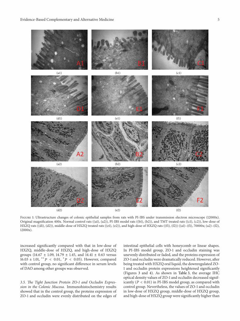
3.3. Ultrastructure of Rat Colonic Epithelial Tissue. In controlgroup, rat intestinal epithelial cell microvilli were arrangedin neat rows. The epithelial cell membrane and the nuclearmembrane were integral.The nucleolus could be seen clearly.In the cytoplasm, both the endoplasmic reticulum and mito-chondria could be seen clearly. The junction of the epithelialcells was tight (Figure 1 (a1), (a2)). However, in PI-IBSmodel group, the microvilli on the surface of epithelial cellsof the intestine were sparse or distributed irregularly withdifferent lengths.There was degeneration or necrosis in a fewintestinal epithelial cells. The junctions among the cells werenot broadened obviously. In addition, mast cells were filledwith high-density particles and some vacuoles in cytoplasmand showed active degranulation state (Figure 1 (b1), (b2)).In TMT group, microvilli showed slight dropout and tightjunctions were unclear in a few of the epithelial cells (Figure 1(c1), (c2)). In low-dose of HXZQ, middle-dose of HXZQ, andhigh-dose of HXZQ groups, the epithelial cell membraneswere intact, the cellular shapes were overall normal, and theregular distribution ofmicrovilli were observed. To conclude,the colonic epithelial ultrastructure was better maintainedcompared to PI-IBS model group (Figure 1).
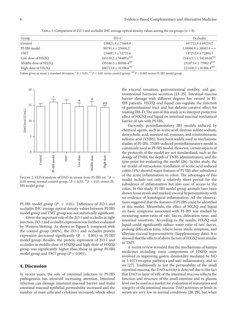
3.4. Diamine Oxidase (DAO) Activity. Figure 2 shows thatthe levels of serum DAO activity in PI-IBS model group
Evidence-Based Complementary and Alternative Medicine 5
(a1)
(a2)
(b1)
(b2)
(c1)
(c2)
(d1)
(d2)
(e1)
(e2)
(f1)
(f2)
Figure 1: Ultrastructure changes of colonic epithelial samples from rats with PI-IBS under transmission electron microscope (12000x).Original magnification 400x. Normal control rats ((a1), (a2)), PI-IBS model rats ((b1), (b2)), and TMT treated rats ((c1), (c2)), low-dose ofHXZQ rats ((d1), (d2)), middle-dose of HXZQ treated rats ((e1), (e2)), and high-dose of HXZQ rats ((f1), (f2)) ((a1)–(f1), 70000x; (a2)–(f2),12000x).
increased significantly compared with that in low-dose ofHXZQ, middle-dose of HXZQ, and high-dose of HXZQgroups (14.67 ± 1.09, 14.79 ± 1.45, and 14.41 ± 0.63 versus16.03 ± 1.01, ∗∗𝑃 < 0.01, ∗𝑃 < 0.05). However, comparedwith control group, no significant difference in serum levelsof DAO among other groups was observed.
3.5. The Tight Junction Protein ZO-1 and Occludin Expres-sion in the Colonic Mucosa. Immunohistochemistry resultsshowed that in the control group, the proteins expression ofZO-1 and occludin were evenly distributed on the edges of
intestinal epithelial cells with honeycomb or linear shapes.In PI-IBS model group, ZO-1 and occludin staining wasunevenly distributed or faded, and the proteins expression ofZO-1 and occludin were dramatically reduced. However, afterbeing treated withHXZQ oral liquid, the downregulated ZO-1 and occludin protein expressions heightened significantly(Figures 3 and 4). As shown in Table 3, the average IHCoptical density values of ZO-1 and occludin decreased signif-icantly (𝑃 < 0.01) in PI-IBS model group, as compared withcontrol group. Nevertheless, the values of ZO-1 and occludinin low-dose of HXZQ group, middle-dose of HXZQ group,and high-dose of HXZQ group were significantly higher than
6 Evidence-Based Complementary and Alternative Medicine
Table 3: Comparison of ZO-1 and occludin IHC average optical density values among the six groups (𝑛 = 8).
Group ZO-1 OccludinControl 158821.4 ± 75669.8 167713.9 ± 69214.7
PI-IBS model 90791.4 ± 25604.2∗
118096.9 ± 30682.4 ∗ ∗
TMT 134687.3 ± 52725.6 147252.4 ± 72804.5
Low-dose of HXZQ 165330.2 ± 58489.6###
214315.1 ± 54218.04###
Middle-dose of HXZQ 195346.5 ± 60506.4###
232473.4 ± 73962.4###
High-dose of HXZQ 206713.8 ± 41338.6###
221420.3 ± 81806.4###
Values given as mean ± standard deviation. ∗𝑃 < 0.05, ∗∗𝑃 < 0.01 versus control group; ###𝑃 < 0.001 versus PI-IBS model group.
0
5
10
15
20
DAO
leve
l (U
/mL)
Con
trol
PI-I
BS m
odel
TMT
Low
dos
e
Mid
dle d
ose
Hig
h do
se
∗# #
##
Figure 2: ELISA analysis of DAO in serum from PI-IBS rat. ∗𝑃 <0.05 versus normal control group; #𝑃 < 0.05, ##𝑃 < 0.01 versus PI-IBS model group.
PI-IBS model group (𝑃 < 0.01). Difference of ZO-1 andoccludin IHC average optical density values between PI-IBSmodel group and TMT group was not statistically significant.
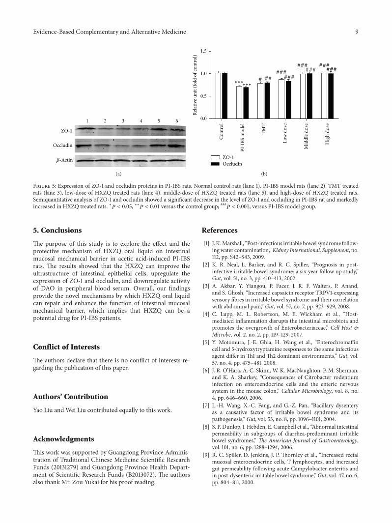
Given the important role of the ZO-1 and occludin in tightjunction, ZO-1 and occludin expressionwas further evaluatedby Western blotting. As shown in Figure 5, compared withthe control group (100%), the ZO-1 and occludin proteinexpression decreased significantly (𝑃 < 0.001) in PI-IBSmodel group. Besides, the protein expression of ZO-1 andoccludin in middle-dose of HXZQ and high-dose of HXZQgroup was significantly higher than those in group PI-IBSmodel group and TMT group (𝑃 < 0.001).
4. Discussion
In recent years, the role of intestinal infection in PI-IBSpathogenesis has attracted increasing attention. Intestinalinfection can damage intestinal mucosal barrier and makeintestinal mucosal epithelial permeability increased and thenumber of mast cells and cytokines increased, which affect
the visceral sensation, gastrointestinal motility, and gas-trointestinal hormone secretion [23–25]. Intestinal mucosabarrier damage with different degrees has existed in PI-IBS patients. HXZQ oral liquid can regulate the functionof gastrointestinal tract and has definite curative effect fortreating IBS-D.The aim of this study is to interpret protectiveeffect of HXZQ oral liquid on intestinal mucosal mechanicalbarrier of rats with PI-IBS.
Currently, postinflammatory IBS models induced bychemical agents, such as acetic acid, dextran sulfate sodium,deoxycholic acid, mustard oil, zymosan, and trinitrobenzenesulfonic acid (TNBS), have been widely used in mechanisticstudies of PI-IBS. TNBS-induced postinflammatory model iscommonly used as PI-IBSmodel. However, certain aspects ofthe protocols of the model are not standardized, such as thedosage of TNBS, the depth of TNBS administration, and thetime point for evaluating the model [26]. In this study, therat model of intracolonic instillation of acetic acid-inducedcolitis (4%) showed major features of PI-IBS after subsidenceof the acute inflammation in colon. The advantages of thismodel include not only a relatively short period for thesubsidence of inflammation but also ease of access to thecolon. In this study, PI-IBS model group animals have beenshown loose stools and marked visceral hypersensitivity withno evidence of histological inflammation. All the observa-tions suggested that the features of PI-IBS could be identifiedin this model. Meanwhile, the effect of HXZQ oral liquidon basic symptoms associated with PI-IBS was studied bymeasuring water ratio of rats’ faeces, defecation time, andintestinal sensitivity. According to the results, HXZQ oralliquid could significantly reduce water ratio of rats’ faeces,prolong defecation time, relieve loose stools symptom, andalleviate visceral hypersensitivity (Supplementary data). It isshowed that the effects of above factors ofHXZQwere similarto TMT.
A recent review revealed that the mechanisms of kampomedicines including some components of HXZQ wereinvolved in improving gastric dysmotility mediated by NOor 5-HT3 receptor pathways and anti-inflammatory, and soon [27]. Traditionally to test the permeability of the smallintestinal mucosa, the DAO activity is detected due to the factthat DAO in layer of villi of the intestinal mucosa reflects thefunction and structure of the small intestine and its plasmalevel can be used as amarker for evaluation ofmaturation andintegrity of the intestinal mucosa. DAO activities or levels inserum are very low in normal conditions [28, 29], but when

Evidence-Based Complementary and Alternative Medicine 7
(a) (b)
(c) (d)
(e) (f)
Figure 3: Immunohistochemical analysis of occludin in colonic mucosa from rats with PI-IBS. Original magnification 400x. Normal controlrats (a), PI-IBS model rats (b), and TMT treated rats (c), low-dose of HXZQ rats (d), middle-dose of HXZQ treated rats (e), and high-doseof HXZQ rats (f). Semiquantitative analysis of occludin-staine areas showed a significant decrease in rats with PI-IBS, and HXZQ couldsignificantly increase these areas.
the intestinal mucosal epithelial cells are damaged, DAO willbe released, and large amount of DAOwill enter the systemiccirculation [30]. DAO is an intracellular enzyme with highactivity existing in intestinal villous cells in mammalians,especially high in the jejunum and ileum [31]. The activityof DAO in intestinal mucosa decreases when the epitheliumare injured; thus DAO activity of intestinal mucosa canindicate the changes in its cellular integrity. In this study,the serum DAO levels of PI-IBS model group were signifi-cantly increased compared with those in HXZQ groups. Nosignificant difference of serum DAO levels between HXZQgroups andNCgroupwas detected.Our results demonstratedthat small intestinal mucosal permeability of the rats with PI-IBS was increased slightly. Therefore, the protective effect ofHXZQ on colonic mucosa in PI-IBS rats may be caused byreducing the intestinal mucosal permeability and/or improv-ing the integrity of the intestinal mucosa. Moreover, the his-tomorphological change of colon mucosa was not observed
in PI-IBS rats, but the changes of the ultrastructure of cellshave been observed. For example, the microvilli distributedunevenly with various lengths, and there was degeneration ornecrosis in few intestinal epithelial cells. These observationsindicated that intestinal mucosamechanical barrier of PI-IBSrats was impaired and intestinal permeability was increased.After administration of HXZQ, the ultrastructure of mucosalepithelial cell was improved, which demonstrates that HXZQcan repair the ultrastructure of the damaged cell, decreasethe permeability, and inhibit inflammatory reaction, thusimproving the intestinal mucosal barrier function.
The intestinal barrier function was maintained by aseries of epithelial TJs, protecting the body from pathogensand other toxic luminal substances. Transmembrane pro-teins (such as occludin, tricellulin, claudins, and junctionaladhesion molecule) interact with cytoplasmic peripheralmembrane proteins (such as ZO-1, ZO-2, ZO-3, and cingulin)that constitute the frames of tight junctions [32]. Recent

8 Evidence-Based Complementary and Alternative Medicine
(a) (b)
(c) (d)
(e) (f)
Figure 4: Immunohistochemical analysis of ZO-1 in colonic mucosa from rats with PI-IBS. Original magnification 400x. Normal control rats(a), PI-IBS model rats (b), and TMT treated rats (c), low-dose of HXZQ rats (d) rats, middle-dose of HXZQ treated rats (e), and high-dose ofHXZQ rats (f). Semiquantitative analysis of ZO-1-staine areas showed a significant decrease in rats with PI-IBS, andHXZQ could significantlyincrease these areas.
studies have proved that increased permeability of the tightjunctions is thought to contribute to PI-IBS [25, 33].The tightjunction seals the space between adjacent epithelial cells, and,in intact gastrointestinal epithelia, tight junction permeabilityis the rate-limiting step that defines the overall epithelialpermeability [34]. ZO-1 and occludin are the key proteinsforming the tight junctions, and their role in barrier per-meability has been thoroughly examined and found crucialfor the regulation of tight junction [35, 36]. Our results haveshown that the expression of ZO-1 and occludin protein inthe colonic mucosa of PI-IBS rats was significantly increasedafter administrating HXZQ. Therefore, we speculate that theultrastructural change is linked to the degradation of ZO-1 and occludin and the subsequent increase in paracellularpermeability. Meanwhile, our results suggest that alterationof tight junction proteins may be involved in the initiationof PI-IBS and HXZQ can upregulate the expression of ZO-1
and occludin protein, which suggests that the tight junctionafforded by occludin and ZO-1 is important in maintainingthe epithelial barrier integrity in response to HXZQ.
In the study, trimebutine maleate was used as the positivecontrol drug. Trimebutine maleate, a weak 𝜇-opioid receptoragonist, has been widely used for the treatment of irritablebowel syndrome to relieve abdominal pain and alter bowelhabits [37, 38]. Moreover, trimebutine maleate is effectivein reducing the colonic muscle hypercontractility of PI-IBSmice [39]. Our results showed that trimebutine maleate hadno obvious effect on improving structure and function ofintestinal mucosal mechanical barrier in PI-IBS rats. Thus,as is shown in the previous research reports, trimebutinemaleate pharmacological mechanisms mainly reflected inregulating the hypo- and hypermotility of the broad gastroin-testinal tract with a dual action depending on the enkephalinereceptor subtype [40].
Evidence-Based Complementary and Alternative Medicine 9
ZO-1
Occludin
𝛽-Actin
1 2 3 4 5 6
(a)
ZO-1Occludin
Con
trol
PI-I
BS m
odel
TMT
Low
dos
e
Mid
dle d
ose
Hig
h do
se
∗∗∗∗∗∗
#####
######
######
####
Relat
ive u
nit (
fold
of c
ontro
l)
1.5
1.0
0.5
0.0
(b)
Figure 5: Expression of ZO-1 and occludin proteins in PI-IBS rats. Normal control rats (lane 1), PI-IBS model rats (lane 2), TMT treatedrats (lane 3), low-dose of HXZQ treated rats (lane 4), middle-dose of HXZQ treated rats (lane 5), and high-dose of HXZQ treated rats.Semiquantitative analysis of ZO-1 and occludin showed a significant decrease in the level of ZO-1 and occluding in PI-IBS rat and markedlyincreased in HXZQ treated rats. ∗𝑃 < 0.05, ∗∗𝑃 < 0.01 versus the control group; ###𝑃 < 0.001, versus PI-IBS model group.
5. Conclusions
The purpose of this study is to explore the effect and theprotective mechanism of HXZQ oral liquid on intestinalmucosal mechanical barrier in acetic acid-induced PI-IBSrats. The results showed that the HXZQ can improve theultrastructure of intestinal epithelial cells, upregulate theexpression of ZO-1 and occludin, and downregulate activityof DAO in peripheral blood serum. Overall, our findingsprovide the novel mechanisms by which HXZQ oral liquidcan repair and enhance the function of intestinal mucosalmechanical barrier, which implies that HXZQ can be apotential drug for PI-IBS patients.
Conflict of Interests
The authors declare that there is no conflict of interests re-garding the publication of this paper.
Authors’ Contribution
Yao Liu and Wei Liu contributed equally to this work.
Acknowledgments
This work was supported by Guangdong Province Adminis-tration of Traditional Chinese Medicine Scientific ResearchFunds (20131279) and Guangdong Province Health Depart-ment of Scientific Research Funds (B2013072). The authorsalso thank Mr. Zou Yukai for his proof reading.
References
[1] J. K.Marshall, “Post-infectious irritable bowel syndrome follow-ingwater contamination,”Kidney International, Supplement, no.112, pp. S42–S43, 2009.
[2] K. R. Neal, L. Barker, and R. C. Spiller, “Prognosis in post-infective irritable bowel syndrome: a six year follow up study,”Gut, vol. 51, no. 3, pp. 410–413, 2002.
[3] A. Akbar, Y. Yiangou, P. Facer, J. R. F. Walters, P. Anand,and S. Ghosh, “Increased capsaicin receptor TRPV1-expressingsensory fibres in irritable bowel syndrome and their correlationwith abdominal pain,” Gut, vol. 57, no. 7, pp. 923–929, 2008.
[4] C. Lupp, M. L. Robertson, M. E. Wickham et al., “Host-mediated inflammation disrupts the intestinal microbiota andpromotes the overgrowth of Enterobacteriaceae,” Cell Host &Microbe, vol. 2, no. 2, pp. 119–129, 2007.
[5] Y. Motomura, J.-E. Ghia, H. Wang et al., “Enterochromaffincell and 5-hydroxytryptamine responses to the same infectiousagent differ in Th1 and Th2 dominant environments,” Gut, vol.57, no. 4, pp. 475–481, 2008.
[6] J. R. O’Hara, A. C. Skinn, W. K. MacNaughton, P. M. Sherman,and K. A. Sharkey, “Consequences of Citrobacter rodentiuminfection on enteroendocrine cells and the enteric nervoussystem in the mouse colon,” Cellular Microbiology, vol. 8, no.4, pp. 646–660, 2006.
[7] L.-H. Wang, X.-C. Fang, and G.-Z. Pan, “Bacillary dysenteryas a causative factor of irritable bowel syndrome and itspathogenesis,” Gut, vol. 53, no. 8, pp. 1096–1101, 2004.
[8] S. P. Dunlop, J. Hebden, E. Campbell et al., “Abnormal intestinalpermeability in subgroups of diarrhea-predominant irritablebowel syndromes,” The American Journal of Gastroenterology,vol. 101, no. 6, pp. 1288–1294, 2006.
[9] R. C. Spiller, D. Jenkins, J. P. Thornley et al., “Increased rectalmucosal enteroendocrine cells, T lymphocytes, and increasedgut permeability following acute Campylobacter enteritis andin post-dysenteric irritable bowel syndrome,”Gut, vol. 47, no. 6,pp. 804–811, 2000.
10 Evidence-Based Complementary and Alternative Medicine
[10] S. Maher, D. J. Brayden, L. Feighery, and S. McClean, “Crackingthe junction: update on the progress of gastrointestinal absorp-tion enhancement in the delivery of poorly absorbed drugs,”Critical Reviews inTherapeutic Drug Carrier Systems, vol. 25, no.2, pp. 117–168, 2008.
[11] C.Mart́ınez, B. Lobo,M. Pigrau et al., “Diarrhoea-predominantirritable bowel syndrome: an organic disorder with structuralabnormalities in the jejunal epithelial barrier,” Gut, vol. 62, no.8, pp. 1160–1168, 2013.
[12] A. W. DuPont, “Postinfectious irritable bowel syndrome,” Clin-ical Infectious Diseases, vol. 46, no. 4, pp. 594–599, 2008.
[13] R. Spiller and K. Garsed, “Postinfectious irritable bowel syn-drome,” Gastroenterology, vol. 136, no. 6, pp. 1979–1988, 2009.
[14] H. Li, C.-Z. Lu, and K.-C. Tang, “Clinical observation ontreatment of SARS with combination of chaihu droplet pill andhuoxiang zhengqi droplet pill,” Zhongguo Zhong Xi Yi Jie He ZaZhi, vol. 24, no. 4, pp. 321–324, 2004.
[15] Y.-H.He, H.-Y. Zhao, Z.-L. Liu et al., “Effects of huoxianzhengqiliquid on enteric mucosal immune responses in mice withBacillus dysenteriae and Salmonella typhimurium induceddiarrhea,”World Journal of Gastroenterology, vol. 12, no. 45, pp.7346–7349, 2006.
[16] Y. Lu, D. Li, and F. Tang, “Mechanism of huoxiang zhengqiextract for regulating the intestinal motility in rat model ofdiarrhea-predominant irritable bowel syndrome,” ZhongguoZhong Xi Yi Jie He Za Zhi, vol. 31, no. 7, pp. 941–945, 2011.
[17] Z. Z. Wang, Z. H. Yang, V. Grinchuk et al., “Anti-inflammatoryand intestinal function-modulating activities of a traditionalChinese herbal formula Houxiang Zhengqi,” Gastroenterology,vol. 142, no. 5, p. S726, 2012.
[18] Y. C. Xie and F. Tang, “Experimental study on protectingintestinal barrier function of Huoxiang Zhengqi soft capsule,”Zhongguo Zhong Yao Za Zhi, vol. 29, no. 5, pp. 456–458, 2004.
[19] Y.-H. He, X.-J. Luo, X.-W. Qian, Z.-P. Wu, and A.-P. Lv, “Effectsof Huoxiang Zhengqi liquid on enteric mucosal immuneresponses in mice with Bacillus dysenteriae and Salmonellatyphimurium induced diarrhea,” Zhongguo Zhongyao Zazhi,vol. 32, no. 22, pp. 2397–2400, 2007.
[20] C. Xie, X. F. Wang, X. J. Qi, L. L. Lu, and H. C. Chan, “Effectof Huoxiang-zhengqi liquid on HCO(3)(-) secretion by intactporcine distal airway epithelium,” Sheng Li Xue Bao, vol. 60, no.1, pp. 90–96, 2008.
[21] J.-H. La, T.-W. Kim, T.-S. Sung, J.-W. Kang, H.-J. Kim, and I.-S. Yang, “Visceral hypersensitivity and altered colonic motiltyafter subsidence of inflammation in a ratmodel of colitis,”WorldJournal of Gastroenterology, vol. 9, no. 12, pp. 2791–2795, 2003.
[22] Q. X. Peng, H. B. Cai, X. G. Sun, X. Li, Z. X. Mo, and J. Shi,“Alocasia cucullata exhibits strong antitumor effect in vivo byactivating antitumor immunity,” PLoS ONE, vol. 8, no. 9, ArticleID e75328, 2013.
[23] K. J. Lee, Y. B. Kim, J. H. Kim, H. C. Kwon, D. K. Kim,and S. W. Cho, “The alteration of enterochromaffin cell, mastcell, and lamina propria T lymphocyte numbers in irritablebowel syndrome and its relationshipwith psychological factors,”Journal of Gastroenterology and Hepatology, vol. 23, no. 11, pp.1689–1694, 2008.
[24] M. Guilarte, J. Santos, I. de Torres et al., “Diarrhoea-predominant IBS patients show mast cell activation and hyper-plasia in the jejunum,” Gut, vol. 56, no. 2, pp. 203–209, 2007.
[25] T. Piche, G. Barbara, P. Aubert et al., “Impaired Intestinal barrierintegrity in the colon of patients with irritable bowel syndrome:
involvement of soluble mediators,” Gut, vol. 58, no. 2, pp. 196–201, 2009.
[26] H.-Y. Qin, J. C. Y. Wu, X.-D. Tong, J. J. Y. Sung, H.-X.Xu, and Z.-X. Bian, “Systematic review of animal models ofpost-infectious/post-inflammatory irritable bowel syndrome,”Journal of Gastroenterology, vol. 46, no. 2, pp. 164–174, 2011.
[27] K. Tominaga and T. Arakawa, “Kampo medicines for gastroin-testinal tract disorders: a review of basic science and clinical evi-dence and their future application,” Journal of Gastroenterology,vol. 48, no. 4, pp. 452–462, 2013.
[28] G. D. Luk, T. M. Bayless, and S. B. Baylin, “Diamine oxidase(histaminase). A circulating marker for rat intestinal mucosalmaturation and integrity,” Journal of Clinical Investigation, vol.66, no. 1, pp. 66–70, 1980.
[29] K. Takagi, M. Nakao, Y. Ogura, T. Nabeshima, and A. Kunii,“Sensitive colorimetric assay of serum diamine oxidase,”ClinicaChimica Acta, vol. 226, no. 1, pp. 67–75, 1994.
[30] W.-B. Song, Y.-H. Lv, Z.-S. Zhang et al., “Soluble intercellularadhesion molecule-1, D-lactate and diamine oxidase in patientswith inflammatory bowel disease,” World Journal of Gastroen-terology, vol. 15, no. 31, pp. 3916–3919, 2009.
[31] J. S. Thompson, W. P. Vaughan, C. F. Forst, D. L. Jacobs, J. S.Weekly, and L. F. Rikkers, “The effect of the route of nutrientdelivery on gut structure and diamine oxidase levels,” Journal ofParenteral and Enteral Nutrition, vol. 11, no. 1, pp. 28–32, 1987.
[32] K. L. Edelblum and J. R. Turner, “The tight junction in inflam-matory disease: communication breakdown,” Current Opinionin Pharmacology, vol. 9, no. 6, pp. 715–720, 2009.
[33] J. K. Marshall, M. Thabane, A. X. Garg, W. Clark, J. Meddings,and S. M. Collins, “Intestinal permeability in patients withirritable bowel syndrome after a waterborne outbreak of acutegastroenteritis in Walkerton, Ontario,” Alimentary Pharmacol-ogy andTherapeutics, vol. 20, no. 11-12, pp. 1317–1322, 2004.
[34] G. Barbara, “Mucosal barrier defects in irritable bowel syn-drome. Who left the door open?” The American Journal ofGastroenterology, vol. 101, no. 6, pp. 1295–1298, 2006.
[35] M. S. Balda, C. Flores-Maldonado, M. Cereijido, and K. Matter,“Multiple domains of occludin are involved in the regulation ofparacellular permeability,” Journal of Cellular Biochemistry, vol.78, no. 1, pp. 85–96, 2000.
[36] R. Rao, “Occludin phosphorylation in regulation of epithelialtight junctions,” Annals of the New York Academy of Sciences,vol. 1165, pp. 62–68, 2009.
[37] J. Fioramonti and L. Bueno, “Centrally acting agents andvisceral sensitivity,”Gut, vol. 51, supplement 1, pp. i91–i95, 2002.
[38] T. Poynard, C. Regimbeau, and Y. Benhamou, “Meta-analysisof smooth muscle relaxants in the treatment of irritable bowelsyndrome,” Alimentary Pharmacology andTherapeutics, vol. 15,no. 3, pp. 355–361, 2001.
[39] Y. Long, Y. Liu, J. Tong, W. Qian, and X. Hou, “Effectivenessof trimebutine maleate on modulating intestinal hypercon-tractility in a mouse model of postinfectious irritable bowelsyndrome,” European Journal of Pharmacology, vol. 636, no. 1–3,pp. 159–165, 2010.
[40] K. Taniyama, I. Sano, S. Nakayama et al., “Dual effect oftrimebutine on contractility of the guinea pig ileum via theopioid receptors,”Gastroenterology, vol. 101, no. 6, pp. 1579–1587,1991.

Submit your manuscripts athttp://www.hindawi.com
Stem CellsInternational
Hindawi Publishing Corporationhttp://www.hindawi.com Volume 2014
Hindawi Publishing Corporationhttp://www.hindawi.com Volume 2014
MEDIATORSINFLAMMATION
of
Hindawi Publishing Corporationhttp://www.hindawi.com Volume 2014
Behavioural Neurology
EndocrinologyInternational Journal of
Hindawi Publishing Corporationhttp://www.hindawi.com Volume 2014
Hindawi Publishing Corporationhttp://www.hindawi.com Volume 2014
Disease Markers
Hindawi Publishing Corporationhttp://www.hindawi.com Volume 2014
BioMed Research International
OncologyJournal of
Hindawi Publishing Corporationhttp://www.hindawi.com Volume 2014
Hindawi Publishing Corporationhttp://www.hindawi.com Volume 2014
Oxidative Medicine and Cellular Longevity
Hindawi Publishing Corporationhttp://www.hindawi.com Volume 2014
PPAR Research
The Scientific World JournalHindawi Publishing Corporation http://www.hindawi.com Volume 2014
Immunology ResearchHindawi Publishing Corporationhttp://www.hindawi.com Volume 2014
Journal of
ObesityJournal of
Hindawi Publishing Corporationhttp://www.hindawi.com Volume 2014
Hindawi Publishing Corporationhttp://www.hindawi.com Volume 2014
Computational and Mathematical Methods in Medicine
OphthalmologyJournal of
Hindawi Publishing Corporationhttp://www.hindawi.com Volume 2014
Diabetes ResearchJournal of
Hindawi Publishing Corporationhttp://www.hindawi.com Volume 2014
Hindawi Publishing Corporationhttp://www.hindawi.com Volume 2014
Research and TreatmentAIDS
Hindawi Publishing Corporationhttp://www.hindawi.com Volume 2014
Gastroenterology Research and Practice
Hindawi Publishing Corporationhttp://www.hindawi.com Volume 2014
Parkinson’s Disease
Evidence-Based Complementary and Alternative Medicine
Volume 2014Hindawi Publishing Corporationhttp://www.hindawi.com