RESEARCH ARTICLE Open Access Salvator Mundi: vestiga ting ... · G R et al. Herit Sci : P 2 12...
12
Gutman Rieppi et al. Herit Sci (2020) 8:39 https://doi.org/10.1186/s40494-020-00382-3 RESEARCH ARTICLE Salvator Mundi: an investigation of the painting’s materials and techniques Nica Gutman Rieppi 1* , Beth A. Price 2 , Ken Sutherland 2,3 , Andrew P. Lins 2 , Richard Newman 4 , Peng Wang 5 , Ting Wang 5 and Thomas J. Tague Jr. 5 Abstract Before the start of its restoration in 2007, the Salvator Mundi was thought to be one of a number of copies of a long- lost Leonardo da Vinci painting, depicting Christ giving a blessing with his right hand while holding a crystal orb in his left. During the restoration treatment, a scientific examination of the painting was carried out to elucidate the painting’s materials and techniques. Microscopic sampling of the painting was necessarily limited, and nine out of ten samples were prepared and analyzed as cross-sections. A number of analytical methods were employed selectively, including stereomicroscopy, visible and fluorescent light microscopy, scanning electron microscopy with energy dis- persive spectroscopy, Raman microspectroscopy, Fourier transform infrared microspectroscopy in transmission mode and with attenuated total reflection. The pigments characterized were lead white, vermilion, red iron oxide earth, red lake, natural ultramarine, lead–tin yellow, umber, and charcoal, carbon and bone blacks. Manganese-containing soda-lime glass was detected in the ground, imprimatura and paint layers, and a walnut oil medium was identified by pyrolysis gas chromatography-mass spectrometry. Cross-section studies revealed aspects of the painting’s stratigra- phy: a size layer, white ground and off-white imprimatura, followed by a complex sequence of paint layers applied by the artist to achieve sophisticated visual effects. Keywords: Salvator Mundi, Panel painting, Cross-section, Raman, SEM, FTIR, Py-GCMS, Pigments © The Author(s) 2020. This article is licensed under a Creative Commons Attribution 4.0 International License, which permits use, sharing, adaptation, distribution and reproduction in any medium or format, as long as you give appropriate credit to the original author(s) and the source, provide a link to the Creative Commons licence, and indicate if changes were made. The images or other third party material in this article are included in the article’s Creative Commons licence, unless indicated otherwise in a credit line to the material. If material is not included in the article’s Creative Commons licence and your intended use is not permitted by statutory regulation or exceeds the permitted use, you will need to obtain permission directly from the copyright holder. To view a copy of this licence, visit http://creativeco mmons.org/licenses/by/4.0/. The Creative Commons Public Domain Dedication waiver (http://creativecommons.org/publicdomain/ zero/1.0/) applies to the data made available in this article, unless otherwise stated in a credit line to the data. Introduction In 2017, the painting Salvator Mundi was sold as a redis- covered work by Leonardo da Vinci by Christie’s auction house (Fig. 1). However, prior to its restoration treatment between 2007 and 2011, the painting was thought to be one of a number of copies of the possible, long-lost Leon- ardo painting, an image of which was depicted in a 1650 etching by Wenceslaus Hollar bearing the Latin inscrip- tion “Leonardus da Vinci pinxit” (“Leonardo painted it”) [1]. After the layers of discolored varnish and overpaint were removed, the paint handling and surface details of the painting were revealed, leading to a reassessment of its authorship by Leonardo scholars [1, 2]. e uncover- ing of the original paint surface, affected by paint loss and abrasion, provided a unique opportunity for examina- tion of the painting’s materials and techniques. 1 Certain aspects of the technical examination, including infrared reflectography and X-radiography, have been discussed in a separate publication, which also gives a detailed account of the restoration treatment [2]. In addition to these forms of examination, during the early stages of the restoration, microscopic samples were obtained from inconspicuous yet relatively intact areas of paint and Open Access *Correspondence: [email protected] 1 Art Analysis & Research, LLC, New York, NY 10018, USA Full list of author information is available at the end of the article 1 Aspects of this research were presented at the CHARISMA conference Leonardo da Vinci’s Technical Practice: Paintings, Drawings and Influ- ence, e National Gallery, London, 13-14 January 2012, co-organized by the Centre de Recherche et de Restauration des Musées de France (C2RMF) and the British Museum, London (Dianne Dwyer Modestini, Beth A. Price, Nica Gutman Rieppi, Ken Sutherland, Robert Simon and omas J. Tague Jr., ‘Leonardo da Vinci’s ‘Salvator Mundi’ Rediscovered’); and at a symposium co- hosted by New York University and Columbia University, February 18, 2012 (Nica Gutman Rieppi, Beth A. Price, Ken Sutherland, omas J. Tague Jr., “Leonardo da Vinci - Recent Technical Findings and Discoveries”).
Transcript of RESEARCH ARTICLE Open Access Salvator Mundi: vestiga ting ... · G R et al. Herit Sci : P 2 12...

Gutman Rieppi et al. Herit Sci (2020) 8:39 https://doi.org/10.1186/s40494-020-00382-3
RESEARCH ARTICLE
Salvator Mundi: an investigation of the painting’s materials and techniquesNica Gutman Rieppi1*, Beth A. Price2, Ken Sutherland2,3, Andrew P. Lins2, Richard Newman4, Peng Wang5, Ting Wang5 and Thomas J. Tague Jr.5
Abstract
Before the start of its restoration in 2007, the Salvator Mundi was thought to be one of a number of copies of a long-lost Leonardo da Vinci painting, depicting Christ giving a blessing with his right hand while holding a crystal orb in his left. During the restoration treatment, a scientific examination of the painting was carried out to elucidate the painting’s materials and techniques. Microscopic sampling of the painting was necessarily limited, and nine out of ten samples were prepared and analyzed as cross-sections. A number of analytical methods were employed selectively, including stereomicroscopy, visible and fluorescent light microscopy, scanning electron microscopy with energy dis-persive spectroscopy, Raman microspectroscopy, Fourier transform infrared microspectroscopy in transmission mode and with attenuated total reflection. The pigments characterized were lead white, vermilion, red iron oxide earth, red lake, natural ultramarine, lead–tin yellow, umber, and charcoal, carbon and bone blacks. Manganese-containing soda-lime glass was detected in the ground, imprimatura and paint layers, and a walnut oil medium was identified by pyrolysis gas chromatography-mass spectrometry. Cross-section studies revealed aspects of the painting’s stratigra-phy: a size layer, white ground and off-white imprimatura, followed by a complex sequence of paint layers applied by the artist to achieve sophisticated visual effects.
Keywords: Salvator Mundi, Panel painting, Cross-section, Raman, SEM, FTIR, Py-GCMS, Pigments
© The Author(s) 2020. This article is licensed under a Creative Commons Attribution 4.0 International License, which permits use, sharing, adaptation, distribution and reproduction in any medium or format, as long as you give appropriate credit to the original author(s) and the source, provide a link to the Creative Commons licence, and indicate if changes were made. The images or other third party material in this article are included in the article’s Creative Commons licence, unless indicated otherwise in a credit line to the material. If material is not included in the article’s Creative Commons licence and your intended use is not permitted by statutory regulation or exceeds the permitted use, you will need to obtain permission directly from the copyright holder. To view a copy of this licence, visit http://creat iveco mmons .org/licen ses/by/4.0/. The Creative Commons Public Domain Dedication waiver (http://creat iveco mmons .org/publi cdoma in/zero/1.0/) applies to the data made available in this article, unless otherwise stated in a credit line to the data.
IntroductionIn 2017, the painting Salvator Mundi was sold as a redis-covered work by Leonardo da Vinci by Christie’s auction house (Fig. 1). However, prior to its restoration treatment between 2007 and 2011, the painting was thought to be one of a number of copies of the possible, long-lost Leon-ardo painting, an image of which was depicted in a 1650 etching by Wenceslaus Hollar bearing the Latin inscrip-tion “Leonardus da Vinci pinxit” (“Leonardo painted it”) [1]. After the layers of discolored varnish and overpaint were removed, the paint handling and surface details of the painting were revealed, leading to a reassessment of its authorship by Leonardo scholars [1, 2]. The uncover-ing of the original paint surface, affected by paint loss and
abrasion, provided a unique opportunity for examina-tion of the painting’s materials and techniques.1 Certain aspects of the technical examination, including infrared reflectography and X-radiography, have been discussed in a separate publication, which also gives a detailed account of the restoration treatment [2]. In addition to these forms of examination, during the early stages of the restoration, microscopic samples were obtained from inconspicuous yet relatively intact areas of paint and
Open Access
*Correspondence: [email protected] Art Analysis & Research, LLC, New York, NY 10018, USAFull list of author information is available at the end of the article
1 Aspects of this research were presented at the CHARISMA conference Leonardo da Vinci’s Technical Practice: Paintings, Drawings and Influ-ence, The National Gallery, London, 13-14 January 2012, co-organized by the Centre de Recherche et de Restauration des Musées de France (C2RMF) and the British Museum, London (Dianne Dwyer Modestini, Beth A. Price, Nica Gutman Rieppi, Ken Sutherland, Robert Simon and Thomas J. Tague Jr., ‘Leonardo da Vinci’s ‘Salvator Mundi’ Rediscovered’); and at a symposium co-hosted by New York University and Columbia University, February 18, 2012 (Nica Gutman Rieppi, Beth A. Price, Ken Sutherland, Thomas J. Tague Jr., “Leonardo da Vinci - Recent Technical Findings and Discoveries”).

Page 2 of 12Gutman Rieppi et al. Herit Sci (2020) 8:39
prepared as cross-sections to examine the layering struc-ture and subsequent, non-original retouching. In 2011, in preparation for the exhibition ‘Leonardo da Vinci: Painter at the Court of Milan’ at the National Gallery, London,
the cross-section studies were resumed and expanded to investigate the material composition of the paint lay-ers. Analyses were undertaken using a complement of instrumental techniques: visible and fluorescent light microscopy (VLM and FLM), scanning electron micros-copy with energy dispersive spectroscopy (SEM-EDS), Raman microspectroscopy, Fourier transform infrared microspectroscopy (MFTIR), FTIR with attenuated total reflection (ATR-FTIR), and pyrolysis gas chromatog-raphy-mass spectrometry (Py-GCMS). The scientific details of these results have not been published previ-ously by the authors; in addition to shedding light on the artist’s materials and techniques, they provide a basis for future technical studies of the Salvator Mundi.
MethodsStereomicroscopy and paint samplingThe paint surface was examined in detail using ster-eomicroscopy (up to 50× magnification), informed by information derived from infrared reflectography [2]. Working under the stereomicroscope, ten micro-scopic samples that appeared to contain original paint layer structures were acquired using a surgical scalpel from locations adjacent to losses. The sampling locations were chosen to represent a range of colors and composi-tional elements. Instrumental analyses were carried out on nine of the samples prepared as cross-sections and on one unmounted sample for supplemental pigment and medium analysis. Sample details are listed in Table 1.
Visible and fluorescent light microscopy (VLM and FLM)Samples for cross-sections were embedded in Bio-Plas-tic® polyester-polystyrene resin (Ward’s Natural Science), cured, and polished with 4000 to 12,000 grade Micro-Mesh abrasives. The cross-sections were viewed in visible
Fig. 1 Salvator Mundi, oil on walnut panel, c. 1500, with approximate sample locations marked (see also Table 1)
Table 1 Sample details
Key: 1 = VLM and FLM, 2 = SEM-EDS, 3 = Raman, 4 = MFTIR, 5 = ATR-FTIR, 6 = PyGC-MSa Sampling coordinates, distance from lower left corner (x/y), mmb Coordinates approximate since sample was taken while the painting was framed
Sample Number Description Locationa Sample Form
1 Flesh highlight, proper right hand [1, 2, 3] 37/295 Cross-section
2 Flesh shadow, proper right hand [1, 2, 3, 5] 48/260 Cross-section
3 Proper left diagonal band, area with underpainted knotwork design [1, 2, 3] 266/129 Cross-section
4 Background (with overpaint) [1, 2, 3] 56/377 Cross-section
5 Highlight of white cuff of sleeve, proper right hand [1, 2, 3] 3.5/184 Cross-section
6 Blue robe [1, 2, 3] 49/16 Cross-section
7 Blue robe, bottom edge [1, 2, 3] 50/12 Cross-section
8 Flesh shadow, proper right hand [1, 2, 3, 5] 48/261 Cross-section
9 Background (with overpaint), left edge [1, 2, 3] 9/249 Cross-section
10 Blue robe [1, 4, 6] 406/154b Not embedded

Page 3 of 12Gutman Rieppi et al. Herit Sci (2020) 8:39
light and with ultraviolet-violet excitation, using a Leitz Laborlux S microscope, equipped with fiber optic lights for VLM and a Leitz D filter cube (355–425 nm excita-tion, 460 nm suppression filter) and 100 W mercury source for FLM. Samples were imaged using a Nikon Digital Sight DS-5M camera with Nikon EclipseNET software.
Scanning electron microscopy with energy dispersive spectroscopy (SEM‑EDS)Paint cross-sections were mounted on carbon tape on an aluminum stub; all were analyzed uncoated in low vac-uum mode except sample 1, which was carbon-coated using a Denton Desk II cold sputter coater with car-bon rod evaporation accessory. Backscattered electron images and EDS spectra were acquired using a JEOL JSM-6460LV SEM equipped with an Oxford Instruments INCA X-sight EDS detector and INCA Energy 200 soft-ware. The accelerating voltage was 20 kV. The carbon coated cross-section was analyzed under high vacuum and the others at 30 Pa. Supplementary, quantitative EDS analysis was undertaken on sample 1 in high vacuum mode, with an accelerating voltage of 20 kV and a beam current of ~ 1 nanoamp. Corning Reference Glasses A, B, C, and D were used as standards.
Fourier transform infrared microspectroscopy (MFTIR and ATR‑FTIR)Representative portions of sample 10 were isolated and flattened on a Spectra-Tech diamond window with a roller tool. Analyses were performed using a Thermo Nicolet Continuum microscope (MCT-A detector) attached to a Nexus 670 spectrometer. The data were col-lected in transmission mode between 4000 and 600 cm−1 at 4 cm−1 resolution and 200 scans per spectrum. Omnic 6.0 software was used for data treatment; spectra were processed using Happ-Genzel apodization. ATR-FTIR analyses were undertaken on cross-section samples 2 and 8. Data were collected using a Bruker Hyperion 3000 FTIR microscope, equipped with MCT detector and a 20× ATR objective with germanium (Ge) crystal. The microscope was attached to a Vertex 70 FTIR spectrome-ter equipped with a mid-IR Globar source. Data were col-lected from 4000 to 650 cm−1 at 4 cm−1 resolution, 512 scans per spectrum, using Blackman-Harris apodization and Mertz phase correction. Data were analyzed using Bruker OPUS software, version 6.5.
Raman spectroscopyCross-section samples were examined using a Bruker Senterra Raman microscope equipped with a 785 nm laser and 50x objective. Data were collected using ~ 3 cm−1 resolution between 70 and 3500 cm−1
with a 20 s integration time and 2 accumulations per spectrum. The power on the samples was ~ 1 mW to prevent transient heating. Area Raman images were col-lected using an Infinity 1 digital video camera and rel-evant spectra extracted from the video images. All data were processed using Bruker OPUS 6.5 software. Fluo-rescence and polarized light illumination facilitated sam-ple observation and characterization.
Pyrolysis gas chromatography mass spectrometry (Py‑GCMS)A portion of the unmounted sample 10 (c. 10 μg) was placed in a Frontier Lab stainless steel sample cup, and 2 μL of a 25% solution of tetramethylammonium hydrox-ide (TMAH) in methanol was added prior to insertion into a Frontier PY-2020iD vertical microfurnace pyro-lyzer, attached to an Agilent 6890 N GC and interfaced to a 5973 N MS. The pyrolyzer furnace temperature was 550 °C. The GC was equipped with J&W DB5MS column (30 m, 0.25 mm i.d., 0.25 μm film); oven temperature was programed from 40 °C, with a 2 min hold, then increased at 20 °C/min to 140 °C, at 15 °C/min to 320 °C, and held isothermally for 11 min; total run time 30 min. The inlet was operated with a split ratio of 1:10, with helium car-rier gas at a flow rate of 1 mL/minute. The MS was run in scan mode (m/z 35–600) with the source at 230 °C and quad at 150 °C. Data were collected and processed using Agilent ChemStation software.
Results and discussionThe results of pigment analysis are summarized in Table 2 and are discussed below in the context of the painting’s construction: that is, panel preparation and paint application; and in relation to its different compo-sitional elements.
Panel preparationThe Salvator Mundi was executed on a walnut panel measuring 655 mm high by 451–456 mm wide [2]. The surface of the wood panel appears to have been prepared by sealing with an unpigmented size, observed as a dis-creet lowermost layer in several cross-sections (Fig. 2). Analysis by ATR-FTIR of this layer in sample 2 indicated a proteinaceous medium, likely animal glue, as sug-gested by NH stretch (3295 cm−1) and amide (1647 and 1533 cm−1) bands in the spectra.
Over the sizing (layer 1 in Fig. 2), a white ground layer was applied (layer 2), followed by a thinner, off-white priming or imprimatura2 (layer 3). The ground layer appears semi-translucent, with yellow discoloration
2 The term imprimatura is used in this paper to describe an overall, thin, tinted preparation layer applied on top of the white ground.

Page 4 of 12Gutman Rieppi et al. Herit Sci (2020) 8:39
apparent along the lower interface adjacent to the under-lying sizing material. Analysis of the ground and impri-matura revealed generally similar compositions, each
containing a mixture of lead white pigment and lipid, likely oil, but with a small amount of lead–tin yellow pig-ment added to the imprimatura. The lead white pigment, comprising basic and neutral lead carbonates, was identi-fied by combined EDS and MFTIR. Diagnostic IR spectral bands include a very strong carbonate stretch centered at 1406 cm−1 with weaker diagnostic bands at 1051, 839 and 678 cm−1 (neutral form) and 3538, 1045 and 682 cm−1 (basic form) [3]. Lead–tin yellow was indicated by the detection of lead (Pb) and tin (Sn) in yellow particles by EDS, with an absence of silicon (Si) implying the type I pigment. IR spectral bands suggesting a lipid/oil material were observed at 2927/2854, 1736 and 1170 cm−1. Impri-matura layers of similar composition—lead white with lead–tin yellow—have been found in Italian Renaissance paintings [4]. The presence of a lead white ground, rather than the more traditional calcium sulfate gesso ground, is less typical for this period. However, several paintings
Table 2 Pigments characterized in the Salvator Mundi
Key: 1 = FLM, 2 = SEM–EDS, 3 = Raman, 4 = MFTIR
Color Pigment Ideal chemical‑mineral name/formula Occurrence
Red Vermilion [2, 3] Mercuric sulfide/HgS Flesh highlights and shadows, warm brown background underlayer, warm brown underlayer of robe, warm gray under-paint of diagonal band, lattice design of diagonal band
Possible red lake with alum-derived sub-strate [1, 2]
– Blue robe, brown background underlayer
Iron oxide earth [2, 3] Hematite, iron oxide/Fe2O3 Flesh shadows, lattice design, blue robe
Yellow Lead tin yellow type I [2] Lead stannate/Pb2SnO4 Imprimatura, lattice design, underpainted knotwork of diagonal band, warm gray underpaint of diagonal band
Blue Ultramarine, natural [2, 3, 4] Lazurite/(Na, Ca)7-8(Al, Si)12(O, S)24 [(SO4), Cl2,(OH)2]
Blue robe
Brown Umber [2] Iron oxide, manganese dioxide/Fe2O3, MnO2 Dark robe underpaint
Black Bone black [2] Carbon, calcium hydroxyapatite (mainly)/C, Ca5(PO4)3(OH)
Initial black background layer, warm brown background underpaint, warm brown underpaint of robe and cuff, gray under-paint of diagonal band, flesh shadows, cuff of sleeve, background glaze layers
Carbon black [3], including charcoal [2] Carbon/C Initial black background layer, warm brown background underpaint, warm brown underpaint of robe and cuff, dark robe underpaint, warm gray underpaint of diagonal band, flesh tones, cuff of sleeve, background glaze layers; lattice design (charcoal)
White Lead white [2, 3, 4] Basic lead carbonate and lead carbonate/2PbCO3.Pb(OH)2 and PbCO3
Ground and imprimatura layers, flesh highlights, cuff of sleeve, warm gray underpaint of diagonal band
Transparent Soda lime glass [2, 4] (Na, K)x(Ca,Mg,Al)ySiO2 Ground and imprimatura layers, initial black background layer, warm brown background underpaint, warm brown underpaint of robe and cuff, blue robe, flesh tones, cuff of sleeve
Quartz [2] Silicon dioxide/SiO2 Blue robe, brown underlayer of robe
Fig. 2 Cross-section sample 2 from shadow in flesh, visible light, showing: (1) glue size, (2) ground (3) imprimatura (4) paint layers

Page 5 of 12Gutman Rieppi et al. Herit Sci (2020) 8:39
from the late fifteenth and early sixteenth century with panels prepared with lead white grounds laid directly over wood have been reported: at the Musée du Lou-vre, the Portrait of Bernardino di Salla (c. 1504–1509) by Giovanni Francesco Carota [5], and at the National Gallery, London, works including the Portrait of a Man Aged 20 (“The Archinto Portrait”), dated 1494, by Marco d’Oggiono and The Virgin and Child (c. 1500) by a Fol-lower of Giovanni Antonio Boltraffio [6]. As with the Salvator Mundi, these paintings were constructed with panels made of walnut, a dense wood that would have been well-suited to accommodate an oil-based prepara-tory layer.
In addition to lead white pigment, the ground and imprimatura layers contain angular-shaped, colorless glass particles of variable size from less than one and up to 30 micrometers dimensions, and of a composition consistent with manganese-containing soda-lime glass, which has been reported elsewhere in Italian Renais-sance paintings [7–11] (Fig. 3). FTIR spectra of the Sal-vator Mundi’s glass particles exhibit a strong, broad Si–O stretch centered at 1072 cm−1. The particles con-tain a large proportion of silicon with lesser quantities of sodium (Na), magnesium (Mg), aluminum (Al), chlorine (Cl), potassium (K), calcium (Ca), manganese (Mn), and iron (Fe) as detected by qualitative EDS. Quantitative EDS analysis indicated the following composition for the glass (expressed as weight percent of oxides, exclud-ing Cl, which is given as elemental weight percent): Na2O (10.1), MgO (3.1), Al2O3 (2.5), SiO2 (65.0), Cl (0.7), K2O (5.2), CaO (9.1), MnO (3.2), and FeO (1.0). The observed Na:Ca:K ratios fit well with examples of soda-lime glass commonly encountered in Italian paintings, rather than the potash glass more typical for Northern Europe [7]. A small quantity of lead also was detected, most likely deriving from the lead white pigment and fatty acid soaps (lead carboxylates) present throughout the paint matrix
in cross-section; the soaps are formed from reaction of the lead white pigment with the oil medium. An IR spec-tral band attributable to the lead soaps was observed at 1523 cm−1. These soaps may contribute to the perceived translucency of the ground layer, an alteration phenom-enon in paintings that has been discussed extensively elsewhere [12].
With regard to the distribution of the glass particles in the Salvator Mundi, the ground layer exhibits a higher concentration than the thinner imprimatura layer, which is mainly lead white-based. Glass also was used exten-sively in various paint layers (see sections below). The implementation of pulverized glass by artists has been well documented in European paintings dated to the fif-teenth- and sixteenth-centuries, especially those from Italy [7–11, 13]. In a survey undertaken by the National Gallery (London), large, coarsely-ground glass particles, as detected in the Salvator Mundi, were found more frequently in Italian rather than northern paintings [7]. Likewise, the implementation of glass as an additive in oil-bound imprimiture was observed only in the Italian paintings [7].
It has been postulated that glass was added to improve the drying properties of oil media, with the siccative properties of soda-lime glass attributed to the presence of manganese, an active drier. Glass may alternatively have been used for aesthetic effect by enhancing the translu-cency of the paint, but in the case of the Salvator Mundi, the glass in the lower ground and imprimatura layers more likely functioned as a siccative since these layers are not visible in the final painted image. It also is possible that the glass was added to adjust the handling and tex-tural properties of the panel preparation materials.
BackgroundCross-section analysis combined with stereomicroscopic examinations and IRR [2] provided information about
Fig. 3 Cross-section sample 1 from flesh highlight, proper right hand (left), showing: (1) ground (2) imprimatura (3) black background paint layer (4) flesh highlight paint layers; backscattered electron (SEM-BSE) image (right) corresponding to area with overlaid box, showing coarse glass particles in the lead white matrix of the ground and imprimatura, as well as the upper paint layers (3–4)

Page 6 of 12Gutman Rieppi et al. Herit Sci (2020) 8:39
the paint stratigraphy of the background of the com-position. This part of the painting appears to have been executed with an initial, thin black paint application (the ‘lay-in’) that was applied directly over the imprimatura and subsequently built up with multiple warm-toned brown and black paint layers. The surviving remnants of the background paint, revealed after the cleaning of the painting, were uniformly dark and exhibited a similar warm black color, suggesting that the background may have been even in tone and hue overall [2].
This complex sequence of layers in the background can be seen in cross-section (Fig. 4). The initial thin layer of black paint (layer 3 in Figs. 3 and 4) visible above the imprimatura was found to contain finely ground carbon (C) and bone black (mainly Ca(PO)4(OH)) pigments in oil. The carbon black was identified from Raman spectra with peaks at 1580 and 1325 cm−1 [14]; bone black was indicated by the detection of calcium and phosphorous (P) by EDS. As with the ground and imprimatura layers, a generous amount of manganese-containing soda-lime glass was incorporated in the black paint. As discussed earlier, the glass may have served a drying function and
to impart a translucent quality to the final paint surface and depth to the painted shadows.
The background was subsequently built up with mul-tiple paint layers, the first of these warm brown in tone and comprising vermilion, carbon black, red lake, and glass particles (layer 4 in Fig. 4). Mercury (Hg) and sulfur (S) indicative of vermilion (HgS) were detected by EDS, and Raman spectra showed characteristic bands for this pigment at 255 and 344 cm−1 [15]. The red lake pigment was not present in sufficient quantity in the cross-section to enable identification by methods available at the time of this study; however, the particles fluoresced orange-pink under UV-violet illumination using FLM, and alu-minum was detected by EDS, suggesting an alum-derived substrate. In the area sampled, several further paint lay-ers, black in color, are situated above the warm brown layer (layers 5). These layers, ranging in thickness from approximately 3 to 5 micrometers, vary in pigment com-position and translucency, containing variable combina-tions of carbon and bone black pigments, and may have served to further darken and attenuate the warmth of the background. With each successive paint application, the background would have become smoother with a greater depth and richness in tonality.
Regarding the initial black lay-in paint of the back-ground, it appears from stereomicroscopic examination and IRR [2] to have been applied around the contour of the figure, leaving Christ’s torso and head in reserve, that is, to be painted later. Interestingly, no reserve appears to have been made for the upper part of the raised hand with the blessing gesture, which was painted over the initial black lay-in paint of the background. This is illus-trated in a cross-section corresponding to one of the fingers located above the figure’s shoulder, in which the black lay-in layer is present on top of the imprimatura and beneath the subsequent flesh paint layers (Fig. 3). When examined with the stereomicroscope, the bound-ary of the black lay-in paint appeared to follow the con-tour of Christ’s proper right shoulder, perhaps indicating that the raised hand was not included in the earliest con-ception of the image, or possibly representing the artist’s oversight. This observation merits further investigation utilizing advanced imaging and mapping techniques.
Christ’s blue robeAnalysis of two cross-sections from the Christ figure’s blue robe, as well as a cross-section from the proper right white sleeve cuff, revealed a lowermost, semi-translucent warm brown paint layer (layer 2 in Figs. 5, 6), similar in appearance and composition to that described above in the background (containing vermilion and carbon black pigments mixed with glass particles), but in this case applied directly to the imprimatura layer rather
Fig. 4 Cross-section sample 4 from background, visible light (top) and ultraviolet fluorescence illumination (bottom), showing: (1) ground, (2) imprimatura, (3) initial black paint layer, (4) warm brown underpaint, (5) three black paint layers. Over these paint layers is a fluorescent, unpigmented layer and an uppermost green layer representing non-original overpaint; this aspect of the painting has been discussed in a separate publication detailing the restoration treatment [2]

Page 7 of 12Gutman Rieppi et al. Herit Sci (2020) 8:39
than on top of a black paint layer. The thickness of this warm brown layer varies in the cross-sections, suggest-ing that it may have been applied unevenly or in localized applications rather than as an overall even lay-in for the robe. The presence of similar brown underpaint in differ-ent compositional elements of the Salvator Mundi—the background and robe—suggest that it may have played a visual role, contributing to the overall warm tone of the final painted image. However, its effect on the over-all appearance of the painting may be exaggerated by the
painting’s abraded condition [2]. The warm underpaint appears to have been applied selectively to areas of the robe and background, and is not present in samples taken from the flesh and the diagonal band of the stole.
The warm brown underpaint of the robe appears to have provided a mid-tone base upon which the sub-sequent paint layers of Christ’s robe were built. In the cross-section (sample 6), a dark layer containing carbon black pigment can be seen superimposed on the warm brown paint (layer 3 in Fig. 5). This layer, which is not continuous across the sample, varies in thickness and appears medium-rich, suggesting that it may have been semi-translucent. Although the function of this black paint in relation to the robe overall is unclear, its appar-ent application prior to the final blue paint suggests that it may have served a modelling function, helping to establish light and shade.
The use of washes of monochrome modelling, rendered on top of preparatory layers prior to the application of the full range of color, is a technique employed in fifteenth and sixteenth century Italy. It may have gained influence through Andrea del Verrocchio’s workshop, and has been identified in Verrocchio’s The Virgin and Child with Two Angels (1467–1469), at the National Gallery, London [16]. Further examples, employing thin applications of brown and/or black paint, include the National Gallery’s The Virgin and Child (1493–1499) by Giovanni Antonio Boltraffio and Salome (c. 1510–1530) by Giovanni Pie-tro Rizzoli (called Giampietrino), as well as the Galeria Nationale di Parma’s La Scapiliata (c. 1500–1505) attrib-uted to Leonardo da Vinci [17]. The prevalence of mono-chrome underpaintings of this type is unclear since they are most evident in rare, unfinished areas of paintings, of which known examples include Giovanni Antonio Bol-traffio’s Esterházy Madonna (c. 1495–1497) in the Szép-müvészeti Múzeum, Budapest [18, 19] and Leonardo’s unfinished works such as the Gallerie degli Uffizi’s Ado-ration of the Magi (c. 1482) [20].
The execution of the drapery of Christ’s robe and man-tle in the Salvator Mundi was continued with subsequent application of blue paint, containing natural ultramarine mixed with red lake and a small amount of a red iron oxide earth pigment, possibly added to enhance the warm tone of the blue (Fig. 5). Analysis of the blue pigment identified lazurite, a mineral component of lapis lazuli. A Raman spectral band was observed at 543 cm−1 [21] and IR bands at 1004 (1114 shoulder) and 2340 cm−1. This imported pigment was considered precious, its labori-ous preparation from the mineral source contributing to its cost [22]. No copper (Cu) was detected in the cross-sections by EDS to suggest azurite [Cu3(CO3)2(OH)2] nor was azurite detected by MFTIR. The iron oxide pigment was suggested by the detection of iron using EDS and
Fig. 5 Cross-section sample 6 from blue robe, visible light (top) and ultraviolet fluorescence illumination (bottom), showing: (1) imprimatura, (2) warm brown underlayer, (3) black or dark gray layer (4) blue paint
Fig. 6 Cross-section sample 5 from cuff of sleeve, proper right hand, visible light (top) and ultraviolet fluorescence illumination (bottom), showing: (1) imprimatura, (2) warm brown underlayer, (3) thin black or gray layer, (4) gray paint layer, (5) pale gray paint layer, (6) white paint layer
Page 8 of 12Gutman Rieppi et al. Herit Sci (2020) 8:39
confirmed by Raman, with spectral bands for hematite located at 225 and 293 cm−1 [23].
Due to the worn and blanched condition of the blue paint of the robe, resulting from harsh past cleaning, along with sampling limitations, a detailed comparison of the highlights, midtones and shadows was not possible [2]. Furthermore, the blue has likely altered in tone, since ultramarine paints are prone to discoloration and red lake often fades over time [24–27]. However, the origi-nal blue probably was not brilliant and may have had a somewhat muted appearance resulting from the underly-ing dark paint and the poor covering power of ultrama-rine pigment, which can be somewhat translucent when used in oil without an added opaque pigment, such as lead white. In the Salvator Mundi, the use of an oil-based medium was determined by combined MFTIR and Py-GCMS analysis of sample 10 taken from the blue robe, which gave results indicative of walnut oil: the fatty acid composition included a high level of azelaic acid and a ratio of palmitic to stearic acid of 2.7 [28]. The ratios of the dicarboxylic acids, which can sometimes provide evi-dence for heat bodying of drying oil, did not provide con-vincing information in this case (ratio of suberic:azelaic acid was 0.23, a lower value than has been associated with heat bodied oils [29]). The single sample analyzed included the blue paint and semi-translucent black and warm brown underlayers, in addition to a small amount of the white preparatory layers; these layers could not be separated for individual analysis. Further sampling and analysis would be necessary to determine if walnut oil was used in other parts of the composition, or if addi-tional oil types or different binding media were employed (Fig. 7).
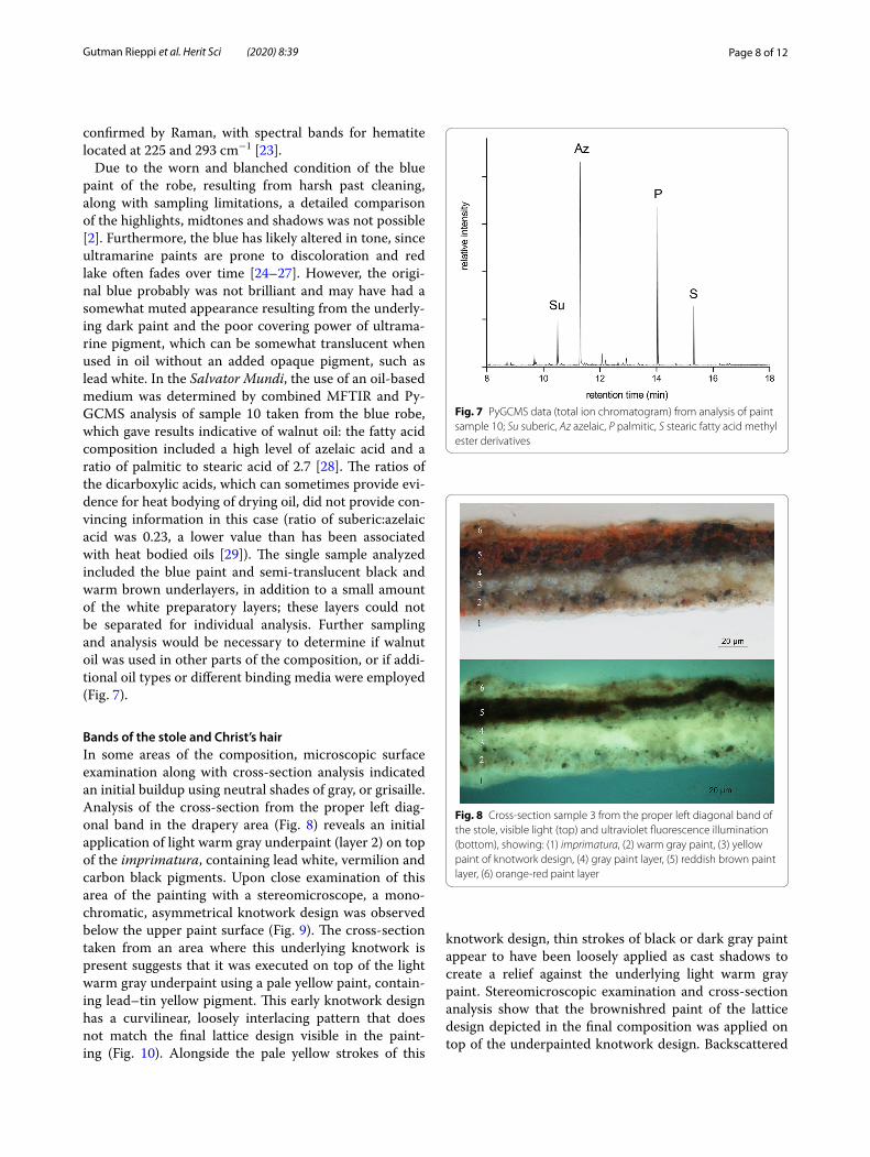
Bands of the stole and Christ’s hairIn some areas of the composition, microscopic surface examination along with cross-section analysis indicated an initial buildup using neutral shades of gray, or grisaille. Analysis of the cross-section from the proper left diag-onal band in the drapery area (Fig. 8) reveals an initial application of light warm gray underpaint (layer 2) on top of the imprimatura, containing lead white, vermilion and carbon black pigments. Upon close examination of this area of the painting with a stereomicroscope, a mono-chromatic, asymmetrical knotwork design was observed below the upper paint surface (Fig. 9). The cross-section taken from an area where this underlying knotwork is present suggests that it was executed on top of the light warm gray underpaint using a pale yellow paint, contain-ing lead–tin yellow pigment. This early knotwork design has a curvilinear, loosely interlacing pattern that does not match the final lattice design visible in the paint-ing (Fig. 10). Alongside the pale yellow strokes of this
knotwork design, thin strokes of black or dark gray paint appear to have been loosely applied as cast shadows to create a relief against the underlying light warm gray paint. Stereomicroscopic examination and cross-section analysis show that the brownishred paint of the lattice design depicted in the final composition was applied on top of the underpainted knotwork design. Backscattered
Fig. 7 PyGCMS data (total ion chromatogram) from analysis of paint sample 10; Su suberic, Az azelaic, P palmitic, S stearic fatty acid methyl ester derivatives
Fig. 8 Cross-section sample 3 from the proper left diagonal band of the stole, visible light (top) and ultraviolet fluorescence illumination (bottom), showing: (1) imprimatura, (2) warm gray paint, (3) yellow paint of knotwork design, (4) gray paint layer, (5) reddish brown paint layer, (6) orange-red paint layer

Page 9 of 12Gutman Rieppi et al. Herit Sci (2020) 8:39
electron images revealed splintery particles in the vermil-ion-containing reddish-brown paint, which are charac-teristic of charcoal (Fig. 11). Cross-section analysis also reveals that the blue paint of the robe is not present in the layer structure of the sample taken from the proper left diagonal band: this, combined with stereomicro-scopic examination, suggests that the bands were part of the conception of the image when the blue paint was applied.
In addition to the area of the proper left diagonal band, evidence was found for another underpainted decorative
design that appears to have been loosely painted for the central plaque, or pendant, on Christ’s chest (not shown). Although it is only visible in certain areas, where upper lying blue paint had been abraded, it differs from the one seen in the final painted image. Further investiga-tion utilizing advanced imaging and mapping techniques (not available at the time of the study) would be useful for elucidating these underpainted designs. While nei-ther of these early designs appear to have been worked up in detail, they nevertheless provide testimony to crea-tive process and give insight to the development of the composition.
Stereomicroscopic examination of areas of the horizon-tal breast band where upper paint layers were abraded also revealed grisaille underpainting in the form of an interwoven lattice pattern similar in design to that ren-dered in the visible composition (Fig. 12). Additional gri-saille underpainting was observed in the lower tresses of Christ’s hair, where the artist applied fine strokes of light gray paint before building up the color (Fig. 13).
Flesh tonesCross-sections from the flesh paint suggest that the approach to establishing the flesh tones differed from that described for the drapery, for which monochromatic and grisaille underpaint appear to have been applied to establish the lights and shadows before the color was added. In the flesh, in contrast, a more brightly colored paint was applied first, on top of the imprimatura, upon which light and shade were established subsequently using multiple layers of paint. Surface examination and cross-section analysis suggest that the materials and approach for different types of flesh paint—highlights,
Fig. 9 Photomicrograph showing underpainted knotwork design present beneath the proper left diagonal band of the stole
Fig. 10 Underpainted knotwork design seen beneath the painted lattice design of the proper left diagonal band of the stole, as observed through stereomicroscopic examination (mapped in red and superimposed over the corresponding area of the painting)
Fig. 11 Backscattered electron (SEM-BSE) image of paint of the lattice design of the proper left diagonal band of the stole, showing charcoal particles

Page 10 of 12Gutman Rieppi et al. Herit Sci (2020) 8:39
shadows and mid-tones—varied. For example, in sam-ple 1 taken from a bright highlight of the raised blessing hand, a base layer of saturated opaque pink, containing lead white, vermilion, glass particles, and a small amount of carbon black, was applied. The warmth of this layer was modified and attenuated with layers of slightly cooler and paler opaque paints, containing the same pigments but in differing proportions (Fig. 3). The highlights were finished with several extraordinarily thin (approximately 4–5 micrometers each) layers of cooler and paler pink paint to create the remarkably smooth surface of the final painted image (Fig. 14). No glass was detected in these uppermost, highlight paint layers.
In contrast to the highlights, the shadowed flesh of the raised hand appears to have been constructed with multiple layers of semi-translucent pigmented glazes. In cross-section 8, taken from the deep shadow of Christ’s raised hand, the lowest brown–red layer is colored most intensely with vermilion and a small amount of carbon black. This layer is followed by multiple darker glaze lay-ers, which appear to be more medium-rich, containing
various mixtures of carbon and bone black particles with some vermilion. The glaze layers are thin, ranging in thickness from approximately 3 to 6 micrometers, and vary in degree of tone, warmth and translucency (Fig. 15). Red lake pigment appears to have been added to some of the layers, especially to the uppermost, thus imparting additional color and warmth. The subtle adjustments in the pigmentation of the glaze layers may have been used to create the modulations and enhance the depth and luminosity of the flesh shadows. The combinations of materials, layering techniques and paint manipulation described may account for the absence of visible brush-strokes, even under stereomicroscopic examination, with a physical smoothness of the paint surface that is most evident in Christ’s face.
ConclusionsThe scientific analysis of samples taken from the Salva-tor Mundi and stereomicroscopic examination of the painted surface have helped to provide an understanding of the material nature of the painting, as well as an insight into the techniques employed and paint handling effects achieved. The Salvator Mundi appears to have been exe-cuted using a fairly limited palette, including lead white, vermilion, red iron oxide earth, red lake, natural ultrama-rine, lead–tin yellow, umber, and charcoal, carbon and bone blacks. However, given the necessarily limited sam-pling performed, the presence of other pigments in the painting cannot be discounted. Further investigation uti-lizing advanced imaging and mapping techniques would be valuable to provide a more comprehensive overview of the pigments used and their distribution. Within each paint layer analyzed, the pigment mixtures are fairly simple, yet sophisticated visual effects were achieved by employing a complex sequence of layers.
Analysis provided insight to the stages of the painting’s creation, with the walnut panel prepared with two lead white-containing layers bound in oil, the second of these tinted with lead–tin yellow. The overall tone of the Salva-tor Mundi may have been set partly through the use of warm brown underpaint, identified in areas of the back-ground and robe, as well as with the lower more brightly hued warm underlayers of the flesh. The underlying dark and grisaille paint applications, as well as the earlier dec-orative designs of the stole, such as the underlying knot-work in the proper left diagonal band, provide insights to an artistic mind actively at work during the various stages of painting.
Examination of cross-sections showed some of the approaches that were used to render the painting’s sub-tle visual effects. For the drapery, light and shade appear
Fig. 12 Photomicrograph of lattice design of the horizontal breast band of Christ’s robe, showing exposed area of grisaille underpainting (indicated by arrows)

Page 11 of 12Gutman Rieppi et al. Herit Sci (2020) 8:39
to have been established in early stages through the use of dark, monochrome and/or grisaille underpaint appli-cations, later enhanced by the layering of color. For the flesh, bright color appears to have been established first, its hue then attenuated by adding subsequent thin, intri-cate layers of light and/or shade. The combination of these approaches, and the addition of pulverized glass to the paint, helped achieved the luminosity, translucency and depth that characterize the Salvator Mundi.
Fig. 13 Photomicrographs of curls of the lower tresses of Christ’s hair, showing exposed grisaille underpainting (left) and a more intact painted curl (right)
Fig. 14 Detail of cross-section sample 1 from flesh highlight, proper right hand, showing: (1) imprimatura (2) black background paint layer (3) flesh highlight paint layers. Several extraordinarily thin layers of cooler and paler pink paint, each approximately 4–5 micrometers thick, are seen uppermost
Fig. 15 Cross-section sample 8 from flesh shadows, visible light (top) and ultraviolet fluorescence illumination (bottom), showing: (1) glue size, (2) ground (3) imprimatura (4) brownish pink paint (5, 6, 7, 8) semi-translucent glaze layers

Page 12 of 12Gutman Rieppi et al. Herit Sci (2020) 8:39
AbbreviationsVLM and FLM: Visible and fluorescent light microscopy; SEM-EDS: Scanning electron microscopy with energy dispersive spectroscopy; MFTIR: Fourier transform infrared microspectroscopy; ATR-FTIR: FTIR with attenuated total reflection; Py-GCMS: Pyrolysis gas chromatography-mass spectrometry.
AcknowledgementsThe authors would like to acknowledge conservator Dianne Dwyer Modestini, Clinical Professor, Kress Program in Paintings Conservation at the Conserva-tion Center of the Institute of Fine Arts, New York University, for her guidance and expertise. This project would also not have been possible without the scholarship and expertise of Robert B. Simon, PhD. The type of wood used for the panel support was identified by George Bisacca, Emeritus Conservator of Paintings, The Metropolitan Museum of Art.
Authors’ contributionsNGR performed stereomicroscopy, VLM and FLM, contextualization of data, and prepared the manuscript. BAP performed MFTIR and prepared the manu-script. KS performed SEM–EDS and Py-GCMS and prepared the manuscript. PAL reviewed data and prepared the manuscript. RN performed quantitative EDS analysis of glass particles. PW and TW performed Raman microspectros-copy. TJT performed ATR-FTIR and Raman microspectroscopy. All authors read and approved the final manuscript.
FundingNot applicable.
Availability of data and materialsAll data generated or analysed during this study are included in this published article.
Competing interestsThe authors declare that they have no competing interests.
Author details1 Art Analysis & Research, LLC, New York, NY 10018, USA. 2 Philadelphia, PA 19101, USA. 3 The Art Institute of Chicago, 111 S. Michigan Ave, Chicago, IL 60603, USA. 4 Museum of Fine Arts, 465 Huntington Avenue, Boston, MA 02115, USA. 5 Bruker Optics Inc., 40 Manning Road, Billerica, MA 01821, USA.
Received: 30 November 2019 Accepted: 7 April 2020
References 1. Syson L. Leonardo Da Vinci: painter at the court of Milan. New Haven: Yale
University Press; 2011. 2. Modestini DD. The Salvator Mundi rediscovered: History, technique and
condition. In: Menu M, editor. Leonardo da Vinci’s technical practice: paintings, drawings and influence. Paris: Hermann; 2014. p. 139–51.
3. Price BA, Pretzel B, Lomax SQ, editors. Infrared and Raman users group spectral database, vol. 1, 2, 2007 edn. Philadelphia: IRUG; 2009.
4. Dunkerton J, Spring M. The development of painting on coloured surfaces in sixteenth-century Italy. In: Roy A, Smith P, editors. Painting Techniques: History, Materials and Studio Practice, Contributions to the Dublin Congress, 7–11 September 1998. London: International Institute for Conservation of Historic and Artistic Works; 1998. p. 120–30.
5. Martin É. The preparation of the panel. In: Mohen J-P, Menu M, Mottin B, editors. Mona Lisa: inside the painting. New York: Abrams; 2006. p. 58.
6. Spring M, Mazzotta A, Roy A, Billinge R, Peggie D. Painting practice in Milan in the 1490s: the influence of Leonardo. Natl Gallery Tech Bull. 2011;32:78–112.
7. Spring M. Colourless powdered glass as an additive in fifteenth- and six-teenth-century European paintings. Natl Gallery Tech Bull. 2012;33:4–26.
8. Spring M. Perugino’s painting materials: analysis and context within sixteenth-century easel painting. In: Brunetti GB, Seccaroni C, Sgamellotti A, editors. Postprints of the workshop on the painting technique of Pietro Vannucci, called il Perugino, organized by INSTM and LabSTECH, Perugia, 14–15 April 2003. Florence: Nardini; 2004. p. 17–24.
9. Roy A, Spring M, Plazzotta C. Raphael’s early work in the National Gallery: paintings before Rome. Natl Gallery Tech Bull. 2004;25:4–35.
10. Berrie B, Matthew L. Material innovation and artistic invention: New materials and new colors in Renaissance Venetian paintings. Scientific Examination of Art: Modern Techniques in Conservation and Analysis. Washington DC: The National Academies Press, 2003. p. 12–26.
11. Tucker M, Passeri I, Sutherland K, Price B. Technique and Pontormo’s Por-trait of Alessandro de’ Medici. In: Strehlke CB, editor. Pontormo, Bronzino, and the Medici: the transformation of the renaissance portrait in Flor-ence. Philadelphia: Philadelphia Museum of Art; 2004. p. 34–54.
12. Noble P. A brief history of metal soaps in paintings from a conservation perspective. In: Casadio F, Keune K, Noble P, Van Loon A, Hendriks E, Centeno SA, Osmond G, editors. Metal soaps in art: conservation and research. Cham: Springer; 2019. p. 1–22.
13. Newman R. Observaciones acerca de los materiales pictóricos de Velázquez en Ciencia e Historia del Arte. In: McKim-Smith G, Newman R, editors. Velazquez en el Prado. Madrid: Museo del Prado; 1993. p. 113–47.
14. Coccato A, Jehlicka J, Moens L, Vandenabeele P. Raman spectroscopy for the investigation of carbon-based black pigments. J Raman Spectrosc. 2015;46:1003–15.
15. Bell IM, Clark RJH, Gibbs PJ. Raman spectroscopic library of natural and synthetic pigments (pre- ≈ 1850 AD). Spectrochim Acta Part A Mol Biomol Spectrosc. 1997;53:2159–79.
16. Dunkerton J. Leonardo in Verrocchio’s workshop: re-examining the tech-nical evidence. Natl Gallery Tech Bull. 2011;32:4–31.
17. Keith L, Roy A. Giampietrino, Boltraffio, and the influence of Leonardo. Nat Gallery Tech Bull. 1996;17:4–19.
18. Chui SA, Phenix A. The Esterházy Madonna in the context of Leonardo da Vinci’s studio practice. In: Menu M, editor. Leonardo da Vinci’s technical practice: paintings, drawings and influence. Paris: Hermann; 2014. p. 169–81.
19. Chui SA, Phenix A. Giovanni Antonio Boltraffio’s Madonna and Child in the context of Leonardo da Vinci’s Milanese studio. Am Inst Conserv Hist Artistic Works Paint Spec Group Postprints. 2011;24:1–12.
20. Bambach C. Leonardo, Michelangelo and notions of the unfinished in art. In: Baum K, Bayer A, Wagstaff A, editors. Unfinished: thoughts left visible. New York: The Metropolitan Museum of Art; 2016. p. 30–41.
21. Bicchieri M, Nardone M, Russo PA, Sodo A, Corsi M, Cristoforetti G, Palleschi V, Salvetti A, Tognoni E. Characterization of azurite and lazurite based pigments by laser induced breakdown spectroscopy and micro-Raman spectroscopy. Spectrochim Acta Part B. 2001;56:915–22.
22. Cennini CA. The Craftsman’s handbook “Il Libro dell’Arte”. Transl. Thomp-son DV, Jr. New York: Dover Publication, Inc.; 1933. p. 36–9.
23. Burgio L, Clark RJ. Library of FT-Raman spectra of pigments, minerals, pig-ment media and varnishes, and supplement to existing library of Raman spectra of pigments with visible excitation. Spectrochim Acta Part A Mol Biomol Spectrosc. 2001;57:1491–521.
24. Bacci M, Cucci C, Del Federico E, Ienco A, Jerschow A, Newman JM, Picollo M. An integrated spectroscopic approach for the identification of what distinguishes Afghan lapis lazuli from others. Vib Spectrosc. 2009;49:80–3.
25. Del Federico E, Schoefberger W, Schelvis J, Kapetanaki S, Tyne L, Jerschow A. Insight into framework destruction in ultramarine pigments. Inorg Chem. 2006;45:1270–6.
26. Del Federico E, Newman J, Tyne L, O’Hern C, Isolani L, Jerschow A. Solid-state NMR studies of ultramarine pigments discoloration. Mater Res Soc Symp Proc. 2007;984:45–50.
27. Saunders D, Kirby J. Light-induced colour changes in red and yellow lake pigments. Natl Gallery Tech Bull. 1994;15:79–97.
28. Mills JS, White R. The organic chemistry of museum objects. 2nd ed. Oxford: Butterworth-Heinemann; 1999. p. 171–2.
29. Mills JS, White R. Analyses of paint media. Natl Gallery Tech Bull. 1983;7:65–7.
Publisher’s NoteSpringer Nature remains neutral with regard to jurisdictional claims in pub-lished maps and institutional affiliations.