Replication Protein A (RPA) Hampers the Processive...
10
Replication Protein A (RPA) Hampers the Processive Action of APOBEC3G Cytosine Deaminase on Single-Stranded DNA Artem G. Lada 1 , Irina S.-R. Waisertreiger 1. , Corinn E. Grabow 1. , Aishwarya Prakash 1¤ , Gloria E. O. Borgstahl 1 , Igor B. Rogozin 2,3 , Youri I. Pavlov 1 * 1 Eppley Institute for Research in Cancer and Allied Diseases, University of Nebraska Medical Center, Omaha, Nebraska, United States of America, 2 National Center for Biotechnology Information, National Library of Medicine, National Institutes of Health, Bethesda, Maryland, United States of America, 3 Institute of Cytology and Genetics, Novosibirsk, Russia Abstract Background: Editing deaminases have a pivotal role in cellular physiology. A notable member of this superfamily, APOBEC3G (A3G), restricts retroviruses, and Activation Induced Deaminase (AID) generates antibody diversity by localized deamination of cytosines in DNA. Unconstrained deaminase activity can cause genome-wide mutagenesis and cancer. The mechanisms that protect the genomic DNA from the undesired action of deaminases are unknown. Using the in vitro deamination assays and expression of A3G in yeast, we show that replication protein A (RPA), the eukaryotic single-stranded DNA (ssDNA) binding protein, severely inhibits the deamination activity and processivity of A3G. Principal Findings/Methodology: We found that mutations induced by A3G in the yeast genomic reporter are changes of a single nucleotide. This is unexpected because of the known property of A3G to catalyze multiple deaminations upon one substrate encounter event in vitro. The addition of recombinant RPA to the oligonucleotide deamination assay severely inhibited A3G activity. Additionally, we reveal the inverse correlation between RPA concentration and the number of deaminations induced by A3G in vitro on long ssDNA regions. This resembles the ‘‘hit and run’’ single base substitution events observed in yeast. Significance: Our data suggest that RPA is a plausible antimutator factor limiting the activity and processivity of editing deaminases in the model yeast system. Because of the similar antagonism of yeast RPA and human RPA with A3G in vitro, we propose that RPA plays a role in the protection of the human genome cell from A3G and other deaminases when they are inadvertently diverged from their natural targets. We propose a model where RPA serves as one of the guardians of the genome that protects ssDNA from the destructive processive activity of deaminases by non-specific steric hindrance. Citation: Lada AG, Waisertreiger IS-R, Grabow CE, Prakash A, Borgstahl GEO, et al. (2011) Replication Protein A (RPA) Hampers the Processive Action of APOBEC3G Cytosine Deaminase on Single-Stranded DNA. PLoS ONE 6(9): e24848. doi:10.1371/journal.pone.0024848 Editor: Sue Cotterill, St. Georges University of London, United Kingdom Received April 13, 2011; Accepted August 19, 2011; Published September 15, 2011 This is an open-access article, free of all copyright, and may be freely reproduced, distributed, transmitted, modified, built upon, or otherwise used by anyone for any lawful purpose. The work is made available under the Creative Commons CC0 public domain dedication. Funding: This work was supported by the NCI Eppley Cancer Center Support Grant [P30CA036727] and in part by NCI grant CA129925 to YIP. Aishwarya Prakash was supported by the University of Nebraska Medical Center graduate fellowship and the Presidential graduate fellowship. The funders had no role in study design, data collection and analysis, decision to publish, or preparation of the manuscript. Competing Interests: The authors have declared that no competing interests exist. * E-mail: [email protected] . These authors contributed equally to this work. ¤ Current address: University of Vermont, Burlington, Vermont, United States of America Introduction Deaminases of the AID/APOBEC superfamily play amazingly diverse roles in vertebrates [1]. APOBEC1 works in lipid metabolism by editing apolipoprotein B mRNA [1,2]. AID is involved in immunoglobulin (Ig) diversification by initiating somatic hypermutation (SHM) and class-switch recombination (CSR) [3]. Members of the APOBEC3 subfamily restrict retroviruses and retrotransposons and have been implicated in the clearance of foreign DNA from human cells [1,2,4,5]. PmCDA1 is involved in immunity in jawless vertebrates [6]. AID/APOBEC enzymes convert cytosines to uracils in their target nucleic acids and therefore are inherent mutators [2,7] and cause single-stranded DNA breaks [8]. Improper targeting of deaminas- es could lead to point mutations and translocations, and ultimately to cancer [9]. Tight regulation of the activity of AID/APOBECs is vitally important for the prevention of genome instability. In agreement with the mutator properties of these enzymes, the expression of deaminases is mutagenic in heterologous hosts, such as bacteria and yeast ([1,2,10] and references therein). To gain insight into the mechanisms of genome protection from deam- inase-dependent mutagenesis, we studied A3G-induced mutagen- esis in live yeast cells and on the DNA of the same reporter in vitro using purified recombinant proteins. Analysis of the data obtained revealed striking differences between these two systems. A3G was non-processive in vivo but processive in vitro. In searching for the PLoS ONE | www.plosone.org 1 September 2011 | Volume 6 | Issue 9 | e24848
Transcript of Replication Protein A (RPA) Hampers the Processive...

Replication Protein A (RPA) Hampers the ProcessiveAction of APOBEC3G Cytosine Deaminase onSingle-Stranded DNAArtem G. Lada1, Irina S.-R. Waisertreiger1., Corinn E. Grabow1., Aishwarya Prakash1¤, Gloria E. O.
Borgstahl1, Igor B. Rogozin2,3, Youri I. Pavlov1*
1 Eppley Institute for Research in Cancer and Allied Diseases, University of Nebraska Medical Center, Omaha, Nebraska, United States of America, 2 National Center for
Biotechnology Information, National Library of Medicine, National Institutes of Health, Bethesda, Maryland, United States of America, 3 Institute of Cytology and Genetics,
Novosibirsk, Russia
Abstract
Background: Editing deaminases have a pivotal role in cellular physiology. A notable member of this superfamily,APOBEC3G (A3G), restricts retroviruses, and Activation Induced Deaminase (AID) generates antibody diversity by localizeddeamination of cytosines in DNA. Unconstrained deaminase activity can cause genome-wide mutagenesis and cancer. Themechanisms that protect the genomic DNA from the undesired action of deaminases are unknown. Using the in vitrodeamination assays and expression of A3G in yeast, we show that replication protein A (RPA), the eukaryotic single-strandedDNA (ssDNA) binding protein, severely inhibits the deamination activity and processivity of A3G.
Principal Findings/Methodology: We found that mutations induced by A3G in the yeast genomic reporter are changes of asingle nucleotide. This is unexpected because of the known property of A3G to catalyze multiple deaminations upon onesubstrate encounter event in vitro. The addition of recombinant RPA to the oligonucleotide deamination assay severelyinhibited A3G activity. Additionally, we reveal the inverse correlation between RPA concentration and the number ofdeaminations induced by A3G in vitro on long ssDNA regions. This resembles the ‘‘hit and run’’ single base substitutionevents observed in yeast.
Significance: Our data suggest that RPA is a plausible antimutator factor limiting the activity and processivity of editingdeaminases in the model yeast system. Because of the similar antagonism of yeast RPA and human RPA with A3G in vitro,we propose that RPA plays a role in the protection of the human genome cell from A3G and other deaminases when theyare inadvertently diverged from their natural targets. We propose a model where RPA serves as one of the guardians of thegenome that protects ssDNA from the destructive processive activity of deaminases by non-specific steric hindrance.
Citation: Lada AG, Waisertreiger IS-R, Grabow CE, Prakash A, Borgstahl GEO, et al. (2011) Replication Protein A (RPA) Hampers the Processive Action of APOBEC3GCytosine Deaminase on Single-Stranded DNA. PLoS ONE 6(9): e24848. doi:10.1371/journal.pone.0024848
Editor: Sue Cotterill, St. Georges University of London, United Kingdom
Received April 13, 2011; Accepted August 19, 2011; Published September 15, 2011
This is an open-access article, free of all copyright, and may be freely reproduced, distributed, transmitted, modified, built upon, or otherwise used by anyone forany lawful purpose. The work is made available under the Creative Commons CC0 public domain dedication.
Funding: This work was supported by the NCI Eppley Cancer Center Support Grant [P30CA036727] and in part by NCI grant CA129925 to YIP. Aishwarya Prakashwas supported by the University of Nebraska Medical Center graduate fellowship and the Presidential graduate fellowship. The funders had no role in studydesign, data collection and analysis, decision to publish, or preparation of the manuscript.
Competing Interests: The authors have declared that no competing interests exist.
* E-mail: [email protected]
. These authors contributed equally to this work.
¤ Current address: University of Vermont, Burlington, Vermont, United States of America
Introduction
Deaminases of the AID/APOBEC superfamily play amazingly
diverse roles in vertebrates [1]. APOBEC1 works in lipid
metabolism by editing apolipoprotein B mRNA [1,2]. AID is
involved in immunoglobulin (Ig) diversification by initiating
somatic hypermutation (SHM) and class-switch recombination
(CSR) [3]. Members of the APOBEC3 subfamily restrict
retroviruses and retrotransposons and have been implicated in
the clearance of foreign DNA from human cells [1,2,4,5].
PmCDA1 is involved in immunity in jawless vertebrates [6].
AID/APOBEC enzymes convert cytosines to uracils in their target
nucleic acids and therefore are inherent mutators [2,7] and cause
single-stranded DNA breaks [8]. Improper targeting of deaminas-
es could lead to point mutations and translocations, and ultimately
to cancer [9]. Tight regulation of the activity of AID/APOBECs is
vitally important for the prevention of genome instability. In
agreement with the mutator properties of these enzymes, the
expression of deaminases is mutagenic in heterologous hosts, such
as bacteria and yeast ([1,2,10] and references therein). To gain
insight into the mechanisms of genome protection from deam-
inase-dependent mutagenesis, we studied A3G-induced mutagen-
esis in live yeast cells and on the DNA of the same reporter in vitro
using purified recombinant proteins. Analysis of the data obtained
revealed striking differences between these two systems. A3G was
non-processive in vivo but processive in vitro. In searching for the
PLoS ONE | www.plosone.org 1 September 2011 | Volume 6 | Issue 9 | e24848

factors that suppress the processivity in vivo, we found that RPA
inhibits both DNA deaminase activity and processivity of the A3G.
Our data demonstrate that RPA may protect genomic DNA from
the destructive activity of editing deaminases.
Results
In the first step of this study, we analyzed the mechanisms and
parameters of the A3G action on genomic loci in yeast. We
expressed human A3G in a S. cerevisiae strain defective for uracil
DNA glycosylase (ung1). Ung1 initiates the base excision repair of
uracil-containing DNA by removing the uracil moiety so the
effects of cytosine deaminases are stronger when Ung1 activity is
absent. A3G production in the ung12 strain leads to about an
eight-fold increase in the frequency of forward mutations at the
URA3 locus (Fig. 1), as determined by the frequency of colonies
resistant to the 5-fluoroorotic acid (5-FOA). The mutagenic effect
of A3G production in yeast suggests that this enzyme is able to
penetrate the nuclei of yeast cells and deaminate cytosines in the
genomic loci. As expected from the cytosine deamination,
sequencing analysis of the URA3 gene from 311 independent
mutant clones revealed that almost all mutations were C to T or G
to A transitions. Most of the substitutions were observed in the
CCC ‘‘hotspot motifs,’’ which is the characteristic feature of A3G
both in vivo and in vitro (Fig. 2, green letters) [11,12]. The vast
majority of the sequenced clones contained a single base
substitution in ,800 bp of the URA3 open reading frame (ORF)
(Fig. 3a). Only two clones among the 311 analyzed contained two
substitutions, both found in the CCC motifs (one clone: C159T
(silent) and G767A (Trp to STOP); another clone: G741A (silent)
and G767A (Trp to STOP)). According to Poisson statistics (p,1),
the mutants with double substitutions result from independent hits
of APOBEC3G. These double hits occurred most likely at
different generations in yeast culture. We concluded that A3G is
not processive in vivo in yeast.
It is established that A3G, as well as AID, is processive in vitro,
which is generally defined by the ability to perform multiple
deaminations upon one encounter with the DNA molecule (also see
Discussion) [13,14,15]. In the next step of this study, we analyzed
A3G activity in vitro using the same URA3 reporter so we could
directly compare these results with the in results obtained with the
heterologous yeast system in vivo. Because AID/APOBEC proteins
act only on ssDNA [1,2], we used a modified in vitro deaminase assay
with a gapped DNA substrate [13]. This substrate consists of a
circular double-stranded DNA vector sequence and a single-
stranded gap containing the URA3 gene sequence. The substrate
is treated with the deaminase and ung2 pyrF2 bacteria is transformed
by the reaction product. Individual transformants are selected and
replica-plated on media with and without uracil to select ura- clones.
This is possible because the yeast URA3 is an ortholog of the pyrF
gene of E.coli and compensates for the pyrF deficiency [16]. We
developed a new DNA polymerization-based approach with the use
of blocking phosphorylated oligonucleotide (see Materials and
Methods and Fig. 4) to construct the circular gapped DNA
substrate. We used two similar substrates that differ in the
orientation of the URA3 reporter and allowed us to examine
targeting of the deaminase to the coding and non-coding strands
of the URA3 gene. Recombinant A3G was purified from the
HEK293T cells transfected with the wild-type human A3G
expression vector (Fig. 5a) [17]. The purified enzyme possessed
robust DNA-binding (Kd<4.561028 M) and deaminase
(1.6 pmol mg21 min21) activity on short oligonucleotides (Fig. 5c,
b, respectively). Incubation of the gapped substrates with the
recombinant A3G resulted in a ,20-fold increase in the frequency
of mutants (4–8% Ura2 clones vs. 0.3% in the control). Multiple C
to T or G to A (depending on the URA3 orientation) transitions were
found in the URA3 coding sequences isolated from the mutant clones
(Fig. 3b, Supporting Table 1). As many as 31 mutations per clone
were found, with an average number of 17.3. A low fraction of
mutant clones, along with the high numbers of base substitutions per
clone obtained in this experiment, is indicative of enzyme processive
action [13,18,19]. According to Poisson statistics (p,1027 according
to the x2 test for the data of the experiment presented in the Table
S1), all substitutions in virtually any single ura- clone result from one
deaminase-substrate encounter event. The majority of base
substitutions were found in the typical A3G hot motifs (Fig. 2, red
letters). The average length of an A3G tract (which is defined as the
distance between first and last substitutions) was 541 nucleotides,
with 672 nucleotides being the maximum. Despite the fact that
single, silent substitutions can not be detected in the selective system,
we observed a highly significant correlation in mutable positions
between the in vivo and in vitro spectra (Pearson linear correlation
coefficient = 0.79, P,1026). A striking difference in the proportion
of multiple deaminations in our in vitro and in vivo experiments
suggests that the high processivity of A3G is lost in vivo (Fig. 3).
The ssDNA in cells are always protected and covered by single-
stranded DNA binding proteins, called Replication Protein A
(RPA) in eukaryotes [20,21]. We hypothesized that RPA protects
the majority of genomic ssDNA from the activity of AID/
APOBEC enzymes. We analyzed the effect of RPA on the
deaminase activity of A3G using oligonucleotide deamination
assay in vitro with pure proteins. Human RPA was purified as
described [22], and yeast RPA was a gift of P. Burgers
(Washington University). DNA-binding activity of both RPA
preps was tested using electrophoretic mobility shift assay (Fig. 5d
and data not shown). Both human (Fig. 6a) and yeast (Fig. 6b)
RPA inhibited A3G activity in a concentration-dependent
manner. A nearly complete inhibition of deamination reaction
was achieved at the concentrations of RPA (,300 nM for yRPA
and ,500 nM for hRPA), where all DNA in the reaction is
expected to be covered with RPA, according to the published
‘‘footprints’’ of the corresponding proteins (Fig. 6c) [21]. This is
similar to the data on the inhibitory effect of RPA on the activity of
AID [23].
Figure 1. A3G is mutagenic in yeast. The frequency of 5-FOA-resistant colonies induced in the LAN-200 yeast strain carrying anA3G expression plasmid or vector alone is shown. One-way ANOVAF-test = 29.99, p = 0.00002.doi:10.1371/journal.pone.0024848.g001
RPA Inhibits Transactions of APOBEC3G
PLoS ONE | www.plosone.org 2 September 2011 | Volume 6 | Issue 9 | e24848

RPA Inhibits Transactions of APOBEC3G
PLoS ONE | www.plosone.org 3 September 2011 | Volume 6 | Issue 9 | e24848
Next, we asked how RPA influences the deaminase activity of
A3G on the gapped substrate containing a long stretch of ssDNA.
The gapped substrate DNA was incubated with A3G in the
presence of various concentrations of human RPA, and analysis of
mutants was done as described previously. The addition of RPA
(100 nM final concentration) to the reaction mix caused a modest
decrease in the frequency of Ura2 clones from 7.6% to 3.7%, but,
importantly, the frequency of clones with multiple mutations
gradually decreased with the increase of RPA concentration
(Fig. 7). The frequency of clones with less than seven substitutions
increased from about 10% in the absence of RPA to 60% with the
100 nM RPA. We concluded that RPA inhibits not only
deamination activity of A3G per se (Fig. 6), but also processivity
of the enzyme (Fig. 7). This observation is consistent with the yeast
in vivo data, where processivity of deaminase is almost absent.
According to our data and previous reports, A3G is highly
mutagenic in yeast, therefore it is able to penetrate the nuclei of
cells [10,24]. In mammalian cells, A3G is localized predominantly
in the cytoplasm (for example, [25] and references therein),
although a small but yet detectable fraction of the protein is found
in the nucleus when A3G is expressed at the endogenous level, and
an even higher level of A3G is found in the nucleus when the
protein is overproduced [26]. A substantial fraction of cytoplasmic
A3G is localized to P-bodies [27,28]. We also found that
overexpressed A3G in the cytoplasm of HEK293T cells is
concentrated in the punctuate bodies (Fig. 8a). We did not detect
A3G in the nuclei using immunofluorescence (Fig. 8a). However,
fractionation of cytoplasmic and nuclear extracts of the same cells
(see Materials and Methods) followed by Western blot analysis
revealed that a small but detectable amount of A3G is found in the
nuclear fraction (Fig. 8b). Moreover, RNAse treatment during the
course of fractionation resulted in the increase of A3G in the
nuclear fraction (Fig. 8b).
Discussion
We have shown that A3G introduces predominantly single base
substitutions per mutant in the yeast genomic locus, whereas it is
highly processive in vitro on the same URA3 gene reporter and
produces clusters of mutations in each mutant (Fig. 3). A3G
introduces multiple mutations both on its natural target, the viral
cDNA, and in Ty retrotransposon cDNA in the yeast system
[24,29,30]. Similar to A3G, AID is processive in vitro and induces
multiple deaminations [13]. In this paper we define processivity in
a broad sense, as the ability of the enzyme to catalyze multiple
reactions per each substrate encounter, before moving to the other
substrate molecule [15]. Tracts of mutations are present in the Ig
genes that undergo somatic hypermutation, but the multiplicity of
mutations is less than in vitro [31]. It is also possible that multiple
substitutions in the immunoglobulin genes could partially result
from consecutive selection of lymphocytes. Multiple substitutions
are found in the oncogenes mistakenly targeted by the AID, but
the number of substitutions per gene is usually less than in Ig genes
[32,33]. Comparison of the spectra of the mutations in the
ultimate targets of deaminases to random genomic reporters
clearly indicates that there is a mechanism protecting the majority
of the genome from destruction by deaminases. Various structural
proteins of chromatin, proteins involved in transcription [31],
DNA replication and repair enzymes could potentially endow this
protection. On the other hand, natural targets of deaminases in vivo
should be found in a special microenvironment (or subcellular
compartment) where certain protective components are missing or
modified.
We hypothesized that the one important difference between our
yeast and in vitro systems is the RPA, which is present in live yeast
cells but not in the in vitro assay. RPA is an eukaryotic ssDNA
binding protein [21]. It binds to the ssDNA with high affinity and
is involved in replication, DNA repair and recombination. RPA
protects ssDNA from damage and prevents secondary structure
formation that could influence different DNA transactions. RPA is
an abundant nuclear protein that covers ssDNA in the nuclei for a
variety of DNA transactions [20,21]. Since RPA and A3G share a
common substrate – ssDNA, it is expected that these two proteins
are competing for the nuclear ssDNA pool. Indeed, when we
added RPA to the oligonucleotide deaminations reaction, we
found that A3G activity is inhibited by both human and yeast RPA
Figure 2. Spectra of mutations induced in the URA3 gene in vivo and in vitro. Red letters over the URA3 sequence indicate mutations foundin vitro in the gapped substrate assay. Multiple substitutions found in ura3 mutants induced by A3G in the gapped substrate in several experimentswere pooled together. Green letters below the URA3 sequence are the mutations induced by the expression of A3G in the LAN-200 yeast strain. C to Tsubstitutions result from the deamination of the non-coding DNA strand, whereas G to A substitutions are the consequence of the coding stranddeaminations. Most of ura3 mutants obtained in yeast contained single base substitutions in the URA3 open reading frame. However, we found twoclones possessing two substitutions each: C159T (silent) and G767A (nonsense) in one clone, and G741A (silent) and G767A (nonsense) in the otherclone. In addition, one clone contained duplication of CAGACA at position 347 (there is CCC motif on the opposite strand just before the duplicatedsequence).doi:10.1371/journal.pone.0024848.g002
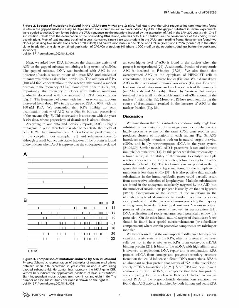
Figure 3. Comparison of mutations induced by A3G in vitro andin vivo. Schematic representation of examples of mutant ura3 allelesobtained upon A3G expression in yeast cells (a) and in vitro usinggapped substrate (b). Horizontal lines represent the URA3 gene ORF;vertical bars indicate the approximate positions of base substitutions.Eight independent mutant clones are shown in (a) and eight in (b). Thenumber of base substitutions per clone is shown on the right (b).doi:10.1371/journal.pone.0024848.g003
RPA Inhibits Transactions of APOBEC3G
PLoS ONE | www.plosone.org 4 September 2011 | Volume 6 | Issue 9 | e24848

(Fig. 6). Moreover, using deaminase assay with gapped DNA
substrate we showed that the number of mutant clones with A3G-
induced multiple substitutions substantially decreased with the
increase in RPA concentration (Fig. 7). This data indicates that
RPA suppresses the processive action of A3G. The mechanism of
deaminase processivity may include one-dimensional sliding and/
or three-dimensional microscopic dissociations and re-associations,
called jumping (reviewed with a particular emphasis on APOBEC
Figure 5. Purification and characterization of proteins used in the study. a. Coomassie-stained 12% SDS-PAGE gel of aliquotes fromdifferent steps of A3G purification from HEK293T cells. Mr – molecular weight marker, 1 – clarified lysate, 2 – flow through, 3–8 – different protein-containing fractions, eluted from the resin. b. Deaminase activity of purified A3G, detected in the oligonucleotide assay with uracil-DNA-glycosylase.In this assay, after deaminase converts cytosine to uracil, uracil-DNA-glycosilase removes uracil from the DNA, leading to formation of the AP site,which is further converted into strand break under conditions of high pH and temperature. 1 – UDG alone, 2 - A3G-expressing HEK293T lysate, 3 –clarified lysate, 4 – clarified lysate treated with RNAse, 5 – flow through, 6–11 – different fractions of purified protein. c and d. DNA-binding activity ofpurified A3G (c) and human RPA (d), detected by electromobility shift assay (EMSA). A3G from fraction 5 (see panel a) was used in this assay. The sameoligonucleotide was used for both proteins (c). The band that corresponds to the free oligonucleotide folded to the secondary structure is indicatedby the asterisk. Note that in (c) this non-specific band migrates similarly to the fastest A3G-shifted band.doi:10.1371/journal.pone.0024848.g005
Figure 4. Construction of circular gapped DNA substrate for the in vitro deaminase assay. a. Scheme of substrate construction (SeeMaterials and Methods for details). b. Agarose gel analysis of the reaction products. The gel was stained with ethidium bromide (EtBr). Mr – molecularweight marker, 1 – original ssDNA, 2 – ssDNA with two oligonucleotides annealed, and 3 – gapped substrate – product of Pfu Ultra reaction. Lanes 1through 3 contain similar amounts of DNA by molarity. The gapped substrate band on lane 3 is much brighter than the ssDNA on lanes 1 and 2because double-stranded DNA binds much more EtBr than the ssDNA of the same length due to intercalation of EtBr in the double helix. The lowerbands on lanes 1 and 3 represent linear DNA species that arise due to damage to the circular ssDNA.doi:10.1371/journal.pone.0024848.g004
RPA Inhibits Transactions of APOBEC3G
PLoS ONE | www.plosone.org 5 September 2011 | Volume 6 | Issue 9 | e24848

deaminases in [15]). AID is capable of both sliding and jumping
([18,19] and references therein). Processivity of A3G may include
sliding, jumping and/or inter-segmental transfer [14,15,17,34,35].
The presence of RPA on the ssDNA will sterically block
deaminase sliding, which contributes a lot to deaminase
processivity regardless of its ability to jump (for example, see
recent model of AID processive action in [19]). Therefore, even
deaminase that can jump will have strongly decreased processivity
in case its sliding is prevented by the RPA. Additionally, cytosines
in the ssDNA regions that are covered with the RPA molecules will
not be accessible to the deaminase independent of whether the
enzyme moves by sliding, jumping or inter-segmental transfer.
Therefore, our data suggest that RPA is a powerful inhibitor of
activity and processivity of deaminases in the nuclei (Fig. 9).
Protection of DNA by RPA may take place in the yeast artificial
system and probably in the nuclei of vertebrate cells. A3G
normally executes its antiviral action in the cytoplasm, where it
performs processive deaminations on retroviral cDNAs. RPA is
not present in the cytoplasm and therefore does not block the anti-
retroviral activity of A3G. Nevertheless, a small but detectable
amount of A3G is present in the nuclei of cells endogenously
expressing this deaminase [26]. The nuclear level of A3G is also
severely increased upon deaminase overproduction. In addition,
A3G can accumulate in the nuclei upon some inhibition of
proteasome [36]. In agreement with the previous reports, we
found by immunofluorescent microscopy that overexpressed A3G
in HEK293T cells is localized in the cytoplasm and is found
predominantly in the punctuate foci, identified before as P-bodies
(Fig. 8a) [27,28]. We were unable to see A3G in the nuclei of the
cells using this method (Fig. 8a). On the other hand, we show by
Western blot that A3G is found in the nuclear fraction of these
cells (Fig. 8b) (also see [26]). Therefore, it is possible that the
nuclear level of A3G is under the limit of detection of fluorescent
microscopy. Moreover, treatment with RNAse A during the
course of extract fractionation resulted in a significant increase of
the A3G nuclear level (Fig. 8b). P-bodies may serve as a storage
facility for A3G, preventing it from promiscuous action on non-
retroviral cellular DNA [27]. RNAse treatment release A3G from
P-bodies [27], which allows it to enter the nucleus by passive
diffusion. The presence of cytoplasmic retention signal [25,37]
allows the protein to be predominantly cytoplasmic even when P-
bodies are destroyed by RNAse treatment (Fig. 8b). Therefore,
multiple mechanisms contribute to the cytoplasmic localization of
A3G. Taken together, our data and previous reports suggest that
the distribution of A3G between the nucleus and cytoplasm is
dynamic and diverse changes in cellular physiology, including
those related to pathological conditions, may lead to an
accumulation of A3G in the nucleus. Therefore, the genomic
DNA of A3G-expressed cells should be protected from the A3G
activity. Similarly, when A3G is overexpressed in yeast, it is able to
penetrate the nuclei and deaminate genomic DNA by single hits.
The results presented in Fig. 3 indicate that the processive action
of A3G is suppressed in the yeast genome, whereas the enzyme is
robustly processive in vitro on the same reporter. In contrast, A3G
introduces multiple substitutions in the yeast retroviral-like Ty
elements. This is not surprising because Ty reverse transcription
takes place in the cytoplasm of yeast cells, where there is no RPA.
It has been suggested that, in addition to intracellular compart-
mentalization, there are additional mechanisms for protection of
the genomic DNA from APOBEC3 proteins [5]. Our data suggest
that RPA is one of these genomic safeguards.
Similar logic could be applied to the regulation of another
deaminase, AID. This enzyme is also processive in vitro [13], and
its activity is strongly inhibited by RPA or SSB [15,23]. AID is
Figure 7. RPA inhibits A3G processivity in vitro. Proportions ofura3 mutant clones with less (white bars) and more (black bars) thanseven substitutions in the ORF are shown as a function of hRPAconcentration. The observed differences between frequencies of cloneswith multiple mutations obtained in different experimental conditionsare significant (P = 0.002 according to the x2 test).doi:10.1371/journal.pone.0024848.g007
Figure 6. RPA inhibits deaminase activity of A3G in vitro. Oligonucleotide deamination assay (Materials and Methods and Fig. 5b) wasperformed with the addition of human (a) and yeast (b) RPA in various concentrations. Relative deaminase activity was calculated as a percentage ofcleaved oligo. Activity data for A3G in the presence of human and yeast RPA were plotted as a function of RPA concentration (c).doi:10.1371/journal.pone.0024848.g006
RPA Inhibits Transactions of APOBEC3G
PLoS ONE | www.plosone.org 6 September 2011 | Volume 6 | Issue 9 | e24848

mutagenic in the yeast system [38] and, similar to A3G, introduces
single base substitutions in the CAN1 reporter gene [10]. AID
shuttles between cytoplasm and the nucleus of human cells [39]
and represents a potential threat to a genome. Because AID
processivity is limited in the Ig genes in SHM and restricted even
more in most of the oncogenes that are being mistargeted by this
deaminase [32,33], it is plausible that AID processive activity is
also regulated by RPA. Active transcription is a prerequisite for
somatic hypermutation, but precise mechanisms that target AID to
the Ig genes are unknown. It has been proposed that RPA plays a
role in the recruitment of AID to the variable and switch regions of
immunoglogulin genes in B-cells [40,41]. In this model, phos-
phorylation of AID by the protein kinase A (PKA) allows the AID-
RPA interaction, which results in the deamination of the target
sites [42]. Precise AID targeting is provided by the recruitment of
PKA to the immunoglobulin genes [43]. Recently, it has been
Figure 9. Model of the inhibitory effect of RPA on the deaminase activity of A3G. (a) In the absence of RPA, for example in the cytoplasm,A3G is able to access ssDNA and slide along it (green arrow), catalyzing multiple deaminations. (b) In the presence of intermediate RPAconcentrations, only a fraction of ssDNA that is not covered by the RPA is accessible to the deaminase. A3G binds to the available ssDNA regions, butfurther sliding and/or jumping is hampered by the RPA bound to ssDNA. Processivity of the enzyme is inhibited much stronger than the deaminationactivity, and the degree of processivity inhibition depends on the extent of coverage of ssDNA by RPA. This scenario likely reflects the situation in thenuclear loci such as the yeast URA3 gene. (c) High concentrations of RPA completely prevent access of A3G to the ssDNA. It results in 100% inhibitionof deamination activity.doi:10.1371/journal.pone.0024848.g009
Figure 8. Nuclear-cytoplasmic distribution of A3G. (a) HEK293T cells transiently transfected with APOBEC3G-expressing plasmid were fixed andstained with DAPI to visualize nuclei and anti-His6 antibodies to visualize APOBEC3G. (b) Western-blot analysis of nuclear and cytoplasmic fractions ofHEK293T cells overexpressing A3G. See Materials and Methods for details.doi:10.1371/journal.pone.0024848.g008
RPA Inhibits Transactions of APOBEC3G
PLoS ONE | www.plosone.org 7 September 2011 | Volume 6 | Issue 9 | e24848

proposed that the combined action of transcription factor Spt5
and RPA recruits phosphorylated AID to the Ig loci. ([44] and
references therein). However, in vitro phosphorylated AID is still
inhibited by RPA [23]. Therefore, it is logical to think that AID is
recruited to the stalled/paused RNA polymerase II complexes
with the aid of Spt5, and this process does not require RPA. There
is probably a mechanism for partial RPA exclusion from natural
AID ssDNA substrates, which allows AID to target cytosines.
Enrichment of RPA in promoters of immunoglobulin genes, that
was demonstrated in [44] and used in support of model where
RPA attract AID to the target loci, can be explained by
recruitment of RPA during the course of DNA repair induced
by cytosine deamination, after AID activity is no longer required.
In the genome, RPA binds to ssDNA generated during DNA
transactions such as replication, DNA repair and transcription.
This, according to our model, prevents deaminase activity on the
cytosines located in the ssDNA regions covered by RPA, and
processivity, primarily by interfering with enzyme sliding. It is
possible that the protection from deaminases by RPA is non-
specific by nature and is executed by steric hindrance due to the
competition of the RPA and deaminases for the same substrate.
The ssDNA that is formed in the course of DNA repair and
recombination could also potentially be protected from deami-
nases by other ssDNA-binding proteins, such as Rad51. A3G
works in the cytoplasm, but is also found in the nucleus, where its
activity has to be prevented. Different parameters such as
transcription activity of particular loci, course of replication and
cell cycle progression, as well as tissue- and cell type-specific
characteristics, can modify the role of RPA in the prevention of
different deaminases access to the genome. Additional studies are
required to better understand the mechanism of RPA-based
genome protection from the inadvertent deaminases-induced
mutagenesis.
Materials and Methods
Yeast strains and techniquesMutant ura3–4 allele was converted to the wild-type one in the
S. cerevisiae strain 1B-D770 ung1::hygB [45] by transformation with
wild type URA3 DNA obtained by PCR. The resulting strain,
named LAN-200 (MATa ade5-1 lys2-Tn5-13 trp1-289 his7-2 leu2-
3,112 ung1::hygB), is suitable for the measurement of the forward
mutation rate at the URA3 locus.
For the mutagenesis experiment, the LAN-200 strain was
transformed with pESC-LEU2 vector and pESC-LEU2-hA3GSc
expressing plasmid [10]. The human A3G gene in this plasmid is
codon-optimized for expression in yeast.
Mutation frequencies were determined by fluctuation analysis as
described earlier [46]. Independent yeast transformants were
grown in a complete minimal medium without leucine to select for
the plasmid. In addition, this media contained galactose and
raffinose instead of glucose, to induce A3G expression. Induced
cultures were plated undiluted on plates containing 5-FOA to
select for ura3 mutants and with dilution on complete plates to
estimate viability. The 5-FOA is converted to the toxic compound
by the orotidine 5-phosphate decarboxylase, which is encoded by
the URA3 gene, therefore only ura3 mutants could grow on the
media containing 5-FOA.
To construct the spectra of mutations induced by A3G in yeast,
patches of LAN-200 transformants originating from single colonies
were replica-plated three times onto fresh medium containing
galactose and raffinose but without leucine. Then they were
replica-plated onto 5-FOA-containing medium to select for ura3
mutants. After five days of incubation, independent 5-FOAR
colonies were colony-purified on 5-FOA medium. Chromosomal
DNA from cells originating from single 5-FOA-resistant colonies
was isolated using a Yeast DNA Extraction Kit (Epicentre).
Subsequent PCR amplification and sequencing was performed as
described previously [38]. Sequences of the primers used for PCR
and sequencing are available upon request.
Cell linesHuman Embryonic Kidney 293T (HEK293T) (Thermo
Scientific catalog number HCL4517).
A3G purificationHEK293T cells were transfected by pcDNA3.1-A3G-Myc-His
expression plasmid using polyethileneimine [47]. Purification was
done according to protocol [17] with the following modification:
buffer containing 500 mM imidazole was used to elute the last
three fractions from the resin.
Oligonucleotide deaminase activity assayOligonucleotide deaminase activity assay was performed
according to the published method [48]. Briefly, 59-Cy5-labeled
oligonucleotide (59-Cy5-TTTTTTTTTTTTTTTATCTTTTT-
TTTTTTACTTTTTTTTTTAAACCCAAATTTTTTTTTTT-
TTTTTTTTTTTTTTTTTTTTTTTT) was incubated with
A3G in the presence of UDG (New England Biolabs). Then
abasic sites were converted to strand breaks by heating at high pH.
The resulting products were resolved on 16% denaturing PAGE.
Gels were scanned using the Typhoon 9410 imaging system (GE
Healthcare). Deamination at the CCC site, which represents the
A3G hot spot, creates a 47 nucleotide product.
Electrophoretic Mobility Shift Assay (EMSA)Electrophoretic Mobility Shift Assay (EMSA) was performed by
standard techniques (see [49], for example). The 59-Cy5 labeled
oligonucleotide (59-Cy5-AAGACCATGACCGCCAGCTCAAG-
TGTAAGTTACATGCATCTCTACCAGAAGTCAGAGGTT-
AGATTAGAGAGTATTT) (Integrated DNA Technologies) was
incubated with various amounts of A3G in reaction buffer (25 mM
Tris-HCl pH 8.0, 50 mM NaCl, 5 mM MgCl2 1 mM DTT, 10%
glycerol) for 15 min at 37uC. Then, samples were loaded on 4%
acrylamide gel (ratio of acrylamide to bis-acrylamide 75:1) and run
in 0.56TBE buffer for 3 h. Gels were scanned using the Typhoon
9410 imaging system (GE Healthcare).
Construction of pyrF- ung- E.coli strainThe ung::tet marker was transferred to the NR16207 strain
(pyrF::kan) from the strain BD2328 (ung::tet) using P1 phage
transduction by Drs. S.G. Kozmin and R. M. Schaaper (NIEHS).
Construction of gapped DNA substrateTwo plasmids, pRS315-URA3 OR1 and OR2, which differ in
the orientation of the URA3 gene, were constructed. Circular
ssDNA originating from these plasmids was purified from bacteria
by standard techniques [50]. Two oligonucleotides are annealed to
this ssDNA, first with a free 39-OH end serving as a primer, the
other with a 39-phosphorylated end blocks DNA synthesis beyond
its annealing site (Fig. 4a). The priming oligonucleotide is extended
using PfuUltra DNA polymerase. PfuUltra has no strand-
displacing activity, therefore, a gapped substrate is formed
(Fig. 4b). The gap size is 1110 and 1320 nucleotides when the
non-coding and coding strand of URA3 is in the ssDNA form,
respectively. Transformation of the E.coli pyrF2 strain with DNA
substrate enables the selection of mutations in the URA3 gene.
RPA Inhibits Transactions of APOBEC3G
PLoS ONE | www.plosone.org 8 September 2011 | Volume 6 | Issue 9 | e24848

A3G activity on gapped DNA substrateSixty ng of substrate DNA was treated with 100 ng of A3G for
10 min in the 10 ml total volume reactions containing 25 mM
Tris-HCl, pH 8.0, 50 mM NaCl, 1 mM DTT, and the products
of the reaction were electroporated into the pyrF- ung- E.coli.
Transformants were selected on LB plates with ampicilin, streaked
on the same type of plates and then replica-plated on Vogel-
Bonner media plates, with and without uracil, to select for ura3
mutants [51]. Dependent on the experiment, 4 to 8 percent of the
clones selected on the LB+Amp plates exhibited a mutant
phenotype, which is indicative of the processive mechanism of
enzyme action according to Poisson statistics (p,1027; see Table
S1) [13,18,19]. The URA3 gene from ura2 clones was sequenced.
Recombinant RPA was added to the deamination reactions where
applicable.
Mutation spectra construction and analysisDNA Star 8 (Lasergene) software was used for sequence
analyses. The x2 test was used to test the hypothesis that A3G
works processively in gapped substrate assay (Table S1) and to
compare the frequencies of clones with multiple mutations
(Figure 7). The same test was used to confirm that APOBEC3G
is non-processive in vivo in yeast. Calculations were done using the
COLLAPSE [52] and STATISTICA programs [52]. The Pearson
linear correlation coefficient was used to compare spectra.
Calculations were done using the program STATISTICA [52].
Immunocytochemistry and fluorescent microscopyHEK293T cells transiently transfected with pcDNA3.1-A3G-
Myc-His plasmid or mock control cells were fixed with methanol-
acetic acid mixture (3:1). All procedures were performed at room
temperature. Mouse anti-His6-tag antibodies (GenScript, USA)
and goat-anti-mouse Cy3-conjugated antibodies (ThermoScienti-
fic, USA) were used for A3G detection. Cells were mounted in
Vectashield mounting medium with DAPI (Vector Laboratories,
Burlingame, CA). Images were captured with a Zeiss Axiovert
200 M microscope (Carl Zeiss, Thornwood, NY) equipped with
an ORCA-ER digital camera (Hamamatsu, Hamamatsu City, JP)
and processed using OpenLab software (Improvision, Boston,
MA).
Nuclear-cytoplasmic fractionation and Western-blotanalysis
HEK293T cells transfected with the pcDNA3.1-A3G-Myc-His
plasmid or mock control cells were collected from 10-cm culture
dishes 26 hours after transfection (about 100% final confluency),
washed once with PBS and resuspended in 300 ml of PBS buffer
containing 1% Triton X-100, 2 mM DTT, protease inhibitors
cocktail set IV (1006 dilution, Calbiochem), 5 mM PMSF, and
incubated for 5 min on ice. Lyzates then were split into two
aliquots, and 10 mg of RNAse A (Qiagen) was added to one of the
aliquots. After incubation for 30 min at 37uC the lyzates were
layered on top of 1 ml of 30% sucrose/PBS/DTT buffer. Samples
were spun at 800 g for 10 min at 4uC and supernatant
(cytoplasmic fraction) was saved. Nuclear pellet was washed once
with PBS and resuspended in 1.3 ml of RIPA buffer containing
2 mM DTT, protease inhibitors cocktail set IV (1006 dilution,
Calbiochem), 5 mM PMSF. After 10 min incubation on ice,
lyzates were spun at 15000 g for 10 min at 4uC, and supernatant
(nuclear fraction) was saved. Both nuclear and cytoplasmic
fractions were used for Western blotting. Mouse Anti-His6-tag
antibodies were used to detect A3G-Myc-His and mouse anti-a-
tubulin antibodies (both from Genscript) were used to confirm
efficient nuclear-cytoplasmic fractionation.
Supporting Information
Table S1 Analysis of mutations introduced by APO-BEC3G into gapped DNA substrate in vitro. Thirty-nine
mutant clones obtained in one experiment are shown. Numbers in
‘‘Substitutions’’ column indicate nucleotide positions in the URA3
ORF. Track size is defined as the distance (bp) between first and
last substitutions. If APOBEC3G works in distributive fashion,
then the frequencies of clones with certain number of substitutions
should follow the Poisson distribution. We have found, on the
contrary, that the observed distribution is strikingly different from
the expected Poisson distribution (p,1027 according to the x2 test
for the data of the experiment presented in the table), confirming
that tracts of mutations found result from processive action of
APOBEC3G.
(DOC)
Acknowledgments
We are thankful to Dr. R. Harris (University of Minnesota) for the vector to
produce the wild-type A3G in human cells; Dr. P. Burgers (Washington
University) for purified yeast RPA; Drs. S.G. Kozmin and R. M. Schaaper
(NIEHS) for the construction of E. coli strains; Drs. L. Fisher and T. Bessho
for help with the PEI cell culture transfection; Drs. K.H. Choi and M.M.
Ouellette for help with EMSA; J. Shaw (University of Nebraska, Kearney)
for help with yeast mutagenesis experiments; K. Wendt for help with
gapped substrate construction; Drs. P. Shcherbakova and F. Kadyrov for
critical reading of the manuscript; and the UNMC Core Sequencing
Facility and University of Chicago Cancer Research DNA Sequencing
Facility for DNA sequencing.
Author Contributions
Conceived and designed the experiments: AGL YIP. Performed the
experiments: AGL ISW CEG AP. Analyzed the data: AGL YIP IBR
GEOB. Contributed reagents/materials/analysis tools: GEOB IBR AP.
Wrote the paper: AGL YIP. Contributed to the editing of the final
manuscript: ISW CEG AP GEOB IBR. Performed whole cell immuno-
fluorescence experiments described in Fig. 8: ISW.
References
1. Conticello SG, Langlois MA, Yang Z, Neuberger MS (2007) DNA deamination
in immunity: AID in the context of its APOBEC relatives. Adv Immunol 94:37–73.
2. Bhagwat AS (2004) DNA-cytosine deaminases: from antibody maturation toantiviral defense. DNA Repair (Amst) 3: 85–89.
3. Muramatsu M, Kinoshita K, Fagarasan S, Yamada S, Shinkai Y, et al. (2000)Class switch recombination and hypermutation require activation-induced
cytidine deaminase (AID), a potential RNA editing enzyme. Cell 102: 553–563.
4. Goff SP (2003) Death by deamination: a novel host restriction system for HIV-1.
Cell 114: 281–283.
5. Stenglein MD, Burns MB, Li M, Lengyel J, Harris RS (2010) APOBEC3
proteins mediate the clearance of foreign DNA from human cells. Nat Struct
Mol Biol 17: 222–229.
6. Rogozin IB, Iyer LM, Liang L, Glazko GV, Liston VG, et al. (2007) Evolution
and diversification of lamprey antigen receptors: evidence for involvement of anAID-APOBEC family cytosine deaminase. Nat Immunol 8: 647–656.
7. Petersen-Mahrt SK, Harris RS, Neuberger MS (2002) AID mutates E. coli suggestinga DNA deamination mechanism for antibody diversification. Nature 418: 99–103.
8. Larson ED, Cummings WJ, Bednarski DW, Maizels N (2005) MRE11/RAD50cleaves DNA in the AID/UNG-dependent pathway of immunoglobulin gene
diversification. Mol Cell 20: 367–375.
9. Okazaki IM, Kotani A, Honjo T (2007) Role of AID in tumorigenesis. Adv
Immunol 94: 245–273.
10. Lada AG, Frahm Krick C, Kozmin SG, Mayorov VI, Karpova TS, et al. (2011)
Mutator effects and mutation signatures of editing deaminases produced in
bacteria and yeast. Biochemistry (Moscow) 76: 131–146.
RPA Inhibits Transactions of APOBEC3G
PLoS ONE | www.plosone.org 9 September 2011 | Volume 6 | Issue 9 | e24848

11. Beale RC, Petersen-Mahrt SK, Watt IN, Harris RS, Rada C, et al. (2004)
Comparison of the differential context-dependence of DNA deamination by
APOBEC enzymes: correlation with mutation spectra in vivo. J Mol Biol 337:
585–596.
12. Yu Q, Konig R, Pillai S, Chiles K, Kearney M, et al. (2004) Single-strand
specificity of accounts for minus-strand deamination of the HIV genome. Nat
Struct Mol Biol 11: 435–442.
13. Pham P, Bransteitter R, Petruska J, Goodman MF (2003) Processive AID-
catalysed cytosine deamination on single-stranded DNA simulates somatic
hypermutation. Nature 424: 103–107.
14. Chelico L, Pham P, Calabrese P, Goodman MF (2006) APOBEC3G DNA
deaminase acts processively 39R59 on single-stranded DNA. Nat Struct Mol Biol
13: 392–399.
15. Chelico L, Pham P, Goodman MF (2009) Stochastic properties of processive
cytidine DNA deaminases AID and APOBEC3G. Philos Trans R Soc
Lond B Biol Sci 364: 583–593.
16. Bach ML, Lacroute F, Botstein D (1979) Evidence for transcriptional regulation
of orotidine-59-phosphate decarboxylase in yeast by hybridization of mRNA to
the yeast structural gene cloned in Escherichia coli. Proc Natl Acad Sci U S A
76: 386–390.
17. Nowarski R, Britan-Rosich E, Shiloach T, Kotler M (2008) Hypermutation by
intersegmental transfer of APOBEC3G cytidine deaminase. Nat Struct Mol Biol
15: 1059–1066.
18. Bransteitter R, Pham P, Calabrese P, Goodman MF (2004) Biochemical analysis
of hypermutational targeting by wild type and mutant activation-induced
cytidine deaminase. J Biol Chem 279: 51612–51621.
19. Pham P, Calabrese P, Park SJ, Goodman MF (2011) An analysis of a single-
stranded DNA scanning process in which AID deaminates C to U haphazardly
and inefficiently to ensure mutational diversity. J Biol Chem.
20. Oakley GG, Patrick SM (2010) Replication protein A: directing traffic at the
intersection of replication and repair. Front Biosci 15: 883–900.
21. Wold MS (1997) Replication protein A: a heterotrimeric, single-stranded DNA-
binding protein required for eukaryotic DNA metabolism. Annu Rev Biochem
66: 61–92.
22. Deng X, Prakash A, Dhar K, Baia GS, Kolar C, et al. (2009) Human replication
protein A-Rad52-single-stranded DNA complex: stoichiometry and evidence for
strand transfer regulation by phosphorylation. Biochemistry 48: 6633–6643.
23. Pham P, Smolka MB, Calabrese P, Landolph A, Zhang K, et al. (2008) Impact
of phosphorylation and phosphorylation-null mutants on the activity and
deamination specificity of activation-induced cytidine deaminase. J Biol Chem
283: 17428–17439.
24. Schumacher AJ, Nissley DV, Harris RS (2005) APOBEC3G hypermutates
genomic DNA and inhibits Ty1 retrotransposition in yeast. Proc Natl Acad
Sci U S A 102: 9854–9859.
25. Stenglein MD, Matsuo H, Harris RS (2008) Two regions within the amino-
terminal half of APOBEC3G cooperate to determine cytoplasmic localization.
J Virol 82: 9591–9599.
26. Stopak K, de Noronha C, Yonemoto W, Greene WC (2003) HIV-1 Vif blocks
the antiviral activity of APOBEC3G by impairing both its translation and
intracellular stability. Mol Cell 12: 591–601.
27. Wichroski MJ, Robb GB, Rana TM (2006) Human retroviral host restriction
factors APOBEC3G and APOBEC3F localize to mRNA processing bodies.
PLoS Pathog 2: e41.
28. Gallois-Montbrun S, Kramer B, Swanson CM, Byers H, Lynham S, et al. (2007)
Antiviral protein APOBEC3G localizes to ribonucleoprotein complexes found in
P bodies and stress granules. J Virol 81: 2165–2178.
29. Harris RS, Bishop KN, Sheehy AM, Craig HM, Petersen-Mahrt SK, et al.
(2003) DNA deamination mediates innate immunity to retroviral infection. Cell
113: 803–809.
30. Dutko JA, Schafer A, Kenny AE, Cullen BR, Curcio MJ (2005) Inhibition of a
yeast LTR retrotransposon by human APOBEC3 cytidine deaminases. Curr
Biol 15: 661–666.
31. Storb U, Shen HM, Nicolae D (2009) Somatic hypermutation: processivity of
the cytosine deaminase AID and error-free repair of the resulting uracils. CellCycle 8: 3097–3101.
32. Shen HM, Peters A, Baron B, Zhu X, Storb U (1998) Mutation of BCL-6 gene
in normal B cells by the process of somatic hypermutation of Ig genes. Science280: 1750–1752.
33. Nilsen H, An Q, Lindahl T (2005) Mutation frequencies and AID activationstate in B-cell lymphomas from Ung-deficient mice. Oncogene 24: 3063–3066.
34. Coker HA, Petersen-Mahrt SK (2007) The nuclear DNA deaminase AID
functions distributively whereas cytoplasmic APOBEC3G has a processive modeof action. DNA Repair (Amst) 6: 235–243.
35. Chelico L, Pham P, Goodman MF (2009) Mechanisms of APOBEC3G-catalyzed processive deamination of deoxycytidine on single-stranded DNA. Nat
Struct Mol Biol 16: 454–455. author reply 455–456.36. Wichroski MJ, Ichiyama K, Rana TM (2005) Analysis of HIV-1 viral infectivity
factor-mediated proteasome-dependent depletion of APOBEC3G: correlating
function and subcellular localization. J Biol Chem 280: 8387–8396.37. Bennett RP, Presnyak V, Wedekind JE, Smith HC (2008) Nuclear Exclusion of
the HIV-1 host defense factor APOBEC3G requires a novel cytoplasmicretention signal and is not dependent on RNA binding. J Biol Chem 283:
7320–7327.
38. Mayorov VI, Rogozin IB, Adkison LR, Frahm C, Kunkel TA, et al. (2005)Expression of human AID in yeast induces mutations in context similar to the
context of somatic hypermutation at G-C pairs in immunoglobulin genes. BMCImmunol 6: 10.
39. Ito S, Nagaoka H, Shinkura R, Begum N, Muramatsu M, et al. (2004)Activation-induced cytidine deaminase shuttles between nucleus and cytoplasm
like apolipoprotein B mRNA editing catalytic polypeptide 1. Proc Natl Acad
Sci U S A 101: 1975–1980.40. Chaudhuri J, Khuong C, Alt FW (2004) Replication protein A interacts with
AID to promote deamination of somatic hypermutation targets. Nature 430:992–998.
41. Cheng HL, Vuong BQ, Basu U, Franklin A, Schwer B, et al. (2009) Integrity of
the AID serine-38 phosphorylation site is critical for class switch recombinationand somatic hypermutation in mice. Proc Natl Acad Sci U S A 106: 2717–2722.
42. Basu U, Chaudhuri J, Alpert C, Dutt S, Ranganath S, et al. (2005) The AIDantibody diversification enzyme is regulated by protein kinase A phosphoryla-
tion. Nature 438: 508–511.43. Vuong BQ, Lee M, Kabir S, Irimia C, Macchiarulo S, et al. (2009) Specific
recruitment of protein kinase A to the immunoglobulin locus regulates class-
switch recombination. Nat Immunol 10: 420–426.44. Yamane A, Resch W, Kuo N, Kuchen S, Li Z, et al. (2011) Deep-sequencing
identification of the genomic targets of the cytidine deaminase AID and itscofactor RPA in B lymphocytes. Nat Immunol 12: 62–69.
45. Lucaccioni A, Pavlov YI, Achilli A, Babudri N (2007) High rate of starvation-
associated mutagenesis in Ung(-) yeast caused by the overproduction of humanactivation-induced deaminase. Curr Genet 52: 239–245.
46. Shcherbakova PV, Kunkel TA (1999) Mutator phenotypes conferred by MLH1overexpression and by heterozygosity for mlh1 mutations. Mol Cell Biol 19:
3177–3183.47. Boussif O, Lezoualc’h F, Zanta MA, Mergny MD, Scherman D, et al. (1995) A
versatile vector for gene and oligonucleotide transfer into cells in culture and in
vivo: polyethylenimine. Proc Natl Acad Sci U S A 92: 7297–7301.48. Bransteitter R, Pham P, Scharff MD, Goodman MF (2003) Activation-induced
cytidine deaminase deaminates deoxycytidine on single-stranded DNA butrequires the action of RNase. Proc Natl Acad Sci U S A 100: 4102–4107.
49. Hellman LM, Fried MG (2007) Electrophoretic mobility shift assay (EMSA) for
detecting protein-nucleic acid interactions. Nat Protoc 2: 1849–1861.50. Sambrook J, Russell D (2001) Molecular Cloning: A Laboratory Manual (Third
edition). NY: Cold Spring Harbor Laboratory Press.51. Miller JH Experiments in Molecular Genetics: Cold Spring Harbor Laboratory,
Cold Spring Harbor, N.Y.
52. Khromov-Borisov NN, Rogozin IB, Pegas Henriques JA, de Serres FJ (1999)Similarity pattern analysis in mutational distributions. Mutat Res 430: 55–74.
RPA Inhibits Transactions of APOBEC3G
PLoS ONE | www.plosone.org 10 September 2011 | Volume 6 | Issue 9 | e24848