Renal hemodynamics, function and oxygenation in critically ill ......2021/03/03 · higher than...
37
1 Renal hemodynamics, function and oxygenation in critically ill patients and after major surgery Sven-Erik Ricksten, Gudrun Bragadottir, Lukas Lannemyr, Bengt Redfors, Jenny Skytte Department of Anesthesiology and Intensive Care Medicine, Sahlgrenska Academy, University of Gothenburg, Sahlgrenska University Hospital, Gothenburg, Sweden. S-41345 Gothenburg, Sweden Corresponding author: Sven-Erik Ricksten, MD, PhD Department of Anesthesiology and Intensive Care Medicine, Sahlgrenska Academy, University of Gothenburg, Sahlgrenska University Hospital, Gothenburg, Sweden. S-41345 Gothenburg, Sweden E-mail: [email protected] Phone: +46 31 3427433, Fax: +46 31 413862 Kidney360 Publish Ahead of Print, published on March 3, 2021 as doi:10.34067/KID.0007012020 Copyright 2021 by American Society of Nephrology.
Transcript of Renal hemodynamics, function and oxygenation in critically ill ......2021/03/03 · higher than...

1
Renal hemodynamics, function and oxygenation in critically ill
patients and after major surgery
Sven-Erik Ricksten, Gudrun Bragadottir, Lukas Lannemyr, Bengt Redfors, Jenny Skytte
Department of Anesthesiology and Intensive Care Medicine, Sahlgrenska Academy,
University of Gothenburg, Sahlgrenska University Hospital, Gothenburg, Sweden.
S-41345 Gothenburg, Sweden
Corresponding author: Sven-Erik Ricksten, MD, PhD
Department of Anesthesiology and Intensive Care Medicine, Sahlgrenska Academy,
University of Gothenburg, Sahlgrenska University Hospital, Gothenburg, Sweden.
S-41345 Gothenburg, Sweden
E-mail: [email protected]
Phone: +46 31 3427433, Fax: +46 31 413862
Kidney360 Publish Ahead of Print, published on March 3, 2021 as doi:10.34067/KID.0007012020
Copyright 2021 by American Society of Nephrology.

2
Introduction
The present review outlines the available data from the work of our group on renal
hemodynamics, function and oxygenation in critically ill patients with acute renal dysfunction
such as in postoperative acute kidney injury, early clinical septic shock, in patients
undergoing cardiac surgery with cardiopulmonary bypass or in patients undergoing liver
transplantation. We also provide information on renal hemodynamics, function and
oxygenation in patients with chronic renal impairment due to congestive heart failure. This
review will argue that the common denominator, for these groups of patients, is that renal
oxygenation is impaired in all groups caused by a lower renal oxygen delivery or a
pronounced increase in renal oxygen consumption.
Determinants of renal oxygen delivery and renal oxygen consumption
Renal oxygen availability is determined by the balance between O2 delivery (RDO2) and O2
consumption (RVO2). RDO2, in turn, is dependent on renal blood flow (RBF) and arterial
oxygen content (CaO2) (Fig. 1). RBF is determined by renal perfusion pressure [in the clinical
situation usually estimated as mean arterial pressure (MAP) minus central venous pressure
CVP)]. Pressure-flow autoregulation of RBF is well developed in the kidney, mediated by
myogenic and tubuloglomerular feedback mechanisms (1). At severe hypotension, however,
the renal autoregulatory capacity is exhausted and RBF becomes pressure-dependent with a
risk of renal ischemia. In high-risk patients, a low MAP during surgery or in the ICU is
directly correlated to an increase in serum creatinine, suggesting acute kidney injury (2-4).
During surgery and in the postoperative period, many patients receive artificial solutions such
as colloids and crystalloids, which induce hemodiluton and a fall in blood oxygen carrying
capacity (CaO2), which in turn may affect RDO2 negatively. In a recent study, the differential
effects of a colloid and a crystalloid fluid on systemic and renal hemodynamics were studied

3
after cardiac surgery. Both fluids improved cardiac output and RBF, but none of the fluids
increased RDO2, due to hemodilution (5).
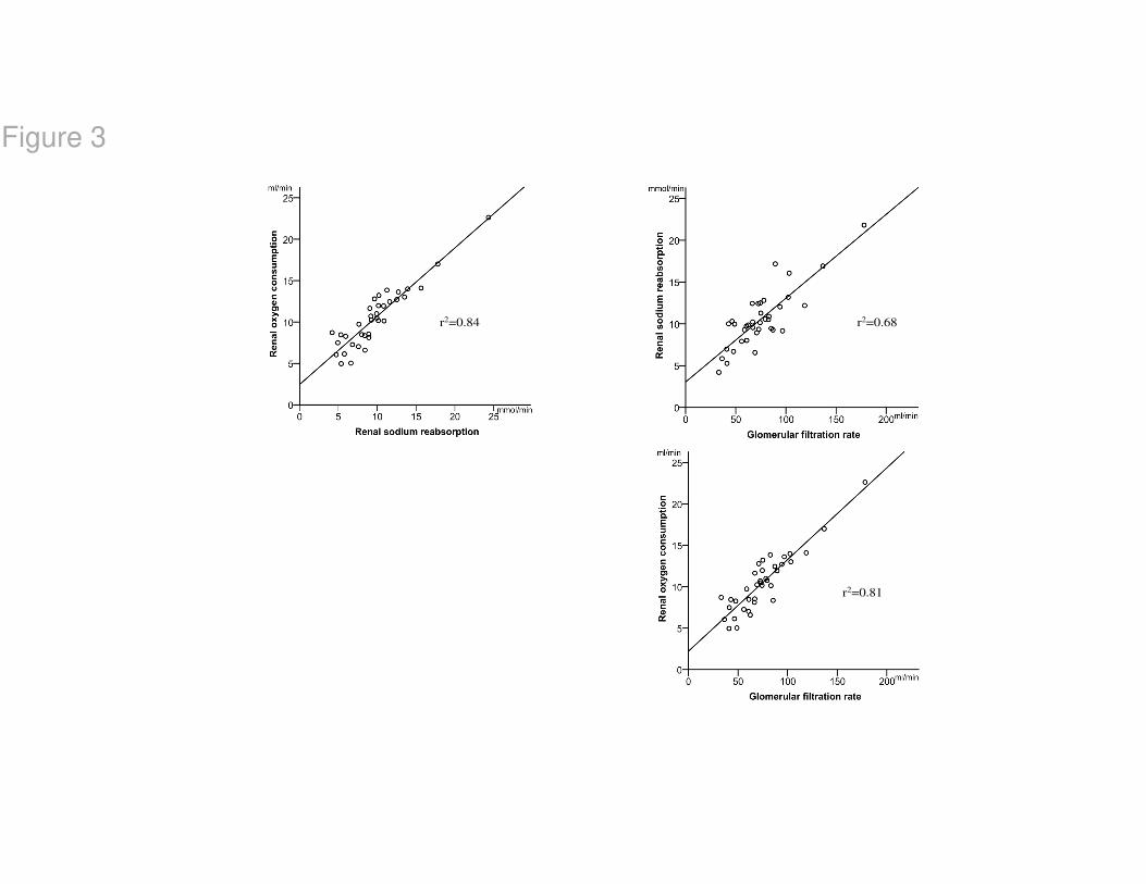
Active tubular sodium reabsorption accounts for 70-80% of RVO2 (6). If you administer
furosemide (bolus 0.5 mg/kg + 0.5 mg/kg/h) to postoperative patients, RVO2 will decrease by
approximately 25% and experimental studies have shown that furosemide causes an increase
in medullary tissue pO2 (7) indicating an improved renal oxygenation. Tubular sodium
reabsorption, in turn, is controlled by glomerular filtration rate (GFR) (Fig 2). If GFR
increases, tubular sodium load will increase causing tubular reabsorption also to increase. It
has been shown in experimental studies (8) and in patients (9) that there is a close linear
correlation between GFR, renal sodium reabsorption and RVO2 (Fig. 3) Thus, GFR is an
important determinant of RVO2. Infusion of atrial natriuretic peptide (ANP) to postoperative
patients, at a dose of 50 ng/kg/min, has been shown to increase GFR by 15%, which was
accompanied by 25% increase in RVO2 (10). In the treatment of acute kidney injury (AKI),
obviously the aim is to increase GFR, but one must bear in mind that any agent that increases
GFR, will also increase RVO2 and vice versa. It is also important to acknowledge that, unlike
other organs where an increase in blood flow will improve oxygenation, an increase in RBF
augments GFR and the filtered load of sodium, resulting in an increase RVO2. Due to this
flow-dependency of RVO2, renal oxygenation will vary little, as long as RBF and GFR
change in parallel.
The major determinants of GFR are the renal perfusion pressure and the
pre/postglomerular resistance ratio. A renal vasodilator that acts preferentially on the
preglomerular resistance vessels increases both RBF and glomerular hydrostatic pressure and
thereby GFR. Two vasodilatory agents have this renal vasodilatory profile; ANP, when used
for treatment of AKI (11), and levosimendan in postoperative patients and in patients with the
cardiorenal syndrome (12, 13) (Fig 4). Vasodilators, such as the inodilators dopamine,
4
dobutamine and milrinone act on both pre- and postglomerular resistance vessels and will
cause a substantial increase in RBF, with no major change in GFR, as dilation of the
postglomerular will increase the “run-off of blood from the glomerulus with minor changes in
upstream glomerular hydrostatic pressure (13-15). The vasoconstrictor vasopressin has been
introduced in the clinical arena for treatment of vasodilatory shock, as e.g. in septic shock
(16). The use of vasopressin in shock treatment is, however, a two-edged sword as
vasopressin, in postoperative patients increases GFR and thereby RVO2, by a vasoconstriction
of postglomerular resistance vessels, accompanied by a fall in RBF and RDO2. Thus,
vasopressin has the potential to induce a renal oxygen supply/demand mismatch (17).
Renal oxygen extraction is a direct measure of renal oxygenation
Renal oxygenation is defined as the ratio between renal oxygen consumption (RVO2) and
renal oxygen delivery (RDO2), i.e. the renal O2 demand/supply relationship. It can easily be
shown that this relationship is equivalent to the renal extraction of O2 (RO2Ex) calculated as:
[(Arterial oxygen content – renal vein oxygen content) / arterial oxygen content], which
requires measurements of arterial and renal vein oxygen saturations. An increase in RO2Ex
means that RVO2 has increased in relation to RDO2, i.e. renal oxygenation has impaired, and
vice versa. When compared to other major organs, RVO2 is relatively high, second only to the
heart. It has been shown in sedated, mechanically ventilated patients that RVO2 is two-thirds
(10 ml/min) that of myocardial oxygen consumption (15 ml/min) (Table 1) (9). Renal blood
flow (RBF), which accounts for approximately 20-25% of cardiac output, is three times
higher than myocardial blood flow in this group of patients. Renal oxygen extraction in the
non-failing kidney is therefore low, 10%, compared with e.g. the heart, in which oxygen
extraction is 55% (Table 1).

5
The relatively high RBF is directed preferentially to the cortex which will optimize the
filtration process and solute reabsorption. The proportion of the cortical flow that is conducted
to outer and inner medulla is only approximately 40% and 20%, respectively (18). The
combination of low medullary perfusion, high oxygen consumption of the medullary thick
ascending limbs (mTAL) and the countercurrent exchange of oxygen within the vasa recta
results in a poorly oxygenated outer medulla (7). The oxygen availability is therefore low in
the outer medulla with an oxygen tissue partial pressure (PO2) of 10-20 mmHg compared to
50 mmHg in the cortex. Thus, as the outer medulla is on the border of hypoxia already under
normal conditions, it is particularly sensitive to prolonged or intermittent episodes of low
RDO2, caused by hypoperfusion or haemodilution, as seen e.g after major surgery (especially
cardiac or vascular surgery) or severe heart failure, common causes of ischemic acute kidney
injury (AKI).
Renal perfusion and oxygenation in clinical AKI
It has provocatively been stated that “acute renal failure is acute renal success” (19, 20), as a
reduction in GFR in AKI should lead to a reduction of the renal reabsorptive workload, thus
preserving medullary oxygenation with a reduced risk of further aggravation of ischemia.
In postoperative sedated, mechanically ventilated uncomplicated patients with no renal
dysfunction, RVO2 is approximately 10-12 ml/min (13) (see Table1) a value, which is slightly
lower than has previously been reported in conscious healthy volunteers. This corresponds to
a mean of 0.82 ml of oxygen per mmol of reabsorbed sodium, which is in line with findings
from previous animal studies (21). In contrast, in high-risk cardiac surgery, complicated by
AKI, patients consumed 1.9 ml O2/mmol reabsorbed sodium. Thus, net-reabsorbing a certain
amount of sodium, consumed 2.4 times more oxygen in the AKI group than in postoperative
patients with no renal impairment (22). The correlation between GFR and RVO2 in patients

6
with early AKI after cardiac surgery vs. those undergoing uncomplicated surgery are shown
in fig. 5. From this figure it can be seen that there is a close correlation between GFR and
RVO2 in both groups of patients. According to the “acute failure is a renal success” –
hypothesis (19, 20), AKI patients should operate on the lower part of the regression line of the
control patients. However, the regression line of the AKI patients is clearly shifted to the left,
i.e. at a certain level of GFR, RVO2 is higher (22). Thus, our findings do not support the
hypothesis put forward by many investigators that “acute renal failure is a renal success”.
One can only speculate on the mechanism behind the increased oxygen utilization for
sodium transport in the AKI patients. A potential explanation could be ischemia-induced loss
of epithelial cell-polarisation and loss of tight junction integrity in AKI, as has been shown in
experimental studies and after human renal transplantation (23, 24). The tubular cells, thus,
lose their ability to efficiently pump in a specific direction, from one compartment to another.
Another explanation for the increased oxygen costs for sodium reabsorption in clinical AKI
may be diminished renal generation of nitric oxide (NO) due to endothelial damage and
down-regulation of endothelial nitric oxide synthase (25-27). NO has been shown to directly
compete with oxygen for mitochondrial respiration suggesting a basal modulatory role for NO
on oxygen consumption (28). Laycock et al showed that blockade of nitric oxide synthase
increased RVO2 in dogs, while reducing GFR and renal sodium reabsorption. Indeed,
inhibition of NO synthesis more than doubled the RVO2/renal sodium reabsorption ratio in
that study.
The resetting of the relationship between GFR and RVO2 in clinical ischemic AKI (fig
5), was accompanied by a severe impairment of the renal oxygen demand/supply relationship,
as demonstrated by the 70% higher renal oxygen extraction, which was caused by a
pronounced vasoconstriction and renal hypoperfusion at a maintained RVO2, compared to
controls, despite a normalized cardiac output and mean arterial pressure. (Table 2) (13).

7
Renal perfusion and oxygenation in early clinical septic shock
Severe sepsis and septic shock is a common cause of ICU admission and is responsible for
AKI (septic AKI) in approximately 40–50% of the critically ill population. Furthermore,
septic AKI is associated with high risk of early hospital mortality (29, 30). There is a
divergent apprehension on the pathophysiology of septic AKI. Schrier and Wang proposed,
based on experimental models, that the predominant early pathogenetic factor is renal
vasoconstriction caused by reflex activation of renal sympathetic activity, the renin-
angiotensin system and increased levels of arginine-vasopressin, thus decreasing both RBF
and GFR, induced by a constriction of afferent arterioles (31). In contrast, in a hyperdynamic
experimental sepsis model, Langenberg et al showed that the fall in creatinine clearance was
accompanied by an increase in RBF and thus explained by a dilation of efferent arterioles
(32).
Recently, we measured renal perfusion, filtration, and oxygenation in patients within 24
hours after arrival to the ICU due to early norepinephrine-dependent septic shock (33). Renal
plasma flow (RPF) was measured using the infusion clearance technique for para-
aminohippuric acid (PAH), corrected by renal extraction of PAH by a renal vein catheter.
Filtration fraction was measured by renal extraction of chromium-51 labeled EDTA (51
Cr-
EDTA). Renal oxygenation was estimated from renal oxygen extraction (Table 3). Patients
undergoing uneventful major cardiac surgery served as a comparator group. We believe that
the comparison between these two groups is relevant since both groups were exposed to
systemic inflammation. Furthermore, both groups were sedated and mechanically ventilated
during the experimental procedure.
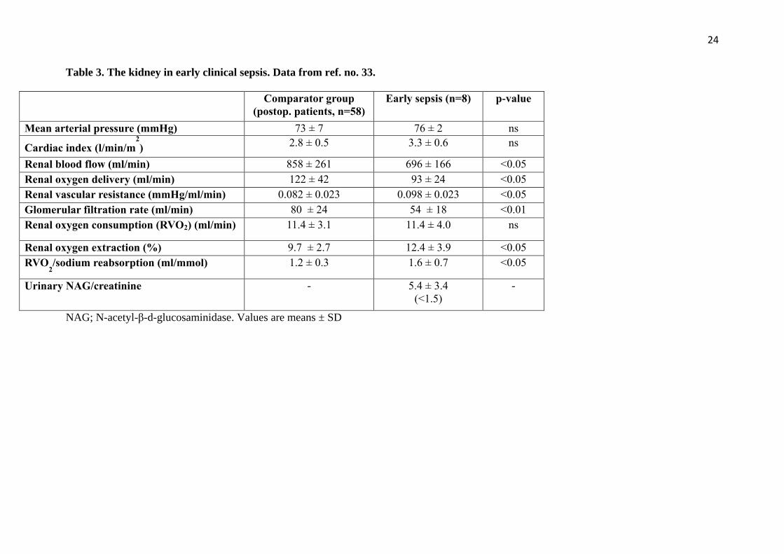
RBF and RDO2 were impaired caused by renal vasoconstriction and a redistribution of
blood flow away from the kidneys when compared to a comparator group (Table 3). This

8
renal vasoconstriction could be explained by an increase, particularly, in the tone of the renal
afferent arterioles as renal filtration fraction (GFR/RPF) was not significantly different
between the two groups, suggestive of a balanced decrease in both GFR and RBF in the septic
patients. Such an increase of the tone of the afferent arterioles in sepsis has been demonstrated
in endotoxemic animals (34). This redistribution of blood flow in septic shock impaired the
renal oxygen supply/demand relationship as evidenced by an increased renal oxygen
extraction. Thus, these results are not in line with data from previous studies on large animals,
showing that AKI in early septic shock is accompanied by renal vasodilation and an increase
in RBF (32). The lower RDO2 may explain early signs of tubular dysfunction/injury seen in
the septic group expressed as a pathologic elevation of the tubular injury marker N-acetyl-b-
D-glucosaminidase (NAG).
Effects of cardiopulmonary bypass on renal perfusion and oxygenation
AKI is a prevalent complication after cardiac surgery with cardiopulmonary bypass (CPB).
The incidence of post–cardiac surgery AKI ranges between 15% and 30%, depending on the
complexity of the procedure (35, 36). Dialysis-dependent AKI, occurring in 2 to 5% of
cardiac surgery patients, carries a mortality between 50% and 80% (37). Renal hypoperfusion
and impaired RDO2 have been considered important pathways in the development of post–
cardiac surgery AKI (38, 39). It has previously been shown that RBF correlates positively to
mean arterial pressure during hypothermic (28o), non-pulsatile CPB, suggesting impaired
autoregulation of RBF (40). A decreased RDO2 may also be caused by hemodilution because
of priming the CPB circuit with cell-free solution, usually a crystalloid. It has been shown that
the degree of hemodilution (38, 41), and a decreased systemic oxygen delivery (42) are
independent risk factors for the development of postoperative AKI.
9
To increase our understanding of the renal effects of cardiac surgery with normothermic
CPB, we measured RBF, RDO2, GFR and RVO2, as well as the renal oxygen supply/demand
relationship (RO2Ex), before, during, and after open cardiac surgery utilizing CPB (43).
Despite a 33% increase in systemic perfusion flow rate, compared to baseline cardiac output,
CPB induced a renal vasoconstriction redistributing blood flow away from the kidneys, which
in combination with hemodilution decreased RDO2 by 20%, while GFR and RVO2 was
unchanged (Fig. 6) Thus, RO2Ex increased by 40-45% indicating a renal oxygen
supply/demand mismatch during CPB. After weaning from CPB, renal oxygenation was
further deteriorated, as RO2Ex increased by 80% from baseline (Fig. 7) due to hemodilution
and an increase in RVO2 (45% compared to baseline) accompanied by a peak seven-fold
increase in the urinary NAG/creatinine ratio indicating tubular injury. There was a positive
correlation between the increase in RO2Ex and the urinary NAG/creatinine ratio, suggesting
that renal hypoxia may have a causative role for the release of the tubular injury marker. The
increase in RVO2 after CPB could be explained by a resetting of the relationship between
renal sodium reabsorption and RVO2. Before CPB, a mean of 0.86 mmol O2 was consumed
per mmol of reabsorbed sodium. In contrast, after CPB, 1.31 mmol O2/mmol sodium
reabsorbed was consumed. Thus, net reabsorbing a certain amount of sodium, consumed 52%
more O2 after compared to before CPB. Such an increased O2 utilization for tubular sodium
transport has previously been described in patients with post-cardiac surgery AKI and most
likely reflects hypoxia-induced tubular dysfunction (see above).
Theoretically, one way to improve renal oxygenation would be to perform CPB at a
higher flow rate than the one traditionally used. This concept was tested by Lannemyr et al,
who studied the effects of varying CPB flow rates (2.4, 2.7 and 3.0 l/min/m2) on renal
oxygenation in 17 patients with normal renal function undergoing cardiac surgery with
normothermic CPB (44). At a flow rate of 2.7 and 3.0 l/min/m2, renal oxygen extraction was
10
12% and 23% lower, respectively, compared with 2.4 L l/min/m2. This corresponds to a 14%
and 30% improvement, respectively, of the renal oxygen supply/demand relationship. One
could therefore speculate that one way to protect the kidneys during CPB would be to use
higher CPB flow rates than those conventionally used. Our group has recently launched a
randomized trial evaluating the renal effects of a high (3.0 l/min/m2) vs. a standard CPB flow
rate (2.4 l/min/m2). The primary end-points are RBF, RDO2 and tubular injury markers
(ClinicalTrials.gov Identifier: NCT04084301).
Renal perfusion and oxygenation after liver transplantation
Acute kidney injury (AKI) is a common complication after liver transplantation, with a
reported incidence of 10–60% (45, 46). Mortality after liver transplantation is reported to be
45–55% in patients developing AKI, compared to 2-6% in patients not developing AKI (45).
The etiology of AKI after liver transplantation is unknown, but is most likely multifactorial.
Hypotension caused by intra-operative blood loss and the post-reperfusion syndrome (47) is
presumably of importance. Furthermore, renal dysfunction may be present prior to
transplantation as seen in patients with hepatorenal syndrome (HRS). In patients with HRS, a
splanchnic vasodilatation is seen. This vasodilation is accompanied by activation of the renin-
angiotensin and the sympathetic nervous system, resulting in increased renal vascular
resistance. As a result, blood flow will be distributed away from the kidneys and hence the
kidneys will receive a decreased oxygen delivery (48, 49). This could be considered as a
potential mechanism causing AKI after liver transplantation.
To gain more insight into the pathophysiological mechanisms behind the development
of AKI in liver transplanted patients, we studied renal hemodynamics, function and
oxygenation in twelve mechanically ventilated and sedated patients early after liver
transplantation (50). RBF was measured by the continuous retrograde renal vein
11
thermodilution technique (51), filtration fraction was measured by renal extraction of 51
Cr-
EDTA and renal oxygenation was estimated from renal oxygen extraction. Patients
undergoing uneventful major cardiac surgery served as a comparator group. We believe that
the comparison between these two groups is relevant since both groups have been exposed to
major surgery and both groups have had contact with material that is not endogenous, with a
consequent systemic inflammation. Furthermore, both groups were sedated and mechanically
ventilated during the experimental procedure, which was, in both groups performed, early
after arrival in the intensive care unit (ICU).
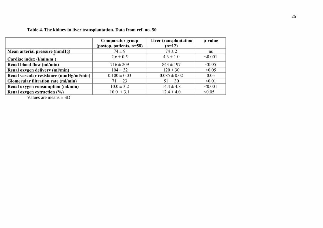
Cardiac output was considerably higher (65%), accompanied by a considerably lower
systemic vascular resistance (-36%) in the liver transplanted patients compared to the
comparator group (Table 4). Despite this hyperdynamic circulation in the liver transplants,
RBF and RDO2 were only moderately elevated (15%). In other words, the ratio between RBF
and cardiac output was almost 30% lower in the liver transplants, suggesting a redistribution
of RBF away from the kidneys. In the liver transplant group, GFR decreased by 40%,
compared to the preoperative value. When compared to the comparator group, GFR and
filtration fraction was 27% and 40%, lower in the liver transplant group, respectively. Serum
creatinine increased by 41% and 48% from baseline on the first and second postoperative day
in the liver transplant group, while in the comparator group, serum creatinine, if anything,
decreased postoperatively. What is the mechanism behind this early fall in GFR after liver
transplantation? The physiological control of GFR is mediated by the balance between the
tone of the afferent and efferent arterioles. A fall in GFR may be caused either by
vasoconstriction of the afferent arterioles or a vasodilation of the efferent arterioles, which
will be accompanied by a decrease or an increase in RBF, respectively. Thus, based on or
findings, we suggest that the fall in GFR together with the increased vasodilation and
increased renal blood flow after liver transplantation, is best explained by a dilation
12
preferentially of the efferent arterioles. Such a renal hemodynamic pattern has previously
been described in animal models of hyperdynamic septic shock, showing renal vasodilation
and loss of GFR, in turn, explained by efferent renal vasodilation (52, 53).
Renal oxygen consumption and extraction were both higher in the liver transplants, 43%
and 33% respectively, when compared to the comparator group. The remarkably high RVO2
seen post-liver transplantation could be explained by the production and release of reactive
oxygen species (ROS) from the liver graft, as a consequence of the ischemia/reperfusion
injury (47). It has been shown that oxidative stress increases mitochondrial oxygen
consumption and reduces tubular epithelial sodium transport, both contributing to an increase
in renal oxygen consumption (54).
Thus, after liver transplantation there is an early decline in renal function despite
hyperdynamic systemic circulation and renal vasodilation, most likely explained by an
efferent arteriolar vasodilation. This was accompanied by an impaired renal oxygenation, as
the pronounced elevation of renal oxygen consumption was not met by a proportional
increase in renal oxygen delivery.
Renal perfusion and oxygenation in patients with congestive heart failure and chronic
renal impairment
Heart failure (HF) affects more than 26 million people worldwide and is a leading cause for
hospitalization in Europe and the US (55). Renal dysfunction caused by HF, also denoted as
the cardiorenal syndrome, is independently associated with increased risk of hospitalization
and cardiovascular death (56). Indeed, renal dysfunction is a stronger predictor of mortality
than New York Heart Association functional class or left ventricular ejection fraction (56.
However, data on renal hemodynamics, GFR, RVO2 and renal oxygenation in clinical HF are
scarce and the therapeutic options for treatment of the cardiorenal syndrome are limited.

13
In a recent study, Lannemyr et measured systemic and renal hemodynamics, GFR,
RVO2 and renal oxygenation in 29 patients with chronic HF (LVEF < 40 %) and impaired
renal function (GFR < 80 ml min-1
1.73m -2
) (13). A pulmonary artery catheter was used for
measurements of systemic hemodynamics. The left renal vein was catheterized and GFR and
RBF were measured using the infusion clearance technique for chromium ethylene diamine
tetraacetic acid (51
CrEDTA) and para-aminohippurate (PAH), corrected for renal extraction of
PAH, respectively.
When compared to a control group, with neither heart failure nor renal dysfunction (33),
renal vascular resistance was 102% higher than in the comparator group, which could explain
the pronounced reduction in RBF (-52%), RDO2 (-46%) and GFR (-48/%) in these patients
(Fig. 8). The ratio RBF/cardiac index was only 18% in the HF patients compared to 31% in
the comparator group, suggesting a marked redistribution of RBF away from the kidneys. The
lower RBF in these patients could be caused by an increased renal vascular resistance caused
by e.g. increased renal sympathetic activity, activation of the renin-angiotensin-aldosterone
system and increased release of vasopressin but also by increased central venous pressure
(=increased renal venous back pressure). Filtration fraction was moderately elevated (17%) in
the HF patients when compared to the comparator group (14%) suggesting that there is a
preferential increase in preglomerular vascular resistance in HF patients compared to controls.
Despite the much lower GFR, RVO2 was only 22% lower than in the comparator group.
Renal oxygen extraction was 15% in the HF patients, a number considerably higher than in
the comparator group (10%). In other words, the pronounced reduction in RDO2 could not
meet renal oxygen requirements causing a chronic impairment of renal oxygenation in the HF
patients. It has been suggested that renal hypoxia could be the common final pathway for
progression to chronic kidney disease (CKD) (54), as chronic hypoxia induces oxidative

14
stress, production of extracellular matrix, collagen deposition, vascular rarefaction and
interstitial fibrosis (54, 58, 59).
Is it possible to measure renal oxygenation non-invasively?
Impaired renal oxygenation measured by renal vein catheterization is associated with tubular
injury and increased postoperative creatinine levels (50, 60). However, invasive
measurements may be technically challenging, and the method is not suitable for general use.
Near-infrared spectroscopy (NIRS) is a noninvasive, optical technique that continuously
measures the difference between oxygenated and deoxygenated hemoglobin within a regional
tissue area, thus obtaining regional oxygen saturation (rSO2) (61). Although measurements of
rSO2 by the NIRS technology for assessment of renal tissue oxygenation in pediatric patients
(62, 63), were found to be feasible, due to the proximity of the kidneys to the skin, the
agreement with renal rSO2 and the invasively measure renal vein oxygen saturation (SrvO2)
has not been determined in an adult population. In a study by Tholén et al, it was shown that
renal rSO2 is correlated to and predicts changes in SrvO2 with a small bias and acceptable
agreement (64). However, there was a drop-out rate of 25%, because of a skin-to-kidney
distance > 4 cm, suggesting that this technique for assessment of renal oxygenation is not
suitable for all patients. There is a need for future studies demonstrating that monitoring of
renal rSO2, guiding interventions to prevent or treat a low renal rSO2, will improve renal
outcome after cardiac surgery in suitable patients, before the technique is recommended for
general use.
Conclusions
In this review we have presented data on renal hemodynamics, GFR, renal oxygen
consumption and renal oxygenation from various groups of patients with renal dysfunction,

15
such as AKI after cardiac surgery, in early sepsis, patients undergoing cardiac surgery with
CPB, liver transplantation and in patients with CRF. Irrespective of the cause of renal failure,
the common denominator, for these groups of patients, is that renal oxygenation is impaired
(Table 5). This was caused by a lower renal oxygen delivery in all groups except for patients
undergoing liver transplantation where impaired renal oxygenation was caused by a
pronounced increase in renal oxygen consumption. Finally, in all groups there was an
increased oxygen utilization for tubular sodium transport, which most likely reflects hypoxia-
induced tubular dysfunction.
Disclosures
L. Lannemyr reports Consultancy Agreements: XVIVO Perfusion AB; Honoraria: Orion
Pharma. All remaining authors have nothing to disclose.
Funding
Swedish State Support for Clinical Research: ALFGBG-75130
Author Contributions
S-E Ricksten: Writing - original draft; Writing - review and editing
G Bragadottir: Methodology; Writing - review and editing
L Lannemyr: Methodology; Writing - review and editing
B Redfors: Methodology; Writing - review and editing
J Skytte: Methodology; Writing - review and editing

16
References
1. Cupples WA, Braam B. Assessment of renal autoregulation. American journal of
physiology 2007; 292:F1105-23.
2. Futier E, Lefrant JY, Guinot PG, Godet T, Lorne E, Cuvillon P, Bertran S, Leone M,
Pastene B, Piriou V, Molliex S, Albanese J, Julia JM, Tavernier B, Imhoff E, Bazin JE,
Constantin JM, Pereira B, Jaber S; INPRESS Study Group. Effect of Individualized vs
Standard Blood Pressure Management Strategies on Postoperative Organ Dysfunction Among
High-Risk Patients Undergoing Major Surgery: A Randomized Clinical Trial
JAMA. 2017; 318:1346-1357.
3. Mathis MR, Naik BI, Freundlich RE, Shanks AM, Heung M, Kim M, Burns ML,
Colquhoun DA, Rangrass G, Janda A, Engoren MC, Saager L, Tremper KK, Kheterpal S,
Aziz MF, Coffman T, Durieux ME, Levy WJ, Schonberger RB, Soto R, Wilczak J, Berman
MF, Berris J, Biggs DA, Coles P, Craft RM, Cummings KC, Ellis TA 2nd, Fleishut PM,
Helsten DL, Jameson LC, van Klei WA, Kooij F, LaGorio J, Lins S, Miller SA, Molina S,
Nair B, Paganelli WC, Peterson W, Tom S, Wanderer JP, Wedeven C; Multicenter
Perioperative Outcomes Group Investigators. Preoperative Risk and the Association between
Hypotension and Postoperative Acute Kidney Injury. Anesthesiology. 2020; 132:461-475.
4. Khanna AK, Maheshwari K, Mao G, Liu L, Perez-Protto SE, Chodavarapu P, Schacham
YN, Sessler DI. Association Between Mean Arterial Pressure and Acute Kidney Injury and a
Composite of Myocardial Injury and Mortality in Postoperative Critically Ill Patients: A
Retrospective Cohort Analysis Crit Care Med. 2019; 47:910-917.
5. Skytte Larsson J, Bragadottir G, Krumbholz V, Redfors B, Sellgren J, Ricksten SE. Effects
of acute plasma volume expansion on renal perfusion, filtration, and oxygenation after cardiac
surgery: a randomized study on crystalloid vs colloid. Br J Anaesth. 2015; 115:736-42.
6. Kiil F, Aukland K, Refsum HE. Renal sodium transport and oxygen consumption. Am J
Physiol. 1961; 201:511-516.
7. Brezis M, Rosen S. Hypoxia of the renal medulla--its implications for disease. The New
England journal of medicine. 1995; 332: 647-655.
8. Torelli G, Milla E, Faelli A, Costantini S. Energy requirement for sodium reabsorption in
the in vivo rabbit kidney. Am J Physiol. 1966; 211:576-80.
9. Ricksten SE, Bragadottir G, Redfors B. Renal oxygenation in clinical acute kidney injury.
Crit Care. 2013; 17:221.
10. Swärd K, Valsson F, Sellgren J, Ricksten SE. Differential effects of human atrial
natriuretic peptide and furosemide on glomerular filtration rate and renal oxygen consumption
in humans. Intensive Care Med. 2005; 31:79-85.
17
11. Valsson F, Ricksten SE, Hedner T, Lundin S. Effects of atrial natriuretic peptide on acute
renal impairment in patients with heart failure after cardiac surgery. Intensive Care Med.
1996; 22:230-6.
12. Bragadottir G, Redfors B, Ricksten SE. Effects of levosimendan on glomerular filtration
rate, renal blood flow, and renal oxygenation after cardiac surgery with cardiopulmonary
bypass: a randomized placebo-controlled study. Crit Care Med. 2013; 41:2328-35.
13. Lannemyr L, Ricksten SE, Rundqvist B, Andersson B, Bartfay SE, Ljungman C, Dahlberg
P, Bergh N, Hjalmarsson C, Gilljam T, Bollano E, Karason K. Differential Effects of
Levosimendan and Dobutamine on Glomerular Filtration Rate in Patients With Heart Failure
and Renal Impairment:A Randomized Double-Blind Controlled Trial. J Am Heart Assoc.
2018; 7:e008455.
14. Redfors B, Bragadottir G, Sellgren J, Swärd K, Ricksten SE. Dopamine increases renal
oxygenation: a clinical study in post-cardiac surgery patients. Acta Anaesthesiol Scand. 2010;
54:183-90.
15. Lannemyr L, Bragadottir G, Redfors B, Ricksten SE. Effects of milrinone on renal
perfusion, filtration and oxygenation in patients with acute heart failure and low cardiac
output early after cardiac surgery. J Crit Care. 2020; 57:225-230.
16. Ukor IF, Walley KR. Vasopressin in Vasodilatory Shock. Crit Care Clin. 2019; 35:247-
261.
17. Bragadottir G, Redfors B, Nygren A, Sellgren J, Ricksten SE. Low-dose vasopressin
increases glomerular filtration rate, but impairs renal oxygenation in post-cardiac surgery
patients. Acta Anaesthesiol Scand. 2009; 53:1052-9.
18. Pallone TL, Robertson CR, Jamison RL. Renal medullary microcirculation. Physiol Rev.
1990 Jul;70:885-920.
19. Thurau K, Boylan JW: Acute renal success. The unexpected logic of oliguria in acute
renal failure. Am J Med 1976; 61:308-315.
20. Gelman S: Ischemic insult, kidney viability, and renal function. Anesth Analg 1998; 86:1-
2
21. Lassen NA, Munck O, Thaysen JH: Oxygen consumption and sodium reabsorption in the
kidney. Acta Physiol Scand 1961; 51:371-384.
22. Redfors B, Bragadottir G, Sellgren J, Swärd K, Ricksten SE. Acute renal failure is NOT
an "acute renal success"--a clinical study on the renal oxygen supply/demand relationship in
acute kidney injury. Crit Care Med. 2010; 38:1695-701.
23. Molitoris BA, Falk SA, Dahl RH. Ischemia-induced loss of epithelial polarity. Role of the
tight junction J. Clin. Invest. 1989; 84:1334-1339.
24. Kwon O, Corrigan G, Myers BD, Sibley R, Scandling JD, Dafoe D, Alfrey E, Nelson WJ.
Sodium reabsorption and distribution of Na+/K+-ATPase during postischemic injury to the
renal allograft. Kidney Int. 1999;55:963-75.
18
25. Wilcox CS. Oxidative stress and nitric oxide deficiency in the kidney: a critical link to
hypertension? Am J Physiol Regul Integr Comp Physiol. 2005; 289:R913-35.
26. Kwon O, Hong SM, Ramesh G. Diminished NO generation by injured endothelium and
loss of macula densa nNOS may contribute to sustained acute kidney injury after ischemia-
reperfusion. Am J Physiol Renal Physiol. 2009; 296:F25-33.
27. Goligorsky MS, Brodsky SV, Noiri E NO bioavailability, endothelial dysfunction, and
acute renal failure: new insights into pathophysiology. Semin Nephrol. 2004 ; 24:316-23.
28. Laycock SK, Vogel T, Forfia PR, Tuzman J, Xu X, Ochoa M, Thompson CI, Nasjletti A,
Hintze TH. Role of nitric oxide in the control of renal oxygen consumption and the regulation
of chemical work in the kidney. Circ Res. 1998; 82:1263-71.
29. Gordon AC, Mason AJ, Thirunavukkarasu N, Perkins GD, Cecconi M, Cepkova M,
Pogson DG, Aya HD, Anjum A, Frazier GJ, Santhakumaran S, Ashby D, Brett SJ; VANISH
Investigators. Effect of Early Vasopressin vs Norepinephrine on Kidney Failure in Patients
With Septic Shock: The VANISH Randomized Clinical Trial. JAMA. 2016; 316:509-18.
30. Nisula S, Kaukonen KM, Vaara ST, Korhonen AM, Poukkanen M, Karlsson S, Haapio M,
Inkinen O, Parviainen I, Suojaranta-Ylinen R, Laurila JJ, Tenhunen J, Reinikainen M, Ala-
Kokko T, Ruokonen E, Kuitunen A, Pettilä V; FINNAKI Study Group. Incidence, risk factors
and 90-day mortality of patients with acute kidney injury in Finnish intensive care units: the
FINNAKI study. Intensive Care Med. 2013; 39:420-8.
31. Schrier RW, Wang W. Acute renal failure and sepsis. N Engl J Med. 2004; 351:159-69.
32. Langenberg C, Wan L, Egi M, May CN, Bellomo R. Renal blood flow in experimental
septic acute renal failure. Kidney Int. 2006; 69:1996-2002.
33. Skytte Larsson J, Krumbholz V, Enskog A, Bragadottir G, Redfors B, Ricksten SE. Renal
Blood Flow, Glomerular Filtration Rate, and Renal Oxygenation in Early Clinical Septic
Shock. Crit Care Med. 2018;46:e560-e566
34. Lugon JR, Boim MA, Ramos OL, et al: Renal function and glomerular hemodynamics in
male endotoxemic rats. Kidney Int. 1989; 36:570-575.
35. Heringlake M, Knappe M, Vargas Hein O, Lufft H, Kindgen-Milles D, Bottiger BW,
Weigand MR, Klaus S, Schirmer U. Renal dysfunction according to the ADQI-RIFLE system
and clinical practice patterns after cardiac surgery in Germany. Minerva anestesiologica.
2006; 72:645-654
36. Englberger L, Suri RM, Li Z, Casey ET, Daly RC, Dearani JA, Schaff HV.
Clinical accuracy of RIFLE and Acute Kidney Injury Network (AKIN) criteria for acute
kidney injury in patients undergoing cardiac surgery. 2011; Crit Care. 15: R16
37. Lassnigg A, Schmidlin D, Mouhieddine M, et al. Minimal changes of serum creatinine
predict prognosis in patients after cardiothoracic surgery: a prospective cohort study. J Am
Soc Nephrol. 2004; 15:1597-1605.

19
38. Ranucci M, Romitti F, Isgro G, Cotza M, Brozzi S, Boncilli A, Ditta A: Oxygen delivery
during cardiopulmonary bypass and acute renal failure after coronary operations. Ann Thorac
Surg. 2005; 80: 2213-20.
39 Ranucci M: Perioperative renal failure: hypoperfusion during cardiopulmonary bypass?
Semin Cardiothorac Vasc Anesth. 2007; 11:265-8.
40. Andersson LG, Bratteby LE, Ekroth R, Hallhagen S, Joachimsson PO, van der Linden J,
Wesslen O. Renal function during cardiopulmonary bypass: influence of pump flow and
systemic blood pressure. Eur J Cardiothorac Surg. 1994; 8:597-602.
41. Karkouti K, Beattie WS, Wijeysundera DN, Rao V, Chan C, Dattilo KM, Djaiani G,
Ivanov J, Karski J, David TE: Hemodilution during cardiopulmonary bypass is an
independent risk factor for acute renal failure in adult cardiac surgery. J Thorac Cardiovasc
Surg 2005; 129: 391-400.
42. Kanji HD, Schulze CJ, Hervas-Malo M, Wang P, Ross DB, Zibdawi M, Bagshaw SM.
Difference between pre-operative and cardiopulmonary bypass mean arterial pressure is
independently associated with early cardiac surgery-associated acute kidney injury. J
Cardiothorac Surg 2010; 5:71-9.
43. Lannemyr L, Bragadottir G, Krumbholz V, Redfors B, Sellgren J, Ricksten SE. Effects of
Cardiopulmonary Bypass on Renal Perfusion, Filtration, and Oxygenation in Patients
Undergoing Cardiac Surgery. Anesthesiology. 2017; 126:205-213.
44. Lannemyr L, Bragadottir G, Hjärpe A, Redfors B, Ricksten SE. Impact of
Cardiopulmonary Bypass Flow on Renal Oxygenation in Patients Undergoing Cardiac
Operations. Ann Thorac Surg. 2019; 107:505-511.
45. Klaus F, Keitel da Silva C, Meinerz G, Carvalho LM, Goldani JC, Cantisani G, Zanotelli
ML, Duro Garcia V, Keitel E. Acute kidney injury after liver transplantation: incidence and
mortality. Transplant Proc. 2014; 46:1819-21.
46. Hilmi IA, Damian D, Al-Khafaji A, Planinsic R, Boucek C, Sakai T, Chang CC, Kellum
JA. Acute kidney injury following orthotopic liver transplantation: incidence, risk factors, and
effects on patient and graft outcomes. British journal of anaesthesia. 2015; 114:919-926.
47. Siniscalchi A, Gamberini L, Laici C, Bardi T, Ercolani G, Lorenzini L, Faenza S. Post
reperfusion syndrome during liver transplantation: From pathophysiology to therapy and
preventive strategies. World J Gastroenterol. 2016; 22:1551-69.
48. Arroyo V, Guevara M, Gines P: Hepatorenal syndrome in cirrhosis: pathogenesis and
treatment. Gastroenterology. 2002; 122:1658-1676.
49. Ruiz-del-Arbol L, Monescillo A, Arocena C, Valer P, Gines P, Moreira V, Milicua JM,
Jimenez W, Arroyo V. Circulatory function and hepatorenal syndrome in cirrhosis.
Hepatology. 2005; 42:439-447.
20
50. Skytte Larsson J, Bragadottir G, Redfors B, Ricksten SE. Renal function and oxygenation
are impaired early after liver transplantation despite hyperdynamic systemic circulation. Crit
Care. 2017; 11;21:87.
51. Swärd K, Valsson F, Sellgren J, Ricksten SE. Bedside estimation of absolute renal blood
flow and glomerular filtration rate in the intensive care unit. A validation of two independent
methods. Intensive Care Med. 2004; 30:1776-82.
52. May CN, Calzavacca P, Ishikawa K, Langenberg C, Wan L, Ramchandra R, Bellomo R.
Novel targets for sepsis-induced kidney injury: the glomerular arterioles and the sympathetic
nervous system. Exp Physiol. 2012; 97:1168-77.
53. Calzavacca P, Bailey M, Velkoska E, Burrell LM, Ramchandra R, Bellomo R, May CN.
Effects of renal denervation on regional hemodynamics and kidney function in experimental
hyperdynamic sepsis. Crit Care Med. 2014; 42:e401-9.
54. Palm F, Nordquist L. Renal oxidative stress, oxygenation, and hypertension. Am J Physiol
Regul Integr Comp Physiol. 2011; 301:R1229-41.
55. Savarese G, Lund LH. Global Public Health Burden of Heart Failure. Card Fail Rev.
2017; 3:7-11.
56. Hillege HL, Nitsch D, Pfeffer MA, Swedberg K, McMurray JJ, Yusuf S, Granger CB,
Michelson EL, Ostergren J, Cornel JH, de Zeeuw D, Pocock S, van Veldhuisen DJ,
Candesartan in Heart Failure: Assessment of Reduction in M and Morbidity I. Renal function
as a predictor of outcome in a broad spectrum of patients with heart failure. Circulation. 2006;
113:671-8.
57. Hillege HL, Girbes AR, de Kam PJ, Boomsma F, de Zeeuw D, Charlesworth A, Hampton
JR and van Veldhuisen DJ. Renal function, neurohormonal activation, and survival in patients
with chronic heart failure. Circulation. 2000; 102:203-10.
58. Singh P, Ricksten SE, Bragadottir G, Redfors B, Nordquist L. Renal oxygenation and
haemodynamics in acute kidney injury and chronic kidney disease. Clin Exp Pharmacol
Physiol. 2013; 40:138-47.
59. Norman JT, Fine LG. Intrarenal oxygenation in chronic renal failure Clin Exp Pharmacol
Physiol. 2006; 33:989-96.
60. Olesen ND, Jørgensen TB, Eiberg J, Helgstrand UJV, Sillesen HH, Cedergreen P, Secher
NH, Nielsen HB. Elevated Renal Oxygen Extraction During Open Abdominal Aortic
Aneurysm Repair Is Related to Postoperative Renal Dysfunction. Semin Cardiothorac Vasc
Anesth. 2018; 22:369-375.
61. Murkin JM, Arango M. Near-infrared spectroscopy as an index of brain and tissue
oxygenation. Br J Anaesth. 2009; 103 Suppl 1:i3-13.
62. Ortmann LA, Fontenot EE, Seib PM, Eble BK, Brown R, Bhutta AT. Use of near-infrared
spectroscopy for estimation of renal oxygenation in children with heart disease. Pediatric
cardiology.2011; 32:748-53.

21
63. Ricci Z, Haiberger R, Tofani L, Romagnoli S, Favia I, Cogo P. Multisite Near Infrared
Spectroscopy During Cardiopulmonary Bypass in Pediatric Patients. Artificial organs. 2015;
39:584-90.
64. Tholén M, Ricksten SE, Lannemyr L. enal Near-Infrared Spectroscopy for Assessment of
Renal Oxygenation in Adults Undergoing Cardiac Surgery: A Method Validation Study. J
Cardiothorac Vasc Anesth. 2020; S1053-0770(20)30403-1.

22
Table 1. Renal and myocardial oxygen/demand supply relationship in postoperative mechanically ventilated patients. Data from ref. no.
10 and 14.
Kidney Heart
Oxygen consumption (ml/min) 10 15
Blood flow (ml/min) 750 250
Oxygen extraction (%) 10 55

23
Table 2. Renal perfusion, filtration and oxygenation in clinical acute kidney injury after cardiac surgery (AKI). Data from ref. no. 22.
Control group
(n=37)
AKI group
(n=12) p-value
Mean arterial pressure (mmHg) 73.9 1.1 73.5 0.7 ns
Cardiac index (L/min/m2) 2.63 0.08 2.77 0.16 ns
Renal blood flow (ml/min) 758 ± 40 477 ± 54 <0.001
Renal vascular resistance (mmHg/ml/min) 0.097 0.005 0.146 0.015 <0.01
Glomerular filtration rate (ml/min) 74.7 4.7 32.3 3.6 <0.001
Sodium reabsorption (mmol/min) 9.7 0.7 4.0 0.4 <0.001
Renal oxygen consumption (ml/min) 10.4 0.6 11.0 1.1 ns
Renal oxygen extraction 0.097 0.004 0.163 0.009 <0.001
Values are means SEM.
24
Table 3. The kidney in early clinical sepsis. Data from ref. no. 33.
Comparator group
(postop. patients, n=58)
Early sepsis (n=8) p-value
Mean arterial pressure (mmHg) 73 ± 7 76 ± 2 ns
Cardiac index (l/min/m2
) 2.8 ± 0.5 3.3 ± 0.6 ns
Renal blood flow (ml/min) 858 ± 261 696 ± 166 <0.05
Renal oxygen delivery (ml/min) 122 ± 42 93 ± 24 <0.05
Renal vascular resistance (mmHg/ml/min) 0.082 ± 0.023 0.098 ± 0.023 <0.05
Glomerular filtration rate (ml/min) 80 ± 24 54 ± 18 <0.01
Renal oxygen consumption (RVO2) (ml/min) 11.4 ± 3.1 11.4 ± 4.0 ns
Renal oxygen extraction (%) 9.7 ± 2.7 12.4 ± 3.9 <0.05
RVO2/sodium reabsorption (ml/mmol) 1.2 ± 0.3 1.6 ± 0.7 <0.05
Urinary NAG/creatinine - 5.4 ± 3.4
(<1.5)
-
NAG; N-acetyl-β-d-glucosaminidase. Values are means ± SD
25
Table 4. The kidney in liver transplantation. Data from ref. no. 50
Comparator group
(postop. patients, n=58)
Liver transplantation
(n=12)
p-value
Mean arterial pressure (mmHg) 74 ± 9 74 ± 2 ns
Cardiac index (l/min/m2
) 2.6 ± 0.5 4.3 ± 1.0 <0.001
Renal blood flow (ml/min) 716 ± 209 843 ± 197 <0.05
Renal oxygen delivery (ml/min) 104 ± 32 120 ± 30 <0.05
Renal vascular resistance (mmHg/ml/min) 0.100 ± 0.03 0.085 ± 0.02 0.05
Glomerular filtration rate (ml/min) 71 ± 23 51 ± 30 <0.01
Renal oxygen consumption (ml/min) 10.0 ± 3.2 14.4 ± 4.8 <0.001
Renal oxygen extraction (%) 10.0 ± 3.1 12.4 ± 4.0 <0.05
Values are means ± SD

26
Table 5. Renal oxygen delivery, function and oxygenation in acute kidney injury, in early clinical sepsis, during and after
cardiopulmonary bypass, after liver transplantation and in chronic renal impairment.
Acute
kidney
injury
Early
sepsis
Cardiopulmonary
bypass (CPB)
Liver
transplantation
Chronic kidney
disease
Renal oxygenation impaired impaired impaired impaired impaired
Renal oxygen delivery decreased decreased decreased increased decreased
Glomerular filtration
rate
decreased decreased unchanged decreased decreased
Renal oxygen
consumption (RVO2)
unchanged unchanged increased (after CPB) increased unchanged
RVO2/sodium
reabsorption
increased increased increased (after CPB) increased not studied

27
Legends
Figure 1: A schematic drawing of the determinants of renal oxygenation. Renal oxygen
availability is determined by the balance between O2 delivery and O2 consumption. Renal
oxygen delivery is dependent on renal blood flow (RBF) and arterial oxygen content (CaO2).
RBF is determined by renal perfusion pressure and renal vascular resistance. At severe
hypotension, the renal autoregulatory capacity is exhausted and RBF becomes pressure-
dependent. A low renal perfusion pressure [(mean arterial pressure (MAP) minus central
venous pressure (CVP)] during surgery or in the ICU is directly correlated to an increase in
serum creatinine.
Figure 2: Tubular sodium reabsorption accounts for 70-80% of renal oxygen consumption
(RVO2). A continuous infusion of e.g. furosemide (0.5 mg/kg/h), will decrease RVO2 by
approximately 25% and medullary tissue pO2 will increase. Tubular sodium reabsorption, in
turn, is controlled by glomerular filtration rate (GFR). If GFR increases, tubular sodium load
will increase and reabsorption will increase. Thus, GFR is a major determinant of RVO2. GFR
is determined by the renal perfusion pressure [(mean arterial pressure (MAP) minus central
venous pressure (CVP)] and the pre/post glomerular resistance ratio. Atrial natriuretic peptide
(ANP) at a dose of 50 ng/kg/min increases GFR by 15% and thereby RVO2 by approximately
25%. In the treatment of acute kidney injury, the aim is to increase GFR, but this will also
increase RVO2.
Figure 3: Demonstrates the close relationships between renal sodium reabsorption, renal
oxygen consumption, and glomerular filtration rate in postoperative patients undergoing
uncomplicated cardiac surgery. Data from ref. no. 22

28
Figure 4: Shows the effects of various renal vasodilators on renal blood flow (RBF) and
glomerular filtration rate (GFR). Note that dopamine, dobutamine and milrinone act on both
afferent and efferent arterioles which will increase RBF with no effects on GFR, while atrial
natriuretic peptide and levosimendan act preferentially on the afferent arterioles, which will
increase both RBF and GFR. MAP; mean arterial pressure, RVP; renal venous pressure
Figure 5: Shows the close correlation between glomerular filtration rate (GFR) and renal
oxygen consumption (RVO2) in uncomplicated post-cardiac surgery patients (controls, n= 37)
and in patients with postoperative acute kidney injury (AKI). Note that the slope of the
regression line is steeper in the AKI group. Thus filtration and reabsorbtion of sodium
consumes approximately 2.5 times more oxygen in the the AKI group. Data from ref. no.22
Figure 6: Shows the effects of cardiopulmonary bypass (CPB) on systemic (DO2I) and renal
(RDO2) oxygen delivery in patients undergoing cardiac surgery. Note that DO2I was well
maintained while RDO2 decreased on CPB (flow rate: 2.4-2.5 l/min/m2). The fall in RDO2
was caused by a decrease in arterial oxygen content, due to hemodilution, and renal
vasoconstriction. Data from ref.no. 44.
Figure 7: Shows the effects of cardiopulmonary bypass (CPB) on renal oxygenation, i.e. the
renal oxygen demand/supply relationship, expressed as renal oxygen extraction. Due to the
fall in renal oxygen delivery, already 30 minutes after the start of CPB renal oxygenation was
impaired, as shown by an increase in renal oxygen extraction. Early after CPB (post CPB),
renal oxygenation was further impiared due to a pronounced increase in renal oxygen
consumption seen after CPB. Data from ref. no. 44.

29
Figure 8: Shows compiled data on renal variables in patients undergoing cardiac surgery
without (control) and with postoperative acute kidney injury (AKI) and from patients with
chronic renal failure (CRF) secondary to congestive heart failure. Note that in CRF there is a
pronounced reduction (-50%) in both renal blood low (RBF) and glomerular filtration rate
(GFR), suggesting a preferential increase in preglomerular vascular resistance. Despite the
lower GFR, renal oxygen consumption is only marginally reduced. Thus, the pronounced
reduction in renal oxygen delivery could not meet renal oxygen requirements causing a
chronic impairment of renal oxygenation in CRF patients. Data from ref. no. 13.
Renal tissue pO2
O2 delivery O2 consumption
RBF
Renal perfusion pressure (MAP-CVP)
Renal vascular resistance
Determinants of renal oxygenation
CaO2
Figure 1

Renal tissue pO2
O2 delivery O2 consumption
RBF
GFR
Renal perfusion pressure (MAP-CVP)
Pre/post glomerular resistance ratio
Determinants of renal oxygenation
CaO2
Tubular sodium reabsorption
Figure 2
r2=0.81
r2=0.84 r2=0.68
Figure 3

MAP RVP
GFR Tubular
reabsorbtion
RBF
Afferent Efferent
Glomerulus
Peritubular
capillaries
Afferent vasodilation; RBF GFR
Efferent vasodilation: RBF GFR
Afferent + efferent vasodilation: RBF GFR
dopamine
dobutamine
milrinone
atrial natriuretic peptide
levosimendan
Figure 4

r2=0.82
r2=0.50
p<0.01
Controls: 0.82 ml O2/mmol
reabsorbed sodium
AKI: 1.9 ml O2/mmol
reabsorbed sodium
Figure 5

Figure 6

Figure 7

0
5
10
15
20
Control AKI CRF
Renal oxygen extraction
0
5
10
15
Control AKI CRF
Renal oxygen consumption
0
20
40
60
80
100
Control AKI CRF
Glomerular filtration rate
0
200
400
600
800
1000
Control AKI CRF
Renal blood flow
Figure 8