Remembering - University of California, San Diego
14
Remembering Larry R. Squire & John T. Wixted LARRY R. SQUIRE, a Fellow of the American Academy since 1995, is Research Career Scientist at the Veterans Affairs San Diego Health- care System and Distinguished Pro- fessor of Psychiatry, Neurosciences, and Psychology at the University of California, San Diego. JOHN T. WIXTED is Distinguished Professor of Psychology at the Uni- versity of California, San Diego. (*See endnotes for complete contributor biographies.) Memory is a large topic, growing out of the fun- damental fact that the experiences we have can modi- fy the nervous system such that our mental life and our behavior can be different than they were in the past. The study of memory ranges widely–from cel- lular and molecular questions about the nature of synaptic change to questions about what memory is, whether it is one thing or many, which brain systems support memory, and how those systems operate. We will consider in particular the structure and organi- zation of memory with a focus on brain systems. The idea that functions of the nervous system can be localized was well accepted by the end of the nine- teenth century. Yet these ideas concerned mainly sen- sory-motor functions and language and did not speak to the topic of memory itself. In the early twentieth century, an influential program of research in the rat concluded that memory is not localized but is dis- tributed through the neocortex (the outer layer of the cerebral hemispheres of the brain of mammals in- 53 No rights reserved. This work was written as part of an author’s of½cial duties as an Employee of the United States Government and is therefore a work of the United States Gov- ernment. In accordance with 17 U.S.C. § 105, no copyright pro- tection is available for such works under U.S. law. doi:10.1162/DAED_a_00317 Abstract: A major development in understanding the structure and organization of memory was the identi½cation of the medial temporal lobe memory system as one of the brain systems that support memory. Work on this topic began in the 1950s with the study of the noted amnesic patient H.M. and culminated in studies of an animal model of human memory impairment in the nonhuman primate. These discov- eries opened new frontiers of research concerned with the functional specialization of structures within the medial temporal lobe, the existence of multiple memory systems, the process of memory consolidation, and the role of neural replay and sleep in the consolidation process. This work also led to new insights about how and where memories are ultimately stored in the brain. All of this research has improved our understanding of how memory is affected by normal aging and why it is so profoundly impaired by the pathological processes associated with dementia.
Transcript of Remembering - University of California, San Diego

Remembering
Larry R. Squire & John T. Wixted
LARRYR. SQUIRE, a Fellow of theAmerican Academy since 1995, isResearch Ca reer Scientist at theVeterans Affairs San Diego Health -care System and Distinguished Pro -fessor of Psychiatry, Neurosci ences,and Psychology at the Universityof California, San Diego.
JOHNT. WIXTED is DistinguishedProfessor of Psychology at the Uni -versity of California, San Diego.
(*See endnotes for complete contributorbiographies.)
Memory is a large topic, growing out of the fun-damental fact that the experiences we have can modi -fy the nervous system such that our mental life andour behavior can be different than they were in thepast. The study of memory ranges widely–from cel-lular and molecular questions about the nature ofsyn aptic change to questions about what memory is,whether it is one thing or many, which brain systemssupport memory, and how those systems operate. Wewill consider in particular the structure and organi -zation of memory with a focus on brain systems.
The idea that functions of the nervous system canbe localized was well accepted by the end of the nine - teenth century. Yet these ideas concerned mainly sen -sory-motor functions and language and did not speakto the topic of memory itself. In the early twen tiethcentury, an influential program of research in the ratconcluded that memory is not localized but is dis-tributed through the neocortex (the outer layer of thecerebral hemispheres of the brain of mammals in -
53
No rights reserved. This work was written as part of anauthor’s of½cial duties as an Employee of the United StatesGovernment and is therefore a work of the United States Gov-ernment. In accordance with 17 U.S.C. § 105, no copyright pro-tection is available for such works under U.S. law.
doi:10.1162/DAED_a_00317
Abstract: A major development in understanding the structure and organization of memory was theidenti½cation of the medial temporal lobe memory system as one of the brain systems that support memory.Work on this topic began in the 1950s with the study of the noted amnesic patient H.M. and culminatedin studies of an animal model of human memory impairment in the nonhuman primate. These discov-eries opened new frontiers of research concerned with the functional specialization of structures withinthe medial temporal lobe, the existence of multiple memory systems, the process of memory consolidation,and the role of neural replay and sleep in the consolidation process. This work also led to new in sightsabout how and where memories are ultimately stored in the brain. All of this research has im proved ourunderstanding of how memory is affected by normal aging and why it is so profoundly impaired by thepathological processes associated with dementia.

volved in higher functions such as sensoryper cep tion, attention, memory, and ac -tion), such that each re gion contributesequivalently to the whole.1 Mem ory wasthought to be distributed and well integrat-ed with intellectual and perceptual func-tions, and no particular brain region wasthought to be dedicated to memory func -tion.
All of this changed in the 1950s whenpro found effects on memory were report -ed following a bilateral medial temporallobe resection (the removal of the innerstruc tures of the temporal lobe) carried outin the patient known as H.M.2 This ex -per imental surgery successfully relievedH.M.’s severe epilepsy, as was intended, butit also resulted in severe and debilitatingforgetfulness, which occurred against abackground of apparently intact intellec -tual and perceptual functions. For exam-ple, the patient could copy a complex draw - ing as well as controls, suggesting that hisability to perceive visual information wasin tact; and he could continuously rehearse(and then repeat back) a string of ½ve orsix digits as well as controls, suggestingthat his “working memory” was also in -tact. But when his attention was diverted,he soon forgot the drawing and the digits.Early descriptions of H.M. can be said tohave inaugurated the modern era of mem -ory research and strongly influenced thedirection of subsequent work. Most signi -½cantly, this work identi½ed for the ½rsttime a particular area of the brain as im -portant for memory.
H.M.’s bilateral lesion included the hip -pocampus, amygdala, and the adjacent pa -ra hippocampal gyrus. The immediateques tion was which structures within thislarge surgical removal were responsible forhis circumscribed memory impairment;that is, which structures and connectionswithin the human temporal lobe have ded -icated memory functions. These mattersbe came understood gradually during the
1980s following the successful develop-ment of an animal model of human amne-sia in the nonhuman primate.3 The impor -tant structures proved to be the hippo cam -pus and the adjacent entorhinal, perirhi-nal, and parahippocampal cortices, whichmake up much of the parahippocampalgy rus (Figure 1).4 (Anatomically relatedstruc tures in the thalamus and hypothal-amus in the diencephalic midline, an areanot part of H.M.’s lesion, are also impor-tant for memory, but these will not be dis -cussed.) Damage limited to the hippocam -pus itself causes moderately severe mem-ory impairment, but the impairment isgreatly exacerbated when the damage ex -tends to and includes the parahippocam-pal gyrus (as was the case with H.M.).5 Inall cases, the disorder is characterized mostprominently by an impaired ability to formnew memories (anterograde amnesia), butalso by dif½culty in accessing some mem-ories acquired before the onset of the im -pairment (retrograde amnesia). Memoriesacquired shortly before the occurrence ofa brain lesion (such as during the previ-ous year) tend to be more impaired thanmem ories acquired in the distant past.Thus, the structures that compose the me -dial temporal lobe memory system are es -sen tial for the initial formation of endur-ing long-term memories as well as for theirmaintenance and retrieval for a time afterlearning. The fact that very remote mem-ory tends to be preserved after medial tem -poral lobe damage indicates that thesestruc tures are not the ultimate repositoryof long-term memory.
Once the important structures of theme dial temporal lobe were identi½ed, thequestion naturally arose whether the dif-ferent structures have specialized roles. Anearly view held that the hippocampus playsan especially important role in spatialmem ory.6 This idea was based on the com -mon ½nding that rodents with selective
54
Remem -bering
Dædalus, the Journal of the American Academy of Arts & Sciences

55
Larry R. Squire & John T. Wixted
144 (1) Winter 2015
Figure 1The Medial Temporal Lobe Memory System
Top: Schematic view of the memory system, which is composed of the hippocampus and the perirhinal, entorhi-nal, and parahippocampal cortices. In addition to the connections shown here, there are also weak projectionsfrom the perirhinal and parahippocampal cortices to the ca1-subiculum border. Bottom: Ventral view of ahuman brain (upper left) and a monkey brain (upper right) and a lateral view of a rat brain (lower center). Themajor cortical components of the medial temporal lobe are highlighted and outlined. The hippocampus is notvisible from the surface and, in the human, lies beneath the structures of the medial temporal lobe. Its anteriorextent lies below the posterior entorhinal and perirhinal cortices, and the main body of the hippocampus liesbeneath the parahippocampal cortex. In the rat, the parahippocampal cortex is termed the post rhinal cortex.Abbreviations: dg, dentate gyrus; ent, entorhinal cortex; ph, parahippocampal cortex; por, post rhinal cortex;pr, perirhinal cortex; s, subicular complex. Source: Adapted from Figure 2 in Larry R. Squire and John T. Wixted,“The Cognitive Neuroscience of Memory since H.M.,” Annual Review of Neuroscience 34 (2011): 259–288.
S
CA1
CA3
DG
ENTORHINALCORTEX
HIPPOCAMPUS
OTHER DIRECTPROJECTIONS
PERIRHINAL CORTEX
PARAHIPPOCAMPAL CORTEX
UNIMODAL AND POLYMODAL ASSOCIATION AREAS
(frontal, temporal, and parietal lobes)
ENT
PR
PH
PR
ENT
PH
PR
ENT
POR

hip pocampal lesions are severely impairedon spatial learning tasks, such as learningto navigate a maze. However, subsequentwork involving humans and monkeys withselective hippocampal lesions demonstrat -ed pronounced spatial and nonspatialmem ory impairment. For example, pa -tients with hippocampal lesions were im -paired in their ability to recognize wordsthat had appeared on an earlier list–a taskwith no obvious spatial component.7 Find -ings like these suggest that the hippocam -pus plays a broader role in memory en -coding and consolidation (the gradual pro -cess by which a temporary, labile memoryis transformed into a more stable, long-lasting form).
Another popular idea about specializa-tion of function within the medial tempo-ral lobe was based on a long-standing psy -chological distinction between familiari-ty and recollection.8 Familiarity involvesknowing only that an item has been pre-viously encountered (for example, whenyou recognize a face but cannot recall whothe person is), and recollection involvesrecalling speci½c details about the priorencounter (such as recalling where andwhen you met the familiar person). Initial-ly, a number of ½ndings were interpretedto mean that hippocampal lesions selec-tively impair the recollection process butleave memory based on familiarity intact.9In addition, neuroimaging studies wereoften interpreted to mean that recollection-based decisions generate elevated activityin the hippocampus, whereas familiarity-based decisions generate elevated activityin other medial temporal lobe structures,particularly the perirhinal cortex.10 How -ever, subsequent studies found that bilat-eral hippocampal lesions in humans havecomparable effects on recollection and fa -miliarity, and neuroimaging studies foundthat both familiarity-based and recollec-tion-based recognition generate elevatedhippocampal activity when both kinds of
memory are strong.11 Thus, the specializa -tion of function within the medial tempor -al lobe does not seem to be informed by thisdistinction.
Because the functions of the differentme dial temporal lobe structures do not ap -parently divide up along the lines of spatialversus nonspatial memory or recollectionversus familiarity, we must look elsewhereto identify functional differences betweenthe structures. An important considerationis the fact that the inputs to each structureare quite different.12 For example, the peri -rhinal cortex receives the majority of itscortical input from areas supporting visu-al object perception. Thus, the perirhi nalcortex may be particularly important forforming memories of visual objects. Sim-ilarly, the parahippocampal cortex receivessigni½cant input from areas supportingspatial processing (for example, the abilityto perceive that objects A and B are closertogether than objects C and D). This areamay therefore be particularly importantfor forming memories about the spatial lo -cations of objects. A growing body of evi-dence is consistent with these ideas.13 Thatis, the functional specialization of differentmedial temporal lobe structures is sensi-bly related to the domain of informationthey process–information that is carriedto these structures from upstream regionssupporting different kinds of perceptualprocessing.14
Within the medial temporal lobe, thehippocampus is the ultimate recipient ofconvergent projections from the entorhi-nal, perirhinal, and parahippocampal cor -tices. Thus, the hippocampus itself is in aposition to play a role in the encoding andconsolidation of all aspects of an experi-ence (its visual, spatial, auditory, and olfac -tory qualities, as well as other contextualinformation). These anatomical facts cantherefore explain why damage to the hip-pocampus results in broad memory im -pairment that covers all modalities and ex -
56
Remem -bering
Dædalus, the Journal of the American Academy of Arts & Sciences

tends across multiple domains. Currentstudies are using new genetic methods inmice and other techniques to analyze theseparate contributions of speci½c con-nections and cell types within the hippo -campus.15
The memory impairment associated withmedial temporal lobe lesions is narrowerthan once thought, because not all forms oflearning and memory are affected. The½rst clue came in 1962 when H.M. wasfound capable of acquiring a motor skill(mirror drawing) over a period of threedays, though he could not recall these pe -r iods of practice. While this ½nding showedthat memory is not unitary, discussions atthe time tended to set aside motor skills asa special case representing a less cognitiveform of memory. The suggestion was thatthe rest of memory is of one piece and isde pendent on medial temporal lobe struc -tures.
Yet during the subsequent years, it wasdiscovered that motor-skill learning is butone example of a large domain of abilitiesthat are independent of the medial tem-poral lobe. An early discovery was that per -ceptual and cognitive skills–not just mo -tor skills–are intact in patients like H.M.Thus, memory-impaired patients acquiredat a normal rate the skill of reading mirror-reversed words, despite poor memory forthe words themselves.16 This ½nding led tothe proposal of a brain-based distinctionbetween declarative and procedural knowl -edge. Declarative knowledge referred toknowledge available as conscious recollec -tions about facts and events. Procedur alknowledge referred to skill-based in forma -tion: knowledge expressed through perfor -mance rather than recollection.
Soon after this discovery was made, thephenomenon of priming was also foundto be spared in amnesia.17 Priming refers toan improved ability to detect or identifystimuli based on a recent encounter with
the same or related stimuli. For example,memory-impaired patients could (likehealthy volunteers) name recently present -ed object drawings one hundred millisec-onds faster than new drawings, despite hav -ing poor memory for the drawings them-selves.18 Perhaps the most compelling evi -dence for the independence of priming andordinary memory ability was that severe-ly amnesic patients can exhibit fully intactpriming for words while performing onlyat chance levels on conventional recogni-tion memory tests for the same words.19
Another important insight was the ideathat the neostriatum (a subcortical regionof the brain that includes the caudate nu -cleus and putamen), and not the medialtem poral lobe, is important for the sort ofgradual, feedback-guided learning thatresults in habit memory.20 For example,memory-impaired patients learned tasksat a normal rate when the outcome of eachlearning trial was determined probabilis-tically, and performance therefore neededto be based on a gut feeling rather than onconscious memory of past events.21 Workwith experimental animals was also thesource of new insights, including the dis-covery in the early 1980s that the cerebel-lum is essential for delay eyeblink condi-tioning,22 a kind of learning entirely pre-served after hippocampal lesions.23 Stillother types of learning, which involve at -tach ing a positive or negative valence to astimulus (as in fear conditioning), dependon the amygdala.24
Given the variety of tasks explored inthese studies and the number of brainstruc tures implicated, an account of mem -ory based on a two-part dichotomy (de -clarative versus procedural) began to seemtoo simplistic. Accordingly, the perspectiveeventually shifted to a framework that ac -commodated more than two memory sys -tems. At that time, the umbrella term “non -declarative memory” was introduced withthe intention of distinguishing between
57
Larry R. Squire & John T. Wixted
144 (1) Winter 2015

de clarative memory (which refers to onememory system) and other types of mem - ory (in which several additional systems areinvolved).25 Figure 2 illustrates this idea.26
Declarative memory is what the termmem ory signi½es when we use it in ev ery - day language. The stored representationsare flexible and thought to be accessible toconscious awareness. Declarative memo-ry is representational; it provides a way tomodel the external world and is ei ther trueor false. In contrast, nondeclara tive mem -ory is neither true nor false: it is disposi-tional and occurs as modi½cations withinspecialized performance systems. Thus,the various memory systems can be dis-tinguished in terms of the different kindsof information they process and the prin-ciples by which they operate. These sys -tems work in parallel to support be hav ior.For example, an aversive event in child -hood (such as being knocked down by alarge dog) can lead to an enduring declar -a tive memory of the event itself (de pen -dent on the hippocampus and relatedstruc tures) as well as a long-lasting, non -declar ative fear of dogs (a phobia, depen -dent on the amygdala) that is experiencedas part of the personality rather than as amemory.
The hippocampus and related structuresin the medial temporal lobe have a time-limited role in the formation and storageof memory. Two lines of work underlie thisidea. First, damage to these structures typ -ically spares remote memory and impairsmore recent memory in a temporallygraded fashion. In humans, hippocampalle sions affect memory for up to a few yearsafter learning. In experimental animals(usu ally rats or mice), similar damage im -pairs memory for up to thirty days afterlearning.27 Thus, long-term, stable mem-ory develops more slowly in humans thanin experimental animals. Discussion in the½eld continues about the possible special
status of spatial memory and autobio-graphical memory in humans and the ideathat these forms of memory might dependon medial temporal lobe structures as longas memory persists.28 Yet there are reportsof patients with medial temporal lobe le -sions in whom remote spatial and autobio -graphical memory has been spared.29
The second line of work involves studiesof experimental animals that track neu ralactivity or structural changes in the hip po -campus and neocortex after learning. Forexample, expression patterns of activity - related genes like c-Fos describe gradual-ly decreasing activity in the hippo cam pusafter learning and parallel increases in ac -tivity in a number of cortical re gions.30
These ½ndings and others describe the in - creasing importance of distributed corticalregions for the representation of mem oryas time passes after learning.31 Similar½nd ings have been obtained in neuroimag -ing studies; for example, when volunteersattempt to recall news events that occurredanywhere from one to thirty years earli-er.32 The idea is not that memory is liter-ally transferred from the hippo campus tothe neocortex. Memory is al ways in theneocortex, but gradual changes occur toincrease the complexity, distribu tion, andconnectivity of memory repre sen tationsamong multiple cortical regions. At thesame time the role of the hippo campusgradually diminishes (Figure 3).
One way to view this process is to sup-pose that a time-and-place-speci½c newmem ory (a so-called episodic memory) isrepresented initially by an ensemble of dis -tributed changes in the neocortex and bychanges in the hippocampus (and anatom -ically related structures) as well. The neo - cor tical ensemble is viable so long as theepisode is maintained within active mem -ory. However, when one’s attention is di -rected elsewhere, a problem arises. Howcan the unique distribution of sites that rep -resent this new memory be revivi½ed by
58
Remem -bering
Dædalus, the Journal of the American Academy of Arts & Sciences

59
Larry R. Squire & John T. Wixted
144 (1) Winter 2015
Figure 3Consolidation of Memory in the Neocortex
Encoding of new information initially engages the hippocampus and a distributed set of specialized cortical areas(left panel). Subsequent reactivation of this hippocampal-cortical network progressively strengthens cortico-cortical connections or establishes new ones (middle panel). Eventually the cortico-cortical connections are suf -½ciently strong and stable for memory to be maintained and retrieved independently of the hippocampus (rightpanel). Source: Paul W. Frankland and Bruno Bontempi, “The Organization of Recent and Remote Mem ories,”Nature Reviews Neuroscience 6 (2005): 119–130.
Figure 2Organization of Mammalian Long-Term Memory Systems
The ½gure lists the brain structures thought to be especially important for each form of declarative and nonde-clarative memory. In addition to its central role in emotional learning, the amygdala is able to modulate thestrength of both declarative and nondeclarative memory. Source: Figure prepared by Larry R. Squire.

unaided recall or after the presentation ofa partial reminder? The notion is that re -mem bering becomes possible because me -dial temporal lobe structures, by way oftheir widespread, divergent connections tothe neocortex, effectively bind together thedistributed neocortical sites that togetherconstitute the new memory. This connec -tivity supports the capacity for remember -ing during the consolidation process untilthe connectivity among the relevant corti-cal sites becomes strong enough to repre-sent a stable memory without the supportof the medial temporal lobe.
A long-standing idea, which has receivedrenewed attention in recent years, is thatretrieval of memory provides an opportu -nity for updating or modulating what wasoriginally learned and even the possibilityof severely disrupting it.33 The process bywhich a long-term memory transiently re -turns to a labile state (and then re-stabi-lizes) has been termed reconsolidation. Al -though it is clear that memory can bemod i½ed or distorted by memory retrieval,questions remain about the conditions un -der which memory can actually be abol-ished. Some studies in experimental ani-mals report that a reactivated memory canbe impaired but that the disruption is tran -sient.34 Other studies in animals reportthat only recent memories (ones that areone or seven days old, but not fourteen ortwenty-eight days old) can be impaired af -ter reactivation.35
Consolidation presumably requires somerelatively long-lasting form of communi-cation between the medial temporal lobeand the neocortex. One proposal for howthis could be accomplished is through thephenomenon of neural replay. Record ingsof neural activity in rodents showed that½ring sequences of hippocampal neu ronsduring waking behavior are then spon ta -neously replayed during subsequent slow-wave sleep.36 Later it was found that hip-
pocampal replay was coordinated with ½r -ing patterns in the visual cortex, which isconsistent with the idea that a dialogue oc -curs between hippocampus and neo cor -tex.37 This coordination could be part ofthe process by which recent memorieseven tually become consolidated remotememories. Interestingly, disrupting replayactivity in rodents during a rest period (½l -led by quiet wakefulness and slow-wavesleep) following spatial learning impairslater memory for the task.38
These studies with rodents led to con -cep tually similar studies with humans. Forex ample, volunteers memorized the loca-tions of card pairs on a computer screenwhile being exposed to a particular odor(the smell of a rose). Later, odor reexpo-sure, spe ci½cally during slow-wave sleep,in creased hippocampal activity (measuredby neuroimaging) and lessened forgettingof the card pair locations following sleep.39
In another study, the hippocampus and pa - ra hippocampal gyrus were active whilepar ticipants learned routes in a virtual real -ity environment and were active again dur - ing subsequent slow-wave sleep.40 Thede gree of activation during slow-wave sleepcorrelated with memory performance thenext day. Studies like these have been in -terpreted to mean that consolidation re -sults from the reactivation of newly en -coded hippocampal representations, speci -½cal ly during slow-wave sleep.41
An important question is whether neu-ral replay and the consolidation processare speci½c to slow-wave sleep or whetherthese events might occur whenever thebrain is not actively encoding new memo -ries, such as during quiet wakefulness.42
In rodents, neural replay can occur duringwakefulness.43 Moreover, in a neuroimag-ing study with humans, coordinated hippo -campal-cortical activity occurred during arest period that followed learning, and thisactivity predicted later memory perfor -mance.44 Accordingly, an intriguing pos-
60
Remem -bering
Dædalus, the Journal of the American Academy of Arts & Sciences

61
Larry R. Squire & John T. Wixted
144 (1) Winter 2015
sibility is that the neural replay activity pro - posed to underlie memory consolidationmay occur whenever the brain is in a quietstate (not just during slow-wave sleep).
Where are memories ultimately storedin the brain? A variety of evidence has con -verged on the view that the different as -pects of remembered information arestored in the same regions of the brain thatinitially perform the processing and analy -sis of that information. According to thisview, remembering a previous experienceconsists of the coordinated reactivation ofthe distributed neocortical regions thatwere activated during initial perceptualpro cessing.45 While the memory is stillnew, this reactivation of distributed cor-tical activity depends on the hippocampusand other medial temporal lobe structures,but once memory is fully consolidated, re -activation can occur within the neocortexitself. Each neocortical region operateswith in a speci½c domain and stores onlythe features of an experience–such as vi -su al, auditory, or spatial information–thatbelong to that domain. Thus, as proposedby psychologist Karl Lashley long ago,mem ories are distributed throughout theneocortex.46 However, contrary to hisview, memory is not uniformly distrib-uted. Some areas are more important forstoring the visual aspects of an experi-ence, and other areas are more importantfor storing other aspects.
An implication of this view is that neo-cortical lesions that selectively impair per -ceptual processing in a particular domain(such as the perceptual processing of col -or) should also cause correspondingly spe - ci½c anterograde and retrograde memoryimpairment within the same domain. Thiscircumstance is illustrated by “The Case ofthe Colorblind Painter,” a case describedby the neurologist Oliver Sacks.47 An ac -complished painter was involved in an au -to mobile accident at the age of sixty-½ve,
which rendered him color-blind. The dis-ability was striking: he could discriminatebe tween wavelengths of light, even thoughthe different wavelengths gave rise to theperception of various shades of gray ratherthan the perception of different colors. Be -cause his condition was acquired (it wasnot congenital), it was possible to interro -gate not only his ability to form new colormemories, but also the status of previ-ously established memories that had onceincluded the subjective experience of color.The case description leaves little doubt thatthe patient’s experience–both going for-ward and looking back–was now com-pletely (and selectively) devoid of color.Al though he retained abstract semanticknowledge of color, he could neither per-ceive nor later remember the color of ob -jects presented to him (anterograde im -pair ment). In addition, he could not sub-jectively experience color in his earlier(and once chromatic) memories. For ex -am ple, he knew that his lawn was green,but he reported that he could no longer vi -sualize it in green when he tried to remem -ber what it once looked like.
Note the difference between the effect ofthis cortical lesion on memory and theeffect of bilateral medial temporal lobe le -sions. With respect to remote memoriesthat have already been fully consolidated,medial temporal lobe lesions have littleeffect. In contrast, focal cortical lesions canselectively abolish one feature (like color)of a long-consolidated memory. With re -spect to new experiences, bilateral medialtemporal lesions lead to severe anterogradeamnesia (no subsequent memory for a re -cent experience). In contrast, focal corticallesions of the kind suffered by the painterprevent the encoding and retrieval of onlyone aspect of the experience (color in hiscase). Because the processing of color inthe painter’s neocortex was impaired, hisexperience of color was eliminated in bothperception and memory.

Selective de½cits in long-term knowl-edge of the kind suffered by the painter arenot limited to perceptual experience. Se -mantic knowledge (knowledge aboutobjects, facts, and word meanings) is alsostored in neocortical regions that can beselectively damaged.48 Thus, damage lim -ited to lateral regions of patients’ temporallobe (close to, but not including, me dialtem poral lobe structures) can disrupt pre-viously stored information–such as whatan animal looks or sounds like. Such pa -tients have dif½culty naming pictures ofan imals and providing information aboutthem. Other patients with damage to theparietal cortex can have dif½culty identi-fying small manipulable objects (likespoons and brushes) and knowing how touse them. Neuroimaging studies supportthe ½ndings from lesion studies and showthat the properties of objects, together withhow they are perceived and used, influencewhich brain areas store long-term knowl-edge about their identity.49
The information in the preceding sec-tions helps illuminate some of the memoryde½cits associated with normal aging anddementia. One of the most common exper -iences associated with normal aging is thedecline in memory function. Often times,the memory dif½culty is characterized aspoor “short-term” memory. In its commonusage, a short-term memory problemmeans having trouble remembering recentexperiences (such as when someone tellsa story for the second time without remem -bering having told it before) while at thesame time having no trouble remember-ing events from decades ago. Older adultswho exhibit these symptoms are having dif -½ culty encoding and consol idating newmemories, while memories that were ac -quired and consolidated long ago are easyto retrieve. These changes in memory abil-ity are related to changes within medialtemporal lobe structures. In experimental
animals, the dentate gyrus within the hip -pocampus is most sensitive to the effectsof aging.50 Studies in humans have report-ed between 1 and 2 percent annual hippo -campal atrophy in non-demented adultsolder than ½fty-½ve years.51 Aerobic exer -cise can reverse age-related volume loss byone to two years.52
Alzheimer’s disease, the most commonform of dementia, is a progressive neuro -degenerative condition. It is a distinct con -dition, not an acceleration of the normalaging process. The ½rst targets of the dis-ease are the entorhinal cortex and the CA1½eld of the hippocampus, which explainswhy memory is especially affected in itsearly stages.53 The rate of hippocampal vol -ume loss is at least 2.5 times greater in Alz -heimer’s disease than in normal aging.54
The disease progresses to involve intellec -tual functions quite broadly. The neocor-tex becomes involved (though sensory andmotor areas are relatively spared) and pa -tients develop dif½culty with language,prob lem solving, calculation, and judg-ment.
Semantic dementia, another progressivedisorder, begins elsewhere in the brain andis associated with a different pattern ofsymp toms.55 This condition prominentlyinvolves atrophy of the anterior and lateraltemporal lobes.56 Unlike patients with Alz -heimer’s disease, these patients have se -vere loss of previously stored and long-consolidated semantic knowledge (that is,loss of conceptual knowledge about ob -jects, facts, and word meanings). Yet theirability to form new memories can be rela -tively spared. Thus, patients could recog-nize which drawings of animals they hadseen recently but failed at tests of concep-tual knowledge about the same items.57
Not just the name of the item is lost–theconcept itself is degraded.
The understanding of memory haschanged in ways that might have seemed
62
Remem -bering
Dædalus, the Journal of the American Academy of Arts & Sciences
63
Larry R. Squire & John T. Wixted
144 (1) Winter 2015
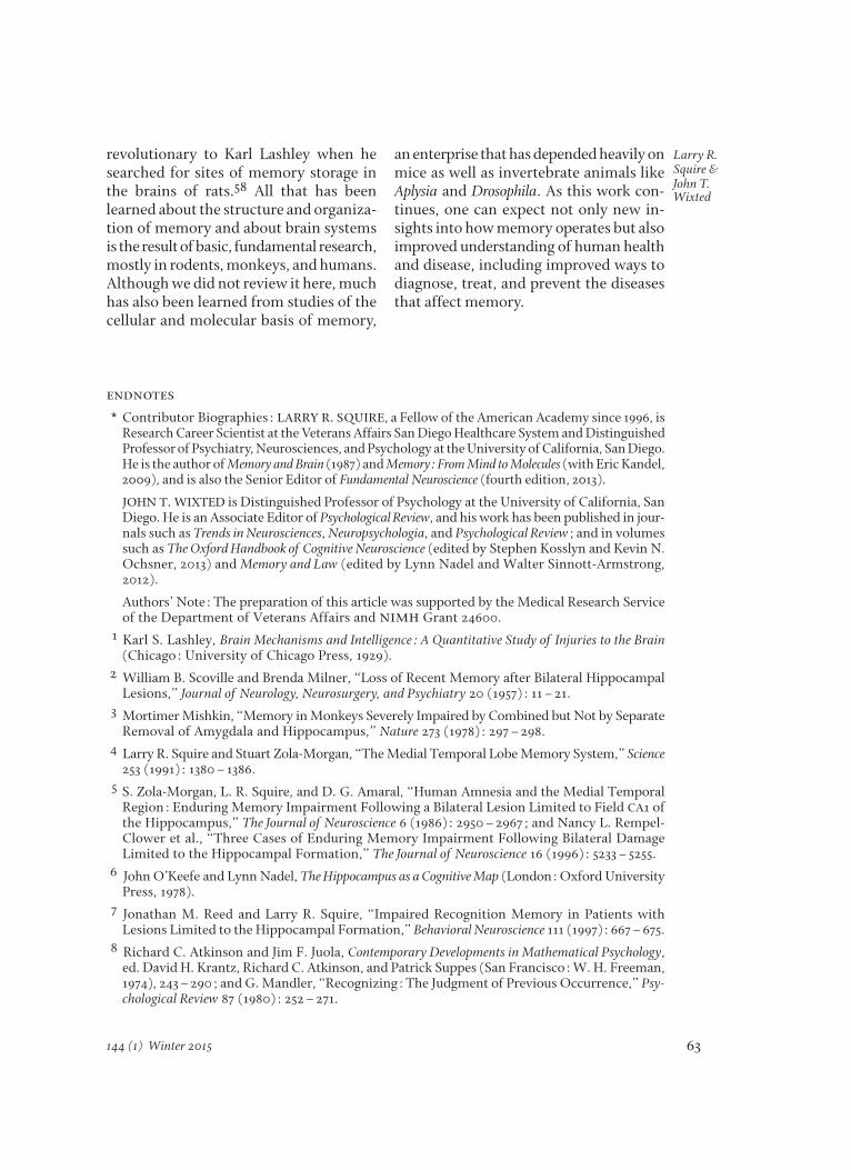
endnotes* Contributor Biographies: LARRY R. SQUIRE, a Fellow of the American Academy since 1996, is
Research Career Scientist at the Veterans Affairs San Diego Healthcare System and DistinguishedProfessor of Psychiatry, Neurosciences, and Psychology at the University of California, San Diego.He is the author of Memory and Brain (1987) and Memory: From Mind to Molecules (with Eric Kandel,2009), and is also the Senior Editor of Fundamental Neuroscience (fourth edition, 2013).
JOHN T. WIXTED is Distinguished Professor of Psychology at the University of California, SanDiego. He is an Associate Editor of Psychological Review, and his work has been published in jour -nals such as Trends in Neurosciences, Neuropsychologia, and Psychological Review; and in volumessuch as The Oxford Handbook of Cognitive Neuroscience (edited by Stephen Kosslyn and Kevin N.Ochsner, 2013) and Memory and Law (edited by Lynn Nadel and Walter Sinnott-Armstrong,2012).
Authors’ Note: The preparation of this article was supported by the Medical Research Serviceof the Department of Veterans Affairs and nimh Grant 24600.
1 Karl S. Lashley, Brain Mechanisms and Intelligence: A Quantitative Study of Injuries to the Brain(Chicago: University of Chicago Press, 1929).
2 William B. Scoville and Brenda Milner, “Loss of Recent Memory after Bilateral HippocampalLesions,” Journal of Neurology, Neurosurgery, and Psychiatry 20 (1957): 11–21.
3 Mortimer Mishkin, “Memory in Monkeys Severely Impaired by Combined but Not by Sepa rateRemoval of Amygdala and Hippocampus,” Nature 273 (1978): 297–298.
4 Larry R. Squire and Stuart Zola-Morgan, “The Medial Temporal Lobe Memory System,” Science253 (1991): 1380–1386.
5 S. Zola-Morgan, L. R. Squire, and D. G. Amaral, “Human Amnesia and the Medial TemporalRegion: Enduring Memory Impairment Following a Bilateral Lesion Limited to Field CA1 ofthe Hippocampus,” The Journal of Neuroscience 6 (1986): 2950–2967; and Nancy L. Rempel-Clower et al., “Three Cases of Enduring Memory Impairment Following Bilateral DamageLimited to the Hippocampal Formation,” The Journal of Neuroscience 16 (1996): 5233–5255.
6 John O’Keefe and Lynn Nadel, The Hippocampus as a Cognitive Map (London: Oxford Univer sityPress, 1978).
7 Jonathan M. Reed and Larry R. Squire, “Impaired Recognition Memory in Patients withLesions Limited to the Hippocampal Formation,” Behavioral Neuroscience 111 (1997): 667–675.
8 Richard C. Atkinson and Jim F. Juola, Contemporary Developments in Mathematical Psychology,ed. David H. Krantz, Richard C. Atkinson, and Patrick Suppes (San Francisco: W. H. Freeman,1974), 243–290; and G. Mandler, “Recognizing: The Judgment of Previous Occurrence,” Psy -chological Review 87 (1980): 252–271.
revolutionary to Karl Lashley when hesearched for sites of memory storage inthe brains of rats.58 All that has beenlearned about the structure and organiza-tion of memory and about brain systemsis the result of basic, fundamental research,mostly in rodents, monkeys, and humans.Although we did not review it here, muchhas also been learned from studies of thecellular and molecular basis of memory,
an enterprise that has depended heavily onmice as well as invertebrate animals likeAplysia and Drosophila. As this work con -tin ues, one can expect not only new in -sights into how memory operates but alsoimproved understanding of human healthand disease, including improved ways todiagnose, treat, and prevent the diseasesthat affect memory.

64
Remem -bering
Dædalus, the Journal of the American Academy of Arts & Sciences
9 Malcolm W. Brown and John P. Aggleton, “Recognition Memory: What Are the Roles of thePerirhinal Cortex and Hippocampus?” Nature Reviews Neuroscience 2 (2001): 51–61.
10 H. Eichenbaum, A. P. Yonelinas, and C. Ranganath, “The Medial Temporal Lobe and Recog-nition Memory,” Annual Review of Neuroscience 30 (2007): 123–152.
11 Z. Song et al., “Impaired Capacity for Familiarity after Hippocampal Damage,” Proceedings ofthe National Academy of Sciences 108 (2011): 9655–9660; Christine N. Smith, John T. Wixted,and Larry R. Squire, “The Hippocampus Supports Both Recollection and Familiarity WhenMem ories Are Strong,” The Journal of Neuroscience 31 (2011): 15693–17502; and Z. Song, A. Jen -eson, and L. R. Squire, “Medial Temporal Lobe Function and Recognition: A Novel Ap proachto Separating the Contribution of Recollection and Familiarity,” The Journal of Neuro science 31(2011): 16026–16032.
12 W. A. Suzuki and D. G. Amaral, “Topographic Organization of the Reciprocal Connectionsbe tween the Monkey Entorhinal Cortex and the Perirhinal and Perihippocampal Cortices,”The Journal of Neuroscience 14 (1994): 1856–1877; and L. R. Squire, “Mechanisms of Memory,”Science 232 (1986): 1612–1619.
13 Elizabeth A. Buffalo, Patrick S. F. Bellgowan, and Alex Martin, “Distinct Roles for Medial Tem -poral Lobe Structures in Memory for Objects and Their Locations,” Learning & Memory 13(2006): 638–643; Bernhard P. Staresina, Katherine D. Duncan, and Lila Davachi, “Perirhinaland Parahippocampal Cortices Differentially Contribute to Later Recollection of Object- andScene-Related Event Details,” The Journal of Neuroscience 31 (2011): 8739–8747; Jackson C. Liang,Anthony D. Wagner, and Alison R. Preston, “Content Representation in the Hu man MedialTemporal Lobe,” Cerebral Cortex 23 (2013): 80–96; and Bernhard P. Staresina, Elisa Cooper, andRichard N. Henson, “Reversible Information Flow across the Medial Tem poral Lobe: The Hip -pocampus Links Cortical Modules During Memory Retrieval,” The Journal of Neuro science 33(2013): 14184–14192.
14 John T. Wixted and Larry R. Squire, “The Medial Temporal Lobe and the Attributes of Mem-ory,” Trends in Cognitive Science 15 (2011): 210–217.
15 Michael A. Yassa and Craig E. L. Stark, “Pattern Separation in the Hippocampus,” Trends inNeurosciences 34 (2011): 515–525; and Xu Liu et al., “Optogenetic Stimulation of a Hippo -campal Engram Activates Fear Memory Recall,” Nature 1038 (2012): 381–385.
16 Neal J. Cohen and Larry R. Squire, “Preserved Learning and Retention of Pattern AnalyzingSkill in Amnesia: Dissociation of Knowing How and Knowing That,” Science 210 (1980):207–209.
17 Endel Tulving and Daniel L. Schacter, “Priming and Human Memory Systems,” Science 247(1990): 301–306; Elizabeth K. Warrington and Rosaleen A. McCarthy, “Categories of Knowl - edge: Further Fractionations and an Attempted Integration,” Brain 110 (1987): 1273–1296;and Daniel L. Schacter and Randy L. Buckner, “Priming and the Brain,” Neuron 20 (1998):185–195.
18 Carolyn Backer Cave and Larry R. Squire, “Intact and Long-Lasting Repetition Priming in Am -nesia,” The Journal of Experimental Psychology: Learning, Memory and Cognition 18 (1992): 509–520.
19 Stephan B. Hamann and Larry R. Squire, “Intact Perceptual Memory in the Absence of Con-scious Memory,” Behavioral Neuroscience 111 (1997): 850–854.
20 Mortimer Mishkin et al., “Memories and Habits: Two Neural Systems,” in Neurobiology ofHuman Learning and Memory, ed. G. Lynch, J. L. McGaugh, and W. M. Weinberger (NewYork: Guilford, 1984), 65–77.
21 Barbara J. Knowlton, Jennifer A. Mangels, and Larry R. Squire, “A Neostriatal Habit LearningSystem in Humans,” Science 273 (1996): 1399–1402.
22 Delay eyeblink conditioning is a form of Pavlovian conditioning in which a conditioned stim -ulus (such as a tone) is presented and remains on until the unconditioned stimulus (such as apuff of air to the eye) is presented. The two stimuli overlap and co-terminate. D. A. McCormick

65
Larry R. Squire & John T. Wixted
144 (1) Winter 2015
et al., “Initial Localization of the Memory Trace for a Basic Form of Learning,” Proceedings ofthe National Academy of Sciences 79 (1982): 2731–2735.
23 Robert E. Clark and Larry R. Squire, “Awareness and the Conditioned Eyeblink Response,” inEyeblink Classical Conditioning, Vol. 1: Applications in Humans, ed. Diana S. Woodruff-Pak andJoseph E. Steinmetz (Norwell, Mass.: Kluwer Academic Publishers, 2000), 229–251; and K. M.Christian and R. F. Thompson, “Neural Substrates of Eyeblink Conditioning: Acquisi tion andRetention,” Learning & Memory 10 (2003): 427–455.
24 Joseph LeDoux, The Emotional Brain (New York: Simon & Schuster, 1996).25 Larry R. Squire and Stuart Zola-Morgan, “Memory: Brain Systems and Behavior,” Trends in
Neurosciences 11 (1988): 170–175.26 For earlier versions of this diagram, see Squire, “Mechanisms of Memory.” 27 Larry R. Squire and Peter J. Bayley, “The Neuroscience of Remote Memory,” Current Opinion in
Neurobiology 17 (2007): 185–196.28 Morris Moscovitch et al., “The Cognitive Neuroscience of Remote Episodic, Semantic and
Spatial Memory,” Current Opinion in Neurobiology 16 (2006): 179–190.29 Squire et al., “The Neuroscience of Remote Memory.” 30 Paul W. Frankland and Bruno Bontempi, “The Organization of Recent and Remote Memo-
ries,” Nature Reviews Neuroscience 6 (2005): 119–130.31 Leonardo Restivo et al., “The Formation of Recent and Remote Memory is Associated with
Time-Dependent Formation of Dendritic Spines in the Hippocampus and Anterior CingulateCortex,” The Journal of Neuroscience 29 (2009): 8206–8214.
32 C. N. Smith and L. R. Squire, “Medial Temporal Lobe Activity During Retrieval of SemanticMemory Is Related to the Age of the Memory,” The Journal of Neuroscience 29 (2009): 930–938.
33 Elizabeth F. Loftus, “Planting Misinformation in the Human Mind: A 30-Year Investigation ofthe Malleability of Memory,” Learning & Memory 12 (2005): 361–366; Jonathan L. Lee, “Re -consolidation: Maintaining Memory Relevance,” Trends in Neuroscience 32 (2009): 413–420;Yadin Dudai, “The Restless Engram: Consolidations Never End,” The Annual Review of Neu-roscience 35 (2012): 227–247; Peggy L. St. Jacques, Christopher Olm, and Daniel L. Schacter,“Neural Mechanisms of Reactivation-Induced Updating that Enhance and Distort Memory,”Proceedings of the National Academy of Sciences 110 (49): 19671–19678; and Karim Nader, GlennE. Schafe, and Joseph E. LeDoux, “Fear Memories Require Protein Synthesis in the Amyg-dala for Reconsolidation after Retrieval,” Nature 406 (2000): 722–726.
34 K. Matthew Lattal and Ted Abel, “Behavioral Impairments Caused by Injections of the ProteinSynthesis Inhibitor Anisomycin after Contextual Retrieval Reverse with Time,” Proceedings ofthe National Academy of Sciences 101 (2004): 4667–4672; and Ann E. Power et al., “AnisomycinIn fused into the Hippocampus Fails to Block ‘Reconsolidation’ but Impairs Extinction: TheRole of Re-Exposure Duration,” Learning & Memory 13 (2006): 27–34.
35 Maria H. Milekic and Cristina M. Alberini, “Temporally Graded Requirement for Protein Syn - thesis Following Memory Reactivation,” Neuron 36 (2002): 521–525.
36 M. A. Wilson and B. L. McNaughton, “Reactivation of Hippocampal Ensemble Memories Dur-ing Sleep,” Science 265 (1994): 676–679.
37 Daoyun Ji and Matthew A. Wilson, “Coordinated Memory Replay in the Visual Cortex andHippocampus During Sleep,” Nature Neuroscience 10 (2007): 100–107.
38 Valérie Ego-Stengel and Matthew A. Wilson, “Disruption of Ripple-Associated HippocampalActivity During Rest Impairs Spatial Learning in the Rat,” Hippocampus 20 (2010): 1–10.
39 Björn Rasch et al., “Odor Cues During Slow-Wave Sleep Prompt Declarative Memory Consoli -da tion,” Science 315 (2007): 1426–1429.

66
Remem -bering
Dædalus, the Journal of the American Academy of Arts & Sciences
40Philippe Peigneux et al., “Are Spatial Memories Strengthened in the Human HippocampusDuring Slow-Wave Sleep?” Neuron 44 (2004): 535–545.
41 Marion Inostroza and Jan Born, “Sleep for Preserving and Transforming Episodic Memory,”Annual Review of Neuroscience 36 (2013): 79–102.
42 Sara C. Mednick et al., “An Opportunistic Theory of Cellular and Systems Consolidation,”Trends in Neurosciences 34 (2011): 504–514.
43 Mattias P. Karlsson and Loren M. Frank, “Awake Replay of Remote Experiences in the Hip-pocampus,” Nature Neuroscience 12 (2009): 913–918.
44 Arielle Tambini, Nicholas Ketz, and Lila Davachi, “Enhanced Brain Correlations During Restare Related to Memory for Recent Experiences,” Neuron 65 (2010): 280–290.
45 E. De Renzi, “Memory Disorders Following Focal Neocortical Damage,” Philosophical Trans-actions of the Royal Society B: Biological Sciences 298 (1982): 73–83; Mortimer Mishkin, “A Mem -ory System in the Monkey,” Philosophical Transactions of the Royal Society B: Biological Sciences298 (1982): 85–92; Larry R. Squire, Memory and Brain (New York: Oxford University Press,1987); and Antonio R. Damasio, “Time-Locked Multiregional Retroactivation: A Systems-Level Proposal for the Neural Substrates of Recall and Recognition,” Cognition 33 (1989):25–62.
46 K. S. Lashley, Brain Mechanisms and Intelligence: A Quantitative Study of Injuries to the Brain (Chi -cago: Chicago University Press, 1929).
47 Oliver Sacks, “The Case of the Colorblind Painter,” in Oliver Sacks, An Anthropologist on Mars:Seven Para doxical Tales (New York: Random House, 1995), 3–41.
48 Warrington and McCarthy, “Categories of Knowledge: Further Fractionations and an At -tempted Integration.”
49 Alex Martin, “The Representation of Object Concepts in the Brain,” Annual Review of Psycho logy58 (2007): 25–45.
50 Scott A. Small et al., “Imaging Correlates of Brain Function in Monkeys and Rats Isolates aHippocampal Subregion Differentially Vulnerable to Aging,” Proceedings of the NationalAcademy of Sciences 101 (2004): 7181–7186.
51 C. R. Jack, Jr., et al., “Rate of Medial Temporal Lobe Atrophy in Typical Aging and Alzheimer’sDisease,” Neurology 51 (1998): 993–999.
52 Kirk I. Erickson et al., “Exercise Training Increases Size of Hippocampus and Improves Mem-ory,” Proceedings of the National Academy of Sciences 108 (2011): 3017–3022.
53 Bradley T. Hyman et al., “Alzheimer’s Disease: Cell-Speci½c Pathology Isolates the Hip-pocampal Formation,” Science 225 (1984): 1168–1170; and M. J. West, P. D. Coleman, D. G.Flood, and J. C. Troncoso, “Differences in the Pattern of Hippocampal Neuronal Loss inNormal Ageing and Alzheimer’s Disease,” Lancet 17 (1994): 769–772.
54 Jack et al., “Rate of Medial Temporal Lobe Atrophy in Typical Aging and Alzheimer’s Disease.”55 John R. Hodges and Kim S. Graham, “Episodic Memory: Insights from Semantic Dementia,”
Philosophical Transactions of the Royal Society B: Biological Sciences 1413 (2001): 1423–1434.56 D. A. Levy, P. J. Bayley, and L. R. Squire, “The Anatomy of Semantic Knowledge: Medial vs.
Lateral Temporal Lobe,” Proceedings of the National Academy of Sciences 101 (2004): 6710–6715;and Karalyn Patterson, Peter J. Nestor, and Timothy T. Rogers, “Where Do You Know WhatYou Know?” Nature Reviews Neuroscience 8 (2007): 976–987.
57 K. S. Graham, J. T. Becker, and J. R. Hodges, “On the Relationship between Knowledge andMemory for Pictures: Evidence from the Study of Patients with Semantic Dementia andAlzheimer’s Disease,” Journal of the International Neuropsychological Society 3 (1997): 534–544.
58 Lashley, Brain Mechanisms and Intelligence.