Reliability of Growth Indicators and Efficiency of...
20
Review Article Reliability of Growth Indicators and Efficiency of Functional Treatment for Skeletal Class II Malocclusion: Current Evidence and Controversies Giuseppe Perinetti and Luca Contardo Department of Medical, Surgical and Health Sciences, School of Dentistry, University of Trieste, Trieste, Italy Correspondence should be addressed to Giuseppe Perinetti; [email protected] Received 1 October 2016; Accepted 14 December 2016; Published 11 January 2017 Academic Editor: Enita Nakaˇ s Copyright © 2017 G. Perinetti and L. Contardo. is is an open access article distributed under the Creative Commons Attribution License, which permits unrestricted use, distribution, and reproduction in any medium, provided the original work is properly cited. Current evidence on the reliability of growth indicators in the identification of the pubertal growth spurt and efficiency of functional treatment for skeletal Class II malocclusion, the timing of which relies on such indicators, is highly controversial. Regarding growth indicators, the hand and wrist (including the sole middle phalanx of the third finger) maturation method and the standing height recording appear to be most reliable. Other methods are subjected to controversies or were showed to be unreliable. Main sources of controversies include use of single stages instead of ossification events and diagnostic reliability conjecturally based on correlation analyses. Regarding evidence on the efficiency of functional treatment, when treated during the pubertal growth spurt, more favorable response is seen in skeletal Class II patients even though large individual responsiveness remains. Main sources of controversies include design of clinical trials, definition of Class II malocclusion, and lack of inclusion of skeletal maturity among the prognostic factors. While no growth indicator may be considered to have a full diagnostic reliability in the identification of the pubertal growth spurt, their use may still be recommended for increasing efficiency of functional treatment for skeletal Class II malocclusion. 1. Background It has been reported decades ago that the growth rate of the mandible is not constant throughout development [1– 3] showing a peak during puberty [1, 2, 4, 5]. However, the intensity, onset, and duration of the pubertal growth peak (including mandibular growth peak) are subjected to note- worthy individual variations [1, 3–5]. A deficient mandibular growth on the sagittal plane is the most frequent diagnostic finding in skeletal (and dental) Class II malocclusion that occurs in up to one-third of the population [6, 7]. us, a therapy able to enhance mandibular growth is indicated in skeletal Class II patients [8]. In this regard, animal studies have shown that forward mandibular displacement enhances condylar growth resulting in significant mandible elongation [9, 10]. Consequently, a wide range of functional appliances (either removable or fixed) have been developed to stimulate mandibular growth by forward posturing of the mandible. To date, the efficiency of functional treatment for skeletal Class II malocclusion is still controversial with reviews reporting very limited [11–13], partial [14–16], or relevant [17– 19] effects of such treatment in terms of induced mandibular growth. Among the reasons for such inconsistencies is the timing, that is, skeletal maturity [18, 20, 21], during which treatment is performed. Clinical trials indicated that the func- tional treatment for skeletal Class II malocclusion is efficient when performed during the pubertal growth spurt [22–26] and without clinically relevant effects when performed before [27–29]. erefore, over the last six decades, efforts have been carried out to find reliable and reproducible indicators of skeletal maturity in individual subjects [5, 20, 30–33]. ese indicators have included radiographic hand and wrist maturational (HWM) methods [30, 34, 35], third finger middle phalanx (MPM) method [36–38], cervical vertebral maturational (CVM) methods [20, 33, 39], dental maturation Hindawi BioMed Research International Volume 2017, Article ID 1367691, 19 pages https://doi.org/10.1155/2017/1367691
Transcript of Reliability of Growth Indicators and Efficiency of...

Review ArticleReliability of Growth Indicators and Efficiency ofFunctional Treatment for Skeletal Class II Malocclusion:Current Evidence and Controversies
Giuseppe Perinetti and Luca Contardo
Department of Medical, Surgical and Health Sciences, School of Dentistry, University of Trieste, Trieste, Italy
Correspondence should be addressed to Giuseppe Perinetti; [email protected]
Received 1 October 2016; Accepted 14 December 2016; Published 11 January 2017
Academic Editor: Enita Nakas
Copyright © 2017 G. Perinetti and L. Contardo.This is an open access article distributed under the Creative Commons AttributionLicense, which permits unrestricted use, distribution, and reproduction in any medium, provided the original work is properlycited.
Current evidence on the reliability of growth indicators in the identification of the pubertal growth spurt and efficiency of functionaltreatment for skeletal Class II malocclusion, the timing of which relies on such indicators, is highly controversial. Regardinggrowth indicators, the hand and wrist (including the sole middle phalanx of the third finger) maturation method and the standingheight recording appear to be most reliable. Other methods are subjected to controversies or were showed to be unreliable. Mainsources of controversies include use of single stages instead of ossification events and diagnostic reliability conjecturally based oncorrelation analyses. Regarding evidence on the efficiency of functional treatment, when treated during the pubertal growth spurt,more favorable response is seen in skeletal Class II patients even though large individual responsiveness remains. Main sources ofcontroversies include design of clinical trials, definition of Class II malocclusion, and lack of inclusion of skeletal maturity amongthe prognostic factors. While no growth indicator may be considered to have a full diagnostic reliability in the identification of thepubertal growth spurt, their use may still be recommended for increasing efficiency of functional treatment for skeletal Class IImalocclusion.
1. Background
It has been reported decades ago that the growth rate ofthe mandible is not constant throughout development [1–3] showing a peak during puberty [1, 2, 4, 5]. However, theintensity, onset, and duration of the pubertal growth peak(including mandibular growth peak) are subjected to note-worthy individual variations [1, 3–5]. A deficient mandibulargrowth on the sagittal plane is the most frequent diagnosticfinding in skeletal (and dental) Class II malocclusion thatoccurs in up to one-third of the population [6, 7]. Thus, atherapy able to enhance mandibular growth is indicated inskeletal Class II patients [8]. In this regard, animal studieshave shown that forward mandibular displacement enhancescondylar growth resulting in significant mandible elongation[9, 10]. Consequently, a wide range of functional appliances(either removable or fixed) have been developed to stimulatemandibular growth by forward posturing of the mandible.
To date, the efficiency of functional treatment for skeletalClass II malocclusion is still controversial with reviewsreporting very limited [11–13], partial [14–16], or relevant [17–19] effects of such treatment in terms of induced mandibulargrowth. Among the reasons for such inconsistencies is thetiming, that is, skeletal maturity [18, 20, 21], during whichtreatment is performed.Clinical trials indicated that the func-tional treatment for skeletal Class II malocclusion is efficientwhen performed during the pubertal growth spurt [22–26]andwithout clinically relevant effects when performed before[27–29].
Therefore, over the last six decades, efforts have beencarried out to find reliable and reproducible indicatorsof skeletal maturity in individual subjects [5, 20, 30–33].These indicators have included radiographic hand and wristmaturational (HWM) methods [30, 34, 35], third fingermiddle phalanx (MPM) method [36–38], cervical vertebralmaturational (CVM)methods [20, 33, 39], dental maturation
HindawiBioMed Research InternationalVolume 2017, Article ID 1367691, 19 pageshttps://doi.org/10.1155/2017/1367691
2 BioMed Research International
[31, 32, 40] and dental emergence [32, 41], chronological age[5, 41], and noninvasive biomarkers from serum [42, 43] orgingival crevicular fluid (GCF) [44, 45].
2. Common Issues related to the Investigationand Use of the Skeletal Maturity Indicators
Current evidence on the reliability of the different growthindicators and consequent definition of treatment timing ishighly controversial. Contrasting results have been reportedon the capability of the growth indicators (mainly the CVMmethod) in the identification of the mandibular growth peak[46–53] and on the efficiency of functional treatment forClass II malocclusion [13, 18]. The investigation on growthindicators has common sources of controversies for all theindicators and specific issues related to each indicator.Herein,common controversial issues to all indicators are listed, whilespecific issues and controversies on the functional treatmentare reported below.
2.1. Stages versus Ossification Events. In using radiographicalindicators of growth phase that are based on sequentialdiscrete stages, an important distinction has to be madebetween stages and ossification events [54, 55]. The stagesare specific periods in the development of a bone that havebeen described in that particular rating method, while anossification event occurs when a given stage matures intothe following one [54, 55]. Of particular clinical relevance,as ossification event is defined as the midpoint betweentwo consecutive stages, a proper identification event requiresserial radiographs. The main limitation raised by the useof single stages resides in the concept that these stageshave variable duration [35, 47, 55, 56] as has been seenfor the HWM [5, 55], MPM [37], and CVM [47, 56]methods, making the prediction of the imminent growthspurt less reliable. Therefore, the exact determination of theimminent growth spurt would require closer monitoring ofthe ossification event, that is, longitudinal recordings, ratherthan being based on a single stage. This aspect is of furtherrelevance considering that fine transitional changes in thehand and wrist or cervical vertebral morphology may beresponsible for determining a pubertal or nonpubertal stage.According to these concepts, longitudinal studies on thecapabilities of the different indicators in the identificationof the mandibular growth peak (or pubertal growth spurt)are to be preferred over cross-sectional ones. From a clinicalstandpoint, whenever possible, serial monitoring should bepreferred over growth prediction based on single staging.
2.2. Correlation Analysis versus Diagnostic Reliability. In spiteof the huge number of studies on growth indicators andpubertal growth spurt, the diagnostic reliability of any of thegrowth indicators in the identification of the peak in standingheight or mandibular growth on an individual basis is yetundetermined. Of note, correlations between parameters donot necessarily imply diagnostic accuracy [57, 58].
One of the reasons underlying this noteworthy lack ofdata may reside in the difficulty of obtaining diagnostic
parameters, such as sensitivity, specificity, and accuracy, fromlongitudinal data in a subset of selected subjects all witha predetermined condition (mandibular growth peak) or adiagnostic outcome (a given HWM/CVM stage). However,the identification of a mandibular growth peak requireslongitudinal data, and it is defined as the greatest growthinterval [21, 37].
To overcome such limitations, a recent study [21] usingalready published data on the CVM method [49] has intro-duced a simple procedure to derive data on diagnosticreliability in the case of longitudinal recordings of growthindicators and mandibular growth. In particular, individualCVM stages and increments in mandibular growth recordedlongitudinally were analysed in a group of subjects accordingto the different predetermined annual (chronological) ageintervals. Therefore, a full diagnostic reliability analysis,including sensitivity, specificity, positive and negative predic-tive values (PPVs and NPVs), and accuracy, of a given CVMstage in the identification of the mandibular growth peakcould be carried out within each age interval group. To dateonly limited longitudinal studies reported on the diagnosticreliability of the CVM [21] and MPM [37] methods in theidentification of the mandibular growth peak. Therefore,longitudinal studies reporting diagnostic reliability shouldbe preferred over investigations using bivariate correlations[59, 60] or even multiple regression analyses [61, 62].
2.3. Definition of Total Mandibular Length. In several studieson the reliability of growth indicators [34, 35, 63–66] or on theefficiency of functional treatment for Class II malocclusion[17, 26, 67, 68] (see below), the landmark Articulare (Ar)was used instead of the landmark Condylion (Co) to assessthe posterior end-point of the mandible. The Ar is definedas the point of intersection of the images of the posteriorborder of the ramal process of the mandible and the inferiorborder of the basilar part of the occipital bone [69]. Theproblem with Ar is that it is not an anatomical landmark thatpertains to the mandible exclusively. On the other hand, thelandmarkAr has the advantage of beingmore easily identifiedas compared to the Co. Even though a previous study [70]reported close correlation between the Ar-Pogonion (Pog)and Co-Pog distances on a sample of 60 cases; other evidence[71, 72] suggested the use of the point Co over Ar as beingmore reliable in terms of mandibular growth recording. Inparticular, the posture of the mandible might also affect theposition of Ar [71]. Yet repeatability analysis on a cross-sectional sample [70] does not provide evidence that, in alongitudinal analysis, increments in mandibular length (asAr-Gn and Co-Gn or Ar-Pog and Co-Pog) would yieldoverlapping patterns of mandibular growth peaks (which aremostly used to validate growth indicators). Therefore, futuredata are warranted to fully elucidate whether the differentlandmarks may be used indifferently.
3. Hand and Wrist Maturation Method
The use of the hand and wrist bones for the assessment ofskeletal maturity has initially been reported by Todd [73]

BioMed Research International 3
Table 1: Description of the stages of the hand and wrist maturation (HWM) method according to Fishman [35].
Stage description AttainmentSMI 1: third finger proximal phalanx, epiphysis as wide as metaphysis
Before the standing height and mandibular growthpeaks (prepubertal)
SMI 2: third finger middle phalanx, epiphysis as wide as metaphysisSMI 3: fifth finger middle phalanx, epiphysis as wide as metaphysisSMI 4: thumb, appearance of adductor sesamoidSMI 5: third finger distal phalanx, epiphysis showing capping towardsthe metaphysis
Generally, at coincidence of the standing height andmandibular growth peaks (pubertal)
SMI 6: third finger middle phalanx, epiphysis showing capping towardsthe metaphysisSMI 7: fifth finger middle phalanx, epiphysis showing capping towardsthe metaphysisSMI 8: third finger distal phalanx, fusion of epiphysis and diaphysis
After the standing height and mandibular growth peaks(postpubertal)
SMI 9: third finger proximal phalanx, fusion of epiphysis and diaphysisSMI 10: third finger middle phalanx, fusion of epiphysis and diaphysisSMI 11: radius, fusion of epiphysis and diaphysisThe method is also referred to as skeletal maturity assessment (SMA). SMI, skeletal maturity indicator.
1
2
5 8
37
4
6 10
11
9
Figure 1: Diagram of the stages of the hand and wrist maturation (HWM) method according to Fishman [35]. The method is also referredto as skeletal maturity assessment (SMA). Blue, prepubertal stages; red, pubertal stages; black, postpubertal stages. See Table 1 for details.Modified from Fishman [35] with permission.
followed by others [30, 74, 75]. In particular, all of thesemethods were based on the assessment of a skeletal age (inyears) according to specific ossification events of the handand wrist. Subsequently, such individual skeletal age had tobe compared with reported norms. For reasons listed below,stage-based procedure for the hand and wrist maturation hasbeen added.Among the different stage-basedHWMmethods[32, 34, 35, 65], the most used nowadays both in researchand clinical practice is likely to be that proposed by Fishman[35], also known as skeletal maturation assessment (SMA).Details of the 11-stage HWM method according to Fishman[20] are summarized in Table 1 and shown in Figure 1, whilemain longitudinal investigations in relation to mandibulargrowth in untreated subjects without major malocclusion aresummarized in Table 2.
3.1. Current Evidence. All the published longitudinal studieson theHWMmethods andmandibular growth peak includedCaucasian [35, 53, 65] and Australian aborigine [34, 76]subjects, and none reported a specific diagnostic reliabilityanalysis. Tofani [65] reported that onset of fusion of distalphalanges are good predictors of mandibular growth peak;however, this study included only females. The study byGrave [34] also reported moderate significant correlationsof the hand and wrist maturation with mandibular growthpeak for both females and males. A further study by Graveand Brown [76] on the same sample reported previously[34], investigating the HWM method with standing height,reported that peak height velocity would occur up to 3 and6 months later, in males and females, respectively, of theattainment of the third finger middle phalanx (MP3) stage

4 BioMed Research International
Table2:Mainlong
itudinalstudies
ontheh
andandwris
tmaturation(H
WM)m
etho
dandmandibu
larg
rowth
peak
inun
treated
subjectswith
outm
ajor
malocclu
sion.
Stud
ySampleo
rigin
and
otherinformation
Samples
izea
ndsex
distrib
ution/ager
ange
Handandwris
tmaturation
assessment
Main
mandibu
lar
parameter(s)
Statisticalanalysis
Mainresults
Clinicalim
plications
accordingto
thea
utho
rs
Tofani
1972
[65]
Broadb
ent-B
olton
grow
thstu
dy20
F/9–
18yrs
Onsetof
fusio
nof
the
firstandthird
finger
distalph
alanges
Ar-Po
g,Ar-Go,
Go-Po
g
Differencesb
etweenpre-
andpo
stpub
ertaland
correlationanalyses
Age
ofon
seto
ffusionof
dista
lphalanges
andthat
form
andibu
larg
rowth
peak
weres
ignificantly
correlated
Onsetof
fusio
nof
dista
lph
alangesa
regood
predictorsof
mandibu
lar
grow
thpeak
Grave
1973
[34]
Austr
alian
aborigines
36F,52
M/8–18y
rsCu
stom
metho
dAr-Po
gCorrelatio
nanalyses
Somem
oderates
ignificant
correlations
weres
eenfor
females
andmales
TheH
WM
metho
dmay
beuseful
inclinicalpractice
Fishman
1982
[35]
DenverC
hild
Research
Stud
yand
ownpractic
e206F,196M/0–25y
rs
Eleven-stage
metho
d(SMIs)a
ccording
toFishman
[35]
(Figure1)
Ar-Gn
Differencesa
mon
gsta
ges
Maxim
umgrow
thincrem
entsweres
een
durin
gsta
ges5
–7
TheS
MIsprovidea
keyto
identifi
catio
nof
maturation
levelw
ithim
portant
clinicalapp
lications
Mellio
netal.2013
[53]
Broadb
ent-B
olton
grow
thstu
dy(a)
50F,50
M/8
and10yrsa
tleastfor
females
and
males,respectively
,with
6to
11annu
alrecordings
Eleven-stage
metho
d(SMIs)a
ccording
toFishman
[35]
(Figure1)
Co-Gn
Actualagea
tonsetand
peak
inmandibu
larg
rowth
used
astheg
oldsta
ndards
againstw
hich
keyages
inferred
from
SMIsmetho
dwas
compared
TheS
MIsshow
edin
males
andfemales
amod
erately
stron
gor
weaker
relatio
nships,respectively
,to
thetim
ingforthe
onset
andpeak
inmandibu
lar
grow
th
TheS
MIsappear
tooff
ertheb
estind
icationthat
peak
grow
thvelocityhas
been
reached
Stud
iesu
singmaturationmetho
dbasedon
ossifi
catio
nevents(stages)arerepresented.A
r,Articulare;Po
g,Po
gonion
;Go,Gon
ion;Gn,Gnathion;Co,Con
dylio
n;SM
Is,skeletalm
aturationindicators(according
toFishman
[35]).No
te.a:itm
ayinclu
desomeC
lassIIsubjects.
BioMed Research International 5
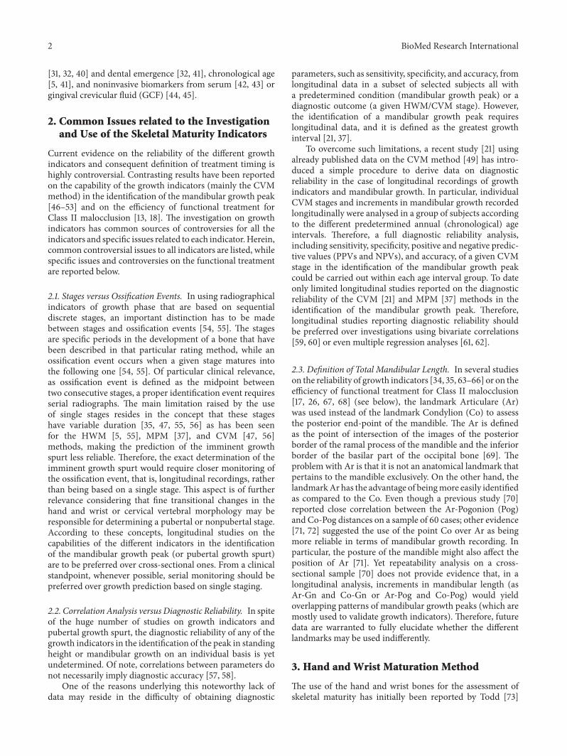
MPS3 MPS4 MPS5MPS1 MPS2
Figure 2:Diagramof the improved third fingermiddle phalanxmaturation (MPM)method according to Perinetti et al. [37]. Blue, prepubertalstages; red, pubertal stages; black, postpubertal stages. See Table 3 for details. Modified from Perinetti et al. [37] with permission.
G (corresponding to the SMI6, See Table 1). In the HWMmethod according to Fishman [35], peak in mandibulargrowth (as Ar-Gn) would occur in stage 6 and 7 for femalesand males, respectively [35]. Further studies correlated thisHWMmethodwith standing height [32, 54, 76]. Similarly, thestudy by Mellion et al. [53] reported for the HWM accordingto Fishman [35] a moderately strong or weaker relationshipsin males and females, respectively. In particular, the HWMmethod assessments had consistently lower errors than eithermean chronologic age or CVM method in the identificationof both the peaks in standing height and mandibular length[53].
Of note, a previous longitudinal study [77] compared theskeletal age of the whole HWM method (according to Todd[73] and Greulich and Pyle [30]) with specific ossificationevents of the first, second, and third finger, referred toas the three-finger maturation assessment. As a result, thethree-finger maturation assessments were shown to maturein slight advancement than the whole HWM assessments.However, this study [77] was based on correlation analysesand differences in skeletal age between methods, lacking atrue diagnostic analysis [78] of concordance or measurementof agreement [79].
3.2. Current Controversies. The Greulich and Pyle method[30] and other similar methods [73–75] have been criticizedin that it may be difficult to set a reference standard,because of the differential rate of maturation in differentbones across individuals of the same population or acrossdifferent population [54, 80]. For this reason, several stan-dards, that is, norms, have been published for the hand andwrist maturation assessment according to the population ofinterest. For more detail, see Greulich and Pyle [30] andTodd [73] for white American subjects, Sutow and Ohwada[74] for Japanese subjects, and Tanner and Whitehouse [75]for British subjects. However, such norms are not alwaysavailable for each population, while another important issuerelates to the secular trends, with successive generationsbecoming taller and reaching puberty at earlier stages [81,82]. Therefore, the staging of skeletal maturity by describingspecific ossification events on the hand-wrist radiograph[32, 34, 35, 53, 65, 66, 83, 84] may be a valid tool asbeing more independent of differences among populationsand secular trends and availability of published standards[80]. The methods based on ossification events [32, 34,35] might thus be considered to have a wider clinicalapplicability.
3.3. Clinical Implication. Even though the number of studiescorrelating theHWMmethodswithmandibular growth peakis limited (Table 2), all of these investigations concluded thatthese methods may be useful in clinical practice. Therefore,the use of the HWM method may be recommended forplanning treatment timing. In spite of this favorable evidence,the HWM method has a main disadvantage residing inthe need of an additional film, with consequent increasedradiation exposure of the whole hand and wrist. This aspectwould prevent a serial recording to monitor closely theossification events, limiting the diagnosis that has to rely onsingle stages.
4. Third Finger Middle PhalanxMaturation Method
Previous studies reported above on the HMW methods [34,54, 76, 85] provided an indication of the possibility for thethird finger middle phalanx maturation to the used alone asan indicator of skeletal maturity. Close concurrence of theattainment of MP3 stage G with the peak height velocity hasbeen reported for both males and females [54, 85]. Similarresults were seen when correlating the third finger middlephalanx maturation with mandibular growth peak [35, 76].Therefore, the use of the sole third fingermiddle phalanx for amaturationalmethod has been proposed [36, 38, 86–88].Thisthird finger middle phalanx maturation (MPM) method [37,78] would thus have the advantage of an easy interpretationof the stages, without double contours or superimposition byother structures. Details of a 5-stageMPMmethod accordingto Perinetti et al. [37] are summarized in Table 3 and showedin Figure 2, while the only longitudinal investigation [37] inrelation to mandibular growth in untreated subjects withoutmajor malocclusion is summarized in Table 4.
4.1. Current Evidence. All of previous investigations [36–38, 78, 86–88] suggested the use of the MPM method inclinical practice. The main advantage of the MPM methodresides in the minimal radiation exposure that would allowclose monitoring of the ossification events by longitudinalrecordings.Therefore, ideal timing of treatment in individualpatients may be identified more precisely as compared towhen information comes from single recording, as for thecase of the HWM and CVM methods. Finally, the MPMmethod is of easy execution and interpretation and may beperformed in any clinical setting with minimal instrumenta-tion. In spite of the potential clinical advantages offered by
6 BioMed Research International
Table 3: Description of the stages of the third finger middle phalanx maturation (MPM) method according to Perinetti et al. [37].
Stage description AttainmentMPS1: epiphysis is narrower than the metaphysis, or epiphysis is as wide asmetaphysis but with both tapered and rounded lateral borders. Epiphysis andmetaphysis are not fused. Reported as MP3-F [32]
More than 1 year before the onset of thepubertal growth spurt [32] or mandibulargrowth peak [37]
MPS2: epiphysis is at least as wide as the metaphysis with sides increasingthickness and showing a clear line of demarcation at right angle, either with orwithout lateral steps on the upper contour. In case of asymmetry between thetwo sides, the more mature side is used to assign the stage. Reported as SMI2 [35]or as MP3-FG [32]
1 year before the pubertal growth spurt[32] or mandibular growth peak [37]
MPS3: epiphysis is either as wide as or wider than the metaphysis with lateralsides showing an initial capping towards the metaphysis. In case of asymmetrybetween the two sides, the more mature side is used to assign the stage. Epiphysisand metaphysis are not fused. Reported as SMI6 [35] or as MP3-G [32]
At coincidence of the pubertal growthspurt [32] or mandibular growth peak[37]
MPS4: epiphysis begins to fuse with the metaphysis although contour of theformer is still clearly recognizable. The capping may still be detectable. Reportedas MP3-H [32]
After the pubertal growth spurt [32] ormandibular growth peak [37]
MPS5: epiphysis is totally fused with the metaphysis. Reported as SMI10 [35] oras MP3-I [32]
At the end of the pubertal growth spurt[32]
Table 4: Main longitudinal studies on the third finger middle phalanxmaturation (MPM)method andmandibular growth peak in untreatedsubjects without major malocclusion.
Study
Sampleorigin and
otherinformation
Sample sizeand sex dis-tribution/age
range
Middlephalanx
maturationassessment
Mainmandibularparameter
Statisticalanalysis Main results
Clinical implicationsaccording to the
authors
Perinetti et al.2016 [37]
Burlingtongrowthstudy
15 F, 20M/9–16 yrs
Five-stagecustommethod(Figure 2)
Co-Gn Diagnosticperformance
Stage 2 had asatisfactory butvariable accuracy
in theidentification of
imminentmandibular growth
peak
The MPMmethodmay be useful intreatment timing
Co, Condylion; Gn, Gnathion.
the MPM method, current evidence is still little. The presentinvestigations [36, 38, 78, 86–88] are limited by the cross-sectional designs in which the MPM method was analyzedin correlation [36, 38, 86–88] or in diagnostic agreement [78]with the CVM method. Indeed, such analyses do not provethe diagnostic reliability of the method in the identificationof the pubertal/mandibular growth peak. The results forthe recent longitudinal study [37] on diagnostic reliability(Table 4) showed that the MPM stage 2 (MPS2) precedes themandibular growth spurt, which is generally concomitant ofMPS3. However, even though the overall diagnostic accu-racy of 0.91 was satisfactory, the overall positive predictivevalue was 0.73, thus meaning that false positives may beencountered. This evidence was mainly due to the durationof the MP2 that in some cases lasted for 2 years and it wasmore evident in the older age groups. Again, the following ofthe ossification events should be preferred instead of basinggrowth prediction on single stages [54].
4.2. Clinical Implications. Although further investigationsare needed, the MPS2 and MPS3 may be considered to be
associated with the onset and maximum mandibular growthpeak, respectively, in most of the subjects, and may there-fore be used for planning treatment timing for functionaltreatments especially for skeletal Class II malocclusion [4].According to the minimal radiation exposure, longitudinalmonitoring is recommended to follow closely the ossificationevents. Finally, a combinational use of theMPMmethodwitha further noninvasive indicator of pubertal growth spurt, thatis, standing height, especially in the older adolescents, mightincrease diagnostic reliability [37].
5. Cervical Vertebral Maturation Method
The CVM method was initially proposed by Lamparski[39] and then modified by others [20, 33, 46, 49]. In thisprocedure, the shape of the first cervical vertebrae is analyzedto carry out information on the different growth phase ofthe subject. In particular, the original method by Lamparski[39] uses vertebrae that can be obscured by the thyroid collarand relied on interstage comparisons, while the subsequentvariants of the CVM method [20, 33, 46, 49] were less or
BioMed Research International 7
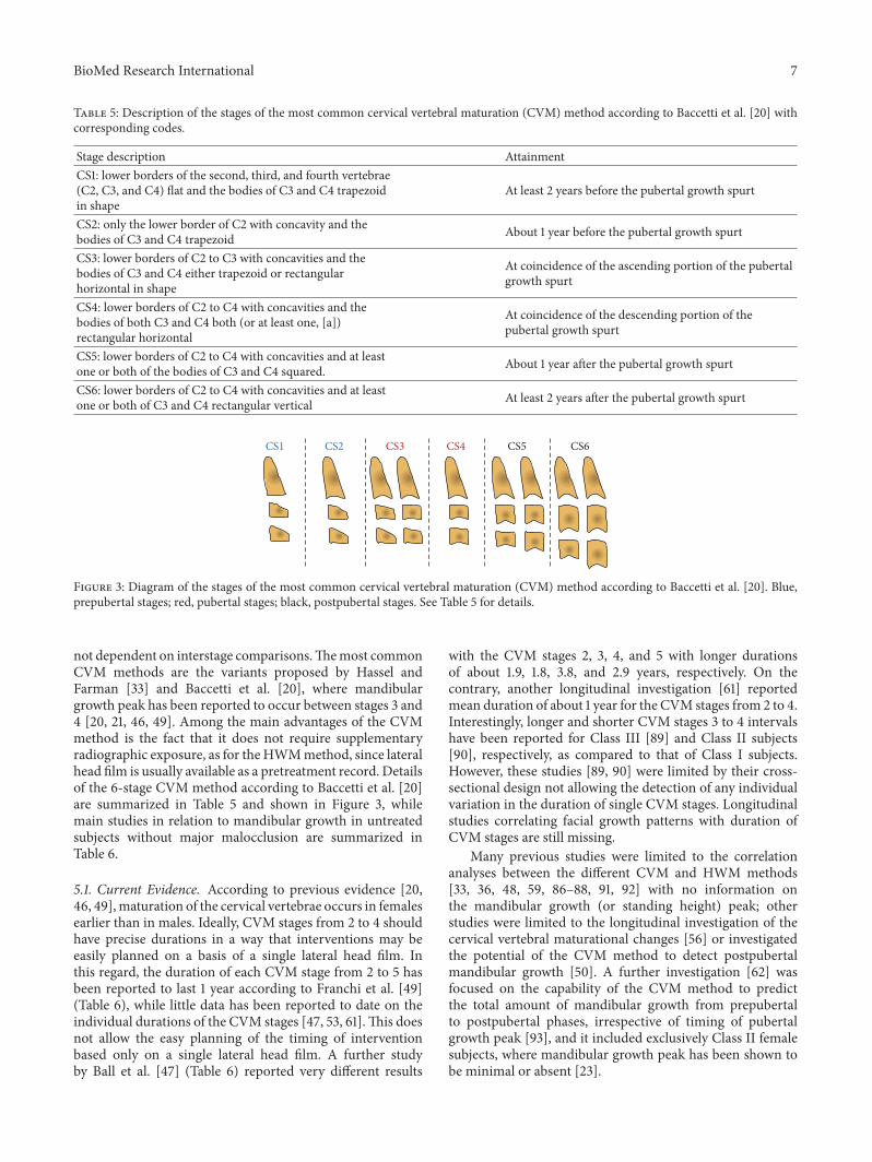
Table 5: Description of the stages of the most common cervical vertebral maturation (CVM) method according to Baccetti et al. [20] withcorresponding codes.
Stage description AttainmentCS1: lower borders of the second, third, and fourth vertebrae(C2, C3, and C4) flat and the bodies of C3 and C4 trapezoidin shape
At least 2 years before the pubertal growth spurt
CS2: only the lower border of C2 with concavity and thebodies of C3 and C4 trapezoid About 1 year before the pubertal growth spurt
CS3: lower borders of C2 to C3 with concavities and thebodies of C3 and C4 either trapezoid or rectangularhorizontal in shape
At coincidence of the ascending portion of the pubertalgrowth spurt
CS4: lower borders of C2 to C4 with concavities and thebodies of both C3 and C4 both (or at least one, [a])rectangular horizontal
At coincidence of the descending portion of thepubertal growth spurt
CS5: lower borders of C2 to C4 with concavities and at leastone or both of the bodies of C3 and C4 squared. About 1 year after the pubertal growth spurt
CS6: lower borders of C2 to C4 with concavities and at leastone or both of C3 and C4 rectangular vertical At least 2 years after the pubertal growth spurt
CS6CS1 CS2 CS3 CS4 CS5
Figure 3: Diagram of the stages of the most common cervical vertebral maturation (CVM) method according to Baccetti et al. [20]. Blue,prepubertal stages; red, pubertal stages; black, postpubertal stages. See Table 5 for details.
not dependent on interstage comparisons.Themost commonCVM methods are the variants proposed by Hassel andFarman [33] and Baccetti et al. [20], where mandibulargrowth peak has been reported to occur between stages 3 and4 [20, 21, 46, 49]. Among the main advantages of the CVMmethod is the fact that it does not require supplementaryradiographic exposure, as for theHWMmethod, since lateralhead film is usually available as a pretreatment record. Detailsof the 6-stage CVM method according to Baccetti et al. [20]are summarized in Table 5 and shown in Figure 3, whilemain studies in relation to mandibular growth in untreatedsubjects without major malocclusion are summarized inTable 6.
5.1. Current Evidence. According to previous evidence [20,46, 49],maturation of the cervical vertebrae occurs in femalesearlier than in males. Ideally, CVM stages from 2 to 4 shouldhave precise durations in a way that interventions may beeasily planned on a basis of a single lateral head film. Inthis regard, the duration of each CVM stage from 2 to 5 hasbeen reported to last 1 year according to Franchi et al. [49](Table 6), while little data has been reported to date on theindividual durations of the CVM stages [47, 53, 61].This doesnot allow the easy planning of the timing of interventionbased only on a single lateral head film. A further studyby Ball et al. [47] (Table 6) reported very different results
with the CVM stages 2, 3, 4, and 5 with longer durationsof about 1.9, 1.8, 3.8, and 2.9 years, respectively. On thecontrary, another longitudinal investigation [61] reportedmean duration of about 1 year for the CVM stages from 2 to 4.Interestingly, longer and shorter CVM stages 3 to 4 intervalshave been reported for Class III [89] and Class II subjects[90], respectively, as compared to that of Class I subjects.However, these studies [89, 90] were limited by their cross-sectional design not allowing the detection of any individualvariation in the duration of single CVM stages. Longitudinalstudies correlating facial growth patterns with duration ofCVM stages are still missing.
Many previous studies were limited to the correlationanalyses between the different CVM and HWM methods[33, 36, 48, 59, 86–88, 91, 92] with no information onthe mandibular growth (or standing height) peak; otherstudies were limited to the longitudinal investigation of thecervical vertebral maturational changes [56] or investigatedthe potential of the CVM method to detect postpubertalmandibular growth [50]. A further investigation [62] wasfocused on the capability of the CVM method to predictthe total amount of mandibular growth from prepubertalto postpubertal phases, irrespective of timing of pubertalgrowth peak [93], and it included exclusively Class II femalesubjects, where mandibular growth peak has been shown tobe minimal or absent [23].

8 BioMed Research International
Table6:Mainlong
itudinalstudies
onthec
ervicalvertebralmaturation(C
VM)m
etho
dandmandibu
larg
rowth
peak
inun
treated
subjectswith
outm
ajor
malocclu
sion.
Stud
ySampleo
rigin
and
otherinformation
Samples
izea
ndsex
distr
ibution/ager
ange
Cervicalvertebral
maturationassessment
Mainmandibu
lar
parameter(s)
Statisticalanalysis
Mainresults
Clinicalim
plications
according
totheA
utho
rsO’Rellyand
Yann
iello
1988
[64]
Broadb
ent-B
olton
grow
thstu
dy13
F/9–
15yrs
Six-sta
geLamparski’s
stand
ards
Ar-Po
g,Ar-Go,
Go-Po
gDifferencesa
mon
gsta
ges
Stages
1–3occurringthey
ear
precedingthep
eakin
most
cases
TheC
VM
canbe
used
toassess
timingof
mandibu
larg
rowth
Franchietal.
2000
[49]
Michigangrow
thstu
dy15
F,9M/7–16y
rsSix-sta
gemod
ified
Lamparski’ssta
ndards
Co-Gn,
Co-Goi,
Goi-G
nDifferencesa
mon
gsta
ges
Totalm
andibu
larlength
show
edtheg
reatestsignificant
increm
entb
etweenstages
3and4
TheC
VM
isav
alid
metho
dfor
thee
valuationof
skele
tal
maturity
andmandibu
lar
grow
thpeak
Guand
McN
amara
2006
[52]
Partof
theM
athews
andWareimplant
sample[143]
13F,7M/≈7–17yrs
Six-stagem
etho
daccordingto
Baccettiet
al.[20](Figure
3)
Co-Gn,
Co-Go,
Go-Me
Differencesa
mon
gsta
ges
Peak
inmandibu
larlength
observed
betweensta
ges3
and
4Not
repo
rted
Chen
etal.2010
[108]
Research
Centre
ofCr
aniofacialGrowth
andDevelo
pmentat
Beijing
University.
55F,32
M/8–18y
rsFo
ur-stage
quantitative
metho
d[94]
Ar-Gn,Ar-Go,
Go-Gn
Differencesa
mon
gsta
ges(as
absoluteand
relativ
egrowth
increm
ent)
Maxim
umgrow
thincrem
ents
weres
eendu
ringsta
geII(b)
with
relativeincrementsmore
consistentthanabsoluteon
es
Use
oftheq
uantitativ
eCVM
metho
disrecommendedfor
treatmentp
lann
ing
Balletal.2011
[47]
Burling
tongrow
thstu
dy90
M/9–18y
rsSix-stagem
etho
daccordingto
Baccettiet
al.[20](Figure
3)Ar-Gn
Differencesa
mon
gstages
ingrou
psof
advanced,average,and
delayedmaturation
Mandibu
larg
rowth
peak
occurred
mainlydu
ringsta
ge4
(which
laste
d3.8y
rs)
TheC
VM
metho
dcann
otpredictthe
onseto
fthe
mandibu
larg
rowth
peak
Mellio
netal.
2013
[53]
Broadb
ent-B
olton
grow
thstu
dy(a)
50F,50
M/8
and10yrs
atleastfor
females
and
males,respectively
,with
6to
11annu
alrecordings
Six-stagem
etho
daccordingto
Baccettiet
al.[20](Figure
3)Co-Gn
Actualagea
tonsetand
peak
inmandibu
lar
grow
thused
astheg
old
stand
ards
againstw
hich
keyages
inferred
from
CVM
metho
dwas
compared
TheC
VM
stagessho
wed
onlya
weakto
mod
erater
elatio
nship
tothetim
ingforthe
onsetand
peak
inmandibu
larg
rowth
Use
oftheC
VM
metho
disno
trecommendedfortreatment
planning
Grayetal.2016
[61]
Burling
tongrow
thstu
dy12
F,13
M/10
–16y
rsSixsta
gemetho
daccordingto
Baccettiet
al.[20](Figure
3)Ar-Gn
Mixed
linearregression
Mandibu
larlengthchanges
weren
otsig
nificantly
associated
with
CVM
stages
TheC
VM
metho
ddo
esno
taccuratelyidentifythe
mandibu
larg
rowth
peak
Perin
ettietal.
2016
[21]
Samea
sFranchi
etal.
[49]
Samea
sFranchi
etal.
[49]
Samea
sFranchi
etal.
[49]
Co-Gn,
Co-Goi,
mMG
Diagn
ostic
perfo
rmance
Stages
3-4have
varia
ble
diagno
stica
ccuracyin
the
identifi
catio
nof
mandibu
lar
grow
thpeak
TheC
VM
canbe
used
inclinicalpractice.Limitatio
nsdu
etotheu
seof
thes
ame
samplefrom
which
them
etho
dwas
deriv
edStud
iesu
singm
aturationmetho
dbasedon
ossifi
catio
nevents(stages)arerepresented.A
r,Articulare;Po
g,Po
gonion
;Go,Gon
ion;Co,Con
dylio
n;Gn,Gnathion;Goi,G
onionintersectio
n;mMG,m
eanmandibu
lar
grow
th([Co-Gn+Goi-G
n]/2).No
te.a:itm
ayinclu
desomeC
lassIIsubjects;
b:sta
geIIequivalent
tostage3
inthe6
-stage
CVM
metho
d.
BioMed Research International 9
However, as for the HWM method, the most relevantinformation may be derived from longitudinal studies inves-tigating the capabilities of these methods in detecting themandibular growth peak, possibly in individual subjects. Pre-vious studies on the CVM method and mandibular growthpeak have reported contrasting results of negligible [47, 48,53, 61, 62] and noteworthy [49, 52, 64, 66] correlations.Interestingly, only few studies [21, 47, 49, 52, 53, 61, 64, 94](Table 6) correlated the CVM method (as stage system)with mandibular growth under longitudinal monitoring.According to this evidence, a total of five studies [21, 49, 52,64, 94] reported mandibular growth peak to occur duringstages 3 and 4, and four [21, 49, 64, 94] of them recommendedthe use of the CVM method in treatment planning. Onestudy [21], however, used the same sample of Franchi etal. [49] from which the CVM method was derived. Theremaining three studies [47, 53, 61] failed to detect a signif-icant correlation between the CVM and mandibular growthpeak and did not recommend the method for treatmentplanning.
5.2. Current Controversies. When reporting on the CVMmethod, the different variants of the method [20, 33, 46, 48,84] have to be taken into account and results should be lim-ited to the investigated methods or parameters [95]. Signifi-cant differences in study designs, cephalometric recordings,and data analysis have to be taken into account when dealingwith clinical usefulness of the CVM method. For instance,apart from the study [21] using the same sample reportedby Franchi et al. [49] (Table 6), the only investigation [66]that has reported on the diagnostic capability of the CVMmethod in the identification of the mandibular growth peakused receiver operating characteristics curves. However, thisstudy [66] was based on a cross-sectional sample and it waslimited to the analysis of the area under the curve, which isnot enough to describe in full the diagnostic reliability of themethod. Therefore, conclusions on the diagnostic reliabilityof the CVM method in the identification of the mandibulargrowth peak have conjecturally been based on differenceamong groups/stages [47, 49, 52, 64, 94], regression analyses[61], or other analyses missing diagnostic capabilities [53].
Another relevant issue when dealing with the CVMmethod resides in its repeatability. The method has beenreported to have poor repeatability [96, 97]. Although thislimitation may be avoided by proper training [98], poorrepeatability has been seen even in studies correlating theCVM method with mandibular growth [62], while longitu-dinal investigations herein considered (Table 6) reported noinformation [52, 53, 64, 94] or good to high repeatability [47,49, 61] in the CVM stage assignment. Finally, when assigningthe CVM stage, it has been suggested that exceptional cases,that is, cases outside the reported norms, may exist [98] andthis may be responsible for doubtful interpretation and poorreproducibility.
5.3. Clinical Implications. As for the HWM method, theCVM methods require films that are usually available asa pretreatment record, while optimal treatment timing is
to be delayed for an undermined term after the diagnosis.Therefore, further reevaluation of the growth phase needsa reexecution of a lateral head film, which would not beindicated. Moreover, the cervical vertebrae might be partiallycovered by the protection collar, which would be necessaryto reduce radiation exposure [99]. Apart from this consider-ation, the use of the CVM method requires proper trainingin stage assignment and knowledge of exception cases [98].Moreover, variability in duration of the CVM stages 2 to 4[47, 56] has been taken into account and functional treatmentrequiring the inclusion of the mandibular growth spurt inthe active treatment period should last until attainment ofCS5 [21]. Future longitudinal studies on diagnostic reliabilityof the CVM method in the identification of the mandibulargrowth peak are still necessary to fully elucidate the clinicalusefulness of the method.
6. Dental Maturation Method
Dental maturity can be assessed by the exfoliation of decidu-ous teeth, such as the secondmolars [100], phases of dentition[101], dental emergence [5, 32], or calcification stages throughthe evaluation of tooth formation [40]. Calcification stages ofthe teeth can be carried out on panoramic radiographs thatare routinely used for different purposes, with mandibularteeth preferred over maxillary ones being less subjectedto superimpositions from other skeletal structures. Evenintraoral radiograph may be used with minimal irradiationto the patient. Therefore, dental maturation has been pro-posed as a further useful method for assessing the growthphase in individual subjects [31]. The most common methodused for scoring dental maturation is the one described byDemirjian et al. [40].This method has the advantage of usingrelative values of the root formation to the crown height,rather than absolute lengths. Foreshortened or elongatedprojections of developing teeth will not affect the reliabilityof this assessment [40]. Details of the dental maturationmethod according to Demirjian et al. [40] are summarizedin Table 7 and shown in Figure 4, while main cross-sectionalstudies of diagnostic reliability using the HWM or CVMmethods in untreated subjects without major malocclusionare summarized in Table 8.
6.1. Current Evidence. The period corresponding to the exfo-liation of the deciduous second molars has been advocatedas favorable for the beginning of a one-phase orthodontictreatment in growing subjects [102]. However, as previouslyreported [100], the exfoliation of the deciduous secondmolars has no significant relationship with the onset of thepubertal growth spurt (Table 8). Similarly, the assessment ofthe phase of dentition (as deciduous, early mixed, mixed, andpermanent) is a simple procedure and has been used to assessthe effects of different treatment timing in Class II patients[103]. However, the only study [101] on diagnostic reliability(Table 6) reported that neither the early mixed nor the mixeddentition phases are valid indicators of the pubertal growthspurt. Therefore, the use of the exfoliation of the deciduoussecond molar or phases of dentition is not recommended

10 BioMed Research International
Table 7: Description of the stages of the most common dental maturation method according to Demirjian et al. [40].
Stage description AttainmentStage D. When (1) the crown formation is complete down to thecementoenamel junction; (2) the superior border of the pulpchamber in the single-root teeth has a definite curved form, with itbeing concave towards the cervical region; the projection of thepulp horns, if present, gives an outline shaped like the top of anumbrella and (3) the beginning of root formation is seen in theform of a spicule
Canine, premolars, and second molar beforethe pubertal growth spurt [28, 57, 110, 112]
Stage E. When (1) the walls of the pulp chamber form straight lines,the continuity of which is broken by the presence of the pulp horn,which is larger than in the previous stage and (2) the root length isless than the crown height
Mostly, canine and first premolar before thepubertal growth spurt [28, 57, 110, 112]
Stage F. When (1) the walls of the pulp chamber form a more orless isosceles triangle, with the apex ending in a funnel shape and(2) the root length is equal to or greater than the crown height
Sometimes, canine before the pubertalgrowth spurt [28, 57, 112]
Stage G. When the walls of the root canal are parallel and its apicalend is still partially open
Canine, premolars, and second molarbefore, during, and after the pubertal growthspurt [28, 57, 110, 112]
Stage H. When (1) the apical end of the root canal is completelyclosed and (2) the periodontal membrane has a uniform widtharound the root and the apex
Second molar after the pubertal growthspurt [28, 57, 112]
Only stages D to H are summarised due to their relevance with the circumpubertal growth phase. In molars, the distal root is considered in assessing the stage[40]. Only results from studies reporting diagnostic reliability analysis are shown regarding the moment of attainment of the different stages for mandibularteeth.
D HE F G
Figure 4: Diagram of the stages of the most common dental maturation method according to Demirjian et al. [40]. Only the stages D to Hare represented due to their relevance with the circumpubertal growth phase. In molars, the distal root should be considered in assessing theG and H stages. Blue, prepubertal stages; grey, any stage; black, postpubertal stages. See Table 7 for details.
for treatment planning. Similarly, dental emergence hasalso been reported to be poorly correlated with pubertalgrowth spurt [5, 32]. Regarding dental calcification stages,high correlations with skeletal maturity have been reportedby most of the investigations performed to date using theCVM [31, 60, 86, 104–109], HWM [106, 110, 111] or MPM[86, 112] methods. As a consequence, most of the studieshave proposed the staging of dental maturation as a reliableindicator of the individual skeletal maturity, which has majordiagnostic implications [31, 60, 106–109, 111, 113–117]. On thecontrary, other studies [104, 105, 110, 112] (Table 8) including ameta-analysis [118] reported a very limited clinical usefulnessof dental maturation in the identification of the pubertalgrowth spurt.
6.2. Current Controversies. The apparent inconsistencyamong all the current investigations on dental and skeletalmaturation (all cross-sectional) resides in the use of properdiagnostic reliability analysis. The present evidence ondiagnostic reliability [104, 105, 110, 112] has revealed that theconclusions reported in previous investigations based oncorrelational analyses [31, 60, 106–109, 111, 113–117] were notactually supported by the results obtained in those studies.The few exceptions seen for early dental developmentalstages, which were reliable in the identification of theprepubertal growth phase [105, 110, 112, 118], would havepoor clinical meaning since early mixed and intermediatemixed dentition may be used instead for the same purpose[5, 32, 101]. Longitudinal studies on the diagnostic reliability

BioMed Research International 11
Table8:Maincross-sectionalstudies
onthedentalem
ergenceanddentalmaturationmetho
daccordingto
Dem
irjianet
al.[40
]and
hand
andwris
torc
ervicalvertebralmaturationin
untre
ated
subjectswith
outm
ajor
malocclu
sion.
Stud
ySamples
izea
ndsex
distrib
ution/ager
ange
Dentalm
aturation
assessment
Skele
talm
aturation
assessment
Statisticalanalysis
Mainresults
Clinicalim
plications
accordingto
thea
utho
rs
Tassietal.
2007
[100]
428(a)
Exfoliatio
nof
the
decidu
oussecon
dmolars
CVM,6-stage
metho
daccordingto
Baccettiet
al.[20]
Sensitivity,specificity,
PPV,
positiveL
HR
Nosig
nificantrelationshipbetween
them
omento
fexfoliatio
nof
decidu
oussecon
dmolarsa
ndthe
onseto
fthe
pubertalgrow
thspurt
Not
recommendedfor
treatmentp
lann
ing
Franchietal.
2008
[101]
500F,500
M/≈6–
14yrs
Early
mixed,m
ixed,late
mixed,and
perm
anent
CVM,6-stage
metho
daccordingto
Baccettiet
al.[20]
Sensitivity,specificity,
PPV,
positiveL
HR
Mixed
dentition
andearly
perm
anent
dentition
aren
otvalid
indicatorsfor
theo
nsetof
thep
ubertalgrowth
spurt
Not
recommendedfor
treatmentp
lann
ing
Perin
ettietal.
2012
[57]
208F,146
M/6.8–17.1
yrs
Mandibu
larteeth
CVM,6-stage
metho
daccordingto
Baccettiet
al.[20]
Sensitivity,specificity,
PPV,
NPV
,accuracy,
positiveL
HR
Dentalm
aturationassessmentis
reliableintheidentificatio
nof
prepub
ertaland
postp
ubertalgrowth
phases
Not
recommendedfor
treatmentp
lann
ing
Perilloetal.
2013
[28]
192F,108
M/6.8–17.1
yrs
Mandibu
larc
aninea
ndsecond
molar
CVM,6-stage
metho
daccordingto
Baccettiet
al.[20]
Sensitivity,specificity,
PPV,
NPV
,accuracy,
positiveL
HR
Com
binedcanine
andsecond
molar
maturationhaslittlerolein
the
identifi
catio
nof
thep
ubertalgrowth
spurt
Not
recommendedfor
treatmentp
lann
ing
Surend
ran
andTh
omas
2014
[112]
71F,79
M/8–16y
rsMandibu
larteeth
MP3
,6-stage
metho
daccordingto
Rajagopal
andKa
nsal[36]
PositiveL
HR
Dentalm
aturationassessmentis
reliableintheidentificatio
nof
prepub
ertaland
postp
ubertalgrowth
phases
Recommendedon
lyfor
planning
treatmentsthat
need
tobe
perfo
rmed
inprepub
ertalp
atients
Cericatoetal.
2016
[110]
314F,262M/7–18y
rsMandibu
larteeth
CVM,6-stage
metho
daccordingto
Hasseland
Farm
an[33]
and
Baccettietal.[20]
PositiveL
HR
Dentalm
aturationassessmentis
reliableintheidentificatio
nof
prepub
ertalgrowth
phases
Not
repo
rted
Onlystu
dies
repo
rtingdiagno
sticperfo
rmance
arerepresented.
Nolong
itudinalstudy
hasb
eenrepo
rted
todate.C
VM,cervicalvertebralmaturation;
PPV,
positivepredictiv
evalue;LH
R,lik
eliho
odratio
;NPV
,negativ
epredictivev
alue;a,other
inform
ationno
tprovided.

12 BioMed Research International
of dental maturation, mainly as calcification stages, in theidentification of the mandibular growth peak are stillmissing.
6.3. Clinical Implications. Irrespective of the mandibulartooth, none of the dental maturation stages may be reliablyused to identify in individual subjects the pubertal growthspurt (Table 8). Other indicators remain preferable for thedetermination of the growth phase in individual growingpatients [118].
7. Other Indicators
7.1. Standing Height. Standing height has been used as anindicator of the pubertal growth spurt from several decadesago [3, 5, 119, 120]. This procedure requires several mea-surements of standing height repeated at regular intervalsto construct an individual curve of growth velocity and hasthe advantage of being noninvasive. The peak in standingheight has been reported to precede [3, 119] or to bein concurrence [120, 121] with the peaks in facial bonesgrowth. Other evidence reported that standing height hadlittle predictive value in determining the growth profile ofany of the mandibular parameters except for Ar-Pog forfemales [63]. Mandibular growth peak has been seen to occurin concurrence with or slightly after the peak in standingheight for males and females, respectively [35]. In a morerecent investigation [53], the peak in stature had a shorterduration and tended to occur a few months before that ofthe face and mandible. Although all of these investigations[3, 5, 32, 34, 49, 53, 119–121] reported a satisfactory degreeof correlation between the standing height and mandibulargrowth, data on diagnostic reliability of standing height peakin the identification of the mandibular growth peak has beenreported only in one study [21]. In particular, a variablediagnostic accuracy (between 0.61 and 0.95) was seen for thestanding height peak in the identification of the mandibulargrowth peak (as greatest annual increments in Co-Gn or inmean value between Co-Gn and Co-Go) [21]. From a clinicalperspective, therefore, the recording of standing height maybe useful, especially in conjunction with other radiographicalindicators.
7.2. Chronological Age. Several investigations [32, 35, 53, 122,123] reported that the average ages at the onset and peakof pubertal growth in stature are about 12 and 14 years inboys and 10 and 12 years in girls. However, a noteworthyvariability was also seen when pubertal growth spurt wasdefined as standing height peak [21, 35, 54, 63, 65, 76, 84,92] or mandibular growth peak [37, 49, 52, 64]. To date,only one cross-sectional study [124] reported on diagnosticperformance of chronologic age in the identification of thepubertal growth phase (according to the CVMmethod [20]).In males, age up to 9 years can reliably identify a prepubertalstage of skeletal development, and in females an age of at least14 years can reliably identify a postpubertal stage. In bothmales and females, chronologic age could not reliably identify
the onset of the pubertal growth phase [20]. Therefore, inspite of the simplicity of the method, its clinical applicabilityas an indicator of the onset of the pubertal growth spurtin the individual patient is limited [20, 21, 32, 37]. Onthe contrary, the study by Mellion et al. [53] reported thatchronological age would have only a slightly greater error,as compared to that of the HWM according to Fishman[35], in the identification of the mandibular growth peakand it is therefore recommended for the treatment planning.However, this only evidence [53] derived from an old sample(Tables 2 and 6) has to be confirmed by further investigation,especially considering that onset of puberty can be influencedby several factors including genetics, ethnicity, nutrition, andsocioeconomic status [82] responsible for a secular trend[81].
7.3. Menarche and Voice Change. Menarche usually occursimmediately after [123, 125] or 1 year after the pubertal growthspurt [5, 126]. According to other evidence [65], menarchewould occur after the mandibular growth peak in the early-and average-maturing girls, while in late-maturing girls itmay generally occur before the mandibular growth peak.However, late-maturing girls would represent a minority ofthe population rendering this indicator useless [65]. Similarly,in boys, the voice change occurs during or after the puber-tal growth spurt [54, 125]. Therefore, these two indicatorsare not usable in planning treatment timing in orthodon-tics.
7.4. Biomarkers. The use of biomarkers has been proposedvery recently as a new aid in assessing individual skeletalmaturity, with the advantage of being related to the physi-ology of the patient and of avoiding the use of radiations.The very scarce data reported to date include molecularconstituents from the serum, such as insulin-like growthfactor I (IGF-I) [42, 43, 127], or from the gingival crevic-ular fluid (GCF), such as alkaline phosphatase (ALP) [41,44] or total protein content [45]. These studies reportedincreased levels of the investigated biomarkers during thepubertal growth spurt as compared to the prepubertal andpostpubertal growth phases [41–44, 127] with the exceptionof the GCF total protein content [45]. However, these studiesfollowed cross-sectional designs and used the CVM methodto assess pubertal growth phase [41–45], with one exceptionwhere a sample of 25 subjects was followed longitudinallyin their mandibular growth [127]. Of particular interest arethe biomarkers from the GCF, since its sampling involves avery simple, rapid, and noninvasive procedure that can beperformed in a clinical setting. However, even though dentalpermutation has been reported not to influence significantlythe GCF ALP activity [128], variability among the subjectsand method errors [129] have to be taken into account.Moreover, optimal gingival conditions without plaque accu-mulation or clinically evident inflammation is necessaryas the GCF ALP activity reflects local tissue inflammation[130]. Future studies on the diagnostic reliability of thesebiomarkers in the identification of the pubertal growth spurtor mandibular growth peak are warranted.

BioMed Research International 13
8. Efficiency of Functional Treatment forSkeletal Class II Malocclusion
8.1. Current Evidence. Herein, to report and evaluate criti-cally current evidence on functional treatment for skeletalClass II malocclusion, data from most recent meta-analyseshas been reviewed. Several meta-analyses on the efficiencyof functional treatment for Class II malocclusion (skeletalor not) [11–19, 131–133] have been published reporting con-trasting results. Some evidence has shown how functionaltreatment for skeletal Class II malocclusion may be effectivein terms of mandibular elongation [17, 18, 132, 133] ordentoalveolar compensation [15, 16]. On the contrary, otherevidence reported minimal effects for such treatment [11, 13,131]. The reason for this apparent inconsistency might residein the different interventions performed [19, 134], in the largevariation in individual responsiveness to functional treat-ment [17, 18] in conjunction with the absence of an analysisof potential prognostic factors [135], type of appliance [14, 17,18, 131, 132], and patient’s compliance for the removable appli-ances. Most recent meta-analyses [14–18, 131, 133] includinguntreated matched Class II control subjects with contrastingoutcomes have been herein summarized (Table 9). In par-ticular, these meta-analyses have been analysed accordingto the main sources of controversies such as design ofclinical trials, definition of Class II malocclusion, and skeletalmaturity.
8.2. Design of Clinical Trials. When performing clinicaltrials on the efficiency of functional treatment for Class IImalocclusion, a relevant ethical issue relates to the leavingof subjects with relevant malocclusions without orthodontictreatment during the pubertal growth spurt. This issue haslimited the execution of randomized clinical trials (RCTs)at this stage of development. Therefore, reviews includingexclusively RCTs [11–13] might have been focused mostly onprepubertal subjects, leaving the potential effects of treatmenton pubertal patients excluded from the analysis. To datethe only exception is for an RCT [24] executed on a groupof pubertal patients reporting clinically relevant effects forfunctional treatment in reducing the entity of the skeletalClass II malocclusion. On the contrary, other most relevantRCTs performed to date included exclusively [27, 29] ormostly [136] prepubertal patients. For this reason, the con-sideration of controlled clinical trials (CCTs) with reasonablemethodological quality has been advocated [137], especiallyconsidering that whenever RCTs are not available for meta-analysis, CCTs or observational studies may be used withessentially similar outcomes [138]. In spite of a previousmeta-analysis including exclusively RCTs [13], themost recent onesherein summarized included both RCTs and CCTs, althoughan attempt has been made in several cases to the inclu-sion of prospective trials over retrospective investigations(Table 9).
8.3. Definition of Class II Malocclusion. A clear distinctionshould be made between skeletal and dentoalveolar ClassII malocclusion. Interestingly, clinical trials [27, 29] on the
efficiency of functional treatment for Class II malocclusionused overjet (equal or above 7mm) as the only diagnosticcriterion for Class II malocclusion. However, such an overjetas a sole diagnostic parameter has been shown to be not fullyreliable in the identification of a skeletal Class IImalocclusion[139]. On the contrary, other trials [140, 141] used specificcephalometric parameters to assure the inclusion of skeletalClass II patients. In the meta-analyses herein reported, trialswere included according to dental parameters alone [15, 16,131], to a combination of ANB angle equal to or above4∘ in combination with at least half-cusp Class II molarrelationship [17, 18], or to nonspecified criteria [14, 133].Therefore, conclusions on the supplementary mandibularelongation consequent to functional treatment should belimited to those trials including true skeletal Class II patientsdue to retrognathic mandible [17, 18].
8.4. Skeletal Maturity. In spite of the previous evidence sug-gesting skeletal maturity as a potential prognostic factor interms of skeletal effects produced by functional treatmentin skeletal Class II patients [4, 25, 134, 142], to date fewclinical trials have focused on the timing of intervention.The assessment of skeletal maturity, with clear distinctionamong prepubertal, pubertal, and postpubertal groups, wasan inclusion criterion only for 2 meta-analyses [17, 18],while it was not considered for all the others [14–16, 131,133]. However, information on skeletal maturity, when avail-able, was extracted in most of the meta-analyses (Table 9).Subgroup analysis for the different growth phases (mainlyprepubertal versus pubertal patients) was performed in 4meta-analyses [15–18], even though it was inconclusive in 1case [15] because of limited data available, while, in anothercase, prepubertal and pubertal patients were pooled [16]. Ofnote, meta-analyses in which skeletal maturation was notconsidered or not analyzable [14, 15, 131] reported minimaleffects of dentoalveolar nature, while meta-analyses evalu-ating specifically [17, 18] or mostly [133] pubertal patientsreported clinically relevant effects in terms of mandibularelongation and reduction of the skeletal Class IImalocclusion(Table 9).
8.5. Other Limitations of the Current Studies. The current in-vestigation on the effects of functional treatment of Class IImalocclusion is inherently hampered by other factors [14–18, 131, 133]. For instance, in spite of the use of annualizedchanges, observational terms may include not only theeffective functional treatment, but also variable periods oftime of retention or of further management of the dentition.Therefore, skeletal changesmight occur not uniformly duringthe entire observational term skewing the analysis of treat-ment outcomes [12]. It is hard to avoid heterogeneity of theselected studies because of small sample sizes, inclusion ofretrospective trials with historical control groups, and similarskeletal outcomes defined by different cephalometric param-eters. Finally, an analysis of the potential responsiveness totreatment according to specific prognostic factors is still notfeasible, and current evidence is mostly focused on the short-term effects.

14 BioMed Research International
Table9:Mostrecentm
eta-analyses
inclu
ding
controlledtrialson
mandibu
lare
ffectsp
rodu
cedby
functio
naltreatmentinClassIIp
atients.
Stud
yInclu
dedtrials
Appliance
Skele
talm
aturity
Furthern
otes
orresults
onskele
talm
aturity
ClinicalIm
plications
onfunctio
nal
treatmentfor
ClassIIm
alocclu
sion
accordingto
thea
utho
rsDesign
Definitio
nof
ClassII
malocclu
sion
Inclu
sion
criterio
nData
extractio
nSubgroup
analysis
Removable
appliances
Ehsani
etal.
2015
[14]
RCTs,C
CTs(prospective
orretro
spectiv
e)Not
specified
Twin-block
No
No
No
Not
repo
rted
Individu
alchangesw
ereo
flim
itedclinical
significance,but
whencombinedreached
clinicalrelevance
Koretsi
etal.
2015
[15]
RCTs,C
CTs(prospective)
Acombinatio
nof
dental
andskele
talp
aram
eters
oron
lydental
parameters
Vario
usNo
Yes
Prepub
ertalversus
pubertal
Com
paris
onsb
etween
pubertalandprepub
ertal
inconclusiv
ebecause
oflim
iteddataavailable
Effectiv
e,althou
ghmaineffectsseem
tobe
mainlydentoalveolarratherthanskele
tal
Perin
ettietal.
2015
[18]
RCTs,C
CTs(prospective
orretro
spectiv
e)ANB>4∘
andClassII
molar
relatio
nship,at
least
Vario
usYes
Yes
Prepub
ertalversus
pubertal
Ann
ualized
supp
lementary
totalm
andibu
lare
long
ation
was
0.9m
mand2.9m
min
prepub
ertaland
pubertal
patients,respectiv
ely.
Effectiv
e,with
clinically
relevant
skele
tal
effectson
lyifperfo
rmed
durin
gthe
pubertalgrow
thph
ase
Fixed
appliances
Al-Jew
air2
015
[131]
RCTs,C
CTs(prospective
orretro
spectiv
e)Molarsinatleastan
end-to-end
relationship
MARA
No
Yes
No
Five
outo
f7stu
dies
inclu
dedsubjectsaton
set
orpu
bertalgrow
thph
ase
Effectsmay
beno
tclin
icallyrelevant
(alth
ough
statisticallysig
nificant)
Perin
ettietal.
2015
[17]
RCTs,C
CTs(prospective
orretro
spectiv
e)ANB>4∘
andClassII
molar
relatio
nship,at
least
Vario
us,
with
orwith
out
FFAs
Yes
Yes
Pubertalversus
postp
ubertal
Supp
lementary
total
mandibu
lare
long
ationwas
2.2m
mand0.4m
min
pubertalandpo
stpub
ertal
patients,respectiv
ely.L
ittle
dataavailableo
nthe
postp
ubertalsub
jects
Effectiv
e,with
clinically
relevant
skele
tal
effectson
lyifperfo
rmed
durin
gthe
pubertalgrow
thph
ase
Yang
etal.2016
[133]
CCTs
(prospectiv
e)Skele
talC
lassII
Herbst
No
Yes
No
Mosto
fthe
subjectswere
treated
durin
gthep
ubertal
grow
thspurt
Effectiv
e,with
relevant
changeso
ndental
discrepancyandskele
talchang
es
Zymperdikas
etal.2016[16]
RCTs,C
CTs(prospective)
Acombinatio
nof
dental
andskele
talp
aram
eters,
oron
lydental
parameters
Vario
usNo
Yes
Prepub
ertaland
pubertal(m
erged)
versus
postp
ubertal
Trendtowards
more
favourablechangesinthe
prepub
ertaland
pubertal
than
inthep
ostpub
ertal
patientsa
lthou
ghno
tstatisticallysig
nificant
Effectiv
e,althou
ghmaineffectsseem
tobe
mainlydentoalveolarratherthanskele
tal
Meta-analyses
publish
edover
thelast2
yearsa
rerepo
rted.N
otes:R
CTs,rand
omized
clinicaltrials;
CCTs,con
trolledclinicaltrials;
MARA
,mandibu
lara
nteriorreposition
ingappliance;FFAs,fullfixed
appliances.
Results
arelim
itedto
thes
hort-te
rmeffects.

BioMed Research International 15
9. Concluding Remarks
Current evidence on both the reliability of growth indicatorsand efficiency of functional treatment for skeletal Class IImalocclusion is still controversial and highly heterogeneous.Although no skeletal maturity indicator may be consideredto have a full diagnostic reliability in the identification of thepubertal growth spurt or mandibular growth peak, treatmenttiming according to available indicators (mainly HWM andCVM methods) has yielded more favorable outcomes interms of mandibular elongation and reduction of the ClassII malocclusion. The use of the HWM or CVM methods (orothers) may still be recommended for treatment planning,even though large individual responsiveness and dentoalve-olar compensations have been reported even in pubertalpatients. Future investigation will have to further elucidatethe controversies reported herein and follow more robustdesigns.
Competing Interests
The authors declare that there is no conflict of interests re-garding the publication of this paper.
References
[1] A. Bjork, “Variations in the growth pattern of the humanmandible: longitudinal radiographic study by the implantmethod,” Journal of Dental Research, vol. 42, no. 1, pp. 400–411,1963.
[2] J. E. Harris, “A cephalometric analysis of mandibular growthrate,” American Journal of Orthodontics, vol. 48, no. 3, pp. 161–174, 1962.
[3] R. S. Nanda, “The rates of growth of several facial componentsmeasured from serial cephalometric roentgenograms,” Ameri-can Journal of Orthodontics, vol. 41, no. 9, pp. 658–673, 1955.
[4] A. Petrovic, J. Stutzmann, and J. Lavergne, “Mechanism of cran-iofacial growth and modus operandi of functional appliances:a cell-level and cybernetic approach to orthodontic decisionmaking,” in Craniofacial GrowthTheory and Orthodontic Treat-ment, D. S. Carlson, Ed., Monograph 23 Craniofacial GrowthSeries, pp. 13–74, Center for Human Growth and Development,University of Michigan, Ann Arbor, Mich, USA, 1990.
[5] A. Bjork and S. Helm, “Prediction of the age of maximumpuberal growth in body height,”The Angle Orthodontist, vol. 37,no. 2, pp. 134–143, 1967.
[6] W. R. Proffit, H. W. Fields Jr., and L. J. Moray, “Prevalence ofmalocclusion and orthodontic treatment need in the UnitedStates: estimates from the NHANES III survey,” The Interna-tional Journal of Adult Orthodontics and Orthognathic Surgery,vol. 13, no. 2, pp. 97–106, 1998.
[7] G. Perinetti, C. Cordella, F. Pellegrini, and P. Esposito, “Theprevalence of malocclusal traits and their correlations in mixeddentition children: results from the Italian OHSAR Survey,”Oral Health and Preventive Dentistry, vol. 6, no. 2, pp. 119–129,2008.
[8] J. A. McNamara Jr., F. L. Bookstein, and T. G. Shaughnessy,“Skeletal and dental changes following functional regulatortherapy on class II patients,” American Journal of Orthodontics,vol. 88, no. 2, pp. 91–110, 1985.
[9] J. A.McNamara Jr., R. J. Hinton, andD. L. Hoffman, “Histologicanalysis of temporomandibular joint adaptation to protrusivefunction in young adult rhesus monkeys (Macaca mulatta),”American Journal of Orthodontics, vol. 82, no. 4, pp. 288–298,1982.
[10] P. Proff, T. Gedrange, R. Franke et al., “Histological andhistomorphometric investigation of the condylar cartilage ofjuvenile pigs after anterior mandibular displacement,”Annals ofAnatomy, vol. 189, no. 3, pp. 269–275, 2007.
[11] E. Marsico, E. Gatto, M. Burrascano, G. Matarese, and G. Cor-dasco, “Effectiveness of orthodontic treatment with functionalappliances on mandibular growth in the short term,” AmericanJournal of Orthodontics and Dentofacial Orthopedics, vol. 139,no. 1, pp. 24–36, 2011.
[12] J. Y. Chen, L. A.Will, and R. Niederman, “Analysis of efficacy offunctional appliances onmandibular growth,”American Journalof Orthodontics and Dentofacial Orthopedics, vol. 122, no. 5, pp.470–476, 2002.
[13] B.Thiruvenkatachari, J. E. Harrison, H. V.Worthington, and K.D. O’Brien, “Orthodontic treatment for prominent upper frontteeth (Class II malocclusion) in children,”Cochrane Database ofSystematic Reviews, no. 11, Article ID CD003452, 2013.
[14] S. Ehsani, B. Nebbe, D. Normando, M. O. Lagravere, andC. Flores-Mir, “Short-term treatment effects produced by theTwin-block appliance: a systematic review and meta-analysis,”The European Journal of Orthodontics, vol. 37, no. 2, pp. 170–176,2014.
[15] V. Koretsi, V. F. Zymperdikas, S. N. Papageorgiou, and M.A. Papadopoulos, “Treatment effects of removable functionalappliances in patients with Class II malocclusion: a systematicreview and meta-analysis,” European Journal of Orthodontics,vol. 37, no. 4, pp. 418–434, 2015.
[16] V. F. Zymperdikas, V. Koretsi, S. N. Papageorgiou, and M. A.Papadopoulos, “Treatment effects of fixed functional appliancesin patients with Class II malocclusion: a systematic review andmeta-analysis,” European Journal of Orthodontics, vol. 38, no. 2,pp. 113–126, 2016.
[17] G. Perinetti, J. Primozic, G. Furlani, L. Franchi, and L. Contardo,“Treatment effects of fixed functional appliances alone or incombination with multibracket appliances: a systematic reviewand meta-analysis,” The Angle Orthodontist, vol. 85, no. 3, pp.480–492, 2015.
[18] G. Perinetti, J. Primozic, L. Franchi, and L. Contardo, “Treat-ment effects of removable functional appliances in pre-pubertaland pubertal Class II patients: a systematic review and meta-analysis of controlled studies,” PLoS ONE, vol. 10, no. 10, ArticleID e0141198, 2015.
[19] G. S. Antonarakis and S. Kiliaridis, “Short-term anteroposteriortreatment effects of functional appliances and extraoral tractionon class IImalocclusion. Ameta-analysis,”TheAngle Orthodon-tist, vol. 77, no. 5, pp. 907–914, 2007.
[20] T. Baccetti, L. Franchi, and J. A. McNamara Jr., “The cervicalvertebral maturation (CVM) method for the assessment ofoptimal treatment timing in dentofacial orthopedics,” Seminarsin Orthodontics, vol. 11, no. 3, pp. 119–129, 2005.
[21] G. Perinetti, L. Contardo, A. Castaldo, J. A. McNamara Jr.,and L. Franchi, “Diagnostic reliability of the cervical vertebralmaturation method and standing height in the identification ofthe mandibular growth spurt,” The Angle Orthodontist, vol. 86,no. 4, pp. 599–609, 2016.
[22] L. Franchi, L. Alvetro, V. Giuntini, C. Masucci, E. Defraia,and T. Baccetti, “Effectiveness of comprehensive fixed appliance

16 BioMed Research International
treatment used with the Forsus Fatigue Resistant Device inClass II patients,”TheAngle Orthodontist, vol. 81, no. 4, pp. 678–683, 2011.
[23] T. Baccetti, L. Franchi, and F. Stahl, “Comparison of 2 com-prehensive Class II treatment protocols including the bondedHerbst and headgear appliances: a double-blind study of con-secutively treated patients at puberty,” American Journal ofOrthodontics & Dentofacial Orthopedics, vol. 135, no. 6, pp.698.e1–698.e10, 2009.
[24] R. Martina, I. Cioffi, A. Galeotti et al., “Efficacy of the Sanderbite-jumping appliance in growing patients with mandibularretrusion: a randomized controlled trial,” Orthodontics andCraniofacial Research, vol. 16, no. 2, pp. 116–126, 2013.
[25] T. Baccetti, L. Franchi, L. R. Toth, and J. A. McNamara Jr.,“Treatment timing for Twin-block therapy,” American Journalof Orthodontics and Dentofacial Orthopedics, vol. 118, no. 2, pp.159–170, 2000.
[26] D. Nalbantgil, T. Arun, K. Sayinsu, and F. Isik, “Skeletal, dentaland soft-tissue changes induced by the Jasper Jumper appliancein late adolescence,” The Angle Orthodontist, vol. 75, no. 3, pp.426–436, 2005.
[27] J. F. Tulloch, C. Phillips, G. Koch, andW.R. Proffit, “The effect ofearly intervention on skeletal pattern in Class II malocclusion:a randomized clinical trial,” American Journal of Orthodonticsand Dentofacial Orthopedics, vol. 111, no. 4, pp. 391–400, 1997.
[28] L. Perillo, A. Femiano, S. Palumbo, L. Contardo, and G.Perinetti, “Skeletal and dental effects produced by functionalregulator-2 in pre-pubertal class II patients: a controlled study,”Progress in Orthodontics, vol. 14, article no. 18, 2013.
[29] K. O’Brien, J. Wright, F. Conboy et al., “Effectiveness ofearly orthodontic treatment with the Twin-block appliance: amulticenter, randomized, controlled trial. Part 1: dental andskeletal effects,”American Journal of Orthodontics &DentofacialOrthopedics, vol. 124, no. 3, pp. 234–243, 2003.
[30] W. W. Greulich and S. I. Pyle, Radiographic Atlas of SkeletalDevelopment of the Hand and Wrist, Stanford University Press,Stanford, Calif, USA, 1959.
[31] S. Chertkow, “Tooth mineralization as an indicator of thepubertal growth spurt,” American Journal of Orthodontics, vol.77, no. 1, pp. 79–91, 1980.
[32] U. Hagg and J. Taranger, “Maturation indicators and thepubertal growth spurt,” American Journal of Orthodontics, vol.82, no. 4, pp. 299–309, 1982.
[33] B. Hassel and A. G. Farman, “Skeletal maturation evaluationusing cervical vertebrae,” American Journal of Orthodontics &Dentofacial Orthopedics, vol. 107, no. 1, pp. 58–66, 1995.
[34] K. C. Grave, “Timing of facial growth: a study of relations withstature and ossification in the hand around puberty,” AustralianOrthodontic Journal, vol. 3, no. 4, pp. 117–122, 1973.
[35] L. S. Fishman, “Radiographic evaluation of skeletal maturation.A clinically oriented method based on hand-wirst films,” TheAngle Orthodontist, vol. 52, no. 2, pp. 88–112, 1982.
[36] R. Rajagopal and S. Kansal, “A comparison of modified MP3stages and the cervical vertebrae as growth indicators,” Journalof Clinical Orthodontics, vol. 36, no. 7, pp. 398–406, 2002.
[37] G. Perinetti, V. Sbardella, and L. Contardo, “Diagnostic relia-bility of the third finger middle phalanx maturation (MPM)method in the identification of the mandibular growth peak,”The European Journal of Orthodontics, 2016.
[38] H. M. Abdel-Kader, “The reliability of dental x-ray film inassessment of MP3 stages of the pubertal growth spurt,” Amer-ican Journal of Orthodontics and Dentofacial Orthopedics, vol.114, no. 4, pp. 427–429, 1998.
[39] D. G. Lamparski, Skeletal Age Assessment Utilizing CervicalVertebrae, The University of Pittsburgh, Pittsburgh, Pa, USA,1972.
[40] A. Demirjian, H. Goldstein, and J. M. Tanner, “A new system ofdental age assessment,” Human Biology, vol. 45, no. 2, pp. 211–227, 1973.
[41] G. Perinetti, T. Baccetti, B. Di Leonardo, R. Di Lenarda, and L.Contardo, “Dentition phase and chronological age in relationto gingival crevicular fluid alkaline phosphatase activity ingrowing subjects,” Progress in Orthodontics, vol. 12, no. 2, pp.100–106, 2011.
[42] M. Masoud, I. Masoud, R. L. Kent Jr., N. Gowharji, and L.E. Cohen, “Assessing skeletal maturity by using blood spotinsulin-like growth factor I (IGF-I) testing,” American Journalof Orthodontics and Dentofacial Orthopedics, vol. 134, no. 2, pp.209–216, 2008.
[43] R. A. R. Ishaq, S. A. Z. Soliman, M. Y. Foda, andM.M. S. Fayed,“Insulin-like growth factor I: a biologic maturation indicator,”American Journal of Orthodontics and Dentofacial Orthopedics,vol. 142, no. 5, pp. 654–661, 2012.
[44] G. Perinetti, T. Baccetti, L. Contardo, and R. Di Lenarda,“Gingival crevicular fluid alkaline phosphatase activity as a non-invasive biomarker of skeletal maturation,” Orthodontics andCraniofacial Research, vol. 14, no. 1, pp. 44–50, 2011.
[45] G. Perinetti, L. Franchi, A. Castaldo, and L. Contardo, “Gingivalcrevicular fluid protein content and alkaline phosphatase activ-ity in relation to pubertal growth phase,” The Angle Orthodon-tist, vol. 82, no. 6, pp. 1047–1052, 2012.
[46] T. Baccetti, L. Franchi, and J. A. McNamara Jr., “An improvedversion of the cervical vertebral maturation (CVM) method forthe assessment of mandibular growth,”The Angle Orthodontist,vol. 72, no. 4, pp. 316–323, 2002.
[47] G. Ball, D. Woodside, B. Tompson, W. S. Hunter, and J.Posluns, “Relationship between cervical vertebral maturationandmandibular growth,”American Journal of Orthodontics andDentofacial Orthopedics, vol. 139, no. 5, pp. e455–e461, 2011.
[48] P. Beit, T. Peltomaki, M. Schatzle, L. Signorelli, and R. Patcas,“Evaluating the agreement of skeletal age assessment basedon hand-wrist and cervical vertebrae radiography,” AmericanJournal of Orthodontics and Dentofacial Orthopedics, vol. 144,no. 6, pp. 838–847, 2013.
[49] L. Franchi, T. Baccetti, and J. A. McNamara Jr., “Mandibulargrowth as related to cervical vertebral maturation and bodyheight,” American Journal of Orthodontics and DentofacialOrthopedics, vol. 118, no. 3, pp. 335–340, 2000.
[50] P. Fudalej and A.-M. Bollen, “Effectiveness of the cervicalvertebral maturation method to predict postpeak circumpu-bertal growth of craniofacial structures,” American Journal ofOrthodontics and Dentofacial Orthopedics, vol. 137, no. 1, pp. 59–65, 2010.
[51] K. Grave and G. Townsend, “Cervical vertebral maturationas a predictor of the adolescent growth spurt,” AustralianOrthodontic Journal, vol. 19, no. 1, pp. 25–32, 2003.
[52] Y. Gu and J. A. McNamara Jr., “Mandibular growth changes andcervical vertebral maturation: a cephalometric implant study,”The Angle Orthodontist, vol. 77, no. 6, pp. 947–953, 2007.
[53] Z. J. Mellion, R. G. Behrents, and L. E. Johnston Jr., “The patternof facial skeletal growth and its relationship to various common

BioMed Research International 17
indexes of maturation,” American Journal of Orthodontics andDentofacial Orthopedics, vol. 143, no. 6, pp. 845–854, 2013.
[54] W. J. B. Houston, “Relationships between skeletal maturityestimated from hand-wrist radiographs and the timing of theadolescent growth spurt,” European Journal of Orthodontics, vol.2, no. 2, pp. 81–93, 1980.
[55] W. J. Houston, J. C. Miller, and J. M. Tanner, “Prediction of thetiming of the adolescent growth spurt from ossification eventsin hand-wrist films,” British Journal of Orthodontics, vol. 6, no.3, pp. 145–152, 1979.
[56] M. Altan, O. Nebioglu Dalcı, and H. Iseri, “Growth of thecervical vertebrae in girls from 8 to 17 years. A longitudinalstudy,” European Journal of Orthodontics, vol. 34, no. 3, pp. 327–334, 2012.
[57] G. Perinetti, L. Contardo, and T. Baccetti, “Gingival crevicularfluid as a source of biomarkers of patient responsivenessto orthodontic treatment,” in Taking Advantage of EmergingTechnologies in Clinical Practice, J. A. McNamara Jr., N. Hatch,and S. D. Kapila, Eds., pp. 197–222, Center for Human GrowthandDevelopment,TheUniversity ofMichigan. NeedhamPress,Ann Arbor, Mich, USA, 2012.
[58] C. Flores-Mir, C. A. Burgess, M. Champney, R. J. Jensen, M.R. Pitcher, and P. W. Major, “Correlation of skeletal maturationstages determined by cervical vertebrae and hand-wrist evalua-tions,”The Angle Orthodontist, vol. 76, no. 1, pp. 1–5, 2006.
[59] R. W. K. Wong, H. A. Alkhal, and A. B. M. Rabie, “Useof cervical vertebral maturation to determine skeletal age,”American Journal of Orthodontics and Dentofacial Orthopedics,vol. 136, no. 4, pp. 484.e1–484.e6, 2009.
[60] I. Rozyło-Kalinowska, A. Kolasa-Rączka, and P. Kalinowski,“Relationship between dental age according to Demirjian andcervical vertebrae maturity in Polish children,” European Jour-nal of Orthodontics, vol. 33, no. 1, pp. 75–83, 2011.
[61] S. Gray, H. Bennani, J. A. Kieser, andM. Farella, “Morphometricanalysis of cervical vertebrae in relation to mandibular growth,”American Journal of Orthodontics and Dentofacial Orthopedics,vol. 149, no. 1, pp. 92–98, 2016.
[62] T. P. Engel, A.-M. Renkema, C. Katsaros, P. Pazera, N. Pandis,and P. S. Fudalej, “The cervical vertebrae maturation (CVM)method cannot predict craniofacial growth in girls with ClassII malocclusion,” European Journal of Orthodontics, vol. 38, no.1, pp. 1–7, 2015.
[63] S. E. Bishara, J. E. Jamison, L. C. Peterson, and W. H. DeKock,“Longitudinal changes in standing height and mandibularparameters between the ages of 8 and 17 years,” AmericanJournal of Orthodontics, vol. 80, no. 2, pp. 115–135, 1981.
[64] M. T. O’Reilly and G. J. Yanniello, “Mandibular growth changesand maturation of cervical vertebrae. A longitudinal cephalo-metric study,”TheAngle Orthodontist, vol. 58, no. 2, pp. 179–184,1988.
[65] M. I. Tofani, “Mandibular growth at puberty,”American Journalof Orthodontics, vol. 62, no. 2, pp. 176–195, 1972.
[66] B.M. Soegiharto, D. R.Moles, and S. J. Cunningham, “Discrim-inatory ability of the skeletal maturation index and the cervicalvertebrae maturation index in detecting peak pubertal growthin Indonesian and white subjects with receiver operatingcharacteristics analysis,” American Journal of Orthodontics andDentofacial Orthopedics, vol. 134, no. 2, pp. 227–237, 2008.
[67] M. O. Oztoprak, D. Nalbantgil, A. Uyanlar, and T. Arun, “Acephalometric comparative study of class II correction withSabbagh universal spring (SUS2) and forsus FRD appliances,”European Journal of Dentistry, vol. 6, no. 3, pp. 302–310, 2012.
[68] N. Kucukkeles and T. Sandalli, “Cephalometric evaluation ofthe therapeutic effects of the Herbst appliance in the treatmentof Class II. Div I. malocclusion,” Journal of Marmara UniversityDental Faculty, vol. 1, no. 3, pp. 230–236, 1992.
[69] A. Bjork, “The face in profile,” Svensk Tandlarkare Tidskrift, vol.40, no. 5, pp. 32–33, 1947.
[70] D. W. Haas, F. Martinez, G. J. Eckert, and N. R. Diers, “Mea-surements of mandibular length: a comparison of articulare vscondylion,” The Angle Orthodontist, vol. 71, no. 3, pp. 210–215,2001.
[71] A. Stickel and H. Pancherz, “Can ‘articulare’ be used in thecephalometric analysis of mandibular length? A methodologicstudy,” European Journal of Orthodontics, vol. 10, no. 1, pp. 362–368, 1988.
[72] C.M.Aelbers and L. R.Dermaut, “Orthopedics in orthodontics:part I, fiction or reality—a review of the literature,” AmericanJournal of Orthodontics and Dentofacial Orthopedics, vol. 110,no. 5, pp. 513–519, 1996.
[73] T. W. Todd, Atlas of Skeletal Maturation, The CV MosbyCompany, St. Louis, Mo, USA, 1st edition, 1937.
[74] W. W. Sutow and W. Ohwada, “Skeletal maturation in healthyJapanese children from 6 to 19 years of age. Comparison withskeletal maturation in American children,” Hiroshima Journalof Medical Sciences, vol. 2, no. 1, pp. 181–193, 1953.
[75] J. M. Tanner and R. W. Whitehouse, Atlas of Children’s Growth:Normal Variation and Growth Disorders, Academic Press, Lon-don, UK, 1st edition, 1982.
[76] K. C. Grave and T. Brown, “Skeletal ossification and theadolescent growth spurt,”American Journal of Orthodontics, vol.69, no. 6, pp. 611–619, 1976.
[77] H. R. Leite, M. T. O’Reilly, and J. M. Close, “Skeletal ageassessment using the first, second, and third fingers of the hand,”American Journal of Orthodontics and Dentofacial Orthopedics,vol. 92, no. 6, pp. 492–498, 1987.
[78] G. Perinetti, L. Perillo, L. Franchi, R. Di Lenarda, and L.Contardo, “Maturation of themiddle phalanx of the third fingerand cervical vertebrae: a comparative and diagnostic agreementstudy,”Orthodontics and Craniofacial Research, vol. 17, no. 4, pp.270–279, 2014.
[79] R. E. Donatelli and S.-J. Lee, “How to report reliability inorthodontic research: part 1,” American Journal of Orthodonticsand Dentofacial Orthopedics, vol. 144, no. 1, pp. 156–161, 2013.
[80] C. Flores-Mir, B. Nebbe, and P.W.Major, “Use of skeletal matu-ration based on hand-wrist radiographic analysis as a predictorof facial growth: a systematic review,” Angle Orthodontist, vol.74, no. 1, pp. 118–124, 2004.
[81] J. Karlberg, “Secular trends in pubertal development,”HormoneResearch, vol. 57, supplement 2, pp. 19–30, 2002.
[82] A.-S. Parent, G. Teilmann, A. Juul, N. E. Skakkebaek, J. Toppari,and J.-P. Bourguignon, “The timing of normal puberty andthe age limits of sexual precocity: variations around the world,secular trends, and changes aftermigration,”Endocrine Reviews,vol. 24, no. 5, pp. 668–693, 2003.
[83] A. M. Silveira, L. S. Fishman, J. D. Subtelny, and D. K.Kassebaum, “Facial growth during adolescence in early, averageand late maturers,” The Angle Orthodontist, vol. 62, no. 3, pp.185–190, 1992.
[84] H.Mitani andK. Sato, “Comparison ofmandibular growthwithother variables during puberty,”TheAngle Orthodontist, vol. 62,no. 3, pp. 217–222, 1992.
18 BioMed Research International
[85] S. Helm, S. Siersbaek-Nielsen, V. Skieller, andA. Bjork, “Skeletalmaturation of the hand in relation tomaximum puberal growthin body height,” Tandlaegebladet, vol. 75, no. 12, pp. 1223–1234,1971.
[86] M.Navlani and P. G.Makhija, “Evaluation of skeletal and dentalmaturity indicators and assessment of cervical vertebral matu-ration stages by height/width ratio of third cervical vertebra,”Journal of Pierre Fauchard Academy (India Section), vol. 27, no.3, pp. 73–80, 2013.
[87] T. Ozer, J. D. Kama, and S. Y. Ozer, “A practical methodfor determining pubertal growth spurt,” American Journal ofOrthodontics and Dentofacial Orthopedics, vol. 130, no. 2, pp.131.e1–131.e6, 2006.
[88] M. Prasad, V. S. K. Ganji, S. A. George, A. K. Talapaneni, andS. K. Shetty, “A comparison between cervical vertebrae andmodified MP3 stages for the assessment of skeletal maturity,”Journal of Natural Science, Biology and Medicine, vol. 4, no. 1,pp. 74–80, 2013.
[89] M. Kuc-Michalska and T. Baccetti, “Duration of the pubertalpeak in skeletal class I and Class III subjects,” The AngleOrthodontist, vol. 80, no. 1, pp. 54–57, 2010.
[90] R. Salazar-Lazo, L. E. Arriola-Guillen, and C. Flores-Mir,“Duration of the peak of adolescent growth spurt in class I andII malocclusion subjects using a cervical vertebrae maturationanalisis,” Acta Odontologica Latinoamericana, vol. 27, no. 2, pp.96–101, 2014.
[91] P. San Roman, J. C. Palma, M. D. Oteo, and E. Nevado, “Skeletalmaturation determined by cervical vertebrae development,”European Journal of Orthodontics, vol. 24, no. 3, pp. 303–311,2002.
[92] T. Uysal, S. I. Ramoglu, F. A. Basciftci, and Z. Sari, “Chronologicage and skeletal maturation of the cervical vertebrae and hand-wrist: is there a relationship?”American Journal of Orthodonticsand Dentofacial Orthopedics, vol. 130, no. 5, pp. 622–628, 2006.
[93] G. Perinetti, J. Primozic, L. Franchi, and L. Contardo, “Cervicalvertebral maturation method: growth timing versus growthamount,” European Journal of Orthodontics, vol. 38, no. 1, pp.111–112, 2016.
[94] L.-L. Chen, T.-M. Xu, J.-H. Jiang, X.-Z. Zhang, and J.-X.Lin, “Quantitative cervical vertebral maturation assessment inadolescents with normal occlusion: amixed longitudinal study,”American Journal of Orthodontics and Dentofacial Orthopedics,vol. 134, no. 6, pp. 720.e1–720.e7, 2008.
[95] G. Perinetti, “Cervical vertebral maturation: is that all?” Amer-ican Journal of Orthodontics and Dentofacial Orthopedics, vol.145, no. 4, p. 417, 2014.
[96] D. B. Gabriel, K. A. Southard, F. Qian, S. D. Marshall, R. G.Franciscus, and T. E. Southard, “Cervical vertebrae maturationmethod: poor reproducibility,” American Journal of Orthodon-tics and Dentofacial Orthopedics, vol. 136, no. 4, pp. 478.e1–478.e7, 2009.
[97] T. S. Nestman, S. D. Marshall, F. Qian, N. Holton, R. G.Franciscus, and T. E. Southard, “Cervical vertebrae maturationmethod morphologic criteria: poor reproducibility,” AmericanJournal of Orthodontics and Dentofacial Orthopedics, vol. 140,no. 2, pp. 182–188, 2011.
[98] G. Perinetti, A. Caprioglio, and L. Contardo, “Visual assessmentof the cervical vertebral maturation stages a study of diagnosticaccuracy and repeatability,”The Angle Orthodontist, vol. 84, no.6, pp. 951–956, 2014.
[99] R. Patcas, L. Signorelli, T. Peltomaki, and M. Schatzle, “Is theuse of the cervical vertebrae maturation method justified to
determine skeletal age? A comparison of radiation dose oftwo strategies for skeletal age estimation,” European Journal ofOrthodontics, vol. 35, no. 5, pp. 604–609, 2012.
[100] N. G. G. Tassi, L. Franchi, T. Baccetti, and E. Barbato, “Diag-nostic performance study on the relationship between theexfoliation of the deciduous second molars and the pubertalgrowth spurt,” American Journal of Orthodontics and Dentofa-cial Orthopedics, vol. 131, no. 6, pp. 769–771, 2007.
[101] L. Franchi, T. Baccetti, L. De Toffol, A. Polimeni, and P.Cozza, “Phases of the dentition for the assessment of skeletalmaturity: a diagnostic performance study,” American Journal ofOrthodontics and Dentofacial Orthopedics, vol. 133, no. 3, pp.395–400, 2008.
[102] A. A. Gianelly, “One-phase versus two-phase treatment,” Amer-ican Journal of Orthodontics and Dentofacial Orthopedics, vol.108, no. 5, pp. 556–559, 1995.
[103] L. Wieslander, “Early or late cervical traction therapy of ClassII malocclusion in the mixed dentition,” American Journal ofOrthodontics, vol. 67, no. 4, pp. 432–439, 1975.
[104] G. Perinetti, R. Di Lenarda, and L. Contardo, “Diagnosticperformance of combined canine and second molar maturityfor identification of growth phase,”Progress inOrthodontics, vol.14, no. 1, 2013.
[105] G. Perinetti, L. Contardo, P. Gabrieli, T. Baccetti, and R.Di Lenarda, “Diagnostic performance of dental maturity foridentification of skeletal maturation phase,” European Journalof Orthodontics, vol. 34, no. 4, pp. 487–492, 2012.
[106] S. Coutinho, P. H. Buschang, and F. Miranda, “Relationshipsbetween mandibular canine calcification stages and skeletalmaturity,” American Journal of Orthodontics and DentofacialOrthopedics, vol. 104, no. 3, pp. 262–268, 1993.
[107] S. Krailassiri, N. Anuwongnukroh, and S. Dechkunakorn,“Relationships between dental calcification stages and skeletalmaturity indicators inThai individuals,”TheAngleOrthodontist,vol. 72, no. 2, pp. 155–166, 2002.
[108] J. Chen, H. Hu, J. Guo et al., “Correlation between dentalmaturity and cervical vertebral maturity,” Oral Surgery, OralMedicine, Oral Pathology, Oral Radiology, and Endodontics, vol.110, no. 6, pp. 777–783, 2010.
[109] S. Kumar, A. Singla, R. Sharma, M. S. Virdi, A. Anupam, andB. Mittal, “Skeletal maturation evaluation using mandibularsecond molar calcification stages,” The Angle Orthodontist, vol.82, no. 3, pp. 501–506, 2012.
[110] G. O. Cericato, A. Franco, M. A. V. Bittencourt, M. A. P.Nunes, and L. R. Paranhos, “Correlating skeletal and dentaldevelopmental stages using radiographic parameters,” Journalof Forensic and Legal Medicine, vol. 42, pp. 13–18, 2016.
[111] T. Uysal, Z. Sari, S. I. Ramoglu, and F. A. Basciftci, “Relation-ships between dental and skeletal maturity in Turkish subjects,”The Angle Orthodontist, vol. 74, no. 5, pp. 657–664, 2004.
[112] S. Surendran and E. Thomas, “Tooth mineralization stages asa diagnostic tool for assessment of skeletal maturity,” AmericanJournal of Orthodontics and Dentofacial Orthopedics, vol. 145,no. 1, pp. 7–14, 2014.
[113] R. H. Sukhia andM. Fida, “Correlation among chronologic age,skeletalmaturity, anddental age,”World Journal ofOrthodontics,vol. 11, no. 4, pp. e78–e84, 2010.
[114] A.M. Sierra, “Assessment of dental and skeletal maturity. A newapproach,” The Angle Orthodontist, vol. 57, no. 3, pp. 194–208,1987.
BioMed Research International 19
[115] L. J. Lopes, T. De Oliveira Gamba, M. A. P. G. Visconti, G.M. B. Ambrosano, F. Haiter-Neto, and D. Q. Freitas, “Utilityof panoramic radiography for identification of the pubertalgrowth period,” American Journal of Orthodontics and Dento-facial Orthopedics, vol. 149, no. 4, pp. 509–515, 2016.
[116] G. Basaran, T. Ozer, and N. Hamamci, “Cervical vertebraland dental maturity in Turkish subjects,” American Journal ofOrthodontics and Dentofacial Orthopedics, vol. 131, no. 4, pp.447.e13–447.e20, 2007.
[117] A. Bagherpour, M. Pousti, and E. Adelianfar, “Hand skeletalmaturity and its correlation with mandibular dental develop-ment,” Journal of Clinical and Experimental Dentistry, vol. 6, no.3, pp. e275–e279, 2014.
[118] G. Perinetti, G. H. Westphalen, M. Biasotto, S. Salgarello, andL. Contardo, “The diagnostic performance of dental maturityfor identification of the circumpubertal growth phases: a meta-analysis,” Progress in Orthodontics, vol. 14, article 8, 2013.
[119] J. K. Bambha, “Longitudinal cephalometric roentgenographicstudy of face and cranium in relation to body height,” Journal ofthe American Dental Association, vol. 63, pp. 776–799, 1961.
[120] C. J. Hunter, “The correlation of facial growth with body heightand skeletalmaturation at adolescence,”TheAngle Orthodontist,vol. 36, no. 1, pp. 44–54, 1966.
[121] E. O. Bergersen, “The male adolescent facial growth spurt:its prediction and relation to skeletal maturation,” The AngleOrthodontist, vol. 42, no. 4, pp. 319–338, 1972.
[122] B. D. Bowden, “Epiphysial changes in the hand/wrist area asindicators of adolescent stage,” Australian Orthodontic Journal,vol. 4, no. 3, pp. 87–104, 1976.
[123] J. M. Tanner, R. H. Whitehouse, E. Marubini, and L. F.Resele, “The adolescent growth spurt of boys and girls of theHarpenden growth study,” Annals of Human Biology, vol. 3, no.2, pp. 109–126, 1976.
[124] T. Baccetti, L. Franchi, L. De Toffol, B. Ghiozzi, and P. Cozza,“The diagnostic performance of chronologic age in the assess-ment of skeletal maturity,” Progress in Orthodontics, vol. 7, no. 2,pp. 176–188, 2006.
[125] U. Hagg and J. Taranger, “Menarche and voice change asindicators of the pubertal growth spurt,” Acta OdontologicaScandinavica, vol. 38, no. 3, pp. 179–186, 1980.
[126] E. H.-H. Lai, J. Z.-C. Chang, C.-C. Jane Yao et al., “Relationshipbetween age at menarche and skeletal maturation stages in Tai-wanese female orthodontic patients,” Journal of the FormosanMedical Association, vol. 107, no. 7, pp. 527–532, 2008.
[127] M. I. Masoud, H. Y. A. Marghalani, I. M. Masoud, andN. F. Gowharji, “Prospective longitudinal evaluation of therelationship between changes in mandibular length and blood-spot IGF-1 measurements,” American Journal of Orthodonticsand Dentofacial Orthopedics, vol. 141, no. 6, pp. 694–704, 2012.
[128] G. Perinetti and L. Contardo, “Gingival crevicular fluid alkalinephosphatase activity in relation to pubertal growth spurt anddental maturation: a multiple regression study,” South EuropeanJournal of Orthodontics and Dentofacial Research, vol. 3, no. 1,pp. 6–11, 2016.
[129] G. Perinetti, B. Di Leonardo, R. Di Lenarda, and L. Con-tardo, “Repeatability of gingival crevicular fluid collection andquantification, as determined through its alkaline phosphataseactivity: implications for diagnostic use,” Journal of PeriodontalResearch, vol. 48, no. 1, pp. 98–104, 2013.
[130] G. Perinetti, M. Paolantonio, B. Femminella, E. Serra, and G.Spoto, “Gingival crevicular fluid alkaline phosphatase activity
reflects periodontal healing/recurrent inflammation phases inchronic periodontitis patients,” Journal of Periodontology, vol.79, no. 7, pp. 1200–1207, 2008.
[131] T. S. Al-Jewair, “Meta-analysis on the mandibular dimensionseffects of the MARA appliance in patients with Class IImalocclusions,”The Angle Orthodontist, vol. 85, no. 4, pp. 706–714, 2015.
[132] L. Perillo, R. Cannavale, F. Ferro et al., “Meta-analysis of skeletalmandibular changes during Frankel appliance treatment,” Euro-pean Journal of Orthodontics, vol. 33, no. 1, pp. 84–92, 2011.
[133] X. Yang, Y. Zhu, H. Long et al., “The effectiveness of the Herbstappliance for patients with Class II malocclusion: a meta-analysis,” European Journal of Orthodontics, vol. 38, no. 3, pp.324–333, 2016.
[134] P. Cozza, T. Baccetti, L. Franchi, L. De Toffol, and J. A.McNamara Jr., “Mandibular changes produced by functionalappliances in Class II malocclusion: a systematic review,”American Journal of Orthodontics and Dentofacial Orthopedics,vol. 129, no. 5, pp. 599.e1–599.e12, 2006.
[135] L. Franchi and T. Baccetti, “Prediction of individualmandibularchanges induced by functional jaw orthopedics followed byfixed appliances in Class II patients,” The Angle Orthodontist,vol. 76, no. 6, pp. 950–954, 2006.
[136] T. T. Wheeler, S. P. McGorray, C. Dolce, M. G. Taylor, and G. J.King, “Effectiveness of early treatment of Class IImalocclusion,”American Journal of Orthodontics and Dentofacial Orthopedics,vol. 121, no. 1, pp. 9–17, 2002.
[137] C. Flores-Mir, “Can we extract useful and scientifically soundinformation from retrospective nonrandomized trials to beapplied in orthodontic evidence-based practice treatments?”American Journal of Orthodontics and Dentofacial Orthopedics,vol. 131, no. 6, pp. 707–708, 2007.
[138] I. Shrier, J.-F. Boivin, R. J. Steele et al., “Should meta-analyses ofinterventions include observational studies in addition to ran-domized controlled trials? A critical examination of underlyingprinciples,” American Journal of Epidemiology, vol. 166, no. 10,pp. 1203–1209, 2007.
[139] M. A. Darendeliler, “Validity of randomized clinical trials inevaluating the outcome of Class II treatment,” Seminars inOrthodontics, vol. 12, no. 1, pp. 67–79, 2006.
[140] R. R. de Almeida-Pedrin, M. Rodrigues de Almeida, R.Rodrigues de Almeida, A. Pinzan, and F. P. C. Ferreira, “Treat-ment effects of headgear biteplane and bionator appliances,”American Journal of Orthodontics and Dentofacial Orthopedics,vol. 132, no. 2, pp. 191–198, 2007.
[141] C. Quintao, I. Helena, V. P. Brunharo, R. C. Menezes, andM. A.O. Almeida, “Soft tissue facial profile changes following func-tional appliance therapy,” European Journal of Orthodontics, vol.28, no. 1, pp. 35–41, 2006.
[142] H. Pancherz, “The mechanism of Class II correction in Herbstappliance treatment. A cephalometric investigation,” AmericanJournal of Orthodontics, vol. 82, no. 2, pp. 104–113, 1982.
[143] J. R. Mathews and W. H. Ware, “Longitudinal mandibulargrowth in children with tantalum implants,” American Journalof Orthodontics, vol. 74, no. 6, pp. 633–655, 1978.

Submit your manuscripts athttps://www.hindawi.com
Hindawi Publishing Corporationhttp://www.hindawi.com Volume 2014
Anatomy Research International
PeptidesInternational Journal of
Hindawi Publishing Corporationhttp://www.hindawi.com Volume 2014
Hindawi Publishing Corporation http://www.hindawi.com
International Journal of
Volume 2014
Zoology
Hindawi Publishing Corporationhttp://www.hindawi.com Volume 2014
Molecular Biology International
GenomicsInternational Journal of
Hindawi Publishing Corporationhttp://www.hindawi.com Volume 2014
The Scientific World JournalHindawi Publishing Corporation http://www.hindawi.com Volume 2014
Hindawi Publishing Corporationhttp://www.hindawi.com Volume 2014
BioinformaticsAdvances in
Marine BiologyJournal of
Hindawi Publishing Corporationhttp://www.hindawi.com Volume 2014
Hindawi Publishing Corporationhttp://www.hindawi.com Volume 2014
Signal TransductionJournal of
Hindawi Publishing Corporationhttp://www.hindawi.com Volume 2014
BioMed Research International
Evolutionary BiologyInternational Journal of
Hindawi Publishing Corporationhttp://www.hindawi.com Volume 2014
Hindawi Publishing Corporationhttp://www.hindawi.com Volume 2014
Biochemistry Research International
ArchaeaHindawi Publishing Corporationhttp://www.hindawi.com Volume 2014
Hindawi Publishing Corporationhttp://www.hindawi.com Volume 2014
Genetics Research International
Hindawi Publishing Corporationhttp://www.hindawi.com Volume 2014
Advances in
Virolog y
Hindawi Publishing Corporationhttp://www.hindawi.com
Nucleic AcidsJournal of
Volume 2014
Stem CellsInternational
Hindawi Publishing Corporationhttp://www.hindawi.com Volume 2014
Hindawi Publishing Corporationhttp://www.hindawi.com Volume 2014
Enzyme Research
Hindawi Publishing Corporationhttp://www.hindawi.com Volume 2014
International Journal of
Microbiology