Regulation of erythropoiesis by hypoxia-inducible factors
13
REVIEW Regulation of erythropoiesis by hypoxia-inducible factors Volker H. Haase ⁎ Department of Medicine, Vanderbilt School of Medicine, Nashville, TN, USA Department of Cancer Biology, Vanderbilt School of Medicine, Nashville, TN, USA Department of Molecular Physiology and Biophysics, Vanderbilt School of Medicine, Nashville, TN, USA abstract article info Keywords: Anemia Chronic mountain sickness Erythropoiesis Erythrocytosis EPO Hepcidin Hypoxia-inducible factors Iron metabolism A classic physiologic response to systemic hypoxia is the increase in red blood cell production. Hypoxia-inducible factors (HIFs) orchestrate this response by inducing cell-type specific gene expression changes that result in increased erythropoietin (EPO) production in kidney and liver, in enhanced iron uptake and utilization and in ad- justments of the bone marrow microenvironment that facilitate erythroid progenitor maturation and prolifera- tion. In particular HIF-2 has emerged as the transcription factor that regulates EPO synthesis in the kidney and liver and plays a critical role in the regulation of intestinal iron uptake. Its key function in the hypoxic regulation of erythropoiesis is underscored by genetic studies in human populations that live at high-altitude and by muta- tional analysis of patients with familial erythrocytosis. This review provides a perspective on recent insights into HIF-controlled erythropoiesis and iron metabolism, and examines cell types that have EPO-producing capability. Furthermore, the review summarizes clinical syndromes associated with mutations in the O 2 -sensing pathway and the genetic changes that occur in high altitude natives. The therapeutic potential of pharmacologic HIF acti- vation for the treatment of anemia is discussed. © 2012 Elsevier Ltd. All rights reserved. 1. Introduction Over 100 years ago Paul Bert and Denis Jourdanet described the association between reduced atmospheric O 2 pressure and elevated rbc numbers in humans and in animals, 1–3 which in 1890, during a high-altitude expedition to the Peruvian Andes led by Francois-Gilbert Viault, was shown to result from an acute physiologic response rather than being an inherited condition. 4 It was the interest in understanding the molecular basis of this erythropoietic response that first led to the discovery of erythropoietin (EPO) and later on to the identification of the molecular machinery that senses pO 2 . The hypoxic induction of EPO serves as a paradigm of O 2 -dependent gene regulation and the search for the transcription factor that regulates EPO resulted in the identification of hypoxia-inducible factor (HIF), which controls a wide spectrum of tissue-specific and systemic hypoxia responses. Recent experimental data indicate that HIF promotes erythropoiesis at multiple levels and coordinates cell type-specific hypoxia responses. These include renal and hepatic EPO synthesis, enhanced iron uptake and utilization, as well as changes in the bone marrow microenvironment that facilitate erythroid progenitor maturation and proliferation. Because of its central role in the hypoxic regulation of erythropoiesis, pharmaco- logical targeting of the HIF O 2 -sensing pathway has therapeutic potential for the treatment of anemia, in particular anemia associated with in- adequate EPO production, e.g. in patients with chronic kidney dis- ease (CKD). This review discusses recent insights into the cellular and molecular mechanisms that underlie O 2 -dependent regulation of EPO synthesis, iron metabolism and erythroid progenitor maturation, and examines their relevance to clinical disorders and anemia therapy. 2. EPO-producing cell types Surgical organ removal in animals identified the kidney as the major site of EPO synthesis in adults. 5 Although initially debated, EPO is pro- duced by peritubular interstitial fibroblasts and not by renal tubular epithelial cells or peritubular endothelial cells. 6–12 Renal EPO-producing cells (REPC) can be typically found in the renal cortex (predominantly juxtamedullary region) and outer medulla (Fig. 1). REPC express ecto- 5′-nucleotidase (CD73) and platelet-derived growth factor receptor β-polypeptide (PDGFRB), 9,13 both are also markers of pericytes and EPO-negative interstitial fibroblasts. 14 Epo expression in tubular epithelial cells appears to be suppressed by GATA transcription factors, in particular GATA-2 and GATA-3, and can be reactivated under normoxic or hypoxic conditions when the GATA core consensus binding sequence upstream of the Epo transcription start site is mutated. 11 The kidney responds to hypoxia by increasing the number of REPC in an O 2 -dependent manner and therefore regulates EPO output through adjustments in REPC num- ber. 8,11 O 2 -dependent Epo transcription is controlled by distinct regulato- ry DNA sequences. These flank the Epo coding sequence on both sides, the kidney-inducibility element in the 5′-region and the liver-inducibility Blood Reviews 27 (2013) 41–53 ⁎ Department of Medicine, Division of Nephrology and Hypertension, Vanderbilt University Medical Center, C-3119A, MCN, 1161 21st Avenue South, Nashville TN 37232, USA. Tel.: +1 615 343 7254; fax: +1 615 322 6854. E-mail address: [email protected]. 0268-960X/$ – see front matter © 2012 Elsevier Ltd. All rights reserved. http://dx.doi.org/10.1016/j.blre.2012.12.003 Contents lists available at SciVerse ScienceDirect Blood Reviews journal homepage: www.elsevier.com/locate/blre
Transcript of Regulation of erythropoiesis by hypoxia-inducible factors
Blood Reviews 27 (2013) 41–53
Contents lists available at SciVerse ScienceDirect
Blood Reviews
j ourna l homepage: www.e lsev ie r .com/ locate /b l re
REVIEW
Regulation of erythropoiesis by hypoxia-inducible factors
Volker H. Haase ⁎Department of Medicine, Vanderbilt School of Medicine, Nashville, TN, USADepartment of Cancer Biology, Vanderbilt School of Medicine, Nashville, TN, USADepartment of Molecular Physiology and Biophysics, Vanderbilt School of Medicine, Nashville, TN, USA
⁎ Department of Medicine, Division of NephrologyUniversity Medical Center, C-3119A, MCN, 1161 21st AveUSA. Tel.: +1 615 343 7254; fax: +1 615 322 6854.
E-mail address: [email protected].
0268-960X/$ – see front matter © 2012 Elsevier Ltd. Alhttp://dx.doi.org/10.1016/j.blre.2012.12.003
a b s t r a c t
a r t i c l e i n f oKeywords:
AnemiaChronic mountain sicknessErythropoiesisErythrocytosisEPOHepcidinHypoxia-inducible factorsIron metabolismA classic physiologic response to systemic hypoxia is the increase in red blood cell production. Hypoxia-induciblefactors (HIFs) orchestrate this response by inducing cell-type specific gene expression changes that result inincreased erythropoietin (EPO) production in kidney and liver, in enhanced iron uptake and utilization and in ad-justments of the bone marrowmicroenvironment that facilitate erythroid progenitor maturation and prolifera-tion. In particular HIF-2 has emerged as the transcription factor that regulates EPO synthesis in the kidney andliver and plays a critical role in the regulation of intestinal iron uptake. Its key function in the hypoxic regulationof erythropoiesis is underscored by genetic studies in human populations that live at high-altitude and bymuta-tional analysis of patients with familial erythrocytosis. This review provides a perspective on recent insights intoHIF-controlled erythropoiesis and ironmetabolism, and examines cell types that have EPO-producing capability.Furthermore, the review summarizes clinical syndromes associated with mutations in the O2-sensing pathwayand the genetic changes that occur in high altitude natives. The therapeutic potential of pharmacologic HIF acti-vation for the treatment of anemia is discussed.
© 2012 Elsevier Ltd. All rights reserved.
1. Introduction
Over 100 years ago Paul Bert and Denis Jourdanet described theassociation between reduced atmospheric O2 pressure and elevatedrbc numbers in humans and in animals,1–3 which in 1890, during ahigh-altitude expedition to the Peruvian Andes led by Francois-GilbertViault, was shown to result from an acute physiologic response ratherthan being an inherited condition.4 It was the interest in understandingthe molecular basis of this erythropoietic response that first led to thediscovery of erythropoietin (EPO) and later on to the identification ofthe molecular machinery that senses pO2. The hypoxic induction ofEPO serves as a paradigm of O2-dependent gene regulation and thesearch for the transcription factor that regulates EPO resulted in theidentification of hypoxia-inducible factor (HIF), which controls a widespectrum of tissue-specific and systemic hypoxia responses.
Recent experimental data indicate that HIF promotes erythropoiesisat multiple levels and coordinates cell type-specific hypoxia responses.These include renal and hepatic EPO synthesis, enhanced iron uptakeandutilization, aswell as changes in the bonemarrowmicroenvironmentthat facilitate erythroid progenitor maturation and proliferation. Becauseof its central role in the hypoxic regulation of erythropoiesis, pharmaco-logical targeting of the HIF O2-sensing pathway has therapeutic potential
and Hypertension, Vanderbiltnue South, Nashville TN 37232,
l rights reserved.
for the treatment of anemia, in particular anemia associated with in-adequate EPO production, e.g. in patients with chronic kidney dis-ease (CKD). This review discusses recent insights into the cellularand molecular mechanisms that underlie O2-dependent regulationof EPO synthesis, ironmetabolism and erythroid progenitormaturation,and examines their relevance to clinical disorders and anemia therapy.
2. EPO-producing cell types
Surgical organ removal in animals identified the kidney as themajorsite of EPO synthesis in adults.5 Although initially debated, EPO is pro-duced by peritubular interstitial fibroblasts and not by renal tubularepithelial cells or peritubular endothelial cells.6–12 Renal EPO-producingcells (REPC) can be typically found in the renal cortex (predominantlyjuxtamedullary region) and outer medulla (Fig. 1). REPC express ecto-5′-nucleotidase (CD73) and platelet-derived growth factor receptorβ-polypeptide (PDGFRB),9,13 both are also markers of pericytes andEPO-negative interstitial fibroblasts.14 Epo expression in tubular epithelialcells appears to be suppressed by GATA transcription factors, in particularGATA-2 and GATA-3, and can be reactivated under normoxic or hypoxicconditions when the GATA core consensus binding sequence upstreamof the Epo transcription start site is mutated.11 The kidney responds tohypoxia by increasing the number of REPC in an O2-dependent mannerand therefore regulates EPO output through adjustments in REPC num-ber.8,11 O2-dependent Epo transcription is controlled by distinct regulato-ry DNA sequences. Theseflank the Epo coding sequence on both sides, thekidney-inducibility element in the 5′-region and the liver-inducibility
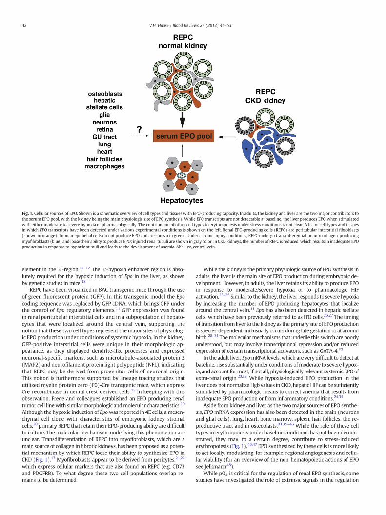
Fig. 1. Cellular sources of EPO. Shown is a schematic overview of cell types and tissues with EPO-producing capacity. In adults, the kidney and liver are the two major contributors tothe serum EPO pool, with the kidney being the main physiologic site of EPO synthesis. While EPO transcripts are not detectable at baseline, the liver produces EPO when stimulatedwith either moderate to severe hypoxia or pharmacologically. The contribution of other cell types to erythropoiesis under stress conditions is not clear. A list of cell types and tissuesin which EPO transcripts have been detected under various experimental conditions is shown on the left. Renal EPO-producing cells (REPC) are peritubular interstitial fibroblasts(shown in orange). Tubular epithelial cells do not produce EPO and are shown in green. Under chronic injury conditions, REPC undergo transdifferentiation into collagen-producingmyofibroblasts (blue) and loose their ability to produce EPO; injured renal tubuli are shown in gray color. In CKD kidneys, the number of REPC is reduced, which results in inadequate EPOproduction in response to hypoxic stimuli and leads to the development of anemia. Abb.: cv, central vein.
42 V.H. Haase / Blood Reviews 27 (2013) 41–53
element in the 3′-region.15–17 The 3′-hypoxia enhancer region is abso-lutely required for the hypoxic induction of Epo in the liver, as shownby genetic studies in mice.18
REPC have been visualized in BAC transgenic mice through the useof green fluorescent protein (GFP). In this transgenic model the Epocoding sequence was replaced by GFP cDNA, which brings GFP underthe control of Epo regulatory elements.11 GFP expression was foundin renal peritubular interstitial cells and in a subpopulation of hepato-cytes that were localized around the central vein, supporting thenotion that these two cell types represent themajor sites of physiolog-ic EPO production under conditions of systemic hypoxia. In the kidney,GFP-positive interstitial cells were unique in their morphologic ap-pearance, as they displayed dendrite-like processes and expressedneuronal-specific markers, such as microtubule-associated protein 2(MAP2) and neurofilament protein light polypeptide (NFL), indicatingthat REPC may be derived from progenitor cells of neuronal origin.This notion is furthermore supported by lineage tracing studies thatutilized myelin protein zero (P0)-Cre transgenic mice, which expressCre-recombinase in neural crest-derived cells.13 In keeping with thisobservation, Frede and colleagues established an EPO-producing renaltumor cell linewith similarmorphologic andmolecular characteristics.19
Although the hypoxic induction of Epowas reported in 4E cells, amesen-chymal cell clone with characteristics of embryonic kidney stromalcells,20 primary REPC that retain their EPO-producing ability are difficultto culture. The molecular mechanisms underlying this phenomenon areunclear. Transdifferentiation of REPC into myofibroblasts, which are amain source of collagen infibrotic kidneys, has beenproposed as a poten-tial mechanism by which REPC loose their ability to synthesize EPO inCKD (Fig. 1).13 Myofibroblasts appear to be derived from pericytes,21,22
which express cellular markers that are also found on REPC (e.g. CD73and PDGFRB). To what degree these two cell populations overlap re-mains to be determined.
While the kidney is the primary physiologic source of EPO synthesis inadults, the liver is the main site of EPO production during embryonic de-velopment. However, in adults, the liver retains its ability to produce EPOin response to moderate/severe hypoxia or to pharmacologic HIFactivation.23–25 Similar to the kidney, the liver responds to severe hypoxiaby increasing the number of EPO-producing hepatocytes that localizearound the central vein.11 Epo has also been detected in hepatic stellatecells, which have been previously referred to as ITO cells.26,27 The timingof transition from liver to the kidney as the primary site of EPOproductionis species-dependent andusually occurs during late gestation or at aroundbirth.28–31 Themolecularmechanisms that underlie this switch are poorlyunderstood, but may involve transcriptional repression and/or reducedexpression of certain transcriptional activators, such as GATA-4.32
In the adult liver, EpomRNA levels, which are very difficult to detect atbaseline, rise substantially under conditions ofmoderate to severe hypox-ia, and account formost, if not all, physiologically relevant systemic EPOofextra-renal origin.23,33 While hypoxia-induced EPO production in theliver does not normalizeHgb values in CKD, hepaticHIF can be sufficientlystimulated by pharmacologic means to correct anemia that results frominadequate EPO production or from inflammatory conditions.24,34
Aside from kidney and liver as the twomajor sources of EPO synthe-sis, EPOmRNA expression has also been detected in the brain (neuronsand glial cells), lung, heart, bone marrow, spleen, hair follicles, the re-productive tract and in osteoblasts.31,35–46 While the role of these celltypes in erythropoiesis under baseline conditions has not been demon-strated, they may, to a certain degree, contribute to stress-inducederythropoiesis (Fig. 1).45,47 EPO synthesized by these cells is more likelyto act locally, modulating, for example, regional angiogenesis and cellu-lar viability (for an overview of the non-hematopoietic actions of EPOsee Jelkmann48).
While pO2 is critical for the regulation of renal EPO synthesis, somestudies have investigated the role of extrinsic signals in the regulation
43V.H. Haase / Blood Reviews 27 (2013) 41–53
of EPO production. Wussow and colleagues postulated the existence ofan O2 sensor in the brain stem, which triggers renal EPO productionthrough release of yet to be identified humoral factors.49 More recently,HIF activation in the skin has been shown to modulate renal andhepatic EPO production indirectly through HIF-1- and nitric oxide(NO)-mediated effects on dermal blood flow, which in turn changedblood flow to kidney and liver.50 Whether alterations in renal bloodflow are responsible for changes in EPO production under these condi-tions is debatable as renal tissue pO2 is kept at relatively constant levelsand is not very sensitive to changes in blood flow as long as they occurwithin physiologic range.51
3. EPO synthesis: a paradigm of hypoxic gene regulation
Human EPO is heavily glycosylated, consists of 165 amino acidsand has a molecular mass of about 30 kDa, 40% of which is derivedfrom its carbohydrate component. Its major action is to promote sur-vival of EPO-dependent colony-forming unit-erythroid (CFU-E) cellsand erythroblasts that have not yet begun to synthesize hemoglobin.Upon ligand binding, the EPO receptor (EPOR), which lacks intrinsiccatalytic function and is hypoxia-inducible, 52–54 associates with tyrosinekinase Janus kinase 2 (JAK2). JAK2 phosphorylates EPOR and providesmultiple docking sites for signal-transducing proteins that contain src ho-mology 2 (SH2) domains. Signaling at the EPOR occurs through multiplepathways, which include the signal transduction and activator of tran-scription (STAT) 5pathway, thephosphatidylinositol-3-kinase/protein ki-nase B (PI-3K/AKT) and mitogen-associated protein kinase/extracellularsignal-related kinase (MAPK/ERK) pathways, as well as protein kinaseC.55
EPO production is primarily stimulated by hypoxia, which, dependingon severity, increases serum EPO levels up to several hundred-fold.56 HIFis a heterodimeric basic helix-loop-helix (bHLH) transcription factor thatbelongs to the PAS (PER/aryl hydrocarbon receptor nuclear translocator(ARNT)/single minded (SIM)) family of transcription factors. It consistsof an O2-sensitive α-subunit and a constitutively expressed β-subunit,also known as the aryl hydrocarbon receptor nuclear translocator(ARNT).57–59 Three HIF α-subunits are known, HIF-1α, HIF-2α andHIF-3α. HIF-1 was first isolated from human Hep3B hepatoma cellsusing DNA sequences that were derived from the 3′-hypoxia enhancerof the EPO gene.60,61 Together with HIF-2α (also known as endothelialPAS domain protein 1 (EPAS1) or HIF like factor, (HLF)), HIF-1α facili-tates O2 delivery and cellular adaptation to hypoxia by stimulating awide spectrum of biological processes that include angiogenesis, anaer-obic glucose metabolism, mitochondrial biogenesis and others.62
HIF-regulated genes are induced following the binding of HIFheterodimers to specific DNA consensus sequences and recruitment oftranscriptional co-factors. HIF-specific DNA elements are found in theregulatory regions of many O2-sensitive genes and are referred to ashypoxia-response elements (HREs) (Fig. 2). While hypoxic suppressionof certain genes has been found to be associated with HIF-1 and/orHIF-2 activation, it is unlikely that HIF acts as a direct transcriptionalrepressor.63 Under normoxia, all three HIF α-subunits are targeted forrapid proteasomal degradation by the von Hippel–Lindau tumor sup-pressor (VHL), which acts as the substrate recognition component ofan E3 ubiquitin ligase.64,65WhereasHIF-1 andHIF-2 heterodimers func-tion as transcriptional activators, splice variants of HIF-3α have beenshown to be inhibitory.66,67 Although HIF-1 and HIF-2 sharemany tran-scriptional targets, certain genes and processes do not appear to beco-regulated. For example, anaerobic glycolysis appears to be pre-dominantly controlled by HIF-1,68 whereas EPO synthesis and ironmetabolism have emerged as HIF-2-regulated processes.24,69–73 Inaddition to canonical HRE-mediated transcription, which requireshetero-dimerization with ARNT, HIF-α modulates cellular signalingpathways through interactionwith proteins that do not contain PAS do-mains. These include, among others, tumor suppressor protein p53, thec-MYC proto-oncogene and the Notch intracellular domain.74–77
Under normal O2 conditions HIF-α-subunits are rapidly degradedfollowing ubiquitylation by theVHL-E3ubiquitin ligase complex, preclud-ing the formation of transcriptionally active heterodimers. VHL-mediatedpoly-ubiquitylation requires hydroxylation of specific proline residues(Pro402 and Pro564 in human HIF-1α; Pro405 and Pro531 in humanHIF-2α), which are localized within its O2-dependent degradation do-main (ODD).78–84 Hydroxylation of HIF-α is carried out by three major2-oxoglutarate (2OG)-dependent oxygenases (prolyl-4-hydroxylase do-main (PHD) proteins), PHD1, PHD2 and PHD3, also known as egl nine ho-molog (EGLN) 2, EGLN1, and EGLN3, respectively. These enzymes belongto a larger family of proteins, in humans there are over 60 members,which couple the oxidative decarboxylation of 2OG to various chemicalprocesses, which aside from O2-sensing, include collagen synthesis andfatty acid metabolism. In mammals, these reactions produce succinateand CO2 and appear to be limited to hydroxylation anddemethylation ini-tiated by hydroxylation.85 HIF 2OG oxygenases function as O2 sensors asthey require molecular O2 for catalysis. Under hypoxia, hydroxylation isinhibited and HIF signaling is activated.86 To add complexity to the regu-lation of this pathway, HIF increases transcription of PHD2 and PHD3. Fur-thermore, protein turnover of PHD1 and PHD3 is hypoxically regulated bySiah proteins, which themselves are hypoxia-inducible.87,88
All three PHDs are expressed in the kidney where they control HIFactivity. Based on immunohistochemistry and RNA analysis their ex-pression levels vary between different renal cell types.89 mRNA tran-scripts of all three PHDs have been detected in FACS-sorted REPC.90 Afourth potential HIF prolyl-hydroxylase, P4H-TM, localizes to the en-doplasmic reticulum membrane and has been shown to hydroxylateHIF-1α-derived peptides, but not type 1 collagen. P4H-TM seems to beimportant for normal kidney function in zebra fish and appears to be in-volved in the renal EPO response in mice.91,92
The transcriptional activity of HIF is modulated by a second hypoxicswitch, which operates within the carboxy-terminal transactivationdomain of HIF-α. Factor Inhibiting HIF (FIH) is a 2OG oxygenase thatcatalyzes the hydroxylation of an asparagine residue within theC-terminal transactivation domain of HIF-α, thereby inhibiting thebinding of co-activators CREB-binding protein (CBP) and p300 tothe HIF transcriptional complex. Conversely, FIH inactivation facilitatesCBP/p300 recruitment and results in increased HIF target gene expres-sion under hypoxia.86 In the kidney, FIH has been detected in REPC,podocytes and in the distal tubule.90,93 While the role of PHDs and FIHin the regulation of HIF activity is well established, alternative hydroxyl-ation targets have been identified and are likely to impact hypoxia andEPO responses in the kidney.85,94,95 Furthermore, it is likely that renalEPO synthesis is modulated by epigenetic changes that are carried outby non-HIF 2OG oxygenases. Although nothing is known about theirrole in renal physiology, 2OG oxygenases, which contain a jumonji do-main, catalyze the demethylation of methylated histones,85 and are like-ly to provide additional functional links between alterations in renal pO2
levels and gene expression.96
4. HIF-2 in control of EPO synthesis
Although in vitro approaches identified HIF-1 as the transcriptionfactor responsible for the hypoxic induction of EPO,97 HIF-2 has nowemerged as themain regulator of EPOproduction in vivo (Fig. 2). Severallines of evidence exist that support this notion: a) the location ofHIF-2α-expressing renal interstitial cells coincides with the location ofREPC12,98; b) genetic studies in mice have demonstrated that renal andliver EPO synthesis is HIF-2- and not HIF-1-dependent, as did siRNAand chromatin immunoprecipitation (ChIP)-based studies in certainEPO-producing cell lines72,99,100; c) genetic analysis of patients withinherited forms of erythrocytosis have revealed mutations in HIF2Α butnot in HIF1Α (see section on HIF pathway mutations in patients withsecondary erythrocytosis); and d) genetic variants of HIF2A have beenassociated with high altitude dwellers who are protected from chronic
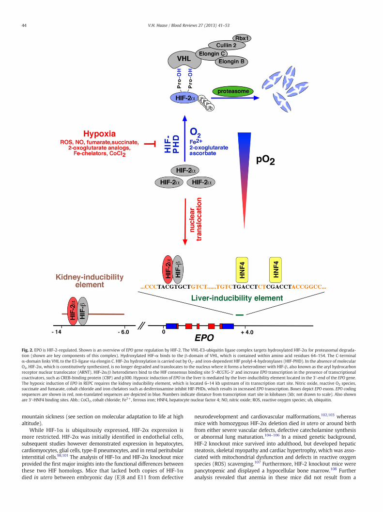
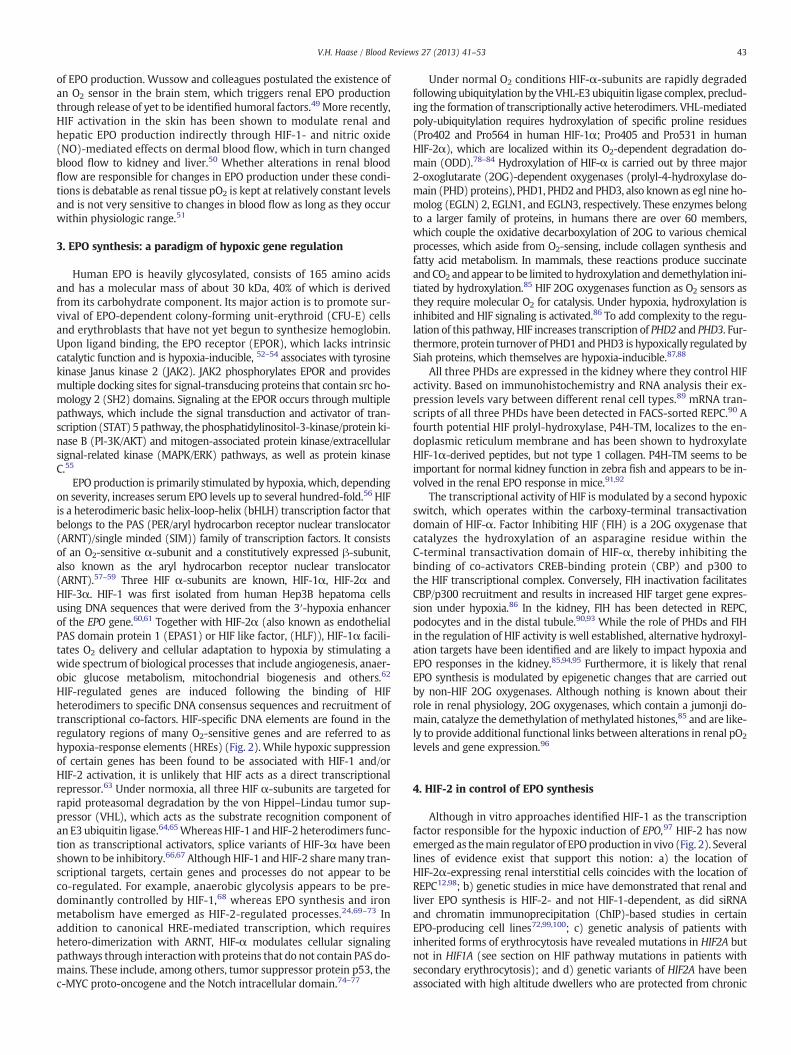
Fig. 2. EPO is HIF-2-regulated. Shown is an overview of EPO gene regulation by HIF-2. The VHL-E3-ubiquitin ligase complex targets hydroxylated HIF-2α for proteasomal degrada-tion (shown are key components of this complex). Hydroxylated HIF-α binds to the β-domain of VHL, which is contained within amino acid residues 64–154. The C-terminalα-domain links VHL to the E3-ligase via elongin C. HIF-2α hydroxylation is carried out by O2- and iron-dependent HIF prolyl-4-hydroxylases (HIF-PHD). In the absence of molecularO2, HIF-2α, which is constitutively synthesized, is no longer degraded and translocates to the nucleus where it forms a heterodimer with HIF-β, also known as the aryl hydrocarbonreceptor nuclear translocator (ARNT). HIF-2α/β heterodimers bind to the HIF consensus binding site 5′-RCGTG-3′ and increase EPO transcription in the presence of transcriptionalcoactivators, such as CREB-binding protein (CBP) and p300. Hypoxic induction of EPO in the liver is mediated by the liver-inducibility element located in the 3′-end of the EPO gene.The hypoxic induction of EPO in REPC requires the kidney inducibility element, which is located 6–14 kb upstream of its transcription start site. Nitric oxide, reactive O2 species,succinate and fumarate, cobalt chloride and iron chelators such as desferrioxamine inhibit HIF-PHDs, which results in increased EPO transcription. Boxes depict EPO exons. EPO codingsequences are shown in red, non-translated sequences are depicted in blue. Numbers indicate distance from transcription start site in kilobases (kb; not drawn to scale). Also shownare 3′-HNF4 binding sites. Abb.: CoCl2, cobalt chloride; Fe2+, ferrous iron; HNF4, hepatocyte nuclear factor 4; NO, nitric oxide; ROS, reactive oxygen species; ub, ubiquitin.
44 V.H. Haase / Blood Reviews 27 (2013) 41–53
mountain sickness (see section on molecular adaptation to life at highaltitude).
While HIF-1α is ubiquitously expressed, HIF-2α expression ismore restricted. HIF-2α was initially identified in endothelial cells,subsequent studies however demonstrated expression in hepatocytes,cardiomyocytes, glial cells, type-II pneumocytes, and in renal peritubularinterstitial cells.98,101 The analysis of HIF-1α and HIF-2α knockout miceprovided the first major insights into the functional differences betweenthese two HIF homologs. Mice that lacked both copies of HIF-1αdied in utero between embryonic day (E)8 and E11 from defective
neurodevelopment and cardiovascular malformations,102,103 whereasmice with homozygous HIF-2α deletion died in utero or around birthfrom either severe vascular defects, defective catecholamine synthesisor abnormal lung maturation.104–106 In a mixed genetic background,HIF-2 knockout mice survived into adulthood, but developed hepaticsteatosis, skeletal myopathy and cardiac hypertrophy, which was asso-ciated with mitochondrial dysfunction and defects in reactive oxygenspecies (ROS) scavenging.107 Furthermore, HIF-2 knockout mice werepancytopenic and displayed a hypocellular bone marrow.108 Furtheranalysis revealed that anemia in these mice did not result from a
45V.H. Haase / Blood Reviews 27 (2013) 41–53
cell-autonomous defect in erythroid precursor maturation, but was dueto inadequate renal EPO production, indicating that HIF-2 was indis-pensable for systemic EPO homoeostasis in adults.70 In a differentmodel, Morita and colleagues showed that local EPO production in theretina was also HIF-2-dependent,69 suggesting a more general role forHIF-2 in the control of EPO regulation.While thesemousemodels dem-onstrated that EPOproduction in adultswasHIF-2-dependent, develop-mental studies highlighted the importance of HIF-1 in the regulation oferythropoiesis during embryonic development. HIF-1-deficient embry-os were characterized by a reduction in myeloidmulti-lineage cells andcommitted erythroid progenitors at E9.5. This was associated withdecreased Epo mRNA levels in the embryo proper but not in the yolksac, while EpoRmRNA was decreased in both tissues.54
The most compelling support for the notion that HIF-2 is the mainregulator of adult EPO synthesis comes from conditional knockout stud-ies in mice. Utilization of a tamoxifen-inducible, ubiquitously expressedCre-recombinase transgene permitted a direct comparison of the effectsof HIF-1 and HIF-2 inactivation on erythropoiesis. Acute postnatal globalablation of HIF-2α, but not of HIF-1α, resulted in anemia, which, similarto HIF-2α germ line inactivation, was responsive to treatment withrecombinant EPO.71 While stimulation of renal EPO production inresponse to hemolysis (phenylhydrazine treatment) was blunted inHIF-2α-ablated mice, postnatal deletion of HIF-1α did not have anynotable effect on erythropoiesis, which suggested that HIF-1 doesnot play a significant role in the regulation of systemic EPO homeostasisat baseline or in response to acute anemia.71 Our laboratory has gener-ated cell type-specific knockout mice to investigate the differences be-tween HIF-1 and HIF-2 in the regulation of renal and hepatic EPOsynthesis. Inactivation of HIF-2α in the kidney completely ablated therenal EPO response in mice subjected to normobaric hypoxia (10% O2
for 10 days), phlebotomy-induced anemic hypoxia, or treatment witha HIF activating compound.24 Cell type-specific inactivation of theVHL-E3 ubiquitin ligase in hepatocytes resulted in HIF-2-, but notin HIF-1-dependent erythrocytosis, while pharmacological PHD in-hibition caused a HIF-2-dependent increase in liver Epo mRNAlevels.72 Consistent with these findings are transgenic studies withnon-degradable mutant isoforms of HIF-1α or HIF-2α. Over-expressionof non-degradable HIF-2α in hepatocytes produced erythrocytosis,whereas over-expression of HIF-1α did not.109 Taken together,multiplegenetic studies in mice provide overwhelming evidence that, in theadult, renal and hepatic EPO synthesis is predominantly HIF-2- andnot HIF-1-regulated. These studies have clearly identified HIF-2 as akey pharmacological target for the treatment of anemia.
HIF-2 transactivation at the EPO HRE involves multiple nuclearfactors that associate with the EPO gene.97,99 One of these factors ishepatocyte nuclear factor-4 (HNF4), which binds to the 3′ EPO hypoxiaenhancer region and is likely to interact with HIF-2 (Fig. 2).99 Similar toHIF-2, the cellular location of HNF4 expression coincides with the sitesof EPO production in liver and kidney. Furthermore, HNF4 is requiredfor the hypoxic induction of EPO in Hep3B cells.99,110,111 The notionthat HIF-2 transactivation depends on the cooperation with other tran-scription factors has been previously suggested and may determinewhether HIF target genes are HIF-1 or HIF-2-regulated, however, specificfactors that are required for HIF-2-dependent EPO induction have not yetbeen identified.112
5. Molecular fine-tuning of HIF-2 regulated erythropoiesis
Post-transcriptional and post-translational modifications of HIF2ΑmRNA and HIF-2α protein that do not involve PHD enzymes havebeen shown to modulate EPO production. The molecular mecha-nisms that underlie these modifications, link cellular metabolism andredox-state to hypoxia-induced erythropoiesis. HIF-2α is acetylatedduring hypoxia and deacetylated by Sirtuin 1, a nicotinamide adeninedinucleotide (NAD)+-dependent protein deacetylase, which increasesHIF-2-dependent EPO synthesis in vitro and in vivo, thus linking cellular
redox and energy state to systemic hypoxia responses.113 Sirtuin1-deficient mice produced significantly lower amounts of fetal liverEpomRNA, and as adults less EPO in response to severe hypoxia. Inter-estingly, caloric restriction, which induces Sirtuin 1 activity, suppressedEPO production in the liver.114,115 Although further studies are neededto clearly define the role of sirtuins in HIF-2-dependent erythropoiesis,these findings highlight the existence of complex functional links be-tween EPO production and cellular energy state.
Additional post-translational modifications, which impact EPO pro-duction and hypoxia-induced erythropoiesis, involve SUMOylation.SUMO (Small Ubiquitin-like Modifier) proteins are structurally relatedto ubiquitin and reversibly modify cellular function and localizationof targeted proteins. An enzyme, which removes SUMO, is SENP(Sentrin/SUMO-specific protease). SENP 1 knockout mice are anemicand die during mid-gestation.116 In this model de-SUMOylation did notoccur, prevented HIF activation under hypoxic conditions and resultedin reduced hepatic EPO production.116 Although SUMOylation was spe-cifically investigated with regard to HIF-1 signaling, the presence of ane-mia suggested that SENP is likely to be involved in the de-SUMOylation ofHIF-2α.
To add more complexity to the regulation of HIF-2 activity, low in-tracellular iron levels have been shown to diminish HIF-2α translationand thus are predicted to limit HIF-2-induced EPO production anderythropoiesis when cellular iron stores are depleted. This feedbackloop makes sense physiologically, as erythropoiesis cannot occur inthe absence of iron. The 5′-untranslated region (UTR) of HIF2Α mRNAcontains an iron-regulatory element (IRE), a stem loop structure thatbinds iron-regulatory protein (IRP) when intracellular iron levels arelow.117 IRPs (IRP1 and IRP2) function as intracellular iron sensors thatcontrol the expression of several iron-sensitive genes, such as transferrinreceptor 1 (TFR1), ferritin and divalentmetal transporter 1 (DMT1).118,119
Iron is incorporated into an iron–sulfur cluster at the center of the proteinand converts IRP1 to an enzyme with aconitase activity. In its aconitaseform IRP1 does not bind to the IRE. In contrast, IRP2 does not convert toan aconitase and is regulated via iron-dependent proteasomal degra-dation.117,120,121 Depending on the location of the IRE stem loop, theIRP/IRE complex either inhibits translation (5′-IRE), or stabilizesmRNAs when the IRE is located in the 3′-UTR (e.g. TFR1 mRNA levelsincreasewhen intracellular iron is low). Since the IRE inHIF2Α is locatedin its 5′-untranslated region, HIF-2α translation is inhibited when ironlevels are low. This in turn limits EPO synthesis and thereby adjustshypoxia-inducibility of erythropoiesis to iron availability.
6. HIF pathwaymutations in patients with secondary erythrocytosis
Mild to moderate perturbations in the HIF O2-sensing pathwaylead to the development of benign erythrocytoses that are associatedwith increased or inappropriately normal serumEPO levels. This is in con-trast to primary erythrocytoses, which are characterized by suppressedserum EPO levels and are caused by molecular defects in erythroid pro-genitor cells or hematopoietic stem cells.122,123 Other forms of secondaryerythrocytosis that associate with increased EPO production result fromchronic hypoxic conditions, such as COPD, right-to-left cardiac shuntsor high altitude, or can be due to EPO-producing tumors.
Abnormalities in the HIF O2-sensing pathway were first observedin patients with Chuvash polycythemia. Chuvash polycythemia is arare autosomal recessive form of secondary erythrocytosis that is en-demic but not limited to Chuvashia, a republic in central EuropeanRussia. It is caused by a homozygousmutation in the VHL tumor suppres-sor at codon 200, R200W, and patients with the Chuvash mutation, whoare ethnically distinct from Chuvashians, have been identified in otherparts of Europe, theUnited States andAsia.124–133 Somepatients are com-pound heterozygotes for the R200W and other VHL mutations.128,129,134
Codon200 is located in the C-terminus of the VHLprotein and lies outsideof the central groove that binds hydroxylatedHIF-α (β-domain core) andoutside of the helical α-domain (amino acid residues 157–189), which
46 V.H. Haase / Blood Reviews 27 (2013) 41–53
interacts with elongin C.135 Genetic alterations in these two core re-gions are strongly associated with the development of VHL disease,an inherited autosomal dominant tumor syndrome. Patients whocarry a VHL germ line mutation are predisposed to the developmentof highly vascularized tumors, which include renal cell carcinoma,hemangioblastomas of the CNS and retina, and pheochromocytomas.136
Chuvash patients, who are homozygous for the R200W allele, are notpredisposed to the development of these tumors. The ability of theR200W VHL species to capture hydroxylated HIF-α for ubiquitylationand subsequent proteasomal degradation is impaired, which is mostlikely due to changes in protein stability or conformation that impingeon the VHL-HIF-α interaction.137 Although individuals with Chuvashpolycythemia are not prone to tumor development, they suffer fromprematuremorbidity andmortality due to pulmonary hypertension, ce-rebrovascular accidents and vertebral hemangiomas.138,139 Evaluationof cardiopulmonary function in a small group of Chuvash patients re-vealed significant abnormalities in respiratory and pulmonary vascularregulation at baseline and in response to hypoxia. Basal ventilation andpulmonary vascular tone were elevated and increases in heart rate andventilation, as well as pulmonary vasoconstrictive responses to mild ormoderate hypoxia were considerably enhanced, indicating that tightregulation of the VHL/HIF axis is required for normal cardiopulmonaryphysiology.140,141 Chuvash patients furthermore display abnormalitiesin metabolic stress responses and cytokine profiles.142–145
Furthermutational analysis of theHIFO2-sensing pathway in patientswith idiopathic erythrocytosis led to the identification of families withheterozygousmutations inHIF2Α, PHD2 orVHL (non-R200W); for a sum-mary of non-R200W VHL mutations the reader is referred to Lee andPercy.134 Interestingly, mutations in HIF-1α have not been described todate, underscoring the importance of HIF-2 in the regulation of EPOsynthesis in humans. Most gain-of-function mutations in HIF-2αare in direct proximity to proline residue 531, which is one of thetwo main hydroxylation sites (the other major hydroxylation site isproline 405).146–153 Biochemical analysis demonstrated that theoriginally identified G537W mutation impaired recognition and hy-droxylation by PHD2 and thus interaction with VHL.154 Two recentlyidentifiedHIF-2α gain-of-functionmutations, A530T andA530V,were as-sociated with polycythemia, paraganglioma and/or somatostatinoma.155
Conversely, several PHD2 missense mutations have been identified thatresulted in diminished hydroxylase activity.156,157 Patients, who wereheterozygous for these mutations were characterized by increased rbcvalues in the presence of inappropriately normal serum EPO levels.156–161
Some of these mutations (P317R, H374R) likely affect iron-chelation atthe catalytic center, which is critical for PHD enzymatic activity. Further-more, H374R was associated with paraganglioma development, indicat-ing that PHD2 may function as a tumor suppressor.157,160
7. Molecular adaptation to life at high altitude
Chronic mountain sickness (CMS), also known as Monge's disease,affects long-termhigh-altitude (>2500 m) residents or natives, and is as-sociated with excessive erythrocytosis (females, Hgb≥19 g/dL; males,Hgb≥21 g/dL), hypoxemia, pulmonary hypertension, right-sided heartfailure and neurologic symptoms, such as headache, fatigue, tinnitus,insomnia, paresthesia and loss ofmemory.162–164 The diseasewasfist de-scribed in high altitude dwellers on the South American Altiplano, whereit affects ~5–15% of the population.162,164 CMS is usually alleviated bydescent to low altitude or by phlebotomy.162,163 While the disease isprevalent in the Andean population, it is less common in native Tibetans,who live at comparable altitude. In contrast, Tibetan residents of HanChinese descent aremuchmore frequently affected byCMS,which repre-sents a major public health burden.164–167 Prevalence of CMS is higher inmen than in women, increases with altitude and age, and is more likelyto develop in the presence of lung diseases, smoking and environmentalpollution.164 The pathogenesis of CMS is thought to result, at least part-ly, from an abnormal, i.e. blunted, ventilatory response.164 Aside from
differences in susceptibility to CMS, native Tibetans and Andeans differin their baseline physiologic responses to high altitude. Native Tibetanshave higher resting ventilation and hypoxic ventilatory response atcomparable altitudes, lower oxygen saturation of arterial hemoglobinand lower hemoglobin concentrations (15.6 g/dL versus 19.2 g/dL inmales)168,169 There is also less intrauterine growth retardation and bet-ter neonatal oxygenation among native Tibetans compared to nativeAndeans or Han Chinese.166,170 Furthermore, differences in energy me-tabolism have been described, which need further characterization.171
These differences in physiologic phenotypes reflect divergence in ge-netic adaptation and selection, which result from differences in lengthof high-altitude habitation (~between 25,000 and 50,000 years fornative residents on the Tibetan plateau, compared to ~10,000 years forthe Andean Altiplano and ~60 years for Tibetan residents of Han Chinesedescent), the degree of geographical isolation (Tibetan plateau>SouthAmerican Altiplano) and gene pool stability.166 Genome-wide searchesfor single-nucleotide polymorphisms (SNP), candidate gene approachesand exome sequencing have identified genetic variations in the HIF2A,PHD2 and PPARA genes that associate with lower hemoglobins in nativeTibetans compared to Han Chinese and with high altitude adaptation inother populations.172–179 Although the precise biological function ofthese alleles is not known, they are predicted to confer adaptation tothe hypoxic environment and to modulate susceptibility to CMS andother high altitude-associated diseases.
8. HIF coordinates EPO production with iron metabolism
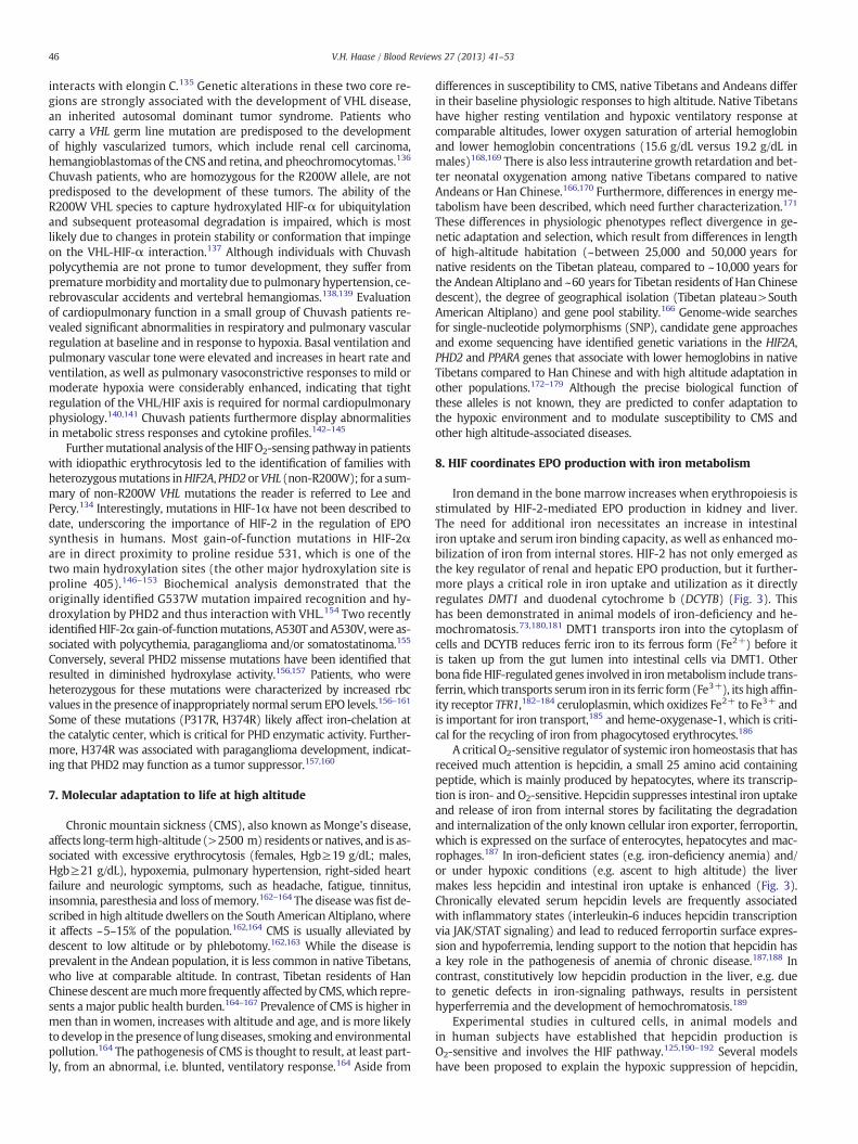
Iron demand in the bone marrow increases when erythropoiesis isstimulated by HIF-2-mediated EPO production in kidney and liver.The need for additional iron necessitates an increase in intestinaliron uptake and serum iron binding capacity, as well as enhanced mo-bilization of iron from internal stores. HIF-2 has not only emerged asthe key regulator of renal and hepatic EPO production, but it further-more plays a critical role in iron uptake and utilization as it directlyregulates DMT1 and duodenal cytochrome b (DCYTB) (Fig. 3). Thishas been demonstrated in animal models of iron-deficiency and he-mochromatosis.73,180,181 DMT1 transports iron into the cytoplasm ofcells and DCYTB reduces ferric iron to its ferrous form (Fe2+) before itis taken up from the gut lumen into intestinal cells via DMT1. Otherbona fideHIF-regulated genes involved in ironmetabolism include trans-ferrin,which transports serum iron in its ferric form (Fe3+), its high affin-ity receptor TFR1,182–184 ceruloplasmin, which oxidizes Fe2+ to Fe3+ andis important for iron transport,185 and heme-oxygenase-1, which is criti-cal for the recycling of iron from phagocytosed erythrocytes.186
A critical O2-sensitive regulator of systemic iron homeostasis that hasreceived much attention is hepcidin, a small 25 amino acid containingpeptide, which is mainly produced by hepatocytes, where its transcrip-tion is iron- and O2-sensitive. Hepcidin suppresses intestinal iron uptakeand release of iron from internal stores by facilitating the degradationand internalization of the only known cellular iron exporter, ferroportin,which is expressed on the surface of enterocytes, hepatocytes and mac-rophages.187 In iron-deficient states (e.g. iron-deficiency anemia) and/or under hypoxic conditions (e.g. ascent to high altitude) the livermakes less hepcidin and intestinal iron uptake is enhanced (Fig. 3).Chronically elevated serum hepcidin levels are frequently associatedwith inflammatory states (interleukin-6 induces hepcidin transcriptionvia JAK/STAT signaling) and lead to reduced ferroportin surface expres-sion and hypoferremia, lending support to the notion that hepcidin hasa key role in the pathogenesis of anemia of chronic disease.187,188 Incontrast, constitutively low hepcidin production in the liver, e.g. dueto genetic defects in iron-signaling pathways, results in persistenthyperferremia and the development of hemochromatosis.189
Experimental studies in cultured cells, in animal models andin human subjects have established that hepcidin production isO2-sensitive and involves the HIF pathway.125,190–192 Several modelshave been proposed to explain the hypoxic suppression of hepcidin,
47V.H. Haase / Blood Reviews 27 (2013) 41–53
including direct HRE-mediated regulation by HIF-1, regulation bydioxygenases, signaling via EPOR or through humoral factors that are re-leased from the bone marrow when erythropoiesis is stimulated.192–196
Other studies have linked hypoxia to iron signaling pathways and haveproposed that hypoxia diminishes signals that normally increase hepcidinproduction in hepatocytes. Activation of signaling through the hemo-chromatosis protein HFE, TFR1, TFR2, or hemojuvelin (HJV), whichacts as a co-receptor for bone morphogenetic protein 6 (BMP6), in-creases hepcidin transcription in a SMAD-dependent fashion.189,197–201
Recent in vivo studies have shown that HIF induces furin, a proproteinconvertase that cleaves HJV and generates a soluble form of HJV,which suppresses hepcidin by antagonizing BMP6 signaling.202,203 Sim-ilarly, transmembrane protease serine 6 (TMPRSS6), also known asmatriptase-2, was reported to be HIF-regulated and is predicted toblunt BMP6/HJV-mediated signals under hypoxic conditions.204–206
Our laboratory has used a genetic approach to dissect the role of HIFin the regulation of hepcidin. We have created conditional knockoutstrains, in which we disengaged HIF activation from EPO synthesisand found that hypoxia/HIF-mediated suppression of hepcidin requiredEPO.207 However, we determined that the induction of EPO synthesisalone was not sufficient to suppress hepcidin in this model. Hepcidinsuppression under conditions of hypoxia and hepatic HIF activationwas dependent on erythropoietic activity in the bone marrow. Ourdata established that HIF activation in hepatocytes suppresses hepcidinindirectly through EPO-mediated stimulation of erythropoiesis andis consistent with previous studies from Pak and colleagues inphlebotomized animals.195 In the context of anemic hypoxia, bothHIF-1 and HIF-2 are activated.24 HIF-2 induces EPO production in kidneyand in liver (depending on the severity of hypoxia), resulting in increasedserum EPO levels and stimulation of erythropoiesis, which subsequentlyleads to the suppression of hepcidin in the liver.208 HIF-2 is a direct regu-lator of both renal and hepatic EPO synthesis, but regulates hepcidin onlyindirectly via stimulation of bone marrow activity (Fig. 3).196,207,209
It is plausible that serum iron levels modulate the suppression ofhepcidin under hypoxic conditions, although this has not been suffi-ciently addressed experimentally. Serum iron and ferritin levels are
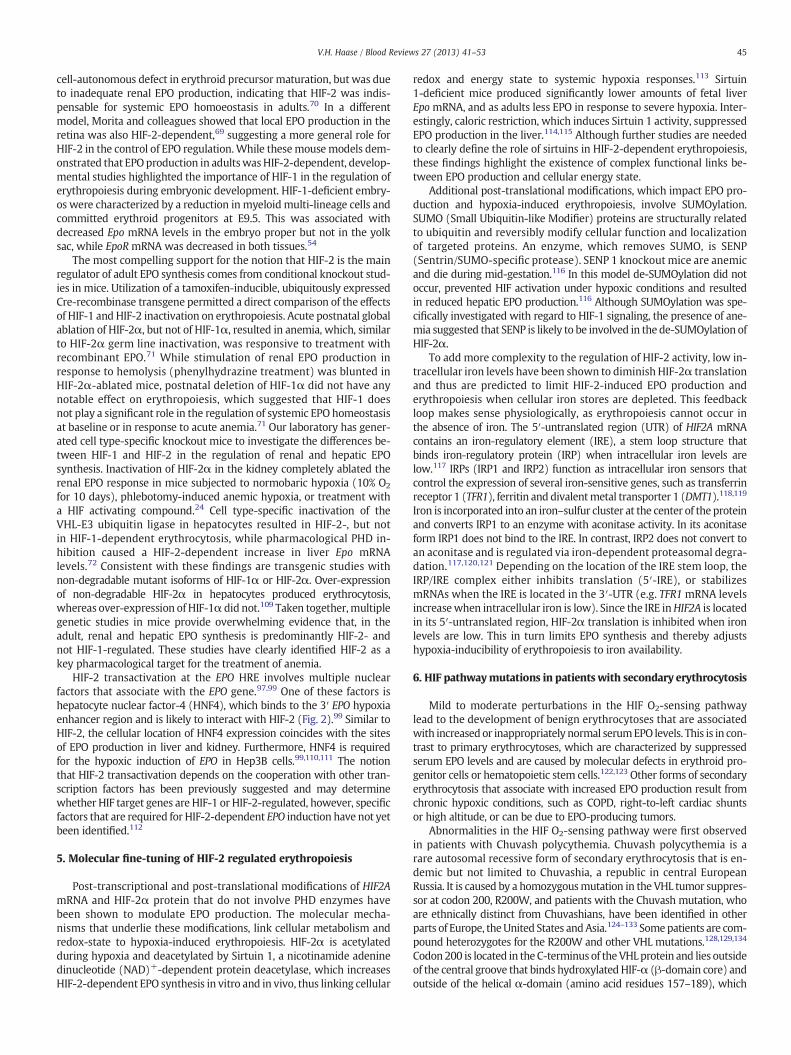
Fig. 3. HIF coordinates EPO production with iron metabolism. Shown is a schematic overvieware indicated in red. HIF-2 activation (either by hypoxia, pharmacologic means or as a resuserum EPO levels and stimulation of erythropoiesis. Iron metabolism is adjusted to match i(DCYTB) reduces ferric iron (Fe3+) to its ferrous form (Fe2+), which is then transported intoboth HIF-2-regulated. Absorbed iron is released into the circulation by ferroportin and is thenbonemarrowand other organs. TF is HIF-regulated and hypoxia increases its serum levels. EPO-levels. As a result of low serum hepcidin, ferroportin (FPN) cell surface expression is increasedhepatocytes and RES cells. Growth differentiation factor 15 (GDF15) is made by erythroid prec
decreased in Chuvash patients and in individuals sojourning at highaltitude for 10–12 days.190,210 Time course analysis, however, indi-cated that the acute decrease of serum hepcidin at high altitude pre-ceded changes in serum ferritin and transferrin saturation, suggestingthat under hypoxic conditions hepcidin regulation is, at least initially,iron-independent.211 Support for this notion also comes from patientswith β-thalassemia, who have low serum hepcidin levels despite ironoverload.212
Growth differentiation factor 15 (GDF-15) and twisted gastrulationhomolog 1 (TWSG1) have been identified as candidate erythrokines, al-though not erythroblast-specific, that have the potential to suppresshepcidin under conditions of increased erythropoietic activity.213–215
GDF15 is an iron- and O2-regulated (HIF-independent) member of theTGF-β superfamily, which is secreted from maturing erythroblasts andhas been shown to suppress hepcidin transcription in primary humanhepatocytes and hepatoma cells (Fig. 3).213,216 While increased GDF15serum levels associate with syndromes of ineffective erythropoiesis,for example α- and β-thalassemia, its role in hepcidin regulationunder physiologic conditions and in other forms of anemia remainsunclear.213,215,217–219 Therefore, it was proposed that GDF15 may be amarker of bone marrow stress or erythroblast apoptosis.215 ElevatedserumGDF15 level have also been found in patients with heart failure,220
which adds complexity to this model. We found that recombinantmurine GDF15 suppressed hepcidin in Hep3B cells at a concentrationof 750 pg/ml.207 This is in contrast to previous reports where higherdoses of GDF15 were needed to achieve hepcidin suppression in humanHuH-7 hepatoma cells and in primary hepatocytes, while low doseGDF15 treatment increased hepcidin.213 While demonstrated in mice,studies in humans receiving recombinant EPO have not yet shown a sig-nificant inverse relationship between serum hepcidin and GDF15 levels,which may relate to the EPO doses administered, study size, complexityof regulation and species-dependent differences.207,221 In the context ofiron-deficiency anemia, Tanno and colleagues found that GDF15 serumlevels were not elevated,222 while Lakhal and colleagues reported thatpatients with low serum iron had elevated GDF15 levels compared toiron-replete controls (mean of 1048 pg/ml vs. 542 pg/ml).216 Similarly,
of changes in iron metabolism as a consequence of HIF activation. HIF-regulated geneslt of mutations) induces renal and hepatic EPO synthesis, which leads to an increase inron demand from increased erythropoiesis. In the duodenum, duodenal cytochrome bthe cytosol of enterocytes by divalent metal transporter-1 (DMT1). DCYTB and DMT1 aretransported in complex with transferrin (TF) to the liver, reticulo-endothelial cells (RES),induced erythropoiesis inhibits hepcidin synthesis in the liver and reduces serumhepcidin(hepcidin promotes ferroportin degradation) and more iron is released from enterocytes,ursor cells and has been shown to suppress hepcidin in hepatocytes.
48 V.H. Haase / Blood Reviews 27 (2013) 41–53
increased serum GDF15 levels were found following DFO treatment,suggesting iron-dependent regulation.216 Furthermore, temporaryincreases in serum GDF15 levels associated with increased serum EPOfollowing ascent to high altitude.211
In addition to regulating iron metabolism, hypoxia has direct effectson the bone marrow. It promotes erythropoiesis by modulating ery-throid progenitor maturation and proliferation.223,224 Hypoxia stimu-lates EPOR expression and regulates components of the hemoglobinsynthesis pathway.52–54,225,226 Hypoxia also modulates the interactionof erythroid progenitors with other cell types and thereby regulatesstem cell maintenance, lineage differentiation and maturation. Recentstudies have highlighted a role for endothelial HIF-2 in this regulation,224
as mice with globally reduced HIF-2 expression displayed a defect in ery-throidmaturation, which was dependent on the expression levels of vas-cular adhesion molecule (VCAM)-1, an integrin receptor that binds verylate antigen-4 (VLA4) on erythroblasts and supports erythroid matura-tion. This finding lendsmore support to the concept that HIF-2 acts a cen-tral regulator of hypoxia-induced erythropoiesis, which coordinates EPOsynthesis with iron metabolism and erythroid progenitor maturation.
9. The HIF axis as a novel therapeutic target for the treatmentof anemia
Over the last 20 years EPO therapy has transformed the lives ofmillions of patients who suffer from anemia. Therapy with recombinantEPO eliminates the need for rbc transfusions, improves cardiovascularfunction and cognitive ability. The US Food and Drug Administration(FDA) approved recombinant EPO in 1989, initially for use in patientswith renal anemia then for use in cancer patients (FDA approval 1993).Since then its administration has become standard of care. However, de-spite its clinical effectiveness and success, recent randomized controlledclinical trials have raised significant safety concerns, resulting in severalblack boxwarnings issued by the FDA. These studies showed that aimingfor normal Hct values in the dialysis patient population (Hct target of42%) increased the risk of serious cardiovascular complications or ad-verse composite outcomes.227 In pre-dialysis CKD patients with or with-out diabetes higher Hgb targets, particularly in patients with poor initialHgb responses, were also associated with increased cardiovascularrisk.228–230 Furthermore, in the Oncology setting high dose recombinantEPO administration was found to be associated with tumor growth andincreased overall mortality, a concerning finding, which currently lackssufficient explanation (for recent reviews on this topic see231,232). Car-diovascular safety concerns in the CKD/ESRD patient population havechanged EPO-prescribing practices, and have resulted in a decrease in re-combinant EPOuse andnot surprisingly in an increase in rbc transfusions(http://www.usrds.org/2012/slides/indiv/v2index.html#/176/). The un-derlying mechanisms for the increase in cardiovascular mortality areunclear, but may relate to the EPO dose administered and the clinicalconditions that associate with EPO hyporesponsiveness. There is muchdebate about what represents a “safe” Hgb target and individual patientneeds and lifestyle choices have to be taken into account when prescrib-ing recombinant EPO. Despite these dilemmas, the clinical success of re-combinant EPO therapy has been a major incentive for the developmentof new erythropoiesis stimulating agents and the design of novel thera-peutics that boost synthesis of endogenous EPO.233
Pharmacological inactivation of PHD enzymes with 2OG analogsincreases serum EPO levels in animal models and in humans, andhas the potential to benefit patients with anemia that results from in-adequate EPO production.24,34,92,234,235 In vitro and in vivo studieshave suggested that pharmacological or genetic targeting of individualPHD enzymes has differential effects on renal and hepatic EPO synthe-sis. Inducible, global deletion of PHD2 in adult mice resulted in severeerythrocytosis from a dramatic increase in renal EPO production(Hct values>80%), as well as other organ pathologies, in particularwhen PHD3 was inactivated simultaneously.236–240 PHD1- andPHD3-deficient mice, which in contrast to conventional PHD2 knockout
mice survive into adulthood, developedmild tomoderate erythrocytosis(Hct of 67% compared to 53% in control mice) only when both enzymeswere inactivated simultaneously, the liver being the source of EPOand not the kidney.25,239 In the liver, genetic or pharmacologic inactiva-tion of all three PHDs, however, is required to produce a strong andsustained erythropoietic response.25,34 This is in contrast to the kid-ney where inactivation of PHD2 alone is sufficient to produce severeerythrocytosis.238,239 While these tissue-specific differences are notwell understood, functional diversity between individual PHDs isexpected, because of differences in cellular localization, hypoxia-inducibility and biochemical behavior (for a review see86,241). Fur-thermore, PHD1 and PHD3 appear to preferentially target HIF-2αin vitro and in vivo, which offers potential for therapeutic exploitationunder conditions in which HIF-1 activation is non-desirable.239,242
Aside from stimulating endogenous EPO synthesis, pharmacologicalinhibition of HIF prolyl-hydroxylation is likely to have beneficial effectson iron uptake and utilization (see section on HIF and ironmetabolism),and may therefore be superior to the administration of recombinantEPO alone, especially in renal anemia patients, who often suffer fromchronic inflammation, functional iron deficiency and EPO resistance.243
The beneficial effects on ironmetabolism aremost likely producedwithsystemic administration of HIF stabilizing PHD inhibitors, which wouldtargetmultiple organs including kidney, liver, gut and the bonemarrow.A potential downside to this approach, however, is that HIF transcriptionfactors regulate a multitude of biological processes, and intermittent HIFactivation over prolonged periods of timemay lead to changes in glucose,fat and cholesterolmetabolism, promote angiogenesis and have other ad-verse effects.244–249 Liver-specific PHD inhibition using siRNA has beenshown to correct Hbg values in preclinical models of renal anemia andanemia of chronic inflammation, and was associated with decreasedhepcidin expression in the liver.34 The latter, however, is most likely areflection of increased erythropoietic activity.207 Whether tissue-specifictargeting of PHD enzymes is as efficacious as systemic therapy remainsto be investigated. Irrespective of tissues targeted, the short-term andlong-term effects of HIF stabilizing compounds on the human body willhave to be carefully evaluated in clinical trials and through well-controlled physiologic studies in normal individuals.
9.1. Practice points
• Recognize the role of HIF-2 as a central regulator of hypoxia-inducederythropoiesis.
• Recognize the role of genetic factors in the pathophysiology of highaltitude sickness.
• Recognize the role of hepcidin in the regulation of iron uptake andutilization.
• Patients with secondary erythrocytosis of unclear etiology should beevaluated for mutations in the HIF O2-sensing pathway.
• HighHgb targets in renal anemia therapy are associatedwith increasedcardiovascular risk; the optimal Hgb target is not known.
9.2. Research agenda
• Molecular and cellular mechanism underlying the pathogenesis ofrenal anemia.
• EPO-associated cardiovascular and oncologic risk.• Regulators of iron metabolism as a therapeutic target in anemiatherapy.
• Physiologic, metabolic and genetic studies of human populationsthat live at high altitude.
• Physiologic studies in patients with mutations in the O2-sensingmachinery.
• HIF andnon-HIF 2OGoxygenases in the regulation hypoxia responses.• HIF prolyl-4-hydroxylases as therapeutic targets.
49V.H. Haase / Blood Reviews 27 (2013) 41–53
Conflict of interest
The author serves on the Scientific Advisory Board of AkebiaTherapeutics, a company that develops prolyl-4-hydroxylase inhibitorsfor the treatment of anemia.
Acknowledgments
The author is supported by the Krick-Brooks chair in Nephrologyand by grants from the National Institutes of Diabetes and Digestiveand Kidney Diseases (NIDDK).
References
[1] Bert P. Sur la richesse en hemoglobine du sang des animaux vivant sur les hautslieux. C R Acad Sci Paris 1882;94:805–7.
[2] Jourdanet D. Influence de la pression de l'air sur la vie de l'homme. Paris: G. Masson;1875.
[3] Bert P. La pression barométrique. Recherches de physiologie expérimentale.Paris: G. Masson; 1878.
[4] Viault F. Sur laugmentation considérable du nombre des globules rouges dans lesang chez les inhabitants des hauts plateaux de l'Amérique du Sud. C R Acad SciParis 1890;111:917–8.
[5] Jacobson LO, Goldwasser E, Fried W, Plzak L. Role of the kidney in erythropoiesis.Nature 1957;179:633–4.
[6] Lacombe C, Da Silva JL, Bruneval P, Fournier JG, Wendling F, Casadevall N, et al.Peritubular cells are the site of erythropoietin synthesis in the murine hypoxickidney. J Clin Invest 1988;81:620–3.
[7] Koury ST, Bondurant MC, Koury MJ. Localization of erythropoietin synthesizingcells in murine kidneys by in situ hybridization. Blood 1988;71:524–7.
[8] Koury ST, Koury MJ, Bondurant MC, Caro J, Graber SE. Quantitation oferythropoietin-producing cells in kidneys of mice by in situ hybridization: corre-lation with hematocrit, renal erythropoietin mRNA, and serum erythropoietinconcentration. Blood 1989;74:645–51.
[9] Bachmann S, Le Hir M, Eckardt KU. Co-localization of erythropoietin mRNA andecto-5'-nucleotidase immunoreactivity in peritubular cells of rat renal cortex in-dicates that fibroblasts produce erythropoietin. J Histochem Cytochem 1993;41:335–41.
[10] Maxwell PH, OsmondMK, Pugh CW, Heryet A, Nicholls LG, Tan CC, et al. Identificationof the renal erythropoietin-producing cells using transgenicmice. Kidney Int 1993;44:1149–62.
[11] Obara N, Suzuki N, Kim K, Nagasawa T, Imagawa S, Yamamoto M. Repression viathe GATA box is essential for tissue-specific erythropoietin gene expression.Blood 2008;111:5223–32.
[12] Paliege A, Rosenberger C, Bondke A, Sciesielski L, Shina A, Heyman SN, et al.Hypoxia-inducible factor-2alpha-expressing interstitial fibroblasts are the onlyrenal cells that express erythropoietin under hypoxia-inducible factor stabilization.Kidney Int 2010;77:312–8.
[13] Asada N, Takase M, Nakamura J, Oguchi A, Asada M, Suzuki N, et al. Dysfunction offibroblasts of extrarenal origin underlies renal fibrosis and renal anemia in mice. JClin Invest 2011;121:3981–90.
[14] Smith SW, Chand S, Savage CO. Biology of the renal pericyte. Nephrol Dial Trans-plant 2012;27:2149–55.
[15] Semenza GL, Traystman MD, Gearhart JD, Antonarakis SE. Polycythemia in trans-genic mice expressing the human erythropoietin gene. Proc Natl Acad Sci U S A1989;86:2301–5.
[16] Semenza GL, Koury ST, Nejfelt MK, Gearhart JD, Antonarakis SE. Cell-type-specificand hypoxia-inducible expression of the human erythropoietin gene in transgenicmice. Proc Natl Acad Sci U S A 1991;88:8725–9.
[17] Semenza GL, Dureza RC, Traystman MD, Gearhart JD, Antonarakis SE. Humanerythropoietin gene expression in transgenic mice: multiple transcription initia-tion sites and cis-acting regulatory elements. Mol Cell Biol 1990;10:930–8.
[18] Suzuki N, Obara N, Pan X, Watanabe M, Jishage K, Minegishi N, et al. Specific con-tribution of the erythropoietin gene 3' enhancer to hepatic erythropoiesis afterlate embryonic stages. Mol Cell Biol 2011;31:3896–905.
[19] Frede S, Freitag P, Geuting L, Konietzny R, Fandrey J. Oxygen-regulated expres-sion of the erythropoietin gene in the human renal cell line REPC. Blood2011;117:4905–14.
[20] Plotkin MD, Goligorsky MS. Mesenchymal cells from adult kidney support an-giogenesis and differentiate into multiple interstitial cell types includingerythropoietin-producing fibroblasts. Am J Physiol Renal Physiol 2006;291:F902–12.
[21] Humphreys BD, Lin SL, Kobayashi A, Hudson TE, Nowlin BT, Bonventre JV, et al.Fate tracing reveals the pericyte and not epithelial origin of myofibroblasts inkidney fibrosis. Am J Pathol 2009;176:85–97.
[22] Kida Y, Duffield JS. Pivotal role of pericytes in kidney fibrosis. Clin Exp PharmacolPhysiol 2011;38:467–73.
[23] Fried W. The liver as a source of extrarenal erythropoietin production. Blood1972;40:671–7.
[24] Kapitsinou PP, Liu Q, Unger TL, Rha J, Davidoff O, Keith B, et al. Hepatic HIF-2 reg-ulates erythropoietic responses to hypoxia in renal anemia. Blood 2010;116:3039–48.
[25] Minamishima YA, Kaelin Jr WG. Reactivation of hepatic EPO synthesis in miceafter PHD loss. Science 2010;329:407.
[26] Koury ST, Bondurant MC, Koury MJ, Semenza GL. Localization of cells producingerythropoietin in murine liver by in situ hybridization. Blood 1991;77:2497–503.
[27] Maxwell PH, Ferguson DJ, Osmond MK, Pugh CW, Heryet A, Doe BG, et al. Ex-pression of a homologously recombined erythopoietin-SV40 T antigen fusiongene in mouse liver: evidence for erythropoietin production by Ito cells. Blood1994;84:1823–30.
[28] Zanjani ED, Ascensao JL, McGlave PB, Banisadre M, Ash RC. Studies on the liver tokidney switch of erythropoietin production. J Clin Invest 1981;67:1183–8.
[29] Eckardt KU, Ratcliffe PJ, Tan CC, Bauer C, Kurtz A. Age-dependent expression ofthe erythropoietin gene in rat liver and kidneys. J Clin Invest 1992;89:753–60.
[30] Moritz KM, Lim GB, Wintour EM. Developmental regulation of erythropoietinand erythropoiesis. Am J Physiol 1997;273:R1829–44.
[31] Dame C, Fahnenstich H, Freitag P, Hofmann D, Abdul-Nour T, Bartmann P, et al.Erythropoietin mRNA expression in human fetal and neonatal tissue. Blood1998;92:3218–25.
[32] Dame C, Sola MC, Lim KC, Leach KM, Fandrey J, Ma Y, et al. Hepatic erythropoie-tin gene regulation by GATA-4. J Biol Chem 2004;279:2955–61.
[33] Fried W, Kilbridge T, Krantz S, McDonald TP, Lange RD. Studies on extrarenalerythropoietin. J Lab Clin Med 1969;73:244–8.
[34] Querbes W, Bogorad RL, Moslehi J, Wong J, Chan AY, Bulgakova E, et al. Treat-ment of erythropoietin deficiency in mice with systemically administeredsiRNA. Blood 2012;120:1916–22.
[35] Marti HH, Wenger RH, Rivas LA, Straumann U, Digicaylioglu M, Henn V, et al.Erythropoietin gene expression in human, monkey and murine brain. Eur JNeurosci 1996;8:666–76.
[36] Marti HH, Gassmann M, Wenger RH, Kvietikova I, Morganti-Kossmann MC,Kossmann T, et al. Detection of erythropoietin in human liquor: intrinsic eryth-ropoietin production in the brain. Kidney Int 1997;51:416–8.
[37] Bernaudin M, Bellail A, Marti HH, Yvon A, Vivien D, Duchatelle I, et al. Neuronsand astrocytes express EPO mRNA: oxygen-sensing mechanisms that involvethe redox-state of the brain. Glia 2000;30:271–8.
[38] Magnanti M, Gandini O, Giuliani L, Gazzaniga P, Marti HH, Gradilone A, et al.Erythropoietin expression in primary rat Sertoli and peritubular myoid cells.Blood 2001;98:2872–4.
[39] Dame C, Bartmann P, Wolber E, Fahnenstich H, Hofmann D, Fandrey J. Erythro-poietin gene expression in different areas of the developing human central ner-vous system. Brain Res Dev Brain Res 2000;125:69–74.
[40] Fandrey J, Bunn HF. In vivo and in vitro regulation of erythropoietin mRNA: mea-surement by competitive polymerase chain reaction. Blood 1993;81:617–23.
[41] Bodo E, Kromminga A, Funk W, Laugsch M, Duske U, Jelkmann W, et al. Humanhair follicles are an extrarenal source and a nonhematopoietic target of erythro-poietin. FASEB J 2007;21:3346–54.
[42] Yasuda Y, Masuda S, Chikuma M, Inoue K, Nagao M, Sasaki R. Estrogen-dependentproduction of erythropoietin in uterus and its implication in uterine angiogenesis.J Biol Chem 1998;273:25381–7.
[43] Kobayashi T, Yanase H, Iwanaga T, Sasaki R, Nagao M. Epididymis is a novel siteof erythropoietin production in mouse reproductive organs. Biochem BiophysRes Commun 2002;296:145–51.
[44] Masuda S, Kobayashi T, Chikuma M, Nagao M, Sasaki R. The oviduct produceserythropoietin in an estrogen- and oxygen-dependent manner. Am J PhysiolEndocrinol Metab 2000;278:E1038–44.
[45] Rankin EB, Wu C, Khatri R, Wilson TL, Andersen R, Araldi E, et al. The HIF signal-ing pathway in osteoblasts directly modulates erythropoiesis through the pro-duction of EPO. Cell 2012;149:63–74.
[46] Miro-Murillo M, Elorza A, Soro-Arnaiz I, Albacete-Albacete L, Ordonez A, Balsa E,et al. Acute Vhl gene inactivation induces cardiac HIF-dependent erythropoietingene expression. PLoS One 2011;6:e22589.
[47] Weidemann A, Kerdiles YM, Knaup KX, Rafie CA, Boutin AT, Stockmann C, et al.The glial cell response is an essential component of hypoxia-induced erythropoi-esis in mice. J Clin Invest 2009;119:3373–83.
[48] Jelkmann W. Erythropoietin after a century of research: younger than ever. Eur JHaematol 2007;78:183–205.
[49] von Wussow U, Klaus J, Pagel H. Is the renal production of erythropoietin con-trolled by the brain stem? Am J Physiol Endocrinol Metab 2005;289:E82–6.
[50] Boutin AT, Weidemann A, Fu Z, Mesropian L, Gradin K, Jamora C, et al. Epidermalsensing of oxygen is essential for systemic hypoxic response. Cell 2008;133:223–34.
[51] Evans RG, Gardiner BS, Smith DW, O'Connor PM. Intrarenal oxygenation: uniquechallenges and the biophysical basis of homeostasis. Am J Physiol Renal Physiol2008;295:F1259–70.
[52] Chin K, Yu X, Beleslin-Cokic B, Liu C, Shen K, Mohrenweiser HW, et al. Productionand processing of erythropoietin receptor transcripts in brain. Brain Res MolBrain Res 2000;81:29–42.
[53] Manalo DJ, Rowan A, Lavoie T, Natarajan L, Kelly BD, Ye SQ, et al. Transcriptionalregulation of vascular endothelial cell responses to hypoxia by HIF-1. Blood2005;105:659–69.
[54] Yoon D, Pastore YD, Divoky V, Liu E, Mlodnicka AE, Rainey K, et al. HIF-1alpha-deficiency results in dysregulated EPO signaling and iron homeostasis inmouse development. J Biol Chem 2006;281:25703–11.
[55] Jelkmann W, Bohlius J, Hallek M, Sytkowski AJ. The erythropoietin receptor innormal and cancer tissues. Crit Rev Oncol Hematol 2008;67:39–61.
[56] Ebert BL, Bunn HF. Regulation of the erythropoietin gene. Blood 1999;94:1864–77.
50 V.H. Haase / Blood Reviews 27 (2013) 41–53
[57] Kewley RJ, Whitelaw ML, Chapman-Smith A. The mammalian basic helix-loop-helix/PAS family of transcriptional regulators. Int J Biochem Cell Biol 2004;36:189–204.
[58] Semenza GL. Regulation of mammalian O2 homeostasis by hypoxia-induciblefactor 1. Annu Rev Cell Dev Biol 1999;15:551–78.
[59] Wenger RH, Stiehl DP, Camenisch G. Integration of oxygen signaling at the con-sensus HRE. Sci STKE 2005;2005:re12.
[60] Wang GL, Jiang BH, Rue EA, Semenza GL. Hypoxia-inducible factor 1 is abasic-helix-loop-helix-PAS heterodimer regulated by cellular O2 tension. ProcNatl Acad Sci U S A 1995;92:5510–4.
[61] Wang GL, Semenza GL. Purification and characterization of hypoxia-induciblefactor 1. J Biol Chem 1995;270:1230–7.
[62] Semenza GL. HIF-1 and mechanisms of hypoxia sensing. Curr Opin Cell Biol2001;13:167–71.
[63] Schodel J, Oikonomopoulos S, Ragoussis J, Pugh CW, Ratcliffe PJ, Mole DR.High-resolution genome-wide mapping of HIF-binding sites by ChIP-seq. Blood2011;117:e207–17.
[64] Maxwell PH, Wiesener MS, Chang GW, Clifford SC, Vaux EC, Cockman ME, et al.The tumour suppressor protein VHL targets hypoxia-inducible factors foroxygen-dependent proteolysis. [see comments] Nature 1999;399:271–5.
[65] Maynard MA, Qi H, Chung J, Lee EH, Kondo Y, Hara S, et al. Multiple splice vari-ants of the human HIF-3alpha locus are targets of the VHL E3 ubiquitin ligasecomplex. J Biol Chem 2003;278:1032–40.
[66] Makino Y, Cao R, Svensson K, Bertilsson G, Asman M, Tanaka H, et al. InhibitoryPAS domain protein is a negative regulator of hypoxia-inducible gene expres-sion. Nature 2001;414:550–4.
[67] Maynard MA, Evans AJ, Shi W, Kim WY, Liu FF, Ohh M. Dominant-negative HIF-3alpha 4 suppresses VHL-null renal cell carcinoma progression. Cell Cycle 2007;6:2810–6.
[68] Hu CJ, Wang LY, Chodosh LA, Keith B, Simon MC. Differential roles ofhypoxia-inducible factor 1alpha (HIF-1alpha) and HIF-2alpha in hypoxic generegulation. Mol Cell Biol 2003;23:9361–74.
[69] Morita M, Ohneda O, Yamashita T, Takahashi S, Suzuki N, Nakajima O, et al.HLF/HIF-2alpha is a key factor in retinopathy of prematurity in associationwith erythropoietin. EMBO J 2003;22:1134–46.
[70] Scortegagna M, Ding K, Zhang Q, Oktay Y, Bennett MJ, Bennett M, et al. HIF-2alpharegulates murine hematopoietic development in an erythropoietin-dependentmanner. Blood 2005;105:3133–40.
[71] Gruber M, Hu CJ, Johnson RS, Brown EJ, Keith B, Simon MC. Acute postnatal ablationof Hif-2alpha results in anemia. Proc Natl Acad Sci U S A 2007;104:2301–6.
[72] Rankin EB, Biju MP, Liu Q, Unger TL, Rha J, Johnson RS, et al. Hypoxia-induciblefactor-2 (HIF-2) regulates hepatic erythropoietin in vivo. J Clin Invest2007;117:1068–77.
[73] Mastrogiannaki M, Matak P, Keith B, Simon MC, Vaulont S, Peyssonnaux C.HIF-2alpha, but not HIF-1alpha, promotes iron absorption in mice. J Clin Invest2009;119:1159–66.
[74] An WG, Kanekal M, Simon MC, Maltepe E, Blagosklonny MV, Neckers LM. Stabi-lization of wild-type p53 by hypoxia-inducible factor 1alpha. Nature 1998;392:405–8.
[75] Ravi R, Mookerjee B, Bhujwalla ZM, Sutter CH, Artemov D, Zeng Q, et al. Regulationof tumor angiogenesis by p53-induced degradation of hypoxia- inducible factor1alpha. Genes Dev 2000;14:34–44.
[76] Koshiji M, Kageyama Y, Pete EA, Horikawa I, Barrett JC, Huang LE. HIF-1alpha in-duces cell cycle arrest by functionally counteracting Myc. EMBO J 2004;23:1949–56.
[77] Gustafsson MV, Zheng X, Pereira T, Gradin K, Jin S, Lundkvist J, et al. Hypoxia re-quires notch signaling to maintain the undifferentiated cell state. Dev Cell2005;9:617–28.
[78] Jaakkola P, Mole DR, Tian YM, Wilson MI, Gielbert J, Gaskell SJ, et al. Targeting ofHIF-alpha to the von Hippel-Lindau ubiquitylation complex by O2-regulatedprolyl hydroxylation. Science 2001;292:468–72.
[79] Ivan M, Kondo K, Yang H, Kim W, Valiando J, Ohh M, et al. HIFalpha targeted forVHL-mediated destruction by proline hydroxylation: implications for O2 sens-ing. Science 2001;292:464–8.
[80] Epstein AC, Gleadle JM, McNeill LA, Hewitson KS, O'Rourke J, Mole DR, et al. C.elegans EGL-9 and mammalian homologs define a family of dioxygenases thatregulate HIF by prolyl hydroxylation. Cell 2001;107:43–54.
[81] Hon WC, Wilson MI, Harlos K, Claridge TD, Schofield CJ, Pugh CW, et al. Structur-al basis for the recognition of hydroxyproline in HIF-1 alpha by pVHL. Nature2002;417:975–8.
[82] Masson N, Willam C, Maxwell PH, Pugh CW, Ratcliffe PJ. Independent function oftwo destruction domains in hypoxia-inducible factor-alpha chains activated byprolyl hydroxylation. EMBO J 2001;20:5197–206.
[83] Yu F, White SB, Zhao Q, Lee FS. HIF-1alpha binding to VHL is regulated bystimulus-sensitive proline hydroxylation. Proc Natl Acad Sci U S A 2001;98:9630–5.
[84] Bruick RK, McKnight SL. A conserved family of prolyl-4-hydroxylases that mod-ify HIF. Science 2001;294:1337–40.
[85] Loenarz C, Schofield CJ. Expanding chemical biology of 2-oxoglutarateoxygenases. Nat Chem Biol 2008;4:152–6.
[86] Kaelin Jr WG, Ratcliffe PJ. Oxygen sensing by metazoans: the central role of theHIF hydroxylase pathway. Mol Cell 2008;30:393–402.
[87] Nakayama K, Frew IJ, Hagensen M, Skals M, Habelhah H, Bhoumik A, et al. Siah2regulates stability of prolyl-hydroxylases, controls HIF1alpha abundance, andmodulates physiological responses to hypoxia. Cell 2004;117:941–52.
[88] Nakayama K, Qi J, Ronai Z. The ubiquitin ligase Siah2 and the hypoxia response.Mol Cancer Res 2009;7:443–51.
[89] Schodel J, Klanke B, Weidemann A, Buchholz B, Bernhardt W, Bertog M, et al.HIF-prolyl hydroxylases in the rat kidney: physiologic expression patterns andregulation in acute kidney injury. Am J Pathol 2009;174:1663–74.
[90] Pan X, Suzuki N, Hirano I, Yamazaki S, Minegishi N, Yamamoto M. Isolation andcharacterization of renal erythropoietin-producing cells from genetically pro-duced anemia mice. PLoS One 2011;6:e25839.
[91] Hyvarinen J, Parikka M, Sormunen R, Ramet M, Tryggvason K, Kivirikko KI, et al.Deficiency of a transmembrane prolyl 4-hydroxylase in the zebrafish leads tobasement membrane defects and compromised kidney function. J Biol Chem2010;285:42023–32.
[92] Laitala A, Aro E, Walkinshaw G, Maki JM, Rossi M, Heikkila M, et al. Transmem-brane prolyl 4-hydroxylase is a fourth prolyl 4-hydroxylase regulating EPO pro-duction and erythropoiesis. Blood 2012;120:3336–44.
[93] Schodel J, Bohr D, Klanke B, Schley G, Schlotzer-Schrehardt U, Warnecke C, et al.Factor inhibiting HIF limits the expression of hypoxia-inducible genes inpodocytes and distal tubular cells. Kidney Int 2010;78:857–67.
[94] Cummins EP, Berra E, Comerford KM, Ginouves A, Fitzgerald KT, Seeballuck F,et al. Prolyl hydroxylase-1 negatively regulates IkappaB kinase-beta, giving in-sight into hypoxia-induced NFkappaB activity. Proc Natl Acad Sci U S A2006;103:18154–9.
[95] Luo W, Hu H, Chang R, Zhong J, Knabel M, O'Meally R, et al. Pyruvate Kinase M2 Is aPHD3-Stimulated Coactivator for Hypoxia-Inducible Factor 1. Cell 2011;145:732–44.
[96] Lendahl U, Lee KL, Yang H, Poellinger L. Generating specificity and diversity inthe transcriptional response to hypoxia. Nat Rev Genet 2009;10:821–32.
[97] Semenza GL, Wang GL. A nuclear factor induced by hypoxia via de novo proteinsynthesis binds to the human erythropoietin gene enhancer at a site required fortranscriptional activation. Mol Cell Biol 1992;12:5447–54.
[98] Rosenberger C, Mandriota S, Jurgensen JS, Wiesener MS, Horstrup JH, Frei U,et al. Expression of hypoxia-inducible factor-1alpha and -2alpha in hypoxicand ischemic rat kidneys. J Am Soc Nephrol 2002;13:1721–32.
[99] Warnecke C, Zaborowska Z, Kurreck J, Erdmann VA, Frei U, Wiesener M, et al.Differentiating the functional role of hypoxia-inducible factor (HIF)-1alpha andHIF-2alpha (EPAS-1) by the use of RNA interference: erythropoietin is aHIF-2alpha target gene in Hep3B and Kelly cells. FASEB J 2004;18:1462–4.
[100] Chavez JC, Baranova O, Lin J, Pichiule P. The transcriptional activator hypoxia in-ducible factor 2 (HIF-2/EPAS-1) regulates the oxygen-dependent expression oferythropoietin in cortical astrocytes. J Neurosci 2006;26:9471–81.
[101] Wiesener MS, Jurgensen JS, Rosenberger C, Scholze CK, Horstrup JH, Warnecke C,et al. Widespread hypoxia-inducible expression of HIF-2alpha in distinct cellpopulations of different organs. FASEB J 2003;17:271–3.
[102] Ryan HE, Lo J, Johnson RS. HIF-1 alpha is required for solid tumor formation andembryonic vascularization. EMBO J 1998;17:3005–15.
[103] Iyer NV, Kotch LE, Agani F, Leung SW, Laughner E, Wenger RH, et al. Cellular anddevelopmental control of O2 homeostasis by hypoxia- inducible factor 1 alpha.Genes Dev 1998;12:149–62.
[104] Tian H, Hammer RE, Matsumoto AM, Russell DW, McKnight SL. Thehypoxia-responsive transcription factor EPAS1 is essential for catecholaminehomeostasis and protection against heart failure during embryonic develop-ment. Genes Dev 1998;12:3320–4.
[105] Peng J, Zhang L, Drysdale L, Fong GH. The transcription factorEPAS-1/hypoxia-inducible factor 2alpha plays an important role in vascularremodeling. Proc Natl Acad Sci U S A 2000;97:8386–91.
[106] Compernolle V, Brusselmans K, Acker T, Hoet P, Tjwa M, Beck H, et al. Loss ofHIF-2alpha and inhibition of VEGF impair fetal lung maturation, whereas treat-ment with VEGF prevents fatal respiratory distress in premature mice. NatMed 2002;8:702–10.
[107] Scortegagna M, Ding K, Oktay Y, Gaur A, Thurmond F, Yan LJ, et al. Multiple organpathology, metabolic abnormalities and impaired homeostasis of reactive oxy-gen species in Epas1-/- mice. Nat Genet 2003;35:331–40.
[108] Scortegagna M, Morris MA, Oktay Y, Bennett M, Garcia JA. The HIF family mem-ber EPAS1/HIF-2alpha is required for normal hematopoiesis in mice. Blood2003;102:1634–40.
[109] Kim WY, Safran M, Buckley MR, Ebert BL, Glickman J, Bosenberg M, et al. Failureto prolyl hydroxylate hypoxia-inducible factor alpha phenocopies VHL inactiva-tion in vivo. EMBO J 2006;25:4650–62.
[110] Blanchard KL, Acquaviva AM, Galson DL, Bunn HF. Hypoxic induction of thehuman erythropoietin gene: cooperation between the promoter and enhancer,each of which contains steroid receptor response elements. Mol Cell Biol1992;12:5373–85.
[111] Galson DL, Tsuchiya T, Tendler DS, Huang LE, Ren Y, Ogura T, et al. The orphan re-ceptor hepatic nuclear factor 4 functions as a transcriptional activator fortissue-specific and hypoxia-specific erythropoietin gene expression and is antago-nized by EAR3/COUP-TF1. Mol Cell Biol 1995;15:2135–44.
[112] Aprelikova O, Wood M, Tackett S, Chandramouli GV, Barrett JC. Role of ETS tran-scription factors in the hypoxia-inducible factor-2 target gene selection. CancerRes 2006;66:5641–7.
[113] Dioum EM, Chen R, Alexander MS, Zhang Q, Hogg RT, Gerard RD, et al. Regulationof hypoxia-inducible factor 2alpha signaling by the stress-responsivedeacetylase sirtuin 1. Science 2009;324:1289–93.
[114] KangMJ, KimHJ, Kim HK, Lee JY, Kim DH, Jung KJ, et al. The effect of age and calorierestriction on HIF-1-responsive genes in aged liver. Biogerontology 2005;6:27–37.
[115] Cohen HY, Miller C, Bitterman KJ, Wall NR, Hekking B, Kessler B, et al. Calorie re-striction promotes mammalian cell survival by inducing the SIRT1 deacetylase.Science 2004;305:390–2.
[116] Cheng J, Kang X, Zhang S, Yeh ET. SUMO-specific protease 1 is essential for stabi-lization of HIF1alpha during hypoxia. Cell 2007;131:584–95.
51V.H. Haase / Blood Reviews 27 (2013) 41–53
[117] Muckenthaler MU, Galy B, Hentze MW. Systemic iron homeostasis and theiron-responsive element/iron-regulatory protein (IRE/IRP) regulatory network.Annu Rev Nutr 2008;28:197–213.
[118] Sanchez M, Galy B, Muckenthaler MU, Hentze MW. Iron-regulatory proteinslimit hypoxia-inducible factor-2alpha expression in iron deficiency. Nat StructMol Biol 2007;14:420–6.
[119] Zimmer M, Ebert BL, Neil C, Brenner K, Papaioannou I, Melas A, et al.Small-molecule inhibitors of HIF-2a translation link its 5'UTR iron-responsive ele-ment to oxygen sensing. Mol Cell 2008;32:838–48.
[120] Salahudeen AA, Thompson JW, Ruiz JC, Ma HW, Kinch LN, Li Q, et al. An E3 ligasepossessing an iron-responsive hemerythrin domain is a regulator of iron homeo-stasis. Science 2009;326:722–6.
[121] Vashisht AA, Zumbrennen KB, Huang X, Powers DN, Durazo A, Sun D, et al. Control ofiron homeostasis by an iron-regulated ubiquitin ligase. Science 2009;326:718–21.
[122] Gordeuk VR, Stockton DW, Prchal JT. Congenital polycythemias/erythrocytoses.Haematologica 2005;90:109–16.
[123] Levine RL. Janus kinase mutations. Semin Oncol 2009;36:S6–S11.[124] Ang SO, Chen H, Gordeuk VR, Sergueeva AI, Polyakova LA, Miasnikova GY, et al.
Endemic polycythemia in Russia: mutation in the VHL gene. Blood Cells Mol Dis2002;28:57–62.
[125] Ang SO, Chen H, Hirota K, Gordeuk VR, Jelinek J, Guan Y, et al. Disruption of ox-ygen homeostasis underlies congenital Chuvash polycythemia. Nat Genet2002;32:614–21.
[126] Percy MJ, McMullin MF, Jowitt SN, Potter M, Treacy M, Watson WH, et al.Chuvash-type congenital polycythemia in 4 families of Asian and Western Euro-pean ancestry. Blood 2003;102:1097–9.
[127] Percy MJ, Beard ME, Carter C, Thein SL. Erythrocytosis and the Chuvash vonHippel-Lindau mutation. Br J Haematol 2003;123:371–2.
[128] Pastore Y, Jedlickova K, Guan Y, Liu E, Fahner J, Hasle H, et al. Mutations of vonHippel-Lindau tumor-suppressor gene and congenital polycythemia. Am J HumGenet 2003;73:412–9.
[129] Pastore YD, Jelinek J, Ang S, Guan Y, Liu E, Jedlickova K, et al. Mutations in theVHL gene in sporadic apparently congenital polycythemia. Blood 2003;101:1591–5.
[130] Liu E, Percy MJ, Amos CI, Guan Y, Shete S, Stockton DW, et al. The worldwide dis-tribution of the VHL 598C>T mutation indicates a single founding event. Blood2004;103:1937–40.
[131] Bento MC, Chang KT, Guan Y, Liu E, Caldas G, Gatti RA, et al. Congenital polycy-themia with homozygous and heterozygous mutations of von Hippel-Lindaugene: five new Caucasian patients. Haematologica 2005;90:128–9.
[132] Cario H, Schwarz K, Jorch N, Kyank U, Petrides PE, Schneider DT, et al. Mutationsin the von Hippel-Lindau (VHL) tumor suppressor gene and VHL-haplotypeanalysis in patients with presumable congenital erythrocytosis. Haematologica2005;90:19–24.
[133] Perrotta S, Nobili B, Ferraro M, Migliaccio C, Borriello A, Cucciolla V, et al. VonHippel-Lindau-dependent polycythemia is endemic on the island of Ischia: iden-tification of a novel cluster. Blood 2006;107:514–9.
[134] Lee FS, Percy MJ. The HIF pathway and erythrocytosis. Annu Rev Pathol 2011;6:165–92.
[135] Stebbins CE, Kaelin Jr WG, Pavletich NP. Structure of the VHL-ElonginC-ElonginBcomplex: implications for VHL tumor suppressor function. Science 1999;284:455–61.
[136] Lonser RR, Glenn GM, Walther M, Chew EY, Libutti SK, Linehan WM, et al. vonHippel-Lindau disease. Lancet 2003;361:2059–67.
[137] Hickey MM, Lam JC, Bezman NA, Rathmell WK, von Simon MC. Hippel-Lindaumutation in mice recapitulates Chuvash polycythemia via hypoxia-induciblefactor-2alpha signaling and splenic erythropoiesis. J Clin Invest 2007;117:3879–89.
[138] Gordeuk VR, Sergueeva AI, Miasnikova GY, Okhotin D, Voloshin Y, Choyke PL,et al. Congenital disorder of oxygen sensing: association of the homozygousChuvash polycythemia VHL mutation with thrombosis and vascular abnormali-ties but not tumors. Blood 2004;103:3924–32.
[139] Bushuev VI, Miasnikova GY, Sergueeva AI, Polyakova LA, Okhotin D, Gaskin PR,et al. Endothelin-1, vascular endothelial growth factor and systolic pulmonaryartery pressure in patients with Chuvash polycythemia. Haematologica2006;91:744–9.
[140] Smith TG, Brooks JT, Balanos GM, Lappin TR, Layton DM, Leedham DL, et al. Mu-tation of von Hippel-Lindau tumour suppressor and human cardiopulmonaryphysiology. PLoS Med 2006;3:e290.
[141] Formenti F, Beer PA, Croft QP, Dorrington KL, Gale DP, Lappin TR, et al. Cardiopul-monary function in two human disorders of the hypoxia-inducible factor (HIF)pathway: von Hippel-Lindau disease and HIF-2{alpha} gain-of-function muta-tion. FASEB J 2011;25:2001–11.
[142] McClain DA, Abuelgasim KA, Nouraie M, Salomon-Andonie J, Niu X, MiasnikovaG, et al. Decreased serum glucose and glycosylated hemoglobin levels in patientswith Chuvash polycythemia: a role for HIF in glucose metabolism. J Mol Med(Berl). 2012 Sep 27. [Epub ahead of print].
[143] Niu X, Miasnikova GY, Sergueeva AI, Polyakova LA, Okhotin DJ, Tuktanov NV,et al. Altered cytokine profiles in patients with Chuvash polycythemia. Am JHematol 2009;84:74–8.
[144] Sergueeva AI, Miasnikova GY, Okhotin DJ, Levina AA, Debebe Z, Ammosova T, et al.Elevated homocysteine, glutathione and cysteinylglycine concentrations in pa-tients homozygous for the Chuvash polycythemia VHL mutation. Haematologica2008;93:279–82.
[145] Formenti F, Constantin-Teodosiu D, Emmanuel Y, Cheeseman J, Dorrington KL,Edwards LM, et al. Regulation of human metabolism by hypoxia-inducible factor.Proc Natl Acad Sci U S A 2010;107:12722–7.
[146] Percy MJ, Furlow PW, Lucas GS, Li X, Lappin TR, McMullin MF, et al. Again-of-function mutation in the HIF2A gene in familial erythrocytosis. N Engl JMed 2008;358:162–8.
[147] Percy MJ, Beer PA, Campbell G, Dekker AW, Green AR, Oscier D, et al. Novel exon12 mutations in the HIF2A gene associated with erythrocytosis. Blood 2008;111:5400–2.
[148] Gale DP, Harten SK, Reid CD, Tuddenham EG, Maxwell PH. Autosomal dominanterythrocytosis and pulmonary arterial hypertension associated with an activat-ing HIF2 alpha mutation. Blood 2008;112:919–21.
[149] Martini M, Teofili L, Cenci T, Giona F, Torti L, Rea M, et al. A novel heterozygousHIF2AM535I mutation reinforces the role of oxygen sensing pathway disturbancesin the pathogenesis of familial erythrocytosis. Haematologica 2008;93:1068–71.
[150] Perrotta S, Della Ragione F. The HIF2A gene in familial erythrocytosis. N Engl JMed 2008;358:1966 [author reply -7].
[151] van Wijk R, Sutherland S, VanWesel AC, Huizinga EG, Percy MJ, Bierings M, et al.Erythrocytosis associated with a novel missense mutation in the HIF2A gene.Haematologica 2010;95:829–32.
[152] Lorenzo FR, Yang C, Ng Tang Fui M, Vankayalapati H, Zhuang Z, Huynh T, et al. Anovel EPAS1/HIF2A germline mutation in a congenital polycythemia withparaganglioma. J Mol Med (Berl). 2012 Oct 23. [Epub ahead of print].
[153] Percy MJ, Chung YJ, Harrison C, Mercieca J, Hoffbrand AV, Dinardo CL, et al. Twonew mutations in the HIF2A gene associated with erythrocytosis. Am J Hematol2012;87:439–42.
[154] Furlow PW, Percy MJ, Sutherland S, Bierl C, McMullin MF, Master SR, et al.Erythrocytosis-associated HIF-2alphamutations demonstrate a critical role for res-idues C-terminal to the hydroxylacceptor proline. J Biol Chem 2009;284:9050–8.
[155] Zhuang Z, Yang C, Lorenzo F, Merino M, Fojo T, Kebebew E, et al. Somatic HIF2Again-of-function mutations in paraganglioma with polycythemia. N Engl J Med2012;367:922–30.
[156] Percy MJ, Zhao Q, Flores A, Harrison C, Lappin TR, Maxwell PH, et al. A familywith erythrocytosis establishes a role for prolyl hydroxylase domain protein 2in oxygen homeostasis. Proc Natl Acad Sci U S A 2006;103:654–9.
[157] Ladroue C, Hoogewijs D, Gad S, Carcenac R, Storti F, Barrois M, et al. Distinct dereg-ulation of the hypoxia inducible factor by PHD2 mutants identified in germlineDNA of patients with polycythemia. Haematologica 2012;97:9–14.
[158] Percy MJ, Furlow PW, Beer PA, Lappin TR, McMullin MF, Lee FS. A novelerythrocytosis-associated PHD2 mutation suggests the location of a HIF bindinggroove. Blood 2007;110:2193–6.
[159] Al-Sheikh M, Moradkhani K, Lopez M, Wajcman H, Prehu C. Disturbance in theHIF-1alpha pathway associated with erythrocytosis: further evidences broughtby frameshift and nonsense mutations in the prolyl hydroxylase domain protein2 (PHD2) gene. Blood Cells Mol Dis 2008;40:160–5.
[160] Ladroue C, Carcenac R, Leporrier M, Gad S, Le Hello C, Galateau-Salle F, et al.PHD2 mutation and congenital erythrocytosis with paraganglioma. N Engl JMed 2008;359:2685–92.
[161] Albiero E, Ruggeri M, Fortuna S, Finotto S, Bernardi M, Madeo D, et al. Isolatederythrocytosis: study of 67 patients and identification of three novel germ-linemutations in the prolyl hydroxylase domain protein 2 (PHD2) gene.Haematologica 2012;97:123–7.
[162] Monge CC, Arregui A, Leon-Velarde F. Pathophysiology and epidemiology ofchronic mountain sickness. Int J Sports Med 1992;13(Suppl. 1):S79–81.
[163] Leon-Velarde F, Maggiorini M, Reeves JT, Aldashev A, Asmus I, Bernardi L, et al.Consensus statement on chronic and subacute high altitude diseases. High AltMed Biol 2005;6:147–57.
[164] Penaloza D, Arias-Stella J. The heart and pulmonary circulation at high altitudes:healthy highlanders and chronic mountain sickness. Circulation 2007;115:1132–46.
[165] Pei SX, Chen XJ, Si Ren BZ, Liu YH, Cheng XS, Harris EM, et al. Chronic mountainsickness in Tibet. Q J Med 1989;71:555–74.
[166] Moore LG, Niermeyer S, Zamudio S. Human adaptation to high altitude: regionaland life-cycle perspectives. Am J Phys Anthropol 1998(Suppl. 27):25–64.
[167] Pei T, Li X, Tao F, Xu H, You H, Zhou L, et al. Burden of disease resulting fromchronic mountain sickness among young Chinese male immigrants in Tibet.BMC Public Health 2012;12:401.
[168] Beall CM, Almasy LA, Blangero J, Williams-Blangero S, Brittenham GM, Strohl KP,et al. Percent of oxygen saturation of arterial hemoglobin among Bolivian Ayma-ra at 3,900-4,000 m. Am J Phys Anthropol 1999;108:41–51.
[169] Beall CM. Tibetan and Andean contrasts in adaptation to high-altitude hypoxia.Adv Exp Med Biol 2000;475:63–74.
[170] Moore LG, Armaza F, Villena M, Vargas E. Comparative aspects of high-altitudeadaptation in human populations. Adv Exp Med Biol 2000;475:45–62.
[171] Ge RL, Simonson TS, Cooksey RC, Tanna U, Qin G, Huff CD, et al. Metabolic insightinto mechanisms of high-altitude adaptation in Tibetans. Mol Genet Metab2012;106:244–7.
[172] Yi X, Liang Y, Huerta-Sanchez E, Jin X, Cuo ZX, Pool JE, et al. Sequencing of 50human exomes reveals adaptation to high altitude. Science 2010;329:75–8.
[173] Xu S, Li S, Yang Y, Tan J, Lou H, Jin W, et al. A genome-wide search for signals ofhigh-altitude adaptation in Tibetans. Mol Biol Evol 2010;28:1003–11.
[174] Beall CM, Cavalleri GL, Deng L, Elston RC, Gao Y, Knight J, et al. Natural selectionon EPAS1 (HIF2alpha) associated with low hemoglobin concentration in Tibetanhighlanders. Proc Natl Acad Sci U S A 2010;107:11459–64.
[175] Simonson TS, Yang Y, Huff CD, Yun H, Qin G, Witherspoon DJ, et al. Genetic evi-dence for high-altitude adaptation in Tibet. Science 2010;329:72–5.
[176] Peng Y, Yang Z, Zhang H, Cui C, Qi X, Luo X, et al. Genetic variations in Tibetanpopulations and high-altitude adaptation at the Himalayas. Mol Biol Evol2010;28:1075–81.
52 V.H. Haase / Blood Reviews 27 (2013) 41–53
[177] Aggarwal S, Negi S, Jha P, Singh PK, Stobdan T, PashaMA, et al. EGLN1 involvement inhigh-altitude adaptation revealed through genetic analysis of extreme constitutiontypes defined in Ayurveda. Proc Natl Acad Sci U S A 2010;107:18961–6.
[178] Bigham A, Bauchet M, Pinto D, Mao X, Akey JM, Mei R, et al. Identifying signa-tures of natural selection in Tibetan and Andean populations using dense ge-nome scan data. PLoS Genet 2010;6.
[179] van Patot MC, Gassmann M. Hypoxia: adapting to high altitude by mutatingEPAS-1, the gene encoding HIF-2alpha. High Alt Med Biol 2011;12:157–67.
[180] Shah YM, Matsubara T, Ito S, Yim SH, Gonzalez FJ. Intestinal hypoxia-inducibletranscription factors are essential for iron absorption following iron deficiency.Cell Metab 2009;9:152–64.
[181] Mastrogiannaki M, Matak P, Delga S, Deschemin JC, Vaulont S, Peyssonnaux C.Deletion of HIF-2alpha in the enterocytes decreases the severity of tissue ironloading in hepcidin knockout mice. Blood 2011;119:587–90.
[182] Rolfs A, Kvietikova I, Gassmann M, Wenger RH. Oxygen-regulated transferrinexpression is mediated by hypoxia-inducible factor-1. J Biol Chem 1997;272:20055–62.
[183] Lok CN, Ponka P. Identification of a hypoxia response element in the transferrinreceptor gene. J Biol Chem 1999;274:24147–52.
[184] Tacchini L, Bianchi L, Bernelli-Zazzera A, Cairo G. Transferrin receptor induction byhypoxia. HIF-1-mediated transcriptional activation and cell-specific post-transcrip-tional regulation. J Biol Chem 1999;274:24142–6.
[185] Mukhopadhyay CK, Mazumder B, Fox PL. Role of hypoxia-inducible factor-1 intranscriptional activation of ceruloplasmin by iron deficiency. J Biol Chem2000;275:21048–54.
[186] Lee PJ, Jiang BH, Chin BY, Iyer NV, Alam J, Semenza GL, et al. Hypoxia-inducible fac-tor-1 mediates transcriptional activation of the heme oxygenase-1 gene in re-sponse to hypoxia. J Biol Chem 1997;272:5375–81.
[187] Ganz T. Molecular control of iron transport. J Am Soc Nephrol 2007;18:394–400.[188] Ganz T. Hepcidin, a key regulator of iron metabolism and mediator of anemia of
inflammation. Blood 2003;102:783–8.[189] Andrews NC. Forging a field: the golden age of iron biology. Blood 2008;112:219–30.[190] Gordeuk VR, Miasnikova GY, Sergueeva AI, Niu X, Nouraie M, Okhotin DJ, et al.
Chuvash polycythemia VHLR200Wmutation is associated with down-regulationof hepcidin expression. Blood 2011;118:5278–82.
[191] Nicolas G, Chauvet C, Viatte L, Danan JL, Bigard X, Devaux I, et al. The geneencoding the iron regulatory peptide hepcidin is regulated by anemia, hypoxia,and inflammation. J Clin Invest 2002;110:1037–44.
[192] Peyssonnaux C, Zinkernagel AS, Schuepbach RA, Rankin E, Vaulont S, Haase VH,et al. Regulation of iron homeostasis by the hypoxia-inducible transcription fac-tors (HIFs). J Clin Invest 2007;117:1926–32.
[193] Braliou GG. Verga Falzacappa MV, Chachami G, Casanovas G, Muckenthaler MU,Simos G. 2-Oxoglutarate-dependent oxygenases control hepcidin gene expression.J Hepatol 2008;48:801–10.
[194] Pinto JP, Ribeiro S, Pontes H, Thowfeequ S, Tosh D, Carvalho F, et al. Erythropoi-etin mediates hepcidin expression in hepatocytes through EPOR signaling andregulation of C/EBPalpha. Blood 2008;111:5727–33.
[195] Pak M, Lopez MA, Gabayan V, Ganz T, Rivera S. Suppression of hepcidin duringanemia requires erythropoietic activity. Blood 2006;108:3730–5.
[196] Volke M, Gale DP, Maegdefrau U, Schley G, Klanke B, Bosserhoff AK, et al. Evidencefor a lack of a direct transcriptional suppression of the iron regulatory peptidehepcidin by hypoxia-inducible factors. PLoS One 2009;4:e7875.
[197] Gao J, Chen J, Kramer M, Tsukamoto H, Zhang AS, Enns CA. Interaction of thehereditary hemochromatosis protein HFE with transferrin receptor 2 is requiredfor transferrin-induced hepcidin expression. Cell Metab 2009;9:217–27.
[198] Nemeth E, Ganz T. The role of hepcidin in iron metabolism. Acta Haematol2009;122:78–86.
[199] Babitt JL, Huang FW,Wrighting DM, Xia Y, Sidis Y, Samad TA, et al. Bone morpho-genetic protein signaling by hemojuvelin regulates hepcidin expression. NatGenet 2006;38:531–9.
[200] Andriopoulos Jr B, Corradini E, Xia Y, Faasse SA, Chen S, Grgurevic L, et al. BMP6is a key endogenous regulator of hepcidin expression and iron metabolism. NatGenet 2009;41:482–7.
[201] Meynard D, Kautz L, Darnaud V, Canonne-Hergaux F, Coppin H, Roth MP. Lack ofthe bone morphogenetic protein BMP6 induces massive iron overload. NatGenet 2009;41:478–81.
[202] Silvestri L, Pagani A, Camaschella C. Furin-mediated release of soluble hemojuvelin: anew link between hypoxia and iron homeostasis. Blood 2008;111:924–31.
[203] McMahon S, Grondin F, McDonald PP, Richard DE, Dubois CM. Hypoxia-enhancedexpression of the proprotein convertase furin is mediated by hypoxia-induciblefactor-1: impact on the bioactivation of proproteins. J Biol Chem 2005;280:6561–9.
[204] Du X, She E, Gelbart T, Truksa J, Lee P, Xia Y, et al. The serine protease TMPRSS6 isrequired to sense iron deficiency. Science 2008;320:1088–92.
[205] Lakhal S, Schodel J, Townsend AR, Pugh CW, Ratcliffe PJ, Mole DR. Regulation oftype II transmembrane serine proteinase TMPRSS6 by hypoxia-inducible factors:new link between hypoxia signaling and iron homeostasis. J Biol Chem2010;286:4090–7.
[206] Silvestri L, Pagani A, Nai A, De Domenico I, Kaplan J, Camaschella C. The serineprotease matriptase-2 (TMPRSS6) inhibits hepcidin activation by cleaving mem-brane hemojuvelin. Cell Metab 2008;8:502–11.
[207] Liu Q, Davidoff O, Niss K, Haase VH. Hypoxia-inducible factor regulateshepcidin via erythropoietin-induced erythropoiesis. J Clin Invest 2012;122:4635–44.
[208] Haase VH. Hypoxic regulation of erythropoiesis and iron metabolism. Am J Phys-iol Renal Physiol 2010;299:F1–F13.
[209] Mastrogiannaki M, Matak P, Mathieu JR, Delga S, Mayeux P, Vaulont S, et al. He-patic HIF-2 down-regulates hepcidin expression in mice through epo-mediatedincrease in erythropoiesis. Haematologica 2012;97:824–34.
[210] Piperno A, Galimberti S, Mariani R, Pelucchi S, Ravasi G, Lombardi C, et al. Modula-tion of hepcidin production during hypoxia-induced erythropoiesis in humans invivo: data from the HIGHCARE project. Blood 2011;117:2953–9.
[211] Talbot NP, Lakhal S, Smith TG, Privat C, Nickol AH, Rivera-Ch M, et al. Regulationof hepcidin expression at high altitude. Blood 2012;119:857–60.
[212] Ginzburg Y, Rivella S. beta-thalassemia: a model for elucidating the dynamicregulation of ineffective erythropoiesis and iron metabolism. Blood 2011;118:4321–30.
[213] Tanno T, Bhanu NV, Oneal PA, Goh SH, Staker P, Lee YT, et al. High levels ofGDF15 in thalassemia suppress expression of the iron regulatory proteinhepcidin. Nat Med 2007;13:1096–101.
[214] Tanno T, Porayette P, Sripichai O, Noh SJ, Byrnes C, Bhupatiraju A, et al. Identifi-cation of TWSG1 as a second novel erythroid regulator of hepcidin expression inmurine and human cells. Blood 2009;114:181–6.
[215] Tanno T, Noel P, Miller JL. Growth differentiation factor 15 in erythroid healthand disease. Curr Opin Hematol 2010;17:184–90.
[216] Lakhal S, Talbot NP, Crosby A, Stoepker C, Townsend AR, Robbins PA, et al. Reg-ulation of growth differentiation factor 15 expression by intracellular iron. Blood2009;113:1555–63.
[217] Tamary H, Shalev H, Perez-Avraham G, Zoldan M, Levi I, Swinkels DW, et al. El-evated growth differentiation factor 15 expression in patients with congenitaldyserythropoietic anemia type I. Blood 2008;112:5241–4.
[218] Ramirez JM, Schaad O, Durual S, Cossali D, Docquier M, Beris P, et al. Growth dif-ferentiation factor 15 production is necessary for normal erythroid differentia-tion and is increased in refractory anaemia with ring-sideroblasts. Br JHaematol 2009;144:251–62.
[219] Finkenstedt A, Bianchi P, Theurl I, Vogel W, Witcher DR, Wroblewski VJ, et al.Regulation of iron metabolism through GDF15 and hepcidin in pyruvate kinasedeficiency. Br J Haematol 2009;144:789–93.
[220] Wang TJ, Wollert KC, Larson MG, Coglianese E, McCabe EL, Cheng S, et al. Prog-nostic utility of novel biomarkers of cardiovascular stress: the framinghamheart study. Circulation 2012;126:1596–604.
[221] Ashby DR, Gale DP, Busbridge M, Murphy KG, Duncan ND, Cairns TD, et al. Eryth-ropoietin administration in humans causes a marked and prolonged reduction incirculating hepcidin. Haematologica 2009;95:505–8.
[222] Tanno T, Rabel A, Lee YT, Yau YY, Leitman SF, Miller JL. Expression of growthdifferentiation factor 15 is not elevated in individuals with iron deficiency second-ary to volunteer blood donation. Transfusion 2010;50:1532–5.
[223] Adelman DM, Maltepe E, Simon MC. Multilineage embryonic hematopoiesisrequires hypoxic ARNT activity. Genes Dev 1999;13:2478–83.
[224] Yamashita T, Ohneda O, Sakiyama A, Iwata F, Ohneda K, Fujii-Kuriyama Y. Themicroenvironment for erythropoiesis is regulated by HIF-2alpha through VCAM-1in endothelial cells. Blood 2008;112:1482–92.
[225] Liu YL, Ang SO, Weigent DA, Prchal JT, Bloomer JR. Regulation of ferrochelatasegene expression by hypoxia. Life Sci 2004;75:2035–43.
[226] Hofer T, Wenger RH, Kramer MF, Ferreira GC, Gassmann M. Hypoxicup-regulation of erythroid 5-aminolevulinate synthase. Blood 2003;101:348–50.
[227] Besarab A, BoltonWK, Browne JK, Egrie JC, Nissenson AR, Okamoto DM, et al. Theeffects of normal as compared with low hematocrit values in patients with car-diac disease who are receiving hemodialysis and epoetin. N Engl J Med1998;339:584–90.
[228] Singh AK, Szczech L, Tang KL, Barnhart H, Sapp S, Wolfson M, et al. Correction ofanemia with epoetin alfa in chronic kidney disease. N Engl J Med 2006;355:2085–98.
[229] Pfeffer MA, Burdmann EA, Chen CY, Cooper ME, de Zeeuw D, Eckardt KU, et al. Atrial of darbepoetin alfa in type 2 diabetes and chronic kidney disease. N Engl JMed 2009;361:2019–32.
[230] Solomon SD, Uno H, Lewis EF, Eckardt KU, Lin J, Burdmann EA, et al. Erythropoi-etic response and outcomes in kidney disease and type 2 diabetes. N Engl J Med2010;363:1146–55.
[231] Besarab A, Frinak S, Yee J. What is so bad about a hemoglobin level of 12 to 13g/dL for chronic kidney disease patients anyway? Adv Chronic Kidney Dis2009;16:131–42.
[232] Spivak JL, Gascon P, Ludwig H. Anemia management in oncology and hematolo-gy. The Oncologist 2009;14(Suppl. 1):43–56.
[233] Macdougall IC, Ashenden M. Current and upcoming erythropoiesis-stimulatingagents, iron products, and other novel anemia medications. Adv Chronic KidneyDis 2009;16:117–30.
[234] Bernhardt WM, Wiesener MS, Scigalla P, Chou J, Schmieder RE, Gunzler V, et al.Inhibition of prolyl hydroxylases increases erythropoietin production in ESRD. JAm Soc Nephrol 2010;21:2151–6.
[235] Hsieh MM, Linde NS, Wynter A, Metzger M, Wong C, Langsetmo I, et al. HIFprolyl hydroxylase inhibition results in endogenous erythropoietin induction,erythrocytosis, and modest fetal hemoglobin expression in rhesus macaques.Blood 2007;110:2140–7.
[236] Takeda K, Cowan A, Fong GH. Essential role for prolyl hydroxylase domain pro-tein 2 in oxygen homeostasis of the adult vascular system. Circulation2007;116:774–81.
[237] Takeda K, Ho VC, Takeda H, Duan LJ, Nagy A, Fong GH. Placental but not heartdefects are associated with elevated hypoxia-inducible factor alpha levels inmice lacking prolyl hydroxylase domain protein 2. Mol Cell Biol 2006;26:8336–46.

53V.H. Haase / Blood Reviews 27 (2013) 41–53
[238] Minamishima YA, Moslehi J, Bardeesy N, Cullen D, Bronson RT, Kaelin Jr WG.Somatic inactivation of the PHD2 prolyl hydroxylase causes polycythemia andcongestive heart failure. Blood 2008;111:3236–44.
[239] Takeda K, Aguila HL, Parikh NS, Li X, Lamothe K, Duan LJ, et al. Regulation of adulterythropoiesis by prolyl hydroxylase domain proteins. Blood 2008;111:3229–35.
[240] Minamishima YA, Moslehi J, Padera RF, Bronson RT, Liao R, Kaelin Jr WG. A feed-back loop involving the Phd3 prolyl hydroxylase tunes the mammalian hypoxicresponse in vivo. Mol Cell Biol 2009;29:5729–41.
[241] Bruick RK. Oxygen sensing in the hypoxic response pathway: regulation of thehypoxia-inducible transcription factor. Genes Dev 2003;17:2614–23.
[242] Appelhoff RJ, Tian YM, Raval RR, Turley H, Harris AL, Pugh CW, et al. Differentialfunction of the prolyl hydroxylases PHD1, PHD2, and PHD3 in the regulation ofhypoxia-inducible factor. J Biol Chem 2004;279:38458–65.
[243] Babitt JL, Lin HY. Mechanisms of Anemia in CKD. J Am Soc Nephrol 2012;23:1631–4.
[244] Semenza GL. HIF-1: upstream and downstream of cancer metabolism. Curr OpinGenet Dev 2009;20:51–6.
[245] Cantley J, Selman C, Shukla D, Abramov AY, Forstreuter F, Esteban MA, et al. De-letion of the von Hippel-Lindau gene in pancreatic beta cells impairs glucose ho-meostasis in mice. J Clin Invest 2009;119:125–35.
[246] Cheng K, Ho K, Stokes R, Scott C, Lau SM, HawthorneWJ, et al. Hypoxia-induciblefactor-1alpha regulates beta cell function in mouse and human islets. J Clin In-vest 2010;120:2171–83.
[247] Rankin EB, Rha J, Selak MA, Unger TL, Keith B, Liu Q, et al. Hypoxia-inducible fac-tor 2 regulates hepatic lipid metabolism. Mol Cell Biol 2009;29:4527–38.
[248] Zehetner J, Danzer C, Collins S, Eckhardt K, Gerber PA, Ballschmieter P, et al.PVHL is a regulator of glucose metabolism and insulin secretion in pancreaticbeta cells. Genes Dev 2008;22:3135–46.
[249] Krishnan J, Suter M, Windak R, Krebs T, Felley A, Montessuit C, et al. Activation ofa HIF1alpha-PPARgamma axis underlies the integration of glycolytic and lipidanabolic pathways in pathologic cardiac hypertrophy. Cell Metab 2009;9:512–24.