Regulacion de Union Estrecha en El Epitelio Respiratorio
12
Hindawi Publishing Corporation BioMed Research International Volume 2013, Article ID 947072, 11 pages http://dx.doi.org/10.1155/2013/947072 Review Article Regulation of Tight Junctions in Upper Airway Epithelium Takashi Kojima, 1 Mitsuru Go, 2 Ken-ichi Takano, 2 Makoto Kurose, 2 Tsuyoshi Ohkuni, 2 Jun-ichi Koizumi, 2 Ryuta Kamekura, 2 Noriko Ogasawara, 2 Tomoyuki Masaki, 2 Jun Fuchimoto, 1 Kazufumi Obata, 1, 2 Satoshi Hirakawa, 1, 3 Kazuaki Nomura, 1, 2 Takashi Keira, 1, 2 Ryou Miyata, 1, 2 Nobuhiro Fujii, 4 Hiroyuki Tsutsumi, 3 Tetsuo Himi, 2 and Norimasa Sawada 1 1 Department of Pathology, Sapporo Medical University School of Medicine, Sapporo 060-8556, Japan 2 Department of Otolaryngology, Sapporo Medical University School of Medicine, Sapporo 060-8556, Japan 3 Department of Pediatrics, Sapporo Medical University School of Medicine, Sapporo 060-8556, Japan 4 Department of Microbiology, Sapporo Medical University School of Medicine, Sapporo 060-8556, Japan Correspondence should be addressed to Takashi Kojima; [email protected] Received 7 July 2012; Accepted 29 August 2012 Academic Editor: Mouldy Sioud Copyright © 2013 Takashi Kojima et al. is is an open access article distributed under the Creative Commons Attribution License, which permits unrestricted use, distribution, and reproduction in any medium, provided the original work is properly cited. e mucosal barrier of the upper respiratory tract including the nasal cavity, which is the �rst site of exposure to inhaled antigens, plays an important role in host defense in terms of innate immunity and is regulated in large part by tight junctions of epithelial cells. Tight junction molecules are expressed in both M cells and dendritic cells as well as epithelial cells of upper airway. Various antigens are sampled, transported, and released to lymphocytes through the cells in nasal mucosa while they maintain the integrity of the barrier. Expression of tight junction molecules and the barrier function in normal human nasal epithelial cells (HNECs) are affected by various stimuli including growth factor, TLR ligand, and cytokine. In addition, epithelial-derived thymic stromal lymphopoietin (TSLP), which is a master switch for allergic in�ammatory diseases including allergic rhinitis, enhances the barrier function together with an increase of tight junction molecules in HNECs. Furthermore, respiratory syncytial virus infection in HNECs in vitro induces expression of tight junction molecules and the barrier function together with proin�ammatory cytokine release. is paper summarizes the recent progress in our understanding of the regulation of tight junctions in the upper airway epithelium under normal, allergic, and RSV-infected conditions. 1. Introduction e epithelium in the upper respiratory consists of pseu- dostrati�ed ciliated columnar epithelial cells, including M cells (membranous or microfold cells), which are specialized for antigen uptake and form a continuous barrier against a wide variety of exogenous antigens [1–5], and dendritic cells (DCs), which take up transported antigens via M cells and present antigens for CD4 + T cells, while they maintain the integrity of the airway epithelial barrier [6–8]. e epithelium plays a crucial role as an interface of adaptive responses and innate responses via tight junctions to prevent inva- sion of inhaled environmental agents such as allergens and pathogens (Figure 1). In addition, in the human nasal mucosa of allergic rhinitis or virus infection, dynamic changes of tight junctions have been known. 2. Tight Junctions in Epithelium e airway epithelium of the human upper respiratory mucosa acts as the �rst physical barrier that protects against inhaled substances and pathogens [9, 10]. e epithelium is a highly regulated and impermeable barrier exclusively formed by tight junctions [9, 10]. Tight junctions, the most apically located of the inter- cellular junctional complexes, inhibit solute and water �ow through the paracellular space (termed the “barrier” func- tion) [11, 12]. ey also separate the apical from the basolat- eral cell surface domains to establish cell polarity (termed the “fence” function) [13, 14]. Recent evidence suggests that tight junctions also participate in signal transduction mechanisms that regulate epithelial cell proliferation, gene expression, differentiation, and morphogenesis [15].
description
:D
Transcript of Regulacion de Union Estrecha en El Epitelio Respiratorio
Hindawi Publishing CorporationBioMed Research InternationalVolume 2013, Article ID 947072, 11 pageshttp://dx.doi.org/10.1155/2013/947072
Review ArticleRegulation of Tight Junctions in Upper Airway Epithelium
Takashi Kojima,1 Mitsuru Go,2 Ken-ichi Takano,2 Makoto Kurose,2 Tsuyoshi Ohkuni,2
Jun-ichi Koizumi,2 Ryuta Kamekura,2 Noriko Ogasawara,2 Tomoyuki Masaki,2
Jun Fuchimoto,1 Kazufumi Obata,1, 2 Satoshi Hirakawa,1, 3 Kazuaki Nomura,1, 2
Takashi Keira,1, 2 RyouMiyata,1, 2 Nobuhiro Fujii,4 Hiroyuki Tsutsumi,3
Tetsuo Himi,2 and Norimasa Sawada1
1 Department of Pathology, Sapporo Medical University School of Medicine, Sapporo 060-8556, Japan2Department of Otolaryngology, Sapporo Medical University School of Medicine, Sapporo 060-8556, Japan3Department of Pediatrics, Sapporo Medical University School of Medicine, Sapporo 060-8556, Japan4Department of Microbiology, Sapporo Medical University School of Medicine, Sapporo 060-8556, Japan
Correspondence should be addressed to Takashi Kojima; [email protected]
Received 7 July 2012; Accepted 29 August 2012
Academic Editor: Mouldy Sioud
Copyright © 2013 Takashi Kojima et al.is is an open access article distributed under the Creative CommonsAttribution License,which permits unrestricted use, distribution, and reproduction in any medium, provided the original work is properly cited.
e mucosal barrier of the upper respiratory tract including the nasal cavity, which is the �rst site of exposure to inhaled antigens,plays an important role in host defense in terms of innate immunity and is regulated in large part by tight junctions of epithelialcells. Tight junction molecules are expressed in both M cells and dendritic cells as well as epithelial cells of upper airway. Variousantigens are sampled, transported, and released to lymphocytes through the cells in nasal mucosa while they maintain the integrityof the barrier. Expression of tight junction molecules and the barrier function in normal human nasal epithelial cells (HNECs)are affected by various stimuli including growth factor, TLR ligand, and cytokine. In addition, epithelial-derived thymic stromallymphopoietin (TSLP), which is a master switch for allergic in�ammatory diseases including allergic rhinitis, enhances the barrierfunction together with an increase of tight junction molecules in HNECs. Furthermore, respiratory syncytial virus infection inHNECs in vitro induces expression of tight junction molecules and the barrier function together with proin�ammatory cytokinerelease. is paper summarizes the recent progress in our understanding of the regulation of tight junctions in the upper airwayepithelium under normal, allergic, and RSV-infected conditions.
1. Introduction
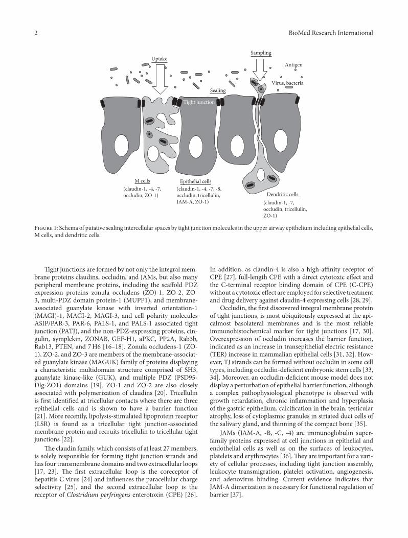
e epithelium in the upper respiratory consists of pseu-dostrati�ed ciliated columnar epithelial cells, including Mcells (membranous or microfold cells), which are specializedfor antigen uptake and form a continuous barrier against awide variety of exogenous antigens [1–5], and dendritic cells(DCs), which take up transported antigens via M cells andpresent antigens for CD4+ T cells, while they maintain theintegrity of the airway epithelial barrier [6–8].e epitheliumplays a crucial role as an interface of adaptive responsesand innate responses via tight junctions to prevent inva-sion of inhaled environmental agents such as allergens andpathogens (Figure 1). In addition, in the human nasalmucosaof allergic rhinitis or virus infection, dynamic changes of tightjunctions have been known.
2. Tight Junctions in Epithelium
e airway epithelium of the human upper respiratorymucosa acts as the �rst physical barrier that protects againstinhaled substances and pathogens [9, 10].e epithelium is ahighly regulated and impermeable barrier exclusively formedby tight junctions [9, 10].
Tight junctions, the most apically located of the inter-cellular junctional complexes, inhibit solute and water �owthrough the paracellular space (termed the “barrier” func-tion) [11, 12]. ey also separate the apical from the basolat-eral cell surface domains to establish cell polarity (termed the“fence” function) [13, 14]. Recent evidence suggests that tightjunctions also participate in signal transduction mechanismsthat regulate epithelial cell proliferation, gene expression,differentiation, and morphogenesis [15].
2 BioMed Research International
Dendritic cells
M cells
Tight junction
Antigen
Virus, bacteria
Epithelial cells
UptakeSampling
Sealing
(claudin-1, -7,occludin, tricellulin,ZO-1)
(claudin-1, -4, -7, -8,occludin, tricellulin,JAM-A, ZO-1)
(claudin-1, -4, -7,occludin, ZO-1)
F 1: Schema of putative sealing intercellular spaces by tight junctionmolecules in the upper airway epithelium including epithelial cells,M cells, and dendritic cells.
Tight junctions are formed by not only the integral mem-brane proteins claudins, occludin, and JAMs, but also manyperipheral membrane proteins, including the scaffold PDZexpression proteins zonula occludens (ZO)-1, ZO-2, ZO-3, multi-PDZ domain protein-1 (MUPP1), and membrane-associated guanylate kinase with inverted orientation-1(MAGI)-1, MAGI-2, MAGI-3, and cell polarity moleculesASIP/PAR-3, PAR-6, PALS-1, and PALS-1 associated tightjunction (PATJ), and the non-PDZ-expressing proteins, cin-gulin, symplekin, ZONAB, GEF-H1, aPKC, PP2A, Rab3b,Rab13, PTEN, and 7H6 [16–18]. Zonula occludens-1 (ZO-1), ZO-2, and ZO-3 are members of the membrane-associat-ed guanylate kinase (MAGUK) family of proteins displayinga characteristic multidomain structure comprised of SH3,guanylate kinase-like (GUK), and multiple PDZ (PSD95-Dlg-ZO1) domains [19]. ZO-1 and ZO-2 are also closelyassociated with polymerization of claudins [20]. Tricellulinis �rst identi�ed at tricellular contacts where there are threeepithelial cells and is shown to have a barrier function[21]. More recently, lipolysis-stimulated lipoprotein receptor(LSR) is found as a tricellular tight junction-associatedmembrane protein and recruits tricellulin to tricellular tightjunctions [22].
e claudin family, which consists of at least 27members,is solely responsible for forming tight junction strands andhas four transmembrane domains and two extracellular loops[17, 23]. e �rst extracellular loop is the coreceptor ofhepatitis C virus [24] and in�uences the paracellular chargeselectivity [25], and the second extracellular loop is thereceptor of Clostridium perfringens enterotoxin (CPE) [26].
In addition, as claudin-4 is also a high-affinity receptor ofCPE [27], full-length CPE with a direct cytotoxic effect andthe C-terminal receptor binding domain of CPE (C-CPE)without a cytotoxic effect are employed for selective treatmentand drug delivery against claudin-4 expressing cells [28, 29].
Occludin, the �rst discovered integral membrane proteinof tight junctions, is most ubiquitously expressed at the api-calmost basolateral membranes and is the most reliableimmunohistochemical marker for tight junctions [17, 30].Overexpression of occludin increases the barrier function,indicated as an increase in transepithelial electric resistance(TER) increase in mammalian epithelial cells [31, 32]. How-ever, TJ strands can be formed without occludin in some celltypes, including occludin-de�cient embryonic stem cells [33,34]. Moreover, an occludin-de�cient mouse model does notdisplay a perturbation of epithelial barrier function, althougha complex pathophysiological phenotype is observed withgrowth retardation, chronic in�ammation and hyperplasiaof the gastric epithelium, calci�cation in the brain, testicularatrophy, loss of cytoplasmic granules in striated duct cells ofthe salivary gland, and thinning of the compact bone [35].
JAMs (JAM-A, -B, -C, -4) are immunoglobulin super-family proteins expressed at cell junctions in epithelial andendothelial cells as well as on the surfaces of leukocytes,platelets and erythrocytes [36].ey are important for a vari-ety of cellular processes, including tight junction assembly,leukocyte transmigration, platelet activation, angiogenesis,and adenovirus binding. Current evidence indicates thatJAM-A dimerization is necessary for functional regulation ofbarrier [37].
BioMed Research International 3
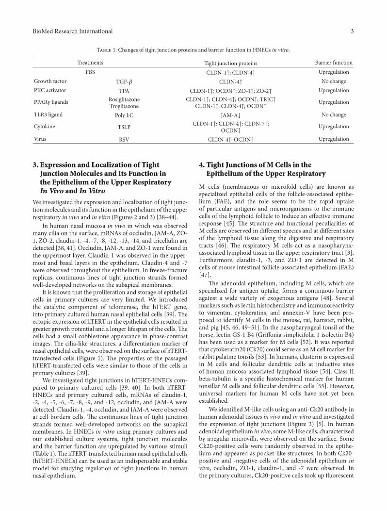
T 1: Changes of tight junction proteins and barrier function in HNECs in vitro.
Treatments Tight junction proteins Barrier functionFBS CLDN-1↑; CLDN-4↑ Upregulation
Growth factor TGF-𝛽𝛽 CLDN-4↑ No changePKC activator TPA CLDN-1↑; OCDN↑; ZO-1↑; ZO-2↑ Upregulation
PPAR𝛾𝛾 ligands RosiglitazoneTroglitazone
CLDN-1↑; CLDN-4↑; OCDN↑; TRIC↑CLDN-1↑; CLDN-4↑; OCDN↑
Upregulation
TLR3 ligand Poly I:C JAM-A↓ No change
Cytokine TSLP CLDN-1↑; CLDN-4↑; CLDN-7↑;OCDN↑
Upregulation
Virus RSV CLDN-4↑; OCDN↑ Upregulation
3. Expression and Localization of TightJunctionMolecules and Its Function inthe Epithelium of the Upper RespiratoryIn Vivo and In Vitro
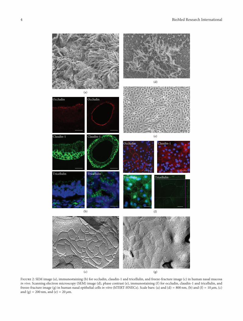
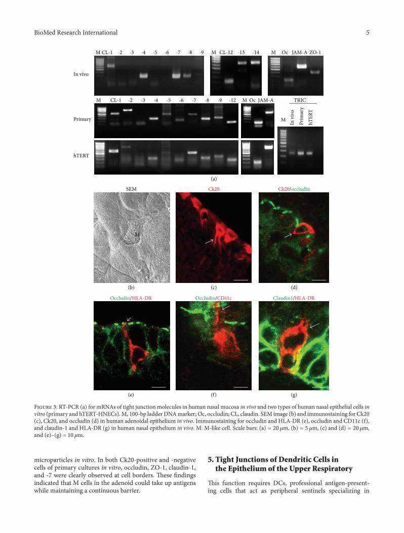
We investigated the expression and localization of tight junc-tionmolecules and its function in the epithelium of the upperrespiratory in vivo and in vitro (Figures 2 and 3) [38–44].
In human nasal mucosa in vivo in which was observedmany cilia on the surface, mRNAs of occludin, JAM-A, ZO-1, ZO-2, claudin-1, -4, -7, -8, -12, -13, -14, and tricellulin aredetected [38, 41]. Occludin, JAM-A, and ZO-1 were found inthe uppermost layer. Claudin-1 was observed in the upper-most and basal layers in the epithelium. Claudin-4 and -7were observed throughout the epithelium. In freeze-fracturereplicas, continuous lines of tight junction strands formedwell-developed networks on the subapical membranes.
It is known that the proliferation and storage of epithelialcells in primary cultures are very limited. We introducedthe catalytic component of telomerase, the hTERT gene,into primary cultured human nasal epithelial cells [39]. eectopic expression of hTERT in the epithelial cells resulted ingreater growth potential and a longer lifespan of the cells.ecells had a small cobblestone appearance in phase-contrastimages. e cilia-like structures, a differentiation marker ofnasal epithelial cells, were observed on the surface of hTERT-transfected cells (Figure 1). e properties of the passagedhTERT-transfected cells were similar to those of the cells inprimary cultures [39].
We investigated tight junctions in hTERT-HNECs com-pared to primary cultured cells [39, 40]. In both hTERT-HNECs and primary cultured cells, mRNAs of claudin-1,-2, -4, -5, -6, -7, -8, -9, and -12, occludin, and JAM-A weredetected. Claudin-1, -4, occludin, and JAM-A were observedat cell borders cells. e continuous lines of tight junctionstrands formed well-developed networks on the subapicalmembranes. In HNECs in vitro using primary cultures andour established culture systems, tight junction moleculesand the barrier function are upregulated by various stimuli(Table 1).e hTERT-transfected human nasal epithelial cells(hTERT-HNECs) can be used as an indispensable and stablemodel for studying regulation of tight junctions in humannasal epithelium.
4. Tight Junctions of M Cells in theEpithelium of the Upper Respiratory
M cells (membranous or microfold cells) are known asspecialized epithelial cells of the follicle-associated epithe-lium (FAE), and the role seems to be the rapid uptakeof particular antigens and microorganisms to the immunecells of the lymphoid follicle to induce an effective immuneresponse [45]. e structure and functional peculiarities ofM cells are observed in different species and at different sitesof the lymphoid tissue along the digestive and respiratorytracts [46]. e respiratory M cells act as a nasopharynx-associated lymphoid tissue in the upper respiratory tract [3].Furthermore, claudin-1, -3, and ZO-1 are detected in Mcells of mouse intestinal follicle-associated epithelium (FAE)[47].
e adenoidal epithelium, including M cells, which arespecialized for antigen uptake, forms a continuous barrieragainst a wide variety of exogenous antigens [48]. Severalmarkers such as lectin histochemistry and immunoreactivityto vimentin, cytokeratins, and annexin-V have been pro-posed to identify M cells in the mouse, rat, hamster, rabbit,and pig [45, 46, 49–51]. In the nasopharyngeal tonsil of thehorse, lectin GS-1 B4 (Griffonia simplicifolia 1 isolectin B4)has been used as a marker for M cells [52]. It was reportedthat cytokeratin20 (Ck20) could serve as anM cell marker forrabbit palatine tonsils [53]. In humans, clusterin is expressedin M cells and follicular dendritic cells at inductive sitesof human mucosa-associated lymphoid tissue [54]. Class IIbeta-tubulin is a speci�c histochemical marker for humantonsillar M cells and follicular dendritic cells [55]. However,universal markers for human M cells have not yet beenestablished.
We identi�edM-like cells using an anti-Ck20 antibody inhuman adenoidal tissues in vivo and in vitro and investigatedthe expression of tight junctions (Figure 3) [5]. In humanadenoidal epithelium in vivo, someM-like cells, characterizedby irregular microvilli, were observed on the surface. SomeCk20-positive cells were randomly observed in the epithe-lium and appeared as pocket-like structures. In both Ck20-positive and -negative cells of the adenoidal epithelium invivo, occludin, ZO-1, claudin-1, and -7 were observed. Inthe primary cultures, Ck20-positive cells took up �uorescent
4 BioMed Research International
(a)
(b)
(c)
(d)
(e)
(f)
(g)
Occludin
Claudin-1
Tricellulin
Occludin
Occludin
Claudin-1
Claudin-1
TricellulinTricellulin Tricellulin
F 2: SEM image (a), immunostaining (b) for occludin, claudin-1 and tricellulin, and freeze-fracture image (c) in human nasal mucosain vivo. Scanning electron microscopy (SEM) image (d), phase contrast (e), immunostaining (f) for occludin, claudin-1 and tricellulin, andfreeze-fracture image (g) in human nasal epithelial cells in vitro (hTERT-HNECs). Scale bars: (a) and (d) = 800 nm, (b) and (f) = 10 𝜇𝜇m, (c)and (g) = 200 nm, and (e) = 20 𝜇𝜇m.
BioMed Research International 5
Occludin/CD11c Claudin1/HLA-DROccludin/HLA-DR
Ck20/occludinCk20SEM
(b) (c)
(a)
(d)
(e) (f) (g)
hT
ER
T
M
M
M
TRIC
hTERT
Primary
In vivo
CL-1
CL-1
-2 -3 -4 -5 -6 -7 -8 -9
-2 -3 -4
M
M
M
M
CL-12 -13 -14
-5 -6 -7 -8 -9 -12
Oc JAM-A
Oc JAM-A
ZO-1
Pri
mar
y
In v
ivo
F 3: RT-PCR (a) for mRNAs of tight junction molecules in human nasal mucosa in vivo and two types of human nasal epithelial cells invitro (primary and hTERT-HNECs).M, 100-bp ladder DNAmarker; Oc, occludin; CL, claudin. SEM image (b) and immunostaining for Ck20(c), Ck20, and occludin (d) in human adenoidal epithelium in vivo. Immunostaining for occludin and HLA-DR (e), occludin and CD11c (f),and claudin-1 and HLA-DR (g) in human nasal epithelium in vivo. M: M-like cell. Scale bars: (a) = 20 𝜇𝜇m, (b) = 5 𝜇𝜇m, (c) and (d) = 20 𝜇𝜇m,and (e)–(g) = 10 𝜇𝜇m.
microparticles in vitro. In both Ck20-positive and -negativecells of primary cultures in vitro, occludin, ZO-1, claudin-1,and -� were clearly observed at cell borders. ese �ndingsindicated that M cells in the adenoid could take up antigenswhile maintaining a continuous barrier.
5. Tight Junctions of Dendritic Cells inthe Epithelium of the Upper Respiratory
is function requires DCs, professional antigen-present-ing cells that act as peripheral sentinels specializing in
6 BioMed Research International
the uptake, processing, and presentation of antigenic mate-rial.
Rescigno et al. discovered a newmechanism for pathogenuptake in the mucosa by which DCs open the tight junctionsbetween epithelial cells and send dendrites outside the epi-thelium to directly sample the pathogen. DCs express tightjunction proteins such as occludin, claudin-1, and ZO-1 topreserve the integrity of the epithelial barrier [56].
e epithelial DC population expresses high levels of theLangerhans cell (LC) marker langerin and the tight junctionproteins claudin-1, -7, and ZO-2 [57]. Claudin-1 is detectedin murine CD207+ LCs residing in the epidermis but not inother skin DCs [58]. In human THP-1 monocytes, mRNAsof occludin, tricellulin, JAM-A, ZO-1, ZO-2, and claudin-4, -7, -8, and -9 can be detected. In mature DCs that havedendrites elongated by treatment with IL-4, GM-CSF, TNF-𝛼𝛼, and ionomycin, mRNA and protein of JAM-A are signi�-cantly increased compared to monocytes [59]. We previouslyreported that in mouse XS52 DCs, claudin-1, -3, -4, -6, -7,-8, and occludin are detected and claudin-7 is induced viaan NF-𝜅𝜅B pathway by thymic stromal lymphopoietin (TSLP)and ligands of Toll-like receptor 2 (TLR2), TLR4, or TLR7/8[60].
In the human nasal mucosa of allergic rhinitis, HLA-DR-, and CD11c-positive DCs express tight junction proteinclaudin-1 and penetrate beyond the apicalmost tight junctionprotein occludin to minimize the increase in permeability ofthe epithelial barrier (Figure 3) [38, 61].
6. TLR3 Ligand Reduced Tight Junctions inthe Epithelium of the Upper Respiratory
TLRs are a component of the innate immune system [62,63]. ey enable the host to recognize a large number ofpathogen-associated molecular patterns such as those of bac-terial lipopolysaccharides (LPS), viral RNA, CpG-containingDNA, and �agellin, among others [64]. TLRs are alsoexpressed in the epithelium of the upper respiratory andmay play a vital role in the immunological outcomes inthese tissues, which produce proin�ammatory cytokines andchemokines upon ligation [65].
In human nasal epithelium in vivo and in vitro, mRNAsfor all 10 known human TLRs are detected in vivo andvitro [66]. TLR3 recognizes viral double-stranded (dsRNA)and its synthetic analogue polyinosinic-polycytidylic acid(poly(I:C)) and stimulates innate immune responses [67].In primary cultures of human adenoid epithelial cells thatexpressed mRNAs of TLR1, 2, 3, 4, 6, 7, and 10, stimulationby the TLR3 ligand poly(I:C), induced production of notonly TNF𝛼𝛼 and IL-8 but also reduced JAM-A expression.echanges were regulated via distinct signaling transductionpathways [44].
e control of TLR3-mediated signaling pathways inhuman nasal epithelium may be important not only ininfection by viral dsRNA but also in autoimmune dis-eases caused by endogenous dsRNA released from necroticcells.
7. TSLP Induced Tight Junctions inthe Epithelium of the Upper Respiratory
e epithelial-derived factor TSLP is an IL-7-like cytokinethat potently induces deregulation of 2 responses, ahallmark feature of allergic in�ammatory diseases such asasthma, atopic dermatitis, and allergic rhinitis [42, 68–70]. TSLP-stimulated CD11c+ DCs induce naïve CD4+ Tcells to differentiate into 2 cells that produce IL-4, IL-5, IL-13, and TNF-𝛼𝛼 [69]. We found high expression ofTSLP in epithelium from patients with allergic rhinitis withrecruitment and in�ltration of CD11c+ DCs [42]. In vitro,TSLP was signi�cantly produced in HNECs by treatmentwith a TLR2 ligand Pam3Cys-Ser-(Lys)4 and a mixture of IL-1𝛽𝛽 and TNF-𝛼𝛼. Treatment with TSLP rapidly enhanced thebarrier function of culturedHNECs togetherwith an increaseof tight junction proteins claudin-1, -4, -7, and occludin.e nasal epithelial-derived TSLP not only activates DCs butalso preserves the epithelial barrier via upregulation of tightjunction proteins to regulate antigen sensitization during theearly stage of allergic rhinitis.
8. The Effects of Lymphocytes on TightJunctional Barrier of the Upper Respiratory
Chronic rhinosinusitis (CRS) is characterized by mucosalin�ammation involving both the nasal cavity and paranasalsinuses [71]. e patients with chronic rhinosinusitis andnasal polyps have a 2-predominant type of in�ammation[72]. e leaky epithelium is present in vivo and in vitroin patients with downregulation of claudin-4 and occludinmRNA in the biopsy specimens [73]. Furthermore, thebarrier function in human primary sinonasal epithelial cellsis decreased by the1 cytokine IFN-𝛾𝛾 and2 cytokine IL-4, whereas 17 cytokine has no effect [73].
On the other hand, B lymphocytes which are responsiblefor the production of IgE play a crucial role in allergic andin�ammatory of upper and lower airways [74]. Nasal polypshave increased numbers of activated eosinophils, mast cells,and IgE [75]. Nasal polyp epithelium from human tissuespecimens has reduced claudin-1 along the basal aspect of themucosal layer, whereas occludin is reduced in the apical andbasal epithelial zones [76]. However, the closed relationshipbetween B cells and tight junctional barrier remains stillunknown.
9. RSV Induced Tight Junctions inthe Epithelium of the Upper Respiratory
e airway epithelium, which has a well-developed barrierregulated by tight junctions, is the �rst line of defense duringrespiratory virus infection. Moreover, it is also known thattight junctions include targets or receptors of viruses suchas claudin-1 and occludin as coreceptors of HCV, JAM as areovirus receptor, and CAR as a coxsackie and adenovirusreceptor [77]. In human nasal epithelial cells, rhinovirusinfection decreases expression of tight junction moleculesZO-1, occludin, and claudin-1 and reduced barrier functionusing primary cultures [78].
BioMed Research International 7
G-proteinG-protein
CLDN-4CLDN-4
FFFF
Control: 24 h RSV: 24 h
(a)
0
10
20
30
40
50
60
70
80
0 24 72
Control
RSV
(hour)
TE
R (
Oh
m c
m2)
(b)
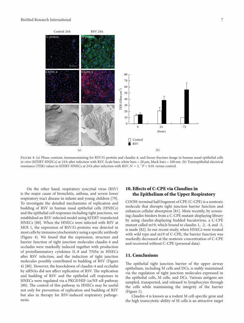
F 4: (a) Phase contrast, immunostaining for RSV/G-protein and claudin-4, and freeze-fracture image in human nasal epithelial cellsin vitro (hTERT-HNECs) at 24 h aer infection with RSV. Scale bars: white bars = 20 𝜇𝜇m, black bars = 100 nm. (b) Transepithelial electricalresistance (TER) values in hTERT-HNECs at 24 h aer infection with RSV.𝑁𝑁 𝑁 𝑁, ∗𝑃𝑃 𝑃 𝑃𝑃𝑃𝑃 versus control.
On the other hand, respiratory syncytial virus (RSV)is the major cause of bronchitis, asthma, and severe lowerrespiratory tract disease in infants and young children [79].To investigate the detailed mechanisms of replication andbudding of RSV in human nasal epithelial cells (HNECs)and the epithelial cell responses including tight junctions, weestablished an RSV-infected model using hTERT-transfectedHNECs [80]. When the HNECs were infected with RSV atMOI 1, the expression of RSV/G-proteins was detected inmost cells by immunocytochemistry using a speci�c antibody(Figure 4). We found that the expression, structure andbarrier function of tight junction molecules claudin-4 andoccludin were markedly induced together with productionof proin�ammatory cytokines I�-8 and TNF𝛼𝛼 in HNECsaer RSV infection, and the induction of tight junctionmolecules possibly contributed to budding of RSV (Figure4) [80]. However, the knockdown of claudin-4 and occludinby siRNAs did not affect replication of RSV. e replicationand budding of RSV and the epithelial cell responses inHNECs were regulated via a PKC𝛿𝛿/HIF-1𝛼𝛼/NF-𝜅𝜅B pathway[80]. e control of this pathway in HNECs may be usefulnot only for prevention of replication and budding of RSVbut also in therapy for RSV-induced respiratory pathoge-nesis.
10. Effects of C-CPE via Claudins inthe Epithelium of the Upper Respiratory
COOH-terminal half fragment of CPE (C-CPE) is a nontoxicmolecule that disrupts tight junction barrier function andenhances cellular absorption [81]. More recently, by screen-ing claudin-binders from a C-CPE mutant-displaying libraryby using claudin-displaying budded baculovirus, a C-CPEmutant called m19, which bound to claudin-1, -2, -4, and -5,is made [82]. In our recent study, when HNECs were treatedwith wild type and m19 of C-CPE, the barrier function wasmarkedly decreased at the nontoxic concentration of C-CPEand recovered without C-CPE (personal data).
11. Conclusions
e epithelial tight junction barrier of the upper airwayepithelium, including M cells and DCs, is stably maintainedvia the regulation of tight junction molecules expressed inthe epithelial cells, M cells, and DCs. Various antigens aresampled, transported, and released to lymphocytes throughthe cells while maintaining the integrity of the barrier(Figure 1).
Claudin-4 is known as a rodent M cell-speci�c gene andthe high transcytotic ability of M cells is an attractive target
8 BioMed Research International
for mucosally delivered vaccines and therapeutics [83, 84].Furthermore, C-CPE which is a nontoxic small moleculethat disrupts tight junction barrier function can be used as acarrier for other substances to speci�c claudin-positive cells[81]. ese may promote development of a novel strategy fordrug delivery system via targeted claudins in upper airwayepithelium.
Tricellular tight junctions form at the convergence ofbicellular tight junctions where three epithelial cells meet inpolarized epithelia, and tricellulin was the �rst marker ofthe tricellular tight junction identi�ed in epithelial cells [21].In various DCs, including human THP-1 cells, mouse XS52cells, and epidermal LCs, tricellulin is also detected [59, 60].e tricellular tight junctionmay be a good penetration pointfor DCs into the epithelium [85]. More recently, it is reportedthat Shigella targets tricellular junctions including tricellulinto spread between cells via noncanonical clathrin-dependentendocytic pathway [86]. us, further study of tricellulinin epithelial cells and DCs of upper airway epithelium maybe important to prevent invasion of inhaled environmentalagents such as allergens and pathogens.
Taken together, these studies of tight junctions in upperairway epithelium should provide new insights not only intopathological conditions but also in the context of new vac-cines and therapeutics against infectious and in�ammatorymucosal diseases in upper airway.
Acknowledgments
e authors are grateful to Dr. Masuo Kondoh (Osaka Uni-versity) for C-CPE. is work was supported by program fordeveloping the supporting system for upgrading educationand research, the Ministry of Education, Culture, SportsScience, and Technology, and the Ministry of Health, Labourand Welfare of Japan.
References
[1] S. T. Holgate, “e airway epithelium is central to the patho-genesis of asthma,” Allergology International, vol. 57, no. 1, pp.1–10, 2008.
[2] Y. Fujimura, “Evidence ofM cells as portals of entry for antigensin the nasopharyngeal lymphoid tissue of humans,” VirchowsArchiv, vol. 436, no. 6, pp. 560–566, 2000.
[3] D. Y. Kim, A. Sato, S. Fukuyama et al., “e airway antigensampling system: respiratory M cells as an alternative gatewayfor inhaled antigens,” e Journal of Immunology, vol. 186, no.7, pp. 4253–4262, 2011.
[4] M. C. Nawijn, T. L. Hackett, D. S. Postma, A. J. M. van Oost-erhout, and I. H. Heijink, “E-cadherin: gatekeeper of airwaymucosa and allergic sensitization,” Trends in Immunology, vol.32, no. 6, pp. 248–255, 2011.
[5] K. I. Takano, T. Kojima, N. Ogasawara et al., “Expression oftight junction proteins in epithelium including Ck20-positiveM-like cells of human adenoids in vivo and in vitro,” Journal ofMolecular Histology, vol. 39, no. 3, pp. 265–273, 2008.
[6] R. M. Steinman, M. Pack, and K. Inaba, “Dendritic cells in theT-cell areas of lymphoid organs,” Immunological Reviews, vol.156, pp. 25–37, 1997.
[7] T. Yamanaka, L. Helgeland, I. N. Farstad, H. Fukushima, T.Midtvedt, and P. Brandtzaeg, “Microbial colonization driveslymphocyte accumulation and differentiation in the follicle-associated epithelium of Peyer’s patches,” e Journal ofImmunology, vol. 170, no. 2, pp. 816–822, 2003.
[8] H. Hammad and B. N. Lambrecht, “Dendritic cells and airwayepithelial cells at the interface between innate and adaptiveimmune responses,” Allergy, vol. 66, no. 5, pp. 579–587, 2011.
[9] S. T. Holgate, “Epithelium dysfunction in asthma,” Journal ofAllergy and Clinical Immunology, vol. 120, no. 6, pp. 1233–1244,2007.
[10] R. P. Schleimer, A. Kato, R. Kern, D. Kuperman, and P. C. Avila,“Epithelium: at the interface of innate and adaptive immuneresponses,” Journal of Allergy and Clinical Immunology, vol. 120,no. 6, pp. 1279–1284, 2007.
[11] B. M. Gumbiner, “Breaking through the tight junction barrier,”e Journal of Cell Biology, vol. 123, no. 6, part 2, pp. 1631–1633,1993.
[12] E. E. Schneeberger and R. D. Lynch, “Structure, function, andregulation of cellular tight junctions,”American Journal of Phys-iology—Lung Cellular and Molecular Physiology, vol. 262, no. 6,pp. L647–L661, 1992.
[13] G. van Meer and K. Simons, “e function of tight junctions inmaintaining differences in lipid composition between the apicaland the basolateral cell surface domains of MDCK cells,” eEMBO journal, vol. 5, no. 7, pp. 1455–1464, 1986.
[14] M. Cereijido, J. Valdés, L. Shoshani, and R. G. Contreras, “Roleof tight junctions in establishing and maintaining cell polarity,”Annual Review of Physiology, vol. 60, pp. 161–177, 1998.
[15] M. S. Balda and K. Matter, “Tight junctions and the regulationof gene expression,” Biochimica et Biophysica Acta, vol. 1788, no.4, pp. 761–767, 2009.
[16] E. E. Schneeberger and R. D. Lynch, “e tight junction: amultifunctional complex,”American Journal of Physiology—CellPhysiology, vol. 286, no. 6, pp. C1213–C1228, 2004.
[17] S. Tsukita, M. Furuse, and M. Itoh, “Multifunctional strands intight junctions,” Nature Reviews Molecular Cell Biology, vol. 2,no. 4, pp. 285–293, 2001.
[18] N. Sawada, M. Murata, K. Kikuchi et al., “Tight junctions andhuman diseases,”Medical ElectronMicroscopy, vol. 36, no. 3, pp.147–156, 2003.
[19] J. M. Anderson, “Cell signalling: MAGUK magic,” CurrentBiology, vol. 6, no. 4, pp. 382–384, 1996.
[20] K. Umeda, J. Ikenouchi, S. Katahira-Tayama et al., “ZO-1 andZO-2 independently determinewhere claudins are polymerizedin tight-junction strand formation,” Cell, vol. 126, no. 4, pp.741–754, 2006.
[21] J. Ikenouchi, M. Furuse, K. Furuse, H. Sasaki, S. Tsukita, andS. Tsukita, “Tricellulin constitutes a novel barrier at tricellularcontacts of epithelial cells,”e Journal of Cell Biology, vol. 171,no. 6, pp. 939–945, 2005.
[22] S. Masuda, Y. Oda, H. Sasaki et al., “LSR de�nes cell corners fortricellular tight junction formation in epithelial cells,” Journal ofCell Science, vol. 124, no. 4, pp. 548–555, 2011.
[23] K. Mineta, Y. Yamamoto, Y. Yamazaki et al., “Predicted expan-sion of the claudin multigene family,” FEBS Letters, vol. 585, no.4, pp. 606–612, 2011.
[24] L. W. Meredith, G. K. Wilson, N. F. Fletcher, and J. A. McK-eating, “Hepatitis C virus entry: beyond receptors,” Reviews inMedical Virology, vol. 22, no. 3, pp. 182–193, 2012.
BioMed Research International 9
[25] S. M. Krug, D. Günzel, M. P. Conrad et al., “Charge-selectiveclaudin channels,”Annals of theNewYorkAcademy Sciences, vol.1257, no. 1, pp. 20–28, 2012.
[26] K. Fujita, J. Katahira, Y. Horiguchi, N. Sonoda, M. Furuse, andS. Tsukita, “Clostridium perfringens enterotoxin binds to thesecond extracellular loop of claudin-3, a tight junction integralmembrane protein,” FEBS Letters, vol. 476, no. 3, pp. 258–261,2000.
[27] J. Katahira, H. Sugiyama, N. Inoue, Y. Horiguchi, M. Matsuda,and N. Sugimoto, “Clostridium perfringens enterotoxin utilizestwo structurally relatedmembrane proteins as functional recep-tors in vivo,”e Journal of Biological Chemistry, vol. 272, no. 42,pp. 26652–26658, 1997.
[28] P. Michl, M. Buchholz, M. Rolke et al., “Claudin-4: a new targetfor pancreatic cancer treatment using clostridium perfringensenterotoxin,” Gastroenterology, vol. 121, no. 3, pp. 678–684,2001.
[29] R. Saeki, M. Kondoh, H. Kakutani et al., “A novel tumor-targeted therapy using a claudin-4-targeting molecule,” Molec-ular Pharmacology, vol. 76, no. 4, pp. 918–926, 2009.
[30] M. Furuse, T. Hirase, M. Itoh et al., “Occludin: a novel integralmembrane protein localizing at tight junctions,” e Journal ofCell Biology, vol. 123, no. 6, pp. 1777–1788, 1993.
[31] M. S. Balda, J. A. Whitney, C. Flores, S. González, M. Cereijido,and K. Matter, “Functional dissociation of paracellular perme-ability and transepithelial electrical resistance and disruptionof the apical-basolateral intramembrane diffusion barrier byexpression of a mutant tight junction membrane protein,” eJournal of Cell Biology, vol. 134, no. 4, pp. 1031–1049, 1996.
[32] K. M. McCarthy, I. B. Skare, M. C. Stankewich et al., “Occludinis a functional component of the tight junction,” Journal of CellScience, vol. 109, no. 9, pp. 2287–2298, 1996.
[33] T. Hirase, J. M. Staddon,M. Saitou et al., “Occludin as a possibledeterminant of tight junction permeability in endothelial cells,”Journal of Cell Science, vol. 110, no. 14, pp. 1603–1613, 1997.
[34] M. Saitou, K. Fujimoto, Y. Doi et al., “Occludin-de�cientembryonic stem cells can differentiate into polarized epithelialcells bearing tight junctions,” e Journal of Cell Biology, vol.141, no. 2, pp. 397–408, 1998.
[35] M. Saitou, M. Furuse, H. Sasaki et al., “Complex phenotype ofmice lacking occludin, a component of tight junction strands,”Molecular Biology of the Cell, vol. 11, no. 12, pp. 4131–4142,2000.
[36] I. Martìn-Padura, S. Lostaglio, M. Schneemann et al., “Junc-tional adhesion molecule, a novel member of the immuno-globulin superfamily that distributes at intercellular junctionsand modulates monocyte transmigration,” e Journal of CellBiology, vol. 142, no. 1, pp. 117–127, 1998.
[37] A. C. Monteiro and C. A. Parkos, “Intracellular mediators ofJAM-A-dependent epithelial barrier function,” Annals of theNew York Academy of Sciences, vol. 1257, no. 1, pp. 115–124,2012.
[38] K. I. Takano, T. Kojima, M. Go et al., “HLA-DR- and CD11c-positive dendritic cells penetrate beyondwell-developed epithe-lial tight junctions in human nasal mucosa of allergic rhinitis,”e Journal of Histochemistry and Cytochemistry, vol. 53, no. 5,pp. 611–619, 2005.
[39] M. Kurose, T. Kojima, J. I. Koizumi et al., “Induction of claudinsin passaged hTERT-transfected human nasal epithelial cellswith an extended life span,” Cell and Tissue Research, vol. 330,no. 1, pp. 63–74, 2007.
[40] J. I. Koizumi, T. Kojima, N. Ogasawara et al., “Protein kinasec enhances tight junction barrier function of human nasalepithelial cells in primary culture by transcriptional regulation,”Molecular Pharmacology, vol. 74, no. 2, pp. 432–442, 2008.
[41] T. Ohkuni, T. Kojima, N. Ogasawara et al., “Expression andlocalization of tricellulin in human nasal epithelial cells in vivoand in vitro,”Medical Molecular Morphology, vol. 42, no. 4, pp.204–211, 2009.
[42] R. Kamekura, T. Kojima, J. I. Koizumi et al., “ymic stromallymphopoietin enhances tight-junction barrier function ofhuman nasal epithelial cells,” Cell and Tissue Research, vol. 338,no. 2, pp. 283–293, 2009.
[43] N. Ogasawara, T. Kojima, M. Go et al., “PPAR𝛾𝛾 agonists upreg-ulate the barrier function of tight junctions via a PKC pathwayin human nasal epithelial cells,” Pharmacological Research, vol.61, no. 6, pp. 489–498, 2010.
[44] T. Ohkuni, T. Kojima, N. Ogasawara et al., “Poly(I:C) reducesexpression of JAM-A and induces secretion of IL-8 and TNF-𝛼𝛼 via distinct NF-𝜅𝜅B pathways in human nasal epithelial cells,”Toxicology and Applied Pharmacology, vol. 250, no. 1, pp. 29–38,2011.
[45] J. Mach, T. Hshieh, D. Hsieh, N. Grubbs, and A. Chervonsky,“Development of intestinal M cells,” Immunological Reviews,vol. 206, pp. 177–189, 2005.
[46] A. Gebert, “e role of M cells in the protection of mucosalmembranes,” Histochemistry and Cell Biology, vol. 108, no. 6,pp. 455–470, 1997.
[47] A. M. Clark and B. H. Hirst, “Expression of junction-asso-ciated proteins differentiates mouse intestinal M cells fromenterocytes,”Histochemistry and Cell Biology, vol. 118, no. 2, pp.137–147, 2002.
[48] M. J. P. van Kempen, G. T. Rijkers, and P. B. van Cauwenberge,“e immune response in adenoids and tonsils,” InternationalArchives of Allergy and Immunology, vol. 122, no. 1, pp. 8–19,2000.
[49] P. J. Giannasca, J. A. Boden, and T. P. Monath, “Targeteddelivery of antigen to hamster nasal lymphoid tissue with M-cell-directed lectins,” Infection and Immunity, vol. 65, no. 10, pp.4288–4298, 1997.
[50] T. Kucharzik, N. Lügering, K. W. Schmid, M. A. Schmidt, R.Stoll, and W. Domschke, “Human intestinal M cells exhibitenterocyte-like intermediate �laments,” Gut, vol. 42, no. 1, pp.54–62, 1998.
[51] P. Verbrugghe, W. Waelput, B. Dieriks, A. Waeytens, J. Vandes-ompele, and C. A. Cuvelier, “Murine M cells express annexin Vspeci�cally,” Journal of Pathology, vol. 209, no. 2, pp. 240–249,2006.
[52] P. Kumar and J. F. Timoney, “Light and electron microscopestudies on the nasopharynx and nasopharyngeal tonsil of thehorse,” Anatomia, Histologia, Embryologia, vol. 30, no. 2, pp.77–84, 2001.
[53] A. Carapelli, M. Regoli, C. Nicoletti, L. Ermini, L. Fonzi, and E.Bertelli, “Rabbit tonsil-associated M-cells express cytokeratin20 and take up particulate antigen,” e Journal of Histochem-istry and Cytochemistry, vol. 52, no. 10, pp. 1323–1331, 2004.
[54] P. Verbrugghe, P. Kujala, W. Waelput, P. J. Peters, and C. A.Cuvelier, “Clusterin in human gut-associated lymphoid tissue,tonsils, and adenoids: localization to M cells and folliculardendritic cells,” Histochemistry and Cell Biology, vol. 129, no. 3,pp. 311–320, 2008.
10 BioMed Research International
[55] J. H. Lee, S. K. Kong, Z. S.Wu et al., “Class II 𝛽𝛽-tubulin is a novelmarker for human tonsillar M cells and follicular dendriticcells,” Journal of Oral Pathology and Medicine, vol. 39, no. 7, pp.533–539, 2010.
[56] M. Rescigno, M. Urbano, B. Valzasina et al., “Dendritic cellsexpress tight junction proteins and penetrate gut epithelialmonolayers to sample bacteria,” Nature Immunology, vol. 2, no.4, pp. 361–367, 2001.
[57] S. S. J. Sung, S. M. Fu, C. E. Rose Jr., F. Gaskin, S. T. Ju, andS. R. Beaty, “A major lung CD103 (𝛼𝛼E)-𝛽𝛽7 integrin-positiveepithelial dendritic cell population expressing langerin andtight junction proteins,” e Journal of Immunology, vol. 176,no. 4, pp. 2161–2172, 2006.
[58] S. C. Zimmerli and C. Hauser, “Langerhans cells and lymphnode dendritic cells express the tight junction componentclaudin-1,” Journal of Investigative Dermatology, vol. 127, no. 10,pp. 2381–2390, 2007.
[59] N. Ogasawara, T. Kojima, M. Go et al., “Induction of JAM-A during differentiation of human THP-1 dendritic cells,”Biochemical and Biophysical Research Communications, vol.389, no. 3, pp. 543–549, 2009.
[60] R. Kamekura, T. Kojima, A. Takashima et al., “ymic stromallymphopoietin induces tight junction protein claudin-7 via NF-𝜅𝜅B in dendritic cells,” Histochemistry and Cell Biology, vol. 133,no. 3, pp. 339–348, 2010.
[61] N. Ogasawara, T. Kojima, M. Go et al., “Epithelial barrier andantigen uptake in lymphoepithelium of human adenoids,” ActaOto-Laryngologica, vol. 131, no. 2, pp. 116–123, 2011.
[62] A. Iwasaki and R. Medzhitov, “Toll-like receptor control of theadaptive immune responses,”Nature Immunology, vol. 5, no. 10,pp. 987–995, 2004.
[63] K. Takeda and S.Akira, “Toll-like receptors in innate immunity,”International Immunology, vol. 17, no. 1, pp. 1–14, 2005.
[64] T. Kawai and S. Akira, “TLR signaling,” Cell Death and Differ-entiation, vol. 13, no. 5, pp. 816–825, 2006.
[65] M. J. Lange, J. C. Lasiter, andM. L. Misfeldt, “Toll-like receptorsin tonsillar epithelial cells,” International Journal of PediatricOtorhinolaryngology, vol. 73, no. 4, pp. 613–621, 2009.
[66] M. J. Lesmeister, M. R. Bothwell, and M. L. Misfeldt, “Toll-like receptor expression in the human nasopharyngeal tonsil(adenoid) and palantine tonsils: a preliminary report,” Interna-tional Journal of Pediatric Otorhinolaryngology, vol. 70, no. 6,pp. 987–992, 2006.
[67] L. Alexopoulou, A. C. Holt, R. Medzhitov, and R. A. Flavell,“Recognition of double-stranded RNA and activation of NF-𝜅𝜅Bby Toll-like receptor 3,” Nature, vol. 413, no. 6857, pp. 732–738,2001.
[68] S. T. Holgate, “Epithelium dysfunction in asthma,” Journal ofAllergy and Clinical Immunology, vol. 120, no. 6, pp. 1233–1244,2007.
[69] V. Soumelis, P. A. Reche, H. Kanzler et al., “Human epithe-lial cells trigger dendritic cell-mediated allergic in�ammationby producing TSLP,” Nature Immunology, vol. 3, no. 7, pp.673–680, 2002.
[70] S. Ying, B. O’Connor, J. Ratoff et al., “ymic stromal lym-phopoietin expression is increased in asthmatic airways andcorrelates with expression of 2-attracting chemokines anddisease severity,” e Journal of Immunology, vol. 174, no. 12,pp. 8183–8190, 2005.
[71] B. F. Marple, J. A. Stankiewicz, F. M. Baroody et al., “Diagnosisand management of chronic rhinosinusitis in adults,” Postgrad-uate Medicine, vol. 121, no. 6, pp. 121–139, 2009.
[72] N. van Bruaene, C. A. Pérez-Novo, T. M. Basinski et al., “T-cellregulation in chronic paranasal sinus disease,” Journal of Allergyand Clinical Immunology, vol. 121, no. 6, pp. 1435.e3–1441.e3,2008.
[73] M. B. Soyka, P. Wawrzyniak, T. Eiwegger et al., “Defectiveepithelial barrier in chronic rhinosinusitis:the regulation oftight junctions by IFN-𝛾𝛾 and IL-4,” Journal of Allergy andClinical Immunology. In press.
[74] J. P. Drolet, H. Frangie, J. Guay, O. Hajoui, Q. Hamid, and B.D. Mazer, “B lymphocytes in in�ammatory airway diseases,”Clinical and Experimental Allergy, vol. 40, no. 6, pp. 841–849,2010.
[75] M. S. Georgy and A. T. Peters, “Nasal polyps,” in Allergy andAsthma Proceedings, vol. 33, chapter 7, supplement 1, pp. 22–23,2012.
[76] G. A. Rogers, K. Den Beste, C. A. Parkos, A. Nusrat, J. M. Del-gaudio, and S. K. Wise, “Epithelial tight junction alterations innasal polyposis,” International Forum of Allergy and Rhinology,vol. 1, no. 1, pp. 50–54, 2011.
[77] J. A. Guttman and B. B. Finlay, “Tight junctions as targets ofinfectious agents,” Biochimica et Biophysica Acta, vol. 1788, no.4, pp. 832–841, 2009.
[78] N. K. Yeo and Y. J. Jang, “Rhinovirus infection-induced alter-ation of tight junction and adherens junction components inhuman nasal epithelial cells,”e Laryngoscope, vol. 120, no. 2,pp. 346–352, 2010.
[79] V. Bitko and S. Barik, “Persistent activation of RelA by respira-tory syncytial virus involves protein kinase C, underphospho-rylated I𝜅𝜅B𝛽𝛽, and sequestration of protein phosphatase 2A bythe viral phosphoprotein,” Journal of Virology, vol. 72, no. 7, pp.5610–5618, 1998.
[80] T. Masaki, T. Kojima, T. Okabayashi et al., “A nuclear factor-𝜅𝜅Bsignaling pathway via protein kinase C 𝛿𝛿 regulates replicationof respiratory syncytial virus in polarized normal human nasalepithelial cells,”Molecular Biology of the Cell, vol. 22, no. 13, pp.2144–2156, 2011.
[81] N. Sonoda,M. Furuse,H. Sasaki et al., “Clostridiumperfringensenterotoxin fragment removes speci�c claudins from tightjunction strands: evidence for direct involvement of claudins intight junction barrier,” e Journal of Cell Biology, vol. 147, no.1, pp. 195–204, 1999.
[82] K.Matsuhisa,M. Kondoh,H. Suzuki, andK. Yagi, “Comparisonof mucosal absorption-enhancing activity between a claudin-3�-4 binder and a broadly speci�c claudin binder,” vol. 423, no.2, pp. 229–233, 2012.
[83] J. Wang, M. Lopez-Fraga, A. Rynko, and D. D. Lo, “TNFRand LT𝛽𝛽R agonists induce follicle-associated epithelium andMcell speci�c genes in rat and human intestinal epithelial cells,”Cytokine, vol. 47, no. 1, pp. 69–76, 2009.
[84] T. E. Rajapaksa, M. Stover-Hamer, X. Fernandez, H. A. Eckel-hoefer, and D. D. Lo, “Claudin 4-targeted protein incorporatedinto PLGA nanoparticles can mediate M cell targeted delivery,”Journal of Controlled Release, vol. 142, no. 2, pp. 196–205, 2010.
[85] A. Kubo, K. Nagao, M. Yokouchi, H. Sasaki, and M. Amagai,“External antigen uptake by Langerhans cells with reorganiza-tion of epidermal tight junction barriers,” Journal of Experimen-tal Medicine, vol. 206, no. 13, pp. 2937–2946, 2009.

BioMed Research International 11
[86] M. Fukumatsu, M. Ogawa, S. Arakawa et al., “Shigella targetsepithelial tricellular junctions and uses a noncanonical clathrin-dependent endocytic pathway to spread between cells,” CellHost and Microbe, vol. 11, no. 4, pp. 325–336, 2012.

Submit your manuscripts athttp://www.hindawi.com
Stem CellsInternational
Hindawi Publishing Corporationhttp://www.hindawi.com Volume 2014
Hindawi Publishing Corporationhttp://www.hindawi.com Volume 2014
MEDIATORSINFLAMMATION
of
Hindawi Publishing Corporationhttp://www.hindawi.com Volume 2014
Behavioural Neurology
EndocrinologyInternational Journal of
Hindawi Publishing Corporationhttp://www.hindawi.com Volume 2014
Hindawi Publishing Corporationhttp://www.hindawi.com Volume 2014
Disease Markers
Hindawi Publishing Corporationhttp://www.hindawi.com Volume 2014
BioMed Research International
OncologyJournal of
Hindawi Publishing Corporationhttp://www.hindawi.com Volume 2014
Hindawi Publishing Corporationhttp://www.hindawi.com Volume 2014
Oxidative Medicine and Cellular Longevity
Hindawi Publishing Corporationhttp://www.hindawi.com Volume 2014
PPAR Research
The Scientific World JournalHindawi Publishing Corporation http://www.hindawi.com Volume 2014
Immunology ResearchHindawi Publishing Corporationhttp://www.hindawi.com Volume 2014
Journal of
ObesityJournal of
Hindawi Publishing Corporationhttp://www.hindawi.com Volume 2014
Hindawi Publishing Corporationhttp://www.hindawi.com Volume 2014
Computational and Mathematical Methods in Medicine
OphthalmologyJournal of
Hindawi Publishing Corporationhttp://www.hindawi.com Volume 2014
Diabetes ResearchJournal of
Hindawi Publishing Corporationhttp://www.hindawi.com Volume 2014
Hindawi Publishing Corporationhttp://www.hindawi.com Volume 2014
Research and TreatmentAIDS
Hindawi Publishing Corporationhttp://www.hindawi.com Volume 2014
Gastroenterology Research and Practice
Hindawi Publishing Corporationhttp://www.hindawi.com Volume 2014
Parkinson’s Disease
Evidence-Based Complementary and Alternative Medicine
Volume 2014Hindawi Publishing Corporationhttp://www.hindawi.com