Regions important for the adhesin activity of Moraxella catarrhalis Hag
13
BioMed Central Page 1 of 13 (page number not for citation purposes) BMC Microbiology Open Access Research article Regions important for the adhesin activity of Moraxella catarrhalis Hag Brian Bullard 1 , Serena Lipski 1 and Eric R Lafontaine* 2 Address: 1 Department of Medical Microbiology and Immunology, University of Toledo Health Sciences Campus, 3055 Arlington Avenue, Toledo, OH, 43614, USA and 2 Department of Infectious Diseases, University of Georgia College of Veterinary Medicine, Athens, GA, 30602, USA Email: Brian Bullard - [email protected] ; Serena Lipski - [email protected]; Eric R Lafontaine* - [email protected] * Corresponding author Abstract Background: The Moraxella catarrhalis Hag protein, an Oca autotransporter adhesin, has previously been shown to be important for adherence of this respiratory tract pathogen to human middle ear and A549 lung cells. Results: The present study demonstrates that adherence of M. catarrhalis isogenic hag mutant strains to the human epithelial cell lines Chang (conjunctival) and NCIH292 (lung) is reduced by 50–93%. Furthermore, expressing Hag in a heterologous Escherichia coli background substantially increased the adherence of recombinant bacteria to NCIH292 cells and murine type IV collagen. Hag did not, however, increase the attachment of E. coli to Chang cells. These results indicate that Hag directly mediates adherence to NCIH292 lung cells and collagen, but is not sufficient to confer binding to conjunctival monolayers. Several in-frame deletions were engineered within the hag gene of M. catarrhalis strain O35E and the resulting proteins were tested for their ability to mediate binding to NCIH292 monolayers, middle ear cells, and type IV collagen. These experiments revealed that epithelial cell and collagen binding properties are separable, and that residues 385– 705 of this ~2,000 amino acid protein are important for adherence to middle ear and NCIH292 cells. The region of O35E-Hag encompassing aa 706 to 1194 was also found to be required for adherence to collagen. In contrast, β-roll repeats present in Hag, which are structural features conserved in several Oca adhesins and responsible for the adhesive properties of Yersinia enterocolitica YadA, are not important for Hag-mediated adherence. Conclusion: Hag is a major adherence factor for human cells derived from various anatomical sites relevant to pathogenesis by M. catarrhalis and its structure-function relationships differ from those of other, closely-related autotransporter proteins. Background Autotransporter proteins form the largest known family of virulence factors expressed by Gram-negative bacteria, and play central roles in processes such as cell-to-cell aggregation, biofilm formation, serum resistance, and adherence to host epithelial cells [1]. The members of this family share structural features that include a surface- exposed passenger domain specifying the biological activ- ity of the protein and a C-terminal transporter module comprised of several β-strands that anchors the autotrans- porter in the outer membrane (OM). Based on the length and structure of this transporter module, autotransporters Published: 3 July 2007 BMC Microbiology 2007, 7:65 doi:10.1186/1471-2180-7-65 Received: 23 January 2007 Accepted: 3 July 2007 This article is available from: http://www.biomedcentral.com/1471-2180/7/65 © 2007 Bullard et al; licensee BioMed Central Ltd. This is an Open Access article distributed under the terms of the Creative Commons Attribution License (http://creativecommons.org/licenses/by/2.0 ), which permits unrestricted use, distribution, and reproduction in any medium, provided the original work is properly cited.
-
Upload
brian-bullard -
Category
Documents
-
view
213 -
download
1
Transcript of Regions important for the adhesin activity of Moraxella catarrhalis Hag

BioMed CentralBMC Microbiology
ss
Open AcceResearch articleRegions important for the adhesin activity of Moraxella catarrhalis HagBrian Bullard1, Serena Lipski1 and Eric R Lafontaine*2Address: 1Department of Medical Microbiology and Immunology, University of Toledo Health Sciences Campus, 3055 Arlington Avenue, Toledo, OH, 43614, USA and 2Department of Infectious Diseases, University of Georgia College of Veterinary Medicine, Athens, GA, 30602, USA
Email: Brian Bullard - [email protected] ; Serena Lipski - [email protected]; Eric R Lafontaine* - [email protected]
* Corresponding author
AbstractBackground: The Moraxella catarrhalis Hag protein, an Oca autotransporter adhesin, haspreviously been shown to be important for adherence of this respiratory tract pathogen to humanmiddle ear and A549 lung cells.
Results: The present study demonstrates that adherence of M. catarrhalis isogenic hag mutantstrains to the human epithelial cell lines Chang (conjunctival) and NCIH292 (lung) is reduced by50–93%. Furthermore, expressing Hag in a heterologous Escherichia coli background substantiallyincreased the adherence of recombinant bacteria to NCIH292 cells and murine type IV collagen.Hag did not, however, increase the attachment of E. coli to Chang cells. These results indicate thatHag directly mediates adherence to NCIH292 lung cells and collagen, but is not sufficient to conferbinding to conjunctival monolayers. Several in-frame deletions were engineered within the hag geneof M. catarrhalis strain O35E and the resulting proteins were tested for their ability to mediatebinding to NCIH292 monolayers, middle ear cells, and type IV collagen. These experimentsrevealed that epithelial cell and collagen binding properties are separable, and that residues 385–705 of this ~2,000 amino acid protein are important for adherence to middle ear and NCIH292cells. The region of O35E-Hag encompassing aa 706 to 1194 was also found to be required foradherence to collagen. In contrast, β-roll repeats present in Hag, which are structural featuresconserved in several Oca adhesins and responsible for the adhesive properties of Yersiniaenterocolitica YadA, are not important for Hag-mediated adherence.
Conclusion: Hag is a major adherence factor for human cells derived from various anatomicalsites relevant to pathogenesis by M. catarrhalis and its structure-function relationships differ fromthose of other, closely-related autotransporter proteins.
BackgroundAutotransporter proteins form the largest known family ofvirulence factors expressed by Gram-negative bacteria,and play central roles in processes such as cell-to-cellaggregation, biofilm formation, serum resistance, andadherence to host epithelial cells [1]. The members of this
family share structural features that include a surface-exposed passenger domain specifying the biological activ-ity of the protein and a C-terminal transporter modulecomprised of several β-strands that anchors the autotrans-porter in the outer membrane (OM). Based on the lengthand structure of this transporter module, autotransporters
Published: 3 July 2007
BMC Microbiology 2007, 7:65 doi:10.1186/1471-2180-7-65
Received: 23 January 2007Accepted: 3 July 2007
This article is available from: http://www.biomedcentral.com/1471-2180/7/65
© 2007 Bullard et al; licensee BioMed Central Ltd. This is an Open Access article distributed under the terms of the Creative Commons Attribution License (http://creativecommons.org/licenses/by/2.0), which permits unrestricted use, distribution, and reproduction in any medium, provided the original work is properly cited.
Page 1 of 13(page number not for citation purposes)

BMC Microbiology 2007, 7:65 http://www.biomedcentral.com/1471-2180/7/65
can be classified as conventional (long module containing12 β-strands) or trimeric (short module containing 4 β-strands) [2]. One of the better characterized trimericautotransporter molecules is the Yersinia enterocoliticaadhesin YadA; YadA and structurally related autotrans-porters are also often referred to as Oca (Oligomericcoiled-coil adhesin) proteins [3]. Because of their struc-ture and role in pathogenesis, autotransporters representpromising targets for anti-infective approaches. Large por-tions of these proteins (the passenger domains) arelocated on the bacterial surface and are therefore amena-ble to recognition by the immune system. In addition,autotransporters play key roles in virulence and targetingthem may interfere with development of disease.
Moraxella catarrhalis is a Gram-negative bacterium thatcauses a wide range of infections, including ~20% of allcases of bacterial otitis media in children [4], up to 10%of lung infections in elderly patients with ChronicObstructive Pulmonary Disease (COPD) [5], sinusitis [6],and conjunctivitis [7]. This organism is a significanthealth concern, and complicating this problem is the factthat most M. catarrhalis clinical isolates display resistanceto antibiotics including β-lactams [8-10]. The develop-ment of a vaccine for M. catarrhalis thus has increasingimportance for the health status of both the elderly andthe very young. This bacterium expresses severalautotransporter proteins that have been well-studiedincluding the trimeric/Oca adhesins UspA1 [3,11-18],UspA2H [11], and Hag/MID [13,15,19-26], the trimeric/Oca serum resistant factor UspA2 [3,11,16,17,27,28], andthe conventional autotransporter adhesin/phospholipaseB McaP [29,30].
Expression of Hag, or its ortholog MID (Moraxella IgD-binding protein), is important for attachment to erythro-cytes as well as A549 (lung pneumocytes) and HMEE(human middle ear epithelial) cells [22-24,26], and Hagdirectly mediates adherence to HMEE cells [26]. In addi-tion, Hag binds immunoglobulin D and forms oligomers[13,19,21,22,24]. The gene coding for this protein isfound in almost all M. catarrhalis isolates studied to date[24,26], and specifies a relatively well-conserved moleculeof ~2,000 residues in length. Expression of Hag is subjectto translational phase variation, via slipped strand mis-pairing in a homopolymeric guanine tract located near thebeginning of the ORF [13,24,26]. Hag has also beenfound to contain a series of degenerate amino acid (aa)repeats similar to those present in the N-terminus of theprototypical Oca adhesin YadA and which specify theadhesive properties of this Y. enterocolitica protein[26,31,32].
In this study, we sought to determine what role Hag playsin M. catarrhalis adherence to lung mucoepidermoid
(NCIH292) and conjunctival (Chang) cells by measuringthe binding of Hag isogenic mutants and of recombinantE. coli expressing wild-type (WT) hag genes to thesehuman cells. We also sought to identify the region(s) ofHag important for adherence using a panel of in-framehag deletion constructs.
ResultsHag expression is important for the adherence of M. catarrhalis to NCIH292 and Chang cells in vitroPrevious work demonstrated that Hag plays an importantrole in the attachment of M. catarrhalis to HMEE cells andA549 human type II pneumocytes [23,26]. To furtherexplore the role of this protein in adherence to humanepithelial cells that are relevant to pathogenesis by M.catarrhalis, a panel of previously-described WT isolatesand cognate hag mutant strains [26] were tested for theirability to bind to NCIH292 lung mucoepidermoid andChang conjunctival cells. Of note, these mutants were pre-viously shown to have reduced adherence to A549 andHMEE cells [26].
Strain TTA37 attached poorly to NCIH292 monolayersand the adherence of its hag mutant, TTA37.Hag, was notsignificantly lower (data not shown). Hag-deficientstrains derived from isolates V1171, O35E, McGHS1 andO12E did not attach as efficiently to NCIH292 cells astheir respective WT progenitors (Fig. 1A). The binding ofV1171.Hag, O35E.TN2 and McGHS1.Hag to these mon-olayers was about a third that of their parent strainsV1171, O35E, and McGHS1, respectively. Though thetwo-fold difference between strains O12E and O12E.Hagwas not statistically significant, the preponderance of ourdata suggests that Hag expression is important for theadherence of M. catarrhalis to NCIH292 lung cells.
Similar results were obtained when the WT and isogenicmutant strains were tested for their ability to bind toChang conjunctival cells. The hag mutants of strainsV1171, O12E and McGHS1 all adhered significantly lessthan their WT progenitors (Fig. 1B). Strain TTA37 attachedwell to Chang cells (unlike NCIH292 cells) and the adher-ence of its Hag deficient mutant TTA37.Hag was reducedby 93%. Strain O35E.TN2 did not show Hag-dependentbinding to Chang cells, though strain O35E did not attachas well to these monolayers as the other WT M. catarrhalisisolates tested. Taken together, these results indicate thatHag expression is important for the binding of M. catarrh-alis to conjunctival cells. Of note, all strains tested in thisstudy were previously shown to express WT levels of theadhesins UspA2H (in the case of strain TTA37), UspA1,McaP, and OMPCD [26], yet we found that Hag deletionsstill substantially reduce adherence.
Page 2 of 13(page number not for citation purposes)

BMC Microbiology 2007, 7:65 http://www.biomedcentral.com/1471-2180/7/65
Hag directly confers binding to NCIH292 cells and murine type IV collagenIt is possible that absence of Hag in the OM of M. catarrh-alis mutants affected the proper display of other adhesins,which themselves mediate adherence to NCIH292 andChang monolayers. To determine whether Hag directlymediates binding to these epithelial cells, recombinant E.coli strains previously shown to express the hag gene prod-uct of M. catarrhalis strain O12E, O35E or V1171 [26]were tested in quantitative attachment assays. Of note,these clones were previously shown to have gained theability to attach to HMEE cells [26]. As shown in Fig. 2A,O12E-, O35E- and V1171-Hag conferred on E.coli theability to bind to NCIH292 cells at levels that were ~5-,~10- and ~15-fold greater than E. coli carrying the controlplasmid pCC1.3, respectively. In contrast, E. coli express-ing these three Hag proteins did not show significantincreases in binding to Chang monolayers (Fig. 2B). Theseresults demonstrate that Hag directly mediates adherenceto NCIH292 cells, but its expression is not sufficient toincrease the binding of recombinant E. coli to Changmonolayers.
Based on sequence and structural similarities, Hagbelongs to the Oca family of autotransporter adhesins[26]. Y. enterocolitica YadA is the prototypical member ofthis family and has been shown to directly mediate adher-
ence to different types of collagen [31-33], including typeIV [33-35]. E. coli cells expressing the Hag protein of M.catarrhalis strain O35E, O12E or V1171 were thereforetested for their ability to bind to murine type IV collagen.As shown in Fig. 2C, E. coli expressing O12E- and V1171-Hag were ~35 times more adherent to collagen than thenegative control while recombinant bacteria expressingO35E-Hag exhibited a ~80-fold increase in binding. Thesedata indicate that Hag proteins from different M. catarrh-alis isolates directly mediate attachment to murine type IVcollagen when expressed in the E. coli background. How-ever, our panel of M. catarrhalis WT and isogenic mutantstrains all bound poorly to collagen-coated wells (datanot shown).
Construction and surface-expression of mutated Hag proteinsTo define the region(s) of Hag important for adherence toepithelial cells and collagen, several in-frame deletionswere engineered in the hag ORF of strain O35E (specifiedby the plasmid pELO35.Hag). Fig. 3 illustrates the variousdeletion constructs that were made. These mutated Hagproteins were tested for their ability to mediate the bind-ing of recombinant E. coli to NCIH292 cells, HMEE cellsand murine collagen (see below). Sarkosyl-insoluble OMproteins were also prepared from recombinant E. coliexpressing these constructs, and tested by immunoblot-
Adherence of M. catarrhalisstrains to NCIH292 (Panel A) and Chang (Panel B) cellsFigure 1Adherence of M. catarrhalisstrains to NCIH292 (Panel A) and Chang (Panel B) cells. Black bars correspond to WT isolates and hag isogenic mutant strains are represented by open bars. The results are expressed as the mean (± standard error) percentage of inoculated bacteria binding to monolayers. The number above each bar represents mean percentage; standard error is shown in parentheses. Asterisks indicate that the difference in adherence between a WT strain and its respective hag mutant is statistically significant.
O35
E
O35
E.T
N2
V11
71
V11
71.H
ag
MC
GH
S1
MC
GH
S1.
Hag
O12
E
O12
E.H
ag
0
10
20
30
40
50
60
70
80
90
100
1.7(0.4)
42.0(1.4)
5.1(0.8)
12.5(0.8)
24.8(8.9)
49.6(6.9)
15.4(1.6)
42.6(4.2)
% a
dh
eren
ce
O35
E
O35
E.T
N2
TT
A37
TT
A37
.Hag
V11
71
V11
71.H
ag
McG
HS
1
McG
HS
1.H
ag
O12
E
O12
E.H
ag
0
10
20
30
40
50
60
70
80
90
100
110
8.8(0.9)
6.6(0.6)
90.6(12.3)
9.6(0.7)
24.4(2.3)
46.5(6.4)
43.1(7.4)
14.5(2.4)
53.5(4.4)
3.7(0.5)
% a
dh
eren
ce
A. NCIH292 (lung) B. Chang (conjunctival)
** *
**
**
McG
HS
1.H
ag
McG
HS
1
Page 3 of 13(page number not for citation purposes)

BMC Microbiology 2007, 7:65 http://www.biomedcentral.com/1471-2180/7/65
ting with Hag-reactive antibodies. As shown in Fig. 4, allmutated Hag proteins were associated with the OM. Thepolyclonal antibodies used in these westerns are againstthe last 607 residues of O35E-Hag (i.e. His.Hag.CT, seeMethods) and encompass the transporter domain (Fig. 3).This portion of Oca adhesins has been shown to beimportant for oligomerization properties, which likely
accounts for the multiple bands reacting with anti-Hagantibodies in Fig. 4.
To verify that these OM-located Hag proteins wereexposed on the bacterial surface, intact recombinant E. colicells were treated with proteinase K and analyzed by West-ern blot using antibodies against Hag. As controls, protei-nase K-treated bacteria were also probed with amonoclonal antibody against the inner membrane-anchored protein TonB [36] to demonstrate that a mini-mal amount of the enzyme transversed across the OM andinto the periplasm of E. coli. Fig. 5 shows the results of arepresentative experiment in which E. coli carrying theplasmids pELO35.Hag, pBBHS2.24 and pBBHS3.20 wereincubated for 15 min on ice in the absence (lanes 1, 3 and5) or presence (lanes 2, 4 and 6) of proteinase K. As shownin Fig. 5A, incubation with the enzyme decreases the reac-tivity of the Hag-specific monoclonal antibody 5D2 to thecell lysates. By contrast, little to no degradation of TonBwas observed under these conditions (Fig. 5B). Longerincubation with proteinase K resulted in the near-com-plete digestion of Hag but also caused partial degradationof TonB (data not shown). The data are therefore consist-ent with the Hag proteins being exposed on the surface ofrecombinant bacteria (personal communication from Dr.Ray Larsen, Bowling Green State University); similarresults were obtained when E. coli expressing the othermutated Hag proteins were treated with this enzyme (datanot shown).
Adhesive properties of recombinant Hag proteins containing in-frame deletionsAs previously reported [26], one striking similaritybetween the Y. enterocolitica adhesin YadA and O35E-Hagis the presence of degenerate aa sequence repeats in theirN-termini, which in YadA form coiled parallel structuresdescribed as a β-roll [31]. O35E-Hag contains 10 such N-terminal repeats between residues 85–274 (see Fig. 3, top)having the consensus sequence GxxSIAIGxx(A/S)xAx. Forcomparison, the YadA β-roll repeats are xxxSVAIGxxSxAx[26]. In this Y. enterocolitica adhesin, the repeated motifsare followed by a "neck" region (HIM domain, Pfamaccession # PF05662), which is also present in O35E-Hagafter the β-roll repeats (aa 312–336 in Fig. 3). Of signifi-cance, the β-roll specifies the adhesive properties of YadA,and an intact neck region appears to be essential for theproper display/stability of the β-roll since mutations ineither area abolishes YadA-dependent binding to humancells and collagen [31-35]. Based on these observations,we hypothesized that the β-roll and neck region located inthe N-terminus of O35E-Hag are responsible for epithelialcell and/or collagen binding functions. To test thishypothesis, the region of Hag encompassing only the β-roll repeats (aa 71–283, construct 2.24 in Fig. 3) or the β-roll structure and its associated neck (aa 71–384, con-
Adherence of recombinant E. coli bacteria expressing WT Hag proteinsFigure 2Adherence of recombinant E. coli bacteria expressing WT Hag proteins. Panels A and B: Adherence is expressed as the mean percentage (± standard error) of inoculated bac-teria binding to monolayers. Panel C: Adherence is expressed as the normalized mean number of bacteria per microscopic fields (± standard error) binding to collagen type IV coated wells. The number above each bar represents the mean; standard error is shown in parentheses. Asterisks indi-cate that the difference in binding between E. coli expressing O35E-Hag (i.e. pELO35.Hag), O12E-Hag (i.e. pBBO12.Hag) or V1171-Hag (i.e. pSV1171.Hag) and the negative control is statistically significant.
0.0
2.5
5.0
7.5
10.0
12.5
0.81(0.16)
4.30(0.38)
7.55(2.10)
11.5(0.95)
0.0
0.5
1.0
1.5
2.0
2.5
0.15(0.02)
0.12(0.01)
0.43(0.29)
0.25(0.04)
0
5
10
15
20
25
30
0.33(0.06)
11.28(1.77)
24.67(1.96)
11.94(1.73)
A.
B.
C.
NCIH292
Chang
Collagen
pCC1.3
(negativecontro
l)
pBBO12.Hag
pELO35.Hag
pSV1171.Hag
*
*
*
*
*
*
% a
dh
eren
ce%
ad
her
ence
Bac
teri
al c
ells
/fie
ld
Page 4 of 13(page number not for citation purposes)
BMC Microbiology 2007, 7:65 http://www.biomedcentral.com/1471-2180/7/65
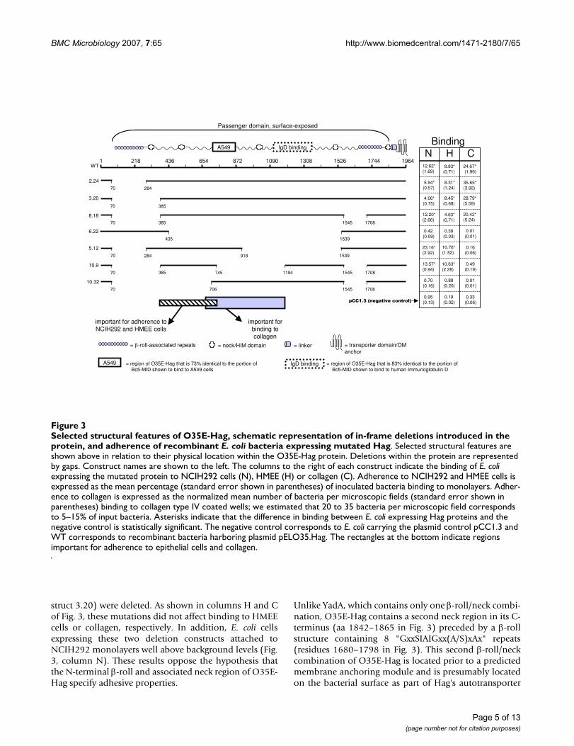
struct 3.20) were deleted. As shown in columns H and Cof Fig. 3, these mutations did not affect binding to HMEEcells or collagen, respectively. In addition, E. coli cellsexpressing these two deletion constructs attached toNCIH292 monolayers well above background levels (Fig.3, column N). These results oppose the hypothesis thatthe N-terminal β-roll and associated neck region of O35E-Hag specify adhesive properties.
Unlike YadA, which contains only one β-roll/neck combi-nation, O35E-Hag contains a second neck region in its C-terminus (aa 1842–1865 in Fig. 3) preceded by a β-rollstructure containing 8 "GxxSIAIGxx(A/S)xAx" repeats(residues 1680–1798 in Fig. 3). This second β-roll/neckcombination of O35E-Hag is located prior to a predictedmembrane anchoring module and is presumably locatedon the bacterial surface as part of Hag's autotransporter
Selected structural features of O35E-Hag, schematic representation of in-frame deletions introduced in the protein, and adher-ence of recombinant E. coli bacteria expressing mutated HagFigure 3Selected structural features of O35E-Hag, schematic representation of in-frame deletions introduced in the protein, and adherence of recombinant E. coli bacteria expressing mutated Hag. Selected structural features are shown above in relation to their physical location within the O35E-Hag protein. Deletions within the protein are represented by gaps. Construct names are shown to the left. The columns to the right of each construct indicate the binding of E. coli expressing the mutated protein to NCIH292 cells (N), HMEE (H) or collagen (C). Adherence to NCIH292 and HMEE cells is expressed as the mean percentage (standard error shown in parentheses) of inoculated bacteria binding to monolayers. Adher-ence to collagen is expressed as the normalized mean number of bacteria per microscopic fields (standard error shown in parentheses) binding to collagen type IV coated wells; we estimated that 20 to 35 bacteria per microscopic field corresponds to 5–15% of input bacteria. Asterisks indicate that the difference in binding between E. coli expressing Hag proteins and the negative control is statistically significant. The negative control corresponds to E. coli carrying the plasmid control pCC1.3 and WT corresponds to recombinant bacteria harboring plasmid pELO35.Hag. The rectangles at the bottom indicate regions important for adherence to epithelial cells and collagen.
= β-roll-associated repeats = neck/HIM domain = linker = transporter domain/OM anchor
important for adherence to NCIH292 and HMEE cells
important for binding to collagen
435 1539
6.22
2181 13081090872 17441526 1964654436WT
28470
2.24
3.20
38570
385 1545 1708
8.1870
385 745 17081194 1545
10.9
70
1539
5.12
284 91870
706 1545 1708
10.3270
A549 IgD binding
Passenger domain, surface-exposed
BindingN H C
12.62* (1.69)
8.83* (0.71)
5.94* (0.57)
4.06* (0.75)
0.42 (0.09)
12.20* (2.66)
23.16* (2.92)
13.57* (0.94)
0.70 (0.16)
8.31* (1.24)
8.45* (0.68)
4.63* (0.71)
0.38 (0.03)
10.76* (1.52)
10.63* (2.28)
0.88 (0.20)
24.67* (1.96)
35.65* (3.92)
28.79* (5.59)
20.42* (5.24)
0.01 (0.01)
0.16 (0.06)
0.49 (0.19)
0.01 (0.01)
0.95 (0.13)
0.19 (0.02)
0.33 (0.06)pCC1.3 (negative control)
A549 IgD binding = region of O35E-Hag that is 83% identical to the portion of Bc5-MID shown to bind to human Immunoglobulin D
= region of O35E-Hag that is 73% identical to the portion of Bc5-MID shown to bind to A549 cells
Page 5 of 13(page number not for citation purposes)

BMC Microbiology 2007, 7:65 http://www.biomedcentral.com/1471-2180/7/65
passenger domain. To determine whether this C-terminalβ-roll structure specifies adhesive properties, residues1546–1707 (encompassing 2 "GxxSIAIGxx(A/S)xAx"motifs) were removed in construct 8.18, which also con-tains the deletion of the N-terminal β-roll/neck area (Fig.3). Expression of this doubly-deleted Hag protein stillconferred on E. coli cells the ability to bind to HMEE cells,collagen and lung cells at or near WT levels (columns N,H and C of Fig. 3). By contrast, recombinant bacteriaexpressing the mutated adhesin construct 6.22, whichretains both β-roll/neck combinations but is missing resi-dues 436–1538 (Fig. 3), no longer adhere to either epithe-lial cells or collagen (columns N, H and C of Fig. 3). Theseresults therefore support rejection of the hypothesis thatthe β-roll structures of O35E-Hag specify adhesive proper-ties as they do in YadA. Our data also suggest that residues385–1545 encompass the collagen and epithelial cellbinding domain(s) of this M. catarrhalis adhesin.
The M. catarrhalis strain Bc5 protein MID, a Hag ortholog,binds human immunoglobulin D on the bacterial surface,and MID residues 962–1200 are responsible for this bind-ing [19,21,24]. Sequence analysis revealed that the regionof O35E-Hag encompassing aa 1113–1353 is 83% identi-cal to the Bc5-MID IgD binding domain (not shown). Fur-thermore, Hag mediates the binding of IgD by M.catarrhalis strain O35E [13]. Thus, the possibility that the
IgD-binding domain of O35E-Hag is responsible foradherence to human epithelial cells and collagen wastested by removing aa 919–1538 (dotted rectangle labeled"IgD binding" at the top of Fig. 3) from the alreadymutated Hag protein 2.24, yielding the construct 5.12 (seeFig. 3). As shown in Fig. 3, deleting the predicted IgDbinding region of O35E-Hag does not adversely affectattachment to epithelial cells (columns N and H) butcompletely abolishes binding to collagen (column C).Adherence to human cells is therefore specified by aregion of O35E-Hag distinct from that important forbinding collagen, making these adhesive properties sepa-rable.
The adhesive properties of constructs 8.18 and 5.12 sug-gest that residues 385–918 contain the portion of O35E-Hag mediating adherence to NCIH292 cells and HMEEcells. Interestingly, previous sequence analysis by our lab-oratory indicated that aa 715–863 of O35E-Hag are 73%identical to residues 764–913 of M. catarrhalis Bc5 MID[23]. This is noteworthy because others showed that apurified and radioactively-labeled recombinant proteinencompassing this particular portion of Bc5 MID directlybinds to A549 cells and erythrocytes [22]. To determinewhat role, if any, these aa have in adherence to NCIH292cells, HMEE cells and collagen, most of the potentialA549/erythrocyte binding site of O35E-Hag (rectanglelabeled "A549"at the top of Fig. 3) was removed from thealready mutated Hag protein 8.18, yielding construct 10.9(see Fig. 3). As shown in columns N and H of Fig. 3, E. coli
Western blot analysis of recombinant E. coli cells treated with proteinase KFigure 5Western blot analysis of recombinant E. coli cells treated with proteinase K. E. coli carrying the plasmids pELO35.Hag (lanes 1 and 2), pBBHS2.24 (lanes 3 and 4) and pBBHS3.20 (lanes 5 and 6) were incubated for 15 min on ice in the presence (lanes 2, 4 and 6) or absence (lanes 1, 3 and 5) of proteinase K. These cells were lysed, resolved by SDS-PAGE, transferred to PVDF membranes and probed with the anti-Hag antibody 5D2 (panel A) or anti-TonB antibody 4F1 (panel B). Molecular weight markers are shown to the left in kDa.
-+ + - +- proteinase K
TonB-reactive antibody
Hag-reactive antibody148
98
64
1 2 3 4 5 6A.
B.
Western blot analysis of Sarkosyl-insoluble OM proteins extracted from E. coli expressing Hag proteinsFigure 4Western blot analysis of Sarkosyl-insoluble OM pro-teins extracted from E. coli expressing Hag proteins. OM preparations were obtained from E. coli carrying the plasmids pCC1.3 (lane 1), pELO35.Hag (lane 2), pBBHS2.24 (lane 3), pBBHS3.20 (lane 4), pBBHS8.18 (lane 5), pBBHS6.22 (lane 6), pBBHS5.12 (lane 7), pBBHS10.9 (lane 8) and pBBHS10.32 (lane 9). These preparations were resolved by SDS-PAGE, transferred to PVDF membranes and probed with antibodies against the purified recombinant protein His.Hag.CT. The figure is a composite of several western blot experiments in which OM preparations of the negative control (i.e. pCC1.3) and the positive control (i.e. pELO35.Hag) were included. Molecular weight markers are shown to the left in kDa. The numbers at the bottom of the western panel represent the predicted molecular weight of each Hag protein.
250148
98
64
1 2 3 4 5 6 7 8 9
201180
170152 87
116106
119 Predicted MW (kDa)
Page 6 of 13(page number not for citation purposes)

BMC Microbiology 2007, 7:65 http://www.biomedcentral.com/1471-2180/7/65
expressing this construct attached to both NCIH292 lungcells and HMEE cells at the same level as recombinant bac-teria expressing the full length adhesin. However, theserecombinant bacteria, like those expressing 5.12, nolonger bound to collagen (column C of Fig. 3). Thus, theresults are consistent with residues 385–745 encompass-ing a portion of O35E-Hag that is crucial for adherence toNCIH292 monolayers and HMEE cells. This hypothesis isalso supported by the results of quantitative attachmentassays with construct 10.32, which was obtained by delet-ing aa 385–705 from the adherence positive Hag con-struct 8.18 (see Fig. 3); recombinant E. coli expressingconstruct 10.32 no longer adhere to either NCIH292 orHMEE cells (Fig. 3). The inability of constructs 5.12, 10.9and 10.32 to mediate the binding of recombinant bacteriato collagen suggests that a region of O35E-Hag startingupstream of aa 706 and extending to at least residue 1194is important for adherence to this extracellular matrix pro-tein (Fig. 3).
DiscussionPrevious reports established that Hag and its orthologMID play important roles in adherence of M. catarrhalis tomiddle ear epithelial cells and A549 pneumocytes
[22,23,26]. Disruption of hag/mid in several M. catarrhalisstrains substantially decreased adherence to both celltypes, while expression of hag in a heterologous E. colibackground increased attachment to middle ear cells atleast 17-fold. The results of the present study extend thesefindings and demonstrate that Hag expression is alsoimportant for adherence to conjunctival (Chang) andlung mucoepidermoid (NCIH292) epithelial cells (Fig.1). These cell lines are relevant to pathogenesis by M.catarrhalis as this organism is a causative agent of conjunc-tivitis, otitis media and lung infections. Furthermore, ourdata reveal that Hag directly mediates binding toNCIH292 monolayers (Fig. 2A). Thus, Hag is a key adher-ence factor for epithelial cells relevant to pathogenesis bythe bacterium, making the protein an attractive target foranti-infective approaches.
Hag/MID exhibit several properties of a good vaccine can-didate. Most M. catarrhalis isolates tested to date contain ahag/mid gene and express its product [13,15,22,24,26].The protein also contains surface-exposed epitopes[13,22,24], making it readily available for recognition bythe immune system, and immunization with a peptideencompassing MID residues 713–964 increases clearance
Table 1: Strains and plasmids
Strain or plasmid Description Source or reference
M. catarrhalis[26]
O35E Wild-type isolate [26]O35E.TN2 Isogenic hag mutant strain of O35E [26]O12E Wild-type isolate [26]O12E.Hag Isogenic hag mutant strain of O12E [26]TTA37 Wild-type isolate [26]TTA37.Hag Isogenic hag mutant strain of TTA37 [26]V1171 Wild-type isolate [26]V1171.Hag Isogenic hag mutant strain of V1171 [26]McGHS1 Wild-type isolate [26]McGHS1.Hag Isogenic hag mutant strain of McGHS1 [26]
E. coliEPI300 Cloning strain EpicentreTUNER Expression strain Novagen
PlasmidspETcoco-1 Protein expression vector, Cmr NovagenpBBCT.77 pETcoco-1 expressing O35E-Hag aa 1358–1964 joined to six N-terminal histidine residues, Cmr This studypCC1.3 Adherence negative plasmid control [48]pCC1 Cloning vector, Cmr EpicentrepELO35.Hag pCC1 expressing the entire O35E hag gene product, Cmr [26]pBBO12.Hag pCC1 expressing the entire O12E hag gene product, Cmr [26]pSV1171.Hag pCC1 expressing the entire V1171 hag gene product, Cmr [26]pBBHS2.24 Deletion derivative of pELO35.Hag, missing aa 71–283 This studypBBHS3.20 Deletion derivative of pELO35.Hag, missing aa 71–384 This studypBBHS8.18 Deletion derivative of pELO35.Hag, missing aa 71–384 and 1546–1707 This studypBBHS6.22 Deletion derivative of pELO35.Hag, missing aa 436–1538 This studypBBHS5.12 Deletion derivative of pELO35.Hag, missing aa 71–283 and 919–1538 This studypBBHS10.9 Deletion derivative of pELO35.Hag, missing aa 71–384, 746–1193 and 1546–1707 This studypBBHS10.32 Deletion derivative of pELO35.Hag, missing aa 71–705 and 1546–1707 This study
Page 7 of 13(page number not for citation purposes)

BMC Microbiology 2007, 7:65 http://www.biomedcentral.com/1471-2180/7/65
of M. catarrhalis from the lungs of infected mice [25]. Ourdata demonstrating that Hag plays a central role in M.catarrhalis adherence, a crucial step in pathogenesis bymost infectious agents [37-39], strengthen the hypothesisthat this OM protein has vaccinogenic potential. Webelieve that targeting functionally relevant regions of Hagin a vaccine will interfere with colonization of susceptibleindividuals by M. catarrhalis. Bacterial adhesins havealready proven to be efficacious vaccine antigens. Forexample, immunization with the cell-binding domain ofthe H. influenzae adhesin Hap elicits production of anti-bodies that reduce attachment to human epithelial cellsand protect mice in a nasopharyngeal colonization model[40]. Similarly, immunization with the adhesin FimH ofuropathogenic E. coli is protective in animal models ofinfection [41,42]. Furthermore, all vaccines licensed foruse in the US against whooping cough contain the Borde-tella pertussis filamentous hemagglutinin adhesin FHA[43].
Also consistent with the vaccinogenic potential of Hag isthe demonstration that saliva collected from healthyadults [44] and children colonized with M. catarrhalis [45]contain antibodies against Hag. COPD patients recover-ing from pulmonary exacerbations caused by this organ-ism were also shown to have increased serum levels ofanti-Hag IgG [17,46]. In addition, Hag is a major target ofnew IgA antibodies purified from the sputum of COPDpatients with M. catarrhalis infections who have success-fully cleared the organism [16]. In fact, Hag is the onlyOM protein out of several tested (i.e. UspA1, UspA2,CopB, TbpB) which elicited an IgA response in all patients[16]. This protective immune response, however, appearsto be strain specific as COPD patients get reinfected withdifferent M. catarrhalis isolates [5]. Hag is a large protein(1964–2335 aa) that exhibits interstrain variability (57–89% identity) [24,26]. These observations emphasize theneed to identify the region(s) of Hag specifying the bestvaccinogenic properties for protection against the major-ity of M. catarrhalis isolates. To achieve this, we sought toidentify the region(s) of Hag specifying adhesive proper-ties by deleting predicted structural domains of the pro-tein. This approach has been successfully used to identifythe cell-binding domains of other autotransporter adhes-ins such as Y. enterocolitica YadA [32] and H. influenzaeHap [47].
The involvement of Hag in adherence by M. catarrhalis iscomplex. While the protein itself is sufficient to directlymediate binding to middle ear [26] and NCIH292 lungcells (Fig. 2A), the exact role of Hag in attachment to A549and Chang cells is not clear. As shown in Fig. 1B, lack ofHag expression in strains TTA37.Hag, V1171.Hag,McGHS1.Hag and O12E.Hag reduces binding to Changmonolayers 70–90%, but Hag expression does not confer
on E. coli the ability to attach to these conjunctival cells(Fig. 2B). These results are similar to those previouslyreported by our laboratory, where it was discovered thatHag expression is important for WT adherence of M.catarrhalis to A549 cells but that expression of the proteinis not sufficient to mediate attachment of recombinant E.coli cells to these pneumocytes [26]. Complicating mattersis the demonstration that purified Bc5-MID, specificallyresidues 713–964, binds to A549 cells [22]. These obser-vations suggest that post-translational modification of theprotein and/or expression of a co-adhesin, neither ofwhich occurs in the heterologous E. coli background, isnecessary for Hag-dependent binding to A549 and Changmonolayers. Interestingly, lack of OMPCD [48] or UspA1[11,12] substantially decreases adherence of M. catarrhalisto A549 and Chang cells, respectively. These moleculesmay thus act in concert with Hag during attachment tothese epithelial cells. The lack of a suitable plasmid vectorfor M. catarrhalis currently precludes us from directlyaddressing these issues in the native genetic background.Only three plasmids, namely pWW102B [49], pWW115[50] and pEMCJH04 [51], have been described for use inM. catarrhalis. These vectors are not amenable for comple-mention of our mutants due to the incompatibility ofantibioticR markers and replication in a restricted numberof M. catarrhalis isolates. For these reasons, we focused ourefforts on identifying region(s) of O35E-Hag importantfor directly mediating adherence to HMEE and NCIH292cells, using E. coli as a host for our experiments.
The adhesive properties of constructs 5.12, 8.18 and 10.9imply that residues 71–384 and 746–1707 of O35E-Hagdo not specify binding to either HMEE or NCIH292 mon-olayers (Fig. 3). These results are particularly interesting inlight of previous reports that the β-roll repeats of YadAspecify binding to both collagen and human epithelialcells [31-35]. A recent study also indicates that the β-rollrepeats found in the N-terminus of the Actinobacillus actin-omycetemcomicans Oca autotransporter adhesin EmaAspecify adhesive properties [52]. The ability of construct8.18 to confer on E. coli the ability to attach to NCIH292cells, HMEE cells and collagen, however, demonstratesthat the β-roll structures of O35E-Hag are not required foradherence. Although all mutated Hag proteins conferringattachment to epithelial cells also have aa 1–70 and1708–1964 in common (Fig. 3), it is unlikely that theseresidues are directly mediating adherence. This belief isbased on previous sequence analysis of O35E-Hag indi-cating that aa 1–66 correspond to a signal sequence andthat the last 100 residues (i.e. 1864–1964) specify a trans-porter domain anchoring the protein in the OM [26]. Fur-thermore, recombinant bacteria expressing the mutatedHag proteins 6.22 and 10.32, both of which specify aa 1–70 as well as 1708–1964 but lack most of residues 385–745, do not bind to HMEE or NCIH292 cells (Fig. 3).
Page 8 of 13(page number not for citation purposes)

BMC Microbiology 2007, 7:65 http://www.biomedcentral.com/1471-2180/7/65
These observations support the hypothesis that aa 385–745 of O35E-Hag contain a major epithelial cell bindingdeterminant.
Forsgren and colleagues previously reported that residues764–913 of MID, which correspond to aa 715–863 ofO35E-Hag, encompass the part of the protein mediatingadherence to A549 cells and erythrocytes [22]. Our resultsdemonstrate that the mutated Hag construct 10.9, lackingmost of this region (see Fig. 3), binds to NCIH292 andmiddle ear cells (columns N and H in Fig. 3). In addition,E. coli carrying the plasmid pBBHS10.32, which specifiesthe entire A549 binding domain (Fig. 3), does not attachto NCIH292 or HMEE cells (columns N and H in Fig. 3).These results suggest that the portion of Hag important foradherence to middle ear and NCIH292 cells is differentfrom that specifying attachment to A549 pneumocytes.Whether the mutated Hag constructs 10.32 or 10.9 medi-ate adherence to A549 cells was not tested since we previ-ously demonstrated that Hag expression is not sufficientto confer recombinant bacteria with the ability to attach tothis particular cell line. Interestingly, the serum of COPDpatients recovering from M. catarrhalis infections containelevated antibody titers against MID residues 367–774[46], which correspond to aa 355–714 of O35E-Hag.These observations support the hypothesis that theNCIH292 and HMEE epithelial cell binding region of Haghas immunogenic potential, which is a desirable charac-teristic of a vaccine candidate.
M. catarrhalis binds to extracellular matrix proteinsincluding fibronectin [27], vitronectin [28], and laminin[18]; to our knowledge, the organism has not previouslybeen reported to bind to collagen. Our data demonstratethat E. coli expressing the hag gene product of three M.catarrhalis WT isolates (i.e. O35E, V1171, and O12E)gained the ability to attach to type IV collagen (Fig. 2C).M. catarrhalis isolates expressing Hag, however, werefound to bind poorly to collagen (data not shown), atleast when grown under the conditions we used. One pos-sible explanation for these conflicting results is that M.catarrhalis cells express an additional surface molecule(not present in the E. coli background) that precludes Hag-mediated binding to collagen. This hypothesis is sup-ported by reports demonstrating that expression of certainsurface structures can adversely affect the biological func-tion of others. For instance, Pearson and colleaguesrecently showed that Hag interferes with UspA1-depend-ent biofilm development [15]. Similarly, the expression oftype 1 fimbriae prevents Ag43-mediated autoaggregationof E. coli cells, even though Ag43 is still expressed at WTlevels on the bacterial surface [53]. Other E. coli examplesinclude capsular polysaccharides blocking the function ofAg43 [54], and the autoaggregation proteins TibA as wellas AIDA-I functioning as surface antagonists of flagellar
motility [55]. Whether M. catarrhalis expresses a moleculeinhibiting Hag-dependent adherence to collagen remainsto be determined. Another possible explanation for theseconflicting results is that Hag is expressed at greater levelsin E. coli compared to M. catarrhalis. The biological rele-vance of M. catarrhalis binding to collagen is currentlyunknown. The molecule is a major component of theextracellular matrix that supports most tissues and type IVcollagen is found in the basal lamina, which is the layeron which the epithelium sits. Hag-mediated binding tocollagen may therefore facilitate adherence of M. catarrh-alis to damaged tissues during infection.
ConclusionHag expression is important for adherence to severalhuman epithelial cells relevant to pathogenesis by M.catarrhalis, including middle ear, lung and conjunctivalcells. This Oca-family adhesin directly mediates bindingto type IV collagen as well as middle ear and NCIH292lung cells, but its expression is not sufficient to conferrecombinant bacteria with the ability to attach to Changconjunctival monolayers. The collagen and epithelial cellbinding properties of Hag are separable and residues 385–705 appear to specify the epithelial cell binding domainof the protein. This portion of Hag is different from thatproposed by other investigators to be crucial for attach-ment to A549 cells (i.e. 715–863). Furthermore, con-served structural features shown to specify the adhesiveproperties of the Oca adhesin YadA (i.e. β-roll), are notimportant for adherence mediated by Hag. These observa-tions suggest that Hag employs different mechanisms toconfer adherence to epithelial cells and collagen.
MethodsBacterial strains, plasmids, tissue culture cell lines, and growth conditionsNCIH292, Chang and HMEE cells were cultured as previ-ously described [23,26,30]. Table 1 lists the bacterialstrains and plasmids used in this study. M. catarrhalis wasgrown on Todd-Hewitt agar plates as previously reported[26]. Recombinant E. coli cells were cultured using Luria-Bertani medium supplemented with chloramphenicol ata concentration of 15 μg/ml (LB+Cm). E. coli EPI300 cellswere grown overnight in 5 ml of LB+Cm at 37°C with agi-tation (200 rpm). The next morning, these cultures weretransferred to flasks containing 20 ml of fresh LB+Cm sup-plemented with 250 μl of Epicentre's 1000 × Copy Con-trol Induction Solution and grown at 37°C with vigorousshaking (300 rpm) for 2 hr (for adherence assays and pro-teinase K treatment experiments) or 5 hr (for purificationof plasmid DNA and protein preparations). To purify therecombinant protein His.Hag.CT (see below), E. coliTUNER cells carrying plasmid pBBCT.77 were culturedovernight in 5 ml of LB+Cm supplemented with glucoseat a concentration of 0.2%. The next morning, these cul-
Page 9 of 13(page number not for citation purposes)

BMC Microbiology 2007, 7:65 http://www.biomedcentral.com/1471-2180/7/65
tures were transferred to 20 ml of fresh LB+Cm+0.2% glu-cose and incubated with shaking at 37°C for 1 hr. Proteinexpression was then induced by adding 25 μl of a 1 MIPTG solution and incubating the cultures with vigorousshaking for 5 hr at 37°C.
Recombinant DNA methodologyPlasmid DNA was purified from recombinant E. coliEPI300 cells using Qiagen's Qiaprep Spin Miniprep Kitwith minor modifications to the manufacturer's instruc-tions: two additional washes with the PE buffer were per-formed and plasmid DNA was eluted from the columnsusing 50 μl and 30 μl of elution buffer, consecutively.Details of the other standard molecular biology tech-niques used in this study can be found elsewhere[23,26,30,56].
Construction of in-frame deletions within hagPrimer sewing was used to introduce deletions into thehag ORF of M. catarrhalis strain O35E that is contained byplasmid pELO35.Hag. Briefly, a 42-mer oligonucleotidewas designed such that the 5'-half specifies the regiondirectly upstream of the area to be removed and the 3'-halfcorresponds to the DNA immediately downstream of thedesired deletion. This oligonucleotide, along with aprimer corresponding to its complement, was then usedto perform inverse PCR with the Stratagene QuickChange®
II XL Site-directed Mutagenesis Kit per the manufacturer'srecommended guidelines. This PCR reaction wasrestricted with DpnI to remove the intact WT plasmidDNA template, purified and electroporated into E. coliEPI300 cells. These bacteria were allowed to recover for 1hr at 37°C in 1 ml of LB medium and spread onto LB+Cmagar plates. Chloramphenicol resistant colonies werescreened by colony PCR to identify clones containing theintended deletion. Colonies were patched onto LB+Cmplates and placed into PCR tubes. These tubes were micro-waved on high-power for 2 min and PCR was performedusing Invitrogen's Taq DNA polymerase and hag-specificprimers flanking the deleted region of hag. This overallstrategy was used to generate plasmids pBBHS2.24,pBBHS3.20, pBBHS5.12, pBBHS6.22, pBBHS8.18,pBBHS10.9, and pBBHS10.32 (Table 1, Fig. 3). All plas-mids were sequenced to verify that they contain only theintended in-frame deletions. Construct 5.12 was found tocontain an aa substitution at residue 1541 (G1541 → V).This mutation does not, however, affect our conclusionsthat residues 385–705 are important for adherence to epi-thelial cells and that aa 706–1194 are involved in colla-gen-binding. This belief is based on the phenotypes ofconstructs 8.18, 10.9 and 10.32 (see Fig. 3), which do notcontain this particular mutation or any other aa substitu-tion.
Production of antibodies against the M. catarrhalis strain O35E-Hag proteinA PCR product corresponding to nt 4072–5892 of theO35E-hag ORF was amplified from chromosomal DNAwith the Invitrogen Platinum Pfx DNA polymerase usingthe oligonucleotide primers HagFQ1358AscI (5'-TTGGCG CGC CTT CAA ATG AAT GTC AAA TCA-3', AscI siteunderlined) and HagRF1964PacI (5'-CCT TAA TTA ACCAAA GTG AAA ACC TGC ACC-3'; PacI site underlined).The amplicon was purified with Epicentre's PCR Precipita-tion Solution, digested with AscI and PacI, and ligated intothe cognate sites of the plasmid vector pETcoco-1 (Nova-gen) using T4 DNA ligase (NEB). This ligation mixturewas introduced into E. coli EPI300 cells by electroporationand chloramphenicol resistant transformants werescreened by colony PCR as described above using primersthat anneal to regions of pETcoco-1 flanking the clonedDNA insert. This approach yielded the plasmid pBBCT.77,which was sequenced to verify that no mutations wereintroduced during PCR as well as to confirm that it speci-fies a protein corresponding to aa residues 1358–1964 ofO35E-Hag fused to six N-terminal histidine residues(encoded by pETcoco-1). This protein was designatedHis.Hag.CT.
The plasmid pBBCT.77 was introduced into chemicallycompetent E. coli TUNER cells (Novagen), and theHis.Hag.CT protein was purified from these recombinantbacteria with the BugBuster HT Protein Extraction Reagentand the His-Bind Resin System under the manufacturer'srecommended conditions (Novagen). For the productionof antibodies, female BALB/c mice were immunized withpurified His.Hag.CT emulsified in Freund's adjuvants(complete and incomplete, Fisher Scientific) as reported[29]. Recovered murine serum was demonstrated to rec-ognize Hag in western blot experiments using whole celllysates of the WT strain O35E and its hag mutantO35E.TN2 (data not shown).
Protein preparations and western blot analysisRecombinant E. coli EPI300 cells were suspended in 5 mlPBS to an optical density of 300 Klett units, pelleted bycentrifugation and resuspended in 5 ml of 10 mM HEPESbuffer. Sarkosyl-insoluble outer membrane (OM) pro-teins were isolated from these suspensions as described[57]. These OM protein preparations were then analyzedby western blot as reported [26], using murine serumagainst His.Hag.CT. In some experiments, the Hag-spe-cific monoclonal antibody 5D2 [13] was used in lieu ofmurine sera against His.Hag.CT. The preparation of wholecell lysates from M. catarrhalis and E. coli has beendescribed elsewhere [58,59]. Equivalent loads of proteinwere analyzed in all experiments.
Page 10 of 13(page number not for citation purposes)

BMC Microbiology 2007, 7:65 http://www.biomedcentral.com/1471-2180/7/65
For the treatment of intact bacteria with Proteinase K [60],recombinant E. coli cells were suspended to an opticaldensity of 300 Klett units (109 CFU/ml) in 5 ml of 200mM Tris-Acetate buffer (pH 8.2) supplemented with 20mM MgSO4. Proteinase K (Novagen) was added to 0.5 mlof bacterial suspensions at a final concentration of 0.5–1.2 μg/ml, and these mixtures were incubated on ice for15 min. Bacteria were next pelleted and resuspended in~100 μl PBS supplemented with EDTA-free ProteaseInhibitor Cocktail (Roche Applied Science) at a final con-centration of 2 ×. SDS-PAGE sample loading buffer wasthen added to each suspension and they were analyzed bywestern blot using Hag-reactive antibodies (see above) orwith the TonB-specific monoclonal antibody 4F1 [36].
Adherence assaysAdherence to epithelial cells was measured using a viablecell count attachment assay previously described by ourlaboratory [23,26,30,48]. For assays involving M. catarrh-alis, bacteria were incubated with epithelial cells for 5 minprior to washing off unbound bacteria. For assays with E.coli, infected monolayers were incubated for 1 hr beforeremoving non-adherent recombinant bacteria. Duplicateattachment assays were performed on at least 3 separateoccasions and the results are expressed as the mean (±standard error) percentage of inoculated bacteria thatadhered to epithelial cells. Binding of E. coli strains to col-lagen was measured using a modification of this attach-ment assay. Recombinant E. coli cells were suspended in 5ml of PBS supplemented with 0.15% gelatin (PBSG) to anoptical density of 230 Klett units, and 25 μl of each sus-pension (~107 CFU) were added to duplicate wells of a 24-well BioCoat murine type-IV collagen tissue culture plate(BD Biosciences) containing 0.5 ml tissue culturemedium without antibiotics. Dilutions of each bacterialsuspension were spread onto LB agar plates and grownovernight to enumerate the number of CFU used to infectthese collagen-coated wells. The inoculated tissue cultureplate was centrifuged at 165 × g for 5 min and placed in a37°C incubator with 7.5% CO2 for 1 hr. Each infectedwell was then washed five times with 0.5 ml PBSG, fixedwith 0.5 ml methanol and stained for 15 min with 0.5 mlof a solution containing 0.035% crystal violet and 5%methanol. Wells were washed three times with deionizedH2O, dried, and photographed on either side of a point inthe center of the well (2 fields/well). The number of bac-terial cells per field was counted by two individuals andeach strain was tested on at least 2 separate occasions. Theresults are expressed as the mean number of bacterial cells(± standard error) per field normalized by the number ofCFU used to inoculate the collagen-coated wells.
Sequence and statistical analysesPlasmids were sequenced at the University of Michigansequencing core. The resulting chromatograms were
assembled using the ChromaTool software (BioTools,Inc.), and sequence analysis was performed using VectorNTI 10.1.1 (Invitrogen). Statistical analyses were per-formed using the Mann-Whitney test provided by theGraphPad Prism Software v. 4.0 (GraphPad). P values lessthan 0.05 were considered statistically significant.
Authors' contributionsBB and ERL contributed equally to the design of experi-ments, interpretation of results and writing of the manu-script. BB was responsible for performing mostexperiments. SL performed some of the adherence assayswith Moraxella catarrhalis strains.
AcknowledgementsWe thank Eric Hansen at the University of Texas Southwestern Medical Center in Dallas for providing M. catarrhalis strains and antibodies as well as Ray Larsen at Bowling Green State University for the TonB antibody and assistance with interpretation of the Proteinase K assays. We also wish to thank Mark Wooten, Christine Akimana, Randall Worth, Robert Blumen-thal, Rachel Balder, and William Grose for their helpful comments on the manuscript. This study was supported by a grant from NIH/NIAID (AI051477) to E.R.L. B.B was supported, in part, by a graduate fellowship from the University of Toledo Health Science Campus.
References1. Henderson IR, Navarro-Garcia F, Desvaux M, Fernandez RC,
Ala'Aldeen D: Type V protein secretion pathway: theautotransporter story. Microbiol Mol Biol Rev 2004,68(4):692-744.
2. Cotter SE, Surana NK, St Geme JW 3rd: Trimeric autotransport-ers: a distinct subfamily of autotransporter proteins. TrendsMicrobiol 2005, 13(5):199-205.
3. Hoiczyk E, Roggenkamp A, Reichenbecher M, Lupas A, Heesemann J:Structure and sequence analysis of Yersinia YadA and Morax-ella UspAs reveal a novel class of adhesins. Embo J 2000,19(22):5989-5999.
4. Cripps AW, Otczyk DC, Kyd JM: Bacterial otitis media: a vac-cine preventable disease? Vaccine 2005, 23(17-18):2304-2310.
5. Murphy TF, Brauer AL, Grant BJ, Sethi S: Moraxella catarrhalis inchronic obstructive pulmonary disease: burden of diseaseand immune response. Am J Respir Crit Care Med 2005,172(2):195-199.
6. Brook I, Foote PA, Frazier EH: Microbiology of acute exacerba-tion of chronic sinusitis. Ann Otol Rhinol Laryngol 2005,114(7):573-576.
7. Bingen E, Cohen R, Jourenkova N, Gehanno P: Epidemiologicstudy of conjunctivitis-otitis syndrome. Pediatr Infect Dis J 2005,24(8):731-732.
8. Jacobs MR, Bajaksouzian S, Windau A, Good CE, Lin G, Pankuch GA,Appelbaum PC: Susceptibility of Streptococcus pneumoniae,Haemophilus influenzae, and Moraxella catarrhalis to 17 oralantimicrobial agents based on pharmacodynamic parame-ters: 1998-2001 U S Surveillance Study. Clin Lab Med 2004,24(2):503-530.
9. Klugman KP: The clinical relevance of in-vitro resistance topenicillin, ampicillin, amoxycillin and alternative agents, forthe treatment of community-acquired pneumonia caused byStreptococcus pneumoniae, Haemophilus influenzae andMoraxella catarrhalis. J Antimicrob Chemother 1996, 38 SupplA:133-140.
10. Manninen R, Huovinen P, Nissinen A: Increasing antimicrobialresistance in Streptococcus pneumoniae, Haemophilus influen-zae and Moraxella catarrhalis in Finland. J Antimicrob Chemother1997, 40(3):387-392.
11. Lafontaine ER, Cope LD, Aebi C, Latimer JL, McCracken GH Jr.,Hansen EJ: The UspA1 protein and a second type of UspA2protein mediate adherence of Moraxella catarrhalis to
Page 11 of 13(page number not for citation purposes)
http://www.ncbi.nlm.nih.gov/entrez/query.fcgi?cmd=Retrieve&db=PubMed&dopt=Abstract&list_uids=8858479
BMC Microbiology 2007, 7:65 http://www.biomedcentral.com/1471-2180/7/65
human epithelial cells in vitro. J Bacteriol 2000,182(5):1364-1373.
12. Lafontaine ER, Wagner NJ, Hansen EJ: Expression of the Morax-ella catarrhalis UspA1 protein undergoes phase variation andis regulated at the transcriptional level. J Bacteriol 2001,183(5):1540-1551.
13. Pearson MM, Lafontaine ER, Wagner NJ, St Geme JW 3rd, Hansen EJ:A hag mutant of Moraxella catarrhalis strain O35E is deficientin hemagglutination, autoagglutination, and immunoglobu-lin D-binding activities. Infect Immun 2002, 70(8):4523-4533.
14. Hill DJ, Virji M: A novel cell-binding mechanism of Moraxellacatarrhalis ubiquitous surface protein UspA: specific target-ing of the N-domain of carcinoembryonic antigen-relatedcell adhesion molecules by UspA1. Mol Microbiol 2003,48(1):117-129.
15. Pearson MM, Laurence CA, Guinn SE, Hansen EJ: Biofilm forma-tion by Moraxella catarrhalis in vitro: roles of the UspA1adhesin and the Hag hemagglutinin. Infect Immun 2006,74(3):1588-1596.
16. Murphy TF, Brauer AL, Aebi C, Sethi S: Antigenic specificity of themucosal antibody response to Moraxella catarrhalis inchronic obstructive pulmonary disease. Infect Immun 2005,73(12):8161-8166.
17. Murphy TF, Brauer AL, Aebi C, Sethi S: Identification of surfaceantigens of Moraxella catarrhalis as targets of human serumantibody responses in chronic obstructive pulmonary dis-ease. Infect Immun 2005, 73(6):3471-3478.
18. Tan TT, Forsgren A, Riesbeck K: The Respiratory PathogenMoraxella catarrhalis Binds to Laminin via Ubiquitous Sur-face Proteins A1 and A2. J Infect Dis 2006, 194(4):493-497.
19. Forsgren A, Brant M, Mollenkvist A, Muyombwe A, Janson H, WoinN, Riesbeck K: Isolation and characterization of a novel IgD-binding protein from Moraxella catarrhalis. J Immunol 2001,167(4):2112-2120.
20. Gjorloff Wingren A, Hadzic R, Forsgren A, Riesbeck K: The novelIgD binding protein from Moraxella catarrhalis induceshuman B lymphocyte activation and Ig secretion in the pres-ence of Th2 cytokines. J Immunol 2002, 168(11):5582-5588.
21. Nordstrom T, Forsgren A, Riesbeck K: The immunoglobulin D-binding part of the outer membrane protein MID fromMoraxella catarrhalis comprises 238 amino acids and a tetra-meric structure. J Biol Chem 2002, 277(38):34692-34699.
22. Forsgren A, Brant M, Karamehmedovic M, Riesbeck K: The Immu-noglobulin D-Binding Protein MID from Moraxella catarrhalisIs Also an Adhesin. Infect Immun 2003, 71(6):3302-3309.
23. Holm MM, Vanlerberg SL, Sledjeski DD, Lafontaine ER: The Hagprotein of Moraxella catarrhalis strain O35E is associatedwith adherence to human lung and middle ear cells. InfectImmun 2003, 71(9):4977-4984.
24. Mollenkvist A, Nordstrom T, Hallden C, Christensen JJ, Forsgren A,Riesbeck K: The Moraxella catarrhalis immunoglobulin D-binding protein MID has conserved sequences and is regu-lated by a mechanism corresponding to phase variation. JBacteriol 2003, 185(7):2285-2295.
25. Forsgren A, Brant M, Riesbeck K: Immunization with the trun-cated adhesin Moraxella catarrhalis immunoglobulin D-bind-ing protein (MID764-913) is protective against M. catarrhalisin a mouse model of pulmonary clearance. J Infect Dis 2004,190(2):352-355.
26. Bullard B, Lipski SL, Lafontaine ER: Hag directly mediates theadherence of Moraxella catarrhalis to human middle earcells. Infect Immun 2005, 73(8):5127-5136.
27. McMichael JC, Fiske MJ, Fredenburg RA, Chakravarti DN, VanDer-Meid KR, Barniak V, Caplan J, Bortell E, Baker S, Arumugham R, ChenD: Isolation and characterization of two proteins fromMoraxella catarrhalis that bear a common epitope. InfectImmun 1998, 66(9):4374-4381.
28. Attia AS, Ram S, Rice PA, Hansen EJ: Binding of vitronectin by theMoraxella catarrhalis UspA2 protein interferes with latestages of the complement cascade. Infect Immun 2006,74(3):1597-1611.
29. Lipski SL, Akimana C, Timpe JM, Wooten RM, Lafontaine ER: TheMoraxella catarrhalis Autotransporter McaP Is a ConservedSurface Protein That Mediates Adherence to Human Epi-thelial Cells through Its N-Terminal Passenger Domain.Infect Immun 2007, 75(1):314-324.
30. Timpe JM, Holm MM, Vanlerberg SL, Basrur V, Lafontaine ER: Iden-tification of a Moraxella catarrhalis outer membrane proteinexhibiting both adhesin and lipolytic activities. Infect Immun2003, 71(8):4341-4350.
31. Nummelin H, Merckel MC, Leo JC, Lankinen H, Skurnik M, GoldmanA: The Yersinia adhesin YadA collagen-binding domainstructure is a novel left-handed parallel beta-roll. Embo J 2004,23(4):701-711.
32. Roggenkamp A, Ackermann N, Jacobi CA, Truelzsch K, Hoffmann H,Heesemann J: Molecular analysis of transport and oligomeriza-tion of the Yersinia enterocolitica adhesin YadA. J Bacteriol2003, 185(13):3735-3744.
33. Heise T, Dersch P: Identification of a domain in Yersinia viru-lence factor YadA that is crucial for extracellular matrix-specific cell adhesion and uptake. Proc Natl Acad Sci U S A 2006,103(9):3375-3380.
34. Roggenkamp A, Ruckdeschel K, Leitritz L, Schmitt R, Heesemann J:Deletion of amino acids 29 to 81 in adhesion protein YadA ofYersinia enterocolitica serotype O:8 results in selective abro-gation of adherence to neutrophils. Infect Immun 1996,64(7):2506-2514.
35. Tahir YE, Kuusela P, Skurnik M: Functional mapping of the Yers-inia enterocolitica adhesin YadA. Identification Of eightNSVAIG - S motifs in the amino-terminal half of the proteininvolved in collagen binding. Mol Microbiol 2000, 37(1):192-206.
36. Larsen RA, Myers PS, Skare JT, Seachord CL, Darveau RP, Postle K:Identification of TonB homologs in the family Enterobacte-riaceae and evidence for conservation of TonB-dependentenergy transduction complexes. J Bacteriol 1996,178(5):1363-1373.
37. Beachey EH: Bacterial adherence: adhesin-receptor interac-tions mediating the attachment of bacteria to mucosal sur-face. J Infect Dis 1981, 143(3):325-345.
38. Hultgren SJ, Abraham S, Caparon M, Falk P, St Geme JW 3rd, Nor-mark S: Pilus and nonpilus bacterial adhesins: assembly andfunction in cell recognition. Cell 1993, 73(5):887-901.
39. St Geme JW 3rd: Bacterial adhesins: determinants of micro-bial colonization and pathogenicity. Adv Pediatr 1997, 44:43-72.
40. Liu DF, Mason KW, Mastri M, Pazirandeh M, Cutter D, Fink DL, StGeme JW 3rd, Zhu D, Green BA: The C-terminal fragment ofthe internal 110-kilodalton passenger domain of the Happrotein of nontypeable Haemophilus influenzae is a potentialvaccine candidate. Infect Immun 2004, 72(12):6961-6968.
41. Langermann S, Mollby R, Burlein JE, Palaszynski SR, Auguste CG,DeFusco A, Strouse R, Schenerman MA, Hultgren SJ, Pinkner JS, Win-berg J, Guldevall L, Soderhall M, Ishikawa K, Normark S, Koenig S:Vaccination with FimH adhesin protects cynomolgus mon-keys from colonization and infection by uropathogenicEscherichia coli. J Infect Dis 2000, 181(2):774-778.
42. Langermann S, Palaszynski S, Barnhart M, Auguste G, Pinkner JS, Bur-lein J, Barren P, Koenig S, Leath S, Jones CH, Hultgren SJ: Preventionof mucosal Escherichia coli infection by FimH-adhesin-basedsystemic vaccination. Science 1997, 276(5312):607-611.
43. http://www.cdc.gov/nip/publications/pink/pert.pdf A: Pertussis.:67-84 [http://www.cdc.gov/nip/publications/pink/pert.pdf].
44. Stutzmann Meier P, Heiniger N, Troller R, Aebi C: Salivary anti-bodies directed against outer membrane proteins of Morax-ella catarrhalis in healthy adults. Infect Immun 2003,71(12):6793-6798.
45. Meier PS, Freiburghaus S, Martin A, Heiniger N, Troller R, Aebi C:Mucosal immune response to specific outer membrane pro-teins of Moraxella catarrhalis in young children. Pediatr InfectDis J 2003, 22(3):256-262.
46. Tan TT, Christensen JJ, Dziegiel MH, Forsgren A, Riesbeck K: Com-parison of the serological responses to Moraxella catarrhalisimmunoglobulin D-binding outer membrane protein and theubiquitous surface proteins A1 and A2. Infect Immun 2006,74(11):6377-6386.
47. Fink DL, Buscher AZ, Green B, Fernsten P, St Geme JW 3rd: TheHaemophilus influenzae Hap autotransporter mediatesmicrocolony formation and adherence to epithelial cells andextracellular matrix via binding regions in the C-terminalend of the passenger domain. Cell Microbiol 2003, 5(3):175-186.
48. Holm MM, Vanlerberg SL, Foley IM, Sledjeski DD, Lafontaine ER: TheMoraxella catarrhalis porin-like outer membrane protein CD
Page 12 of 13(page number not for citation purposes)
http://www.ncbi.nlm.nih.gov/entrez/query.fcgi?cmd=Retrieve&db=PubMed&dopt=Abstract&list_uids=9712790
http://www.ncbi.nlm.nih.gov/entrez/query.fcgi?cmd=Retrieve&db=PubMed&dopt=Abstract&list_uids=8698473
http://www.ncbi.nlm.nih.gov/entrez/query.fcgi?cmd=Retrieve&db=PubMed&dopt=Abstract&list_uids=8698473
http://www.ncbi.nlm.nih.gov/entrez/query.fcgi?cmd=Retrieve&db=PubMed&dopt=Abstract&list_uids=8631714
http://www.ncbi.nlm.nih.gov/entrez/query.fcgi?cmd=Retrieve&db=PubMed&dopt=Abstract&list_uids=8631714
http://www.ncbi.nlm.nih.gov/entrez/query.fcgi?cmd=Retrieve&db=PubMed&dopt=Abstract&list_uids=8631714
http://www.ncbi.nlm.nih.gov/entrez/query.fcgi?cmd=Retrieve&db=PubMed&dopt=Abstract&list_uids=7014727
http://www.ncbi.nlm.nih.gov/entrez/query.fcgi?cmd=Retrieve&db=PubMed&dopt=Abstract&list_uids=7014727
http://www.ncbi.nlm.nih.gov/entrez/query.fcgi?cmd=Retrieve&db=PubMed&dopt=Abstract&list_uids=7014727
http://www.ncbi.nlm.nih.gov/entrez/query.fcgi?cmd=Retrieve&db=PubMed&dopt=Abstract&list_uids=8098994
http://www.ncbi.nlm.nih.gov/entrez/query.fcgi?cmd=Retrieve&db=PubMed&dopt=Abstract&list_uids=8098994
http://www.ncbi.nlm.nih.gov/entrez/query.fcgi?cmd=Retrieve&db=PubMed&dopt=Abstract&list_uids=9265967
http://www.ncbi.nlm.nih.gov/entrez/query.fcgi?cmd=Retrieve&db=PubMed&dopt=Abstract&list_uids=9265967
http://www.ncbi.nlm.nih.gov/entrez/query.fcgi?cmd=Retrieve&db=PubMed&dopt=Abstract&list_uids=9110982

BMC Microbiology 2007, 7:65 http://www.biomedcentral.com/1471-2180/7/65
Publish with BioMed Central and every scientist can read your work free of charge
"BioMed Central will be the most significant development for disseminating the results of biomedical research in our lifetime."
Sir Paul Nurse, Cancer Research UK
Your research papers will be:
available free of charge to the entire biomedical community
peer reviewed and published immediately upon acceptance
cited in PubMed and archived on PubMed Central
yours — you keep the copyright
Submit your manuscript here:http://www.biomedcentral.com/info/publishing_adv.asp
BioMedcentral
is an adhesin for human lung cells. Infect Immun 2004,72(4):1906-1913.
49. Wang W, Attia AS, Liu L, Rosche T, Wagner NJ, Hansen EJ: Devel-opment of a shuttle vector for Moraxella catarrhalis. Plasmid2006, 55(1):50-57.
50. Wang W, Hansen EJ: Plasmid pWW115, a cloning vector foruse with Moraxella catarrhalis. Plasmid 2006, 56(2):133-137.
51. Hays JP, Eadie K, Verduin CM, Verbrugh H, van Belkum A: A novelplasmid (pEMCJH03) isolated from Moraxella catarrhalis pos-sibly useful as a cloning and expression vector within this spe-cies. Plasmid 2005, 53(3):263-268.
52. Ruiz T, Lenox C, Radermacher M, Mintz KP: Novel surface struc-tures are associated with the adhesion of Actinobacillus actin-omycetemcomitans to collagen. Infect Immun 2006,74(11):6163-6170.
53. Hasman H, Chakraborty T, Klemm P: Antigen-43-mediated auto-aggregation of Escherichia coli is blocked by fimbriation. J Bac-teriol 1999, 181(16):4834-4841.
54. Schembri MA, Dalsgaard D, Klemm P: Capsule shields the func-tion of short bacterial adhesins. J Bacteriol 2004,186(5):1249-1257.
55. Ulett GC, Webb RI, Schembri MA: Antigen-43-mediated autoag-gregation impairs motility in Escherichia coli. Microbiology 2006,152(Pt 7):2101-2110.
56. Sambrook J Russell, D.W.: Molecular Cloning: A LaboratoryManual (Third Edition). Third Edition edition. Cold Spring Har-bor Laboratory Press; 2001.
57. Carlone GM, Thomas ML, Rumschlag HS, Sottnek FO: Rapid micro-procedure for isolating detergent-insoluble outer mem-brane proteins from Haemophilus species. J Clin Microbiol 1986,24(3):330-332.
58. Cope LD, Lafontaine ER, Slaughter CA, Hasemann CA Jr., Aebi C,Henderson FW, McCracken GH Jr., Hansen EJ: Characterizationof the Moraxella catarrhalis uspA1 and uspA2 genes and theirencoded products. J Bacteriol 1999, 181(13):4026-4034.
59. Patrick CC, Kimura A, Jackson MA, Hermanstorfer L, Hood A,McCracken GH Jr., Hansen EJ: Antigenic characterization of theoligosaccharide portion of the lipooligosaccharide of non-typable Haemophilus influenzae. Infect Immun 1987,55(12):2902-2911.
60. Larsen RA, Thomas MG, Postle K: Protonmotive force, ExbB andligand-bound FepA drive conformational changes in TonB.Mol Microbiol 1999, 31(6):1809-1824.
Page 13 of 13(page number not for citation purposes)
http://www.ncbi.nlm.nih.gov/entrez/query.fcgi?cmd=Retrieve&db=PubMed&dopt=Abstract&list_uids=3489731
![BMC Microbiology BioMed Central · 2017. 8. 27. · Moraxella catarrhalis is an exclusively human, mucosal res-piratory tract commensal and pathogen causing between 5% [1] and 20%](https://static.fdocuments.in/doc/165x107/60b9d1b25ab06638794a37be/bmc-microbiology-biomed-central-2017-8-27-moraxella-catarrhalis-is-an-exclusively.jpg)