Ref : MS ID 101090 Investigation of interactions between ... · Campos, Natália Bernardi Videira,...
54
1 Ref : MS ID 101090 Investigation of interactions between DNA and Nuclear Receptors: a review of the most used methods. Juliana Fattori, Nathalia de Carvalho Indolfo, Jéssica Christina Lóis de Oliveira Campos, Natália Bernardi Videira, Aline Villanova Bridi, Tábata Renée Doratioto, Michelle Alexandrino de Assis, Ana Carolina Migliorini Figueira# Authors’ affiliation: Brazilian Biosciences National Laboratory (LNBio), Brazilian Center for Research in Energy and Materials (CNPEM), P.O. Box 6192, Campinas-SP, Brazil #Correspondent author: [email protected] Abbreviations: AF-1, Activation Function-1; AR, Androgen Receptor; DBD, DNA Binding Domain; ER, Estrogen Receptor; ERE, Estrogen Response Element; ERRα-1, Estrogen Related Receptor α 1; FXR, Farnesoid X Receptor; GR, Glucocorticoid Receptor; GRE, Glucocorticoid Response Element; LBD, Ligand Binding Domain; LXR, Liver X Receptor; NR, Nuclear Receptors; PPARβ, Peroxisome Proliferator-Activated Receptor β; PPARγ, Peroxisome Proliferator-Activated Receptor γ; PR, Progesterone Receptor; RAR, Retinoic Acid Receptor; RE, Response Element; RXR, Retinoid X Receptor; TR, Thyroid Hormone Receptor; VDR, Vitamin D Receptor; VDRE, Vitamin D Receptor Responsive Elements.
Transcript of Ref : MS ID 101090 Investigation of interactions between ... · Campos, Natália Bernardi Videira,...

1
Ref : MS ID 101090
Investigation of interactions between DNA and Nuclear Receptors: a review of the
most used methods.
Juliana Fattori, Nathalia de Carvalho Indolfo, Jéssica Christina Lóis de Oliveira
Campos, Natália Bernardi Videira, Aline Villanova Bridi, Tábata Renée Doratioto,
Michelle Alexandrino de Assis, Ana Carolina Migliorini Figueira#
Authors’ affiliation: Brazilian Biosciences National Laboratory (LNBio), Brazilian
Center for Research in Energy and Materials (CNPEM), P.O. Box 6192, Campinas-SP,
Brazil
#Correspondent author: [email protected]
Abbreviations: AF-1, Activation Function-1; AR, Androgen Receptor; DBD, DNA
Binding Domain; ER, Estrogen Receptor; ERE, Estrogen Response Element; ERRα-1,
Estrogen Related Receptor α 1; FXR, Farnesoid X Receptor; GR, Glucocorticoid
Receptor; GRE, Glucocorticoid Response Element; LBD, Ligand Binding Domain; LXR,
Liver X Receptor; NR, Nuclear Receptors; PPARβ, Peroxisome Proliferator-Activated
Receptor β; PPARγ, Peroxisome Proliferator-Activated Receptor γ; PR, Progesterone
Receptor; RAR, Retinoic Acid Receptor; RE, Response Element; RXR, Retinoid X
Receptor; TR, Thyroid Hormone Receptor; VDR, Vitamin D Receptor; VDRE, Vitamin D
Receptor Responsive Elements.

2
Abstract
Nuclear receptors (NRs) comprise a superfamily of proteins modulated by ligands
that regulate the expression of target genes. These proteins share a multi domain
structure harboring an N-terminal domain, a highly conserved DNA binding domain,
and a ligand binding domain, which has ligand dependent activation function. They play
key roles in development, metabolism and physiology being closely related to diseases.
Most of the knowledge about this superfamily emerges from investigations on new
ligands and are mostly centered in the ligand binding domain. However, more
investigation focusing on interactions between DNA and DNA binding domain are
necessary to shed light on important roles of NRs’ participation in transcriptional
mechanisms and in specific genes network. Here, our goal is to discuss some nuances of
NRs-DNA interaction, describing details of the most used techniques in this sort of
study, such as gel shift (EMSA), DNA footprinting, reporter gene assay, ChIP-Seq, 3C,
and fluorescence anisotropy. Additionally, we aim to provide tools, presenting
advantages and disadvantages of these common methods, when choosing the most
suitable one to study NRs-DNA interactions to answer specific questions.
Keywords: nuclear receptors, DNA binding domain, protein-DNA interaction, ChiP-
seq, DNA footprinting, EMSA, fluorescence anisotropy.

3
Introduction
Nuclear receptors (NRs) are members of a superfamily of transcription factors
modulated by ligands, which regulate the expression of target genes and, therefore, play
key roles in development, metabolism, physiology, and disease [1, 2, 3]. The activity of
the majority of NRs is induced by ligands or small lipophilic molecules such as steroids,
hormones, vitamins, and fatty acids. However, this family also possesses the orphan
nuclear receptors, for which ones ligands remain unknown or could not exist, presenting
unknown activity and suggesting that NRs’ activity can also be regulated by other
processes [2, 4, 5].
Traditionally, NRs act as homo or heterodimers and share the same mechanism of
action. In a classical view, unliganded NRs may be located in the cytoplasm, bound to
chaperones, or in the nucleus, bound to DNA, where they recruit corepressor
complexes, which associate with HDAC (histone deacetyltransferases) to inhibit gene
expression. Ligand addition induces translocation of cytoplasmatic NRs to the nucleus
and promotes conformational changes in the NR structure, allowing corepressor
dissociation and the recruitment of coactivator complexes that have HAT (histone
acetyltransferase) activity to activate the transcription of target genes [1, 6].
However, alternative mechanisms of action have been described for this class of
proteins, which includes both activation by post-translational modifications and
association with other transcription factors (TFs) [7]. For instance, the PPARγ
sumoylation is a critical event that signals corepressor recruitment, even after PPARγ
ligand binding, keeping inflammatory gene promoters in a transrepressed state [6, 8].
Moreover, the interaction between different transcriptional factors (TFs), resulting in
cooperative enhancing or inhibition of gene expression, also named crosstalk, is an
alternative NR action that explains, for example, the negative regulation of TSHβ gene

4
by TR [7], or the blocked activity of NF-kβ by different NRs, such as GR, ER, PR and
AR [6].
Apart from sharing the general action mechanism, all NRs also present a similar
modular structure including an amino-terminal domain, which contains the activation
function-1 (AF-1) ligand independent, a core DNA binding domain (DBD) that is
responsible for recognizing and binding to DNA sequences, linked by a hinge to the
ligand binding domain (LBD), which binds to ligands and recruits coregulators (Figure
1) [1].
Figure 1. Typical domain organization of NRs, showing, in purple, the N-terminal domain,
harboring the activation function ligand independent (AF-1); in light brown, the DNA binding
domain, responsible for DNA binding; in light green, the hinge, which connects DBD and LBD;
in yellow, the ligand binding domain (LBD) with the activation function ligand dependent (AF-
2); and, in pink, the C-terminal portion.
Discovery of the NRs and most of the knowledge about them came from
physiological investigations on hormones and ligands [9-12]. The identification of
natural or synthetic ligands shed light over NRs roles in human physiology, where they
are considered drug targets [13]. Meanwhile, there is still a lot to be understood about
NRs in the context of specific gene networks’ transcription [14]. In other words, there
are many studies focusing on NRs binding to ligands, seeking for molecules that can
modulate them, and less studies focusing on how NRs bind to DNA.
In fact, the protein’s ability to recognize DNA sequences, associated with the
capacity of binding to specific sites across the genome, is a hallmark in gene regulation
and maintenance [15, 16]. Initially, some codes for protein-DNA recognition were
5
reported based on hydrogen bonding patterns between amino acids and nucleotides, in
the DNA major groove. Also, arginine side chain was considered important to make
these contacts [17]. Recently, the development of new technologies, such as
computational simulations and high-throughput spectroscopic techniques, are
uncovering new complexities of protein-DNA interactions, displaying that simple
recognition codes for this interactions are much more complex. In this way, it is known
that there are multiple modes of DNA binding to proteins, and variables such as spacers
of DNA binding sites, multimeric protein binding and alternative structural
conformations should be considered together with cooperativity, allostery and cofactor
presence [15]. Specially to nuclear receptors, the main interactions between protein and
DNA are made by the DNA binding domain. Albeit other interactions with N-terminal
domain and LBD have been reported, they are specific to few receptors and still should
not have been considered as functions [2, 3, 5].
A brief review about the DNA binding domain structure
For nuclear receptors, DNA recognition is controlled by the highly conserved
DNA binding domain (or C domain). Structurally, the DBD is a very compact globular
domain, generally composed by 100 amino acids which are organized in two main
perpendicular helices, with conserved cysteines required for high-affinity DNA binding
(Figure 2) [1]. The first DBD structure determination was in 1990 through nuclear
magnetic resonance (NMR) [18]. However, since the first crystallographic structure of
DBD on DNA was solved, in 1991 [19], X-ray crystallography has become a pivotal
method in unraveling DBD-DNA interaction at atomic levels [20].

6
Figure 2. Illustration of the Nuclear Receptors’ DBD. DBD is shown in dimeric form (in purple
and pink) with two zinc ions (in gray) coordinated to each monomer bound to DNA (PDB:
1HCQ modified). On the right side, details of the two zinc fingers, each one being coordinated
by four cysteines. Helix 1 or the recognition α-helix shown in light pink comprises the P box.
The second zinc finger comprises residues forming the highlighted D box or dimerization
interface. Finally, helix 2 is shown in red in the carboxi-terminal extension (CTE). Adapted
from 21.
As shown in Figure 2, the DNA binding domain of all NRs presents two helices
that pack together through their hydrophobic faces in a perpendicular way, forming the
core domain with the two zinc fingers, contributing to the DBD integral fold [21]. The
first α-helix, termed recognition helix, directly inserts itself to the DNA major groove,
making contacts with the DNA phosphate backbone, forming a crucial substructure for
the DNA-protein binding [21]. Amino acid residues at the base of the first zinc finger
constitute the so-called P box that is involved in the discrimination of the response
elements. Furthermore, residues in the second zinc finger form the D box and are
involved in dimerization [1] and the C-terminal extension of DBD harbors T- and A-
boxes, which contribute to contacts in the flanking DNA core recognition sequences [1,
22-25].
Protein-DNA binding interfaces are also extended by ordered water molecules
which make specific interactions between protein side chains and DNA functional
groups. Apart from the specific interactions, there are several non-specific ones formed
between basic side chains of the DBD and the DNA phosphodiester backbone [21-24].

7
Despite mediating DNA specificity, α-helix 1 contributes little to discrimination
of target-site symmetry. It has been shown that α-helix 2 is the main responsible for
selecting symmetry for DNA recognition motifs, which also projects across the minor
groove of the DNA, creating additional contacts that stabilize DBD-DNA binding.
Additionally, some studies suggest that the specificity in DBD-DNA recognition is
derived not only from the DNA sequence, but also from its geometry [21-24].
DBD- DNA binding and specificity.
Although some NRs can bind to DNA as monomers, it was shown that the most
common DNA binding happens when NRs are organized as homo or heterodimers, this
being the best geometry array for recognition of binding sites [21]. Generally, binding
to DNA occurs in specific DNA motifs, also termed DNA response elements (REs). It
has also been postulated that the formation of homo and heterodimers between receptors
DBDs occurs simultaneously to element response recognition [21].
In general, the REs are composed by hexameric sequences arranged in distinct
configurations, including inverted, everted and direct repeats. The specificities of one
RE, to which each NR binds to, affects directly its function, and whether the receptor
will work as an activator or repressor of the transcription [21]. The response elements
may be located in regulatory sequences, generally found in the 5' region of the target
gene, near the core promoter according to the NRs classical mechanism of action [1].
However, recent studies with new technologies posit that the majority of REs are found
distally from promoters, as enhancers within transcription initiation site, and also,
within intergenic and intronic regions [26, 27].
Typically, the consensus RE is composed by variations of two half sites, which
are in turn composed by hexameric sequences of 5'-PuAGGTCA-3'. These half sites
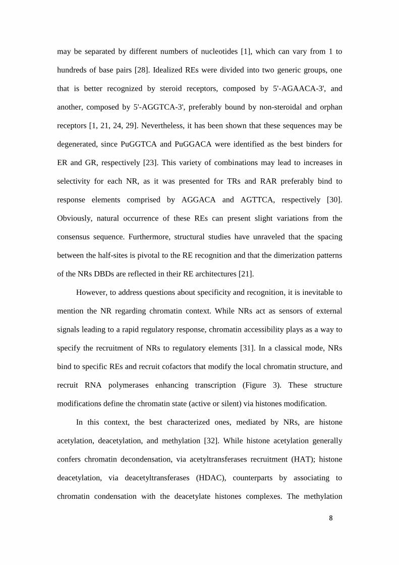
8
may be separated by different numbers of nucleotides [1], which can vary from 1 to
hundreds of base pairs [28]. Idealized REs were divided into two generic groups, one
that is better recognized by steroid receptors, composed by 5'-AGAACA-3', and
another, composed by 5'-AGGTCA-3', preferably bound by non-steroidal and orphan
receptors [1, 21, 24, 29]. Nevertheless, it has been shown that these sequences may be
degenerated, since PuGGTCA and PuGGACA were identified as the best binders for
ER and GR, respectively [23]. This variety of combinations may lead to increases in
selectivity for each NR, as it was presented for TRs and RAR preferably bind to
response elements comprised by AGGACA and AGTTCA, respectively [30].
Obviously, natural occurrence of these REs can present slight variations from the
consensus sequence. Furthermore, structural studies have unraveled that the spacing
between the half-sites is pivotal to the RE recognition and that the dimerization patterns
of the NRs DBDs are reflected in their RE architectures [21].
However, to address questions about specificity and recognition, it is inevitable to
mention the NR regarding chromatin context. While NRs act as sensors of external
signals leading to a rapid regulatory response, chromatin accessibility plays as a way to
specify the recruitment of NRs to regulatory elements [31]. In a classical mode, NRs
bind to specific REs and recruit cofactors that modify the local chromatin structure, and
recruit RNA polymerases enhancing transcription (Figure 3). These structure
modifications define the chromatin state (active or silent) via histones modification.
In this context, the best characterized ones, mediated by NRs, are histone
acetylation, deacetylation, and methylation [32]. While histone acetylation generally
confers chromatin decondensation, via acetyltransferases recruitment (HAT); histone
deacetylation, via deacetyltransferases (HDAC), counterparts by associating to
chromatin condensation with the deacetylate histones complexes. The methylation
9
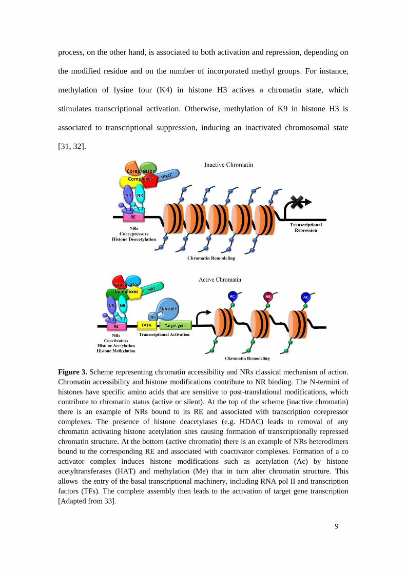
process, on the other hand, is associated to both activation and repression, depending on
the modified residue and on the number of incorporated methyl groups. For instance,
methylation of lysine four (K4) in histone H3 actives a chromatin state, which
stimulates transcriptional activation. Otherwise, methylation of K9 in histone H3 is
associated to transcriptional suppression, inducing an inactivated chromosomal state
[31, 32].
Figure 3. Scheme representing chromatin accessibility and NRs classical mechanism of action.
Chromatin accessibility and histone modifications contribute to NR binding. The N-termini of
histones have specific amino acids that are sensitive to post-translational modifications, which
contribute to chromatin status (active or silent). At the top of the scheme (inactive chromatin)
there is an example of NRs bound to its RE and associated with transcription corepressor
complexes. The presence of histone deacetylases (e.g. HDAC) leads to removal of any
chromatin activating histone acetylation sites causing formation of transcriptionally repressed
chromatin structure. At the bottom (active chromatin) there is an example of NRs heterodimers
bound to the corresponding RE and associated with coactivator complexes. Formation of a co
activator complex induces histone modifications such as acetylation (Ac) by histone
acetyltransferases (HAT) and methylation (Me) that in turn alter chromatin structure. This
allows the entry of the basal transcriptional machinery, including RNA pol II and transcription
factors (TFs). The complete assembly then leads to the activation of target gene transcription
[Adapted from 33].
10
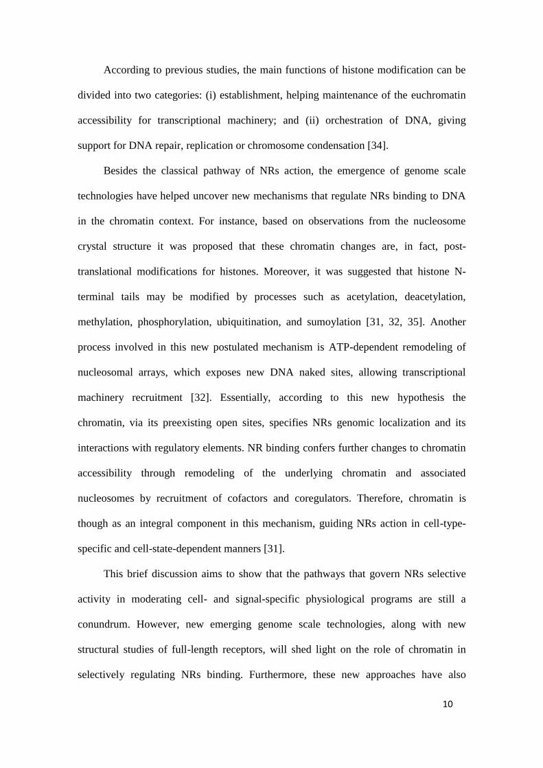
According to previous studies, the main functions of histone modification can be
divided into two categories: (i) establishment, helping maintenance of the euchromatin
accessibility for transcriptional machinery; and (ii) orchestration of DNA, giving
support for DNA repair, replication or chromosome condensation [34].
Besides the classical pathway of NRs action, the emergence of genome scale
technologies have helped uncover new mechanisms that regulate NRs binding to DNA
in the chromatin context. For instance, based on observations from the nucleosome
crystal structure it was proposed that these chromatin changes are, in fact, post-
translational modifications for histones. Moreover, it was suggested that histone N-
terminal tails may be modified by processes such as acetylation, deacetylation,
methylation, phosphorylation, ubiquitination, and sumoylation [31, 32, 35]. Another
process involved in this new postulated mechanism is ATP-dependent remodeling of
nucleosomal arrays, which exposes new DNA naked sites, allowing transcriptional
machinery recruitment [32]. Essentially, according to this new hypothesis the
chromatin, via its preexisting open sites, specifies NRs genomic localization and its
interactions with regulatory elements. NR binding confers further changes to chromatin
accessibility through remodeling of the underlying chromatin and associated
nucleosomes by recruitment of cofactors and coregulators. Therefore, chromatin is
though as an integral component in this mechanism, guiding NRs action in cell-type-
specific and cell-state-dependent manners [31].
This brief discussion aims to show that the pathways that govern NRs selective
activity in moderating cell- and signal-specific physiological programs are still a
conundrum. However, new emerging genome scale technologies, along with new
structural studies of full-length receptors, will shed light on the role of chromatin in
selectively regulating NRs binding. Furthermore, these new approaches have also
11
helped deciphering some new roles of NR cofactors in regulating DNA methylation,
histone post-translational modifications, and chromatin remodeling [31]. These events
show that, from a global view, transcription selectively regulated by NRs through
chromatin is a really well orchestrated dance among chromatin-histones, NRs,
transcription factors, corepressors, coactivators and modifying enzymes.
Moreover, the explanation of how NR-DNA specificity is achieved, considering
that all NRs use a highly conserved DBD core region to specifically choose to bind to
some DNA sequences, is still obscure and need to be further investigated. Also, the
chromatin context, conformational modifications and further contacts between NRs and
DNA should be considered in this discussion. However, it is important to highlight that
this issue is crucial to the understanding of transcription regulation and studies with full-
length NRs and chromatin are key points to answering this.
Regarding this issue, some reviews about DNA binding domain and hormone
response elements have been reported recently [4, 14, 31], herein we focus on the
description and applications of the most used analytical techniques to identify and study
NR-DNA interaction. Our main goal in this review is to discuss advantages and
disadvantages of the most applicable methodologies to NRs, providing a range of
evidences that may assist in better methodological choice for the study of NRs.
Electrophoretic Mobility Shift Assay (EMSA)
Electrophoretic mobility shift assay (EMSA), also known as gel shift is one
method largely applied to study gene regulation and NR-DNA interactions. This method
provides valuable information about sort of regulatory proteins involved in gene
expression, and may be adapted to a wide choice of cultured cell lines, DNA sequences
and transcription factors. In general, it is based on the propriety of retarded mobility in
12
the gel, characteristics of protein-DNA complexes, when compared to free protein and
DNA [36, 37].
Basically, gel shift assay consists in three key steps: binding reactions between
DNA and protein; native electrophoresis and probe detection. The DNA fragment used
in this method is the target sequence, which is usually from restriction fragment, or a
PCR product, or even, is obtained by DNA synthesis. The DNA labelling before is the
most common approach, allowing its specific detection after electrophoresis.
Traditionally, DNA probes have been radiolabeled with ³²P, but due to the concerns
involving this kind of material, numerous nonradioactive labelling methods for
performing EMSA have been developed, such as fluorescent probes that can be detected
in-gel using an appropriate imaging system. Besides that, hapten-modified DNA probes
can be visualized via secondary detection reagents, such as streptavidin or anti-DIG
antibodies, in systems with enzymatic substrates similar to those used for Western
blotting. Regarding to the binding reaction components, there are many unique
requirements for different nucleic acid binding proteins, so there is no universal set of
reaction conditions for EMSA assays, which must be investigated in the literature for
each specific case. Also important, the gels used in this method are non-denaturing
TBE-polyacrylamide gels or TAE-agarose gels [38].
EMSA is a reference technique that presents a great diversity of applications. In
some cases, it may be applied in a simpler way to show DNA-NR binding events [39],
or even to present that a heterologous expressed NR is able to binding to DNA [40].
But, in another way, more sophisticated questions have been answer using this sort of
experiment. For instance, it was applied to show some particularities of TR isoforms in
DNA binding, presenting that TRβ0 can bind as trimers to a subset of naturally-
occurring DNA elements, and not just as homo- or heterodimers with Retinoid X

13
Receptor (RXR), as it was thought [41]. Also, gel shift was employed to analyze the
RXR/TR and RXR/PPAR heterodimerization and DNA binding [42]. In this study, the
authors assessed the capability of these proteins to bind DNA in consensus target sites,
with the heterodimers RXR/PPAR or RXR/TR efficiently bind to DR1 and DR4,
respectively [42].
Other reports illustrate the use of EMSA in the study of nuclear receptors,
identifying consensus DNA sequences that bind to different NR complexes. The
interactions between coactivators and NRs on REs were studied by EMSA, searching
for interactions among Vitamin D Receptor (VDR), coactivators and response elements.
In this study, the effect of SRC-1 and TRAM-1 coactivators on VDR homodimer and
VDR/RXR heterodimer was analyzed, showing that VDR form stable homodimers after
interaction with the coactivators on the VDRE DR3 (direct repeat spaced by 3 base
pairs) and DR4 and DR5 REs may support these interactions, but in a weaker way [43].
In another example, a novel approach was developed to isolate large complexes of
proteins associated with the DNA-bound estrogen receptor α (ERα) using an agarose-
based EMSA, in order to understand how ERα regulates transcription of estrogen-
responsive genes [44]. This method was adapted to other nuclear receptors and their
responsive elements to provide better understanding of how they regulate gene
transcription of certain genes.
Additionally, recent EMSA were applied to study specificity in DNA–NR
binding. It was reported that artificial DNA binding sites based on “AGGTCA” half-
sites confer high affinity, but poor specificity, and that spacing alone does not account
for the divergent DNA recognition properties of TRs and RARs, as it has been proposed
[30]. In this case, the gel shift assay was used to explore the ability of TRα or RARα to
bind to artificial DR4 and DR5 response elements comprised of AGGTCA consensus

14
half-sites, and compare the binding profiles obtained from consensus REs to the two
naturally occurring response elements, αMHC promoter (DR4) and the βRAR promoter
(DR5). Although these elements were bound by their cognate receptors less strongly
than were the artificial AGGTCA elements, they were bound with much greater
specificity. Therefore, they conclude that half-site spacing contributes to DNA
recognition by these receptors, but is not the dominant discriminatory factor under these
conditions [30]. All these results indicates the presence of particular abilities that
different NRs have to discriminate between various promoter regions in similar arrays
(as DRs), but with sequences that diverge slightly from the consensus.
All these examples, together with many others which apply EMSAs for NRs
investigation [45-47], illustrate the wide application of this technique in the study of
NR-DNA interaction. Although having some limitations such as samples that are not in
the chemical equilibrium during the electrophoresis step, or that rapid dissociation
during experiment may prevent detection of complexes, or even, the little direct
information about the localization of the nucleic acid sequences; this assay is, in
general, a rapid and sensitive method in the detection of protein-DNA interactions. The
basic technique is simple to perform and highly sensitive, mainly with the use of
radioisotope-labeled nucleic acids, allowing assays to be performed with small content
of protein and nucleic acid.
Additionally, when this high sensitivity is not required, variants using
fluorescence, chemiluminescence and immunohistochemical detection are also
available, avoiding the danger of radiation. Nucleic acids can be used in a wide range of
sizes and structures and proteins can also include a ranging from small oligopeptides to
transcription complexes. Another advantage is the possibility of using highly purified
proteins and crude cell extracts, which accounts in large part for the continuing
15
popularity of this assay [48]. In this way, the electrophoretic mobility shift assay is still
a reference method generally used in the study of in vitro binding of nuclear receptors to
DNA response elements.
DNA Footprinting
Another important technique abundantly used to study interactions between NRs
and DNA is DNA footprinting, which was one of the first's techniques applied NRs
field in this concern [49]. This is an in vitro assay that investigates protein binding to
specific DNA sites, studying protein-DNA interactions outside and inside the cell
environment [50]. It is based on the fact that when a transcription factor is bound to
DNA, it is protected from degradation by nucleases, providing its "footprinting" on
DNA sequence [50]. Traditionally, this experiment was used in the studies on NRs to
identify their binding sites in DNA. In the past, the DNA region of interest, harboring
one or more transcription factors, was radioactive labeled, followed by DNAse I
treatment, which digest unprotected or free DNA. Following, the digested DNA was
separated by polyacrylamide gel and visualized on X-rays films [50]. Nowadays
instead, some other approaches have been associated to this method, such as the use of
fluorescent probes over radioactive DNA labelling; or the use of polymerase chain
reaction (PCR) to amplify specific DNA regions over electrophoresis, in the searching
for the protected DNA sequences [51]. Additionally, application of synchrotron X-ray
footprinting has been used to study time-resolved structural changes of nucleic acid
conformation and protein-nucleic acid complexes. In this case, nucleases are substituted
by X-ray to produce the cleavage patterns in DNA [52].
Remarkably, one of the first applications of this technique on NRs was the
definition of specific REs, such as the elucidation of Glucocorticoid Response Elements

16
(GREs) in tyrosine aminotransferase gene promoter, which is regulated by GR in the
presence of ligands [53]. Other applications include the regulation of tyrosine
hydroxylase by Nurr1 orphan receptor [54], or even, the binding of NGFI-B in the
steroid 21-hydroxylase (21-OHase) gene promoter [55]. Some other regulatory regions
in gene promoters were also found using footprinting, such as the regulation of growth
hormones, GH1 and GC, by TR. In this case, authors found two regions that were
selectively regulated by TR in strains of rat pituitary cells [56]. In the same way, other
reports showed that the prostate antigen gene promoter was found to be regulated by AR
[57], GR, PR [58], and also, by TR and ER [59, 60].
DNA footprinting also solved questions about how a NR recognizes specific
promoters, considering that many receptors can bind to the same RE sequences. For
example, the responsive elements composed by TGACC and TGTTCT sequences,
known as glucocorticoid receptor REs, were found upstream several genes regulated by
PR and AR. A more detailed investigation of these sequences, by using DNA
footprinting, showed that one of these sequences were regulating rat probasin gene
promoter by AR. This study also suggested that AR binds to another sequence in this
same promoter with a different affinity, but the highest androgen induction was reached
when both sites are filled with AR in a cooperative and mutually dependent manner
[61].
Later, other studies using DNA footprinting revealed conformational changes in
DNA upon NR binding, as it was observed for estrogen related receptor α 1 (ERRα-1)
binding to a silencer element (S1), down regulating the action of human aromatase gene
promoter. In this case, DNA footprinting was used to confirm the previous data
obtained by gel shift mobility assay and presented the exact binding site for ERRα-1.
However, it showed some intriguing conformational changes of DNA upon binding of
17
ERRα-1[62], another important feature obtained by EMSA.
It is undoubted that DNA footprinting is a fairly standardized technique and has
numerous applications in NRs studies as above showed. However, the development of
these experiments has been presented as quite laborious and it is a costly expensive
method, generating hard data to be analyzed. To overcome these problems some
improvements have been made, such as the development of softwares for quantitative
analysis of gel images, to reduce the time of data analysis [63]. Also, digital approaches
to evaluate regulatory protein occupancy on genomic DNA, using massively parallel
DNA sequencing have changed the paradigm of the absence of informatics tools
specifically designed for footprinting analysis, which makes it a less tedious, time
consumable and overwhelmed experiment [64].
These improvements and updates in DNA footprinting methods allowed for the
identification of protein-binding footprints with high resolution on a genome scale.
Digital DNase I Analysis is one of these advances, providing a perspective of the
genome mapping, quantifying the accessibility of chromatin and the NRs occupancy
[65, 66]. The approach is based on techniques of chromatin analysis that have been
developed and widely used to detect regulatory regions, which uses data generated from
chromatin by DNase I digestion with parallel massive sequencing [65, 67]. In the
context of nuclear receptors, the Digital DNase I was applied to map GR accessibility in
chromatin regarding genome wide scale [68]. According to this qualitative analysis
[69], GR was qualified as a NR capable of autonomous binding to genomic DNA target
sites, resulting in local chromatin remodeling, and 95% of these sites were recognized
as preexisting accessible sites in chromatin [68].
However, even with the advances in technology, to infer accurately the location of
these footprints remains a challenging computational task. To avoid this, the

18
development of dynamic Bayesian network had improved the identification and
statistical calculation of protein binding sites from the genome digital footprinting data
in a probabilistic framework [70].
Parallel to this, the development of new DNA labelling methodologies, avoiding
the radiolabelling, combined with new bioinformatics tools for data processing, and,
moreover the development of in silico footprinting [71] have greatly aided the use of
DNA footprinting, which continues to be a powerful tool to answer many questions,
specially about the NR-DNA interaction. On the other hand, it is important to mention
that the identification of REs depends on different factors, like cell type and treatment
conditions, which makes this process more complicated. Nowadays, the advent of
chromatin immunoprecipitation assays made it possible to identify large genomic
fragments to which ones NRs binds directly and indirectly, evaluating cellular contexts
[72]. The insertion of this technology on genome wide scale after the advent of Digital
DNase I analysis updated this assay, which still may be associated to different methods
in the elucidation of more complex questions concerning about NR-DNA interactions.
Transactivation assays – Transfection and reporter gene assays
In addition to the techniques mentioned above, a widely used in vivo method to
measure NR activity is the reporter gene assay. In general, this method is based on to
insert a DNA construction inside the cell, which owns a RE followed by a reporter gene.
This is a fundamental tool to monitor cellular events associated with gene expression,
regulation and signal transduction. After the development of this assay, researchers have
acquired one sensitive, reliable and convenient assay, providing one efficient report of
the activation of particular NRs and their effects on gene expression [73, 74].

19
Aiming to understand the biological role of NRs in gene expression by using this
assay, primarily it is import to monitor the protein in the cellular context. In order to
achieve this, the first step would be the introduction of the target gene inside the cell;
otherwise, it is still possible to consider endogenous levels of NR, which is one of many
advantages of this technique [75]. However, when the introduction of interest gene is
required, transfection is a widely used procedure and a powerful analytical tool for
study gene and protein function and regulation [75]. There are two major transfection
types: stable, where gene is integrated to genome; and transient, where a plasmid
containing the gene supports expression for short periods of time [76, 77].
In the first one, transgene becomes as dependent as other genes of transcription
regulation machinery, which could be considered a disadvantage if compared with the
second one, but it is more similar to the cell real conditions. Within the first scenario, a
natural in vivo system, transgene may be associated or not with histones in a dense
chromatin waiting to be transcripted and translated [31, 32]. In contrast, in the second
method, plasmids probably are in a supercoiled shape, but the regulation sequence
access is still easier because of the absence of histones and chromatinized DNA [77,
78], being this approach widely applied for fast transactivation assays. On the other
hand, it is an artificial system for cell metabolism. Overall, both transfections types
have advantages and disadvantages, and the best choice between them should be done
after some questioning about cell toxicity, transfection efficiency, effects on normal
physiology, and also reproducibility [75].
The second step for reporter assays is the choice of reporter gene since there are
several types of systems available today. Remarkably, some important features have to
be considered in the reporter’s choice: effectiveness and sensitivity, level of expression,
stability of expressed protein and, background, due to endogenous protein [73, 79].
20
Historically, chloramphenicol acetyltransferase (CAT) was the first reporter gene
to be used. However, it has become obsolete nowadays mostly because of the fast decay
of the enzymatic activity [74, 79]. To overcome this, luciferase (LUC), which is more
stable than CAT, is widely used. LUC assay is highly sensitive, it requires fewer cells
than CAT assay, and its response can be measured within 25 hours after transfection.
Nevertheless, it remains a simple, rapid, and sensitive method for NR activity on
promoters [80]. Overall, Renilla luciferase together with firefly luciferase reporters are
considered the most efficient ones, emitting the highest bioluminescence signal,
allowing for detection of subattomoles amounts of enzyme. Both are excellent markers
for gene expression, as they lack post-translational modifications, have absent
endogenous proteins or enzymes, and exhibit fast enzymatic interactions.
Moreover, luciferase reporter gene system can be used for monitoring gene
expression in vivo. One example was the generation of the ERE-LUC transgenic mice
[81], which had the luciferase reporter gene under the control of an estrogen-responsive
element (ERE-LUC). In this report the luciferase activity in estrogen cycles indicated
that the highest transcriptional activity of ER occurred during proesterus in reproductive
tissue [81]. These ERE-LUC model mice also facilitated kinetics, monitoring and
quantitative analysis of ER activity in specific tissues [74].
Later, a novel reporter assay system was developed as an improvement of
luciferase reporter gene system. This assay, termed the tricolor reporter in vitro assay
system, consists in the use of green- and red-emitting Phrixothrix luciferases, as dual
reporters; and of blue-emitting Renilla luciferase, as internal control. This system was
developed firstly to study the RAR-related orphan receptor alpha (RORα), and was
successfully employed to verify the clock effects of gene products on the enhancer
elements of Bmal1 and Per1 promoters [82].
21
As already mentioned, the reporter gene technique is widely applied and may
provide data to solve distinct questions. One example was the study highlighting the
need to investigate the (anti-) androgenic activity of compounds in dependence of the
cellular and promoter context [83]. In this study, the reporter activity of plasmids
containing AR response elements derived from the human secretory component (rat
probasin gene), as well as, the GREs, were evaluated together with mouse mammary
tumor virus promoter [83].
Interestingly, another application of reporter gene method encompasses its use as
adjuvant to corroborate protein structural hypothesis. For example, to understand TR
LBD conformation and its conformational changes after ligand binding, experiments of
H/D exchange MS were performed and suggested a new regulation step in coactivator
recruitment [84]. This hypothesis was tested by site direct mutagenesis and TR
transactivation assays, showing that the detected changes in TR conformation are
important, influencing the activation of this receptor [84].
The development of a new reporter gene assay to test NR-coregulators
interactions inside the cells using a chimeric system was also described as another
application of this assay for NR, combining reporter gene assay with two-hybrid
mammalian system [85, 86, 87]. Additionally, reporter gene assay technique is also
capable of searching and understanding novel NRs’ interactions with DNA itself,
investigating gene regulation by NRs inside promoter regions. Moreover, it is common
to use reporter gene along with EMSA and ChIP-chip to discover and validate where
transcription factors are binding to [88]. As an example, a recent report shows that TM
(thrombomodulin) expression and activity were up-regulated by FXR activation in
vascular endothelial cells [89]. FXR activation significantly enhanced the transcriptional
activity of human TM gene promoter, as seen in reporter gene assays, and EMSA and

22
Chip-chip indicated that FXR induced TM expression by binding to a novel FXR-
responsive element.
Remarkably, there are a considerable number of studies in the literature searching
for selective ER modulators with balanced high affinity for ERα and ERβ, which could
act as therapeutics for the treatment of hormone-response breast cancer, osteoporosis
and many other diseases using these reporter gene assays [90-92]. One of them, aimed
to discover novel selective ligands for ERβ, through development and characterization
of a cell-based Gal4-ERβ β-lactamase reporter gene assay (GERTA) for ligand-induced
activation of the human ERβ. This assay was optimized for screening in an ultra
highthroughput, using 3,456-well nanoplate format, and it was successfully used to
screen a large compound collection for ERβ agonists [91]. Alternatively, one study
focused on the development of second- and third-generation selective ER modulators,
with the goal of reducing toxicity and improving tissue-selective efficacy, developing a
new cell-based ERα-transactivation assay, where ERα-specific antagonists were
screened after only 4h of incubation time, using a fully automated ultra high throughput
screen, and a number of valuable leads were identified [92].
So far, there are some different methods to understanding the activity of NRs in
DNA response elements, and novel ones are under development right now. Among all,
the most widely used and easiest to standardize in mammal cells continues to be
transactivation using luciferase enzyme system. One of the reasons for this preference is
it reproducibility and the easily way to perform measurements. However, it is important
to mention that LUC activity does not always imply in a direct interaction of NR-DNA,
and this activity may be a response to an indirect interaction via different partners
through crosstalk, for example, which may be confirmed by EMSA or DNA
footprinting assay.

23
Nevertheless, LUC assays are excellent markers for gene expression and
altogether, the discussed characteristics turn luciferase reporter gene assay into the most
sophisticated and robust method used nowadays to study nuclear receptor interactions
with DNA in vivo. Here we show that this assay may be applied to answer a large
spectrum of questions, as the analysis of NR activity in different cell contexts, or new
promoters; or as the evaluation of interactions with coregulator and new ligands; and,
even, to confirm structural hypothesis. Also important, this assay allows the study of
NRs in naked DNA reporters or in chromatinized DNA, according to the chosen
transfection method, which, depending on the objective of the study, may present more
realistic results, closer to cellular environment.
Chromatin Immunoprecipitation sequencing (ChIP-Seq)
While EMSA and DNA footprinting are mostly used for in vitro analyses of NR
and DNA interactions, another technique used to investigate this interaction in cellular
context, as reporter gene assay, is the Chromatin Immunopreciptation – Sequencing, or
ChIP-Seq. Nowadays, as far as we know, this is one of the most used assays for
investigation of NRs-DNA binding.
ChIP-Seq is applied to map NR binding sites in genomic-wide scale throughout
the DNA, without significant cellular modification [93], applied to map chromatin
modifications and nucleosome positions [94]. The method is an evolution of ChIP [95]
and ChIP-chip procedures, which initially were based on applications of specific
primers and DNA microarrays, towards next generation DNA sequencing (NGS),
allowing the study through the whole genome [96]. The first ChIP-Seq application
mapped histone modifications in the entire genome, identifying DNA binding sites, and

24
the location of different histone methylation patterns in lysine and arginine residues, in
human CD4CT cells [97].
ChIP-Seq and ChIP–chip are currently the two main competing technologies for
the genome-wide identification of chromatin immunoprecipitated material [98]. The
general principle of ChIP is immunoprecipitation of specific proteins cross-linked
together with their associated DNA. In classical protocols DNA-proteins complexes
from cells extracts are cross-linked and the chromatin is fragmented by sonication.
During basic ChIP procedure, after DNA purification, it is performed a PCR with
specific primers for known DNA sequences [95]. In the ChIP-chip procedure, purified
DNA is applied in a microarray plate, which allows the positive recognition of several,
but limited, known DNA sequences. Differently, in ChIP-Seq procedures, the purified
DNA is submitted to the NGS and most of the binding sites are identified.
When compared with the other two ChIP methodologies, ChIP-Seq is the one that
provides the large amount of results. In a comparison between ChIP-chip and ChIP-Seq,
the latter has higher signal-to-noise ratios, is less expensive, and requires lower amounts
of DNA for genome analysis [93]. Also, the sequencing step did not limit ChIP-Seq to
the few binding sites, allowing analysis of the whole genome [99]. Therefore, based on
ChIP-Seq advantages and, as it is largely used nowadays in NRs field, our review will
focus on this ChIP method.
The importance of ChIP-Seq assay is totally related to understand how a huge
number of transcriptional factors in living cells can interact with some specific DNA
sequences, improving our knowledge about the functional sites. Several studies show
ChIP-Seq experiments applied in the search of NRs binding sites in DNA, using distinct
cell types. For example, ChIP-Seq together with ChIP found 8,848 GR binding sites in
mouse adipocytes treated whit synthetic glucocorticoid Dexamethasona [71]. Also in

25
adipocytes, more than 5,000 PPARγ binding sites were isolated [100]. In human lung
adenocarninoma cells, it was identified more than 4,000 GR binding sites [101], while
in human breast cancer cell lines it was found more than 10,000 ER binding sites [102]
and 20,000,000 PPARβ/δ binding sites [103].
Importantly, we observe in the above mentioned examples that the diversity of
cell types and experimental conditions results in data variability in ChIP-Seq, getting
impossible to define regular guidelines appropriate to a more general rule [104].
Although, taking together the ChIP-Seq results from distinct NRs and cell lines and, the
identification of overlapping or unique DNA binding sites, can lead to new therapeutic
strategies [105].
ChIP-Seq also has been extensively used to map the in vivo genome-wide binding
(cistrome) of NRs in both normal and cancer cells due to evidences that NRs play a
differential role in cancer cells [106]. ChIP-seq assays have confirmed these evidences
as presented in studies of AR in prostate cancer cell [107], and of ER [108], GR [109]
and PPARβ [110] in breast cancer cells. In these investigations authors found new
insights into the DNA sequences, in which ones NRs can bind and identify cooperating
transcription factors. Also, they identified potential NRs regulated genes that are not
seen in normal cells, providing enlightenment into the biological processes regulated by
them. This kind of application may elucidate meta-analysis data of the same cancer cell
line and generating consensus cistrome and expression profiles, which can be used to
understand the pathologies and guide new therapeutics developments for cancer
treatment [105].
As therapeutics targeting for NRs are mainly ligands, ChIP-Seq may also be
applied to identify and characterize NRs behavior in presence and absence of them. As
example, it was observed that triiodothyronine hormone (T3) treatment altered TRβ1

26
binding at distinct genomic sites, also changing expression patterns, suggesting a new
mechanism of regulation of target genes by TRβ1 [111]. Another recent development of
ChIP-Seq allowed perform this assay with a low input of cells, as it was showed in ERα
ChIP-Seq that was successfully performed with an input of only 5,000 cells by using
single tube linear amplification (LinDA) [112].
Besides the large amount of examples of ChIP-Seq, it is important to highlight the
difficulty to find specific antibodies for NRs. Essentially, the success of the experiment
depends on validations and use of highly specific antibodies [104]. An example
showing the efforts in validation of specific antibodies could be seen in the case of LXR
genome-wide mapping of binding sites studies [113]. One alternative to solve this issue
may be outlined with a tag-based approach, by using transgenesis to express NRs
tagged with an epitope or a tag as EGFP. By this way, the only required antibody would
be the one specific to the tag. This approach allows the use of just one antibody to study
several proteins [114], as it was shown in ChIP-Seq study of 24 NRs expressed in breast
cancer cell lines [105].
Another important point of ChIP-Seq application for NRs is the huge amount of
data generated by NGS and the requirement of both bioinformatics and statistics tools,
to process the data and turn them into understandable results. Moreover, big amounts of
data generated by ChIP-Seq studies are available on databanks and can be analyzed in
parallel. The development of novel bioinformatics analyses can compare some patterns
of REs, distance distributions of transcription start sites of known genes, evolutionary
conservations and collaborating partners of several NRs [106], also generating
consensus cistromes and expression profiles among NRs.
However, albeit the binding sites can be predicted by in silico studies, this kind of
prediction still did not achieve sufficient success or accuracy, qualifying ChIP-Seq as

27
one technique capable to perform an effective detection of NRs genome-wide in vivo
binding sites or cistromes [106]. As example, despite some computational analyses
determined a potential of 105 – 10
6 binding sites to VDR responsive elements (VDRE)-
like sequence motifs, ChIP-Seq showed that less than 1000 sites were generally
occupied by the VDR in the absence of ligand and between 2000 and 8000 sites were
occupied following vitamin D treatment [96].
In another recent example, the comparison between ChIP and ChIP-Seq assays of
human and mice had demonstrated a common feature of NRs in recognizing relatively
short AT-rich motifs [104]. In addition, it was verified NRs binding to introns and distal
intergenetic regions far away from transcription start sites [106], counterpoint the
classical statement which suggests REs may be located in the 5' region of the target
gene, closely to the core promoter.
Other evident and specific limitation of ChIP-seq is that it only provides
information about the NRs binding sites and regions nearby, which may difficult the
identification of genes that are under particular regulation. Moreover, it has been shown
that many NRs binding sites land in distal intergenic regions or introns, according to
recent data in the literature. The AR ChIP-seq in prostate cancer cells suggested AR
binding in non-promoter regions and action through chromatin loopings [115]. Despite
the difficulties to use ChIP-seq to predict target genes that are not straightforward [106]
and, as some binding sites are distal from gene promoter; it is difficult to predict what
distal NRs binding sites are non-functional fortuitous binding sites, and what are
involved in transcriptional activity through a remote control mechanism [116]. This sort
of difficult could be overcome by the application of recently developed methods, such
as chromosome conformation capture (3C), which have been performed to observe

28
long-range chromatin interactions between DNA elements engaged in transcriptional
regulation [117].
Overall, taking together, the advantages of ChIP-seq includes the capacity of
whole genome analysis of responsive elements both in vivo and in vitro, the low input
of cells requires, the reliability of results compared with in silico predictions, the higher
signal to noise ratio, the large amount of results, the capacity of understand network
regulations that will generate new therapeutic approaches. With the development of
high-throughput sequencing platforms, like Illumina, Genome Analyzer and SOLiD,
and with the availability of ChIP-grade antibodies; ChIP-Seq has become one of the
most widely used methods for determining de novo functional elements in the
sequenced genome [93].
Chromosome Conformation Capture (3C)
The 3C is a technique that investigates chromosomes’ organization in a cell’s
natural state. It was originally developed in 2002 aiming the identification, location and
mapping physical interactions between genetic elements located throughout the human
genome [117]. Basically, it is based on binding proximity to investigate the interaction
between any two genomic loci, in the same or different chromosomes, revealing their
relative spatial disposition [117]. Knowledge about structural properties and spatial
organization of chromosomes is important for the understanding of the regulation of
gene expression. A chromosomal region that folds in order to bring an enhancer and
associated transcription factors within close proximity of a gene is an example of how
chromosomal interactions can influence gene expression, as it was shown firstly in the
beta-globin locus [118]. Also, the development of 3C technique enables researchers to
investigate this kind of interaction/regulation.
29
The general 3C procedure comprises the isolation of intact nuclei and fixation to
cross-link proteins to other proteins and DNA. The interacting segments will be
physically bound via cross-linking and digested with restriction enzymes. Following
this step, these bound fragments are subjected to ligation at very low DNA
concentration, which favors the ligation of relevant DNA fragments over the random
ones. Finally, cross-linking is reversed and individual ligation products are detected and
quantified by the polymerase chain reaction (PCR), using locus-specific primers [117,
119].
One of the first applications of 3C in NRs studies investigated ER in breast cancer
cells [120]. In this study, it was observed that carbonic anhydrase XII gene, which is
widely related to breast cancer, is regulated by estrogen via ERα. Also, applying 3C
authors observed this regulation involves a distal region giving new insights into CA12
regulation mechanism and its strong relationship with ER in breast cancer [120]. In
another example, 3C method was used to identify distal chromosomal regions which
interact with GR-induced Lipocalin2 (Lcn2) gene [121]. Through these studies, it was
observed GR activation in the Ciz1-Lcn2 locus by long range interactions, suggesting a
relationship between a chromatin looping and gene regulation tissue specific [121].
Moreover, recent studies have found that the distal-binding AR transcription
complex, including AR, associated transcription factors and coactivators, regulates the
expression of several AR target genes involved in prostate cancer growth, through
chromatin looping. By using a global 3C assay, future studies should address whether
such a long-range combinatorial regulation can be generalized to include other AR-
dependent genes in the genome [115].
At last, a recent study used 3C to investigate whether PPARγ locus position is
changed during cell differentiation over other adipogenic genes. These results allowed
30
the observation that the genome organization is remodeled in response to adipogenic
signaling [122].
Besides 3C advantages, such as detecting remote chromatin interactions between
DNA elements engaged in transcriptional regulation, to overcome the ChIP-seq
limitation and to understand a specific protein-DNA interaction that have role in gene
expression; this method also presents limitations. Some of them are the requirement of
prior knowledge of different complexes to choose the best DNA primers [123], which
may be limited to detection of one-point or partial sites in genome. In addition, it is
incapable of de novo detection of genome-wide chromatin interactions, also presenting
low signal-to-noise ratios [116].
Due to these limitations, improvements of 3C resulted into new technologies, such
as 4C, 5C and 6C. Circular chromosome conformation capture (4C) was developed to
overcome the requests of previous knowledge of the different complexes identity by
applying the maternally inherited H19 imprinting control region primer present near to
the target sequence, during ligation [123]. Chromosome conformation capture-Carbon
Copy (5C) is a high-throughput 3C approach, which employs microarrays or
quantitative DNA sequencing using 454-technology as detection methods [124].
Another improvement is combined 3C-ChIP-Cloning assay (6C) that combines the
standard looping approaches previously defined with an immunopreciptation step to
investigate involvement of a specific protein that may mediate long-range chromatin
interactions [125]. These merge of ChIP and 3C aims to reduce noisy and increase
specificity for chromatin interaction detection; however, new approaches to separate
chromatin complexes from non-specific chromatin fragments are necessary to overcome
high levels of false positives [116].

31
Overall, a common problem found in 3C and its derivatives methods is the
frequent random collisions of chromosomal regions to one another, which means that
the detection of a product does not always indicate a specific interaction between two
regions. Therefore, a specific interaction between two regions is only confirmed when
the interaction occurs at a higher frequency than with neighboring DNA. Another
disadvantage of these techniques (3C, 4C, 5C e 6C) is the requirement of a large
number of cells, especially in the high-throughput methodologies. Experiments using
the 4C technique, for example, routinely process ten million cells for analysis on a
single microarray. However, in contrast to 3C and 5C, the 4C method does not require
the prior knowledge of both interacting chromosomal regions [119].
Finally, 3C technology now becomes a standard method for studying the
relationship between nuclear organization and transcription in the native cellular state. It
allows researchers to analyze the folding of chromatin in the native cellular state at a
resolution beyond that provided by current microscopy techniques. Moreover,
considering the shape of the genome is thought to play an important part in the
coordination of transcription and, more specifically that NRs mechanism of action
involves REs distant from target genes, 3C emerges as a remarkably method in NRs
field. Furthermore, together with ChIP-Seq, 3C technologies are indicated assays to
discover new relationship between NRs and DNA, or to monitor previously described
interactions and, also, to study particularities on genome.
Fluorescence Anisotropy
Apart from the techniques described above, there is also another in vitro method
largely used for identify and characterize protein-DNA interactions, termed
Fluorescence Polarization (FP), or Fluorescence Anisotropy (FA). Classically, this sort

32
of experiment is able to measure interactions between molecules in solution, in a
quantitative way, informing whether they physically are capable to interact. More
detailed, it is a biophysical measurement, based on the timescale of rotational mobility
of biological macromolecules and excited-state lifetime. Essentially, when a molecule is
excited by polarized light, the fluorescence emission will be depolarized in relation to
the incident light, mainly due to rotation mobility of molecules in solution [126]. For
example, fluorescence polarization increases as rotational mobility decreases, or
indirectly, as the size of the molecule increases. Therefore, FP is affected by molecular
size, viscosity of the medium and temperature. The FP experimental set up involves a
vertical polarizing filter for the exciting monochromatic light, which makes that only
the properly oriented molecules in the vertically polarized plane to absorb light and
become excited. The emitted light from these excited molecules is then measured in
both the horizontal and vertical planes. In this sort of measurements the calculated
parameters are polarization (P) and anisotropy (r), obtained from the equations below:
P = IVV - IVH
IVV + IVH
r = IVV - IVH
IVV + 2IVH
where, IVV is the fluorescence emission intensity measured in the plane parallel to the
plane of vertically polarized excitation and IVH is the fluorescence emission intensity,
measured in the plane perpendicular to the plane of vertically polarized excitation [126].
Furthermore, applying Hill approach to fit the anisotropy data, as showed in the
following equation, one can determine dissociation constant (Kd) and Hill cooperativity
coefficient (n) [127].
33
robs = ri + (rf – ri)*{[(NR + n)/ (kn)]/ [1 + (NR
n/k
n)]}
Where, robs is the observed anisotropy at total protein concentration NR; ri and rf
are the lower and upper anisotropy values; k is the Kd value and n is the Hill
cooperativity coefficient. There are other plots which could be applied to fit anisotropy
data; however, it was observed [127b-130] for the majority of NR-DNA interactions
that the occupancy of some binding sites may affects the affinity for the unfilled ones.
Therefore the Hill approach, which accounts for the possibility that not all receptor sites
are independent (cooperativity) may be properly applied in these systems.
Many examples of Fluorescence Anisotropy application in studies of DNA-NRs
interactions can be found since the beginning of these studies until nowadays [131]. One
of them applied fluorescence anisotropy assays to verify the interaction of RAR: RXR
heterodimer with DNA in the presence of ligands was also investigated by FP [132]. In
this study presence or absence of both agonists (retinoic acid and 9-cis retinoic acid) did
not influence the heterodimer affinity to DNA. However, when they tested a number of
other antagonists, it was noticed that the DNA binding was destabilized directly, or by
destabilizing the heterodimer [132]. Later, the TR DBD homodimerization was also
investigated with the same assay, leading to the conclusion that the DBD is also
responsible for TR dimerization [133].
Specificity is another variable investigated by FP, as it was reported in an affinity
study of ER by different ERE. Firstly, estradiol (E2) roles and salt dependence in ER-
ERE binding was studied, presenting almost the same binding affinity in presence or
absence of E2, suggesting ER-DNA binding is E2 independent. Moreover, the more
complete is protein constructs, more able it is to distinguish among the two different
DNA sequences, allowing the conclusion that other regions of the protein, besides
DBD, are important in ERE binding and specificity [134].

34
Following the same line, other detailed report aiming to deciphering whether
minimal TR domains are capable to distinguish among different DNA sequences was
performed [130]. In this study, FP assays investigated the affinity of three different
constructs of TR (containing just Helix 1, or just DBD, or both DBD and LBD) to four
different arrays of AGGTCA (PAL0, IP-6, DR-4 and DR-1) [130]. Based on this study,
we found firstly that TR binds to DNA as dimer. We verify that only a small peptide
derived from the DBD (Helix 1) is sufficient for recognition of the DNA, the entire
DBD is sufficient to bind with high affinity to F2, PAL and DR-4, but the highest
specificity was achieved when LBD is present in the protein, defining differences in
Kds, in low nM range [130]. These results confirm that more complete NRs better
distinguish among different REs, indicating that other domains may be important for
selectivity of NRs in DNA binding, as it was shown for ER [134].
Apart of this, some interactions NR-protein and NR-Protein-DNA also were
defined by fluorescence anisotropy, as it was shown in a study involving ER,
transcription intermediary factor 1-alpha (hTIF1α) and ERE binding [135]. It was
performed a trial of anisotropy of ER interacting with the ERE sequence, and the
interaction of this complex with ER-DNA-hTIF1α was verified. It was found that ER
interacts with the hTIF1α bound to DNA in hormone dependent manner and that,
especially in the absence of E2, the hTIF1α interacts better with ERβ than with ERα,
being DNA is not requested in ER-hTIF1α binding.
With similar purposes, in 2010, we reported a fluorescence anisotropy study of
binding affinities of TR and GATA2 on TSHβ promoter, to postulate a model of
interaction where in absence of ligand (T3) thyroid hormone receptor binds to its TRE,
while GATA2 binds to GATA-RE. However, in presence of T3, TR-TRE bond is

35
weakened, facilitating the interaction of TR with GATA2 zinc finger domain, which, in
turns, binds to GATA-RE [129].
Interestingly, FP studies have shown particularities in the measured systems, like
the addition of ligands and ions, which may changes affinities of some NRs to specific
DNA arrays, helping to elucidate mechanisms of actions of NRs in DNA [129,
130, 134]. As a memorable technique, fluorescence anisotropy has been widely applied
for testing interactions between different molecules. Among several advantages
presented by FP, this assay is also very well employed in high-throughput screening
assays [136]. Similar technologies for large-scale identification of pharmaceutical
compounds and environmental have been currently presented [137, 138], whose
compounds that alter the ability of ER to bind to DNA are searched, since this
interaction is known as a good target for cancer treatment [138].
There are no doubts that the application of fluorescence anisotropy in studies of
DNA-protein interactions may present several advantages over other standard methods
of assessing these interactions. On the other hand, it is clear that this sort of experiment
may present disadvantages, like the need for purified protein and labeled samples,
which is quite laborious, but it is feasible for different sorts of proteins, such as NRs. In
addition this is an artificial in vitro assay that not considers physiological conditions,
like the cell environment. Apart from this, FP is a quantitative technique that provides
definition of thermodynamic parameters for biological systems and allows inferring
about physical conditions for biomolecules interaction systems. The measurements are
performed in solution and in the binding reaction equilibrium; opposed to methods such
as EMSA and DNA Footprinting, which involves separation of free and bound ligand,
disturbing this equilibrium. Also, in contrast to DNA Footprinting and EMSA, no
hazardous radioactive waste is generated. FP has a lower limit of detection, in sub-

36
nanomolar range, therefore low quantity of sample is needed. Also, all components are
in solution, requiring relatively simple instrumentation, which makes this method more
applicable than calorimetry. Moreover, this technique is generally applicable and does
not need molecules with dissimilar size to measure equilibrium binding, allowing real-
time measurements for kinetic assays. Several conditions of buffer, ionic strength and
others can be tested rapidly [139] and it is feasible to test more physiological like
systems, as the NRs binding to reconstituted chromatin.
Overview of discussed methods for detect NR-DNA interactions
This mini-review aimed to discuss some of the most common techniques used to
study DNA-protein interactions, as summarized in Table 1. Although other methods,
such calorimetry and surface plasmon resonance, may also be applied to study these
interactions [140, 141], the techniques discussed above have been used to elucidate
details of NRs-DNA interactions since they were first characterized. Here we present
applicability, advantages, and disadvantages of such methods, discussing some details
and providing evidences found in literature, aiming to help choosing the best one to
perform NR experiments.
Among all the discussed assays, DNA Footprinting and EMSA were largely used
in the beginning of the investigations regarding interactions between NR and DNA.
However, the development of new technologies and instruments allowed the outbreak of
modern techniques, their applications have been decreasing. This is also due to the use
of radioactive reagents, which is an inconvenient of these two techniques, and due to the
fact that these experiments have been considered quite laborious. Apart from these
inconveniences, both techniques are very informative and provide specific information
of DNA binding, especially for NR. While EMSA is applied to verify NR binding to

37
specific DNA sequences in vitro, DNA footprinting provides information on which
sequences NRs are bound to, taking into account cellular context. As already discussed,
the development of fluorescent and quimioluminescent probes makes easier the
manipulation of samples for both assays, avoiding radioactivity. Specifically to DNA
Footprinting, the advent of Digital DNase I Analysis increased its usability, facilitating
analysis of huge amount of data.
Parallel to this, ChIP-seq and transactivation assays are applied to cell systems,
abundantly investigating NR-DNA interactions. Transfection/reporter gene assays are
used since the beginning of this sort of investigation, suffering constantly
improvements. In other words, it seems that this technique will continue to be applied
for many years as one way to measure activity of NRs. Despite this assay not even
being applied in most physiological conditions, due to the fact that many times the RE
and the reporter gene are artificially inserted in the cell environment, this is still the best
way to verify NRs behavior. Remarkable, ChIP-seq starts to be applied to NR-DNA
interaction investigation more recently, being developed from ChIP and ChIP-chip
assays. Nowadays, it is one of the most used techniques to investigate the binding sites
of NRs in chromatin context, allowing for the identification of roles in gene regulation
by NRs. However, as discussed before, this assay also presents some limitations, as
many reports have shown that NRs binds to distal sites from gene promoters, making it
difficult to predict NR transcriptional activity through a remote control mechanism. In
this context, the development of 3C technique increased the possibilities of observation
of long-range chromatin interactions between DNA elements engaged in transcriptional
regulation by NRs. Also, combining both methodologies applied to NRs (6C) could
result in a huge advance to a better understanding of transcriptional regulation mediated
by NRs.

38
Finally, fluorescence polarization might be considered an outside point in this
review, as it is a more biophysical and in vitro technique, which may be judged as
artificial. But it is important to consider that this has been used in NRs research for
many years, providing valuable information as some thermodynamic parameters,
quantifying affinities for different DNA sequences and also providing some clues about
selectivity. Regarding all its advantages discussed above and its application to HTS
systems, the FP makes investigation faster, and may be combined with other cell assays
like ChIP-seq and transactivation assays. Together with the above mentioned
techniques, FP added to other structural methods (such as NMR and X-ray
crystallography), which are very important and useful in studying NR-DNA interaction,
may broaden horizons in the best understanding of RN-DNA interactions, answering
important questions about gene regulation and transcriptional networks regulated by
NRs.
Table 1. Advantages and disadvantages of techniques on studies of NR-DNA interaction
TECHNIQUE APPLICATION ADVANTAGES DISADVANTAGES
EMSA Investigation of interaction
between NR and specific DNA sequences.
Simple Technique Rapid and Sensitive
Radioactive labeling Rapid dissociation of
complexes
DNA Footprinting
Identify interactions with DNA. Whole genome analysis
In vitro Radioactive labelling
X-rays films
Reporter Gene
Quantitative measure of NR activation/repression.
In vivo Easy to standardize
Expensive NR could act indirectly
Chip-seq Identification of DNA binding
sites. Whole genome analysis
In vitro and in vivo Require specific antibody
Huge amount of data
3C Identification and mapping NR-
DNA physical interactions
Detection of remote chromatin interactions in native cellular state
Require prior knowledge Low signal-to-noise ratios
Fluorescence Anisotropy
Quantitative measurement of affinity of interaction of NR-DNA.
Quantitative Fast
Require pure protein Fluorescent labeling

39
Conclusion
This review aimed to discuss different methodologies that may be applied to the
study of nuclear receptors and their interactions with DNA. Apart from all the
particularities of this superfamily of proteins, we presented different methodological
aspects of each technique that can be applied to NRs, aiming the better understanding
different aspects of its interaction with DNA, which could improve overall knowledge
of some of its roles in transcriptional regulation.
Acknowledgments
Authors thanks FAPESP for financial support (Grant #2013/08743-2). We also
knowledge T. M .A. Moreira for reviewing this manuscript.
References
1. Aranda, A.; Pascual, A. (2001) Nuclear hormone receptors and gene expression.
Physiol. Rev. 81, 1269–1304.
2. Huang, P.; Chandra, V.; Rastinejad, F. (2010) Structural overview of the nuclear
receptor superfamily: insights into physiology and therapeutics. Annu. Rev. Physiol. 72,
247–272.
3. Nwachukwu, J. C.; Nettles, K. W. (2012) The nuclear receptor signalling scaffold:
insights from full-length structures., EMBO J. 31, 251–253.
4. Helsen, C.; Kerkhofs, S.; Clinckemalie, L.; Spans, L.; Laurent, M.; Boonen, S.;
Vanderschueren, D.; Claessens, F. (2012) Structural basis for nuclear hormone receptor
DNA binding. Mol. Cell. Endocrinol. 348, 411–417.
5. Mangelsdorf, D.J.; Thummel, C.; Beato, M.; Herrlich, P.; Schütz, G.; Umesono,
K.; Blumberg, B.; Kastner, P.; Mark, M.; Chambon, P.; Evans, R.M. (1995) The nuclear
receptor superfamily: the second decade. Cell 83, 835-839.
6. Bosscher, K.D.; Berghe, W.V.; Haegeman, G. (2006) Cross-talk between nuclear
receptors and nuclear factor kB. Oncogene 25, 6868–6886.

40
7. Santos, M.G.; Fairall, L.; Schwabe, J.W.R. (2011) Negative regulation by nuclear
receptors: a plethora of mechanisms. Trends Endocrin. Met. 22, 87-93.
8. Pascual, G.; Fong, A.L.; Ogawa, S.; Gamliel, A.; Li, A.C.; Perissi, V.; Rose, D.W.;
Willson, T.M.; Rosenfield, M.G.; Glass, C.K. A SUMOylation-dependent pathway
mediates transrepression of inflammatory response genes by PPAR-gamma (2005)
Nature 437, 759–763.
9. Jensen, E.V. (1962) On the mechanism of estrogen action. Perspect. Biol. Med. 6, 47-
59.
10. Evans, R.M. (1988) The steroid and thyroid hormone receptor superfamily. Science
240, 889-895.
11. O’Malley, B.W. (1971). Mechanisms of action of steroid hormones. N. Engl. J.
Med. 284, 370–377.
12. Yamamoto, K.R. (1985). Steroid receptor regulated transcription of specific genes
and gene networks. Annu. Rev. Genet. 19, 209–252.
13. a) Overington, J.P.; Al-Lazikani, B.; Hopkins, A.L. (2002) How many drug targets
are there? Nat. Rev. Drug Discov. 5, 993-996. b) Hopkins, A.L.; Groom, C.R. (2002)
The druggable genome. Nat. Rev. Drug Discov. 1, 727-730. c) Mandrekar-Colucci,
S.; Landreth, G.E. (2011) Nuclear receptors as therapeutic targets for Alzheimer's
disease. Expert Opin. Ther. Tar. 15, 1085-1097. d) Landreth, G.; Jiang, Q.; Mandrekar,
S.; Heneka M. (2008) PPARgamma agonists as therapeutics for the treatment of
Alzheimer's disease. Neurotherapeutics 5, 481-489. e) Makishima, M. (2005) Nuclear
receptors as targets for drug development: regulation of cholesterol and bile acid
metabolism by nuclear receptors. J. Pharmacol. Sci. 97, 177-183.
14. White, D.C.; Laperrière, D.; Mader, S. (2011) Evolution of the repertoire of nuclear
receptor bindings sites in genomes. Mol. Cell. Endocrinol. 334, 76-82.
15. Siggers, T.; Gordân, R. (2013) Protein–DNA binding: complexities and multi-
protein codes. Nucleic Acids. Res., 1-13.
16. Rohs, R.; West, S.M.; Sosinsky, A.; Liu, P.; Mann, R.S.; Honig, B. (2010) The Role
of DNA shape in protein-DNA recognition. Nature 461, 1248-1253.17. Seeman, N.C.;
Rosenberg, J.M.; Riche, A. (1976) Sequence-specific recognition of double helical
nucleic acids by proteins. Proc. Natl. Acad. Sci. 73, 804-808.
18. Schwabe, J.W.; Neuhaus, D.; Rhodes, D. (1990) Solution structure of the DNA-
binding domain of the oestrogen receptor. Nature 348, 458-461.
41
19. Luisi, B.F.; Xu, W.X.; Otwinowski, Z.; Freedman, L.P.; Yamamoto, K.R.; Sigler,
P.B. (1991) Crystallographic analysis of the interaction of the glucocorticoid receptor
with DNA. Nature 352, 497-505.
20. Brélivet, Y.; Rochel, N.; Moras, D. (2012) Structural analysis of nuclear receptors:
from isolated domains to integral proteins. Mol. Cell. Endocrinol. 348, 466-473.
21. Chasse, S.A., Rastinejad, F. (2001) Physical structure of nuclear receptor-DNA
complexes. Methods Mol. Biol. 176, 91-103.
22. Rastinejad, F.; Huang, P.; Chandra, V.; Khorasanizadeh, S. (2013) Understanding
nuclear receptor form and function using structural biology. J. Mol. Endocrinol. 51, T1-
T21.
23. Germain, P.; Altucci, L.; Bourguet, W.; Rechette-Egly, C.; Gronemeyer, H. (2003)
Nuclear receptor superfamily: Principles of signaling. Pure Appl. Chem. 75, 1619-1664.
24. Khorasanizadeh, S.; Rastinejad, F. (2001) Nuclear-receptor interactions on DNA-
response elements. Trends Biochem. Sci. 26, 384-390.
25. Pawlak, M.; Lefebvre, P.; Staels, B. (2012) General molecular biology and
architecture of nuclear receptors. Curr. Top. Med. Chem. 12, 486-504.
26. O'Hara, B.; de la Rosa, D.A.; Rajendran, V.M. (2014) Multiple Mineralocorticoid
Response Elements Localized in Different Introns Regulate Intermediate Conductance
K+ (Kcnn4) Channel Expression in the Rat Distal Colon. PLoS ONE 9, e98695.
27. Carroll, J.S.; Meyer, C.A.; Song, J.; Li, W.; Geistlinger, T.R.; Eeckhoute, J.;
Brodsky, A.S.; Keeton, E.K.; Fertuck, K.C.; Hall, G.F.; Wang, Q.; Bekiranov, S.;
Sementchenko, V.; Fox, E.A.; Silver, P.A.; Gingeras, T.R.; Liu, X.S.; Brown, M. (2006)
Genome-wide analysis of estrogen receptor binding sites. Nat. Genet. 38, 1289-1297.
28. Kato, S.; Sasaki, H.; Masushige, S.; Tora, L.; Chambon, P.; Gronemeyer, H. (1995)
Widely spaced, directly repeated PuGGTCA elements act as promiscuous enhancers for
different classes of nuclear receptors. Mol. Cell. Biol. 15, 5858-5867.
29. Claessens, F.; Gewirth, D.T. (2004) DNA recognition by nuclear receptors. Essays
Biochem. 40, 59-72.
30. Phan, T. Q.; Jow, M. M.; Privalsky, M. L. (2010) DNA recognition by thyroid
hormone and retinoic acid receptors: 3-4-5 rule modified. Mol. Cell. Endocrinol. 319,
88–98.
31. Biddie, S.C.; John, S. (2014) Conversing with chromatin: the language of nuclear
receptors. Mol. Endocrinol. 28, 3-15.
42
32. Kishimoto, M.; Fujiki, R.; Takezawa, S.; Sasaki, Y.; Nakamura, T.; Yamaoka, K.;
Kitagawa, H.; Kato, S. (2006) Nuclear Receptor Mediated Gene Regulation through
Chromatin Remodeling and Histone Modifications. Endocrine Journal 53, 157-172.
33. a) Zhang, X.; Ho, S.-M. (2011) Epigenetics meets endocrinology. J. Mol.
Endocrinol. 46, R11-R32. b) Gadaleta, R.M.; Magnani, L. (2014) Nuclear receptors and
chromatin: an inducible couple. J. Mol. Endocrinol. 52, R137-R149. c) King, M.W. The
Medical Biochemistry page. http://www.themedicalbiochemistrypage.org (accessed
June 30, 2014).
34. Kouzarides, T. (2007) Chromatin Modifications and Their Function. Cell 128, 693-
705.
35. Luger, K.; Mäder, A.W.; Richmond, R.K.; Sargent, D.F.; Richmond, T.J. (1997)
Crystal structure of the nucleosome core particle at 2.8 A resolution. Nature 389, 251–
260.
36. Hendrickson, W. (1985). BioTechniques 3, 346-354.
37. Revzin, A. (1989). BioTechniques 7, 346-354.
38. Chernov, I.P.; Akopov, S.B.; Nikolaev, L.G.; Sverdlov, E.D. (2006) Identification
and mapping of DNA binding proteins target sequences in long genomic regions by
two-dimensional EMSA. BioTechiniques 41, 91-96.
39. a) Lazar, M.A.; Berrodin, T.J.; Harding, H.P. (1991) Differential DNA binding by
monomeric, homodimeric, and potentially heterodimeric forms of thyroid hormone
receptor. Mol. Cell. Biol. 11, 5005-5015. b) Ribeiro, R.C.J.; Kushner, P.J.; Apriletti,
J.W.; West, B.L.; Baxter, J.D. (1992) Thyroid hormone alters in vitro DNA binding
monomers and dimmers of thyroid hormone receptors. Mol. Endocrinol. 6, 1142-1152.
c) Shulemovich, K.; Dimaculangan, D.D.; Katz, D.; Lazar, M.A. (1995) DNA bending
by thyroid hormone receptor: influence of half-site spacing and RXR. Nucleic Acids
Res. 23, 811-818. d) Wu, Y.; Yang, Y.-Z.; Koenig, R.J. (1998) Protein-protein
interaction domains and the heterodimerization of thyroid hormone receptor variant α2
with retinoid X receptors. Mol. Endocrinol. 12, 1542-1550. d) Velasco, L.F.R.; Togashi,
M.; Walfish, P.G.; Pessanha, R.P.; Moura, F.N.; Barra, G.B.; Nguyen, P.; Rebong, R.;
Yuan, C.; Simeoni, L.A.; Ribeiro, R.C.J.; Baxter, J.D.; Webb, P.; Neves, F.A.R. (2007)
Thyroid hormone response element organization dictates the composition of active
receptor. J. Biol. Chem. 282, 12458-12466. e) Chen, Y.; Young, M.A. (2010) Structure

43
of a thyroid hormone receptor DNA-binding domain homodimer bound to an inverted
palindrome DNA response element. Mol. Endocrinol. 24, 1650-1664.
40. Figueira, A.C.M.; Dias, S.M.G.; Santos, M.A.M.; Apriletti, J.W.; Baxter, J.D.;
Webb, P.; Neves, F.A.R.; Simeoni, L.A.; Ribeiro, R.C.J.; Polikarpov, I. (2006) Human
Thyroid Receptor Forms Tetramers in Solution, Which Dissociate Into Dimers Upon
Ligand Binding. Cell Biochem. Biophys. 44, 453-462.
41. Mengeling, B.J.; Lee, S.; Privalsky, M.L. (2008) Coactivator Recruitment is
Enhanced by Thyroid Hormone Receptor Trimers. Mol. Cell. Endocrinol. 280, 47-62.
42. Mulero, M.; Perroy, J.; Federici, C.; Cabello, G.; Ollendorff, V. (2013) Analysis of
RXR/THR and RXR/PPARG2 Heterodimerization by Bioluminescence Resonance
Energy Transfer (BRET). PLoS ONE 8, e84569.
43. Takeshita, A.; Ozawa, Y.; Chin W. W.(2000) Nuclear receptor coactivators facilitate
Vitamin D Receptor homodimer action on direct repeat hormone response elements.
Endocrinology 141, 1281-1284.
44. Schultz-Norton, J.R.; Ziegler, Y.S.; Likhite, V.S.; Nardulli, A.M. Isolation of
Proteins Associated with the DNA-Bound Estrogen Receptor alpha. (2009) Methods
Mol. Biol. 590, 209-221.
45. Read, J.T.; Cheng, H.; Hendy, S.C.; Nelson, C.C.; Rennie, P.S. (2009) Receptor-
DNA interactions: EMSA and footprinting. Methods Mol. Biol. 505, 97-122.
46. Greiner, E.F.; Kirfel, J.; Greschik, H.; Huang, D.; Becker, P.; Kapfhammer, J.P.;
Schüle, R. (2000) Differential ligand-dependent protein–protein interactions between
nuclear receptors and a neuronal-specific cofactor. Proc. Natl. Acad. Sci. 97, 7160-
7165.
47. Eloranta, J.J.; Zaïr, Z.M.; Hiller, C.; Häusler, S.; Stieger, B.; Kullak-Ublick, G.A.
(2009) Vitamin D3 and Its Nuclear Receptor Increase the Expression and Activity of the
Human Proton-Coupled Folate Transporter. Mol. Pharmacol. 76, 1062-1071.
48. Hellman, L. M.; Fried, M. G. (2007) Electrophoretic Mobility Shift Assay (EMSA)
for detecting protein-nucleic acid interactions. Nat. Protoc. 2, 1849–1861.
49. a) Wilson, T.E.; Mouw, R.A.; Weaver, C.A.; Milbrandt, J. Parker, K.L. (1993) The
Orphan Nuclear Receptor NGFI-B Regulates Expression of the Gene Encoding Steroid
21-Hydroxylase. Mol. Cell. Biol. 13, 861-868. b) Chumsakul, O.; Nakamura, K.;
Kurata, T.; Sakamoto, T.; Ogasawara, N.; Oshima, T.; Shikawa, S. (2013) High-
44
Resolution Mapping of In vivo Genomic Transcription Factor Binding Sites Using In
situ DNase I Footprinting and ChIP-seq. DNA Res. 20, 325–337.
50. a) Galas, D.; Schmitz A. (1978) DNAse footprinting: a simple method for the
detection of protein-DNA binding specificity. Nucleic Acids Res. 5, 3157-3170.
b) Ralston, A. (2008) Do transcription factors actually bind DNA? DNA footprinting
and gel shift assays. Nature Education 1, 121.
51. Hampshire, A.; Rusling, D.; Broughton-Head, V.; Fox, K. (2007) Footprinting: A
method for determining the sequence selectivity, affinity and kinetics of DNA-binding
ligands. Methods 42, 128–140.
52. Sclavi, B.; Woodson, S.; Sullivan, M.; Chance, M.R.; Brenowitz, M. (1997) Time-
resolved synchrotron X-ray “footprinting”, a new approach to the study of nucleic acid
structure and function: application to protein-DNA interactions and RNA folding. J.
Mol. Biol. 266, 144-159.
53. Becker, P.B.; Gloss, B.; Schmid, W.; Strähle, U.; Schütz, G. (1986) In vivo protein-
DNA interactions in a glucocorticoid response element require the presence of the
hormone. Nature 324 ,686-688.
54. Sakurada, K.; Ohshima-Sakurada, M.; Palmer, T.D.; Gage, F.H. (1999) Nurr1, an
orphan nuclear receptor, is a transcriptional activator of endogenous tyrosine
hydroxylase in neural progenitor cells derived from the adult brain. Development
126, 4017-4026.
55. Wilson, T.E.; Padgett, K.A.; Johnston, M.; Milbrandt, J. (1993) A genetic method
for defining DNA-binding domains: application to the nuclear receptor NGFI-B. Proc.
Natl. Acad. Sci. 90, 9186–9190.
56. Ye, Z.S.; Samuels, H.H. (1987) Cell- and Sequence-specific Binding of Nuclear
Proteins to 5’-Flanking DNA of the Rat Growth Hormone Gene. J. Biol. Chem. 262,
6313-6317.
57. Riegman, P.H.; Vlietstra, R.J.; van der Korput, J.A.; Brinkmann, A.O.; Trapman, J.
(1991) The promoter of the prostate-specific antigen gene contains a functional
androgen responsive element. Mol. Endocrinol. 5, 1921-1930.
58. Truss, M.; Chalepakis, G.; Beato, M. (1990) Contacts between steroid hormone
receptors and thymines in DNA: An interference method (DNA footprint/base
recognition/glucocorticoid receptor/progesterone receptor/murine mammary tumor
virus). Proc. Natl. Acad. Sci. 87, 7180-7184.

45
59. Driscoll, M.D.; Klinge, C.M.; Hilf, R.; Bambara, R.A. (1996) Footprint analysis of
estrogen receptor binding to adjacent estrogen response elements. J. Steroid. Biochem.
Mol. Biol. 58, 45-61.
60. Klinge, C.M.; Bodenner, D.L.; Desai, D.; Niles, R.M.; Traish, A.M. (1997) Binding
of type II nuclear receptors and estrogen receptor to full and half-site estrogen response
elements in vitro. Nucleic Acid Res. 25, 1903-1912.
61. Kasper, S.; Rennie, P.; Bruchovsky, N.; Sheppard, P.; Cheng, H.; Lin, L.; Shiu, R.;
Snoek, R.; Matusik, R. (1994) Cooperative binding of androgen receptors to two DNA
sequences is required for androgen induction of the probasin gene. J. Biol. Chem. 269,
31763-31769.
62. Yang, C.; Zhou, D.; Chen, S. (1998) Modulation of Aromatase Expression in the
Breast Tissue by ERR a-1 Orphan Receptor. Cancer. Res. 58. 5695-5700.
63. Das, R.; Laederach, A.; Pearlman, S.M.; Herschlag, D.; Altman, R.B. (2005) SAFA:
semi-automated footprinting analysis software for high-throughput quantification of
nucleic acid footprinting experiments. RNA 11, 344-354.
64. Hesselberth, J.R.; Chen, X.; Zhang, Z.; Sabo, P.J.; Sandstrom, R.; Reynolds, A.P.;
Thurman, R.E.; Neph, S.; Kuehn, M.S.; Noble, W.S.; Fields, S.; Stamatoyannopoulos,
J.A. (2009) Global mapping of protein-DNA interactions in vivo by digital genomic
footprinting. Nat. Methods 6, 283-289.
65. Hesselberth, J.R.; Chen, X.; Zhang, Z.; Sabo, P.J.; Sandstrom, R.; Reynolds, A.P.;
Thurman, R.E.; Neph, S.; Kuehn, M.S.; Noble, W.S.; Fields, S.; Stamatoyannopoulos,
J.A. (2009) Global mapping of protein-DNA interactions in vivo by digital genomic
footprinting. Nat. Methods 6, 283–289.
66. Sekimata, M.; Melgosa, M.P.; Miller, S.A.; Weinmann, A.S.; Sabo, P.J.; Sandstrom,
R.; Dorschner, M.O.; Stamatoyannopoulos, J.A.; Wilson, C.B. (2009) CCCTC-binding
factor and the transcription factor T-bet orchestrate T helper 1 cell-specific structure and
function at the interferon-gamma locus. Immunity 31, 551–564.
67. Dorschner, M.O.; Hawrylycz, M.; Humbert, R.; Wallace, J.C.; Shafer,
A.; Kawamoto, J.; Mack, J.; Hall, R.; Goldy, J.; Sabo, P.J.; Kohli, A.; Li, Q.; McArthur,
M.; Stamatoyannopoulos, J.A. (2004) High-throughput localization of functional
elements by quantitative chromatin profiling. Nat. Methods 1, 219–225.

46
68. John, S.; Sabo, P.J.; Thurman, R.E.; Sung, M.; Biddie, S.C.; Johnson, T.A.; Hager,
G.L.; Stamatoyannopoulos, J.A. (2011) Chromatin accessibility pre-determines
glucocorticoid receptor binding patterns. Nat. Gen. 43, 264 – 270.
69. Becker, P.; Renkawitz, R.; Schütz, G. (1984) Tissue-specific DNaseI hypersensitive
sites in the 5′-flanking sequences of the tryptophan oxygenase and the tyrosine
aminotransferase genes. EMBO J. 3, 2015–2020.
70. Chen, X.; Hoffman, M.M.; Bilmes, J.A.; Hesselberth, J.R.; Noble, W.S. (2010) A
dynamic Bayesian network for identifying protein-binding footprints from single
molecule-based sequencing data. Bioinformatics 26, i334–i342.
71. Turner, J.D.; Pelascini, L.P.; Macedo, J.A.; Muller, C.P. (2008) Highly individual
methylation patterns of alternative glucocorticoid receptor promoters suggest
individualized epigenetic regulatory mechanisms. Nucleic Acids. Res. 36, 7207–7218.
72. De Bruyn, R.; Bollen, R.; Claessens, F. (2011) Identification and characterization of
androgen response elements. Methods Mol. Biol. 776, 81-93.
73. Naylor, L. H. (1999) Reporter Gene Technology: The future looks bright. Biochem.
Pharmacol. 58, 749-757.
74. Jiang, T.; Xing, B.; Rao, J. (2008) Recent developments of biological reporter
technology for detecting gene expression. Biotechnol. Genet. Eng. Rev. 25, 41-76.
75. Kim, T. K.; Eberwine, J.H. (2010) Mammalian cell transfection: the present and the
future. Anal. Bioanal. Chem. 397, 3173-3178.
76. Recillas-Targa, F. (2006) Multiple strategies for gene transfer, expression,
knockdown, and chromatin influence in mammalian cell lines and transgenic animals.
Mol. Biotechnol. 34, 337-354.
77. Glover, D.J.; Lipps, H.J.; Jans, D.A. (2005) Towards safe, non-viral therapeutic
gene expression in humans. Nat. Rev. Genet. 6, 299-310.
78. Nelson, D. L.; Lehninger, A. L.; Cox, M. M. Lehninger principles of biochemistry,
4th
ed.; W.H. Freeman: New York, 2005; pp. 930-938.
79. Bronstein, I.; Fortin, J.; Stanley, P.E.; Stewart, G.S.; Kricka, L.J. (1994)
Chemiluminescent and Bioluminescent Reporter Gene Assays. Anal. Biochem. 219,
169-181.
80. Williams, T.M.; Burlein, J.E.; Ogden, S.; Kricka, L.J.; Kant, J.A. (1988) Advantages
of firefly luciferase as a reporter gene: Application to the interleukin-2 gene promoter.
Anal. Biochem. 176, 28-32.

47
81. Ciana, P.; Raviscioni, M.; Mussi, P.; Vegeto, E.; Que, I.; Parker, M.G.; Lowik, C.;
Maggi, A. (2003) In vivo imaging of transcriptionally active estrogen receptors. Nat.
Med. 9, 82-86.
82. Nakakima, Y.; Ikeda, M.; Kimura, T.; Honma, S.; Ohmiya, Y.; Honma, K. (2004)
Bidirectional role of orphan nuclear receptor RORalpha in clock gene transcriptions
demonstrated by a novel reporter assay system. FEBS Lett. 565, 122-126.
83. Simon, S.; Mueller, S.O. (2006) Human reporter gene assays: Transcriptional
activity of the androgen receptor is modulated by the cellular environment and promoter
context. Toxicology 220, 90-103.
84. Figueira, A.C.; Saidemberg, D.M.; Souza, P.C.; Martínez, L.; Scanlan, T.S.; Baxter,
J.D.; Skaf, M.S.; Palma, M.S.; Webb, P.; Polikarpov, I. (2011) Analysis of agonist and
antagonist effects on thyroid hormone receptor conformation by hydrogen/deuterium
exchange. Mol. Endocrinol. 25, 15-31.
85. Tyree, C.M.; Klausing, K. (2003) The mammalian two-hybrid assay for detection of
coactivator-nuclear receptor interactions. Methods Mol. Med. 85, 175-183.
86. a) Pan, C.; Liu, Y.-P.; Li, Y.-F.; Hu, J.-X.; Zhang, J.-P.; Wang, H.-M.; Li, J.; Xu, L.-
C. (2012) Effects of cypermethrin on the ligand-independent interaction between
androgen receptor and steroid receptor coactivator-1. Toxicology 299, 160-164. b)
Sheng, Z.G.; Tang, Y.; Liu, Y.X.; Yuan, Y.; Zhao, B.Q.; Chao, X.J.; Zhu, B.Z. (2011)
Low concentrations of bisphenol a suppress thyroid hormone receptor transcription
through a nongenomic mechanism. Toxicol. Appl. Pharmacol. 259, 133-142. c) Nakka,
M.; Agoulnik, I.U.; Weigel, N.L. (2012) Targeted disruption of the p160 coactivator
interface of androgen receptor (AR) selectively inhibits AR activity in both androgen-
dependent and castration-resistant AR-expressing prostate cancer cells. Int. J. Biochem.
Cell Biol. 45, 763-772.
87. Nagy, L.; Kao, H.Y.; Chakravarti, D.; Lin, R.J.; Hassig, C.A.; Ayer, D.E.;
Schreiber, S.L.; Evans, R.M. (1997) Nuclear receptor repression mediated by a complex
containing SMRT, mSin3A, and histone deacetylase. Cell 89, 373-380.
88. a) Ang, J.; Sheng, J.; Lai, K.; Wei, S.; Gao, X. (2013) Identification of Estrogen
Receptor-Related Receptor Gamma as a Direct Transcriptional Target of Angiogenin.
PLoS ONE 8, e7148. b) Swami, S.; Krishnan, A.V.; Peng, L.H.; Lundqvist, J.; Feldman,
D. (2013) Transrepression of the estrogen by calcitriol in human breast cancer cells via
two negative vitamin D response elements. Endocr. Relat. Cancer 20, 565-577. c) Fang,

48
F.; Zheng, Q.; Zhang, J.; Dong, B.; Zhu, S.; Huang, X.; Wang, Y.; Zhao, B.; Li, S.;
Xiong, H.; Chen, J.; Wu, N.; Song, S.W.; Chang, C.; Yang, Y. (2013) Testicular orphan
nuclear receptor 4-associated protein 16 promotes non-small cell lung carcinoma by
activating estrogen receptor β and blocking testicular orphan nuclear receptor 2. Oncol.
Rep. 29, 297-305.
89. He, X.; Xu, Z.; Wang, B.; Zheng, Y.; Gong, W.; Huang, G.; Zhang, L.; Li, Y.; He,
F. (2013) Upregulation of thrombomodulin expression by activation of farnesoid X
receptor in vascular endothelial cells. Eur. J. Pharmacol. 718, 283-289.
90. a) Katzenellenbogen, B.S.; Katzenellenbogen, J.A. (2000) Estrogen receptor
transcription and transactivation: Estrogen receptor alpha and estrogen receptor beta –
regulation by selective estrogen receptor modulators and importance in breast cancer.
Breast Cancer Res. 2, 335–344. b) Henke, B.R.; Consler, T.G.; Go, N.; Hale, R.L.;
Hohman, D.R.; Jones, S.A.; Lu, A.T.; Moore, L.B.; Moore, J.T.; Orband-Miller, L.A.;
Robinett, R.G.; Shearin, J.; Spearing, P.K.; Stewart, E.L.; Turnbull, P.S.; Weaver, S.L.;
Williams, S.P.; Wisley, G.B.; Lambert, M.H. (2002) A new series of estrogen receptor
modulators that display selectivity for estrogen receptor beta. J. Med. Chem. 45, 5492–
5505. c) Meyers, M.J.; Sun, J.; Carlson, K.E.; Marriner, G.A.; Katzenellenbogen, B.S.
(2001) Estrogen receptor beta potency-selective ligands: structure–activity relationship
studies of diarylpropionitriles and their acetylene and polar analogues. J. Med. Chem.
44, 4230–4251. d) Harris, H.A.; Albert, L.M.; Leathurby, Y.; Malamas, M.S.;
Mewshaw, R.E.; Miller, C.P.; Kharode, Y.P.; Marzolf, J.; Komm, B.S.; Winneker, R.C.;
Frail, D.E.; Henderson, R.A.; Zhu, Y.; Keith, J.C. (2003) Evaluation of an estrogen
receptor beta agonist in animal models of human disease. Endocrinology 144, 4241–
4249.
91. Peekhaus, N.T.; Ferrer, M.; Chang, T.; Kornienko, O.; Schneeweis, J.E.; Smith,
T.S.; Hoffman, I.; Mitnaul, L.J.; Chin, J.; Fisher, P.A.; Bizzard, T.A.; Birzin, E.T.;
Chan. W.; Inglese, J.; Stulovici. B.; Rohrer, S.P.; Schaeffer, J.M. (2003) A beta-
Lactamase-Dependent Gal4-Estrogen Receptor beta Transactivation Assay for the
Ultra-High Throughput Screening of Estrogen Receptor beta Agonists in a 3,456-Well
Format. Assay Drug Dev. Technol. 1, 789-800.
92. Shi, X.; Zheng, W.; Schneeweis, J.E.; Fisher, P.A.; Strulovici, B.; Peekhaus, N.T.
(2005) A Short-Incubation Reporter-Gene Assay for High-Throughput Screening of
Estrogen Receptor-alpha AntagonistS. Assay Drug Dev. Technol. 3, 393-400.

49
93. Raha, D.; Hong, M.; Snyder, M. (2010) ChIP-Seq: A Method for Global
Identifiation of Regulatory Elements in the Genome. Curr. Protoc. Mol. Biol. 21, 1-14.
94. Sikora-Wohlfeld, W; Ackermann, M; Christodoulou, E.; Singaravelu, K.; Beyer, A.
(2013) Assessing Computational Methods for Transcription Factor Target Gene
Identification Based on ChIP-seq Data. PLoS Comput. Biol. 9, e1003342.
95. Kuo, M.; Allis, C. (1999). In Vivo Cross-Linking and Immunoprecipitation for
Studying Dynamic Protein: DNA Associations in a Chromatin Environment. Methods
19, 425-433.
96. Pike, J; Meyer, M. (in press) Fundamentals of vitamin D hormone-regulated gene
expression. J. Steroid Biochem. Mol. Biol. doi: 10.1016/j.jsbmb.2013.11.004.
97. Barski, A.; Cuddapah, S.; Cui, K.; Roh, T.; Schones, D.; Wang, Z.; Wei, G.;
Chepelev, I.; Zhao, K. (2007) High-resolution profiling of histone methylations in the
human genome. Cell 129, 823–837.
98. Wu, J.; Smith, L.; Plass, C.; Huang, T. (2006) ChIP–chip comes of age for genome-
wide functional analysis. Cancer Res. 66, 6899–6902.
99. Hoffman, B; Jones, S. (2009) Genome-wide identification of DNA–protein
interactions using chromatin immunoprecipitation coupled with flow cell sequencing. J.
Endocrinol. 201, 1-13.
100. Nielsen, R.; Pedersen, T.; Hagenbeek, D.; Moulos, P.; Siersbaek, R.; Megens,
E.; Denissov, S.; Børgesen, M.; Francoijs, K.; Mandrup, S.; Stunnenberg, H. (2008)
Genome-wide profiling of PPARgamma:RXR and RNA polymerase II occupancy
reveals temporal activation of distinct metabolic pathways and changes in RXR dimer
composition during adipogenesis. Genes Dev. 22, 2953–2967
101. Reddy, T.; Pauli, F.; Sprouse, R.; Neff, N.; Newberry, K.; Garabedian, M.; Myers,
R. (2009) Genomic determination of the glucocorticoid response reveals unexpected
mechanisms of gene regulation. Genome Res. 19, 2163–2171.
102. Welboren, W.; van Driel, M.; Janssen-Megens, E.; van Heeringen S.; Sweep F.;
Span, P.; Stunnenberg, H. (2009) ChIP-Seq of ERalpha and RNA polymerase II defines
genes differentially responding to ligands. EMBO J. 28, 1418–1428.
103. Adhikary, T; Brandt, D; Kaddatz, K; Stockert, J; Naruhn, S; Meissner, W;
Finkernagel, F; Obert, J; Lieber, S; Scharfe, M; Jarek, M; Toth, P; Scheer, F;
Diederich, W; Reinartz, S; Grosse, R; Müller-Brüsselbach, S; Müller, R (2013) Inverse

50
PPARb/d agonists suppress oncogenic signaling to the ANGPTL4 gene and inhibit
cancer cell invasion. Oncogene 32, 5241-5252
104. Furey, T. (2012) ChIP-seq and beyond: new and improved methodologies to detect
and characterize protein-DNA interactions. Nat. Rev. Genet. 13, 840-852.
105. Kittler, R.; Zhou, J.; Hua, S.; Ma, L.; Liu, Y.; Pendleton, E.; Cheng, C.; Gerstein,
M.; White, K. (2013) A comprehensive nuclear receptor network for breast cancer cells.
Cell Rep. 3, 538–551.
106. Tang, Q.; Chen, Y.; Meyer, C.; Geistlinger, T.; Lupien, M.; Wang, Q.; Liu,T.;
Zhang, Y.; Brown, M.; Liu, X. (2011) A comprehensive view of nuclear receptor
cancer cistromes. Cancer Res. 71, 6940-6947.
107. a) Massie, C.E.; Mills, I.G. (2011) Global identification of androgen response
elements. Methods Mol. Biol. 776, 255-73. b) Cheng, K.R.; Cheung, E. (2013)
Sequencing the transcriptional network of androgen receptor in prostate cancer.
Cancer Lett. 340, 254-60.
108. a) Li, Q.; Wang, H.; Yu, L.; Zhou, J.; Chen, J.; Zhang, X.; Chen, L.; Gao, Y.; Li,
Q. (2014) ChIP-seq predicted estrogen receptor biding sites in human breast cancer cell
line MCF7. Tumor Biol. 35, 4779-4784. b) Zwart, W.; Koornstra, R.; Wesseling,
J.; Rutgers, E.; Linn, S.; Carroll, J.S. (2013) A carrier-assisted ChIP-seq method for
estrogen receptor-chromatin interactions from breast cancer core needle biopsy samples.
BMC Genomics 14, 232. c) Ding, M.; Wang, H.; Chen, J.; Shen, B., Xu, Z. (2012)
Identification and functional annotation of genome-wide ER-regulated genes in
breast cancer based on ChIP-Seq data. Comput Math Methods Med. 2012, 568950.
109. Pan, D.; Kocherginsky, M.; Conzen, S.D. (2011) Activation of the
glucocorticoid receptor is associated with poor prognosis in estrogen receptor-negative
breast cancer. Cancer Res. 71, 6360-6370.
110. Adhikary, T.; Brandt, D.T.; Kaddatz, K.; Stockert, J.; Naruhn, S.; Meissner,
W.; Finkernagel, F.; Obert, J.; Lieber, S.; Scharfe, M.; Jarek, M.; Toth, P.M.; Scheer,
F.; Diederich, W.E.; Reinartz, S.; Grosse, R.; Müller-Brüsselbach, S.; Müller, R. (2013)
Inverse PPARβ/δ agonists suppress oncogenic signaling to the ANGPTL4 gene and
inhibit cancer cell invasion. Oncogene 32, 5241-5252.
111. Ramadoss, P; Abraham, B; Tsai, L; Zhou, Y; Costa-E-Sousa, R; Ye, F; Bilban, M;
Zhao, K; Hollenberg, A. (2014) Novel Mechanism of Positive versus Negative

51
Regulation by Thyroid Hormone Receptor β1 (TRβ1) Identified by Genome-wide
Profiling of Binding Sites in Mouse Liver). J. Biol. Chem. 289, 1313-1328.
112. Shankaranarayanan, P.; Mendoza-Parra, M.A.; van Gool, W.; Trindade, L.M.;
Gronemeyer, H. (2012) Single-tube linear DNA amplification for genome-wide studies
using a few thousand cells. Nat. Protoc.7, 328-339.
113. Boergesen, M.; Pedersen, T.; Gross, B.; van Heeringen, S.; Hagenbeek, D.;
Bindesbøll, C.; Caron, S.; Lalloyer, F.; Steffebseb, K.; Nebb, H.; Gustafsson, J.;
Stunnenberg, H.; Staels, B.; Mandrup. S. (2012) Genome-wide profiling of liver X
receptor, retinoid X receptor, and peroxisome proliferator-activated receptor α in mouse
liver reveals extensive sharing of binding sites. Mol. Cell. Biol. 32, 852-867.
114. Poser, I.; Sarov, M.; Hutchins, J.; Hériché, J.; Toyoda, Y.; Pozniakovsky, A.;
Weigl, D.; Nitzsche, A.; Hegemann, B.; Bird, A. (2008). BAC Trans-geneOmics: a
high-throughput method for exploration of protein function in mammals. Nat. Methods
5, 409–415.
115. Wu, D.; Zhang, C.; Shen, Y.; Nephew, K.; Wang, Q. (2011) Androgen receptor-
driven chromatin looping in prostate cancer. Trends Endocrin. Metab. 22, 474-480.
116. Fullwood, M.; Ruan, Y. (2009) ChIP-based methods for the identification of long-
range chromatin interactions. J. Cell. Biochem. 107, 30-9.
117. Dekker, J.; Rippe, K.; Dekker, M.; Kleckner, N. (2002) Capturing chromosome
conformation. Science 295, 1306-1311.
118. Tolhuis, B.; Palstra, R.J.; Splinter, E.; Grosveld, F.; de Laat, W. (2002) Looping
and interaction between hypersensitive sites in the active beta-globin locus. Mol.
Cell. 10, 1453–1465.
119. Simonis, M.; Kooren, J.; de Laat, W. (2007) An evaluation of 3C-based methods to
capture DNA interaction. Nat. Methods 4, 895-901.
120. Barnett, D.H.; Sheng, S.; Charn, T.H.; Waheed, A.; Sly, W.S.; Lin, C.Y.; Liu, E.T.;
Katzenellenbogen, B.S. (2008) Estrogen receptor regulation of carbonic anhydrase XII
through a distal enhancer in breast cancer. Cancer Research 68, 3505-3520.
121. Hakin, O.; John, S.; Qun Ling, J.; Biddie, S.; Hoffman, A.; Hager, G. (2009)
Glucocorticoid receptor activation of the Ciz1-Lcn2 locus by long range interactions. J.
Biol. Chem. 284, 6048-6052.
122. LeBlanc, S.E.; Wu, Q.; Barutcu, A.R.; Xiao, H.; Ohkawa, Y.; Imbalzano, A.N.
(2014) The PPARγ locus makes long-range chromatin interactions with selected tissue-

52
specific gene loci during adipocyte differentiation in a protein kinase A dependent
manner. PLoS ONE 9, 1-12
123. Zhao, Z.; Tavoosidana, G.; Sjolinder, M.; Gondor, A.; Mariano, P.; Wang, S.;
Kanduri, C.; Lezcano, M.; Sandhu, K.; Singh, U.; Pant, V.; Tiwari, V.; Kurukuti, S.;
Ohlsson, R. (2006) Circular chromosome conformation capture (4C) uncovers extensive
networks of epigenetically regulated intra- and interchromosomal interactions. Nat.
Genet. 38, 1371-1377.
124. Dostie, J.; Richmond, T.; Arnaout, R.; Selzer, R.; Lee, W.; Honan, T.; Rubio, E.;
Krumm, A.; Lamb, J.; Nusbaum, C.; Green, R.; Dekker, J. (2006) Chromosome
Conformation Capture Carbon Copy (5C): a massively parallel solution for mapping
interactions between genomic elements. Genome Res. 16, 1299–1309.
125. Tiwari, V. K.; Cope, L.; McGarvey, K.; Ohm, J.; Baylin, S. (2008) A novel 6C
assay uncovers Polycomb-mediated higher order chromatin conformations. Genome
Res. 18, 1171–1179.
126. Goldys, E.M. Fluorescence Applications in Biotechnology and the Life Sciences.
Wiley-Blackwell: New Jersey, 2009. 24p.
127. a) Hill, A.V. (1910) The possible effects of the aggregation of the molecules of
haemoglobin on its oxygen dissociation curve. J. Physiol. 40, 4–7. b) Lima, L.M.T.R.;
Silva, J.L. (2004) Positive contribution of hydration on DNA binding by E2c protein
from papillomavirus. J. Biol. Chem. 279, 47968-47974.
128. Fluorescence Polarization Technical Resource Guide, 4th
ed., Invitrogen Co.,
Madison, WI, 2006, 7-10.
129. Figueira, A.C.M.; Polikarpov, I.; Veprintsev, D.; Santos, G.M. (2010)
Dissecting the Relation between a Nuclear Receptor and GATA: Binding Affinity
Studies of Thyroid Hormone Receptor and GATA2 on TSHβ Promoter. PLoS ONE 5,
e12628.
130. Figueira, A.C.M.; Lima, L.M.T.R.; Lima, L.H.F.; Ranzani, A.T.; Mule, G.S.;
Polikarpov, I. (2010) Recognition by the Thyroid Hormone Receptor of Canonical DNA
Response Elements. Biochemistry 49. 893-904.
131. a) Wang, S.; Zhang, C.; Nordeen, S.K.; Shapiro, D.J. (2007) In vitro fluorescence
anisotropy analysis of the interaction of full-length SRC1a with estrogen receptors alpha
and beta supports an active displacement model for coregulator utilization. J. Biol.
Chem. 282, 2765-2775. b) Zhang, C.; Nordeen, S.K.; Shapiro, D.J. (2013) Fluorescence

53
Anisotropy Microplate Assay to Investigate the Interaction of Full-Length Steroid
Receptor Coactivator-1a with Steroid Receptors. Methods Mol. Biol. 977, 339-351. c)
Pogenberg, V.; Guichou, J.F.; Vivat-Hannah, V.; Kammerer, S.; Pérez, E.; Germain,
P.; de Lera, A.R.; Gronemeyer, H.; Royer, C.A.; Bourguet, W. (2005) Characterization
of the interaction between retinoic acid receptor/retinoid X receptor (RAR/RXR)
heterodimers and transcriptional coactivators through structural and fluorescence
anisotropy studies. J. Biol. Chem. 280, 1625-1633. d) Boyer, M.; Poujol, N.; Margeat,
E.; Royer, C.A. (2000) Quantitative characterization of the interaction between purified
human estrogen receptor α and DNA using fluorescence anisotropy. Nucleic Acids Res.
28, 2494-2502.
132. Poujol, N.; Margeat, E.; Baud, S.; Royer, C.A. (2003) RAR antagonists diminish
the level of DNA binding by the RAR/RXR heterodimer. Biochemistry 42, 4918-4925.
133. Chen, Y.; Young, M.A. (2010) Structure of a Thyroid Hormone Receptor DNA-
Binding Domain Homodimer Bound to an Inverted Palindrome DNA Response
Element. Mol. Endocrinol. 24, 1650-1664.
134. Ozers, M.S.; Hill, J.J.; Kerry, E.; Wood, J.R.; Nardulli, A.M.; Royer, C.A.;
Gorski, J. (1997) Equilibrium Binding of Estrogen Receptor with DNA Using
Fluorescence Anisotropy. J. Biol. Chem. 272, 30405-30411.
135. Thénot, S.; Bonnet, S.; Boulahtouf, A.; Margeat, E.; Royer, C.A.; Borgna, J.;
Cavaillès, V. (1999) Effect of Ligand and DNA Binding on the Interaction between
Human Transcription Intermediary Factor 1α and Estrogen Receptors. Mol. Endocrinol.
13, 2137-2150.
136. Wang, S.Y.; Ahn, B.S.; Harris, R.; Nordeen, S.K.; Shapiro, D.J. (2004)
Fluorescence anisotropy microplate assay for analysis of steroid receptor-DNA
interactions. BioTechniques 37. 810-817.
137. Bolger, R.; Wiese, T.E.; Ervin, K.; Nestich, S.; Checovich, W. (1998) Rapid
Screening of Environmental Chemicals for Estrogen Receptor Binding Capacity.
Environ. Health Perspect. 106, 551-557.
138. Mao, C.; Patterson, N.M.; Cherian, M.T.; Aninye, I.O.; Zhang, C.; Montoya, J.B.;
Cheng, J.; Putt, K.S.; Hergenrother, P.J.; Wilson, E.M.; Nardulli, A.M.; Nordeen, S.K.;
Shapiro, D.J. (2008) A new small molecule inhibitor of estrogen receptor alpha binding
to estrogen response elements blocks estrogen-dependent growth of cancer cells. J. Biol
Chem. 283, 12819-12830.

54
139. Lundblad, J.R.; Laurance, M.; Goodman, R.H. (1996) Fluorescence Polarization
Analysis of Protein-DNA and Protein-Protein Interactions. Mol. Endocrinol. 10, 607-
612.
140. a) Cheskis, B.; Freedman, L.P. (1996) Modulation of nuclear receptor interactions
by ligands: kinetic analysis using surface plasmon resonance. Biochemistry 35, 3309-
3318. b) Cheskis, B.J.; Karathanasis, S.; Lyttle, C.R. (1997) Estrogen receptor ligands
modulate its interaction with DNA. J. Biol. Chem. 272, 11384-11391. c) Neo, S.J.; Su,
X.D.; Thomsen, J.S. (2009) Surface Plasmon resonance study of cooperative
interactions of estrogen receptor alpha and transcriptional factor Sp1 with composite
DNA elements. Anal. Chem. 81, 3344-3349.
141. a) Deegan, B.J.; Seldeen, K.L.; McDonald, C.B.; Bhat, V.; Farooq, A. (2013)
Binding of the ERα nuclear receptor to DNA is coupled to proton uptake. Biochemistry
49, 5978-5988. b) Low, L.Y.; Hernández, H.; Robinson, C.V.; O’Brien, R.; Grossmann,
J.G.; Ladbury, J.E.; Luisi, B. (2002) Metal-dependent folding and stability of nuclear
hormone receptor DNA-binding domains. J. Mol. Biol. 319, 87-106. c) Cutress, M.L.;
Whitaker, I.G.; Mills, I.G.; Stewart, M.; Neal, D.E. (2008) Structural basis for nuclear
import of the human androgen receptor. J. Cell Sci. 121, 957-968. d) Putcha, B.D.;
Fernandez, E.J. (2009) Direct interdomain interactions can mediate allosterism in
thyroid receptor. J. Biol. Chem. 284, 22517-22521.