RED-CELL AND PLASMA LIPIDS IN ACANTHOCYTOSIS
14
RED-CELL AND PLASMA LIPIDS IN ACANTHOCYTOSIS Peter Ways, … , Claude F. Reed, Donald J. Hanahan J Clin Invest. 1963;42(8):1248-1260. https://doi.org/10.1172/JCI104810. Research Article Find the latest version: https://jci.me/104810/pdf
Transcript of RED-CELL AND PLASMA LIPIDS IN ACANTHOCYTOSIS

RED-CELL AND PLASMA LIPIDS IN ACANTHOCYTOSIS
Peter Ways, … , Claude F. Reed, Donald J. Hanahan
J Clin Invest. 1963;42(8):1248-1260. https://doi.org/10.1172/JCI104810.
Research Article
Find the latest version:
https://jci.me/104810/pdf

Journal of Clinical InvestigationVol. 42, No. 8, 1963
RED-CELL ANDPLASMALIPIDS IN ACANTHOCYTOSIS*
By PETERWAYS,t CLAUDEF. REED, AND DONALDJ. HANAHANWITH THE
TECHNICAL ASSISTANCE OF DOLORESDONG, SUSAN PALMER,MARIONMURPHY,AND GERALDINEROBERTS
(From the Departments of Biochemistry and Medicine, University of Washington, Seattle,Wash., and the Department of Medicine, University of Rochester School of Medicine
and Dentistry, Rochester, N. Y.)
(Submitted for publication January 18, 1963; accepted April 11, 1963)
The rare syndrome of acanthocytosis was firstdescribed in 1950 by Bassen and Kornzweig (1)and two years later by Singer, Fisher, and Perl-stein (2). The principal manifestations reportedwere steatorrhea, "atypical" retinitis pigmentosa,progressive neurological deficits, and "thorny"red cells. The most complete case reports to dateare those of Meir, Schwartz, and Boshes (3) andRey (4), who well review the clinical aspects.The erythrocytes of affected individuals have char-acteristic, spikelike excrescences, a normal or de-creased life-span, probably normal osmotic fragil-ity, normal acid fragility, increased susceptibilityto mechanical trauma, and an increased rate ofdestruction in lysolecithin hemolysis tests (2-5).
Recently, it has been demonstrated electropho-retically, immunologically, and by ultracentrifu-gation that beta or low-density lipoproteins areabsent or very low in acanthocytosis (4, 6, 7), thusexplaining the low levels of plasma total lipid,cholesterol, and phospholipid noted earlier (3, 8,9). Striking vacuolization of the intestinal ab-sorptive cell has also been described, and histo-chemical techniques have shown lipid in thesevacuoles (4, 6, 7).
On the basis of these findings, it has been sug-gested that acanthocytosis is primarily a disease oflipid or lipoprotein metabolism and that the prin-cipal clinical manifestations (neurological, hema-
* This study was assisted by grants from the AmericanHeart Association, supported by the Washington StateHeart Association; by grants from the Life InsuranceMedical Research Fund, the Multiple Sclerosis Society,and the U. S. Public Health Service (H-7326); and bycontract DA-49-007-MD-632 with the Research and De-velopment Division, Office of the Surgeon General, De-partment of the Army.
t Established Investigator, American Heart Association.This study was begun during tenure of U. S. PublicHealth Service postdoctoral research fellowship HF 9957.
tological, and gastrointestinal) are all secondaryto the absence of low-density lipoprotein (6, 7).This is still conjectural, however, and the bio-chemical defect responsible for the disease has notbeen elucidated.
The clinical and chemical abnormalities of acan-thocytosis suggested that it might be an ideal syn-drome in which to seek abnormalities in red-celllipids. Values previously reported for total red-cell lipids in one case (3) and for red-cell choles-terol and total phospholipid (6) in another wereprobably normal (normal values were not given),but the possibilities of qualitative changes werenot investigated. In the present study, red-celland plasma lipids from three patients with thedisorder were examined, and abnormalities in boththe distribution of phospholipids and in the con-tent of esterified linoleic acid have been found.Part of these studies have been previously re-ported in preliminary form (10), and subsequentlyPhillips (11) has reported partially confirmatorystudies on red-cell and plasma lipids in acantho-cytosis.
METHODS
Patient 1 was a preadolescent male,1 not previouslyreported, with well-documented acanthocytosis (see Ap-pendix). Patient 22 was reported in abstract form byMabry, DiGeorge, and Auerbach (7, 12). Patient 3 3
was reported by Mier and associates (3). Table I liststhe principal manifestations of acanthocytosis found ineach of these patients.
Blood was drawn into acid citrate dextrose,4 refrig-erated at 40 C, and transported as directly as possible to
1 Under the care of Dr. Charles U. Lowe, Buffalo, N. Y.2 Original material kindly furnished by Dr. Arthur
McElfresh of St. Christopher's Hospital, Philadelphia, Pa.3 Blood obtained through the kindness of Dr. M. Mier,
Chicago, Ill.4 Formula A. National Institutes of Health, Bethesda,
Md.
1248

RED-CELL AND PLASMALIPIDS ACANTHOCYTOSIS
C:0.0
0 00 C4
CO~~~~~~~Uo
.410o ~ CoCO
. C CU
4,S
0~~~~C 02 '..CC.2 0 40 tic o.>o.2 -
C6 7 24 ..- 0 @2
20~ ~ ~ ~ ~ ~~~~6 c
0~ ~ ~~~~~~4 41
co0 0v0
CO) coco.' ~
02
A <v~2.C
04 04~~22Cl)~~ ~ ~ ~ ~
C) Oa
4i~~~~~~~~~~~~~~~~~~
C40 24>2042.- ~~~4.i
~~ ~ ~ ~ ~ ~ ~ 4
.6co ~ ~ ~ ~ ~ ~ ~ ~ ~ ~~~~
Cd>.4) 4iU ~ 4CU - 2.
0 R m- S
co 0 01 c
to .- 02 co...4, >~~~~~~~~~~~~~~~~~~~~~~~~~~4
C 4.2l.O0 0 cd%0 4
0
4i~~ ~ ~ ~ ~ ~ *4-)
*~~~~~~~~~~
1249
1l

PETER WAYS, CLAUDEF. REED, AND DONALDJ. HANAHAN
one of our laboratories. The longest interval betweenthe time of blood collection and red-cell extraction (orin one instance, freezing) was 15 hours. Wehave dem-onstrated that there is no significant change in red-celltotal lipids, lipid phosphorus, phospholipid distribution, orfatty acid composition during storage at 40 C in acidcitrate dextrose for this length of time. Plasma and redcells were separated by centrifugation at 40 C, and thecells washed three times and resuspended in cold 0.9%NaCl to a hematocrit of approximately 50. After mixing,samples were pipetted for red-cell counts, lipid extrac-tions, and hematocrit. The extraction and subsequentanalysis of total lipid, cholesterol, and phospholipids inthe Rochester laboratory have been described (13). Inthe Seattle laboratory, red cells from Patient 1 weresimilarly extracted. In Patient 3, however, after the re-extraction with chloroform, nonlipid impurities were re-moved by dissolving the lipid in chloroform:methanol:0.1M KCl 8:4:3 (final relative volumes). After shaking,cooling to 4° C, and rewarming to 250 C, clean separa-tion occurred, and the chloroform layer was recovered.All extractions were performed in duplicate.
In Seattle, lipids were fractioned on silicic acid col-umns (2 g silicic acid and 1 g of Johns-Manville Hi-Flow Super Cell per 1 mg lipid phosphorus). Neutrallipids were removed with chloroform. The first phospho-lipid peak, eluted with chloroform: methanol (C: M), 9: 1(vol: vol), comprised 1 to 2%o of the total lipid phos-phorus. Although not fully characterized, it has the mo-bility on paper chromatograms of phosphatidic acid or"polyglycerol phosphatide" (14). The next peak, elutedwith C: M, 5: 1, or 6: 1, contained primarily phospha-tidyl ethanolamine with some phosphatidyl serine.5 Thethird peak, eluted with the first four to five column vol-umes of C: M or ethyl acetate: methanol, 5: 4, was pre-dominantly phosphatidyl serine by paper chromatography,but also contained phosphatidyl inositol and other com-pounds as yet unidentified. Continuing the same solvent,a large fraction was then eluted, the first two-thirds of itchromatographically pure lecithin. Subsequently, a mix-ture of lecithin and sphingomyelin appeared, the solventswere changed to C: M, 1: 9, and the final peak, approxi-mately 90% sphingomyelin and 10% lecithin, emerged.Each peak containing more than one phospholipid was re-chromatographed on paper by the method of Reed, Swisher,Marinetti, and Eden (13), and lipid phosphorus waseluted quantitatively. These data, in addition to molarnitrogen: phosphorus and molar fatty acid ester: phos-phorus ratios, were used to arrive at the final phospho-lipid partition.
Phospholipid fatty acids were methylated by dissolvingthe lipid in 0.5 ml benzene, adding 2 ml of absolute metha-
5 While the presence of free serine and ethanolamine inhydrolyzed material of this fraction has been confirmedin normal cells (15), this analysis was not carried out inthe acanthocytes. On both silicic acid-impregnated paperand thin-layer plates, however, the intact lipid gave onlytwo Ninhydrin-positive spots with the mobility of phos-phatidyl serine and phosphatidyl ethanolamine.
nol and 0.04 ml of concentrated sulfuric acid, and thenrefluxing 4 hours at 60° to 70° C. The methyl esterswere extracted with petroleum ether, washed with waterfour times, dried over anhydrous sodium sulfate, and thendissolved in redistilled hexane. Gas-liquid chromatogra-phy -of the methyl esters was performed on either aBarber-Colman model 15 or a Pye-Argon gas chromato-graph with ethylene glycol succinate as stationary phaseat column temperatures of 1650 to 1720 C and gas inletpressures of 12 to 20 pounds per square inch. Methylesters of myristic, palmitic, stearic, oleic, and linoleic acidspure by thin-layer and gas-liquid chromatography wereused to standardize the instrument, and the molar per-centages for these five compounds are correct to ± 2%o.In calculating methyl arachidonate and longer-chainmethyl esters for which standards were not available, themolar percentage of each ester was assumed to be propor-tional to its peak area when the Pye-Argon equipmentwas used, and proportional to peak area divided by molec-ular weight with the Barber-Colman instrument (flame de-tector. Red-cell ghosts were prepared in 20-milliosmolarpIhosphate buffer at pH 7.4 by the method of Dodge,MIitchell, and Hanahan (16), and red-cell counts weredone on a Coulter electronic counter. Fatty acid esterswere quantitated by dissolving the lipid in chloroform andmeasuring infrared absorption at 5.75,u in 1-mm NaClcells with a Perkin-Elmer model 21 infrared spectro-photometer. Calculations were made from the opticaldensity of appropriate standards. This method has beenshown to be linear between 2 and 22 ,uEq fatty acid esterper ml for triolein, dimyristoyl lecithin, and cholesterolpalmitate. Plasmalogens (17), ultracentrifugation ofplasma lipoproteins (18), lipid phosphorus, nitrogen,total solids, and cholesterol (15) were done by establishedprocedures.
Certain of the total lipid analyses and phospholipid par-titions were performed in both laboratories on differentsamples of the same blood. When such data have beenaveraged, this is indicated in the tables.
RESULTS
Red-cell and plasma lipid studies in patients withacanthocytosisTotal lipids and phospholipid distribution. Pre-
vious publications have characterized and quanti-tated the major lipid classes in human red cells (13,15, 19-21). More than two-thirds is phospho-lipid, 25% neutral lipid (primarily cholesterolwith some glycerides and cholesterol esters), and8 to 10%o is not yet fully characterized, but is highin carbohydrate. When normal red-cell phospho-lipids are partitioned, lecithin is found to be themost al)undant of the red-cell phosphatides, sphin-gomyelin and ethanolamine phosphoglycerides areprese1lt in almost equal amounts, serine phospho-glycerides comprise 12 to 18% of the total phos-
1250

RED-CELL AND PLASMALIPIDS IN ACANTHOCYTOSIS 1251
TABLE II
Red-cell lipids in acanthocytosis*
Normal Acanthocytosis Family of Patient 1
Mean 1 SD Patient it Patient 2 Patient 3 Father Mother Brother
Total lipid per cell, 4.83 [13J .25 5.37 4.60 4.98 4.90 5.08 5.12mg X 10-10
Lipid phosphorus per cell, 1.16 [13] .06 1.15 .97 1.20 1.26 1.25 1.20mg X 10-11
Total cholesterol per cell, 1.18 [ 9] .06 1.28 1.23 1.09 1.23 1.30 1.11mg X 10-10
Plasmalogen per cell, 5.01 [ 9] .51 3.40 3.14 4.75 5.67 5.11pM X 10-11
Lipid phosphorus distribu- 11]tion, %of total recovered
Lecithin 29.5 1.5 19.9 18.4 15.0 28.8 32.4 30.5
Sphingomyelin 23.8 1.7 31.4 36.2 28.5 25.3 23.2 22.5
Ethanolamine phospho-glycerides 25.7 2.6 22.9 19.5 24.0 23.6 21.0 25.1
Serine phosphoglycerides 15.0 1.6 15.0 15.9 17.5 14.4 15.9 15.9
Other 5.9 1.7 9.0 9.3 15.0 9.0 7.6 6.4
Total recovery 95 3 93 101 96 105 108 107
* Normal values given are from the Seattle laboratory. Numbers in brackets after normal mean values represent the number of individualsupon whom the values are based. Normal values from the Rochester laboratory have been published and are comparable (13).
t Values for Patient 1 are averaged from assays done in both laboratories, those for Patient 2 were all done in Rochester, and those for Patient3 and the family study, all in Seattle.
pholipid, and the remainder is phosphatidyl inosi-tol, lysolecithin, and other minor components.
In Table II, the values obtained for total lipid,total lipid phosphorus, total cholesterol, and plas-malogens per red cell in three patients with acan-
thocytosis are compared with the correspondingnormal values. In Patient 2, lipid phosphorus and
total solids were low. This patient also has sickle-cell trait, but it is not known whether this couldaccount for these differences. The values for red-cell plasmalogens were significantly decreased inthe two cases assayed.
Quantitation of each phospholipid class (TableII) revealed a consistent and significant decrease
TABLE III
Plasma lipids and lipoproteins in acanthocytosis*
Ultracentrifugal fraction
Plasma lipids Density <1.063 Density >1.063Phospholipid partitiont
Lipid Lipid Lipidphos- Choles- Sphingo- phos- Choles- phos- Choles-
Subject, Total phorus terol Lecithin myelin Other phorus terol phorus terol
mg/100 ml %of total phospholipid mg/100 ml mg/100 mlPatient
1 1.8 34 55 29 162.0 37 .06 2.2 1.97 27
2 180 4.2 59 49 47 4.03 96 1.3 26 42.5 35.5 22 .004 .8
Family of Patient 1
Father (age 35): 7.9 193 65.0 21.6 13.49.3 206 3.5 127 5.6 68
Normal It 9.3 200 3.3 111 5.3 63Mother (age 35)+ 10.0 200 70.2 19.4 11.4 2.9 101 6.2 77Normal 2$ 10.3 187 3.0 98 6.5 71Brother 6.9 181 64.9 21.5 13.6 2.3 123
Normal. Havel (17)Men 8.7 172 4.0 125 4.6 46Women 9.2 178 3.7 116 5.4 57
* All analyses were performed in Seattle except those on Patient 2 (Rochester) and the phospholipid partition in Patient 3 (average of Rochesterand Seattle values).
t Percentage of total lipid phosphorus recovered from the chromatograms.: The ultracentrifugal analyses on the father and mother were performed simultaneously with normal subjects 1 and 2, respectively.

PETERWAYS, CLAUDEF. REED, AND DONALDJ. HANAHAN
;.. ..
a9.j id _ .....................
.~~~~~~AG
PAwTr4TIo2 AS
.~~~~~~~~~~~~~~.......
..~~~~~~~~~~~~~~~~~~~~~~~~~~~~~~~~~~~~ ............
'"rIt-- NT1'S As
3^ATEMtJ,§$ As'.
_s............______
....._..
FIG. 1. IMMUNOELECTROPHORESISOF SERUMOF PATIENT 1. Top trough containshorse antihuman globulin serum; subsequent troughs contain the same serum absorbedwith serial dilutions of patient's serum. Normal human serum was placed in eachwell. The plate was stained with lipid crimson and trifalgic acid. The more concen-trated patient's serum absorbed all the antibodies except those to beta lipoprotein, asthe prominent lipid-staining band in the low-density region shows.
in the relative and absolute amount of lecithinpresent, and a corresponding increase in sphingo-myelin. Similar changes were found in the dis-tribution of plasma phospholipids (Table III).In normal plasma, lecithin is two to four times asplentiful as sphingomyelin (22, 23), but in acan-thocytosis, both plasma lecithin and sphingomyelinwere very scarce, and the lecithin: sphingomyelinratio averaged only 5:4.
In Patients 1 and 3, serum lipoprotein distribu-tion was studied by ultracentrifugation (TableIII). The amount of lipid phosphorus and choles-terol detected in the upper layer at a density of1.063 after centrifugation was minimal and couldhave resulted from mixing in the tube during theslicing procedure. As noted by others, the totallevels of serum lipid phosphorus and cholesterolin these patients are compatible with decreasedlevels of high-density lipoprotein, in addition toabsence of low-density lipoprotein (4, 6).
Paper and cellulose-acetate electrophoresis were
performed on the serum of Patient 1. No lipid-staining material was seen in the low-density lipo-protein area. Figure 1 demonstrates even moreconclusively the absence of low-density lipopro-tein. Immunoelectrophoresis of normal serumwas performed against a horse antiglobulin serumabsorbed in troughs 2, 3, 4, and 5 with decreasingconcentrations of the patient's serum. In contrastto the unabsorbed antiserum, which produced nu-merous protein bands in addition to the beta-lipoprotein band, the next two show only thebeta-lipoprotein band, indicating that the patient'sserum was unable to absorb the antibodies to thelow-density lipoprotein. As the patient's serumwas diluted more, the other reaction bands wereagain seen.
Fatty acid composition. Red-cell total phospho-lipid was transesterified, and the resulting methylesters were analyzed (Table IV). The strikingchange was in the percentage of linoleic acid,which in the acanthocytes was not in excess of
1- Mr.
1252
;.Z,;....!. I 9 I s v - F'T I
1.1 4 A6s.PI
V.....
RED-CELL AND PLASMALIPIDS IN ACANTHOCYTOSIS
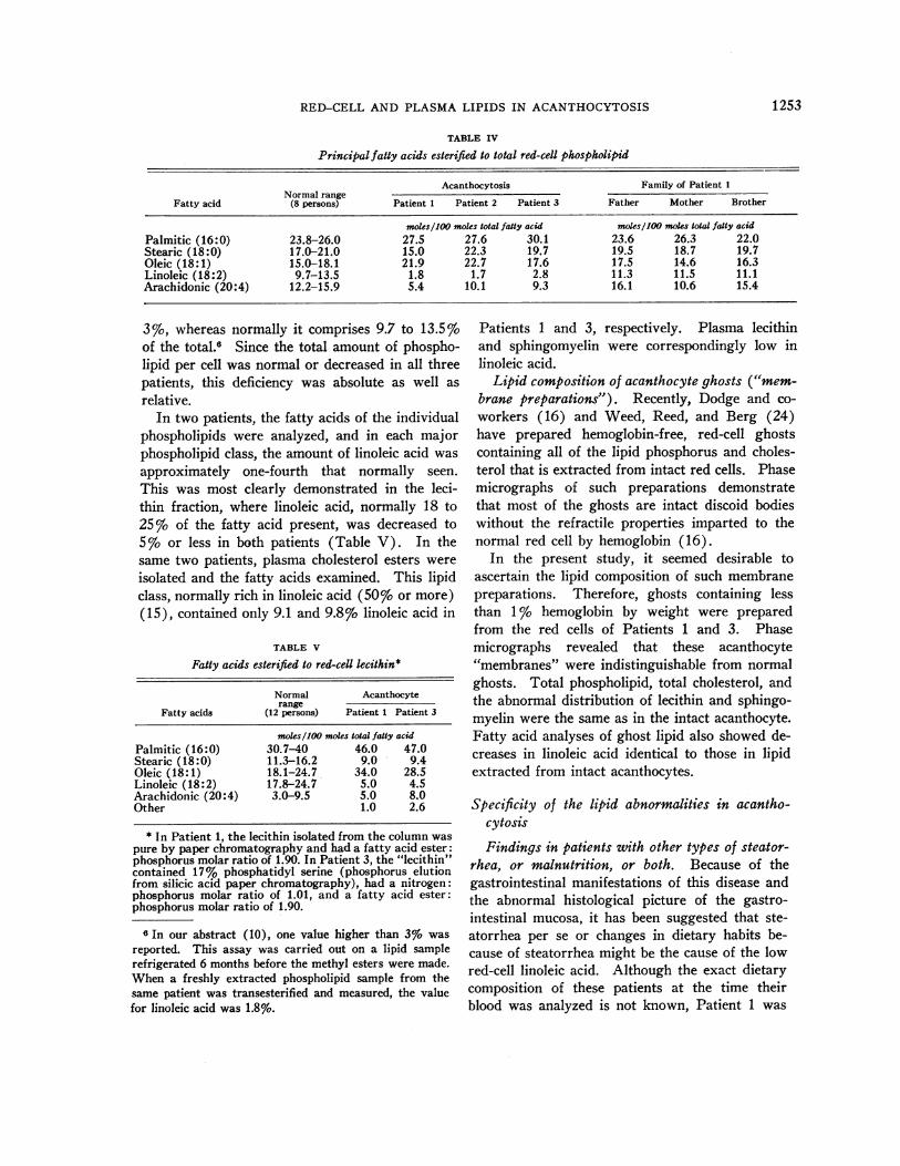
TABLE IV
Principalfatty acids esterified to total red-cell phospholipid
Acanthocytosis Family of Patient 1Normal range
Fatty acid (8 persons) Patient 1 Patient 2 Patient 3 Father Mother Brother
mokls/100 moles total fatty acid moles/100 moles total fatty acidPalmitic (16:0) 23.8-26.0 27.5 27.6 30.1 23.6 26.3 22.0Stearic (18:0) 17.0-21.0 15.0 22.3 19.7 19.5 18.7 19.7Oleic (18:1) 15.0-18.1 21.9 22.7 17.6 17.5 14.6 16.3Linoleic (18:2) 9.7-13.5 1.8 1.7 2.8 11.3 11.5 11.1Arachidonic (20:4) 12.2-15.9 5.4 10.1 9.3 16.1 10.6 15.4
3%, whereas normally it comprises 9.7 to 13.5%of the total.8 Since the total amount of phospho-lipid per cell was normal or decreased in all threepatients, this deficiency was absolute as well asrelative.
In two patients, the fatty acids of the individualphospholipids were analyzed, and in each majorphospholipid class, the amount of linoleic acid wasapproximately one-fourth that normally seen.This was most clearly demonstrated in the leci-thin fraction, where linoleic acid, normally 18 to25%o of the fatty acid present, was decreased to5%o or less in both patients (Table V). In thesame two patients, plasma cholesterol esters wereisolated and the fatty acids examined. This lipidclass, normally rich in linoleic acid (50%o or more)(15), contained only 9.1 and 9.8%0 linoleic acid in
TABLE V
Fatty acids esterified to red-cell lecithin*
Normal Acanthocyterange
Fatty acids (12 persons) Patient 1 Patient 3
mokls/100 moles total fatty acidPalmitic (16:0) 30.7-40 46.0 47.0Stearic (18: 0) 11.3-16.2 9.0 9.4Oleic (18:1) 18.1-24.7 34.0 28.5Linoleic (18:2) 17.8-24.7 5.0 4.5Arachidonic (20:4) 3.0-9.5 5.0 8.0Other 1.0 2.6
* In Patient 1, the lecithin isolated from the column waspure by paper chromatography and had a fatty acid ester:phosphorus molar ratio of 1.90. In Patient 3, the "lecithin"contained 17% phosphatidyl serine (phosphorus elutionfrom silicic acid paper chromatography), had a nitrogen:phosphorus molar ratio of 1.01, and a fatty acid ester:phosphorus molar ratio of 1.90.
6 In our abstract (10), one value higher than 3%o wasreported. This assay was carried out on a lipid samplerefrigerated 6 months before the methyl esters were made.When a freshly extracted phospholipid sample from thesame patient was transesterified and measured, the valuefor linoleic acid was 1.8%.
Patients 1 and 3, respectively. Plasma lecithinand sphingomyelin were correspondingly low inlinoleic acid.
Lipid composition of acanthocyte ghosts ("mem-brane preparations"). Recently, Dodge and co-workers (16) and Weed, Reed, and Berg (24)have prepared hemoglobin-free, red-cell ghostscontaining all of the lipid phosphorus and choles-terol that is extracted from intact red cells. Phasemicrographs of such preparations demonstratethat most of the ghosts are intact discoid bodieswithout the refractile properties imparted to thenormal red cell by hemoglobin (16).
In the present study, it seemed desirable toascertain the lipid composition of such membranepreparations. Therefore, ghosts containing lessthan 1 %o hemoglobin by weight were preparedfrom the red cells of Patients 1 and 3. Phasemicrographs revealed that these acanthocyte"membranes" were indistinguishable from normalghosts. Total phospholipid, total cholesterol, andthe abnormal distribution of lecithin and sphingo-myelin were the same as in the intact acanthocyte.Fatty acid analyses of ghost lipid also showed de-creases in linoleic acid identical to those in lipidextracted from intact acanthocytes.
Specificity of the lipid abnormalities in acantho-cytosis
Findings in patients with other types of steator-rhea, or malnutrition, or both. Because of thegastrointestinal manifestations of this disease andthe abnormal histological picture of the gastro-intestinal mucosa, it has been suggested that ste-atorrhea per se or changes in dietary habits be-cause of steatorrhea might be the cause of the lowred-cell linoleic acid. Although the exact dietarycomposition of these patients at the time theirblood was analyzed is not known, Patient 1 was
1253

PETER WAYS, CLAUDEF. REED, AND DONALDJ. HANAHAN
TABLE VI
Red-cell lipids in patients with malabsorption, or malnutrition, or both
Linoleic-Lipid Phospholipid distribution* acidphos- Choles- -- esterified to
Patient Age Diagnosis phorus terol PE PS Lec. SP Other phospholipid
years mg X 10-'0/cell %of total phospholipid moles/100 moles
totalfatlyacid
1 10 Cystic fibrosis .123 1.25 28.3 23.8 9.82 70 Celiac sprue .156 2.17 35.7 34.3 25.1 5.1 7.73 40 Steatorrhea, sec- .123 30.8 21.2 25.7 15.3 7.2 5.8
ondary to smallbowel resection
4 60 Folic acid deficiency, .110 1.40 21.9 18.4 32.4 15.1 12.4 5.0malnutrition
5 50 Chronic relapsing .155 1.73 22.1 16.4 28.1 23.1 10.3 6.0pancreatitis withsteatorrhea andmalnutrition
6 12 Steatorrhea and .123 1.22 23.2 16.5 31.3 25.2 4.0growth failure, un-known etiology
* Percentage of total lipid phosphorus recovered; PE = phosphatidyl ethanolamine, PS = phosphatidyl serine,Lec. = lecithin, and SP = sphigomyelin.
only eating 19 g of fat daily 1 year after thesestudies. Similarly, Patient 3 is described asavoiding fat (3). To evaluate the possibility thatthe red-cell and plasma lipid abnormalities mightbe nonspecific, red-cell lipids from other patientswith malnutrition, or steatorrhea of other etiology,or both, were analyzed (Table VI). The per-centage of red-cell linoleic acid was reduced inseveral instances, but in no case was this changeof the magnitude seen in the patients with acan-thocytosis. In contrast to acanthocytosis, thelecithin: sphingomyelin ratio was not reversed inthe red cells of any of these patients. In fact, intwo of the five studied (Patients 2 and 4), therewas actually an increase in the amount of lecithinnormally found, and in one of these, a correspond-ing decrease in the amount of sphingomyelin.
Lipid analysis of red-cell populations high inreticulocytes. One of the difficulties inherent instudies of red-cell metabolism is the necessity ofdealing with cells heterogeneous with respect toage. When average cellular age is shortened byaccelerated in vivo destruction, as it appears tobe in some cases of acanthocytosis (3, 4), it couldbe argued that any changes found in red-cell lipidswere merely a function of decreased mean cellularage.
In five instances, the erythrocyte phospholipiddistribution of patients with reticulocytosis was de-termined by paper chromatography (Table VII).There was no significant difference between thisgroup and normal mature cells in the percentagedistribution of the major erythrocyte phosphatides.The fatty acid distribution of the red-cell phos-phatides was determined in two patients of thisgroup, and it indicates that young cells are lowerin linoleic acid than a cell population of normalage distribution.
Family studies
Since previous investigations have suggestedthat acanthocytosis is an inherited disease (1, 2,4, 6), the father, mother, and brother of Patient1 in the present investigation were studied. Red-cell morphology, total lipid, phospholipid distri-b)ution, and plasmalogen levels were normal inall three family members (Table II), as was thefatty acid composition of total red-cell phospho-lipid (Table IV).
Plasma lipid and lipoprotein values for the fam-ily members are shown in Table III. The par-ents had normal total lipid values, total cholesterol,lipid phosphorus, and phospholipid distribution.Analysis of serum fractions separated by ultra-
1254

RED-CELL AND PLASMALIPIDS IN ACANTHOCYTOSIS
centrifugation gave results comparable to normalsera examined simultaneously and to publishednormal data (18, 25). In addition, immuno-,starch gel, and cellulose acetate electrophoreses alldemonstrated normally staining bands of low-density lipoprotein in samples from the father andmother. The brother, however, had low choles-terol and slightly low lipid phosphorus on one
examination. A normal percentage of this lipidwas found in the density layer of less than 1.063,and as with the parents, none of the electrophoreticstudies was abnormal. Repeated analyses of thebrother's plasma on two subsequent occasions gave
values of 6.55 and 6.90 mg per 100 ml for lipidphosphorus, and 170 and 181 mg per 100 ml forcholesterol.
DISCUSSION
The present study establishes that red cells inthe hereditary syndrome of acanthocytosis con-
tain increased amounts of sphingomyelin, are de-ficient in lecithin and plasmalogens, and containvery little esterified linoleic acid. These same
deviations in phospholipid composition and linoleicacid content were found in hemoglobin-free ghosts,indicating that the lipid composition of the mem-
brane itself is abnormal.The significance of these observations for acan-
thocytosis depends largely on their peculiarity to
this syndrome. Linoleic acid, which probably can-
not be synthesized by mammals, can be reducedexperimentally in animal erythrocytes to levels as
low as those found in the acanthocyte. Thus, in
rats (26) and monkeys (27-29), decreased per-
centages (absolute levels were not measured) ofred-cell linoleate follow prolonged dietary restric-tion of linoleic acid. Similarly, our analyses ofred cells from other subjects with steatorrhea, or
malnutrition, or both, generally showed reductionof red-cell linoleate. In these, however, the de-creases were not to the extent observed in acan-
thocytosis. There is another interesting differencebetween the deficient animals and the patientswith malabsorption (whether due to acanthocyto-sis or not). In the experimentally deficient ani-mals, there is marked increase of a red-cell fattyacid with the chromatographic retention character-istics of eicosatrienoic acid (27-29). This in-crease is now considered an integral feature ofessential fatty acid deficiency, but it was not seen
in either the patients with acanthocytosis or thosewith malabsorption of other etiology.
In contrast to the low levels of linoleate, theabnormal phospholipid distribution found in acan-
thocytosis has not, to our knowledge, been de-scribed previously in human red cells. Althoughphospholipid studies were not performed in theanimal work discussed above, analyses of red cellsfrom patients with other types of steatorrhea in-dicate that an increase in red-cell sphingomyelinand a decrease in lecithin are not functions of mal-absorption or fat deprivation per se. This evi-dence suggests that a defect in linoleate absorptionor metabolism is not the primary defect in acan-
thocytosis, but that the abnormalities in phospha-tide distribution may be specific and consequentlyof more fundamental importance.
TABLE VII
Lipid analysis of young red cells
Phospholipid distributionEsterified
Reticulo- Sphingo- linoleicPatient Diagnosis cytes Lecithin myelin Other acid
%of total %of total moles/cells 100 moles
total fattyacid
1 Myeloproliferative disease with 10 32.2 25.8 42.0 6.0gastrointenstinal bleeding
2 Treated folic acid deficiency 22 31.1 27.0 42.3 5.13 Hereditary spherocytosis (pre- 16 32.0 21.0 47.0
splenectomy)4 Acquired hemolytic anemia 23 29.0 20.0 51.0
(Coombs-positive)5 Treated pernicious anemia 20 27.0 19.0 54.0
1255

PETERWAYS, CLAUDEF. REED, AND DONALDJ. HANAHAN
Erythrocyte plasmalogens, which were also de-creased in the acanthocyte, were determined inonly two of the other types of malabsorption, andon a per cell basis were normal. Since in normalred cells 8 to 10% of total plasmalogens are inthe lecithin fraction (19), part of their decreasein acanthocytes may be related to the deficiencyin red-cell lecithin. It can, however, be calcu-lated that the observed decrease is greater thanthat expected even if all of the lecithin plasmalo-gens were absent. Further studies are required toelucidate the association between low plasmalo-gens and the phospholipid abnormality.
Another factor influencing the linoleic acid con-tent of red cells is their average age. Reticulocytecounts, bone marrow examinations, and chromiumsurvival curves were available for two of our pa-tients. They indicated that the acanthocyte prob-ably does not survive normally in vivo, and simi-lar findings have been reported by others (3-5).Since the average age of red cells is known to in-fluence their fatty acid composition (30), reticulo-cyte-rich blood from several persons was exam-ined by the same techniques used to analyze theacanthocytes. In these subjects the reticulocytecount was two to six times higher than in any ofthe patients with acanthocytosis. Although a de-crease in linoleic acid content was observed, itdid not reach the low values found in acantho-cytosis. Therefore, the linoleic acid level observedin Patients 1 and 3 is probably lower than wouldbe expected on the basis of cellular age alone, butmay be consistent with decreased mean cellularage in a patient also on low-fat intake. The reticu-locyte studies, however, again support the speci-ficity of the phospholipid abnormality, since thelecithin and sphingomyelin distribution of red-cellpopulations high in reticulocytes is the same as inthe mature cell.
In any cell bathed continuously with plasma, itis reasonable to suppose that the original mem-
brane lipid composition might be susceptible toalteration by the environment. An exchange be-tween plasma and red-cell lipid phosphorus equalto 10% of lipid phosphorus in the cell daily hasnow been documented (31), and previous workershave shown rapid exchange of plasma and red-cell cholesterol both in vivo and in vitro (32, 33).Since total plasma lipid in acanthocytosis is un-
usually low, and plasma cholesterol, lecithin, andsphingomyelin are deficient in absolute amountand altered in distribution, it is conceivable thatthe replenishment of membrane lipid in this dis-ease could be seriously impaired, with resultantchanges in red-cell phospholipids. On the otherhand, defective membrane synthesis in the mar-row, or the combination of defective synthesis andexchange, might produce the same defects. Noneof the present data indicate which possibility ismost likely. The morphology of marrow cells inthis disease, however, has invariably been normal(2-4). Also, DiGeorge, Mabry, and Auerbachhave reported normalization of cell morphologyafter 4 to 6 weeks of Lipomul therapy (12). Far-quhar has noted immediate restoration of theacanthocyte to normal red-cell shape in vitro afterthe addition of Lipomul (34), and Switzer hasshown that Tween 80 is the component of Lipo-mul which produces this change (35). Thesefindings are compatible with, although they arenot direct evidence for, an important environ-mental contribution to the abnormalities observed.The phospholipid composition of young red cellsand other tissues is now under study in an effortto clarify the relative importance of environmentand de novo synthesis in the abnormalities ofacanthocyte lipid.
The plasma lipid abnormalities in acanthocytosisdeserve further comment. Both this study and thework of Phillips (11) show that the very low lev-els of plasma cholesterol and total phospholipidare accompanied by an increase in the percentageof sphingomyelin and a corresponding decrease inlecithin-a change qualitatively identical to thatin the acanthocyte. Phillips (25), however, in anearlier study and others (36, 37) have shown thatin normal blood the lipoproteins of density greaterthan 1.063 contain relatively more lecithin thanthose of density less than 1.063. Thus, the rela-tive increase in plasma sphingomyelin found inacanthocytosis is opposite to what might havebeen expected in a condition where the low-density plasma lipoproteins are decreased or ab-sent. Hence, the high-density lipoproteins inacanthocytosis are not only low in absolute amount(see Results), but qualitatively abnormal as well-a finding which suggests that the phospholipidabnormalities may play a more basic role in the
1256

RED-CELL AND PLASMALIPIDS IN ACANTHOCYTOSIS
pathogenesis of the disease than previously sup-posed.
These and other studies (6, 11) have disclosedseveral chemical abnormalities in the red cells andplasma of afflicted persons that theoretically mightprovide genetic markers for detecting heterozygouscarriers of the disease. Also, if any one of thedefects (abnormal distribution of phospholipids,low linoleic acid, decreased low-density lipopro-teins, or low red-cell plasmalogens) were presentin the heterozygote to the exclusion of the others,this could provide an important clue to the basicbiochemical defect in acanthocytosis. While Saltand his colleagues were able to demonstrate half-normal concentrations of beta lipoprotein in theparents of their patient (6), neither we nor Rey(4) has been able to confirm these findings. Like-wise, our plasma lipid assays and red-cell analyseshave not demonstrated any qualitative or quanti-tative abnormality that would seem indicative ofa heterozygous defect in either of the parents.The discrepancy between Salt's data and our ownmay indicate that this is an abnormality of vari-able penetrance, or that more than one type ofacanthocytosis exists.
The older view of the red-cell membrane as abimolecular leaflet of lipid in combination withprotein, as originally espoused by Ponder andothers (38), has gained new credence from Rob-inson's concept of the unit membrane evolvedthrough electron microscopic study of cell struc-ture (39). In this synthesis, protein, lipid, andcarbohydrate all play a major role in the struc-ture of membranes. Potentially, abnormalities inany of these entities might be responsible fordefects of red-cell structure or function. Thepresent studies have demonstrated defects of lipidcomposition in erythrocytes that are both func-tionally and morphologically abnormal. Althoughit remains to be established that this is a cause-and-effect relationship, the prospect is intriguing.
Finally, it is tempting to speculate that the lipidand lipoprotein abnormalities of acanthocyte mem-branes may reflect changes in lipid compositionof other cell membranes and are, consequently,related to the other clinical manifestations, par-ticularly the neurological disease. The morpho-logical abnormality of the red cells in acanthocy-tosis provided impetus for investigation of their
lipid composition. The finding that red-cell lipidsare abnormal in one disease with overt and even-tually disabling neurological manifestations shouldprovide an incentive to the careful study of thered-cell membrane in other neurological syn-dromes of obscure etiology.
SUMMARY
The red-cell and plasma lipids from three pa-tients with acanthocytosis have been studied anddefinite abnormalities found. 1) In the presenceof normal or reduced values for total lipid, lipidphosphorus, and cholesterol, the distribution ofred-cell or ghost phospholipids is altered, withan increase in sphingomyelin and a decrease inlecithin. Sphingomyelin was the predominantphospholipid in all three sets of red cells studied.2) The plasma phospholipids were reduced intotal amount to less than 20% of normal valuesand also manifested significant percentage in-creases in sphingomyelin, with correspondinglyless lecithin. 3) The esterified linoleic acid con-tent of red-cell total phospholipid, individual red-cell phospholipids, and various plasma lipid frac-tions was decreased to 25 %or less of the amountnormally found. 4) In patients with steatorrheaof other etiology, red-cell linoleic acid was de-creased, but not to the extent noted in acantho-cytosis, and this change was never associated witha reversal in the amounts of lecithin and sphingo-myelin. 5) Similarly, analysis of reticulocyte-richblood revealed lower levels of linoleic acid, butnormal distribution of phospholipids. 6) Studiesof red-cell and plasma lipids and plasma lipopro-teins in the parents and a sibling of one patientfailed to reveal any abnormalities. 7) These re-sults suggest that the alterations in phospholipiddistribution seen in this syndrome may be spe-cific, but that the low levels of linoleic acid couldbe caused by decreases in available exogenouslinoleate, or a younger than normal populationof red cells, or both.
ACKNOWLEDGMENTS
The authors are indebted to Dr. James T. Dodge forperforming one set of ghost analyses; to Dr. John Jonesfor several of the serum lipoprotein assays; and to Drs.Eloise Giblet and Alex Kaplan for the immunoelectro-phoretic and electrophoretic determinations. Drs. Jones,
1257

PETER WAYS, CLAUDEF. REED, AND DONALDJ. HANAHAN
Clement E. Finch, and Ernest Simon offered numeroushelpful suggestions during the preparation of the manu-script. Finally, we thank Drs. Arno Motulsky and CyrusE. Rubin, who aroused our curiosity in the syndrome ofacanthocytosis, and Drs. Angelo DiGeorge, Charles Lowe,Arthur McElfresh, and Manuel Mier for making ma-terial from their patients available to us.
APPENDIX
Report of Patient 1
Growth and development. This white male child wasborn in 1954, the second son of Jewish parents. A promi-nent abdomen and rather thin extremities were noted atbirth. During the first two months of life, he requiredhospitalization for vomiting, which subsided after numer-ous qualitative manipulations in his diet. He grew slowlyand was significantly delayed in sitting, standing, andwalking. At 31 years of age, his head circumference was2.5 SD less than normal, and his length and weight were1.5 SD less than normal. A skeletal survey at that timereported no retardation of bone age, but subsequently thechild has remained short and thin for his age. Now 8years old, he is in the third percentile for both heightand weight.
Gastrointestinal disease. At birth the boy began tohave frequent, loose, pasty, foul-smelling bowel move-ments, projectile emesis, and a protuberant abdomen.During his first hospitalization (at 2 months), he im-proved on a diet of skim milk and molasses. Because of"malnutrition," however, he was hospitalized at 2' yearsof age at Babies' Hospital in New York under the careof Dr. James A. Wolff. During this hospitalization,abnormal red cells were first noted, and the diagnosis ofacanthocytosis with "celiac-like syndrome" was made byDr. Wolff. On a "regular diet," the diarrhea subsided,to return only sporadically. Evaluation at the BuffaloChildren's Hospital about a year later (at 3!. years) re-vealed a flat vitamin A tolerance curve and normal glucosetolerance. At age 6, a small-bowel biopsy (Dr. CharlesLowe) revealed vacuolization of intestinal absorptive cellsidentical on hematoxylin-eosin-stained sections to thatreported by Salt and co-workers shortly thereafter (6).A superficial dietary history at the time of initial red-celllipid studies suggested that he was ingesting "normal"amounts of fat. Subsequently, more accurate calcula-tions indicate that he has been taking only 10 to 15% ofcalories as fat. He continues to have occasional epi-sodes of watery diarrhea lasting 1 to 2 days, occurringabout once a month. Upper gastrointestinal and small-bowel X rays in 1962 revealed no specific abnormalities.
Neurological disease. When he was hospitalized at 2Ayears, no "neurological nor ophthalmological abnormal-ity" was found. One year later at Buffalo Children'sHospital, absent deep tendon reflexes in the lower ex-
tremities were noted, with minimal reflexes, if any, in theupper extremities. Definite ataxia was not observed.Spinal fluid and electroencephalogram were normal. Sub-
sequently, according to the parents, muscular coordinationhas been poor. In 1961, ptosis and strabismus of the lefteye were noted, and they have persisted. In retrospect,the mother believes he may have had a "squint" at age 2that subsequently disappeared. Since 1960, mild fecalstress incontinence and an inordinate ability to retainurine have been noted. Neurological examination inJune, 1962, revealed an ataxic gait, decreased muscletone in all extremities, complete absence of deep tendonreflexes, and a questionable left extensor plantar response.Vibratory sensation was absent at and below the iliaccrest and decreased in the elbows and wrists. Positionsense and deep pain perception was decreased bilaterally,and the heel-to-knee maneuver was abnormal bilaterally.Sensations of pin prick and light touch were intactthroughout, and there were no trophic changes of theskin. An alternating exotropia was present without in-equality in pupillary size or abnormality of pupillaryresponse. Vision was equal in both eyes, and no fielddefect could be demonstrated. Retinoscopic examinationwas normal.
Hematological disease. Since the abnormal red cellswere first identified at age 21, they have been seen repeat-edly. Peripheral white cells, platelets, and on two occa-sions, all cellular elements of the marrow have been mor-phologically normal. In June, 1957, hematocrit andreticulocyte count were normal for the patient's age, butin April, 1958, reticulocytes were 2.2%o despite normalhematocrit. Chromium tj at that time was 22 days (nor-mal, 26 to 27 days).7 The red-cell and plasma lipids werefirst analyzed in May, 1960, with the results reported inthis publication. In July and August, 1962, hematocritswere 34 to 35%o. Reticulocyte counts were 4 to 5%o, andthe chromium tt was again slightly shortened.
ADDENDUM
Farquhar and Ahrens (40) have now reported a levelof 3% linoleic acid in the phospholipids of another pa-tient with acanthocytosis. Watson (41) has shown thatred-cell lipids of essential fatty acid-deficient rats con-tain very little linoleic acid, but are evidently normal inphospholipid distribution.
REFERENCES
1. Bassen, F. A., and A. L. Kornzweig. Malformationof the erythrocytes in a case of atypical ret:nitispigmentosa. Blood 1950, 5, 381.
2. Singer, K., B. Fisher, and M. Perlstein. Acanthro-cytosis: a genetic erythrocytic malformation.Blood 1952, 7, 577.
3. Mier, M., S. 0. Schwartz, and B. Boshes. Acan-throcytosis, pigmentary degeneration of the retinaand ataxic neuropathy: a generally determined
7 The results of these studies were kindly furnished byDr. Clare Shumway, Buffalo, N. Y.
1258

RED-CELL AND PLASMALIPIDS IN ACANTHOCYTOSIS
syndrome with associated metabolic disorder. Blood1960, 16, 1586.
4. Rey, J. L'absence congenitale de beta-lipjprotdines.Paris, R. Foulon, 1961.
5. Druez, G. Un nouveau cas d'acanthocytose. Dys-morphie erythrocytaire congenitale avec retinite,troubles nerveux et stigmates degeneratifs. Rev.Hemat. 1959, 14, 3.
6. Salt, H. B., 0. H. Wolff, J. K. Lloyd, A. S. Fos-brooke, A. H. Cameron, and D. V. Hubble. Onhaving no beta-lipoprotein: a syndrome comprisinga-beta-lipoproteinaemia, acanthocytosis, and steator-rhoea. Lancet 1960, 2, 325.
7. Mabry, C. C., A. M. DiGeorge, and V. H. Auerbach.Studies concerning the defect in a patient withacanthrocytosis (abstract). Clin. Res. 1960, 8,371.
8. Jampel, R. S., and H. F. Falls. Atypical retinitispigmentosa, acanthrocytosis, and heredodegenera-tive neuromuscular disease. Arch. Ophthal. 1958,59, 818.
9. Friedman, I. S., H. Cohn, M. Zymaris, and M. G.Goldner. Hypocholesteremia in idiopathic steator-rhea. Arch. intern. Med. 1960, 105, 112.
10. Ways, P., C. F. Reed, and D. J. Hanahan. Abnor-malities of erythrocyte and plasma lipids in acan-thocytosis (abstract). J. dlin. Invest. 1961, 40,1088.
11. Phillips, G. B. Quantitative chromatographic analy-sis of plasma and red cell lipids in patients withacanthocytosis. J. Lab. clin. Med. 1962, 59, 357.
12. DiGeorge, A. M., C. C. Mabry, and V. H. Auerbach.A specific disorder of lipid transport (acanthro-cytosis): treatment with intravenous lipids (ab-stract). Amer. J. Dis. Child. 1961, 102, 580.
13. Reed, C. F., S. N. Swisher, G. V. Marinetti, andE. G. Eden. Studies of the lipids of the erythro-cyte. I. Quantitative analysis of the lipids ofnormal human red blood cells. J. Lab. clin. Med.1960, 56, 281.
14. Marinetti, G. V., J. Erbland, and E. Stotz. Phospha-tides of pig heart cell fractions. J. biol Chem.1958, 233, 562.
15. Hanahan, D. J., R. M. Watts, and D. Pappajohn.Some chemical characteristics of the lipids of hu-man and bovine erythrocytes and plasma. J. LipidRes. 1960, 1, 421.
16. Dodge, J. T., C. Mitchell, and D. J. Hanahan. Thepreparation and chemical characteristics of hemo-globin-free ghosts of human erythrocytes. Arch.Biochem. 1963, 100, 119.
17. Norton, W. T. Potentiometric iodometric determina-tion of plasmalogen. Biochim. biophys Acta(Amst.) 1960, 38, 340.
18. Havel, R. J., H. A. Eder, and J. H. Bragdon. Thedistribution and chemical composition of ultracen-trifugally separated lipoproteins in human serum.J. clin. Invest. 1955, 34, 1345.
19. Farquhar, J. W. Human erythrocyte phosphoglycer-ides. I. Quantification of plasmalogens, fatty acidsand fatty aldehydes. Biochim. biophys. Acta(Amst.) 1962, 60, 80.
20. Phillips, G. B., and N. S. Roome. Phospholipids ofhuman red blood cells. Proc. Soc. exp. Biol.(N. Y.) 1959, 100, 489.
21. De Gier, J., and L. L. M. Van Deenen. Some lipidcharacteristics of red cell membranes of variousanimal species. Biochim. biophys. Acta (Amst.)1961, 49, 286.
22. Phillips, G. B. The isolation and quantitation of theprincipal phospholipid components of human se-rum using chromatography on silicic acid. Bio-chim. biophys. Acta (Amst.) 1958, 29, 594.
23. Nye, W. H. R., C. Waterhouse, and G. V. Marinetti.The phosphatides of human plasma. I. Normalvalues determined by paper and column chroma-tography. J. dlin. Invest. 1961, 40, 1194.
24. Weed, R. I., C. F. Reed, and G. Berg. Is hemo-globin an essential structural component of humanerythrocyte membranes? J. dlin. Invest. 1963, 42,581.
25. Phillips, G. B. The phospholipid composition ofhuman serum lipoprotein fractions separated byultracentrifugation. J. clin. Invest. 1959, 38, 489.
26. Caster, W. O., and R. T. Holman. Statistical studyof the relationship between dietary linoleate andthe fatty acids of heart and blood lipids. J. Nutr.1961, 73, 337.
27. Portman, 0. W., S. B. Andrus, D. Pollard, and D.Bruno. Effects of long-term feeding of fat-freediets to Cebus monkeys. J. Nutr. 1961, 74, 429.
28. Fitch, C. D., J. S. Dinning, L. A. Witting, and M. K.Horwitt. Influence of dietary fat on the fatty acidcomposition of monkey erythrocytes. J. Nutr. 1961,75, 409.
29. Greenberg, L. D., and H. D. Moon. Alterations inthe blood fatty acids in single and combined defi-ciencies of essential fatty acids and vitamin B, inmonkeys. Arch. Biochem. 1961, 94, 405.
30. Munn, J. I. Studies of lipids in human red cells.Brit. J. Haemat. 1958, 4, 344.
31. Reed, C. F. Studies of in vivo and in vitro exchangeof erythrocyte and plasma phospholipids (ab-stract). J. clin. Invest. 1959, 38, 1032.
32. Eckles, N. E., C. B. Taylor, D. J. Campbell, andR. G. Gould. The origin of plasma cholesteroland the rates of equilibration of liver, plasma, anderythrocyte cholesterol. J. Lab. clin. Med. 1955,46, 359.
33. Hagerman, J. S., and R. G. Gould. The in vitrointerchange of cholesterol between plasma and redcells. Proc. Soc. exp. Biol. (N. Y.) 1951, 78, 329.
34. Farquhar, J. Personal communication.35. Switzer, S., and H. A. Eder. Interconversion of
acanthocytes and normal erythrocytes with deter-gents (abstract). J. clin. Invest. 1962, 41, 1404.
1259

PETER WAYS, CLAUDEF. REED, AND DONALDJ. HANAHAN
36. Steele, J. M., and H. J. Kayden. The nature of the
phospholipids in human serum and atheromatousvessels. Trans. Ass. Amer. Phycns 1955, 68, 249.
37. Nelson, G. J., and N. K. Freeman. The phospholipid
and phospholipid fatty acid composition of humanserum lipoprotein fractions. J. biol. Chem. 1960,235, 578.
38. Ponder, E. Hemolysis and Related Phenomena. New
York, Grune & Stratton, 1948.
39. Robinson, J. D. The membrane of the living cell.Scientific American 1962 (April), 206, 65.
40. Farquhar, J. W., and E. H. Ahrens, Jr. Effects ofdietary fats on human erythrocyte fatty acid pat-terns. J. clin. Invest. 1963, 42, 675.
41. Watson, W. C. The morphology and lipid composi-tion of the erythrocytes in normal and essential-fatty-acid-deficient rats. Brit. J. Haemat. 1963, 9,32.
1260


![Determination of Algal Cell Lipids Using Nile Red - BioTek · Determination of Algal Cell Lipids Using Nile Red ... cedure that use Nile blue are employed [3]. Puri-fied Nile red](https://static.fdocuments.in/doc/165x107/5af310607f8b9a154c8c5bee/determination-of-algal-cell-lipids-using-nile-red-biotek-of-algal-cell-lipids.jpg)