Recurrent Inverted Papilloma: Diagnosis with ...
7
1445 MS AJNR Am J Neuroradiol 20:1445–1451, September 1999 Recurrent Inverted Papilloma: Diagnosis with Pharmacokinetic Dynamic Gadolinium-Enhanced MR Imaging Ping H. Lai, Chien F. Yang, Huay B. Pan, Ming T. Wu, Sau T. Chu, Luo P. Ger, Wen C. Huang, Cheng C. Hsu, and Chung N. Lee BACKGROUND AND PURPOSE: Dynamic gadolinium-enhanced MR imaging has been used successfully to identify post-treatment recurrence or postoperative changes in rectal and cer- vical carcinoma. Our purpose was to evaluate the usefulness of dynamic gadolinium-enhanced MR imaging for distinguishing recurrent inverted papilloma (IP) from postoperative changes. METHODS: Fifteen patients with 20 pathologically proved lesions (recurrent IP, 12; fibrosis or granulation tissue, eight) were enrolled in the study. Three observers, blinded to pathologic results, independently evaluated conventional MR images, including T1-weighted (unenhanced and postcontrast), proton-density–weighted, and T2-weighted spin-echo images. Results then were determined by consensus. Dynamic images were obtained using fast spin-echo sequences at 5, 30, 60, 90, 120, 150, 180, and 300 seconds after the injection of gadolinium-diethylene- triamine penta-acetic acid. Time-signal intensity curves of suspected lesions were analyzed by a pharmacokinetic model. The calculated amplitude and tissue distribution time were used to characterize tissue, and their values were displayed as a color-coded overlay. RESULTS: T2-weighted images yielded a sensitivity of 67%, a specificity of 75%, and an accuracy of 70% in the diagnosis of recurrent IP. Contrast-enhanced T1-weighted images yield- ed a sensitivity of 75%, a specificity of 50%, and an accuracy of 65%. Pharmacokinetic analysis showed that recurrent IP had faster (distribution time, 41 versus 88 seconds) and higher (am- plitude, 2.4 versus 1.2 arbitrary units) enhancement than did fibrosis or granulation tissue. A cut-off of 65 seconds for distribution time and 1.6 units for amplitude yielded a sensitivity of 100% and a specificity of 100% for diagnosing recurrent IP. CONCLUSION: Dynamic MR imaging can differentiate accurately recurrent IP from post- operative changes and seems to be a valuable diagnostic tool. Inverted papilloma (IP) is a common neoplasm of the nasal cavity. This benign epithelial neoplasm usually arises within the nasal vault and, less com- Received, October 26, 1998; accepted after revision, April 26, 1999. Supported by grants from NSC 87-2314B-075B-003 and VGHKS 87–46. Presented at the annual meeting of the American Society of Neuroradiology, Philadelphia, May 1998. From the Departments of Radiology (P.H.L., C.F.Y., H.B.P., M.T.W.), Otolaryngology (S.T.C.), and Medical Education and Research and Biostatistics (L.P.G.), Veterans General Hospi- tal-Kaohsiung, National Yang-Ming College, Taiwan, ROC; the Department of Hospital Management (W.C.H.), Chia-Nan College of Pharmacy and Science, Tainan, Taiwan, ROC; and the Institute of Computer and Information Engineering (C.C.H., C.N.L.), National Sun Yat-Sen University, Kao- hsiung, Taiwan, ROC. Address reprint requests to Ping H. Lai, MD, Department of Radiology, Veterans General Hospital-Kaohsiung, 386 Ta- Chung First Rd, Kaohsiung, Taiwan 813. q American Society of Neuroradiology monly, in the paranasal sinuses. Patients typically present with nasal obstruction, epistaxis, or nasal discharge. Surgical intervention usually provides a favorable outcome. There is, however, a high rate of recurrence (15%278%) associated with IP (126). When symptoms recur, a biopsy is neces- sary to confirm the recurrence of IP. Because com- plete en bloc excision is necessary for treatment of a recurrent tumor, a precise preoperative diagnosis is beneficial for surgical planning. Biopsy may be unreliable, however, as sampling errors can occur because of the chronic inflammation surrounding sinonasal neoplasms (124). Furthermore, endo- scopic examination cannot access several critical areas such as the frontal sinus, fovea ethmoidalis, cribriform plate, superior-lateral wall of the sphe- noid sinus, and medial-inferior wall of the orbit (1, 4, 5). MR imaging has been used to evaluate sinonasal IP (7). Yousem et al (7) found that IP lesions usu- ally show intermediate signal intensity on T2-
Transcript of Recurrent Inverted Papilloma: Diagnosis with ...

1445
MS
AJNR Am J Neuroradiol 20:1445–1451, September 1999
Recurrent Inverted Papilloma: Diagnosis withPharmacokinetic Dynamic Gadolinium-Enhanced
MR Imaging
Ping H. Lai, Chien F. Yang, Huay B. Pan, Ming T. Wu, Sau T. Chu, Luo P. Ger, Wen C. Huang, Cheng C. Hsu, andChung N. Lee
BACKGROUND AND PURPOSE: Dynamic gadolinium-enhanced MR imaging has been usedsuccessfully to identify post-treatment recurrence or postoperative changes in rectal and cer-vical carcinoma. Our purpose was to evaluate the usefulness of dynamic gadolinium-enhancedMR imaging for distinguishing recurrent inverted papilloma (IP) from postoperative changes.
METHODS: Fifteen patients with 20 pathologically proved lesions (recurrent IP, 12; fibrosisor granulation tissue, eight) were enrolled in the study. Three observers, blinded to pathologicresults, independently evaluated conventional MR images, including T1-weighted (unenhancedand postcontrast), proton-density–weighted, and T2-weighted spin-echo images. Results thenwere determined by consensus. Dynamic images were obtained using fast spin-echo sequencesat 5, 30, 60, 90, 120, 150, 180, and 300 seconds after the injection of gadolinium-diethylene-triamine penta-acetic acid. Time-signal intensity curves of suspected lesions were analyzed bya pharmacokinetic model. The calculated amplitude and tissue distribution time were used tocharacterize tissue, and their values were displayed as a color-coded overlay.
RESULTS: T2-weighted images yielded a sensitivity of 67%, a specificity of 75%, and anaccuracy of 70% in the diagnosis of recurrent IP. Contrast-enhanced T1-weighted images yield-ed a sensitivity of 75%, a specificity of 50%, and an accuracy of 65%. Pharmacokinetic analysisshowed that recurrent IP had faster (distribution time, 41 versus 88 seconds) and higher (am-plitude, 2.4 versus 1.2 arbitrary units) enhancement than did fibrosis or granulation tissue. Acut-off of 65 seconds for distribution time and 1.6 units for amplitude yielded a sensitivity of100% and a specificity of 100% for diagnosing recurrent IP.
CONCLUSION: Dynamic MR imaging can differentiate accurately recurrent IP from post-operative changes and seems to be a valuable diagnostic tool.
Inverted papilloma (IP) is a common neoplasm ofthe nasal cavity. This benign epithelial neoplasmusually arises within the nasal vault and, less com-
Received, October 26, 1998; accepted after revision, April 26,1999.
Supported by grants from NSC 87-2314B-075B-003 andVGHKS 87–46.
Presented at the annual meeting of the American Society ofNeuroradiology, Philadelphia, May 1998.
From the Departments of Radiology (P.H.L., C.F.Y., H.B.P.,M.T.W.), Otolaryngology (S.T.C.), and Medical Education andResearch and Biostatistics (L.P.G.), Veterans General Hospi-tal-Kaohsiung, National Yang-Ming College, Taiwan, ROC;the Department of Hospital Management (W.C.H.), Chia-NanCollege of Pharmacy and Science, Tainan, Taiwan, ROC; andthe Institute of Computer and Information Engineering(C.C.H., C.N.L.), National Sun Yat-Sen University, Kao-hsiung, Taiwan, ROC.
Address reprint requests to Ping H. Lai, MD, Departmentof Radiology, Veterans General Hospital-Kaohsiung, 386 Ta-Chung First Rd, Kaohsiung, Taiwan 813.
q American Society of Neuroradiology
monly, in the paranasal sinuses. Patients typicallypresent with nasal obstruction, epistaxis, or nasaldischarge. Surgical intervention usually provides afavorable outcome. There is, however, a high rateof recurrence (15%278%) associated with IP(126). When symptoms recur, a biopsy is neces-sary to confirm the recurrence of IP. Because com-plete en bloc excision is necessary for treatment ofa recurrent tumor, a precise preoperative diagnosisis beneficial for surgical planning. Biopsy may beunreliable, however, as sampling errors can occurbecause of the chronic inflammation surroundingsinonasal neoplasms (124). Furthermore, endo-scopic examination cannot access several criticalareas such as the frontal sinus, fovea ethmoidalis,cribriform plate, superior-lateral wall of the sphe-noid sinus, and medial-inferior wall of the orbit (1,4, 5).
MR imaging has been used to evaluate sinonasalIP (7). Yousem et al (7) found that IP lesions usu-ally show intermediate signal intensity on T2-

AJNR: 20, September 19991446 LAI
TABLE 1: Characteristics, imaging results, and final diagnoses of patients with suspected recurrence of inverted papilloma
Patient(no.) Age (y)/Sex Prior Therapy
Period afterPrior Tx
(mo.) DiagnosisSI Related toCSF on T2 T1 1 Gd
1234*
69/M41/M62/M66/M
MMMMMMMMMM
1215156060
RecurrenceRecurrenceRecurrenceRecurrencePostop changes
Isointense (FN)Isointense (FN)Hypointense (TP)Hypointense (TP)Hyperintense (TN)
Marked, homogeneous (FN)Marked, homogeneous (FN)Moderate, inhomogeneous (TP)Mild, inhomogeneous (TP)Marked, homogeneous (TN)
567*
75/M58/M34/M
MMMMFESSMMMM
698
2727
RecurrencePostop changesRecurrenceRecurrencePostop changes
Hypointense (TP)Hypointense (FP)Hypointense (TP)Hypointense (TP)Isointense (TN)
Moderate, inhomogeneous (TP)Moderate, inhomogeneous (FP)Mild, inhomogeneous (TP)Mild, inhomogeneous (TP)Marked, homogeneous (TN)
89*
10*
63/M70/M
65/M
MMMMMMFESSFESS
7161655
RecurrenceRecurrencePostop changesRecurrencePostop changes
Isointense (FN)Isointense (FN)Isointense (TN)Hypointense (TP)Hyperintense (TN)
Mild, inhomogeneous (TP)Marked, homogeneous (FN)Moderate, inhomogeneous (FP)Moderate, inhomogeneous (TP)Marked, homogeneous (TN)
1112131415
58/M63/M45/M60/F52/M
MMMMMMFESSMM
10169
1511
RecurrencePostop changesPostop changesRecurrencePostop changes
Hypointense (TP)Hypointense (FP)Hyperintense (TN)Hypointense (TP)Isointense (TN)
Mild, inhomogeneous (TP)Moderate, inhomogeneous (FP)Marked, homogeneous (TN)Moderate, inhomogeneous (TP)Mild, inhomogeneous (FP)
Note.—Tx, treatment; SI, signal intensity; MM, medial maxillectomy; FN, false negative; TP, true positive; TN, true negative; FP, false positive;FESS, functional endoscopic surgery.
* Patient had two different lesions.
weighted images. Postoperative imaging of IP be-comes more complicated because repair withgranulation and fibrosis occurs after surgery. Nev-ertheless, dynamic gadolinium-enhanced MR im-aging with pharmacokinetic analysis has been usedsuccessfully to distinguish recurrent tumors frompostoperative changes in various diseases (8210).Therefore, in this retrospective study, we tested theaccuracy of gadolinium-enhanced MR imagingwith pharmacokinetic analysis in distinguishing re-current IP from postoperative inflammatorychanges (fibrosis or granulation tissue).
Methods
Patients
Our observations were based on 16 MR imaging examina-tions of 15 patients (14 men and one woman; age range,34270 years) with 20 suspected recurrent IP lesions. One pa-tient had undergone two MR imaging examinations after twooperations, respectively (Table 1). Four patients each had twosuspected lesions. All patients had histories of surgical treat-ment for sinonasal IP, 13 by lateral rhinotomy and medial max-illectomy, and three by functional endoscopic surgery. MRstudies were performed because of the clinical endoscopic orCT findings of soft-tissue lesions in the nasal cavity or para-nasal sinus or both. Biopsy specimens for histologic evalua-tions were obtained by lateral rhinotomy and medial maxillec-tomy (12 patients), functional endoscopic surgery (twopatients), or both separately (one patient). The median intervalbetween surgery and MR examination was 11.5 months (range,5260 months).
Imaging Schemes
All images were obtained with a 1.5-T imager with a quad-rature head coil. Conventional MR studies were performed in
the axial and coronal planes. T1-weighted images were ob-tained with parameters of 600/20/2 (TR/TE/excitations). LongTR images with or without frequency-selective fat saturationwere obtained with 220023000/18236/1 (proton-density[PD]-weighted) and 220023000/802108/1 (T2-weighted).The matrix size was 256 3 192, and the section thickness was5 mm. Postcontrast T1-weighted images with or without fre-quency-selective fat saturation were acquired in the coronaland axial planes after dynamic contrast-enhanced imaging.
Dynamic MR images were obtained using the fast spin-echotechnique and the following parameters: 400/17/1 (TR/effec-tive TE/excitations); echo train length, 4; matrix, 256 3 192;field of view, 24 3 18 cm; and section thickness, 5 mm. Thistechnique yields an imaging acquisition time of 15 secondsand four sections, positioned at the level where the main partof the lesion has been identified on the unenhanced image. Theinjection was performed through a 20-gauge catheter insertedin an antecubital vein before the start of the study. The dy-namic images (0 seconds) were obtained before the bolus in-jection of the contrast agent. Dynamic imaging was repeated5 and 30 seconds after rapid manual bolus injection (2 mL /second) of 0.1 mmol of gadolinium-diethylenetriamine penta-acetic acid/kg of body weight followed by a 10-mL flush ofnormal saline (time zero was the end of the flush). Imagingthen was performed every 30 seconds during the first 180 sec-onds and at 300 seconds. Thus, a total of nine sets of dynamicimages were obtained. Conventional MR and dynamic MR im-ages first were reviewed independently by three radiologistswho were blinded to the histologic diagnosis. The three re-viewers were in agreement on the diagnosis in all cases.
MR Image Analysis
For conventional MR imaging, the MR images were eval-uated for signal intensity and contrast enhancement pattern ac-cording to the methods presented by Yousem et al (7). Beforecontrast enhancement, the signal intensity of the lesions wascompared with that of fat, muscle, and CSF in T1-, PD-, andT2-weighted images. After contrast enhancement, the lesions

AJNR: 20, September 1999 RECURRENT INVERTED PAPILLOMA 1447
FIG 1. Two-dimensional color table. The value of the amplitude(A) is from 0 to 3 and that of the distribution time (Tc) is from 0to 120. The four-corner colors of the color table are yellow, red,blue, and green. The colors of other elements are generated us-ing bilinear interpolation according to their position on the colortable. The color mapping of the ROI is determined on the basisof the value of A and Tc in each pixel. Note that at values above3, A is set to 3; at values above 120, Tc is set to 120.
were assessed for their homogeneity and degree of enhance-ment as compared with the nasal turbinate mucosa. Enhance-ment was classified as marked (enhancing as much as that ofmucosa), moderate (definite enhancement but not as strikingas that of mucosa), or mild (slight increase over baseline signalintensity after the administration of contrast agent, much lessthan that of mucosa). We developed criteria adapted from aprevious report (7) for the diagnosis of IP. IP lesions weredefined as appearing hypointense relative to CSF and hyper-intense relative to muscle on T2-weighted images and as show-ing mild to moderate inhomogeneous enhancement after theadministration of contrast agent. Postoperative changes (ie,granulation or fibrosis) were defined as appearing iso- to hy-perintense relative to CSF on T2-weighted images and asshowing marked enhancement after the administration of con-trast agent (11, 12).
For dynamic MR imaging, to obtain the time-signal intensitycurves, regions of interest (ROIs) were placed within the 20suspected lesions by one reader who had no knowledge of thefinal pathologic diagnosis. The size of the ROIs that was cho-sen in a lesion varied with the size and shape of the suspectedlesion that appeared in the nine sets of images. Once deter-mined, the ROI was consistently used to determine the meansignal intensity in the nine sets of images. The change in thesignal intensity of an ROI placed on the mouth-floor musclewas used for reference. The relative signal increase (RSI) thenwas calculated as Scm (t)/S0, where Scm (t) 5 postcontrast sig-nal at time t, and S0 5 unenhanced signal. We used a three-parameter mathematical model for pharmacokinetic analysis,as described by Brix et al (13) and Greenstein et al (14): RSI(t) 5 1 1 A [1 2 exp (2t / Tc)] 2 Ct, where RSI (t) 5postcontrast signal at time t divided by the unenhanced signal,A 5 amplitude of enhancement, Tc 5 distribution time (timeconstant for arrival of contrast material), and C 5 first-orderwashout rate. The pharmacokinetic parameters A, Tc, and Cwere calculated by means of a nonlinear least-squares fittingprogram written in Matlab software. Because the signal inten-sity and the distribution time of uptake of contrast media aredescribed mainly by A and Tc, we focused our pharmacoki-netic analysis on these two variables for tissue characterizationof recurrent tumors and postoperative changes.
For further processing, the pharmacokinetic imaging datasets were transferred to a workstation. To reduce the numberof images and facilitate visual interpretation, a color-codingtable (Fig 1) was applied that used a reference matrix to over-lay color-coded parameters of both amplitude and distributiontime. Overlaying the color-coded parameters on the conven-tional images allows the functional information regarding thecontrast medium enhancement to be combined with the cor-responding morphologic images. To automate this approach,we developed a software package to process the dynamic im-ages on a pixel-by-pixel basis for each section. The softwareprogram mainly consists of six components: 1) an MR imageformat transformation, 2) a motion correction module, 3) anROI boundary detection module, 4) a quantitative evaluationmodule, 5) a quantitative evaluation of dynamic MR data, and6) a color representation of ROI. The automated data process-ing requires approximately 30 minutes and can be performedin a clinical setting.
Statistics
The MR image readings were analyzed to evaluate the re-lationship between different diagnoses. We used Student’s t test(for equal variances) or Wilcoxon rank sums test (for unequalvariances) to compare the RSI of recurrent neoplasms andpost-treatment inflammatory tissues at different time periods.The dynamic MR imaging data for the lesions were testedretrospectively using Student’s t test and a two-sample Wil-coxon test for the difference in the values of the pharmacoki-netic parameters. After the lesions were classified, their meanpharmacokinetic parameter values of amplitude and distribu-
tion time were displayed in a two-dimensional scatter plot. Thecriteria selected to determine optimal threshold values on thescatter plot were the greatest overall accuracy and low rate offalse-positive results (8). Finally, the sensitivity, specificity,and accuracy of the discriminatory power of each of the cri-teria, the signal intensity on T2-weighted images, the postcon-trast studies, and the dynamic contrast imaging werecalculated.
Results
Conventional MR ImagingClassification of the lesions on the basis of the
signal intensity on T2-weighted images resulted intwo of eight postoperative changes in lesions beingmisclassified as recurrence and four of 12 recurrentlesions being misclassified as postoperativechanges. This yielded a sensitivity of 67%, a spec-ificity of 75%, and an accuracy of 70%. For thecontrast studies, four of eight postoperativechanges in lesions were misclassified as recur-rences and three of 12 recurrent lesions were mis-classified as postoperative changes. This yielded asensitivity of 75%, a specificity of 50%, and anaccuracy of 65% (Table 2).
Dynamic MR ImagingFigure 2 shows the time-signal intensity curves
of recurrent tumors and postoperative changes. Thesignal intensities of both postoperative changes andrecurrent IP increased to a similar final magnitude,but there were marked differences in relative en-hancement immediately after contrast injection.Statistical analysis revealed a significantly higher

AJNR: 20, September 19991448 LAI
TABLE 2: Efficacy of signal intensity on T2-weighted images andpostcontrast T1-weighted images for detecting recurrent invertedpapilloma.
MethodSensitivity
(%)Specificity
(%)Accuracy
(%)
Signal intensity on T2-weightedimage
Post Contrast T1-weighted image6775
7550
7065
Note.—Of 20 suspected lesions, 12 were classified as tumor recur-rence and eight classified as postoperative changes; classification con-firmed by histologic examination of surgically obtained specimens.
FIG 2. RSI indices (mean 6 1 standard error) for recurrent IP(open bullets), postoperative changes (solid bullets), and mouth-floor muscle (inverted pyramid) are shown. Note the differencesin the magnitude and time course of enhancement. Both typesof lesions enhance to a similar final magnitude, but there aremarked differences in relative enhancement immediately aftercontrast injection (P , .01 by Student’s t test or Wilcoxon ranksums test).
RSI in recurrent tumors than in postoperativechanges at 30, 60, 90, 120, 150, and 180 seconds(P , .01).
Two representative cases that illustrate the di-agnostic usefulness of the dynamic scan are pre-sented in Figures 3 and 4. Pharmacokinetic analysisshowed that recurrent tumors had higher amplitudeof enhancement (amplitude, 2.4 6 0.7 [mean 6SD] arbitrary units) than did postoperative changes(1.2 6 0.4 arbitrary units) (P 5 .0005, Wilcoxontest). In addition, recurrent tumors showed shorterdistribution times of contrast enhancement (41 618 seconds) than did postoperative changes (88 623 seconds) (P 5 .0012, Wilcoxon test).
Analyzing the scatter plots of the pharmacoki-netic parameters, we found that selecting a thresh-old amplitude of 1.6 units yielded a diagnostic ac-curacy of 95% (sensitivity, 100%; specificity,88%), whereas selecting a distribution time of 65seconds yielded an accuracy of 90% (sensitivity,100%; specificity, 75%). By combining the twothresholds, we were able to discriminate recurrent
tumors from postoperative changes in all cases(sensitivity, 100%; specificity, 100%) (Table 3)(Fig 5).
DiscussionWe have found that dynamic contrast-enhanced
MR imaging has greater sensitivity and specificitythan does conventional MR imaging for discrimi-nating recurrent tumors from postoperative changesafter surgical resection of IP. Recurrent tumors hadhigher amplitude and shorter distribution time thandid postoperative changes.
Several recent studies have attempted to differ-entiate lesions of the sinonasal cavity by means ofMR imaging signal intensity characteristics and en-hancement patterns. Most sinonasal tumors haveintermediate signals on T2-weighted images (7, 11,12, 15). In contrast, T2-weighted images of inflam-matory processes predominantly show bright signalintensity, often as bright as that of mucosa or CSF(7), reflecting the rich water content caused by in-terstitial edema, serous fluid, or mucous secretions.Such characteristics have been found in mucosalinflammation, retention cysts, mucoceles, and po-lyposis (11, 15, 16) and help T2-weighted imagesto distinguish sinonasal tumors from inflammatorytissues in most cases (15, 16). Furthermore, T2-weighted images can also delineate the tumorboundaries with the surrounding inflammatorytissue.
Postoperative imaging of IP is, however, a com-plicated and time-consuming procedure. During theimmediate postoperative period after surgery, thedominant healing reaction is active inflammationwith a high water content. Nevertheless, once fi-brosis becomes a significant component of the ma-turing postoperative reaction, the combined lowsignal intensity of the scar tissue and the brightsignal of any active inflammation results in anoverall tissue signal of an intermediate intensity onT2-weighted images and enhancement on postcon-trast images (15). As such, this tissue cannot bedifferentiated confidently from recurrent tumor (Fig4). In this study, we found that some recurrent tu-mors had bright T2-weighted signal intensities andshowed changes similar to those of inflammatorytissues (Fig 3). We postulated that this was causedby tumor in combination with postoperativechanges. The results of our T2-weighted and post-contrast imaging studies thus showed a lower ac-curacy. Therefore, differentiating a recurrent tumorfrom granulation tissue or scarring in a postopera-tive patient is always problematic with convention-al T2-weighted and contrast-enhanced imaging.
The usefulness of pharmacokinetic analysis ofcontrast enhancement using a two-compartmentmodel has been repeatedly successful in identifyingpost-treatment conditions such as rectal carcinomaand cervical carcinoma (8210). The distributiontime is a tissue-specific transport parameter that de-scribes the velocity of contrast medium transfer be-

AJNR: 20, September 1999 RECURRENT INVERTED PAPILLOMA 1449
FIG 3. A case of postoperative recurrent IP. Patient 2 was referred because of the presence of a polypoid mass in the left superiormaxillary sinus wall. This patient had a left nasal fossa IP, which was initially treated with en bloc excision by lateral rhinotomy-medialmaxillectomy 15 months before the study was conducted.
A, Conventional MR images (upper left, T1-weighted image [600/20/2]; upper right, PD-weighted image [2500/18/1]; lower left, T2-weighted image [2500/90/1]; lower right, postcontrast T1-weighted image). The T1-weighted image shows isointensity with muscle. TheT2-weighted image shows an area of homogeneous hyperintensity in the left superior maxillary sinus wall; the signal intensity is isoin-tense to CSF. Postcontrast studies show homogeneously marked enhancement as compared with enhanced mucosa. The diagnosis ofthe conventional images is ‘‘postoperative changes.’’
B, ROI analysis shows a color-coded image suggestive of recurrence.C, Two-parameter scatter diagram shows amplitude (A) plotted against the distribution time (Tc). The A and Tc of the pixels corre-
sponding to the ROI are shown.
tween the plasma compartment and the interstitiumof the lesion. This parameter is influenced by re-gional blood flow and vascular permeability. Le-sions such as recurrent IP have a short distributiontime, which is an indication that they harbor high-grade vascularity or endothelial permeability orboth. Our finding that tumors have significantlyshorter distribution time values than postoperativechanges support theories that tumors harbor vas-cularity and large openings in the endothelium be-cause of damage of the basement membranes oftumor vessels (17).
The amplitude, which describes the degree ofcontrast enhancement, is mainly influenced by theinterstitial volume. The influence of the native re-laxation time is negligible because tumors andpostoperative changes have similar values. We did
not find any reports in the literature on the use ofMR imaging for evaluating the interstitial space ofIP. Nonetheless, published data (18, 19) suggestthat the interstitial space of other tumors is signif-icantly larger than that of normal tissue, allowingmore space for the transport and accumulation ofcontrast agent molecules. Additionally, a study byNugent and Jain (20) showed quantitatively that theinterstitial space for the transport of molecules ingranulation tissue is considerably smaller than thatin neoplastic tissue.
This study has several limitations. First, the fastspin-echo, dynamic MR sequence, although lesssusceptible to artifacts than a fast gradient-echo se-quence, is limited by the number of sections perscan (four sections per scan in the present study).This limitation may result in a false-negative result,
AJNR: 20, September 19991450 LAI
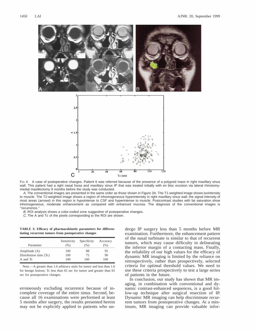
FIG 4. A case of postoperative changes. Patient 6 was referred because of the presence of a polypoid mass in right maxillary sinuswall. This patient had a right nasal fossa and maxillary sinus IP that was treated initially with en bloc excision via lateral rhinotomy-medial maxillectomy 9 months before the study was conducted.
A, The conventional images are presented in the same order as those shown in Figure 3A. The T1-weighted image shows isointensityto muscle. The T2-weighted image shows a region of inhomogeneous hyperintensity in right maxillary sinus wall; the signal intensity ofmost areas (arrows) in this region is hypointense to CSF and hyperintense to muscle. Postcontrast studies with fat saturation showinhomogeneous, moderate enhancement as compared with enhanced mucosa. The diagnosis of the conventional images is‘‘recurrence.’’
B, ROI analysis shows a color-coded zone suggestive of postoperative changes.C, The A and Tc of the pixels corresponding to the ROI are shown.
TABLE 3: Efficacy of pharmacokinetic parameters for differen-tiating recurrent tumors from postoperative changes
ParameterSensitivity
(%)Specificity
(%)Accuracy
(%)
Amplitude (A)Distribution time (Tc)A and Tc
100100100
8875
100
9590
100
Note.—A greater than 1.6 arbitrary units for tumor and less than 1.6for benign lesions; Tc less than 65 sec for tumor and greater than 65sec for postoperative changes.
erroneously excluding recurrence because of in-complete coverage of the entire sinus. Second, be-cause all 16 examinations were performed at least5 months after surgery, the results presented hereinmay not be explicitly applied to patients who un-
dergo IP surgery less than 5 months before MRexamination. Furthermore, the enhancement patternof the nasal turbinate is similar to that of recurrenttumors, which may cause difficulty in delineatingthe inferior margin of a contacting mass. Finally,the reliability of our high values for the efficacy ofdynamic MR imaging is limited by the reliance onretrospectively, rather than prospectively, selectedcriteria for optimal threshold values. We need touse these criteria prospectively to test a large seriesof patients in the future.
In conclusion, our study has shown that MR im-aging, in combination with conventional and dy-namic contrast-enhanced sequences, is a good fol-low-up technique after surgical resection of IP.Dynamic MR imaging can help discriminate recur-rent tumors from postoperative changes. At a min-imum, MR imaging can provide valuable infor-

AJNR: 20, September 1999 RECURRENT INVERTED PAPILLOMA 1451
FIG 5. Two-parameter scatter diagram showing amplitude (A)plotted against the distribution time (Tc). The recurrent IP (dia-monds) are localized in the left upper corner, displaying a com-bination of high amplitude and short distribution time. Tumor re-currence can be defined by a combination of amplitude greaterthan 1.6 arbitrary units and distribution time less than 65 sec-onds. The postoperative changes (solid bullets) are character-ized by low amplitude (,1.6 arbitrary units) and long distributiontime (.65 seconds). Only one postoperatively changed lesiondisplayed high amplitude. In this case, the lesion still can be dis-tinguished from a recurrent tumor on the basis of distributiontime; however, if only the amplitude were considered, the lesionwould be misclassified as a tumor.
mation regarding what sites should be targeted forbiopsy.
References1. Rice DH. Radiology. In Donald PJ, Gluckman JL, Rice DH, eds.
The Sinuses. 1st ed. New York: Raven Press; 1995;83–982. Furuta Y, Shinohara T, Sano K. Molecular pathologic study of
human papillomavirus infection in inverted papilloma andsquamous cell carcinoma of the nasal cavities and paranasalsinuses. Laryngoscope 1991;101:79–85
3. Kamel RH. Transnasal endoscopic medial maxillectomy in in-verted papilloma. Laryngoscope 1995;105:847–853
4. Lawson W, Ho BT, Shaari CM, Biller HF. Inverted papilloma. Areport of 112 cases. Laryngoscope 1995;105:282–288
5. Vrabec DP. The inverted schneiderian papilloma. A 25-yearstudy. Laryngoscope 1994;104:582–605
6. Meyers EN, Fernau JL, Johnson JT, Tabet JC, Barnes L. Manage-ment of inverted papilloma. Laryngoscope 1990;100:481–490
7. Yousem DM, Fellows DW, Kennedy DW, et al. Inverted papillo-ma. Evaluation with MR imaging. Radiology 1992;185:501–505
8. Muller-Schimpfle M, Brix G, Layer G, et al. Recurrent rectalcancer. Diagnosis with dynamic MR imaging. Radiology 1993;189:881–889
9. Hawighorst H, Knapstein PG, Weikel W, et al. Cervical carci-noma. Comparison of standard and pharmacokinetic imaging.Radiology 1996;201:531–539
10. Hess T, Muller-Schimpfle M, Brix G, et al. Dynamic Gd-DTPAenhanced MRI in the follow-up of cervical carcinoma after com-bined radio/chemotherapy. Adv MRI Contrast 1994;12:553–558
11. Lanzieri CF, Shah M, Krauss D, Lavertu P. Use of gadolinium-enhanced MR imaging for differentiating mucoceles from ne-oplasms in the paranasal sinuses. Radiology 1991;178:425–428
12. Som PM, Dillon WP, Sze G, et al. Benign and malignant sino-nasal lesions with intracranial extension. Differentiation withMR imaging. Radiology 1989;172:763–766
13. Brix G, Semmler W, Port R, Schad LR, Layer G, Lorenz WJ.Pharmacokinetic parameters in CNS Gd-DTPA enhanced MRimaging. J Comp Assist Tomogr 1991;15:621–628
14. Greenstein Orel S, Schnall MD, LiVolsi VA, Troupin RH. Sus-picious breast lesions. MR imaging with radiologic-pathologiccorrelation. Radiology 1994;190:485–493
15. Som PM, Shapiro MD, Biller HF, et al. Sinonasal tumors andinflammatory tissues. Differentiation with MR imaging. Radi-ology 1988;167:803–808
16. Som PM, Dillon WP, Fullerton GD, et al. Chronically obstructedsinonasal secretions. Observations on T1 and T2 shortening.Radiology 1989;172:515–520
17. Folkman J. Tumor angiogenesis. Therapeutic implication. NEngl J Med 1971;285:1182–1186
18. Bakay L. The extracellular space in brain tumors. Brain 1970;93:693–698
19. Gullino PM, Grantham FH, Clark SH. The interstitial waterspace of tumors. Cancer Res 1965;25:727–731
20. Nugent LJ, Jain RK. Extracellular diffusion in normal and neo-plastic tissue. Cancer Res 1984;44:238–244
![[PPT]Inverted Papilloma by Grace Fidia · Web viewDiagnosis Klinis: Cervical syndrome, brachialgia, chepalgiakronis, carpal tunnel syndrome Diagnosis Topis : radiks dan neuron Diagnosis](https://static.fdocuments.in/doc/165x107/5add76577f8b9ae1408d021b/pptinverted-papilloma-by-grace-fidia-viewdiagnosis-klinis-cervical-syndrome.jpg)