Inverted papilloma nose and its management
20
Inverted Papilloma of Nose Management 6/29/2010 Otolaryngology online Dr. T. Balasubramanian
-
Upload
dr-t-balasubramanian -
Category
Documents
-
view
7.886 -
download
3
description
This e book discusses the etiopathogenesis of inverted papilloma of nose and its management
Transcript of Inverted papilloma nose and its management
Inverted Papilloma of Nose Management 6/29/2010 Otolaryngology online Dr. T. Balasubramanian
Otolaryngology online
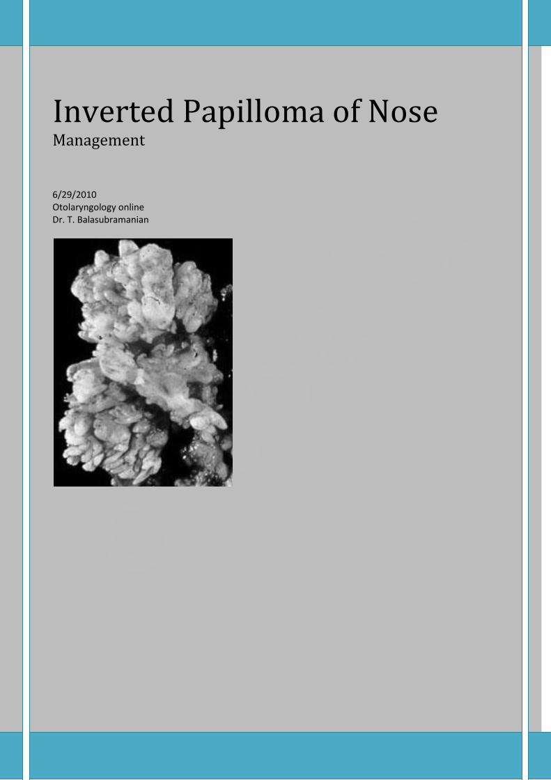
Inverted papilloma nose
Dr. T. Balasubramanian
Introduction: Inverted papilloma is a benign lesion occurring in the nasal cavity and
paranasal sinuses. Even though these tumors are classified as benign they are known
to cause local destruction, known to recur and also can undergo malignant
transformation to squamous cell carcinoma.
History: Ward in 1854 described the macroscopic features of papilloma of nose. He
used the term papillomatous neoplasm to describe this lesion. Billroth in 1855 used
the term villous carcinoma to describe inverted papilloma because of its propensity to
destroy local tissues and recurrence after surgery. Hopmann in 1883 used the terms
hard and soft papilloma to ascertain the stoma : epithelium ratio. This classification
of course was not useful because the number of epithelial layers varied within the
various areas of the same specimen.
Ringertz in 1938 coined the term inverted papilloma after recognizing the
characteristic endophytic growth pattern demonstrated by this type of papilloma.
Kramer and Som in 1935 used the term genuine papilloma of the nasal cavity.
Berendes in 1966 after taking cognizance of the destructive properties of this lesion
used the term Malignant papilloma to indicate this mass. Hyams in 1971 classified
nasal papillomas as inverted papilloma (to indicate papillomas with endophytic
growth) and fungiform papilloma. He also included a third group cylindrical
papilloma to accommodate the variations seen in these papillomas. Batsakis in 1987
used the term inverted Schneiderian papilloma indicating its origin from the
Schneiderian membrane (nasal mucosa). Michaels in 1996 regarded the three types
of nasal papilloma as three completely distinct entities whereas Eggers in 2005
considered these three types of nasal papillomas as hybrid lesions.
Synonyms: As indicated above various synonyms have been used to indicate
inverted papilloma of nose. They include:
1. Schneiderian papilloma
2. Inverted papilloma
3. Benign papilloma of nose
4. Cylindroma
5. Malignant papilloma of nose
Otolaryngology online
Definition: The mucosal lining of nose and paranasal sinuses is known as
Schneiderian membrane in memory of Victor conrod Schnider who described its
histology. Papillomas arising from this membrane are very unique in that they are
found to be growing inwards and hence the term inverted papilloma. These
papillomas are unique in their history, biology and location. Papillomas involving the
vestibule is not included in this group because histologically, biologically and
behaviour wise it is different.
It should be borne in mind that the lining mucosa of nose and paranasal
sinuses is unique Embryologically in the sense that it is derived from the ectoderm, in
contrast to the lining epithelium of laryngobronchial tree which is derived from
endoderm.
Incidence:
Inverted papilloma of nose is one of the commonest benign tumors involving the nose
and paranasal sinuses. It constitutes about 5% of all tumorous lesions of the nose and
sinuses.
Histology:
Inverted papillomas arise from reserve / replacement cells located at the basement
membrane of the mucosa. The stimulus for this proliferation is largely unknown.
Human papilloma virus have been implicated as an etiological agent. Progressive
proliferation of these cells results in thickening of the nasal epithelium. The nasal
epithelium assumes an inverting, fungiform or a combination of both. On rare
occasions the papilloma may be composed entirely of cylindrical cells hence the term
cylindroma is also used. W.H.O. Has histologically classified nasal papillomas into
three types according to its histology. These include:
1. Exophytic papilloma – originates mainly from the nasal septum
2. Inverted papilloma – Typically arises from the lateral nasal wall or within the
maxillary sinus
3. Columnar cell papilloma – commonly arises from lateral nasal wall
Etiology:
Very little is known regarding the etiology of inverted papilloma. Studies have
implicated Human papilloma virus as the probable etiology. Human papilloma virus
DNA have been isolated from nasal papilloma cells. It should also be pointed out
that HPV DNA have not been identified in all the papilloma cells. This could be
accounted when the possibility of degradation of HPV DNA is taken into
Otolaryngology online
consideration. Other suggested causes could be chronic inflammation, smoking,
allergy and occupational exposure to noxious agents.
Age group affected: Majority of these patients fall in the age group between 50 – 70
years.
Sex predisposition: It is three times more common in males than in females.
Classification:
Anatomic classification:
Inverted papilloma can be classified according to its site of occurrence i.e. lateral wall
and septal papillomas. They show differences in their behaviour patterns. The septal
papillomas remain confined to the nasal septum and may very rarely involve the roof
and floor of the nasal cavity. Carcinomatous transformation is rare in septal
papillomas.
Papilloma of lateral wall is known to involve multiple sites i.e. floor, roof of nasal
cavity, para nasal sinuses and naso lacrimal duct. Carcinomatous transformation is
common in this variety.
Histological classification:
Papillary form: Also known as fungiform papilloma. This type shows epithelial
proliferation over a thin core of connective tissue. Inversion of epithelial cells is not
evident in this type.
Inverted papilloma (classic): In this type the epithelial growth is predominantly
directed into the underlying stroma. The stroma is not breached in these patients. If
the stroma is breached then malignant transformation should be suspected.
Columnar cell papilloma: This is a variant of papilloma of nasal cavity. The cells of
this type of papilloma resemble columnar epithelium. Hence the term cylindroma has
been used. This variant is known for its increased recurrence rate and malignant
transformation.
The predominant cell types in these papillomas are ofcourse epidermoid in nature. In
all the above described types of nasal papillomas excessive keratinization is rare. If
the features suggest excessive keratinization then malignant transformation should be
suspected. Microscopic mucous cysts have been identified in all these types.
Intercellular bridges also can be clearly demonstrated in all these types. Loss of these
bridges again is suggestive of malignant transformation.
Characteristic attributes of inverted papilloma:
Otolaryngology online
The following are the characteristic attributes of inverted papilloma of nose.
1. Tendency to recur even after complete surgical removal of mass
2. Its destructive capacity
3. Presence of associated nasal polypi
4. Malignant transformation
Symptoms: Include
Unilateral nasal obstruction – This occurs when the mass is sufficiently large to cause
airway obstruction.
Nasal discharge – This is due to retained secretions in the nasal cavity and the
excessive mucous secretions from mucoid glands present in the nasal mucosa.
Epistaxis – Commonly unilateral and occurs unprovoked. Usually self limiting in
nature.
Head ache – Is caused due to blockage of the normal sinus drainage. If the head ache
is intense and nocturnal then malignant transformation eroding the skull base should
be suspected.
Sinusitis & swelling involving the nose – This is usually due to the mass obstructing
the sinus drainage. Swelling is seen in the alar region (flaring of the ala).
Anosmia – This is very rare and is seen only in patients with bilateral mass lesions.
Hearing impairment – Is caused when the mass expands into the naso pharynx to
involve the eustachean tube. This can also cause tinnitus rarely.
Epiphora – This is caused due to blockage of naso lacrimal duct at the inferior meatus
Numbness over cheek – Due to involvement of infraorbital nerve
Altered speech – Occurs when the mass involves the nasopharynx
Proptosis – Is seen in patients in whom the lamina papyracea has been breached
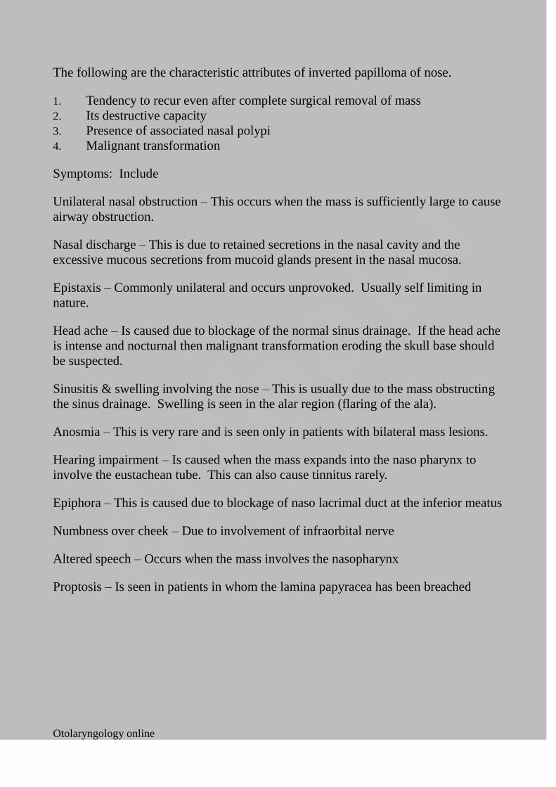
Otolaryngology online
Clinical photograph of a patient with inverted papilloma right nasal cavity
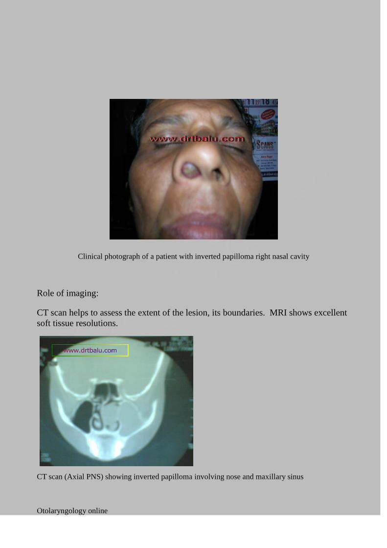
Role of imaging:
CT scan helps to assess the extent of the lesion, its boundaries. MRI shows excellent
soft tissue resolutions.
CT scan (Axial PNS) showing inverted papilloma involving nose and maxillary sinus
Otolaryngology online
Imaging helps in staging of the disease as well as in deciding the optimal
management modality.
Krouse devised the staging system which involved clinical features and imaging:
T1 – In this stage the tumor is confined to the nasal cavity. The tumor involves only
one wall of the nasal cavity without involvement of sinuses.
T2 – In this stage the tumor is limited to the medial and superior portions of the
maxillary sinus with or without the involvement of the nasal cavity. If the nasal
cavity is involved the ethmoidal sinus is also involved.
T3 – In this stage the tumor involves the lateral, inferior, anterior or posterior walls of
maxillary sinus, the sphenoid sinus, or the frontal sinus. Ethmoidal sinuses and nasal
cavity may / may not be involved.
T4 – In this stage the tumor extends outside the confines of nose / paranasal sinuses
to involve adjacent contiguous structures like orbit, skull base or pterygomaxillary
space.
This staging process helps in deciding the optimal management modality.
Management:
Inverted papilloma of nose is a surgically manageable condition. With the advent of
nasal endoscope it has made the radical surgical procedures like lateral rhinotomy and
other external approaches redundant. The aim of the surgery is eradication of the
complete disease in the first attempt itself. Remnants if any will cause recurrence of
the lesion. The cavity left after surgery should be easily accessible from outside in
case there is recurrence of the lesion.
All T1& T2 lesions can be managed by conventional endoscopic sinus surgery.
Endoscope can be used to perform anterior ethmoidectomy, middle meatal
antrostomy and posterior ethmoidectomy. Middle turbinate can also be partially
resected to improve access.
T3 and T4 lesions need a combination of external and endoscopic approaches to
eradicate. The medial wall of maxilla will have to be removed to get access to the
mass (medial maxillectomy).
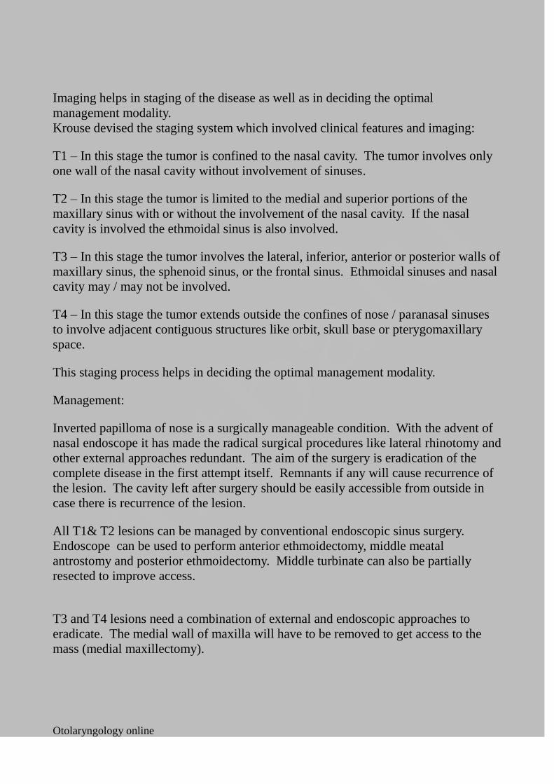
Otolaryngology online
Post OP CT of a patient who has under gone endoscopic resection for inverted papilloma
Surgically uresectable masses and masses that have undergone malignant
transformation should be subjected to irradiation.
Types of endoscopic techniques in the management of inverted papilloma:
Three different types of endoscopic techniques are available to the surgeon for
managing inverted papillomas. The decision on the technique is arrived at on
detailed study of pre op images and intra op endoscopic findings. Surgeon should
bear in mind that enbloc endoscopic resection is possible only when the lesion is
limited to ethmoido nasal complex and does not completely fill the nasal cavity. It is
ofcourse possible to remove the nasal component of the lesion using a debrider before
using the endoscope. The key to successful surgical extirpation of the mass is
performing the dissection of the diseased mucosa along the sub periosteal plane.
Even the underlying bone should be drilled using a diamond burr in order to
eliminate potential microscopic lesions that may be present in the bone.
Otolaryngology online
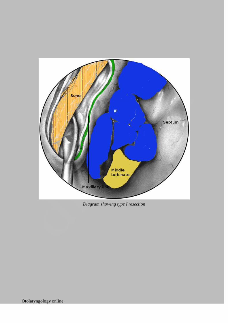
Type I resection: This is indicated in the management of inverted papillomas
involving middle meatus, ethmoid, superior meatus and even lesions protruding into
the maxillary sinus cavity without involvement of sinus mucosa. If the mass
completely fills the nasal cavity it should first be debulked exposing certain vital
surgical landmarks namely the maxillary line, insertion of middle turbinate and the
choana.
Maxillary line: Is the anterior attachment of uncinate process to the ascending /
frontal process of maxilla. This is a very useful intranasal landmark for various
surgical procedures. This is a curvilinear line of mucosal eminence projecting from
just anterior to the superior attachment of middle turbinate extending along the lateral
nasal wall up to the root of the inferior turbinate. In the axial plane the midpoint of
this maxillary line corresponds to the superior aspect of the maxillary ostium. While
performing uncinectomy the incision should be placed just behind this line. The
lacrimal fossa where the lacrimal sac is situated is bisected by this line and hence can
be used as a reliable surgical landmark for identification of lacrimal sac. The mucosa
over the maxillary line is incised using laser / angled knife. The underlying bone is
drilled using a diamond burr. This exposes the ethmoidal infundibulum which is
usually filled by the lesion. The lesion is encircled in a centripetal fashion laterally
along the lamina papyracea anteriorly up to the posterior ethmoid cells and superiorly
along the roof of the ethmoid. The middle turbinate is usually included along with
the specimen by resecting it close to its attachment to skull base. While resecting the
middle turbinate the sphenopalatine vessels should be identified and cauterized to
prevent troublesome bleeding. The superior turbinate should be removed only if the
inverted papilloma extends to involve the posterior ethmoid air cells. Dissection in
the olfactory fossa is not indicated normally, but should be carefully performed if the
inverted papilloma extends medial to the turbinates. Overzealous dissection in this
area can easily lead to troublesome CSF leaks. The mucosa covering the anterior
wall of sphenoid sinus is included in the resection.
Frontal sinusotomy is also performed using true cut instruments. This is usually done
to check the status of frontal sinus mucosa.
Otolaryngology online
Diagram showing type I resection
Otolaryngology online
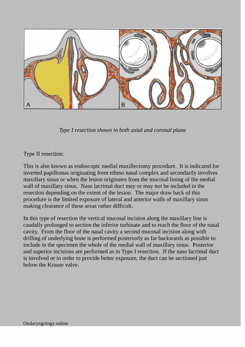
Type I resection shown in both axial and coronal plane
Type II resection:
This is also known as endoscopic medial maxillectomy procedure. It is indicated for
inverted papillomas originating from ethmo nasal complex and secondarily involves
maxillary sinus or when the lesion originates from the mucosal lining of the medial
wall of maxillary sinus. Naso lacrimal duct may or may not be included in the
resection depending on the extent of the lesion. The major draw back of this
procedure is the limited exposure of lateral and anterior walls of maxillary sinus
making clearance of these areas rather difficult.
In this type of resection the vertical mucosal incision along the maxillary line is
caudally prolonged to section the inferior turbinate and to reach the floor of the nasal
cavity. From the floor of the nasal cavity a second mucosal incision along with
drilling of underlying bone is performed posteriorly as far backwards as possible to
include in the specimen the whole of the medial wall of maxillary sinus. Posterior
and superior incisions are performed as in Type I resection. If the naso lacrimal duct
is involved or in order to provide better exposure, the duct can be sectioned just
below the Krause valve.
Otolaryngology online
Figure showing Type II resection. Note the inferior turbinate has been resected
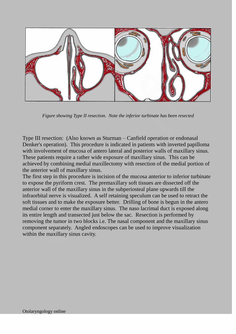
Type III resection: (Also known as Sturman – Canfield operation or endonasal
Denker's operation). This procedure is indicated in patients with inverted papilloma
with involvement of mucosa of antero lateral and posterior walls of maxillary sinus.
These patients require a rather wide exposure of maxillary sinus. This can be
achieved by combining medial maxillectomy with resection of the medial portion of
the anterior wall of maxillary sinus.
The first step in this procedure is incision of the mucosa anterior to inferior turbinate
to expose the pyriform crest. The premaxillary soft tissues are dissected off the
anterior wall of the maxillary sinus in the subperiosteal plane upwards till the
infraorbital nerve is visualized. A self retaining speculum can be used to retract the
soft tissues and to make the exposure better. Drilling of bone is begun in the antero
medial corner to enter the maxillary sinus. The naso lacrimal duct is exposed along
its entire length and transected just below the sac. Resection is performed by
removing the tumor in two blocks i.e. The nasal component and the maxillary sinus
component separately. Angled endoscopes can be used to improve visualization
within the maxillary sinus cavity.
Otolaryngology online
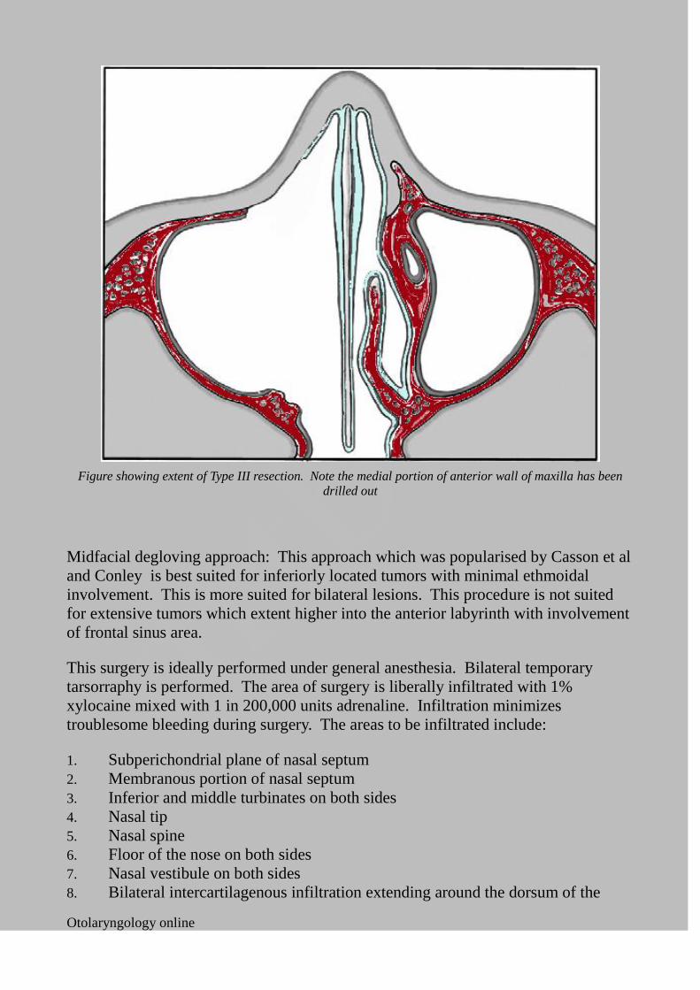
Figure showing extent of Type III resection. Note the medial portion of anterior wall of maxilla has been
drilled out
Midfacial degloving approach: This approach which was popularised by Casson et al
and Conley is best suited for inferiorly located tumors with minimal ethmoidal
involvement. This is more suited for bilateral lesions. This procedure is not suited
for extensive tumors which extent higher into the anterior labyrinth with involvement
of frontal sinus area.
This surgery is ideally performed under general anesthesia. Bilateral temporary
tarsorraphy is performed. The area of surgery is liberally infiltrated with 1%
xylocaine mixed with 1 in 200,000 units adrenaline. Infiltration minimizes
troublesome bleeding during surgery. The areas to be infiltrated include:
1. Subperichondrial plane of nasal septum
2. Membranous portion of nasal septum
3. Inferior and middle turbinates on both sides
4. Nasal tip
5. Nasal spine
6. Floor of the nose on both sides
7. Nasal vestibule on both sides
8. Bilateral intercartilagenous infiltration extending around the dorsum of the
Otolaryngology online
nose, and the anterior wall of maxilla on both sides, up to the glabella of frontal bone.
9. Transcutaneous injection into the orbit along its medial wall
10. Sublabial infiltration from the third molar across the midline to the opposite
third molar
11. Trans oral greater palatine injection is also given
The procedure is started with complete transfixion incision, which is connected to
bilateral intercartilagenous incisions. Elevation of soft tissue from the nasal dorsum
is performed through the intercartilagenous space. The soft tissue elevation over
dorsum of nose is continued over the anterior wall of maxilla on both sides.
Elevation of soft tissue should also continue over the glabella and frontal bone.
Supero laterally the elevation should extend up to the medial canthal region. The
intercartilagenous incision is extended laterally and caudally across the floor of the
vestibule to be connected with the transfixation incision. This results in a full
circumvestibular incision on both sides.
After the transnasal incisions are completed the sublabial incision is performed. It
extends from the first molar on oneside across the midline up to the first molar on the
opposite side. This incision can be extended up to the third molar if more exposure is
needed. The incision is carried down the submucosa, and muscles over anterior wall
of maxilla. At the pyriform aperture region this incision is connected to intranasal
incisions. Periosteal elevators are used to elevate the soft tissue over the anterior
walls of both maxilla up to the level of the orbital rim taking care to protect the
infraorbital vessels and nerve. The entire midfacial skin is stripped from the dorsum
of the nose and anterior wall of maxilla. This flap includes the lower lateral
cartilages, columella with its medial crura. The elevation is continued till the level of
glabella superiorly and medial canthus laterally. The bony nasal pyramid and the
attached upper lateral cartilages are exposed completely. Two rubber drains (Penrose
type) are passed through the nose and upper lip and are used to retract the midfacial
flap along with the upper lip. Once in every 15 minutes one of the drain should be
released to allow blood supply to the middle portion of the upper lip.
Otolaryngology online
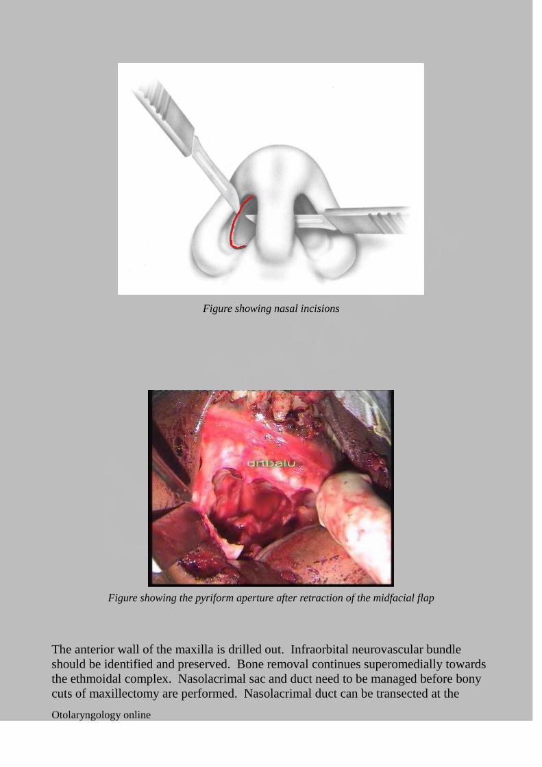
Figure showing nasal incisions
Figure showing the pyriform aperture after retraction of the midfacial flap
The anterior wall of the maxilla is drilled out. Infraorbital neurovascular bundle
should be identified and preserved. Bone removal continues superomedially towards
the ethmoidal complex. Nasolacrimal sac and duct need to be managed before bony
cuts of maxillectomy are performed. Nasolacrimal duct can be transected at the
Otolaryngology online
orbital floor level.
The whole anterior wall of maxillary sinus is drilled out including the lateral portion
of nasal bone including the edge of the pyriform aperture.
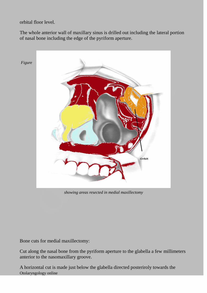
Figure
showing areas resected in medial maxillectomy
Bone cuts for medial maxillectomy:
Cut along the nasal bone from the pyriform aperture to the glabella a few millimeters
anterior to the nasomaxillary groove.
A horizontal cut is made just below the glabella directed posteriroly towards the
Otolaryngology online
frontoethmoid suture line.
Antero posterior cut along the fronto ethmoidal suture line.
Oblique cut of the orbital floor from the orbital rim medial to the infraorbital foramen
extending postero medially to join the fronto ethmoid cut in the posterior ethmoid
region. All these bone cuts should include the attached soft tissues.
The posterior attachment to the ascending process of palatine bone is severed using a
heavy scissors.
Complications of midfacial degloving:
1. Anesthesia over infraorbital nerve area
2. Epiphora
3. Nasal valve stenosis
Medial maxillectomy:
Can also be performed using lateral rhinotomy incision.
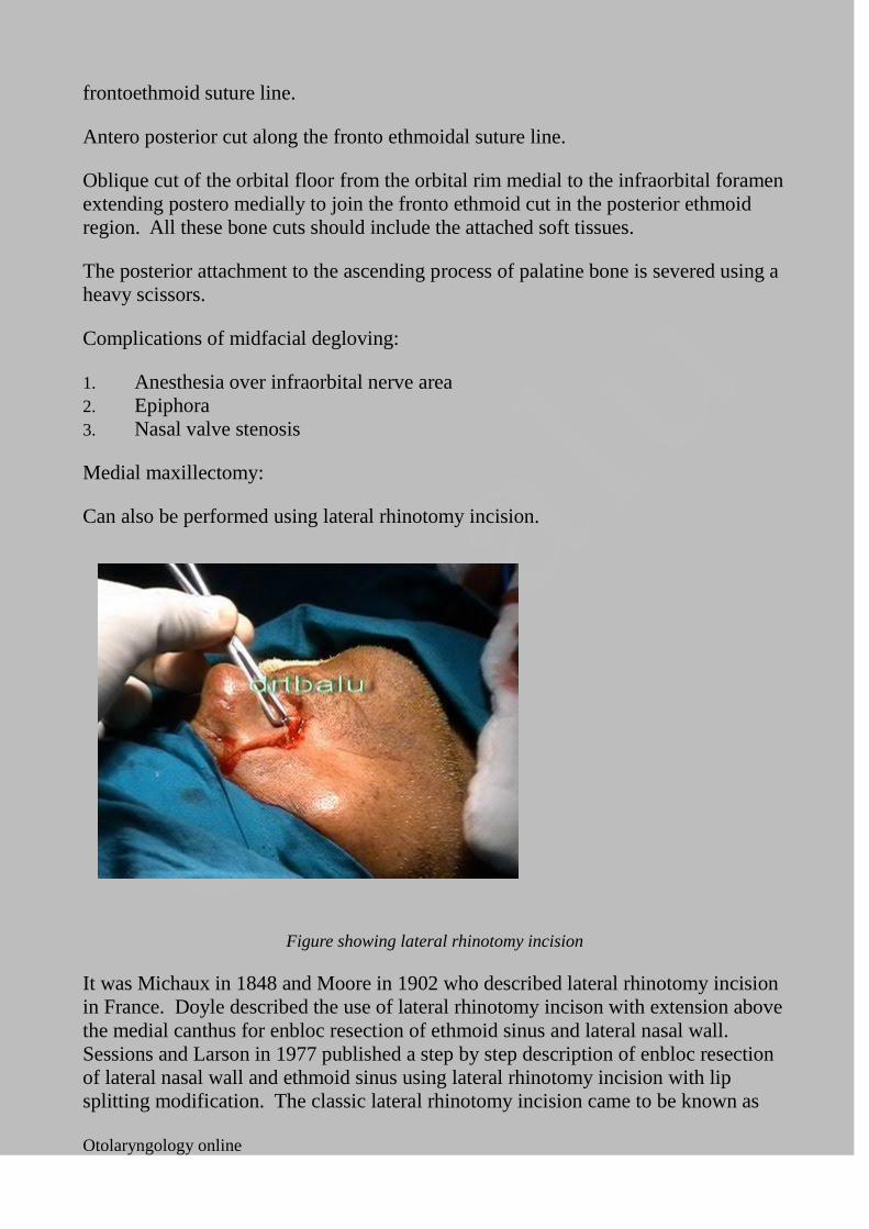
Figure showing lateral rhinotomy incision
It was Michaux in 1848 and Moore in 1902 who described lateral rhinotomy incision
in France. Doyle described the use of lateral rhinotomy incison with extension above
the medial canthus for enbloc resection of ethmoid sinus and lateral nasal wall.
Sessions and Larson in 1977 published a step by step description of enbloc resection
of lateral nasal wall and ethmoid sinus using lateral rhinotomy incision with lip
splitting modification. The classic lateral rhinotomy incision came to be known as
Otolaryngology online
“Moore's lateral rhinotomy incision”.
The lateral rhinotomy incision begins just below the medial aspect of eyebrow, slants
downwards across the midpoint between the nasion and the medial aspect of
palpebral fissure, courses inferiorly along the naso facial junction into the ala facial
crease across the nasal sill towards the pyriform aperture. There may be brisk
bleeding in the region of medial canthus from the angular and canthal vessels which
should be ligated. While the incision passes over the medial canthal area if a small
notch is added or a lazy s incision is given it facilitates accurate approximation of
skin thereby preventing post op web formation.
While dissecting at the level of medial canthus care should be taken to mark the
position of medial canthal tendon which inserts on to the nasal process of maxilla
anchoring the eyelids and lacrimal apparatus to the bone. Proper repositioning of this
area is very important in order to prevent development of post op complications like
telecanthus and defective lacrimal pump mechanism. The medial canthal tendon
should be detached by elevating the incised periosteum of the frontal process of
maxilla using a Freer's elevator after marking its position. If need arises for this bone
to be removed then a permanent transnasal mattress suture should be placed through
burr holes in the opposite nasal bone.
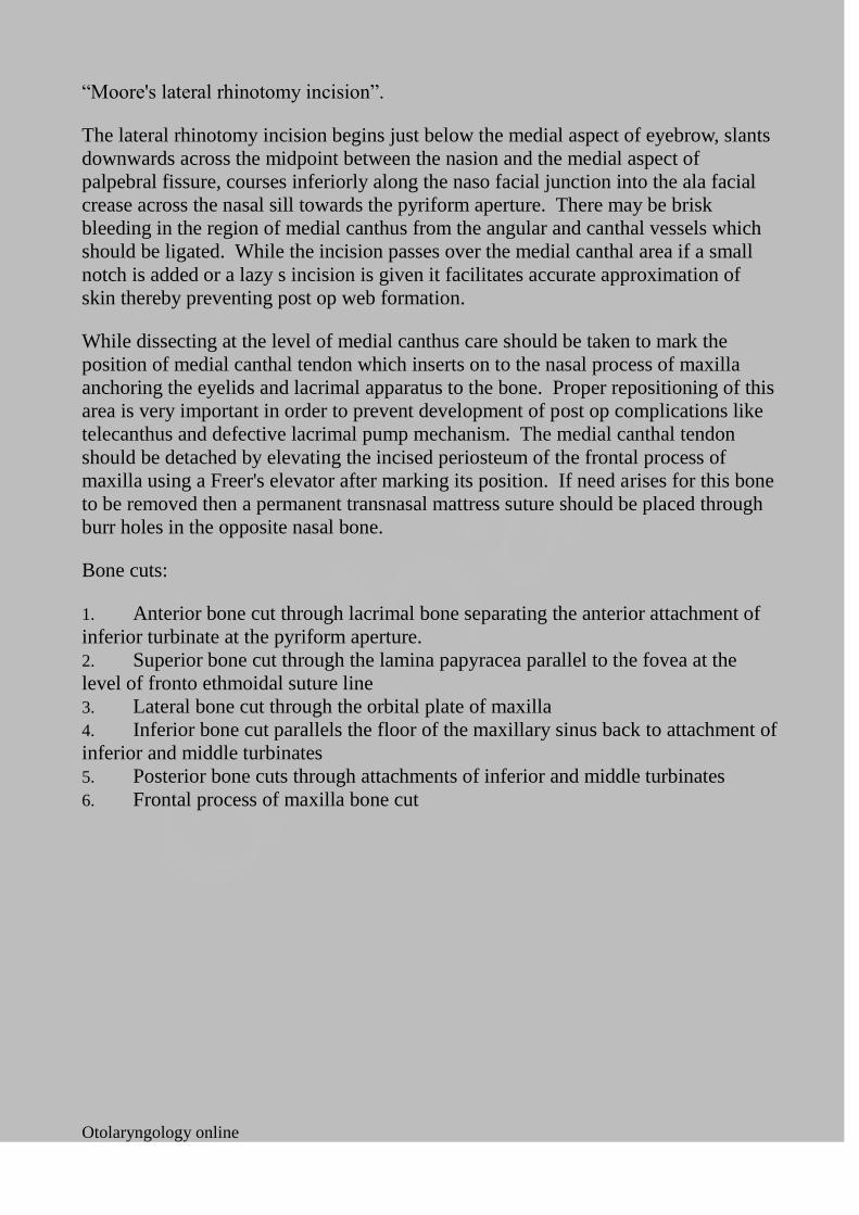
Bone cuts:
1. Anterior bone cut through lacrimal bone separating the anterior attachment of
inferior turbinate at the pyriform aperture.
2. Superior bone cut through the lamina papyracea parallel to the fovea at the
level of fronto ethmoidal suture line
3. Lateral bone cut through the orbital plate of maxilla
4. Inferior bone cut parallels the floor of the maxillary sinus back to attachment of
inferior and middle turbinates
5. Posterior bone cuts through attachments of inferior and middle turbinates
6. Frontal process of maxilla bone cut
Otolaryngology online
Figure
showing bone cuts in lateral rhinotomy
Complications of medial maxillectomy:
1. Lid oedema
2. Asymmetry of palpebral fissures
3. Diplopia
4. Enophthalmos
5. Transient blindness due to retinal artery spasm
6. Orbital hemorrhage
7. CSF leaks

Otolaryngology online