Reactions of Chlorambucil and its main metabolite, Phenylacetic
60
TURUN YLIOPISTON JULKAISUJA ANNALES UNIVERSITATIS TURKUENSIS SARJA - SER. A I OSA - TOM. 401 ASTRONOMICA - CHEMICA - PHYSICA - MATHEMATICA TURUN YLIOPISTO UNIVERSITY OF TURKU Turku 2009 Reactions of Chlorambucil and its main metabolite, Phenylacetic Acid Mustard, with 2’-deoxyribonucleosides and Calf Thymus DNA by Diana Florea-Wang
Transcript of Reactions of Chlorambucil and its main metabolite, Phenylacetic

TURUN YLIOPISTON JULKAISUJAANNALES UNIVERSITATIS TURKUENSIS
SARJA - SER. A I OSA - TOM. 401
ASTRONOMICA - CHEMICA - PHYSICA - MATHEMATICA
TURUN YLIOPISTOUNIVERSITY OF TURKU
Turku 2009
Reactions of Chlorambucil and its main metabolite, Phenylacetic Acid Mustard,
with 2’-deoxyribonucleosides and Calf Thymus DNA
by
Diana Florea-Wang

From the Department of ChemistryUniversity of TurkuTurku, Finland
Custos
Docent Jari HovinenPerkinElmerTurku, Finland
Reviewers
Professor Seppo AuriolaDepartment of Pharmaceutical ChemistryUniversity of KuopioKuopio, Finland
Professor Mikko OivanenDepartment of ChemistryUniversity of HelsinkiHelsinki, Finland
Opponent
Professor Lajos KovacsDepartment of Medicinal ChemistryUniversity of SzegedSzeged, Hungary
ISBN 978-951-29-4041-7 (PRINT)ISBN 978-951-29-4042-4 (PDF)ISSN 0082-7002Painosalama Oy – Turku, Finland 2009

Abstract 3
AbsTRACT
Chlorambucil is an anticancer agent used in the treatment of a variety of cancers, especially in chronic lymphocytic leukemia, and autoimmune diseases. Nevertheless, chlorambucil is potentially mutagenic, teratogenic and carcinogenic.
The high antitumor activity and high toxicity of chlorambucil and its main metabolite, phenylacetic acid mustard, to normal tissues have been known for a long time. Despite this, no detailed chemical data on their reactions with biomolecules in aqueous media have been available.
The aim of the work described in this thesis was to analyze reactions of chlorambucil with 2’-deoxyribonucleosides and calf thymus DNA in aqueous buffered solution, at physiological pH, and to identify and characterize all adducts by using modern analyzing methods. Our research was also focused on the reactions of phenylacetic acid mustard with 2’-deoxynucleosides under similar conditions.
A review of the literature consisting of general background of nucleic acids, alkylating agents and ultraviolet spectroscopy used to identify the purine and pyrimidine nucleosides, as well as the results from experimental work are presented and discussed in this doctoral thesis.

4 Preface
PRefACe
This thesis is based on experimental work carried out at the Laboratory of Organic Chemistry and Chemical Biology at the Department of Chemistry, University of Turku during the years 2002-2008. The Graduate School of Organic Chemistry and Chemical Biology, Medical Research Fund of the Tampere University Hospital and Magnus Ehrnrooth Foundation are gratefully acknowledged for financial support.
I wish to express my deepest thanks and appreciation to Professor Harri Lönnberg for being so kind and giving me the opportunity of continuing my studies in this department in the moment when my English was poor and expressing myself was difficult, for guiding me during the last stage of my PhD studies and finding financial support when I most needed. Words are never enough to show my gratitude to you!
I am also grateful to my supervisor, Docent Jari Hovinen, for his great knowledge in chemistry, ideas, guidance, moral support, patience, everlasting energy and very special talent in doing things quickly and well. Thank you for showing me that chemistry is more alive than I ever thought before!
I am indebted to Professors Seppo Auriola and Mikko Oivanen for their careful reviewing of my thesis and their valuable suggestions.
I wish to express my gratitude to all my closest co-authors: Mr. Kristo Hakala for the high quality MS spectra, fruitful discussions concerning mass spectrometry and great attitude to life, Dr. Agnieszka Pawlowicz for her guidance in the experimental work on DNA reactions and release of inexhaustible energy, Dr. Jari Sinkkonen for great detective work done with NMR spectroscopy and enjoyable discussions NMR related or not, Professor Juhani Vilpo for his excellent knowledge in medicinal chemistry, Dr. Leif Kronberg for interesting discussions on DNA adduction and Professor Jorma Mattinen for his strong knowledge in the area of NMR and valuable interpretation of complicated NMR spectra.
I would also like to thank to Dr. Satu Mikkola, who showed a lot of patience to me and who taught me useful skills in the laboratory work and HPLC technique. One can always enjoy of your good sense of humor and positive attitude!
My warm gratitude goes also to my dear friends and colleagues, Dr. Niangoran Koissi, Mrs. Heidi Korhonen, Mrs. Mihaela Turcu, Mrs. Marika and Mr. Tuomas Karskela, Dr. Attila Jancso, Dr. Mikko Ora, Dr. Helmi Neuvonen and Dr. Anna Polishchuk, for their long lasting friendship, valuable advices, delightful discussions and encouragement in times of doubt.
Special thanks go also to Dr. Päivi Poijärvi-Virta, Mrs. Maarit Laine, Mrs. Tiina Buss, Mrs. Anu Kiviniemi, Mrs. Emilia Kiuru, Ms. Anna Leisvuori, Dr. Kaisa Ketomäki, Dr. Pasi Virta, Dr. Tuomas Lönnberg, Dr. Erkki Nurminen, Professor Pertti Ayräs, Dr. Kari Neuvonen, Dr. Martti Dahlqvist and Mrs. Mia Helkearo for creating a lovely working atmosphere and for their optimistic attitude, very enjoyable and informative

Preface 5
conversations. Ms. Kirsti Wiinamäki is as well thanked for teaching me Finnish and being so kind every time we met. You all have cheered me up in a certain moment during these years and I am really lucky to know you!
The workshop personnel, Mr. Kari Loikas and Mr. Mauri Nauma, deserve my sincere thanks for giving their very best in reminding to the computers or other devices that they should work properly. Dr. Petri Ingman is also thanked for his open mind, pleasant discussions and very easy to collaborate with. In addition, I thank to Mrs. Kirsi Laaksonen for checking patiently and providing quickly the needed chemicals, as well as to Mr. Jaakko Hellmann for helping me with the NMR solvents. Many thanks go also to the ladies working in the departmental secretariat Mrs. Mailis Lankinen, Mrs. Leena Mattila and Mrs. Heli Granlund.
In particular, I would like to thank my parents, Maria and Corneliu. I am grateful to you for what I have become and for your eternal love and care! Many thanks also go to my brothers, Victor and Adrian, and their families. You all bring happiness, wisdom in my life and, although life keeps us so busy all the time, I am really lucky and happy to have you nearby! Thank you for taking care of me and supporting me at any time I needed!
Last but not least, my deepest thanks and love go to my husband, Qi, and to our daughter, Isabel, for bringing more love, joy and excitement in my life. Without your support and understanding, and your happiness and beauty, I would have neither the strength nor determination to undertake a task as the research in a vast domain as chemistry. You are the best of my life!
Diana Florea-Wang
Turku, September 2009
6 Contents
TAble Of CONTeNTs
AbsTRACT .............................................................................................................................. 3
PRefACe .................................................................................................................................. 4
TAble Of CONTeNTs .......................................................................................................... 6
lisT Of ORigiNAl PubliCATiONs ................................................................................. 8
AbbReviATiONs .................................................................................................................. 9
1. iNTRODuCTiON ............................................................................................................101.1. The structure and biological significance of deoxyribonucleic acid ...........101.2. Alkylating agents and alkylation of purine and pyrimidine nucleosides ..12
1.2.1. Classification of alkylating agents .................................................................131.2.2. DNA crosslink ..........................................................................................................171.2.3. Sites of alkylation in purine and pyrimidine deoxynucleosides
and DNA .....................................................................................................................191.2.4. Basics of carcinogenesis and introduction to alkylating agents
as carcinogens .........................................................................................................191.2.5. Stability of alkylated DNA adducts .................................................................211.2.6. DNA-alkylation repair processes .....................................................................211.2.7. Biological activity of DNA-alkylation adducts ...........................................22
1.3. Ultraviolet spectroscopy used in characterization of purine and pyrimidine nucleoside adducts ....................................................................................22
2. AiMs Of The Thesis ...................................................................................................25
3. ResulTs AND DisCussiON .......................................................................................263.1. Chlorambucil versus phenylacetic acid mustard ................................................263.2. Reactions of chlorambucil and phenylacetic acid mustard
in the absence of 2’-deoxyribonucleosides ............................................................283.3. Reactions of chlorambucil and phenylacetic acid mustard
with 2’-deoxyribonucleosides ......................................................................................293.3.1. Reactions of chlorambucil and phenylacetic acid mustard
with 2’-deoxyadenosine ......................................................................................303.3.2. Reactions of chlorambucil and phenylacetic acid mustard
with 2’-deoxyguanosine ......................................................................................353.3.3. Reactions of chlorambucil and phenylacetic acid mustard
with 2’-deoxycytidine and 2’-deoxy-5-methylcytidine ..........................403.3.4. Reactions of chlorambucil and phenylacetic acid mustard
with thymidine ........................................................................................................443.4. Reactions of chlorambucil with calf thymus DNA ...............................................45
4. suMMARy AND CONClusiONs .................................................................................49

Contents 7
5. exPeRiMeNTAl seCTiON...........................................................................................505.1. General ....................................................................................................................................505.2. Chromatographic methods .............................................................................................505.3. Spectroscopic and spectrometric methods .............................................................505.4. Quantitative product analyses ......................................................................................515.5. Stability of the end products..........................................................................................525.6. Dimroth rearrangement ..................................................................................................525.7. Kinetic measurement .......................................................................................................525.8. Reactions of chlorambucil and phenylacetic acid mustard
with 2’-deoxyribonucleosides .......................................................................................535.9. Reactions of chlorambucil with calf thymus DNA ...............................................53
6. RefeReNCes ..................................................................................................................54
ORigiNAl PubliCATiONs ...............................................................................................61

8 List of Original Publications
lisT Of ORigiNAl PubliCATiONs
This doctoral thesis is based on the following original publications (referred in the text with roman numerals):
I. Diana Florea-Wang, Elina Haapala, Jorma Mattinen, Kristo Hakala, Juhani Vilpo and Jari Hovinen. Reactions of N,N-bis(2-chloroethyl)-p-aminophenylbutyric Acid (Chlorambucil) with 2’-Deoxyadenosine. Chem. Res. Toxicol. 2003, 16, 403-408.
II. Diana Florea-Wang, Elina Haapala, Jorma Mattinen, Kristo Hakala, Juhani Vilpo and Jari Hovinen. Reactions of N,N-bis(2-chloroethyl)-p-aminophenylbutyric Acid (Chlorambucil) with 2’-Deoxycytidine, 2’-Deoxy-5-methylcytidine, and Thymidine. Chem. Res. Toxicol. 2004, 17, 383-391.
III. Diana Florea-Wang, Inna Ijäs, Kristo Hakala, Jorma Mattinen, Juhani Vilpo and Jari Hovinen. Reactions of {4-[Bis(2-chloroethyl)amino]phenyl}acetic Acid (Phenylacetic Acid Mustard) with 2’-Deoxyribonucleosides. Chem. & Biodiv. 2007, 4, 406-423.
IV. Diana Florea-Wang, Agnieszka Pawlowicz, Jari Sinkkonen, Leif Kronberg, Juhani Vilpo and Jari Hovinen. Reactions of N,N-Bis(2-chloroethyl)-p-aminophenylbutyric acid (Chlorambucil) with Calf Thymus DNA. Chem. & Biodiv. 2009, 6, 1002-1013.

Abbreviations 9
AbbReviATiONs
Ade adenine
alkylA alkyladenine
alkylG alkylguanine
alkylT alkylthymine
CLB 4-[N,N-bis(2-chloroethyl)-p-aminophenyl]butyric acid; chlorambucil
Cyt cytosine
COSY correlation spectroscopy
dAdo 2’-deoxyadenosine
dCtd 2’-deoxycytidine
dGuo 2’-deoxyguanosine
dMeCtd 2’-deoxy-5-methylcytidine
Gua Guanine
IARC International Agency for Research on Cancer
NOESY Nuclear Overhauser effect spectroscopy
PAM {4-[bis(2-chloroethyl)amino]phenyl}acetic acid; phenylacetic acid mustard
Thd thymidine
Thy thymine
Ura uracil
UV ultraviolet

10 Introduction
1. iNTRODuCTiON
1.1. Thestructureandbiologicalsignificanceofdeoxyribonucleicacid
Structure of deoxyribonucleic acid (DNA). DNA is a nucleic acid, a long-chain polymer made up of a linear array of monomers called nucleotides.1 Each nucleotide is constructed from a heterocyclic nitrogen base, a pentose sugar, and one or more phosphate residues. The most common bases are monocyclic pyrimidines and bicyclic purines. The DNA contains two purines, adenine (Ade) and guanine (Gua), and two pyrimidines, cytosine (Cyt) and thymine (Thy) (Chart 1).2
N
N
N
N N
N
1
2
3
4
5
6
1
2
34
56 7
8
9
H
pyrimidine basepurine base
Cytosine Thymine
N
N
H
O
NH2
HN
N
H
O
O
CH3N
N N
N
H
NH2
HN
N N
N
H
O
H2N
Adenine Guanine
Chart 1. Structures, numbering of atoms and names of DNA bases.
The structure of the nucleosides consists of the base bonded to carbon-1 of the pentose sugar molecule by a N-β-glycosidic bond. The sugar component of DNA is in furanose form and it is a β-D-2-deoxyribose. Successive monomer units in nucleic acids are connected through a phosphate residue which is attached to the hydroxyl on the carbon-5’ of one unit and the hydroxyl on the carbon-3’ of the next unit. Thus, the nucleic acids are assembled on a backbone made up of the pentose units linked by phosphate esters (Chart 2).3

Introduction 11
O
H
OP-O
O
O
base
5'-end
O
HO
OP-O
O
O
base
3'-end
5'
1'
3'
5'
3'
1'
Chart 2. Phosphate ester backbone: from carbon-5’ of a pentose to the carbon-3’ of the next pentose, and further.
The genetic information is encoded in DNA and transmitted from a parent cell to its daughter cells through hydrogen-bonding interaction between specific pairs of nucleobases, known as Watson-Crick base-pairing. Bases which contain amino groups and carboxyl groups are ideal for hydrogen bonding. Every purine base forms a stable hydrogen-bonded pair with a specific pyrimidine base: adenine forms a base pair through two hydrogen bonds to thymine, while guanine forms a base pair through three hydrogen bonds to cytosine (Chart 3).
Gua ......... Cyt ...... ThyAde
N
NN
N
NHH
N N
O
H
O
CH3
2-deoxyribose
2-deoxyribose
N N
O
N
H
H H
N
NO
NHH
N
N
13
5
79
1
3 5
13
5
1
3
57
9
2-deoxyribose
2-deoxyribose
Chart 3. Watson-Crick base-pairing in DNA.
The three-dimensional model of DNA structure consists of two complementary polynucleotide chains held together by hydrogen bonds between the paired bases (Figure 1). The two strands are anti-parallel: one strand is arranged 3’ 5’ from left to right, while the other goes in the opposite direction, 5’ 3’ from left to right.2,4

12 Introduction
figure 1. DNA double helix model.
BiologicalrelevanceofDNA. The biological properties of the DNA are based on the precise interstrand hydrogen bonding described above. Deoxyribonucleic acids give essential functions in all living organisms, wherein the most vital functions are the long-term storage and transmission of genetic information with high fidelity from one generation to the next.5,6
The DNA is exposed to a large variety of harmful physical and chemical agents that have a constant damaging effect. There are estimations of 10000 lesions per day taking place in human genes in every cell.7 Ultraviolet component of sunlight, X-rays, genotoxins present in foods or cigarette smoke, chemotherapeutic agents (i.e. alkylating agents), oxygen radicals resulted from the normal cellular metabolism may direct to mutations that increase the threat of cancer.8
1.2. Alkylatingagentsandalkylationofpurineandpyrimidinenucleosides
Alkylation is the transfer of a reactive alkyl group from one molecule to another molecule where electron density is high.
Nucleic acid bases are prone to structural modification by nucleophiles as well as by electrophiles and these modifications represent the basis of chemical carcinogenesis. In most of the alkylation reactions, the nucleobases play the role of the nucleophile that will react with electrophiles, such as alkyl halides (R-X), alkanesulfonates (R’-SO2-OR’’) and dialkyl sulfates (R’O-SO2-OR’’) (Scheme 1); in this case, purines are alkylated more readily than pyrimidines. Alternatively, pyrimidines may also serve as electrophiles when strong nucleophiles, such as hydrazines (H2N-NHR), alkoxyamines (H2NOR) and bisulfite ion (HSO3¯),9 attack their C-6 position (Scheme 2).

Introduction 13
N
N
NH2
O
2-deoxyribose
CH3CH2 I
N
N
NH2
O
2-deoxyribose
CH3CH2
scheme 1. Reaction of 2’-deoxycytidine with ethyl iodide.10
HN
N
NH2
O
HSO3 HN
N
NH2
OH
SO3
HN
N
O
OH
SO3
- NH3 HN
N
O
O
- HSO3
2-deoxyribose 2-deoxyribose 2-deoxyribose 2-deoxyribose
scheme 2. Reaction of 2’-deoxycytidine with bisulfite ion.9
As discussed above, alkylating agents are reactive electrophiles. They can react with the nucleophilic (electron-rich) sites of cellular macromolecules, such as DNA and proteins, causing DNA damage by strand breaks, DNA-DNA crosslinks and DNA-protein crosslinks.11 Alkylating agents are important research topic due to their controversy: promising anticancer drugs, but also possible carcinogens, mutagens and teratogens.
1.2.1. ClassificationofalkylatingagentsThe classification of alkylating agents is not an easy task due to the many different aspects used in classifying them, such as their provenience, number of functional groups, mode of action, nucleophilic or electrophilic character/the mechanism of their action, chemical and structure similarities.
There are endogenous and environmental alkylating agents, as well as alkylating drugs. Endogenous agents are formed during the metabolism. S-Adenosylmethionine,12 that is a coenzyme implicated in methyl group transfer in biochemical reactions, and nitrosoamines and related compounds are few examples of the endogenous alkylating agents.
Environmental alkylating agents are found in the air, water and foods. Humans are most exposed to N-nitroso compounds formed in tobacco smoke. 13 Different halocarbons are present in the air in small but detectable concentrations. Chloromethane gas is generated by plants, fungi, industrially and it is one of the halocarbons present in the highest percentage in the atmosphere.13 Bromo-compounds (i.e. bromoethane) are more abundant in marine environment. There are also different food mutagens, such as aflatoxins, polycyclic aromatic hydrocarbons, and heterocyclic amines.14 The oldest anti-cancer drugs are alkylating agents and they are still important in the treatment of different types of cancer.15

14 Introduction
According to the number of the functional groups, alkylating agents are divided into three groups: monofunctional alkylating agents, such as methanesulfonates16,17,18 [i.e. methyl methanesulfonate, MMS (1)] and methylnitrosourea19,20 (2); bifunctional alkylating agents, as for example bis(chloroethyl)nitrosourea21,22,23 (3), bischloroethyl sulfide24 (4), bis(chloromethyl) ether25 (5), epichlorohydrin26 (6), nitrogen mustards; and cyclic alkylating agents, such as 2-chlorooxirane27,28 (7) (Chart 4). There are not strict limits between these three categories of alkylating agents (see compound 6 that is both, bifunctional and cyclic). However, most of mutagens and carcinogens are simple mono- or bi-functional alkylating agents.
H3CS
O
OO
CH3
1
N
O
NH2
N
O
CH32
N
O
NH
NO
Cl Cl
3
S
Cl
ClCl
Cl
4
O
Cl
ClCl
Cl
5
Cl
O
6
O
Cl
7
Chart 4. Structural formula of different types of alkylating agents.
Nitrogen mustards, such as 4-[N,N-bis(2-chloroethyl)-p-aminophenyl]butyric acid (chlorambucil, CLB, 8), cyclophosphamide29 (9), melphalan30 (10) and mechlorethamine (11) (Chart 5), are typical bifunctional alkylating agents and they are few of the oldest anticancer agents in clinical use.31 They have the tendency especially to form interstrand crosslink between two dGuo molecules in their N7 position.32
11
9
108
ClN
Cl
CH3
ClN
Cl
NH2
O
OH
ClN
Cl
OH
O
O NHP
NClCl
O
Chart 5. Structures of nitrogen mustards: chlorambucil (8), cyclophosphamide (9), melphalan (10) and mechlorethamine (11).
Alkylating agents can be divided also by their mode of action into two categories: alkylating agents that react directly with nucleic acids and are known as primary carcinogens; and alkylating agents that react after metabolic activation.33 Most of the alkylating agents are directly acting carcinogens, such as dimethyl sulfate (DMS, 12), MMS (1), 2-methylaziridine (13), 1,3-propanesultone (14), epichlorohydrin34 (6), and old anticancer agents as myleran (15), CLB (8) and cyclophosphamide (9) (Chart 6).

Introduction 15
S
O
O
O
OCH3H3C
12
HN
CH3
13
S O
OO
14
O
SO
OS
O
CH3
O
H3CO 15
Chart 6. Structures of directly acting agents.
Quinone methides are known as bioreductive alkylating agents.35,36 A quinine methide precursor is activated, forming a very reactive intermediate that can react with DNA. The antibiotic mitomycin C37 (16, Scheme 1) is a known example of this class due to its high anti-tumor activity; 16 binds covalently to DNA upon reductive activation38 and it is the only quinone-containing alkylating agent that was approved for general use.
Scheme 1 shows example of reactions of direct acting alkylating agent, triethylenethiophosphoramide,31 and metabolically activated alkylating agent, mitomycin C,39,40,41 with DNA.
N
O
H2N
H3C
O
CH2OCONH2
NH
OCH3
mitomycin C (16)
bioreduction
N
OH
H2N
H3C
OH
CH2OCONH2
NH
activated mitomycin C
+ DNA
N
O
H2N
H3C
O
mitomycin C - DNA
NH2
DNA
rearrangement
N
O
H2N
H3C
O
CH2OCONH2
NH2
2,7-diaminomitosene
reductively activated
2,7-diaminomitosene - DNA
N
O
H2N
H3C
O NH2
DNA
N P N
N
S
triethylenethiophosphoramide
+ DNAN P N
N
S
triethylenethiophosphoramide - DNA
H
DNA
+ DNA
+ DNAH
N P N
N
S
triethylenethiophosphoramide - DNA
H
DNADNA
scheme 1. Reaction pathways of direct acting and metabolically activated alkylating agents.
Dialkylnitrosamines are other examples of alkylating agents that are metabolically activated to reactive species.42
Alkylating agents are electrophiles that are able to react at a variety of sites on DNA molecules following mainly the rules of electrophilicity and nucleophilicity.33 There are

16 Introduction
SN1 and SN2 alkylating agents, depending on which mechanism they react with DNA. Two basic mechanisms of alkylation are generally accepted: the first-order nucleophilic substitution (SN1) and the second-order nucleophilic substitution (SN2). SN1 is referring to unimolecular reaction, while SN2 is corresponding to bimolecular reaction. In a nucleophilic substitution, an electron-rich attacking nucleophile replaces a leaving group from a carbon atom, using its lone pair of electrons to form a new bond to the carbon atom. A typical SN1 and SN2 reaction mechanism is shown in Scheme 2.
C
R' R
R''
:NuSN1
SN2 C L
R' R
R''Nu:
Nu C
R''
R' R
Lδ δ
Nu C
RR'
R''
+ :L
transition state
rate-limiting stepC L
R' R
R''
Nu C
RR'
R''
NuC
RR'
R''
+
scheme 2. A representative depiction of the SN1 and SN2 reaction mechanism, in which L is the leaving group and Nu is the nucleophile.
In SN1 substitution reaction, the first step is the formation of an electrophilic planar carbocation intermediate; this takes place slowly and it represents the rate-limiting step. The covalently bonded adduct is rapidly formed from this intermediate and the nucleophile. The stability of the carbocation and/or nature of the leaving group determine the reactivity of the electrophile.33
Strong electrophiles, as for example methyl iodide, dimethylsulfate, are very reactive and they react by following the SN2 mechanism. The SN2 reaction involves an attack of the electrophilic carbon atom by a nucleophile from the opposite site of the leaving group. These types of reactions are dependent on steric accessibility.33
The electrophiles can be divided into hard electrophiles and soft electrophiles. Hard electrophiles that have large dipole moments react at the oxygens of the DNA bases, while soft electrophiles that have small dipole moment react at nitrogens of the DNA bases. Hard electrophiles, as for example N-methyl-N-nitrosourea, are SN1-like alkylating agents, whereas soft electrophiles, as for example DMS, MMS, alkyl halides, react preferably with endocyclic sp2-hybridized nitrogen atoms of nucleosides43 in a SN2-like fashion.
Platinum compounds are also an important class of chemotherapeutic alkylating agents. Cisplatin (17) and carboplatin (18) are few examples of these types of compounds (Chart 7). They are neutral complexes that move freely into the cells until their two chlorides are exchange by water and form bi-positive charged molecules that are stuck into the cells. They can form intrastrand crosslink N7 dGuo – N7 dGuo.

Introduction 17
H2NPt
H2N
Cl
Cl
17 18
Pt
O
O
O
O
H2N
H2N
Chart 7. Chemical structures of platinum compounds.
Cyclopropylpyrroleindoles derivatives, such as duocarmycin A44 (19), are a class of alkylating agents and antitumor antibiotics that exhibit their biological effects through a reversible, sequence-selective minor groove alkylation of DNA.45,46 The reversibility of drug – DNA adducts might be the main reason for which these antitumor alkylation agents act selectively on the tumor cells. Scheme 3 shows the regeneration of intact drugs from their covalent DNA adducts.47,48
HN
OCOOCH3
H3C
O N
H
HNOCH3
OCH3H3CO19
H+
N
N N
N
NHH
N N
CH3
OH
O
HN
OH3COOC
H3C
HO
NNH
OCH3
OCH3
OCH3
+ N
N N
N
NHH
N N
CH3
OH
O +
OO
scheme 3. Reversible DNA alkylation reaction of duocarmycin A.
1.2.2. DNAcrosslinkThe antitumor activity of bifunctional alkylating agents is associated to their capability to induce DNA-DNA crosslinks within the DNA duplex.49 Bifunctional alkylating agents can also react with the nucleophilic sites within proteins, giving rise to DNA-protein crosslinks. 50 These crosslinks are also responsible for the large cytotoxic potential of the alkylating agents.51,52
The alkylation reaction on DNA generates different types of products: monoalkylated adducts and crosslinks, which can be intrastrand crosslinks and interstrand crosslinks (Figure 2).

18 Introduction
figure 2 . Type of products in DNA alkylation.15
The interstrand crosslink arises from the covalent binding of the alkylating agent to both strands of the double helix (Scheme 4) and it is considered to be the most toxic lesion.53 In order to form DNA crosslink, alkylating agents have to contain two or more reactive sites (to be multifunctional) and two reactive nucleophilic sites of DNA must be present in close proximity. Monoalkylated adducts and intrastrand crosslinked products may be formed when the alkylating agent is smaller than the width of the minor groove of the DNA strand, while the interstrand product may be formed when the alkylating agent is longer than the width of the minor groove of the DNA strand. Monoalkylated adducts appear when an alkylating agent that already has reacted with DNA additionally reacts with an external nucleophile, such as water or a buffer component. Thus, the type of product that is formed depends on the nucleophiles present in the system as well as on the structure of the alkylating agent and DNA.
ClN
CH3
Cl
DNA
N
Cl
CH3
monoadduct
DNAN
CH3
Cl
H2O
DNAN
HO
CH3
monoadduct
DNA
N
DNA
CH3
interstrand crosslink
DNA
Cl
N
Cl
CH3
mechloroethamine
DNA
scheme 4. Example of DNA interstrand crosslink formation in the case of mechloroethamine.54,55 Chloride is the leaving group and DNA holds the nucleophilic site.
The DNA interstrand cross-linking may disrupt crucial cellular processes including the DNA replication and transcription53,55 and that may have a lethal influence on cells.

Introduction 19
1.2.3. SitesofalkylationinpurineandpyrimidinedeoxynucleosidesandDNA
Alkylating agents can react at any of the nucleophilic sites (endocyclic and exocyclic nitrogens, exocyclic oxygens) of the purine and pyrimidine bases of nucleic acids in aqueous solution at neutral pH56,57 (Chart 8). Furthermore, the oxygen atoms of the phosphate internucleotide linkages are prone to alkylation.58 However, there are two exceptions: the nitrogen in position 9 of the purines and the nitrogen atom in position 1 of pyrimidines.
N
NN
N
NH2
Adenosine
NH
NN
N
O
NH2
Guanosine
N
N
NH2
O
NH
N
O
O
H3C
Cytidine Thymidine
SN1 SN1
SN1SN1
SN2SN2
SN2
SN2
SN2
SN2
1
234
56
7
89
1
23
4
56
7
89 2
1
34
5
61
2
34
5
6
SN2
SN2
SN2
SN2
SN2
Chart 8. Alkylation sites of the nucleic acid bases and the main type of substitution at each nucleophilic site.
It is generally known that the base-paired positions of nucleic acids are shielded from modifications.59 This is basically the case with the nitrogen atoms but not with the oxygens.60 Base-paired oxygens, such as O2 of Cyt, O6 of Gua, O4 of Thy, possess an extra pair of electrons that is free to react even in dsDNA.61,60
In the case of cytidine, O2-alkylation was considered to be impossible due to the instability of the resulting adduct.62 In 1976, Singer62 reported for the first time the O2-alkylcytidine derivatives that were obtained as major products in the reaction of cytidine with N-methyl-N-nitrosourea and N-ethyl-N-nitrosourea at neutral pH, in aqueous media. It was also observed that weaker carcinogens, such as dimethylsulfate, do not alkylate O2 position of cytidine.
A very important factor in determining the site of alkylation is the nature of the alkylating group (i.e. ethyl vs. methyl).10 The N7 of guanine base is the principal site of alkylation of nucleic acids bases with alkylating agents.10
1.2.4. Basicsofcarcinogenesisandintroductiontoalkylatingagentsascarcinogens
Based on extensive empirical observations,63 as well as on mathematical model,64 human carcinogenesis is a multistep process in which three phases, initiation, promotion and progression, follow each other in a sequential manner. The nature of these stages is very complex and they are described in a simplistic mode in the following text. However, we have to bear in mind that carcinogenesis is far from a simple process, but it consists of multiple genetic alterations of cells, involves multiple steps, and is affected by multiple environmental factors.65

20 Introduction
There are many different endogenous and exogenous carcinogenic factors to which humans are exposed: chemicals, physical agents, radiation, viruses and bacteria.66 The identification of carcinogens is a complicate process that involves the scientific evaluation of human epidemiological studies, animal bioassays, and mechanistic and other relevant data. 67 According to the IARC Monographs, there are more than 400 agents classified as carcinogenic to humans, probably carcinogenic to humans or possibly carcinogenic to humans.68
The exposure to mutagens is followed by the initiation step which starts with the alterations of DNA due to spontaneous or carcinogen-induced genetic changes, epigenetic modifications, or inherent genetic mutations.66 The alterations in specific genes will cause the change in the initiated cell’s response to its microenvironment, giving a possible growth advantage compared to normal cells.69 In initiation phase, the genetic cellular modifications take place very slowly and with small or unobservable changes in the cellular or tissue morphology. In this stage, a permanent inclination to develop cancer exists and it increases by time, and further tumor will develop only if the environmental conditions changes in such a way that further evolution of tumor is favored.
The initiation is followed by the promotion stage in which a clonal expansion of initiated cells takes place. The formation of tumor is stimulated by a non-mutagenic external factor, as for example wounding or inflammation.70 A non-malignant tumor is formed and, without additional stimulus, the tumor may regress.64 This step is associated with hyperproliferation, apoptosis, tissue remodeling and inflammation.71
In the progression phase, the tumor goes through a malignant transformation with unlimited and invasive growth. Progression does not involve external stimulus usually. This stage is associated with alterations in gene expression and supplementary genetic damage because of progressive genomic instability.72
Carcinogens can be classified into directly acting agents and metabolically activated agents. The direct carcinogens are reacting with nucleic acids without enzymatic activation, while the metabolic activated carcinogens, known also as procarcinogens, require metabolic activation. The last DNA-reactive carcinogenic species of procarcinogens are electophilic and this is a general characteristic of all procarcinogens’ metabolic activation.73 Additionally, the damage of DNA can be caused by many directly acting carcinogens through their electrophilic intermediates.74
A further classification will divide the directly acting compounds into nonalkylating and alkylating agents. Nonalkylating agents change base pairing by deamination or a shift in tautomeric equilibria, while the alkylating agents substitute a proton with an alkyl moiety. Alkylating agents, including melphalan, chlorambucil, chlornaphazine, mustard gas, muleran and cyclophosphamide, are few of the known human carcinogens.75,76

Introduction 21
Alkylating agents are used widely in cancer chemotherapy. However, they may damage the DNA and be mutagenic and carcinogenic. They are known to alkylate DNA at various sites on bases, sugars and phosphate groups. The O6 position of guanine is the major mutagenic and lethal lesion among all the alkylation sites; the O6-alkylguanine is predominantly repaired by O6-alkylguanine-DNA alkyltransferase. 77
1.2.5. StabilityofalkylatedDNAadductsUnder physiological conditions, many alkylating agents bind to various heteroatoms, such as nitrogens and oxygens, of genomic DNA.33 The DNA adducts are formed by the covalent binding of alkyl groups to nucleophilic sites on DNA and they show different levels of stability. On one hand, for example, 3-alkylpurine nucleosides and 7-alkylpurine nucleosides are easily depurinated at neutral pH because of the lability of their glycosidic bond. On other hand, same and other adducts can also be removed enzymatically by different pathways: by glycosylase mediated excision repair or by transferase enzymes specific for the removal of only the alkyl group.58,78,79
It was observed that only a small percentage of adducts are stable and stay bound to DNA for a long time33 and also that adducts formed in vitro are usually more stable than adducts formed in vivo.78
1.2.6. DNA-alkylationrepairprocessesIt is generally believed that the alkylating agents/drugs bind covalently to DNA and lead to misreading of the DNA code, cross-linking of DNA,80 and single-stranded and double-stranded breaks of DNA.81 Most of the modifications of nucleic acid bases can be repaired in vivo by repair enzymes, but not all, and cell may die without a proper repair of the lesion.82
In living cells, there are two types of basic repair processes of lesions/damages induced by DNA alkylation. The first process consists of the direct base repair in which the modifying alkyl group is transferred directly to the repair protein. For example the O6-methylguanine-DNA methyltransferases and 3-methyladenine-DNA glycosylases protect the cells from the killing effect of alkylating agents.80 In the second type of repair, the modified base is removed by a glycosylase; an apurinic or apyrimidinic site is formed and this will be repaired by an excision-repair process.33 There is base excision repair and, in some extend, the nucleotide excision repair (NER). Bifunctional alkylating agents, known anti-cancer drugs, cause complex DNA lesions that involve complex repair mechanism that depends on the type of lesion.7,13 For example, NER is repairing the DNA damages caused by UV light, while base excision repair finds minor damages that take place at nucleobases and sugar moieties.7 However, other repair systems do exist.

22 Introduction
1.2.7. BiologicalactivityofDNA-alkylationadductsSince O6 site of guanine was suggested to be important for mutation induction,83 many scientific groups started to analyze the biological activity of DNA-adducts. It is considered that O-alkylations, such as O6-alkylG and O4-alkylT, are highly mutagenic and genotoxic.84 O6-Alkylguanine and O4-alkylthymidine are of great biological importance85,86 and show promutagenic potential in both RNA and DNA in vitro assays58 and they were found also in vivo.87,88 O6-AlkylG pairs with thymine producing G A transitions, while O4-alkylT pairs with guanine producing T A transitions.57 The alkylation of the O2 position of all deoxypyrimidines causes a high destabilization of the glycosidic bond;89 these adducts are much more labile than their parent compounds.
In the case of DNA methylation, probably the most significant products are O6-methylguanine and 3-methyladenine. The first is a miscoding base and the second is a cell-killing lesion.90 N-alkylations, as for example 1-alkylA and 3-alkylA, are less mutagenic, but they are cytotoxic.91 In general, 1-alkylpurines and 3-alkylpyrimidines were found to inhibit DNA synthesis and to contribute to cytotoxicity.58
7-AlkylG is the most observed lesion in DNA and RNA and itself is not harmful. The situation changes when 7-alkylG goes through spontaneous depurination and enzymatic removal, giving arise to cytotoxic abasic sites.13
SN1 alkylating agents are highly mutagenic due to their ability to react with the DNA sites containing oxygens and therefore causing the formation of mispairing adduct, especially O6-alkylguanine and O4-alkylthymine.92
1.3. Ultravioletspectroscopyusedincharacterizationofpurineandpyrimidinenucleosideadducts
There are several types of molecular excitations, but four of them are especially important to chemists because they give information related to the molecular structure of a particular compound. These are the absorption of infrared radiation (IR), which tells about the type of functional group that is present in the molecules, the absorption of ultraviolet and visible light (UV), which proves the compounds that contain π bonds, nuclear magnetic resonance (NMR), which gives information about the carbon skeletons of molecules and the number of hydrogen atoms present on each carbon atom, and mass spectrometry (MS), which elucidates the elemental composition and the structure of a molecule. UV spectroscopy is especially useful in determining the site of base modifications because of the extensive literature data available that can be used as a reference material. In addition, the method is experimentally very easy.
Molecular absorption in the ultraviolet region of the spectrum depends on the electronic transitions of the molecule. 93,94,95,96,97 Most UV absorptions by organic molecules are attributed to transitions involving the excitation of an electron from

Introduction 23
the highest occupied molecular orbital to the next higher energy orbital, the lowest unoccupied orbital.
Absorption spectra of nucleosides and their derivatives. It is known that the sugar and phosphate components of nucleotides do not have any significant UV absorption above 230 nm, which means that the nucleotides and nucleosides have UV spectra alike to those of their constituent bases.98 Nucleic acid bases and their derivatives show a strong UV absorption in the range from 240 to 300 nm, 99 which greatly facilitates the detection and quantification of nucleosides.
In nucleic acid studies, three different parameters of absorption spectroscopy may be utilized: molar absorptivity, wavelength of maximum absorption, and hypochromicity.96 The absorption maximum is close to 260 nm 100 and the molar absorptivity to 104 M-1cm-1. Hypochromic effect appears when a nucleic acid base is inserted into a polymeric structure and its absorptivity is reduced.
Nucleosides have pKa-values in the normal pH-range from 0 to 14 (Ado at 3.7, Guo at 2.0 and 9.5, Ctd at 4.3, and Thd at 9.7) and the wavelengths of the absorption maxima (λmax) and the respective molar absorptivities (ε-values) are changed on passing these pH values. 99 However, at pH 7 all nucleosides are neutral.
UV spectroscopy used in the characterization of adducts. UV spectroscopy is a useful tool for detection of base modifications and characterization of the adducts that are formed. Characterization of the adducts is usually verified by other spectroscopic techniques, such as mass spectroscopy and nuclear magnetic resonance spectroscopy.
Figure 3 includes examples of UV spectra of nucleosides and their modified counterparts. Usually, knowing the value of maximum absorption (λmax) of a compound, it is easy to observe if a modification took place on that compound by checking the λmax of the new formed products. If the value is different, an adduct has been formed. Sometimes also the shape of the spectra may indicate the presence of the modification.

24 Introduction
220 240 260 280 300-0.1
0.0
0.1
0.2
0.3
0.4
0.5
0.6
0.7
0.8
0.9
1.0
UV a
bsor
banc
y
Wavelength (nm)
A
220 240 260 280 300 320
0.0
0.2
0.4
0.6
0.8
1.0
UV a
bsor
banc
y
Wavelength (nm)
B
220 240 260 280 300 320
0.0
0.1
0.2
0.3
0.4
0.5
0.6
0.7
0.8
0.9
1.0
UV a
bsor
banc
y
Wavelength (nm)
C
220 240 260 280 300 320
0.0
0.1
0.2
0.3
0.4
0.5
0.6
0.7
0.8
0.9
1.0
UV
abs
orba
ncy
Wavelength (nm)
D
figure 3. Neutral UV spectra of (A) adenosine (λmax = 259, —) and N6-ethyladenosine (λmax = 268, …), (b) guanosine (λmax = 253, —) and N6-ethylguanosine (λmax = 248, 280 …), (C) cytidine (λmax = 271, —) and N3-ethylcytidine (λmax = 279, …), and (D) thymidine (λmax = 267, —) and O4-ethylthymidine (λmax = 279, …) recorded in H2O. Spectra adapted from Ref. 101.

Aims of the Thesis 25
2. AiMs Of The Thesis
Chlorambucil (CLB) is an aromatic nitrogen mustard and an alkylating agent originally synthesized by Everett et al. in 1953.102 It has been mainly used in the chemotherapy of chronic lymphocytic leukemia.103,104,82,105 Other clinical applications include Hodgkin’s lymphoma, non-Hodgking’s lymphoma,106 Waldenström’s macroglobulineamia,107 trophoblastic neoplasms,108 polycythemia vera,109 ovarian carcinoma,110 breast cancer and some other tumors. It can also be used as an immunosuppressive drug for autoimmune and inflammatory conditions, as for example nephrotic syndrome.111
Like other alkylating agents, CLB binds covalently to DNA,112 RNA and proteins. The covalent binding of CLB to DNA may cause misreading of the DNA code, cross-linking of DNA, single-stranded and double-stranded breaks of DNA,81 and death of the cell.82 It is known that therapeutic alkylating agents, including CLB, may generate second tumors in patients who have taken these drugs as a treatment for their primary cancer.113 CLB, although used extensively in cancer chemotherapy, is itself potentially mutagenic,114,115 teratogenic,116 and carcinogenic, 54,77 and an increased incidence of acute leukemias and other secondary malignancies has been reported in patients who have received this drug.117 However, exhaustive chemical data on CLB reactions with biomolecules in aqueous solution have not existed.
The main aim of the thesis was to identify, characterize and quantify adducts formed in the reactions of chlorambucil with calf thymus DNA in aqueous solution at physiological pH. Therefore, for an easier understanding, the reactions of chlorambucil with nucleosides were followed, alkylation products were characterized and quantified and the results were used further as references for the reaction of chlorambucil with single- and double-stranded DNA. Also the reactions of phenylacetic acid mustard (PAM), chlorambucil’s main metabolite, with nucleosides were studied.
The goals were achieved by detailed study on the reactions of chlorambucil with nucleosides118,119 and single-stranded and double-stranded DNA,120 and the reaction of phenylacetic acid mustard with nucleosides.121 Adducts were isolated and purified by chromatography; then they were characterized by chromatographic, spectroscopic and spectrometric methods. The identification of the products formed in DNA treated with chlorambucil is essential to be able to understand better the properties of chlorambucil as a human carcinogen, the molecular mechanism of its antitumor effect and to make a better use of it in clinical chemotherapy.

26 Results and Discussion
3. ResulTs AND DisCussiON
This section deals with general knowledge related to chlorambucil and phenylacetic acid mustard and with the results obtained and described in the original publications.118,119,120,121 CLB and PAM behave similarly in their reactions with nucleosides and give same type of products. Therefore, the reactions of CLB at physiological pH were studied in the presence and absence of 2’-deoxyribonucleosides and used as examples, being mainly described in the following text; exception makes the case of dGuo, in which its reaction with PAM will be described.
3.1. Chlorambucilversusphenylaceticacidmustard
CLB is an orally administrated drug that is in clinical use for several decades and yet its optimal use is not established.122 CLB is a drug used often as a reference when new chemotherapeutical drugs, especially purine analogues as fludarabine,123,124 cladribine125,126 and pentostatin,127,128 are developed and tested clinically. CLB can be purchased from different chemical suppliers. Phenylacetic acid mustard is the metabolite of CLB and it was synthesized by different methods for many years;102,129,130 PAM is not commercially available.
Chlorambucil undergoes fast gastrointestinal absorption and it is nearly entirely metabolized. CLB is metabolized mainly by β-oxidation of the butyric acid side chain131 to {4-[bis(2-chloroethyl)amino]phenyl}acetic acid (PAM, 21) through the intermediate (E)-4-{4-[bis(2-chloroethyl)amino]phenyl}but-3-enoic acid (20) (Scheme 5).132,133 PAM is further metabolized to its monodechloroethylated derivative (22).134
8
N
OH
O
ClCl
β-oxidation
20
N
OH
O
ClCl
21
N
OH
ClCl
O
22
N
OH
ClH
O
scheme 5. Metabolism of chlorambucil
As other aromatic and aliphatic nitrogen mustards, CLB and PAM decompose in aqueous media and form covalent bonds with a variety of nucleophiles. The mechanism of decomposition of CLB and PAM is described in Scheme 6 and it consists of an intramolecular, rate-determining attack of an unprotonated N-atom to form an aziridinium ion intermediate, followed by attack of an external nucleophile.135,136,137,138,139

Results and Discussion 27
NCl Cl
(CH2)nCOOH
NCl Cl
(CH2)nCOOH
HN
Cl Cl
(CH2)nCOOH
NCl Nu
(CH2)nCOOH
NNu Nu
(CH2)nCOOH
HN
Nu Nu
(CH2)nCOOH
rate-limiting step
where n=1 for PAMn=3 for CLB
-H
Nu
NNu Cl
(CH2)nCOOH
Nu
scheme 6. Mechanism of decomposition of CLB and PAM
The rate-limiting step (Scheme 6) shows that the reactivity of CLB/PAM is not influenced by the external nucleophiles, but by the internal nucleophile which is the nitrogen atom. The external nucleophile then traps the cyclic aziridinium ion intermediate obtained.
It was observed that the rate of decomposition of CLB and PAM is significantly slower in the presence of chloride ion in the solution, 131,140 which is in accordance to the behavior of other aromatic nitrogen mustards.137,139,141 Thus, chloride ion has a stabilizing effect on CLB/PAM. An obvious stabilizing effect on CLB is encountered in the case of high concentration of H+ (lower pH) and chloride in human gastric juice.131
Effects of the fluid matrices on the decomposition of CLB and PAM, as well as the cellular incorporation and protein binding of the drug were studied in vitro.131,140 In the absence of nucleophiles (i.e. unbuffered water, cacodylic acid buffer and perchloric acid solution), the main reaction is CLB/PAM hydrolysis yielding monohydroxy derivative as an intermediate and dihydroxy derivative as the stable end product. In the presence of nucleophiles (i.e. phosphate-buffered solution, formic acid and acetic acid buffers), extra products are formed from the ions (i.e. phosphate, or acetate, or formate) present in the reaction mixtures with the aziridinium ions derived from CLB/PAM.
It is known that approximately 99 % of CLB binds immediately to plasma protein.142 Albumin is the main plasma protein that participates in this binding in human blood.143 CLB binds non-covalently to the hydrophobic pockets of albumin, which explains the decrease of decomposition (hydrolysis) of the drug in plasma and the considerable stabilization effect on the drug. The decrease in the decomposition of CLB due to the binding to albumin is proven also by the fact that the aziridinium ion formation is not favorable in hydrophobic surroundings and that the cellular incorporation of CLB increases when the concentration of plasma protein decreases.140

28 Results and Discussion
CLB and PAM have positive effect, as killing the tumor cells, and negative effects, as being mutagenic, teratogenic and carcinogenic. However, the manner of their action is unclear.80
It was revealed that the pharmacological activity of CLB, its analogs and metabolites depends of a variety of facts and some controversy is present.134 Some studies show approximately twice stronger acute toxicity of PAM compared to CLB in mice.132,134,144,145 More disputed is the antitumor activity of PAM that is similar to CLB132 or two-fold higher than CLB.134,144 In addition, PAM has similar cytotoxic activity as CLB against the human tumor cells in vitro,146 but higher teratogenic and cytotoxic effect in a rat-embryo model.147 However, the therapeutic index of the metabolite is similar134 to or two-fold lower132 than the therapeutic index of the parent drug. A reason for the lower therapeutic index may be the type of metabolism involved.148 The bifunctional alkylators, including CLB and PAM, are known to exert their cytotoxic action through the DNA-cross linking.149
CLB and PAM are SN2 type of alkylators, which means that they react mainly with the endocyclic nitrogens of the purine and pyrimidine nucleosides. The half life of PAM in aqueous solution at neutral pH is slightly shorter than the one of CLB140, 150 and suggests that PAM is slightly more reactive compared to CLB.
Based on their chemical structure, PAM is slightly more soluble in water than CLB.
3.2. Reactionsofchlorambucilandphenylaceticacidmustardintheabsenceof2’-deoxyribonucleosides
Since the predominant reaction of CLB and PAM in aqueous solution is their hydrolysis, the identification of the major products of hydrolysis was necessary prior to studies on the reactions of CLB and PAM with 2’-deoxyribonucleosides.
CLB and PAM were allowed to react in a nonnucleophilic buffer (cacodylic acid, 50 % base) at pH 6.8 and 37 ̊C for 24 hours and the reactions were followed by HPLC techniques. The products of CLB and PAM hydrolysis, respectively, were characterized by HPLC-MS as well as by spiking with authentic samples synthesized according to the literature.140,151
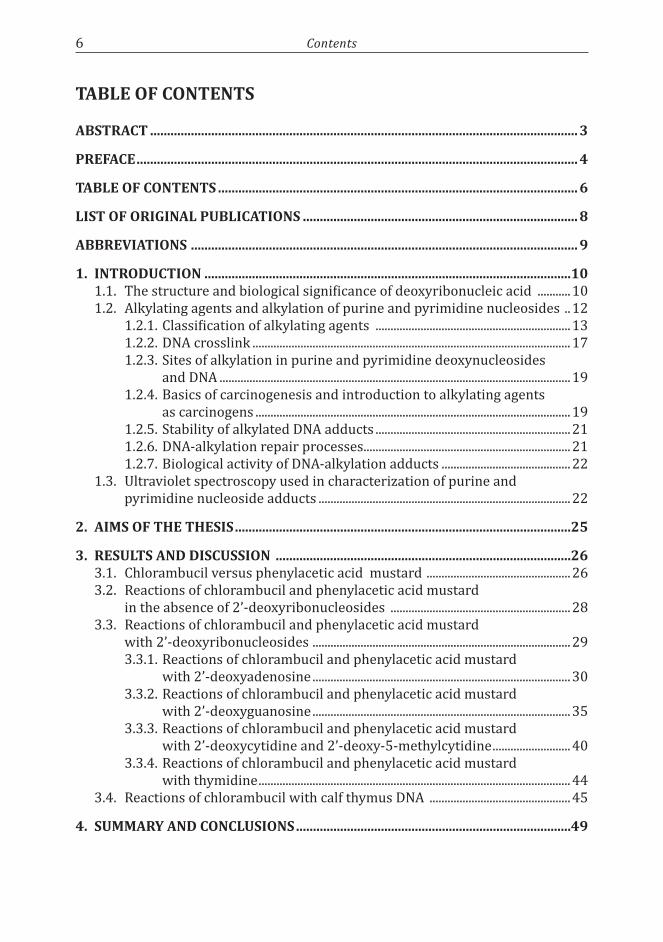
Products of CLB and PAM hydrolysis are shown in Chart 9. In the case of CLB, the major product was N,N-bis(2-hydroxyethyl)-p-aminophenyl butyric acid (23) with a molecular ion of 268. The second most abundant hydrolysis product was an ester (25) with a molecular ion of 517. Other products formed were esters 26 and 27 with a molecular ion of 780, and an ether derivative 24 with a molecular ion of 517; 24 was stable under basic conditions, which proved this structure to consist of an ether bond. The structures of 25-27, containing an ester linkage, were confirmed by saponification: all of them formed compound 23.
Results and Discussion 29
NOHHO
(CH2)n
NOHHO
(CH2)n ON
O
OH
(CH2)n
NOHHO
(CH2)n ON
O
OH
(CH2)n ON
O
OH
(CH2)n
NOHHO
(CH2)n ON
O
O
(CH2)n
2526
27
NOHO
(CH2)n OH
O
NOH
(CH2)n OH
O
OH
O
OH
O
24
(CH2)n
O
OH
O
N
OH
HO
23
OH
O
where n=1 for PAMn=3 for CLB
Chart 9. Main products of the hydrolysis of CLB and PAM
The pseudo-first-order rate constants were determined based on the hydrolysis of CLB and PAM at physiological conditions. The rate of decomposition of the two mustards were similar, as the kobs for the disappearance of CLB and PAM were 5.84×10⁻⁴ s-1 and 4,44×10⁻⁴ s-1, respectively. The effect of an external nucleophile, imidazole, on the product distribution was also studied. The results are incorporated in Table 1.
Table 1. Pseudo first-order rate constants (kobs) of hydrolysis and mole fraction (χ) of the diols of CLB and PAM as the function of [imidazole]; where diols refer to 4-(4-(bis(2-hydroxyethyl)amino)phenyl)butanoic acid and 2-(4-(bis(2-hydroxyethyl)amino)phenyl)acetic acid for CLB and PAM, respectively.
Alkylatingagent kobs [10-4 s-1]
χ[%]ofdiolinthepresence of 0.05 M
imidazole
χ[%]ofdiolinthepresence of 0.10 M
imidazoleClb 5.86 ± 0.11 0.51 0.21PAM 4.44 ± 0.03 0.52 0.21
3.3. Reactionsofchlorambucilandphenylaceticacidmustardwith2’-deoxyribonucleosides
2.5 mM CLB (8) and PAM (21), respectively, were allowed to react in the presence of 16.1 mM 2’-deoxynucleosides in 0.2 M cacodylic acid buffer (50 % base, pH 6.8) for 24 hours at 37 ̊C. However, the concentration of CLB in the reaction with 2’-deoxyadenosine (dAdo) was 0.6 mM. The nucleosides used in these studies were dAdo, 2’-deoxyguanosine (dGuo), 2’-deoxycytidine (dCtd), 2’-deoxy-5-methylcytidine (dMeCtd), and thymidine (Thd). CLB and PAM reacted with various heteroatoms of the nucleosides and numerous adducts were obtained, although the main reaction observed was the hydrolysis of CLB and PAM.
The reactions were followed by HPLC techniques. After the reactions were completed, the reaction mixtures were injected directly onto the column and analyzed by analytical HPLC. Then the products from the reaction mixtures were

30 Results and Discussion
separated by semipreparative HPLC and the obtained fractions were collected and characterized by means of UV, HPLC-MS, ESI-MS and NMR.
3.3.1. Reactions of chlorambucil and phenylacetic acid mustard with 2’-deoxyadenosine
The HPLC trace on the reaction of CLB with dAdo after 24 hours is shown in Figure 4. The substances marked as 28-34 are CLB - dAdo adducts, compounds 23 and 25 are products of CLB hydrolysis, while the peaks marked with asterisks are unidentified impurities.
figure 4. HPLC trace of the reaction of 0.6 mM CLB with 16 mM dAdo in 0.2 M cacodylic acid buffer (50 % base) after 24 hours at 37 ̊C.
Adducts 28 and 32 (Chart 10) gave the same molecular ion of 501 that referred to the structures were the alkylation had taken place at one site of dAdo. The presence of the fragment ion 385.3 in both adducts indicated the loss of the carbohydrate moiety and proved that the site of alkylation was at the adenine moiety. Several facts were taken into account to clarify whether the alkylation had occurred at the endocyclic or exocyclic nitrogen atom (N1 or N6, respectively) of dAdo: a) the intensity of 385.3 ion was larger in the MS spectrum of 32 than in the spectrum of 28; b) in the ultraviolet absorption spectra, the maximum absorption of the 28 is red-shifted comparing to 32 at neutral pH (Figure 5), which means that λmax value of 28 is lower than of 32; this is in accordance with previous literatures related to the UV spectral properties152, 101 for endocyclic and exocyclic N-atoms, respectively.

Results and Discussion 31
N
NN
N
NH
O
OH
NOH
28
HO
(CH2)nCOOH
N
NN
N
HN
O
OH
32
HO
NOH
(CH2)nCOOH
where n=1 for PAMn=3 for CLB
Chart 10. Structures of N1- and N6-alkylated adduct of CLB/PAM – dAdo reactions.
Wave length (nm)240 260 280 300
Absorbance
0
20
40
60
80
100
figure 5. UV spectra of substance 28 marked with dashed line and substance 32 marked with solid line.
The site of alkylation of compounds 28 and 32 was verified by comparison to the available literature data on 1H NMR.153 The resonance of the adenine-linked methylene protons is different when the alkylation is on endocyclic nitrogen or exocyclic nitrogen of adenine. In the case of CLB, the chemical shift of the proton from Ade-CH2 was shifted from 4.42 ppm for N1-alkylated adduct to 3.80 ppm for N6-alkylated derivative.
Based on the facts described above, 28 was assigned as an N1-adduct and 32 as an N6-adduct of dAdo. Substance 28 can be also assigned as N1-alkylated product based on the Dimroth rearrangement154,155,156 of 28 to 32 when treated with aqueous base, reaction that is characteristic for N1-alkyladenine nucleosides. This transformation is a base-catalyzed reaction.

32 Results and Discussion
It is known that different alkylating agents alkylate adenosine at N1 and N6 site.157 N6 adduct of the reaction of CLB with dAdo was the result of the direct alkylation of N6 site of the nucleoside, and not a result of the Dimroth rearrangement of the N1 adduct; that was demonstrated by the very slow rearrangement of N1 to N6 derivative under the reaction conditions employed.
Compound 31 (Chart 11) had the same molecular ion of 501, as 28 and 32, in the case of reaction of dAdo with CLB. This adduct was identified as a carbohydrate derivative based on its mass spectra that shows a fragment ion of 366, which represents the loss of adenine base. A similar spectrum was observed previously150 for an adduct of 2’-deoxyguanosine in which the alkylation had taken place at the 2’-deoxyriboside moiety. Because of steric reasons, substance 31 was tentatively assigned as 5’-O-alkylated adduct, and not as 3’-O adduct.
N
NN
N
NH2
O
OH
31
ON
HO
(CH2)nCOOHwhere n=1 for PAM
n=3 for CLB
Chart 11. Structures of 5’-O-alkylated adduct of CLB/PAM – dAdo reactions.
Adducts 29 and 30 (Chart 12) had the same molecular ion mass that referred to an adduct where the alkylation had taken place at only one site of the adenine base. In the case of CLB – dAdo reactions, the MH+ was 385 for compounds 29 and 30. This information and the fact that the alkylation at N3 or N7 of 2’-deoxyadenosine labilizes the N-glycosidic bond81, 101, 158 led us to the believe that 29 and 30 were N3- and N7-alkylated derivative.
29N
NN
N
NH2
N
HO
30
(CH2)nCOOH
N
NN
N
NH2
N
HO(CH2)nCOOH
where n=1 for PAMn=3 for CLB
Chart 12. Structures of N3- and N7-alkylated adduct of CLB/PAM – dAdo reactions

Results and Discussion 33
The final assignment was based on NMR experiments (1H- and 13C-NMR, COSY, NOESY, HSQC, HMBC) which were carried out to assign unequivocally the site of alkylation within these two adducts. Spectra were recorded in D2O or DMSO-d6 at 25 ̊C. Figure 6 shows the 1H NMR spectra of 29 and 30 in DMSO-d6. According to literature, 159,160 the Δδ of the chemical shifts of the H-2 and H-8 protons is larger with 3-alkyladenine than with the corresponding 7-alkyladenine. In the current case, Δδ of 29 was 0.36 ppm and Δδ of 30 0.13 ppm. The carbon chemical shifts in the 13C NMR spectra for 29 and 30 also showed clear differences between the alkylation at N3 or N7 of adenine.
figure 6. 1H NMR of N3-alkylated adenine (up) and N7-alkylated adenine (down); run in DMSO-d6 at 25 ̊C.
While the 1H- and 13C-NMR experiments together provided substantial evidence for structural characterization of the N3- and N7-adducts, the NOE correlation brought the most conclusive information for the identification of the alkylation site. It was crucial that the NOESY experiment was done in DMSO-d6, so that the NH2-protons and the correlation with them could be observed. Substance 30 showed correlation between the protons of NH2 of adenine and the protons of Ade-CH2 due to their vicinity in space (Figure 7) and it was assigned as N7-alkylated adduct, while substance 29 did not present this type of correlation and it was assigned as the N3-alkylated adduct.

34 Results and Discussion
figure 7. The significant NOE correlation in N7-alkylated adenine (30).
There are many reactive chlorambucil dimers, such as monochloro, dichloro and trichloro derivatives of 25, in the reaction mixtures and this could be an explanation for the appearance of esters in the reaction mixture. Substances 33 and 34 (Chart 13) were tentatively assigned as the esters of N1 adduct of 2’-deoxyadenosine based on their mass spectra and their lability under basic conditions due to hydrolysis of the ester function (saponification) present in their structure.
N
NN
N
NH
O
OH
NO
33
HO
(CH2)n
ON
OH
OH
(CH2)nCOOH
N
NN
N
NH
O
OH
NOH
34
HO
(CH2)n
O
O
N
OH
(CH2)nCOOH
where n=1 for PAMn=3 for CLB
Chart 13. Structures of N1-adducts derived from the reaction of CLB dimers/PAM dimers with N1-adduct of dAdo.

Results and Discussion 35
Cross-links, in which one chlorambucil molecule bridged two dAdo molecules, were not observed.
One extra minor adduct was produced by the reactions of PAM and CLB with dAdo. Its proposed structure is shown in Chart 14 and, based on the isotopic distribution observed in MS data, it might be N1-alkylated adduct of dAdo that still has a chlorine atom attached to ethyl chain of the molecule. It is not known why the second chlorine atom was not displaced.
N
NN
N
NH
O
OH
NCl
15
HO
(CH2)nCOOH
where n=1 for PAMn=3 for CLB
Chart 14. Structure of N1-adduct with a Cl-atom left present in PAM-dAdo reaction.
Hence, many adducts were detected and, as expected,31 the N1-alkylated derivative was the main adduct among them. Other alkylation sites observed in the reaction of CLB/PAM with dAdo were N6, N7, N3 and 5’-O.
3.3.2. Reactions of chlorambucil and phenylacetic acid mustard with 2’-deoxyguanosine
The reaction of CLB with dGuo had been in the attention of our laboratories before.150 In this thesis work, the reaction of phenylacetic acid with 2’-deoxyguanosine was studied and the products characterization was based on the previous literature.150
The HPLC trace for the reaction of PAM with dGuo after 24 hours is shown in Figure 8. Several adducts (35-45) were identified and characterized by means of HPLC-MS and 1H NMR.

36 Results and Discussion
figure 8. HPLC trace of the reaction of 2.5 mM PAM with 16.1 mM dGuo in 0.2 M cacodylic acid buffer (50 % base) after 24 hours at 37 ̊C. Note: 23, 25 are products of PAM hydrolysis and peaks marked with * are impurities.
The most abundant adduct formed was 35 (Chart 15). Its structure was analyzed by MS which showed the molecular ion 373 and a fragment ion 222 that corresponds to the mass of the CME substituent (Chart 15). The presence of ion 222 indicates the alkylation on guanine base. 1H NMR analyses proved that 35 is the N7-adduct of guanine. This is as expected31 because the N7 site is the most nucleophilic site of guanine base and thus, the site most prone to alkylation.
NH
N
NO
NH2N
N
HO(CH2)nCOOH
35 where n=1 for PAMn=3 for CLB
CME is 2-((4-(carboxymethyl)phenyl)(2-hydroxyethyl)amino)-ethyl substituen, when n=1
CPE is 2-((4-(3-carboxypropyl)phenyl)(2-hydroxyethyl)amino)-ethyl substituent, when n=3
Chart 15. MS spectra and structure of N7-alkylated guanine from the reaction of PAM with dGuo.

Results and Discussion 37
The second most abundant adduct was 36 that had a molecular ion of 506 (Chart 16) and it was assigned as a deglycosylated dimer based on its MS data.
NHN
NO
NH2
N
N
NHN
N N
O
H2N
(CH2)nCOOH
36where n=1 for PAMn=3 for CLB
Chart 16. MS spectra and structure of N7,N7-bis alkylated guanine from the reaction of PAM with dGuo.
Four products (37-40) had the same molecular ion of 489 (Chart 17). They were tentatively identified according to their MS data (Figure 9 a-d). Compound 37 had a fragment ion of 338 that indicated the loss of unmodified guanine base. This means that the reaction with the alkylating agent had taken place at the carbohydrate moiety, most likely at 5’-O. Substance 37 was identified as the 5’-O-alkylated adduct.
Fragment ion 373 is present in the mass spectra of compounds 38, 39 and 40, indicating the loss of the sugar moiety, and that the alkylation had taken place at the guanine base. Thus, the alkylation site could to be N1, N2, N3 or O6 site of guanine base. Compounds 39 and 40 had a fragment ion 222 indicating the cleavage of the CME group. Based on our observations, the absence of that fragment was characteristic to the compounds were the alkylation had taken place at their endocyclic N atom, while its presence was an indication that the alkylation had taken place at one of the exocyclic heteroatoms of the guanine base. In the case of 39, the intensity of the signal at 222 was much higher than with 40 and, on the basis of the previous studies of CLB with dGuo,150 that could be characteristic to the O6-alkylated compound which is more labile. The final identification of 39 as an O6-alkylated adduct and 40 as an N2-alkylated adduct was based additionally on the lability of 39 under basic conditions, which was observed previously in the case of O-alkyl groups.89 Substance 38 was assigned as an N1-alkylated derivative based on its fragment ion 373 and the missing fragment ion 222. The N3-alkylated adduct was detected neither in PAM-dGuo reaction nor in CLB-dGuo reaction and one may speculate that this might be due to the large size of PAM/CLB molecule that make difficult for such molecule to react with the sterically hindered N3 site of dGuo.

38 Results and Discussion
However, knowing that PAM/CLB reacts with the N3 position of dAdo (see section 3.3.1.) and that dAdo has same large size as dGuo, probably additional factors beside steric effect are involved.
NH
N
N
O
NH2N
O
OH
ON
HO
(CH2)nCOOH
37
N
N
N
O
NH2N
O
OH
HO
NOH
(CH2)nCOOH
38
N
N
N
O
NH2N
O
OH
HO
NOH
(CH2)nCOOH
39
NH
N
N
O
NH
N
O
OH
HON
OH
(CH2)nCOOH
40 where n=1 for PAMn=3 for CLB
Chart 17. Structures of 5’-O- (37), N1- (38), O6- (39), and N2- (40) alkylated derivatives of reactions between dGuo and PAM/CLB.

Results and Discussion 39
figure 9. MS spectra of 5’-O adduct (a), N1 adduct (b), O6 adduct (c), and N2 adduct (d).
Substances 41-45 (Chart 18) all showed the same molecular ion of 594 in the case of reaction of dGuo with PAM. Compound 41 was identified as a N7,N9-bis adduct. 44 and 45 were tentatively assigned as the N7 adducts derived from the reactions of two different PAM dimers with N7 of guanine; the corresponding derivatives were not observed in the reaction of dGuo with CLB. The formation of N7,N9-bis alkylated adduct was expected, since its presence had previously been reported.161 Compounds 42 and 43 could not be identified on the basis of their MS data, but they might be the isomeric adducts of 44 and 45 or positively charged dialkylated guanine analogs of 41.

40 Results and Discussion
NH
N
N
O
NH2N+
N
HO
N
HO(CH2)nCOOH
(CH2)nCOOH
41NH
N
N
O
NH2N
N
O
O
NOHHO
OH
O
44
NH
N
N
O
NH2N
N
HO
OO
N
OHHO
O
45
where n=1 for PAMn=3 for CLB
Chart 18. Structures of N7,N9-bis alkylated adduct (41), and esters of N7-alkylated guanine derivatives (44, 45) formed by reactions between dGuo and CLB, dGuo and PAM.
Thus, many adducts were detected. N7-alkylated derivative was the main adduct among them. Other alkylation sites observed in the reaction of CLB/PAM with dGuo were 5’-O, N1, O6, N2 and N9.
3.3.3. Reactions of chlorambucil and phenylacetic acid mustard with 2’-deoxycytidine and 2’-deoxy-5-methylcytidine
The HPLC trace of the reaction of 2’-deoxycytidine with chlorambucil after 24 hours is presented in Figure 10 and the corresponding HPLC trace of the reaction of 5-methyl-2’-deoxycytidine with chlorambucil is shown in Figure 11. dCtd and dMeCtd, which differ from one to another only by a methyl group in C-5 position, exhibit quite similar alkylation sites in their reactions with CLB/PAM. The structures of these adducts can be seen in Chart 19. The reaction of CLB with dCtd and dMeCtd is taken as an example.
figure 10. HPLC trace of the reaction of 2.5 mM CLB with 16.1 mM dCtd in 0.2 M cacodylic acid buffer (50 % base) after 24 hours at 37 ̊C.

Results and Discussion 41
Four of the detected adducts (46a, 47a, 49a and 53) gave the same molecular ion 477, which referred to a monomeric adduct of dCtd. The alkylation on the cytosine base of 46a is proven by the presence of the fragment ion of 361, which shows the loss of the sugar moiety. Further, the 1H resonance of the methylene group linked to the pyrimidine moiety exhibits values (4.37 and 4.08 ppm) comparable to those cases where the alkylation had occurred at the endocyclic nitrogen.153 Hence, compound 46a was assigned as the N3 derivative.
HOO
OH
N
N
NH
O
NOH
OO
OH
N
N
NH2
ON
HO
HOO
OH
N
N
NH
ON
OH
HOO
OH
N
N
O
NOH
O
50a,b
47a,b
49a,b
46a,b
HOO
OH
N
N
NH
O
NO
O
NOH
HO
HOO
OH
N
N
NH
O
NOH
(CH2)nO
NO
OH
51a,b
52a,b
R
R
R
R
R
N
N
NH2
ON
OH
R
48a,b
where a: R = H; b: R = CH3n=1 for PAMn=3 for CLB
R
HOO
OH
N
N
NH2
O
NHO
53
(CH2)nCOOH (CH2)nCOOH (CH2)nCOOH
(CH2)nCOOH
(CH2)nCOOH
(CH2)nCOOH
(CH2)3COOH
(CH2)nCOOH
(CH2)n
Chart 19. Structures of the products of the reactions between dCtd and CLB (46a to 52a, 53), and dMeCtd and CLB (46b to 52b).
Substance 47a was characterized by the MS data. The mass spectrum showed a strong fragment ion 250, which indicated that the alkylation had occurred on the exocyclic oxygen150 (see 3.3.2.). 47a additionally had the fragment ions 361 and 366, which referred to the loss of alkylated cytosine base and alkylated sugar moiety, respectively. This could be explained by alkylation of O2 and migration of the alkyl group from O2 to 5’-O of the deoxyribose moiety in the spectrometer during the MS analysis. The corresponding O2-derivative was not observed in the reaction of PAM with dCtd.
42 Results and Discussion
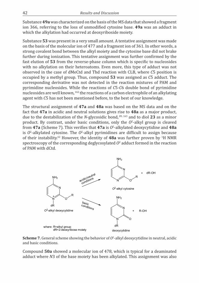
Substance 49a was characterized on the basis of the MS data that showed a fragment ion 366, referring to the loss of unmodified cytosine base. 49a was an adduct in which the alkylation had occurred at deoxyriboside moiety.
Substance 53 was present in a very small amount. A tentative assignment was made on the basis of the molecular ion of 477 and a fragment ion of 361. In other words, a strong covalent bond between the alkyl moiety and the cytosine base did not brake further during ionization. This tentative assignment was further confirmed by the fast elution of 53 from the reverse-phase column which is specific to nucleosides with no alkylation on their heteroatoms. Even more, this type of adduct was not observed in the case of dMeCtd and Thd reaction with CLB, where C5 position is occupied by a methyl group. Thus, compound 53 was assigned as C5 adduct. The corresponding derivative was not detected in the reaction mixtures of PAM and pyrimidine nucleosides. While the reactions of C5-C6 double bond of pyrimidine nucleosides are well known, 162 the reactions of a carbon electrophile of an alkylating agent with C5 has not been mentioned before, to the best of our knowledge.
The structural assignment of 47a and 48a was based on the MS data and on the fact that 47a in acidic and neutral solutions gives rise to 48a as a major product, due to the destabilization of the N-glycosidic bond, 89, 163 and to diol 23 as a minor product. By contrast, under basic conditions, only the O2-alkyl group is cleaved from 47a (Scheme 7). This verifies that 47a is O2-alkylated deoxycytidine and 48a is O2-alkylated cytosine. The O2-alkyl pyrimidines are difficult to assign because of their instability.62 However, the identity of 48a was further proven by 1H NMR spectroscopy of the corresponding deglycosylated O2 adduct formed in the reaction of PAM with dCtd.
dR
N
N
NH2
OR+
O2-alkyl deoxycytidine
acidic/neutral
conditions N
N
NH2
OR
O2-alkyl cytosine
+ dR-OH
dR
N
N
NH2
O
deoxycytidine
+ R-OH
basicconditions
where: R=alkyl groupdR=2-deoxyribose moiety
scheme 7. General scheme showing the behavior of O2-alkyl deoxycytidine in neutral, acidic and basic conditions.
Compound 50a showed a molecular ion of 478, which is typical for a deaminated adduct where N3 of the base moiety has been alkylated. This assignment was also

Results and Discussion 43
supported by the coelution of 50a with the product raised from the treatment of pure 46a with basic solution.
Based on their molecular ion of 726 and lability under basic conditions, substances 51a and 52a were tentatively assigned as esters formed from the reaction of CLB dimer with N3 of dCtd.
The HPLC trace of the reaction of chlorambucil with 5-methyl-2’-deoxycytidine is shown in Figure 11. The products formed in this reaction are similar to those obtained by the reaction of CLB with dCtd (Chart 19).
figure 11. HPLC trace of the reaction of 2.5 mM CLB with 16.1 mM dMeCtd in 0.2 M cacodylic acid buffer (50 % base) after 24 hours at 37 ̊C.
The major product was the N3 adduct 46b, as judged by 1H NMR and MS analyses. The carbohydrate derivative 49b, the deaminated N3 adduct 50b, and the N3 esters 51b and 52b were present in comparable quantities in the reaction mixtures. The O2 adducts, including O2-alkylated nucleoside and the corresponding nucleobase, were present in higher amount than in the reaction with dCtd: the relative abundance of (47b+48b) was 7.5 % compared to the relative abundance of (47a+48a) that was 2.4 %; no explanation could be given for this difference.
The relative abundance of the O2-alkylated products in the reaction of dMeCtd with PAM was also much higher than in the case of dCtd: 8.3 % vs. 2.6 %. The deaminated derivative, in turn, was present in a very small amount. It even was very difficult to be detected.
No cross-linked derivative where a single CLB or PAM molecule was connected to two dCtd or two dMeCtd molecules could be detected. No alkylation at N4 of pyrimidine nucleosides was observed. In this respect the results differ from those of the earlier studies of cytidine with ethylating agents10 or 2’-deoxycytidine with epoxides,164 in which the N4-alkylated adduct was formed at neutral pH.
The N3-alkylated dCtd and dMeCtd were relatively stable under the reaction conditions employed. Even so, they deaminated considerably to their corresponding

44 Results and Discussion
derivatives 50a and 50b at neutral pH, especially in the case of CLB. This was surprising because the spontaneous deamination of cytosine nucleoside is known to be slow. 162,165 Possibly the N3-alkylation enhances the deamination since the rates of deamination were similar for the N3 derivatives in the reactions of dCtd and dMeCtd with CLB; in this situation, the corresponding N3 derivative of dCtd-PAM deaminated more readily than the corresponding dMeCtd-PAM. This is also unexpected because it is known that dMeCtd is more susceptible to deamination than dCtd.162
N3 derivatives of dCtd and dMeCtd were treated in aqueous base and only the deaminated products were observed, in contrast to other cytosine derivatives166 which gave also products of Dimroth rearrangement.
Compound 54 is probably a nonnucleosidic impurity, as suggested by its UV and MS spectrum.
3.3.4. Reactions of chlorambucil and phenylacetic acid mustard with thymidine
Thymidine is the least reactive nucleoside among the purine and pyrimidine nucleosides studied in this thesis work. The HPLC trace of the reaction of chlorambucil with thymidine is shown in Figure 12 and the products formed in this reaction can be seen in Chart 20.
figure 12. HPLC trace of the reaction of 2.5 mM CLB with 16.1 mM Thd in 0.2 M cacodylic acid buffer (50 % base) after 24 hours at 37 ̊C.

Results and Discussion 45
O
OH
HO
N
N
O
O
H3C NOH O
OH
O
N
NH
O
O
H3C
NHO
50b
55
(CH2)nCOOH
(CH2)nCOOHwhere n=1 for PAM
n=3 for CLB
Chart 20. Structures of products of thymidine – chlorambucil reaction
Product 50b was characterized by means of 1H NMR, MS/MS, HPLC-MS analyses and by coelution with the deaminated N3 derivative of 5-methyl-2’-deoxycytidine. 50b is the N3 alkylated derivative and it is the main alkylation site of thymidine, as expected.167
The second abundant adduct, 55, was assigned as a carbohydrate derivative on the basis of the HPLC-MS analysis. No other adducts were detected in the case of thymidine.
3.4. ReactionsofchlorambucilwithcalfthymusDNA
The reactions of CLB with single-stranded and double-stranded calf thymus DNA were performed in 0.2 M cacodylic acid (50 % base) at pH 6.8, at 37 ̊C for 24 hours. Then, the DNA was enzymatically hydrolyzed to 2’-deoxynucleosides. On the basis of NMR, UV, HPLC, LC-ESI-MS/MS studies, as well as by spiking with standard samples obtained in our previous work (i, ii), the major DNA-CLB adducts were assigned as N1 adduct of 2’-deoxyadenosine (28), N3 adduct of 2’-deoxyadenosine (29), N3 adduct of 2’-deoxycytidine (46a) and N7 base adduct of 2’-deoxyguanosine (56) (Figure 13, Chart 21).

46 Results and Discussion
0 5 10 15 20
0
10
20
30
40
50
60
7056
46a29
mAU
min
28
figure 13. HPLC trace of the reaction of chlorambucil (2.5 mM) with calf thymus single-stranded DNA in 0.2 M cacodylic acid buffer (50 % base) after 24 hours at 37 ̊C.
Several minor products were detected and characterized by coelution with authentic samples and by MS. Among these adducts were N6 adduct of 2’-deoxyadenosine (32), N7-alkylated nucleoside (58) and N7,N7-bis (57) adducts of 2’-deoxyguanosine, deaminated N3 (50a) and O2-alkylated (47a) adducts of 2’-deoxycytidine, and N3 adduct of thymidine (50b). All these products were present in the hydrolysates of both, single-stranded and double-stranded DNA. 57 was the only crosslink detected.
Other adducts (60, 31, 49a) were tentatively characterized as “sugar” derivatives based on their MS data. They might be 3’-O-alkylated nucleoside, knowing that the 5’-O of DNA is phosphorylated. On other hand, the DNA does not have many free 3’-hydroxy groups and these products may be artifacts.

Results and Discussion 47
NH
N
N
O
NH2N
N
N
ArN
HN
NO
H2N
56
NH
N
N
O
NH2N
NAr
HO
57
NH
N
N
O
NH2N
O
OH
HO
+
NAr
58
N
NN
NNH
O
OH
HO
NOH
28
N
NN
NNH2
32
N
NN
NHN
O
OH
HO
NOH
Ar
29
O
OH
HON
N
NH
O
NOH
Ar
46a
50aO
OH
HON
N
O
O
NOH
Ar
BaseO
O
HO
NAr
HO
Ar
Base = Gua; 60Base = Ade; 31Base = Cyt; 49a
O
OH
HON
N
NH
ON
OH
Ar
47a
O
OH
HON
N
O
O
NOH
Ar
50b
H3C
NH
N
N
O
NH2N
NAr
HO
NAr
HO
+
59
NAr
HO
where Ar = (CH2)3COOH
OH
Chart 21. Structures of products of reactions of CLB with ssDNA and dsDNA
N7,N9-Bis alkylated derivative of guanine (41) was also observed, as well as other minor adducts.
These reactions were performed in non-nucleophilic buffer, which means that a CLB molecule has only two options: to react with water (hydrolyses) or to react with DNA. Important to remember is also that the reactivity and concentration of nucleophile determines the site of reaction.
Single-stranded DNA was approximately 3.4 times more reactive than double-stranded DNA. The reactivity order in ssDNA and dsDNA was partially different than in the case of nucleobases (Table 2). The most reactive site in ssDNA was N1 of Ade, then N7 of Gua, N3 of Cyt and N3 of Ade. The order of reactivity, from most reactive site to less reactive site, in dsDNA was: N3 of Ade, N7 of Gua, N3 of Cyt and N1 of Ade, last two being approximately equally reactive.

48 Results and Discussion
Table 2. The order of reactivity of the nucleophilic sites of nucleobases with CLB.
more reactive less reactiveNucleosides Gua-N7 Ade-N1 Cyt-N3 Ade-N7 / Ade-N3ssDNA Ade-N1 Gua-N7 Cyt-N3 Ade-N3dsDNA Ade-N3 Gua-N7 Cyt-N3 / Ade-N1 Cyt-N3 / Ade-N1
Surprising was that N3 of Ade was the most reactive site in dsDNA, and not N7 of Gua. It is known from different studies on the reaction of chlorambucil with DNA that the principal site of alkylation is N7 of guanine168,169,170 and the second site is N3 of adenine.171 The reason for this situation is that guanine N7 is situated in the major groove and easily accessible for different alkylating agents, while adenine N3 is accommodated with very little distortion of the DNA helix.172
Also surprising was that N1 of Ade was the most reactive site in ssDNA, and N7-alkylated adduct of Ade could not be detected in ssDNA or in dsDNA.
It is known that the nucleophilic sites of nucleobases participating to the base pairing in dsDNA are partly protected from alkylation. That could be observed also in our case, where the N3 of Cyt and N1 of Ade were less reactive in dsDNA than in ssDNA. Exception is offered by the O2-alkylated adduct of dCtd which was stable enough to be found also in the hydrolysate of dsDNA.
The molar yields of the main adducts were calculated from the original amount of 2-deoxyribonucleosides (Table 3).
Table 3. Yields of the major adducts present in the DNA hydrolysate.
Adducts mol/106 mol of normal nucleotides gua-N7 dAdo-N1 Ade-N3 dCtd-N3
ssDNA 320 390 224 234dsDNA 90 3 245 4

Summary and Conclusions 49
4. suMMARy AND CONClusiONs
CLB (8) was found to be hydrolytically slightly less stable than its metabolite PAM (21). CLB and PAM reacted with different heteroatoms of the 2’-deoxyribonucleosides, even though the predominant reaction observed was the hydrolysis of 8 and 21. The overall reactivity of CLB towards dGuo,150 dAdo, dCtd, dMeCtd, and Thd was approximately 24, 7, 10, 10, and < 1 %, respectively, based on the decrease of the products of CLB hydrolysis. PAM’s overall reactivity towards dGuo, dAdo, dCtd, dMeCtd, and Thd was ca. 25, 10, 10, 10, and < 1 %, respectively, based on the decrease of the products of PAM hydrolysis. One difference in the reactivity of CLB and PAM is that the N3 dCtd-PAM adduct was deaminated easier than the corresponding CLB derivative.
The order of reactivity of the nucleophilic sites of nucleobases with chlorambucil is in accordance with previous data,173 while the situation was different in the case of ssDNA and dsDNA: nucleosides Gua-N7 > Ade-N1 > Cyt-N3 > Ade-N7 = Ade-N3, ssDNA Ade-N1 > Gua-N7 > Cyt-N3 > Ade-N3, and dsDNA Ade-N3 > Gua-N7 > Cyt-N3 = Ade-N1.
This thesis work was concentrated at first on the analyses of the nucleosides’ reactions with CLB and PAM and the data obtained from them was used as example for the reactions of DNA with CLB. Many adducts were detected and characterized in our work and they may be of biological importance. The medical implication of DNA damage caused by alkylation is becoming more and more important.13 For example, in the clinical chemotherapy, identifying the CLB adducts formed with DNA, as well as the system used in their repair, help in understanding the drug resistance and its side effects.174 CLB and PAM have similar chemical behavior, as we have showed,121 and this is in accordance with the previous suggestions that these two mustards cause DNA lesions with similar effectivines.80,175

50 Experimental Section
5. exPeRiMeNTAl seCTiON
5.1. General
2’-Deoxyribonucleosides (dGuo, dAdo, dCtd, dMeCtd, and Thd), cacodylic acid, N,N-bis(2-chloroethyl)-p-aminophenylbutyric acid, calf thymus DNA (Type I: sodium salt, highly polymerized), nuclease P1 from Penicillium citrinum, alkaline phosphatase (Escherichia coli Type III), acid phosphatase (wheat germ Type I) and bis[2-hydroxyethyl]iminotris[hydroxymethyl]methane hydrochloride (Bis-tris) buffer were purchased from Sigma, and they were used as received. N,N-Bis(2-hydroxyethyl)-p-aminophenylbutyric acid and {4-[bis(2-chloroethyl)amino]phenyl}acetic acid were synthesized as described previously.130,140 All inorganic reagents were of ACS grade or better.
5.2. Chromatographicmethods
AnalyticalHPLC analyses were performed on a Hewlett-Packard 1090 Instrument equipped with a diode array detector and autosampler. The reversed-phase column in use was Hypersil ODS2 C18, 4.6 mm × 250 mm with particle size of 5 µm (i), or Waters Symmetry C18, 4.6 mm × 150 mm with particle size of 3.5 µm (ii, iii, iv). The columns were eluted with different gradients of acetonitrile in 10 mM ammonium acetate containing 0.1 % of acetic acid, and with a flow rate of 1 mL min-1. The solvents for chromatography were filtered and degassed prior use. The reaction mixtures were injected directly to the HPLC.
Preparative HPLC analyses (isolation of adducts) were performed on a Merck-Hitachi instrument consisting of a L-6200A Intelligent pump, L-4000 UV detector, and D-2500 Chromato-Integrator. Hypersil C18 reversed-phase column with dimensions of 10 mm × 250 mm, and particle size of 8 µm (i, ii), or Hypersil ODS C18 column, 10 × 250 mm, and particle size of 5 µm (iii), or Waters SymmetryPrep C18 with diamentions of 7.8 mm × 150 mm and particle size of 7 µm (iii, iv) were used for these analyzes. Same buffers as in analytical HPLC were used. The flow rate of the eluents (buffers) was 4 mL min-1 (i, ii), or 2 mL min-1 (iii, iv), or 2.5 mL min-1 (iv).
5.3. Spectroscopicandspectrometricmethods
All adducts were characterized by nuclear magnetic resonance spectroscopy, ultraviolet spectroscopy and mass spectrometry.
NMRanalyses were performed on JEOL Alpha 500 instrument operating at 500.0 MHz for 1H (i), Bruker Avance 600 spectrometer (ii, iii) and Bruker Avance 500 spectrometer (iv) equipped with BBO-5mm-Zgrad probe operating at 500.13 and 125.77 MHz for 1H and 13C, respectively. Isolated pure compounds were dissolved in D2O and the signal of HDO was used as an internal reference in i, ii, iii. DMSO-d6

Experimental Section 51
was used as a solvent for the NMR samples in iv and its instrument’s value was used as an internal reference.
Depending on the amount of isolated product and of how much information was required from that specific sample, different NMR experiments were performed: 1H NMR, 13C NMR, COSY, NOESY, HSQC, HMBC.
The HPLC instrument was equipped with a diode-array detector that recorded the UVspectra of adducts as the compounds eluted from the reversed-phase liquid chromatography column.
The MSanalyses were performed on several mass-spectrometry instruments: a Perkin-Elmer Sciex API-365 Triple-Quadrupole LC/MS/MS system, equipped with a PE 200 Micro pump and a PE Series 200 Autosampler (i); a Perkin-Elmer Sciex API-365 Triple-Quadrupole LC/MS/MS system, equipped with two PE 200 Micro pumps, a PE Series 200 Autosampler, and an Applied Biosystems 783A UV detector connected to a LKB 2210 two channel recorder (ii); an Applied Biosystems API-4000 Triple-Quadrupole LC/MS/MS system, equipped with a Shimadzu-Prominence HPLC system and an Applied-Biosystems-783A UV detector connected to an LKB-2210 two-channel recorder (iii); an Agilent 1100 Series LS/MSD Trap SL instrument, equipped with an electrospray source (iv); and Micro LC triple-quadrupole mass spectrometer, equipped with an electrospray interface (iv).
Same RP-HPLC columns and buffers used in the analytical HPLC analyses were used also in the MS analyses.
A variety of mass-spectrometry studies were done in order to obtain a consistent characterization of products: HPLC/MS, MS/MS, HPLC/ESI-MS, HPLC/ESI-MS/MS.
5.4. Quantitativeproductanalyses
Overall reactivity of chlorambucil and phenylacetic acid mustard withnucleosides. CLB was allowed to react in the presence and absence of 2’-deoxyribonucleosides over night in cacodylic acid buffer at 37 ̊C. The reaction mixtures were analyzed by analytical HPLC and the amount of nonnucleosidic products 23-27 was quantitated by HPLC at 267 nm (isosbestic point of the reaction of CLB and PAM with nonchromophoric nucleophiles139) in the absence of nucleosides. Then the amount of 2’-deoxyribonucleosides alkylation was calculated based on the decrease of the mole fraction of 23-27 in the presence of nucleosides. Similar procedure was used in the case of PAM too.
CLB and PAM were also allowed to react in the presence and absence of 1H-imidazole in cacodylic acid buffer (50 % base, 1.0 M NaClO4, pH 7.4), in the presence of 2’-deoxyribonucleosides. The mole fraction of all products (CLB and PAM with imidazole, CLB and PAM with nucleosides) were calculated as described above.
Relative adduct levels. The relative levels of the most-relevant adducts formed by reaction of CLB or PAM with nucleosides were calculated considering that all

52 Experimental Section
adducts of each nucleoside have same extinction coefficient. Reaction mixtures were run on analytical HPLC and the yield of the most abundant adduct of each nucleoside was considered as 100 %; other adducts were calculated by comparison with it.
Determinationofadducts’yieldswithcalfthymusDNA. The yields of the major adducts of CLB reaction with ssDNA and dsDNA where determined based on the quantitative 1H NMR analysis.176,177,178
Dry isolated adducts were dissolved in a known amount of D2O in the case of CLB-dAdo and CLB-dCtd, and in DMSO-d6 in the case of CLB-dGuo. A known amount of a 5 % triethylamine solution in D2O and DMSO-d6, respectively was added to each of the adduct sample as an internal standard and 1H NMR was run. The concentration of adduct in each NMR sample was calculated by comparing the integral of one proton signal of adduct to the integral of the CH3 signal of triethylamine, of which concentration was known. An exact volume of NMR sample was diluted with a suitable volume of water and run on a HPLC instrument equipped with a diode array detector. The UV detection used was at 267 nm and the quantitative determination of adducts was done by comparing the peak area of these HPLC standard samples with the peak area of adducts in the reaction mixtures. The original amount of 2’-deoxyribonucleosides in the reaction mixture was used to calculate the molar yields of adducts.
5.5. Stabilityoftheendproducts
CLB and PAM were allowed to react with nucleosides in 0.2 M cacodylic acid buffer, 37 ̊C for 24 hours. The products were isolated by preparative HPLC. The collected fractions were concentrated and then dissolved in 0.1 M NaOH, 0.1 M HCl, or 0.2 M cacodylic acid (50 % base); after 24 hours at 37 ̊C, samples were analyzed by HPLC.
5.6. Dimrothrearrangement
CLB and PAM were allowed to react with 2’-deoxyadenosine overnight in the 0.2 M cacodylic acid buffer (50 % base, pH 6.8) at 37 ̊C. The N1 adduct was separated by semipreparative HPLC from the reaction mixture, diluted 10-fold with cacodylic acid buffer or 0.1 M NaOH and kept overnight at 37 ̊C (i) or room temperature (iii). HPLC was used to follow the reactions. The mixture was neutralized with aqueous HCl prior HPLC analysis.
5.7. Kineticmeasurement
Reactions were carried out in a nonnucleophilic buffer (0.2 M cacodylic acid buffer, 50 % base, pH 6.8) at 37 ̊C for 24 hours in the presence and absence of dAdo, dGuo, dCtd, dMeCtd, and Thd (16.1 mM). The initiation of the reactions was done by adding a stock solution of CLB or PAM in acetonitrile so that a final concentration of

Experimental Section 53
0.6 mM CLB (i) or 2.5 mM CLB (iii) and 2.5 mM PAM (iii) was obtained. The final reaction mixtures included 1 % (v/v) acetonitrile.
In the case of PAM, the ionic strength of the cacodylic acid buffer was adjusted to 1.0 M with NaClO4.
Reactions were performed in stoppered bottles and aliquots withdrawn at suitable intervals from the reaction mixture were cooled down in ice-water bath, then stored at -20 ̊C and melted before the high-performance liquid chromatography analysis. Same RP-HPLC columns, buffers, gradients and flow rate as in the analytical HPLC analyses were used.
5.8. Reactionsofchlorambucilandphenylaceticacidmustardwith2’-deoxyribonucleosides
Chlorambucil (0.6 mM in i, 2.5 mM in ii) and phenylacetic acid mustard (2.5 mM in iii) were allowed to react overnight in a nonnucleophilic buffer (aqueous 0.2 M cacodylic acid, 50 % base, pH 7.4) at 37 °C in the presence of dGuo, dAdo, dCtd, dMeCtd, and Thd (16.1 mM), respectively. The reaction mixtures were analyzed by HPLC techniques after 24 hours and main adducts formed were isolated by preparative HPLC, then characterized and identified based on their UV spectra, HPLC/MS, HPLC/ESI/MS, HPLC/MS/MS data and/or 1H NMR spectra.
5.9. ReactionsofchlorambucilwithcalfthymusDNA
The reactions of CLB with calf thymus DNA were performed in similar conditions to the reactions of CLB with nucleosides, with one exception: concentration of DNA was different than the concentration of nucleosides.
Single-stranded (ss) DNA was obtained from double-stranded (ds) DNA by heating the solution of dsDNA to 100 °C for 10 min and then cooling it down rapidly on ice.179 Chlorambucil was allowed to react with ssDNA and dsDNA, respectively in 0.2 M cacodylic acid buffer (50 % base) overnight at physiological pH and 37 °C. The modified DNA was enzymatically hydrolyzed to 2’-deoxyribonucleosides by following the methods described in the literature.178 The precipitation of DNA was done by adding 5 M NaCl solution, cold ethanol and cooling the DNA solution to -20 ̊C. The mixtures were centrifuged and the recovered DNA was washed with cold 70 % ethanol, cold ethanol and then dissolved in water. The DNA was reprecipitated from the solution adding cold ethanol, cooling to -20 ̊C and recovered by centrifugation. The recovered DNA was enzymatically hydrolyzed by dissolving it in 100 mM Bis-tris-MgCl2 buffer, adding Nuclease P1, bacterial alkaline phophatase, wheat germ acid phosphatase and incubating for several hours at 37 ̊C. The enzymatically digested mixtures were filtered while centrifuging, then the ultrafiltrates were collected, evaporated to dryness, reconstituted in 20 % methanol solution and analyzed by HPLC and LC-ESI-MS/MS.

54 References
6. RefeReNCes1 McMurry, J. Organic Chemistry, Thomson Learning, 2004, chapter 28.
2 Nelson, D. L. and Cox, M. M. Lehninger Principles of Biochemistry, W.H. Freeman & Co, 4th Edition, 2005, chapter 8.
3 Schmid, G. H. Organic Chemistry, Mosby-Year Book, Inc., 1996, chapter 27.
4 Wade Jr., L. G. Organic Chemistry, Pearson Education, Inc., 6th Edition, 2006, chapter 23.
5 Heftmann, E. Chromatography, 6th edition: Fundamentals and applications of Chromatography and related differential migration methods, 2004, 906.
6 He, Y., Liu, H., Chen, Y., Tian, Y., Deng, Z., Ko, S. H., Ye, T. and Mao, C. Microsc. Res. Tech. 2007, 70, 522-529.
7 Hoeijmakers, J. H. J. Maturitas 2001, 38, 17-23.
8 Hoeijmakers, J. H. J. Nature 2001, 411, 366-374.
9 Hayatsu, H. J. Biochem. (Tokyo) 1996, 119, 391-395.
10 Sun, L. and Singer, B. Biochemistry 1974, 13, 1905-1913.
11 Aguilar-Mahecha, A., Hales, B. F. and Robaire, B. Biol. Reprod. 2005, 72, 1297-1304.
12 Barrows, L. R., and Magee, P. N. Carcinogenesis 1982, 3, 349-351.
13 Drabløs, F., Feyzi, E., Aas, P. A., Vaagbø, C. B., Kavli, B., Bratlie, M. S., Peňa-Diaz, J., Otterlei, M., Slupphaug, G. and Krokan, H. E. DNA Repair, 2004, 3, 1389-1407.
14 Goldman, R. and Shields, P. G. J. Nutr. 2003, 133, 965S-973S.
15 Hurley, L. H. Nat. Rev. Cancer 2002, 2, 188-200.
16 Lundin, C., North, M., Erixon, K., Walters, K., Jenssen, D., Goldman, A. S. H. and Helleday, T. Nucleic Acids Res. 2005, 33, 3799-3811.
17 Lindahl, T. DNA Repair 2004, 3, 1521-1530.
18 Eder, E., Kütt, W. and Deininger, C. Chem. Biol. Interact. 2001, 137, 89-99.
19 Nagao, T., Morita, Y., Ishizuka, Y., Wada, A. and Mizutani, M. Teratogenesis Carcinog. Mutagen. 1991, 11, 1-10.
20 Wada, A. and Nagao,T. Cong. Anom. 1994, 34, 65-70.
21 IARC. Overall Evaluation of Carcinogenicity. IARC Monographs on the Evaluation of Carcinogenic Risk of Chemicals to Humans, Supplement 7. Lyon, France: International Agency for Research on Cancer, 1987, 440.
22 Fischhaber, P. L., Gall, A. S., Duncan, J. A. and Hopkins, P. B. Cancer Res. 1999, 59, 4363-4368.
23 Tong, W. P., Kohn, K. W. and Ludlum D. B. Cancer Res. 1982, 42, 4460-4464.
24 Dillman, J. F. III, Hege, A. I., Phillips, C. S., Orzolek, L. D., Sylvester, A. J., Bossone, C., Henemyre-Harris, C., Kiser, R. C., Choi, Y. W., Schlager, J. J. and Sabourin, C. L. J. Pharmacol. Exp. Ther. 2006, 317, 76-87.
25 Shooter, K. V. Chem. Biol. Interact. 1975, 11, 575-588.
26 Romano, K. P., Newman, A. G., Zahran, R. W. and Millard, J. T. Chem. Res. Toxicol. 2007, 20, 832-838.

References 5527 Guengerich, F. P., Langouët, S., Mican, A. N., Akasaka, S., Müller, M. and Persmark, M. Exocyclic DNA adducts
in mutagenesis and carcinogenesis), IARC Scientific Publications No. 150, Singer, B. and Bartsch, H. Ed., 1990, 137.
28 Scherer, E., Van der Laken, C. J., Gwinner, L. M., Laib, R. J. and Emmelot, P. Carcinogenesis 1981, 2, 671-677.
29 Iglesias-Guerra, F., Romero, I., Alcudia, F. and Vega-Pérez, J. M. Carbohydr. Res. 1998, 308, 57-62.
30 Österlund, C., Lilliehöök, B., Ekstrand Hammarström, B., Sandström, T. and Bucht, A. J. Appl. Toxicol. 2005, 25, 328-337.
31 Hemminki, K. DNA adducts: identification and biological significance, IARC Scientific Publications No. 125, Hemminki, K., Dipple, A., Shuker, D. E. G., Kadlubar, F. F., Segerbäck, D. and Bartsch, H. Ed., 1994, 313.
32 Wang, F., Li, F., Ganguly, M., Marky, L. A., Gold, B., Egli, M. and Stone, M. P. Biochemistry 2008, 47, 7147-7157.
33 Beranek, D. T. Mutat. Res. 1990, 231, 11-30.
34 Romano, K. P., Newman, A. G., Zahran, R. W. and Millard, J. T. Chem. Res. Toxicol. 2007, 20, 832-838.
35 Gustafson, D. L. and Pritsos, C. A. Cancer Res. 1993, 53, 5470-5474.
36 Beall, H. D. and Winski, S. L. Frontiers in Bioscience [Electronic Publication] 2000, 5.
37 Tomasz, M., Lipman, R., McGuinness, B. F. and Nakanishi, K. J. Am. Chem. Soc. 1988, 110, 5892-5896.
38 Adikesavan, A. K., Barrios, R. and Jaiswal, A. K. Cancer Res. 2007, 67, 7966-7971.
39 Palom, Y., Belcourt, M. F., Tang, L.-Q., Mehta, S. S., Sartorelli, A. C., Pritsos, C. A., Pritsos, K. L., Rockwell, S. and Tomasz, M. Biochem. Pharmacol. 2001, 61, 1517-1529.
40 Seow, H. A., Penketh, P. G., Baumann, R. P. and Sartorelli, A. C. Quinones and Quinone Enzymes, Part B. In Methods in Enzymology, vol. 382, 2004, 221.
41 Wang, P., Song, Y., Zhang, L., He, H. and Zhou, X. Curr. Med. Chem. 2005, 12, 2893-2913.
42 Yang, C. S., Tu, Y. Y., Koop, D. R. and Coon, M. J. Cancer Res. 1985, 45, 1140-1145.
43 York, J. L. J. Org. Chem. 1981, 46, 2171-2173.
44 Lin, C. H. and Patel, D. J. J. Mol. Biol. 1995, 248, 162-179.
45 Boger, D. L., Searcey, M., Tse, W. C. and Jin, Q. Bioorg. Med. Chem. Lett. 2000, 10, 495-498.
46 Boger, D. L. and Yun, W. J. Am. Chem. Soc. 1993, 115, 9872-9873.
47 Asai, A., Nagamura, S., Saito, H., Takahashi, I. and Nakano, H. Nucleic Acids Res. 1994, 22, 88-93.
48 Boger, D. L. and Johnson, D. S. Angew. Chem. Int. Ed. Eng. 1996, 35, 1438-1474.
49 Loeber, R., Michaelson, E., Fang, Q., Campbell, C., Pegg, A. E. and Tretyakova, N. Chem. Res. Toxicol. 2008, 21, 787-795.
50 Barker, S., Weinfeld, M. and Murray, D. Mutat. Res. 2005, 589, 111-135.
51 Osborne, M. R. and Lawley, P. D. Chem. Biol. Interact. 1993, 89, 49-60.
52 Hansson, J., Lewensohn, R., Ringborg, U. and Nilsson, B. Cancer Res. 1987, 47, 2631-2637.
53 Millard, J. T., Spencer, R. J. and Hopkins, P. B. Biochemistry 1998, 37, 5211-5219.

56 References 54 Povirk, L. F., and Shuker, D. E. a review Mutat. Res. 1994, 318, 205-226.
55 Rink, S. M., Solomon, M. S., Taylor, M. J., Rajur, S. B., McLaughlin, L. W. and Hopkins, P. B. J. Am. Chem. Soc. 1993, 115, 2551-2557.
56 Blackburn, G. M., Gait, M. J., Loakes, D. and Williams, D. M. Covalent Interactions of Nucleic Acids with Small Molecules. In Nucleic Acids in Chemistry and Biology, Oxford University Press, 2nd edition, 1996, 283.
57 Singer, B. Regul. Toxicol. Pharm. 1996, 23, 2-13.
58 Saffhill, R., Margison, G. P. and O’Connor, P. J. Biochim. Biophys. Acta 1985, 823, 111-145.
59 Brown, D. M. Basic Principles in Nucleic Acid Chemistry, vol.II, Academic Press, New York, 1974, chapter 1.
60 Bodell, W. J. and Singer, B. Biochemistry 1979, 18, 2860-2863.
61 Singer, B., Bodell, W. J., Cleaver, J. E., Thomas, G. H., Rajewsky, M. F. and Thon, W. Nature 1978, 276, 85-88.
62 Singer, B. FEBS Lett. 1976, 63, 85-88.
63 Rubin, H. Adv. Cancer Res. 2003, 90, 1-62.
64 Vincent, T. L. and Gatenby R. A. Int. J. Oncol. 2008, 32, 729-737.
65 Sugimura, T., Terada, M., Yokota, J., Hirohashi, S. and Wakabayashi, K. Environ. Health Perspect. 1992, 98, 5-12.
66 Rogers, C. J., Colbert, L. H., Greiner, J. W., Perkins, S. N. and Hursting, S. D. review Sports Med. 2008, 38, 271-296.
67 Battershill, J. M. Hum. Exp. Toxicol. 2005, 24, 547-558.
68 IARC. World Cancer Report 2008, Edited by Boyle, P. and Levin, B.; Lyon, France: International Agency for Research on Cancer, 2008.
69 Yuspa, S. H., Shields, P. G. Etiology of cancer: chemical factors. In: Cancer: principles and practices of oncology, Devita, V. T., Hellman, S. H., Resenberg, S. A. editors, 1997, 185.
70 Balkwill, F., Charles, K. A. and Mantovani, A. Cancer Cell 2005, 7, 211-217.
71 Slaga, T. J. Mechanisms involved in two-stage carcinogenesis in mouse skin. In Mechanism of tumor promotion, Slaga, T. J. editor, Boca Raton (FL): CRC Press, 1984, 1.
72 Pitot, H. C. Jpn. J. Cancer Res. 1989, 80, 599-607.
73 MacLeod, M. C. and Slaga, T.J. Cancer Bull. 1995, 47, 492-498.
74 Miller, E. C. and Miller, J. A. Cancer 1981, 47, 2327-2345.
75 Lewis, D. F. V., Bird, M. G. and Jacobs, M. N. Hum. Exp. Toxicol. 2002, 21, 115-122.
76 Ennever, F. K., Noonan, T. J. and Rosenkranz, H. S. Mutagenesis 1987, 2, 73-78.
77 Abril, N., Luque-Romero, F. L., Christians, F. C., Encell, L. P., Loeb, L. A. and Pueyo, C. Carcinogenesis 1999, 20, 2089-2094.
78 Singer, B. J. Natl. Cancer Inst. 1979, 62, 1329-1339.
79 Rossi, S. C., Conrad, M.,Voigt, M. and Topal, M. D. Carcinogenesis, 1989, 10, 373-377.
80 Salmelin, C., Hovinen, J. and Vilpo, J. Mutat. Res. 2000, 467, 129-138.

References 5781 Singer, B. and Kusmierek, J. T. Annu. Rev. Biochem. 1982, 52, 655-693.
82 Lind, M. J., and Ardiet, C. Cancer Surv. 1993, 17, 157-188.
83 Loveless, A. Nature 1969, 223, 206-207.
84 Margison, G. P., Santibaňez-Koref, M. F. and Povey, A. C. Mutagenesis 2002, 17, 483-487.
85 Pegg, A. E. and Singer, B. Cancer Invest. 1984, 2, 223-238.
86 Singer, B. and Essigmann, J. M. Carcinogenesis 1991, 12, 949-955.
87 Goth-Goldstein, R. Cancer Res. 1980, 40, 2623-2624.
88 Swenberg, J. A., Dyroff, M. C., Bedell, M. A., Popp, J. A., Huh, N., Kirstein, U. and Rajewsky, M. F. Proc. Natl. Acad. Sci. 1984, 81, 1692-1695.
89 Singer, B., Kröger, M., and Carrano, M. Biochemistry 1978, 17, 1246-1250.
90 Lindahl, T., Sedgwick, B., Sekiguchi, M. and Nakabeppu, Y. Annu. Rev. Biochem. 1988, 57, 133-157.
91 Wyatt, M. D., Allan, J. M., Lau, A. Y., Ellenberger, T. E. and Samson, L. D. BioEssays 1999, 21, 668-676.
92 Sedgwick, B. Nat. Rev. Mol. Cell Biol. 2004, 5, 148-157.
93 Williams, D. H. and Fleming, I. Spectroscopic methods in organic chemistry, McGraw-Hill International (UK) Limited, 5th Edition, 1995, chapter 1.
94 Willard, H. H., Merritt Jr., L. L., Dean, J. A. and Settle Jr., F. A. Instrumental methods of analysis, Wadsworth, Inc., 6th Edition, 1981, chapter 1 and 3.
95 Sternhell, S. and Kalman, J. R. Organic Structures from Spectra, John Wiley & Sons Ltd., 1987, Part B.
96 Bloomfield, V. A., Crothers, D. M. and Tinoco Jr., I. Electronic and Vibrational Spectroscopy. In Nucleic acids: structures, properties, and functions, University Science Book, 2000, 165.
97 Silverstein, R. M. and Bassler, G. C. Spectrometric identification of organic compounds, John Wiley & Sons, Inc., 2nd edition, 1967, chapter 5.
98 Blackburn, G. M., Gait, M. J., Loakes, D. and Williams, D. M. DNA and RNA Structure. In Nucleic Acids in Chemistry and Biology, The Royal Society of Chemistry, 3rd edition, 2006, 13.
99 Handbook of Biochemistry. Selected Data for Molecular Biology, Sober, H. A. and Harte, R. A. editors, 2nd edition, 1970, section G.
100 Kochetkov, N. K. and Budovskii, E. I. Photochemistry of nucleic acids and their components. In Organic Chemistry of Nucleic Acids, Part B, Plenum Press, 1972, 543.
101 Singer, B. and Grunberger, D. Molecular Biology of Mutagens and Carcinogens, Plenum Press, New York, 1983.
102 Everett, J. L., Roberts, J. J. and Ross, W. C. J. J. Chem. Soc. 1953, 2386-2392.
103 Faguet, C. B. J. Clin. Oncol. 1994, 12, 1974-1990.
104 Cheson, B. D. Chronic Lymphocytic Leukemia. Scientific Advances and Clinical Developments, Marcel Dekker Inc., New York, 1993.
105 Willis, C. R., Goodrich, A., Park, K., Waselenko, J. K., Lucas, M., Reese, A., Diehl, L. F., Grever, M. R., Byrd, J. C. and Flinn, I. W. Ann. Hematol. 2006, 85, 301-307.
106 Woehrer, S., Raderer, M., Kaufmann, H., Hejna, M., Chott, A., Zielinski, C. and Drach, J. Onkologie 2005, 28, 73-78.

58 References 107 Kyle, R. A., Greipp, P. R., Gertz, M. A., Witzig, T. E., Lust, J. A., Lacy, M. Q., and Therneau, T. M. Brit. J.
Haematol. 2000, 108, 737-742.
108 Hansen, L. A. and Clayton, B. D. Drug Intel. Clin. Phar. 1984, 18, 569-576.
109 Berlin, N. I. and Wasserman, L. R. J. Lab. Clin. Med. 1997, 130, 365-373.
110 McCully, K. S., Narayansingh, G. V., Cumming, G. P., Sarkar, T. K., and Parkin, D. E. Scot. Med. J. 2000, 45, 51-53.
111 Polenakovic, M., Grcevska, L. and Dzikova, S. Macedonian Academy of Sciences and Arts, Section of Biological and Medical Sciences 2006, 27, 5-12.
112 Bank, B. B., Kanganis, D., Liebes, L. F. and Silber, R. Cancer Res. 1989, 49, 554-559.
113 Dedrick, R. L. and Morrison, P. F. Cancer Res. 1992, 52, 2464-2467.
114 Tarasov, V. A., Abilev, S. K., Velibokov, R. M. and Aslanyan, M. M. Russ. J. Genet. 2003, 39, 1191-1200.
115 Barnett, L. B. and Lewis, S. E. Mutat. Res. 2003, 543, 145-154.
116 Padmanabhan, R. and Samad, P. A. Reprod. Toxicol. 1999, 13, 189-201.
117 Reynolds, J. E. F. Martindale the Extra Pharmacopoeia, Royal Pharmaceutical Society, London, 31st edition, 1996, 550.
118 Florea-Wang, D., Haapala, E., Mattinen, J., Hakala, K., Vilpo, J. and Hovinen, J. Chem. Res. Toxicol. 2003, 16, 403-408.
119 Florea-Wang, D., Haapala, E., Mattinen, J., Hakala, K., Vilpo, J. and Hovinen, J. Chem. Res. Toxicol. 2004, 17, 383-391.
120 Florea-Wang, D., Pawlowicz, A., Sinkkonen, J., Kronberg, L., Vilpo, J. and Hovinen, J. Chem. & Biodiv. 2009, 6, 1002-1013.
121 Florea-Wang, D., Ijäs, I., Hakala, K., Mattinen, J., Vilpo, J. and Hovinen, J. Chem. & Biodiv. 2007, 4, 406-423.
122 O’Brien, S., Del Giglio, A. and Keating, M. Blood 1995, 85, 307-318.
123 Rai, K. R., Peterson, B. L., Appelbaum, F. R., Kolitz, J., Elias, L., Shepherd, L., Hines, J., Threatte, G. A., Larson, R. A., Cheson, B. D. and Schiffer, C. A. New Engl. J. Med. 2000, 343, 1750-1757.
124 Morrison, V. A., Rai, K. R., Peterson, B. L., Kolitz, J. E., Elias, L., Appelbaum, F. R., Hines, J. D., Shepherd, L., Martell, R. E., Larson, R. A. and Schiffer, C. A. J. Clin. Oncol. 2001, 19, 3611-3621.
125 Robak, T., Blonski, J. Z., Kasznicki, M., Blasinska-Morawiec, M., Krykowski, E., Dmoszynska, A., Mrugala-Spiewak, H., Skotnicki, A. B., Nowak, W., Konopka, L., Ceglarek, B., Maj, S., Dwilewicz-Trojaczek, J., Hellmann, A., Urasinski, I., Zdziarska, B., Kotlarek-Haus, S., Potoczek, S. and Grieb, P. Blood 2000, 96, 2723-2729.
126 Robak, T., Blonski, J., Kasznicki, M., Gora-Tybor, J., Dmoszynska, A. and Skotnicki, A. Haematologica 2005, 90, 994-996.
127 Willis, C. R., Goodrich, A., Park, K., Waselenko, J. K., Lukas, M., Reese, A., Diehl, L. F., Grever, M. R., Byrd, J. C. and Flinn, I. W. Ann. Hematol. 2006, 85, 301-307.
128 Oken, M., Lee, S., Kay, N., Knospe, W. and Cassileth, P. Leukemia Lymphoma 2004, 45, 79-84.
129 Gravatt, G. L, Baguley, B. C., Wilson, W. R. and Denny, W. A. J. Med. Chem. 1994, 37, 4338-4345.
130 Hovinen, J. Acta Chem. Scand. 1996, 50, 1174-1176

Original Publications 59131 Pettersson-Fernholm, T., Vilpo, J., Kosonen, M., Hakala, K. and Hovinen, J. J. Chem. Soc., Perkin Trans. 2
1999, 2183-2187.
132 McLean, A., Newell, D., Baker, G., and Connors, T. Biochem. Pharmacol. 1980, 29, 2039-2047.
133 Mitoma, C., Onodera, T., Takegoshi, T. and Thomas, D. Xenobiotica 1977, 7, 205.
134 Lee, F. Y. F., Coe, P. and Workman, P. Cancer Chemother. Pharmacol. 1986, 17, 21-29.
135 Benn, M. H., Kazmaier, P. and Watanatada, C. J. Chem. Soc., Chem. Commun. 1970, 1685-1686.
136 Owen, W. R., and Stewart, P. J. J. Pharm. Sci. 1979, 68, 992.
137 Cullis, P. M., Green, R. E. and Malone, M. E. J. Chem. Soc., Perkin Trans. 2 1995, 1503-1511.
138 Chatterji, D. C., Yager R. L., and Gallelli, J. F. J. Pharmacol. Sci. 1982, 71, 50-54.
139 Hovinen, J., Pettersson-Fernholm, T., Lahti, M. and Vilpo, J. Chem. Res. Toxicol. 1998, 11, 1377-1381.
140 Löf, K., Hovinen, J., Reinikainen, P., Vilpo, L. M., Seppälä, E. and Vilpo, J. A. Chem. Biol. Interact. 1997, 103, 187-198.
141 Kundu, G. C., Schullek, J. R. and Wilson, I. R. Pharmacol. Biochem. Behav. 1994, 49, 621-624.
142 Goodman and Gilman’s The Pharmacological Basis of Therapeutics, Hardman, J. G. and Limbird, L. E. editors, McGraw-Hill, New York, 9th edition, 1995.
143 Peters, T. Jr. All About Albumin, Academic Press, San Diego, 1996.
144 Godenèche, D., Madelmont, J. C., Moreau, M. F., Plagne, R. and Meyniel, G. Cancer Chemother. Pharmacol. 1980, 5, 1-9.
145 McLean, A., Woods, R. L., Catovsky, D. and Farmer, P. Cancer Treat. Rev. [Suppl.] 1979, 6, 33-42.
146 Goodman, G. E., McLean, A., Alberts, D. S. and Chang, S. Y. Br. J. Cancer 1982, 45, 621-623.
147 Mirkes, P. E. and Greenaway, J. C. Teratology 1982, 26, 135-143.
148 McLean, A., Newell, D. and Barker, G. Biochem. Pharmacol. 1976, 25, 2331-2335.
149 Friedberg, E., Walker, G. and Siede, W. DNA Repair and Mutagenesis, ASM Press, Washington, DC, 1995.
150 Haapala, E., Hakala, K., Jokipelto, E., Vilpo, J. and Hovinen, J. Chem. Res. Toxicol. 2001, 14, 988-995.
151 Foster, A. B., Jarman, M., Ross, W. C. J. and Tisdale, M. J. J. Med. Chem. 1972, 15, 869-870.
152 Singer, B., Sun, L. and Fraenkel-Conrat, H. Biochemistry 1974, 13, 1913-1920.
153 Kim, D. –H., Humphreys, G., and Guengerich, F. P. Chem. Res. Toxicol. 1990, 3, 587-594.
154 Segal, A., Mate, U. and Solomon, J. J. Chem. Biol. Interact. 1979, 28, 333-344.
155 Fujii, T. and Itaya, T. a review updated Heterocycles 1998, 48, 359-390.
156 Barlow, T., Takeshita, J. and Dipple, A. Chem. Res. Toxicol. 1998, 11, 838-845.
157 Sack, G. H., Jr., Fenselau, C., Kan, M.–N. N., and Kan, L. S. J. Org. Chem. 1978, 43, 3932-3936.
158 Itaya, T. and Matsumuto, H. J. Chem. Soc. Chem. Commun. 1984, 858-859.
159 Iyer, R. S., Voehler, M. W. and Harris, T. M. J. Am. Chem. Soc. 1994, 116, 8863-8869.
160 Townsend, L. B., Robins, R. K., Loeppky, R. N. and Leonard, N. J. J. Am. Chem. Soc. 1964, 86, 5320-5325.

60 Original Publications 161 Lawley, P. D. Chemical Carcinogens, Vol. 1, ACS Monograph Series 182, American Chemical Society,
Washington, DC, Searle, C. E. editor, 1984.
162 Kusmierek, J., Käppi, R., Neuvonen, K., Shugar, D., and Lönnberg, H. Acta Chem. Scand. 1989, 43, 196-202.
163 Singer, B. Cancer Res. 1986, 46, 4879-4885.
164 Koskinen, M., Calebiro, D., and Hemminki, K. Chem. Biol. Interact. 2000, 126, 201-213.
165 Lönnberg, H., Suokas, P., Käppi, R., and Darzynkiewicz, E. Acta Chem. Scand. 1986, B40, 196-202.
166 Organic Chemistry of Nucleic Acids. Part B., Plenum Press, London, Kochetkov, N. K. and Budovskii, E. I. editors, 1972.
167 Backman, J., Sjöholm, R., and Kronberg, L. Chem. Res. Toxicol. 2004, 17, 1652-1658.
168 Kallama, S. and Hemminki, K. Acta Pharmacol. Toxicol. 1984, 54, 214-220.
169 Mattes, W. B., Hartley, J. A. and Kohn, K. W. Nucleic Acids Res. 1986, 14, 2971-2987.
170 Kohn, K. W., Hartley, J. A. and Mattes, W. B. Nucleic Acids Res. 1987, 15, 10531-10549.
171 Bank, B. B. Biochem. Pharmacol. 1992, 44, 571-575.
172 Bauer, G. B. P., Wang, P., Kellogg, G. E., Abraham, D. J. and Povirk, L. F. Proc. Am. Assoc. Cancer. Res. 1993, 34, 136.
173 Povirk, L. F. and Shuker, D. E. a review Mutation Res. 1994, 318, 205-226.
174 Panasci, L., Paiement, J.-P., Christodoulopoulus, G., Belenkov, A., Malapetsa, A. and Aloyz, R. Clin. Cancer Res. 2001, 7, 454-461.
175 Sedgwick, B., Bates, P. A., Paik, J., Jacobs, S. C. and Lindahl, T. DNA Repair 2007, 6, 429-442.
176 Munter, T., Cottrell, L., Hill, S., Kronberg, L., Watson, W. P., and Golding, B. T. Chem. Res. Toxicol. 2002, 15, 1549-1560.
177 Pawlowicz, A. J., and Kronberg, L. Chem. Biodivers. 2008, 5, 177-188.
178 Pawlowicz, A. J., Munter, T., Zhao, Y. and Kronberg, L. Chem. Res. Toxicol. 2006, 19, 571-576.
179 Selzer, R. R. and Elfarra, A. A. Carcinogenesis 1999, 20, 285-292.


![DIULLC2016 [Mode de compatibilité] · CLL11: Study design 47 R A N D O M I Z E 2:1:2 Chlorambucil x 6 cycles (control arm) Rituximab + chlorambucil x 6 cycles](https://static.fdocuments.in/doc/165x107/5b9b657109d3f22d2a8d1ea3/diullc2016-mode-de-compatibilite-cll11-study-design-47-r-a-n-d-o-m-i-z-e.jpg)

![Co-metabolic formation of substituted phenylacetic acids by styrene … · 2016. 12. 31. · phenylacetic acid [9,11]. Because of the versatile applications of phenylacetic acids,](https://static.fdocuments.in/doc/165x107/60e1c7ae50274300851b1332/co-metabolic-formation-of-substituted-phenylacetic-acids-by-styrene-2016-12-31.jpg)