Rapid Apoptosis Monitoring Using Gemini’s Bio’s Moxi GO II ...
7
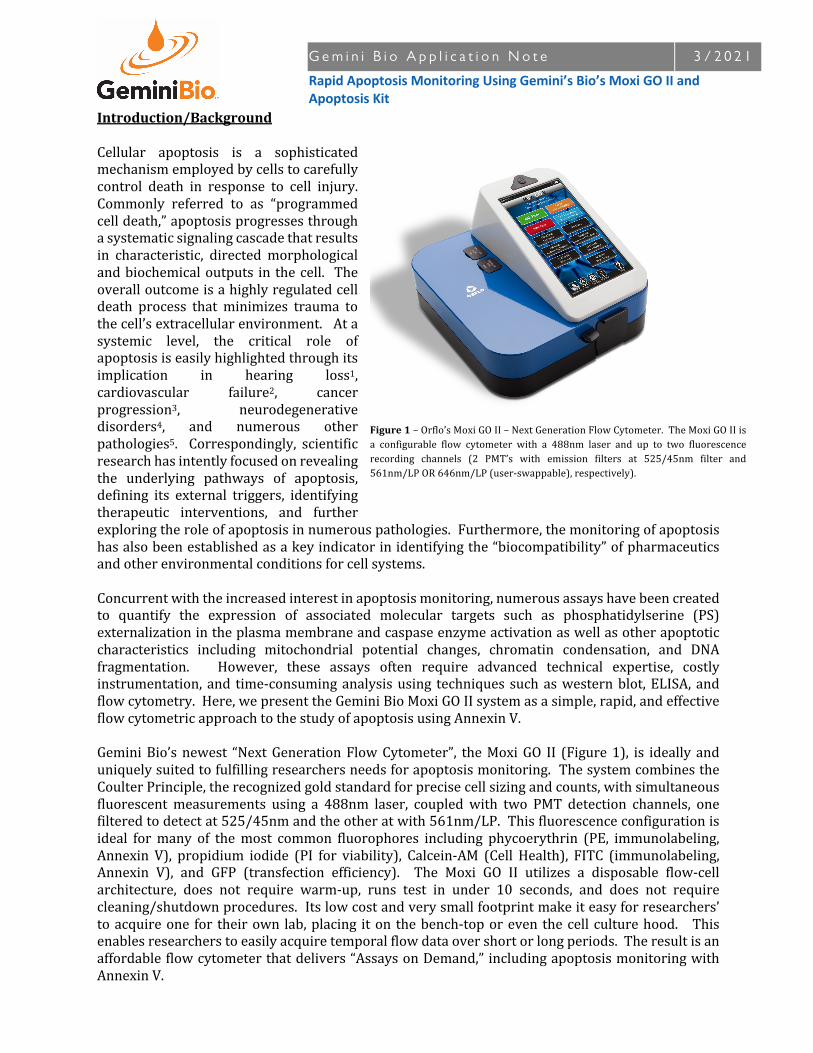
Gemini Bio Application Note 3/2021 Rapid Apoptosis Monitoring Using Gemini’s Bio’s Moxi GO II and Apoptosis Kit Introduction/Background Cellular apoptosis is a sophisticated mechanism employed by cells to carefully control death in response to cell injury. Commonly referred to as “programmed cell death,” apoptosis progresses through a systematic signaling cascade that results in characteristic, directed morphological and biochemical outputs in the cell. The overall outcome is a highly regulated cell death process that minimizes trauma to the cell’s extracellular environment. At a systemic level, the critical role of apoptosis is easily highlighted through its implication in hearing loss 1 , cardiovascular failure 2 , cancer progression 3 , neurodegenerative disorders 4 , and numerous other pathologies 5 . Correspondingly, scientific research has intently focused on revealing the underlying pathways of apoptosis, defining its external triggers, identifying therapeutic interventions, and further exploring the role of apoptosis in numerous pathologies. Furthermore, the monitoring of apoptosis has also been established as a key indicator in identifying the “biocompatibility” of pharmaceutics and other environmental conditions for cell systems. Concurrent with the increased interest in apoptosis monitoring, numerous assays have been created to quantify the expression of associated molecular targets such as phosphatidylserine (PS) externalization in the plasma membrane and caspase enzyme activation as well as other apoptotic characteristics including mitochondrial potential changes, chromatin condensation, and DNA fragmentation. However, these assays often require advanced technical expertise, costly instrumentation, and time-consuming analysis using techniques such as western blot, ELISA, and flow cytometry. Here, we present the Gemini Bio Moxi GO II system as a simple, rapid, and effective flow cytometric approach to the study of apoptosis using Annexin V. Gemini Bio’s newest “Next Generation Flow Cytometer”, the Moxi GO II (Figure 1), is ideally and uniquely suited to fulfilling researchers needs for apoptosis monitoring. The system combines the Coulter Principle, the recognized gold standard for precise cell sizing and counts, with simultaneous fluorescent measurements using a 488nm laser, coupled with two PMT detection channels, one filtered to detect at 525/45nm and the other at with 561nm/LP. This fluorescence configuration is ideal for many of the most common fluorophores including phycoerythrin (PE, immunolabeling, Annexin V), propidium iodide (PI for viability), Calcein-AM (Cell Health), FITC (immunolabeling, Annexin V), and GFP (transfection efficiency). The Moxi GO II utilizes a disposable flow-cell architecture, does not require warm-up, runs test in under 10 seconds, and does not require cleaning/shutdown procedures. Its low cost and very small footprint make it easy for researchers’ to acquire one for their own lab, placing it on the bench-top or even the cell culture hood. This enables researchers to easily acquire temporal flow data over short or long periods. The result is an affordable flow cytometer that delivers “Assays on Demand,” including apoptosis monitoring with Annexin V. Figure 1 – Orflo’s Moxi GO II – Next Generation Flow Cytometer. The Moxi GO II is a configurable flow cytometer with a 488nm laser and up to two fluorescence recording channels (2 PMT’s with emission filters at 525/45nm filter and 561nm/LP OR 646nm/LP (user-swappable), respectively).
Transcript of Rapid Apoptosis Monitoring Using Gemini’s Bio’s Moxi GO II ...
G e m i n i B i o A p p l i c a t i o n N o t e 3 / 2 0 2 1
Rapid Apoptosis Monitoring Using Gemini’s Bio’s Moxi GO II and Apoptosis Kit
Introduction/BackgroundCellular apoptosis is a sophisticatedmechanismemployedbycellstocarefullycontrol death in response to cell injury.Commonly referred to as “programmedcelldeath,”apoptosisprogressesthroughasystematicsignalingcascadethatresultsin characteristic, directed morphologicalandbiochemicaloutputs in thecell. Theoveralloutcomeisahighlyregulatedcelldeath process that minimizes trauma tothecell’sextracellularenvironment.Atasystemic level, the critical role ofapoptosisiseasilyhighlightedthroughitsimplication in hearing loss1,cardiovascular failure2, cancerprogression3, neurodegenerativedisorders4, and numerous otherpathologies5. Correspondingly, scientificresearchhasintentlyfocusedonrevealingthe underlying pathways of apoptosis,defining its external triggers, identifyingtherapeutic interventions, and furtherexploringtheroleofapoptosisinnumerouspathologies.Furthermore,themonitoringofapoptosishasalsobeenestablishedasakeyindicatorinidentifyingthe“biocompatibility”ofpharmaceuticsandotherenvironmentalconditionsforcellsystems. Concurrentwiththeincreasedinterestinapoptosismonitoring,numerousassayshavebeencreatedto quantify the expression of associated molecular targets such as phosphatidylserine (PS)externalizationintheplasmamembraneandcaspaseenzymeactivationaswellasotherapoptoticcharacteristics including mitochondrial potential changes, chromatin condensation, and DNAfragmentation. However, these assays often require advanced technical expertise, costlyinstrumentation, and time-consuminganalysisusing techniques suchaswesternblot,ELISA, andflowcytometry.Here,wepresenttheGeminiBioMoxiGOIIsystemasasimple,rapid,andeffectiveflowcytometricapproachtothestudyofapoptosisusingAnnexinV.GeminiBio’snewest “NextGenerationFlowCytometer”, theMoxiGO II (Figure1), is ideally anduniquelysuitedtofulfillingresearchersneedsforapoptosismonitoring.ThesystemcombinestheCoulterPrinciple,therecognizedgoldstandardforprecisecellsizingandcounts,withsimultaneousfluorescentmeasurements using a 488nm laser, coupled with two PMT detection channels, onefilteredtodetectat525/45nmandtheotheratwith561nm/LP.Thisfluorescenceconfigurationisideal formany of themost common fluorophores including phycoerythrin (PE, immunolabeling,Annexin V), propidium iodide (PI for viability), Calcein-AM (Cell Health), FITC (immunolabeling,Annexin V), and GFP (transfection efficiency). The Moxi GO II utilizes a disposable flow-cellarchitecture, does not require warm-up, runs test in under 10 seconds, and does not requirecleaning/shutdownprocedures.Itslowcostandverysmallfootprintmakeiteasyforresearchers’toacquireone for theirown lab,placing iton thebench-toporeven thecell culturehood. Thisenablesresearcherstoeasilyacquiretemporalflowdataovershortorlongperiods.Theresultisanaffordableflowcytometerthatdelivers“AssaysonDemand,”includingapoptosismonitoringwithAnnexinV.
Figure1–Orflo’sMoxiGOII–NextGenerationFlowCytometer.TheMoxiGOIIisa configurable flow cytometer with a 488nm laser and up to two fluorescencerecording channels (2 PMT’s with emission filters at 525/45nm filter and561nm/LPOR646nm/LP(user-swappable),respectively).

G e m i n i B i o A p p l i c a t i o n N o t e 3 / 2 0 2 1
Rapid Apoptosis Monitoring Using Gemini’s Bio’s Moxi GO II and Apoptosis Kit
ExampleData–ResultsandDiscussionOneofthemostwell-characterized,early-stagemarkersinthecellularapoptosisprogressionisthetranslocationofthemembranephospholipid,phosphatidylserine(PS),fromtheinnertotheouterleafletoftheplasmamembrane.ThedetectionofPStranslocationisachievedthroughtheuseofAnnexinV,acellularproteinwithahighaffinityforPS.BecauseAnnexinViscell-impermeant,onlycellswithapoptosis-relatedexposureofPS,orcellswithcompromisedmembranes(necroticorlate-stageapoptoticcells),arelabeledwiththisprobe.Consequently,byconjugatingAnnexinVtoasuitablefluorophore(e.g.FITCfortheMoxiGOII),cellscanbelabeledforPSinordertodistinguishAnnexinV+(apoptotic)fromAnnexinV-(healthy)populations.Figure2aprovidesarepresentative,user-generatedscreenshotforaFITC-AnnexinVlabeledJurkatsamplethatwasprocessedontheMoxiGOII.ThelargefluorescenceshiftsfortheAnnexinV+vs.AnnexinV-andPI+vs.PI-cellpopulationsareclearlyresolvableintheMoxiGOIIoutput.Inthiscase,thecellsamplewaspre-treatedthrougha4hourincubationin20µMcamptothecin,acelltoxinknowntointerferewithDNAreplicationand,asresult,induceapoptosis.Thatpopulationwasthenmixedwith50%heat-killedJurkatstogenerateanecroticpopulationformorecompletegraphicalrepresentationsofthepossibleapoptosisstates.Theresultingmixturereflectsthe46.6%late-stage/necroticcells(upper-right,mostlyheat-killed),the38%healthycells(lower-right,nostain),andthe14.7%early-stageapoptoticcells(top-left,camptothecin-treatmentinducedapoptosis).Figure2balsopresentsthequadrant-gateddataintabularandbar-chartformatforeasierinterpretation/displayoftheresults.Boththequadrantchartandtablechartcanbeuser-printedinBMPformattogeneratedocumentimages(aswasdoneforthisappnote).TheMoxiGOIIhasbuilt-infunctionalityforrapidlycomparingtheresultsoftwoassaysfrombothvisualandquantitativeperspectives.Figure2cprovidesanexampleofthecomparisonoftheAnnexinV+measurementsoftheJurkatcellswhenfirstexposedtocamptothecin(t=0hour)andafterprolonged(t=6.5
Figure2–a.)TypicalMoxiGOIIapoptosis(FITC–AnnexinV)scatterplot(FITCvs.PI)output for Jurkatcells treatedwithcamptothecin(20µM,4hours)mixedwith50%heat-killedcells.Thequadrantplotsshowsthethreeexpectedstatesfortheculture: Live, Early-Stage Apoptosis, and Late-Stage Apoptosis. b.) Data can bepresented in tabular/bar chart format for easy interpretation of states c.) Thesystemiscapableofgeneratingcomparison/overlaysofdatafortwotests.Shownis an overlay of the Annexin V fluorescence vs. cell size scatter plots for acamptothecin-treated(separatefromabove)Jurkatsampleatthet=0(grayscatterplot) and t=6.5 hour (colored scatter plot) time pointsd.) This data can also bedisplayedasFITCfluorescencehistogramsforthet=0hour(graycurve)andt=6.5hour(redcurve)samples.
G e m i n i B i o A p p l i c a t i o n N o t e 3 / 2 0 2 1
Rapid Apoptosis Monitoring Using Gemini’s Bio’s Moxi GO II and Apoptosis Kit
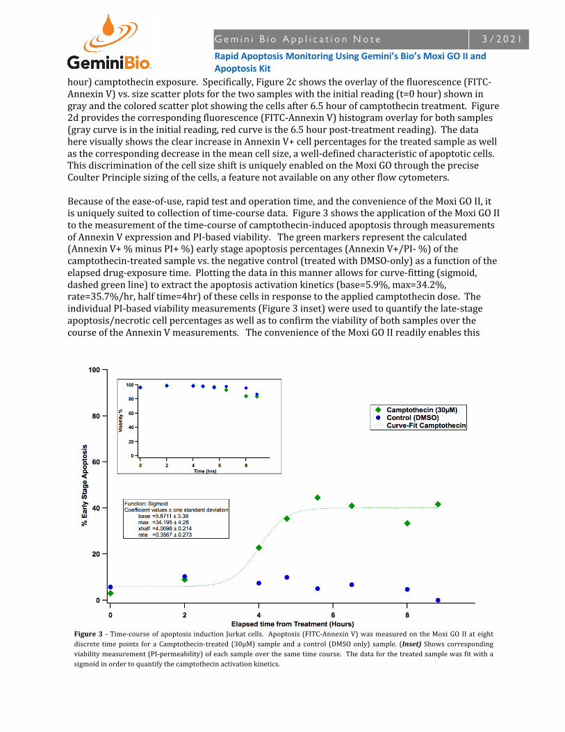
hour)camptothecinexposure.Specifically,Figure2cshowstheoverlayofthefluorescence(FITC-AnnexinV)vs.sizescatterplotsforthetwosampleswiththeinitialreading(t=0hour)showningrayandthecoloredscatterplotshowingthecellsafter6.5hourofcamptothecintreatment.Figure2dprovidesthecorrespondingfluorescence(FITC-AnnexinV)histogramoverlayforbothsamples(graycurveisintheinitialreading,redcurveisthe6.5hourpost-treatmentreading).ThedataherevisuallyshowstheclearincreaseinAnnexinV+cellpercentagesforthetreatedsampleaswellasthecorrespondingdecreaseinthemeancellsize,awell-definedcharacteristicofapoptoticcells.ThisdiscriminationofthecellsizeshiftisuniquelyenabledontheMoxiGOthroughthepreciseCoulterPrinciplesizingofthecells,afeaturenotavailableonanyotherflowcytometers.Becauseoftheease-of-use,rapidtestandoperationtime,andtheconvenienceoftheMoxiGOII,itisuniquelysuitedtocollectionoftime-coursedata.Figure3showstheapplicationoftheMoxiGOIItothemeasurementofthetime-courseofcamptothecin-inducedapoptosisthroughmeasurementsofAnnexinVexpressionandPI-basedviability.Thegreenmarkersrepresentthecalculated(AnnexinV+%minusPI+%)earlystageapoptosispercentages(AnnexinV+/PI-%)ofthecamptothecin-treatedsamplevs.thenegativecontrol(treatedwithDMSO-only)asafunctionoftheelapseddrug-exposuretime.Plottingthedatainthismannerallowsforcurve-fitting(sigmoid,dashedgreenline)toextracttheapoptosisactivationkinetics(base=5.9%,max=34.2%,rate=35.7%/hr,halftime=4hr)ofthesecellsinresponsetotheappliedcamptothecindose.TheindividualPI-basedviabilitymeasurements(Figure3inset)wereusedtoquantifythelate-stageapoptosis/necroticcellpercentagesaswellastoconfirmtheviabilityofbothsamplesoverthecourseoftheAnnexinVmeasurements.TheconvenienceoftheMoxiGOIIreadilyenablesthis
Figure3 -Time-courseofapoptosis induction Jurkatcells. Apoptosis(FITC-AnnexinV)wasmeasuredontheMoxiGOIIateightdiscrete timepoints for aCamptothecin-treated (30µM) sample anda control (DMSOonly) sample. (Inset) Shows correspondingviabilitymeasurement(PI-permeability)ofeachsampleoverthesametimecourse.Thedataforthetreatedsamplewasfitwithasigmoidinordertoquantifythecamptothecinactivationkinetics.

G e m i n i B i o A p p l i c a t i o n N o t e 3 / 2 0 2 1
Rapid Apoptosis Monitoring Using Gemini’s Bio’s Moxi GO II and Apoptosis Kit
typeofprolongedtime-basedanalysis,anapproachthatcanbeprohibitivelyexpensive(bothcostandtime)inlabsthatarenototherwiseequippedwithcontinuallyoperatedflowinstrumentation.ConclusionsThedatainthisstudyshowstheversatilityoftheMoxiGOIIinmonitoringandquantifyingapoptosis.SpecificallyresearcherscanmeasuretheapoptosisstateofcellsbymonitoringthetranslocationofPSfromtheinnertotheouterleafletoftheplasmamembraneusingAnnexinV.CellviabilitycanbecorrespondinglyassessedusingPI.Furthermore,thesystemprovidesthecapabilityformonitoringthe characteristic size shift of the apoptotic cells (through precise Coulter Principle-basedmeasurements).OneofthemostpowerfulfeaturesoftheMoxiGOIIinstrumentistheease-of-useandconvenienceinthecollectionofdata.Withanabilitytoruntestswithouttheneedforsystemwarm-up,maintenance, or shutdownprocedures, theMoxi GO II is ideally suited to time courseexperiments, includingthemonitoringofapoptosis. Finally,astheMoxiGOIItouchscreenGUIisdesignedtomakeeventhemostcomplexflowanalysisaccessibletoresearchers,regardlessoftheirflow expertise. These features shouldmake theMoxi GO II indispensable in any lab performingapoptosismeasurementsorothercell-basedflowcytometrytechniques.

G e m i n i B i o A p p l i c a t i o n N o t e 3 / 2 0 2 1
Rapid Apoptosis Monitoring Using Gemini’s Bio’s Moxi GO II and Apoptosis Kit
References1. TeruKamogashira,ChisatoFujimoto,andTatsuyaYamasoba,“ReactiveOxygenSpecies,Apoptosis,
andMitochondrialDysfunctioninHearingLoss,”BioMedResearchInternational,v.2015,ArticleID617207,7pages,2015
2. PeterM.KangandSeigoIzumo,“ApoptosisandHeartFailure–ACriticalReviewoftheLiterature,”CirculationResearch,2000,v86,1107-1113
3. ScottW.LoweandAthenaW.Lin,“ApoptosisinCancer,”,Carcinogenesis,2000,v21(3),485-495.4. MarkP.Mattson,“ApoptosisinNeurodegenerativeDisorders,”Nature,Nov.2000,v1,120-129.5. B.Favaloro,N.Allocati,V.Graziano,C.DiIio,andV.DeLaurenzi,“RoleofApoptosisinDisease,”Aging,
May2012,v4(5),330-349.
MethodsCellCultureJurkatE6-1(ATCC)werecultured(37°C,5%CO2)inRPMI-1640supplementedwith10%FBS,1mMSodium Pyruvate, and 10mM HEPES (all Life Tech.). For apoptosis induction, 3µL of 10mMcamptothecin(Tocris)stock(inDMSO)wasaddedpermlofculturemedia(30µMfinalcamptothecinconcentration).Forthenegativecontrol,DMSOanequivalentvolumeofDMSOwassubstitutedforthecamptothecintreatment.Forgenerationofanecroticcellpopulation,healthyJurkatcellswereheat-killedbyplacinga15mlcentrifugevialswithcellsincubationina60°Cwaterbathfor10min+.The necrotic populationwasmixed 1:1 with a Camptothecin treated (20µM, 4hr) population togeneratea“threequadrant”Apoptosisexample.
FITC-AnnexinVAssayDual-labeldata(Figure2aand2b)werelabeledwithFITC-AnnexinV(GeminiBio’sVMoxiGoII488validatedkit,Catalog#MXA701)andPropidiumIodide(2µg/ml)followingGeminiBio’s“MoxiGO–EarlyStageApoptosisMonitoringwithAnnexinV”protocol(below).Afterpreparation,cellswererunontheMoxiGOIIsystem(GeminiBioCat#MXG112).Forthetimecoursedataandoverlaydataa1PMT/channelversionoftheMoxiGOIIsystemwasused.OtherthanthePMTconfiguration,thesystemhasthesamearchitectureastheMoxiGOIIsoperformancewouldbeequivalent.Forthosesamples,aseparateprepwassimultaneouslystainedwithPItogetthecorrespondingviabilitydata.Forthetime-coursedata,cellswereassayedontheMoxiGO(1PMTversion)ateightdiscretetimepointsovera~9hourperiod. AnnexinVandPImeasurementsweremadeateachtimepointforboththecamptothecin-treatedandnegativecontrolsamples.Screenshots&DataAnalysisScreenshotswereallgenerateddirectlyfromtheusingthebuilt-insystemscreenshotfunctionality(exportedscreenshotsappearonsystemdriveasBMPs).Datacomparisons/overlayswereallperformedon-unitusingthebuilt-insystemfunctionalityforcomparingsavedtests.FinalimagecroppingandarrangementwasperformedusingPhotoshop(Adobe)andIllustrator(Adobe).TimecoursedatawasextractedbyloadingtheMoxiGOFCSfilesintoFlowJo10.2(TreeStar),runningonMacOSX10.11.SummarydataforapoptosisandviabilitypercentageswereloadedintotheIGORPro(v6.37,Wavemetrics,Inc)analysispackageforcurve-fittingandfinalgraphgeneration.

G e m i n i B i o A p p l i c a t i o n P r o t o c o l 8 / 2 0 1 7
MoxiGO–EarlyStageApoptosisMonitoringwithAnnexinV
Instrument/Cassettes:
§ MoxiGOIINextGenerationFlowCytometer(GeminiBio-Products,Cat#MXG102)§ TypeS+Cassettes(GeminiBio-Products,Cat#MXC030/MXC032)
Reagents/Components:• MoxiCyteApoptosisKit(GeminiBio-Products,Cat#MXA701).Containing:
o Reagent#1:FITC–AnnexinVo Reagent#2:PropidiumIodideo Reagent#3:AnnexinV–BindingBuffer
Protocol:Notes:
• Forcomparisonpurposes,itcanbeusefultogenerateapositivecontrolbyinducingapoptosiswithapharmacologicalagent(e.g.30μMCamptothecintreated,4+hours,37°CforJurkatcells).
• Processasampleofhealthy,untreated,cellsforuseasanegativecontrol.1. Isolatecellstoasingle-cellsuspension.Note:Ifnecessary,useaprotease(e.g.Accutase,GemBio
Cat#400-158)and/orpipettetriturationtobreakaparttheclusters.2. (Optional)Forimprovedstainingresults,particularlywithadherentcells,pre-Washcells1x
(300xg,5min)withPBSorequivalent.3. PelletCells(300xg,5min)Re-suspendpelletto~1x10⁶cells/mlinReagent#3:AnnexinV
BindingBuffer.4. Aliquot100μlofcellstoamicrocentrifugetube(~1x10⁵totalcells).Mixwellbefore
aliquoting.5. Add5µLofReagent#1:FITC-AnnexinVconjugate.Note:While5µLshouldworkformostcell
samples,itmaybenecessarytotitratetheReagent#2volumetooptimizethesignal.6. Add5μlofReagent#2:PropidiumIodide(PI)7. Gentlyvortex(3-4setting)thecellsandincubatefor15minutesatroomtemperature(25°C),
protectedfromlight.8. Add390µLofReagent#3:AnnexinVBindingBuffertoalltubes.9. RunonMoxiGOIIusingthe“Apoptosis(AnnexinV-FITC&PI)”appwithin15minutesof
staining.Protectfromlight.Notes:a. Thiskitwasdesignedtobeusedwiththe646nm/LPfilterinstalledintheback(PMT2)
slotoftheMoxiGOII.Usingthe561nm/LPfilterwillrequirecompensatingforspillover(FITCintoPMT2)andpossiblyloweringthePIconcentrationsothatitisnottoobright.
b. Adjustsizegatestodefinethecellpopulation.c. Touch“Next”viewPMTvsPMTdisplayoftheFITCAnnexin(PMT1)vs.PI(PMT2)
fluorescence.Adjustthegatemarkerstoidentifytherelevantcellsub-populations/d. Oncegated,touch“Summary”forabarchart/tablesummaryviewofthedata.

G e m i n i B i o A p p l i c a t i o n P r o t o c o l 8 / 2 0 1 7
MoxiGO–EarlyStageApoptosisMonitoringwithAnnexinV
http://www.gembio.com