Radiation Safety Training Manual 2016 - University Research
88
Radiation Safety Training Manual 2016
Transcript of Radiation Safety Training Manual 2016 - University Research

Radiation Safety Training Manual 2016

Rad Safety Training Manual, R4 (October, 2016) ii
RADIATION SAFETY TRAINING PROGRAM
Radiation Safety Training is required and mandated by the Colorado Department of Public Health and Environment (CDPHE) regulations. The training program, in its current phase, is the minimum required training for an institution the size of CU Denver | Anschutz. The university employs a diverse group of trainees ranging from summer student workers to those with years of experience using Radioactive Materials (RAM). The Committee on Ionizing Radiation (CIR) hopes that the Radiation Safety Training Program raises the awareness level of the university community in regard to the safe use of RAM and will dispel undue concerns or misconceptions associated with the use of RAM. Overview of the Radiation Safety Certification Program Radiation safety training is available through online and instructor led sessions. Calculators are allowed and personnel whose native language is not English may use dictionaries. Successful completion of radiation safety exams requires a minimum score of 80%. Online training is through the SkillSoft platform, accessed through the university’s employee access portal. Instructor led sessions are typically held once per month, subject to change. For the current schedule and registration go to the Environmental Health and Safety website (http://www.ucdenver.edu/research/EHS/Radiation/Radiation/RADTrainReg/Pages/RadTrainReg.aspx). Principal Investigator (RAM Authorized PI) A Principal Investigator (PI) must complete the online CU: Radiation Safety Initial Training (Radiation Worker – Part I) and the instructor led Radiation Worker – Part II, as well as the PI Responsibilities for RAM Use module in order to obtain authorization from the CIR to procure and use RAM in their laboratory. Any new PI may apply to the CIR for authorization to procure RAM before the training is complete, but no authorization will be granted to a new PI until the training requirement is satisfied. Radiation Worker A Radiation Worker must complete the online CU: Radiation Safety Initial Training (Radiation Worker – Part I) and the instructor led Radiation Worker – Part II to be considered an authorized worker under a PI’s RAM Authorization. No new PI or Radiation Worker may work with RAM until the applicable training requirement has been completed. Annual Refresher (for PIs and Rad Workers) Principal investigators holding active RAM authorizations and all authorized radiation workers must complete refresher training annually. Reminder notices are sent at the beginning of the month training is due and must be completed by the last day of the month.
Rad Safety Training Manual, R4 (October, 2016) iii
Failure to complete the training by the last day of the month the training is due results in placing a RAM procurement restriction on the PIs laboratory and may lead to administrative action by the CIR, or confiscation of RAM from the lab. Credit for training received at other institutions or employers No credit will be given for training received at other institutions due to the difficulty of evaluating the content and effectiveness of such training. Order of completing radiation worker training Radiation Worker training is arranged in two parts that corresponds to the order in which the material should be completed for proper understanding. While it is recommended that the training is completed in order, it is not mandatory they be completed in order.

Rad Safety Training Manual, R4 (October, 2016) iv
Rad Safety Training Manual, R4 (October, 2016) v
Information Available from the Internet
Environmental Health and Safety Website http://www.ucdenver.edu/research/EHS/Pages/EHS.aspx The EHS website contains Radiation Safety information related to RAM use in laboratories to include:
Scheduling and information for Radiation Worker training
Answers to some frequently asked questions about the radiation safety program
Radiation Safety forms
Current versions of the Radiation Safety Manual, Radioactive Waste Disposal Manual and Radiation Safety Training Manual.
The EHS website is updated periodically. Questions or comments about the website may be directed to the EHS main office at 303-724-0345.
Rad Safety Training Manual, R4 (October, 2016) vi
Forward
This manual is intended to assist persons employed at CU Denver | Anschutz in preparing for radiation worker testing administered to Radiation Workers and Principal Investigators by the Environmental Health and Safety Department. The information contained in Sections 1-4 supplements the information contained in the required online and instructor led course to become a Radiation Worker. Section 5 contains guidance for pregnant workers and radiation. Section 6 provides information for PIs who wish to use radioactivity in their lab. As mentioned above, PIs must also complete the PI Responsibilities for RAM Use module to meet the training requirements for becoming an authorized RAM PI. This manual and its references do not necessarily contain an explicit answer for every question on the examinations. These materials do provide sufficient background to enable most workers and investigators to achieve good scores on the examinations with a reasonable amount of study. This manual is not a substitute for the hands-on experience and specialized training in safe use of radioactive materials that a worker should receive in a radioactive materials laboratory. Neither this manual nor the successful completion of certification testing by a worker relieves a responsible authorized principal investigator from providing On-The-Job training to workers under his or her radioactive materials authorization. In fact, documented Radiation Safety On-The-Job training is a requirement reviewed during laboratory inspections. At the very least, as stipulated in the university Radiation Safety Manual, every worker should have access to, and be familiar with, the PI’s authorization documents and supporting applications under which that worker will use specific radioactive materials. Principal investigators seeking authorization for the first time at CU Denver | Anschutz should also note that the Committee on Ionizing Radiation will generally require documented formal training and experience in radioisotope use, in addition to passing the examinations before granting an authorization. This manual is not represented as constituting a course of training in radiation safety that will satisfy the training requirements of any particular radioactive materials license or regulatory agency. Portions of this manual are adapted from training materials used by the U.S. Department of Energy (DOE), The Nuclear Regulatory Commission (NRC), and from the public domain. Additional radiation safety training may be required for users of sealed sources, radiation generating devices or other categories which are not specifically covered in this manual. Questions or comments about this guide or any other training requirement should be directed to Riad Safadi, Environmental Health and Safety Department, F-484 (ext. 4-0234).

Rad Safety Training Manual, R4 (October, 2016) vii
Radiation Safety Study Guide
Section Title Content
1 Fundamentals of Ionizing Radiation and Radiation Protection
Characteristics of Ionizing Radiation, Types of Ionizing Radiation, Properties and Modes of Decay, Effects of Ionizing Radiation on Matter, Natural and Manmade Sources of Radiation, Internal and External Exposure Control, Units of Radiation Dose and Dose Equivalent, Properties of Common Biomedical Research, Radionuclides, Personal Protective Equipment, Personal Monitoring Devices, Ventilation Control and the Use of Fume Hoods
2 Radiation Protection Surveys, Calculations and Practice
Radioactive Decay Calculations, Basic Detector Theory, Selection and Use of Radiation Survey and Counting Instruments, Calculation of Radioactivity from Survey or Counting Measurements, Measurements and Calculation of Radiation Dose Rates, Determining Content of Wastes, Control of Contamination.
3 Radioactive and Mixed Wastes: Regulations and Policies
Radioactive Forms and Definitions, Segregation by Form and Radionuclide/ Half-life, Waste Minimization, Acceptable containers and Labels, Sterilization and Classification of Infectious Radwastes, Special Requirements for Mixed Wastes, Sewage Disposal of H-3 and Trace Quantities, Posting and labeling of Containers and Areas, Security of Radioactive Materials, Contamination Control and Surveys, Control of External Dose Rates, Radioactive Materials Accounting, Transportation of Radioactive Materials
4 Health Effects of Ionizing Radiation
Radiosensitivity, Biological Mechanisms of Damage, Stochastic and Non-stochastic Effects, Risk Coefficients, Maximum Permissible Doses and ALARA Principle, Radiotoxicity and Annual Limits on Intake, Pregnancy/ Fertility Issues
5 Radiation Work and Pregnancy
A guide for PIs and workers
6 Principal Investigator Responsibilities for RAM Use
Committee Authorization Process, Acquisition and Transfer of Radioactive Materials, Authorization and Decommissioning of Laboratory Areas, Conditions on Housekeeping and Maintenance Services
Rad Safety Training Manual, R4 (October, 2016) viii
THREE CARDINAL RULES FOR WORKING WITH RADIOACTIVE MATERIALS
The contents in this manual are all relevant to laboratory use of radioactive materials, but there are three simple rules among the most important. You should always keep the following rules uppermost in your mind. If you follow these three rules, you will naturally protect yourself, and you will cover most of the important regulatory compliance issues as well. Rule No. 1: Keep your radioactive material where it belongs. Keep your radionuclides well-marked throughout your experiment, and keep them in a secured area inside your laboratory. Take appropriate precautions against spreading contamination to you and others, by using good hygiene. This includes wearing gloves, lab coats, and eye protection. It also includes covering surfaces with absorbent paper, confining your use to marked areas of the lab, and routinely checking your laboratory surfaces for contamination. You should use a survey instrument to check yourself, especially your hands, for contamination after every use of radioactive material. Rule No. 2: Tell us if you have a problem. If you think you have gotten some radioactive material on your skin or clothes, or if you have a spill that has any potential for being spread, especially if it gets on the floor, call Environmental Health and Safety at 303-724-0345 immediately. We are here to help you. It is far better to report such events promptly, than to attempt to conceal them, and to have them somehow come to light at a later time. Rule No. 3: Document where your radioactive material ends up. You should know where the radioactive material ends up when you use it, in terms of what fraction typically appears in each waste form (solid lab trash, aqueous liquids, scintillation vials, animal tissue) that you generate. If you don't know, ask your PI, or we can help you. You must document this information on waste tickets and on Radionuclide Accounting Sheets when you request waste pickup, including an entry on both sink logs and waste tags for amounts of H-3 that you dispose into the sewer. This information is absolutely vital to our program. We routinely check the wastes that are submitted to us for disposal to verify what is in them. There are many other important safety and regulatory issues associated with radioactive materials use. However, these three cardinal rules, if followed, will prevent many problems for researchers and for the university Environmental Health and Safety department.

Rad Safety Training Manual, R4 (October, 2016) 1
Section 1
FUNDAMENTALS OF IONIZING RADIATION AND RADIATION PROTECTION Activity The rate of decay of a radioactive substance; the number of atoms that disintegrate per unit time. The units used to represent activity are the Curie and the Becquerel. It is important to recognize that the unit of activity refers to the number of disintegrations per unit time and not necessarily to the number of particles given off per unit time by the radionuclide. For example, 1 uCi (microCurie) of P-32 undergoes 2.22 x 10⁶ transformations per minute, 100% of which are beta particles at 1.7 MeV (energy level). Specific Activity Specific Activity is defined as the activity per unit mass of a radioactive substance and is given in units such as curies per gram (Ci/g) or Becquerels per kilogram (Bq/kg). Remember that the Curie originated from the number of emissions from one gram of radium every second. Thus, the activity of one gram of radium is equivalent to one curie. Therefore, the specific activity of radium is 1 Ci/g. It is important to note that when applied to radionuclides other than radium, the unit Curie does not specify what mass of the material is required. Since one curie of activity equals 37 billion dps, the mass of the material required to produce this number of dps will be a function of the decay rate of the atoms of the material (i.e., the disintegration constant) and the number of atoms of the material per gram (i.e., gram atomic mass[weight]). For example, a curie of pure Co-60 (half-life 5.27 years) would have a mass less than 0.9 milligrams, whereas a curie of natural U-238 (half-life 4.5E9 years) would require over two metric tons of the metal. Obviously, the shorter the half-life of a radionuclide, the greater its specific activity. Radioactive Decay Radioactive nuclides can regain stability by nuclear transformation (radioactive decay). Radioactive decay is the disintegration of the nucleus of an unstable nuclide (also called radionuclide) by spontaneous emission of charged particles, neutrons, and/or photons. During radioactive decay, the atom will give off particles or energy in order to stabilize the ratio of protons to neutrons and to release any excess energy from the nucleus. During the process of nuclear decay, a radionuclide emits radiation. The radiation emitted can
be particulate (alpha - beta - ) or wavelike (gamma - , [photon]) or both. The eventual end product of radioactive decay will be a stable atom. The activity of any sample of radioactive material decreases or decays at a fixed rate which is characteristic of that particular radionuclide. No known physical or chemical factors (e.g., temperature, pressure, dissolution or combination) influence this rate. The rate may be characterized by observing the fraction of activity that remains after successive time intervals. The time that is required for the activity present to be reduced

Rad Safety Training Manual, R4 (October, 2016) 2
to one-half is called the half-life. If successive half-lives are observed, we can see a reduction each time by a fraction of one-half, and the effect will be cumulative; In other words,
one half-life reduces to (0.5)1 or 0.5
two half-lives will reduce to (0.5) * (0.5) = (0.5)2 or 0.25
three half-lives will reduce to (0.5) * (0.5) * (0.5) = (0.5)3 or 0.125 UNITS OF RADIOACTIVITY You should memorize the information on the Curie in the following table, from which the others can be derived, because it is so fundamental to understanding the relationship between counting machine results for radioactive samples and the amount of radioactivity that is present in them.
Table 1 Units of Radioactivity
Unit Disintegrations per second (DPS)
Disintegrations per minute (DPM)
1 Curie 3.7 x 10¹⁰ 2.22 x 10¹²
1 milliCurie 3.7 x 10⁷ 2.22 x 10⁹
1 microCurie
3.7 x 10⁴ 2.22 x 10⁶
1 DPS = 1 Becquerel The actual calculation of the relationship between units of radioactivity (Curie or subdivision thereof, or Becquerel or multiple thereof) and radiation exposure or dose (Roentgen, rad, or Gray) is beyond the scope of the examination, but you should know that the relationship depends on the following factors:
1. Types and energies of radiation(s) emitted by the radioisotope in question and the percentage of disintegrations which result in each such emission;
2. The distance between the radioactive source and the point of measurement (1/r2 dependence, known as "inverse square law").
3. The attenuation provided by any shielding between the source and the point of measurement;
4. The exposure or dose produced by a given quantity of each radiation discussed in item 1, above.
MODES OF RADIOACTIVE DECAY
Rad Safety Training Manual, R4 (October, 2016) 3
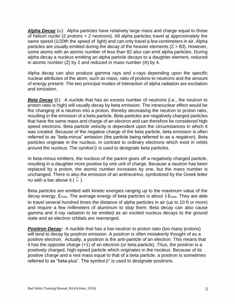
Alpha Decay (): Alpha particles have relatively large mass and charge equal to those of helium nuclei (2 protons + 2 neutrons). All alpha particles travel at approximately the same speed (1/20th the speed of light) and can only travel a few centimeters in air. Alpha particles are usually emitted during the decay of the heavier elements (Z > 83). However, some atoms with an atomic number of less than 82 also can emit alpha particles. During alpha decay a nucleus emitting an alpha particle decays to a daughter element, reduced in atomic number (Z) by 2 and reduced in mass number (A) by 4. Alpha decay can also produce gamma rays and x-rays depending upon the specific nuclear attributes of the atom, such as mass, ratio of protons to neutrons and the amount of energy present. The two principal modes of interaction of alpha radiation are excitation and ionization.
Beta Decay (): A nuclide that has an excess number of neutrons (i.e., the neutron to proton ratio is high) will usually decay by beta emission. The intranuclear effect would be the changing of a neutron into a proton, thereby decreasing the neutron to proton ratio, resulting in the emission of a beta particle. Beta particles are negatively charged particles that have the same mass and charge of an electron and can therefore be considered high speed electrons. Beta particle velocity is dependent upon the circumstances in which it was created. Because of the negative charge of the beta particle, beta emission is often referred to as “beta-minus” emission (the particle being referred to as a negatron). Beta particles originate in the nucleus, in contrast to ordinary electrons which exist in orbits
around the nucleus. The symbol is used to designate beta particles. In beta-minus emitters, the nucleus of the parent gives off a negatively charged particle, resulting in a daughter more positive by one unit of charge. Because a neutron has been replaced by a proton, the atomic number increases by one, but the mass number is unchanged. There is also the emission of an antineutrino, symbolized by the Greek letter
nu with a bar above it ( ). Beta particles are emitted with kinetic energies ranging up to the maximum value of the decay energy, Emax. The average energy of beta particles is about 13 Emax. They are able
to travel several hundred times the distance of alpha particles in air (up to 10 ft or more) and require a few millimeters of aluminum to stop them. Beta decay can also cause gamma and X-ray radiation to be emitted as an excited nucleus decays to the ground state and as electron orbitals are rearranged. Positron Decay: A nuclide that has a low neutron to proton ratio (too many protons) will tend to decay by positron emission. A positron is often mistakenly thought of as a positive electron. Actually, a positron is the anti-particle of an electron. This means that it has the opposite charge (+1) of an electron (or beta particle). Thus, the positron is a positively charged, high-speed particle which originates in the nucleus. Because of its positive charge and a rest mass equal to that of a beta particle, a positron is sometimes
referred to as “beta-plus”. The symbol + is used to designate positrons.
Rad Safety Training Manual, R4 (October, 2016) 4
With positron emitters, the parent nucleus changes a proton into a neutron and gives off a positively charged particle. This results in a daughter nucleus less positive by one unit of charge. Because a proton has been replaced by a neutron, the atomic number decreases by one and the mass number remains unchanged. Electron Capture: For radionuclides having a low neutron to proton ratio, another mode of decay can occur known as orbital electron capture (EC). In this radioactive decay process, the nucleus captures an electron from an orbital shell of the atom, usually the K shell, since the electrons in that shell are closest to the nucleus. The nucleus might conceivably capture an L shell electron, but K shell electron capture is much more probable. The mode of decay is frequently referred to a K-capture. The electron combines with a proton to form a neutron, followed by emission of a neutrino. Electrons from higher energy shell levels immediately move in to fill the vacancies in the inner, lower-energy shells. The excess energy emitted in these moves results in a cascade of characteristic X-ray photons. Either positron emission or electron capture can be expected in nuclides with a low neutron to proton ratio. The intranuclear effect of either mode of decay would be to change a proton into a neutron, thus increasing the neutron to proton ratio. Gamma Emission: Gamma emission is another type of radioactive decay. Nuclear
decay reactions ( , , Electron Capture) resulting in a transmutation generally leave the resultant nucleus in an excited state. Nuclei, thus excited, may reach an unexcited or ground state by emission of a gamma ray. Gamma rays are a type of electromagnetic radiation similar to visible light, radio waves and microwaves but are capable of ionizing matter. They behave as small bundles or packets of energy, called photons, and travel at the speed of light. Gamma rays have no mass or charge and can travel thousands of feet in air at the speed of light. The symbol
is used to designate gamma radiation. For all intents and purposes, gamma radiation is the same as X-rays. Gamma rays are usually of higher energy (MeV), whereas X-rays are usually in the keV range. The basic difference between gamma rays and X-rays is their origin; gamma rays are emitted from the nucleus of unstable atoms, while X-rays originate in the electron shells. X-rays can be produced by machines and in such cases, the X-rays can be of any energy and travel hundreds of meters in air. The basic difference between gamma rays and visible light is their frequency.
Rad Safety Training Manual, R4 (October, 2016) 5
EFFECTS OF IONIZING RADIATION ON MATTER All radiation possesses energy either inherently (electromagnetic radiation) or as kinetic energy of motion (particulate radiations). Absorption of radiation is the process of transferring this energy to atoms of the medium through which the radiation is passing. To say that radiation interacts with matter is to say that it is either scattered or absorbed. The mechanisms of the absorption of radiation are of fundamental interest in the field of radiological health primarily for the following reasons:
1. Absorption in body tissues may result in physiological injury.
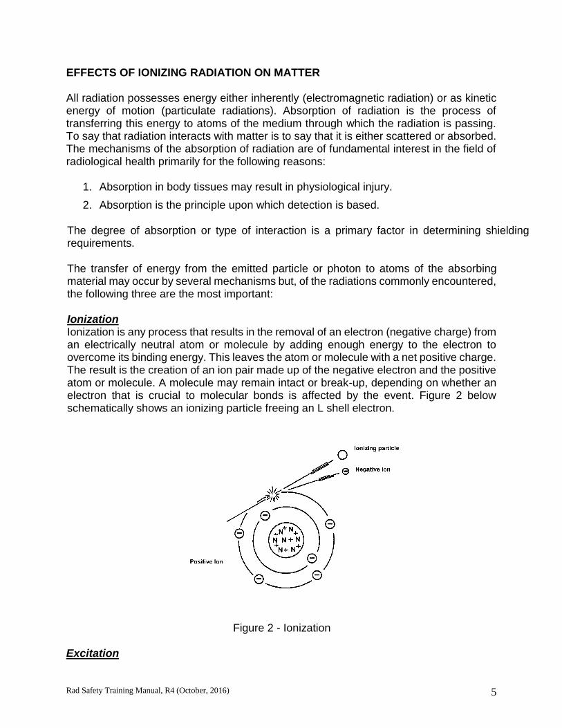
2. Absorption is the principle upon which detection is based. The degree of absorption or type of interaction is a primary factor in determining shielding requirements. The transfer of energy from the emitted particle or photon to atoms of the absorbing material may occur by several mechanisms but, of the radiations commonly encountered, the following three are the most important: Ionization Ionization is any process that results in the removal of an electron (negative charge) from an electrically neutral atom or molecule by adding enough energy to the electron to overcome its binding energy. This leaves the atom or molecule with a net positive charge. The result is the creation of an ion pair made up of the negative electron and the positive atom or molecule. A molecule may remain intact or break-up, depending on whether an electron that is crucial to molecular bonds is affected by the event. Figure 2 below schematically shows an ionizing particle freeing an L shell electron.
Figure 2 - Ionization
Excitation
Rad Safety Training Manual, R4 (October, 2016) 6
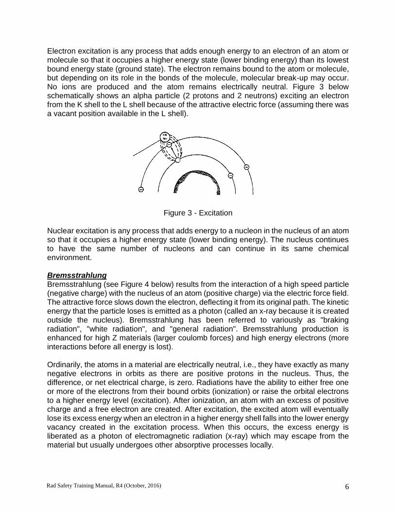
Electron excitation is any process that adds enough energy to an electron of an atom or molecule so that it occupies a higher energy state (lower binding energy) than its lowest bound energy state (ground state). The electron remains bound to the atom or molecule, but depending on its role in the bonds of the molecule, molecular break-up may occur. No ions are produced and the atom remains electrically neutral. Figure 3 below schematically shows an alpha particle (2 protons and 2 neutrons) exciting an electron from the K shell to the L shell because of the attractive electric force (assuming there was a vacant position available in the L shell).
Figure 3 - Excitation
Nuclear excitation is any process that adds energy to a nucleon in the nucleus of an atom so that it occupies a higher energy state (lower binding energy). The nucleus continues to have the same number of nucleons and can continue in its same chemical environment. Bremsstrahlung Bremsstrahlung (see Figure 4 below) results from the interaction of a high speed particle (negative charge) with the nucleus of an atom (positive charge) via the electric force field. The attractive force slows down the electron, deflecting it from its original path. The kinetic energy that the particle loses is emitted as a photon (called an x-ray because it is created outside the nucleus). Bremsstrahlung has been referred to variously as "braking radiation", "white radiation", and "general radiation". Bremsstrahlung production is enhanced for high Z materials (larger coulomb forces) and high energy electrons (more interactions before all energy is lost). Ordinarily, the atoms in a material are electrically neutral, i.e., they have exactly as many negative electrons in orbits as there are positive protons in the nucleus. Thus, the difference, or net electrical charge, is zero. Radiations have the ability to either free one or more of the electrons from their bound orbits (ionization) or raise the orbital electrons to a higher energy level (excitation). After ionization, an atom with an excess of positive charge and a free electron are created. After excitation, the excited atom will eventually lose its excess energy when an electron in a higher energy shell falls into the lower energy vacancy created in the excitation process. When this occurs, the excess energy is liberated as a photon of electromagnetic radiation (x-ray) which may escape from the material but usually undergoes other absorptive processes locally.

Rad Safety Training Manual, R4 (October, 2016) 7
Nuclei also have various possible energy states of the nucleons above the ground or lowest bound energy state. The nucleus can be excited but nuclear excitation occurs only for neutrons or other radiations of relatively high energies. Following nuclear excitation analogous to atomic electron excitation above, the nucleus will eventually return to the ground state and release the excess energy in photons of electromagnetic radiation (gamma rays).
Figure 4 - Bremsstrahlung
Linear Energy Transfer Another measure of energy deposited in an absorber by a charged particle is the Linear Energy Transfer (LET). The LET is the average energy locally deposited in an absorber resulting from a charged particle per unit distance of travel (MeV/cm). The LET is therefore a measure of the local concentration of energy per path length resulting from ionization effects. Biological damage from radiation results from ionization; therefore, the LET is used for calculating quality factors in the calculation of “dose equivalent”. Alpha Absorption As alpha particles travel through matter the strong positive charge attracts electrons and pulls them out of the atomic orbits in other atoms. When an alpha particle causes an electron to be pulled from its orbit the atom is said to be ionized. However, the alpha particle travels at a speed that does not allow the electrons to become attached to the alpha particle. Because of the double positive charge and the large mass, alpha particles produce a large number of ion pairs per unit of distance traveled. In air, an alpha particle may produce 1,000 ion pairs per millimeter traveled. When an alpha particle interacts with other particles to produce “ionization events” it slows down. Then the electrons it has pulled free may attach to the alpha particle, thus forming a helium atom that is no longer capable of causing ionization. Alpha particles typically expend all their energy creating ion pairs after traveling only a few centimeters in air and much shorter distances in dense matter, such as human tissue.
Rad Safety Training Manual, R4 (October, 2016) 8
Thus they are said to have high Linear Energy Transfer rates (LET). A thin sheet of paper or the dead outer layer of the body’s skin will stop most alpha particles. Therefore, alpha radiation is only dangerous when it is internalized in the body through inhalation or ingestion or contamination of open wounds. Beta Absorption The rest mass of a beta particle is the same as that of an orbital electron. Its mass is very much smaller than the mass of the nuclei of the atoms making up the absorbing medium. Since negatively charged beta particles and orbital electrons have like charges, they experience an electrostatic repulsion when in the vicinity of one another. Because the rest masses are equal, the interaction between these two electrons is somewhat similar to the collisions between billiard balls. Therefore, a beta particle may lose all of its energy in a single collision. In such an interaction, the target electron acquires such high kinetic energy it effectively becomes an ionizing particle similar to the incoming electron. Normally, however, a beta particle loses its energy in a large number of ion-ization and excitation events in a manner analogous to the alpha particle. Due to the smaller size and charge of the electron, however, there is a lower probability of beta radiation interacting in a given medium; consequently, the range of a beta particle is considerably greater than an alpha of comparable energy. A beta particle has a charge opposite to that of the atomic nucleus; therefore, an electrostatic attraction will be experienced as the beta approaches the nucleus. Since the mass of an electron is small compared with that of a nucleus; large deflections of the beta can occur in such collisions particularly when electrons of low energies are scattered by high atomic number elements (high positive charge on the nucleus). As a result, a beta usually travels a tortuous, winding path in an absorbing medium. Like an alpha particle, a beta particle may transfer energy through ionization and excitation. In addition, a beta may have a Bremsstrahlung interaction with an atom that results in the production of x-rays. Figure 4 below schematically shows a Bremsstrahlung interaction. In this case, a high energy beta penetrates the electron cloud surrounding the nucleus of the atom, and experiences the strong electrostatic attractive force of the positively charged nucleus. This results in a change in velocity/kinetic energy of the particle and the emission of a Bremsstrahlung x-ray. The energy of the x-ray emitted depends on how much deflection of the beta particle occurred, which in turn, depends on how close the electron came to the nucleus. Therefore, a spectrum of different energy x-rays are observed from the many different Bremsstrahlung encounters an electron will have before it loses all of its energy. Because it is much less likely for a close encounter with the nucleus than a distant encounter, there are lower energy x-rays than high energy x-rays (maximum energy is the energy of the beta particle). Bremsstrahlung becomes an increasingly important mechanism of energy

Rad Safety Training Manual, R4 (October, 2016) 9
loss as the initial energy of the beta increases, and the atomic number of the absorbing medium increases. Beta particles resulting from radioactive decay may be emitted with an energy varying from practically zero up to a maximum energy. Each beta particle will have a range in an absorber based on its energy. After entering a medium, there will be beta particles with different energies. Therefore, determining the number of betas found at a given depth in an absorber and the number of x-rays produced is complex and a function of the energy distribution of the betas. Gamma Absorption X- and gamma rays differ only in their origin, and an individual x-ray could not be distinguished from an individual gamma ray. Both are electromagnetic waves, and differ from radio waves and visible light waves only in having much shorter wavelengths. The difference in name is used to indicate a different source: gamma rays are of nuclear origin, while x-rays are of extra-nuclear origin (i.e., they originate in the electron cloud surrounding the nucleus). Both x-rays and gamma rays have zero rest mass, no net electrical charge, and travel with the speed of light. They are basically only distortions in the electromagnetic field of space, and can be viewed as packets of energy (quanta) that interact with atoms to produce ionization even though they themselves possess no net electrical charge. Photons, when they strike an absorber, can be completely absorbed and impart energy to the absorber or can scatter in a different direction with reduced energy and impart the remaining energy to the absorber that is struck. Gamma Interaction with Matter There are three major mechanisms by which gamma rays lose energy by inter-acting with matter. The Photoelectric Effect: The photoelectric effect is an all-or-none energy loss. The gamma ray, or photon, imparts all of its energy to an orbital electron of some atom. The gamma photon, since it consisted only of energy in the first place, simply vanishes. Figure 5 schematically shows a photoelectric interaction. The energy is imparted to the orbital electron in the form of kinetic energy of motion, over-coming the attractive force of the nucleus for the electron
Figure 5: Photoelectric Interaction

Rad Safety Training Manual, R4 (October, 2016) 10
(the binding energy) and usually causing the electron to fly from its orbit with considerable velocity. Thus, an ion-pair results. The high velocity electron, which is called a photoelectron, is a directly ionizing particle and typically has sufficient energy to knock other electrons from the orbits of other atoms, and it goes on its way producing secondary ion-pairs until all of its energy is expended. The probability of photoelectric effect is maximum when the energy of the photon (gamma) is equal to the binding energy of the electron. The tighter an electron is bound to the nucleus, the higher the probability of photoelectric effect, so most photoelectrons are inner-shell electrons. The photoelectric effect is seen primarily as an effect of low energy photons with energies near the electron binding energies of high Z materials whose inner-shell electrons have high binding energies. Compton Scattering: In Compton scattering there is a partial energy loss for the incoming gamma ray. The gamma ray interacts with an orbital electron of some atom and only part of the energy is transferred to the electron. Figure 6 schematically shows a Compton interaction also called Compton scattering.
The gamma ray continues on with less energy and in a different direction to conserve momentum in the collision. The high velocity electron now referred to as a Compton electron, produces secondary ionization in the same manner as does the photoelectron and the "scattered" -gamma ray continues on until it loses more energy in another gamma ray interaction. By this mechanism of interaction, photons in a beam may be randomized in direction and energy, so that scattered radiation may appear around corners and behind "shadow" type shields. The probability of a
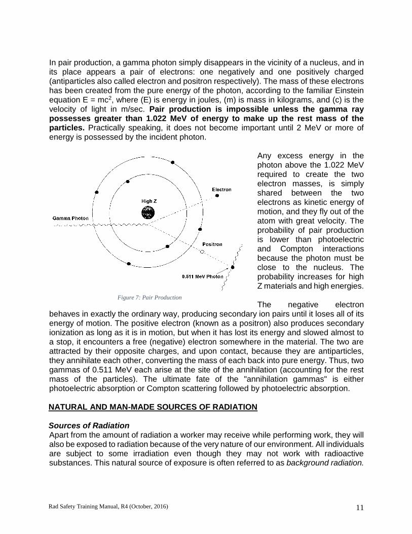
Compton interaction increases for loosely bound electrons. Therefore, most Compton electrons are valence electrons. Compton scattering is primarily seen as an effect of medium energy photons. Pair Production: Pair production occurs when all of energy of the photon is converted to mass. This conversion of energy to mass only occurs in the presence of a strong electric field, which can be viewed as a catalyst. Such strong electric fields are found near the nucleus of atoms and are stronger for high Z materials. Figure 7 below schematically shows pair production and the fate of the positron when it combines with an electron (its anti-particle) at the end of its path.
Figure 6: Compton Scattering
Rad Safety Training Manual, R4 (October, 2016) 11
In pair production, a gamma photon simply disappears in the vicinity of a nucleus, and in its place appears a pair of electrons: one negatively and one positively charged (antiparticles also called electron and positron respectively). The mass of these electrons has been created from the pure energy of the photon, according to the familiar Einstein equation E = mc2, where (E) is energy in joules, (m) is mass in kilograms, and (c) is the velocity of light in m/sec. Pair production is impossible unless the gamma ray possesses greater than 1.022 MeV of energy to make up the rest mass of the particles. Practically speaking, it does not become important until 2 MeV or more of energy is possessed by the incident photon.
Any excess energy in the photon above the 1.022 MeV required to create the two electron masses, is simply shared between the two electrons as kinetic energy of motion, and they fly out of the atom with great velocity. The probability of pair production is lower than photoelectric and Compton interactions because the photon must be close to the nucleus. The probability increases for high Z materials and high energies. The negative electron
behaves in exactly the ordinary way, producing secondary ion pairs until it loses all of its energy of motion. The positive electron (known as a positron) also produces secondary ionization as long as it is in motion, but when it has lost its energy and slowed almost to a stop, it encounters a free (negative) electron somewhere in the material. The two are attracted by their opposite charges, and upon contact, because they are antiparticles, they annihilate each other, converting the mass of each back into pure energy. Thus, two gammas of 0.511 MeV each arise at the site of the annihilation (accounting for the rest mass of the particles). The ultimate fate of the "annihilation gammas" is either photoelectric absorption or Compton scattering followed by photoelectric absorption. NATURAL AND MAN-MADE SOURCES OF RADIATION Sources of Radiation Apart from the amount of radiation a worker may receive while performing work, they will also be exposed to radiation because of the very nature of our environment. All individuals are subject to some irradiation even though they may not work with radioactive substances. This natural source of exposure is often referred to as background radiation.
Figure 7: Pair Production

Rad Safety Training Manual, R4 (October, 2016) 12
Studies of the nature and origin of this source of exposure to man has revealed three main components: 1) external radiation (which includes the radioactivity of the earth's surface, air and water), 2) internal radiation, and 3) radioactivity from radon gas. Man-made sources can influence the contribution from some of these sources. The amount which each of these factors contributes varies with the locale. NATURAL BACKGROUND RADIATION SOURCES External Sources of Radiation Cosmic Radiation: Much work has been carried out in the study of cosmic radiation. This factor in background levels was discovered during attempts to reduce background. Experiments showed that radiation was really coming from outer space. The name cosmic rays are given to this high energy radiation.
Taking into account the dose variation with altitude and the population distribution with altitude, the average yearly dose equivalent rate to the U.S. population from cosmic radiation is estimated to be 33 mrem (330 uSv). This dose equivalent rate is affected slightly with latitude and altitude. For example, in mile high Denver, Colorado, the yearly dose is about 50 mrem (500 uSv). Terrestrial Radiation: The presence of certain small amounts of radioactivity in the soil adds to the background levels to which man is exposed. The amount of radioactive materials found in soil and rocks varies widely with the locale. The main contribution to the background (external dose) is the gamma ray dose from radioactive elements, chiefly of the uranium and thorium series, and lesser amounts from radioactive K-40 and Rb-87. The amount of exposure one is subjected to depends upon the concentration in the soil and the type of soil. In the U.S., three broad areas have been found. These are: the coastal region along the Atlantic Ocean and the Gulf of Mexico, the Colorado Plateau region, and the remainder of the country. The yearly whole body dose equivalent rates in these areas range from 15-35 mrem, 75-140 mrem, and 35-75 mrem, respectively. When absorbed dose rate measurements are weighted by population, and averaged over the entire U.S., the yearly average from soil is estimated at 21 mrem (210 uSv). Internal Sources of Radiation Since small amounts of radioactive substances are found throughout the world in soil and water, some of this activity is transferred to man by way of the food chain cycle.

Rad Safety Training Manual, R4 (October, 2016) 13
In the human body, K-40 is the most abundant isotope. Rb-87, Ra-226, U-238; Po-210 and C-14 are also found in the body. The amount in food varies greatly, so that intake is quite dependent on diet. However, variations in diet seem to have little effect on the body content. The U.S. annual average dose equivalent for all internal emitters (food chain) in the body is 39 mrem (390 uSv). Radon as a source of Radioactivity The background that is found in air is due mainly to the presence of radon and thoron gas, formed as daughter products of elements of the uranium and thorium series. The decay of U-238 proceeds to Ra-226. When Ra-226 emits an alpha as it decays, the gas Rn-222 is formed, which is called radon. In the thorium chain, the decay of Ra-224 results in the gaseous product Rn-220, which is called thoron. The major source of exposure from radon in air occurs when the daughter products attach themselves to aerosols and are inhaled. This leads to an internal dose to the lungs. As for external exposure, the external gamma dose rate from Rn-222 and Rn-220 is estimated to be less than 5 % of the total external terrestrial dose rate. The contribution of inhaled radon gas to the annual average effective dose equivalent is included as an inhaled radionuclide. The U.S. annual average dose equivalent for various inhaled radionuclides (primarily radon) is estimated at 230 mrem (2.3 mSv). MAN-MADE RADIATION SOURCES Nuclear Fallout The term “fallout” has been applied to debris that settles to the earth as the result of a nuclear blast. This debris is radioactive and thus a source of potential radiation exposure to man. Radioactive fallout is not considered naturally occurring but is definitely a contributor to background radiation sources.
Medical Exposures The exposure to the U.S. population from X-rays used in medical and dental procedures is the largest source of man-made radiation. It is estimated that more than 300,000 X-ray units are in use in the U.S., and that about 2/3 of the U.S. population is exposed.
Rad Safety Training Manual, R4 (October, 2016) 14
Computed tomography (CT) improvements have resulted in increased use, resulting in an increased contribution of total dose from medical procedures. In addition to the exposure from X-rays and CT scans, nuclear medicine programs use radiopharmaceuticals for diagnostic purposes. Some radionuclides are used to treat cancer. It has been estimated that more than 10 million doses are administered each year. Many isotopes are also used in biomedical and other types of research. Computed tomography (CT) scans contribute the largest dose 147 mrem (1.47 mSv) of all the medical exposures. The average annual effective dose equivalent in the U.S. for diagnostic X-rays and nuclear medicine are 33 mrem (330 uSv) and 77 mrem (770 uSv), respectively. This gives a combined average annual effective dose equivalent from all sources of medical exposures of 300 mrem (3.0 uSv). Consumer Products There are a number of consumer products and miscellaneous sources of radiation exposure to the U.S. population found in consumer products. Items such as television sets, luminous-dial watches, smoke detectors, static eliminators, tobacco products, airport luggage inspection systems, building materials and many other sources have been studied. The estimated annual average whole body dose equivalent to the U.S. population from consumer products is approximately 13 mrem (130 uSv). The major portion of this exposure (approximately 70%) is due to radioactivity in building materials. Nuclear Facilities Sources of radiation from nuclear reactors consist of neutrons, gamma rays and possible exposures from contamination or environmental releases. The NRC has been tasked by the federal government to calculate doses for populations living within 50 miles of a nuclear facility. Three radionuclides released during routine operations, which contribute to the population dose, are H-3, C-14, and Kr-85. Current estimates of the yearly average dose equivalent in the U.S. from environmental releases are < 1 mrem (10 uSv). Occupational Exposure to Radiation Radiation levels above natural background radiation exposure levels that are caused by exposure to radioactive materials and sources encountered on the job are called occupational exposure. Summary As shown below the average person receives an annual radiation dose of about 620 mrem (6.2 mSv). By age 20, the average person will accumulate over 12 rems (120 mSv) of dose. By age 50, the total dose is up to 31 rems (310 mSv). After 70 years of exposure this dose is up to 43 rems (430 mSv).
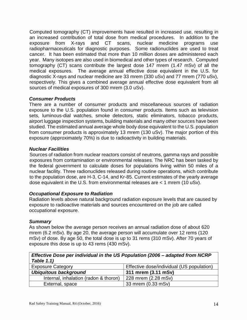
Effective Dose per individual in the US Population (2006 – adapted from NCRP Table 1.1)
Exposure Category Effective dose/individual (US population)
Ubiquitous background 311 mrem (3.11 mSv)
Internal, inhalation (radon & thoron) 228 mrem (2.28 mSv)
External, space 33 mrem (0.33 mSv)

Rad Safety Training Manual, R4 (October, 2016) 15
Internal, ingestion 29 mrem (0.29 mSv)
External, terrestrial 21 mrem (0.21 mSv)
Medical 300 mrem (3.0 mSv)
CT 147 mrem (1.47 mSv)
Nuclear Medicine 77 mrem (0.77 mSv)
Interventional fluoroscopy 43 mrem (0.43 mSv)
Conventional radiography & fluoroscopy
33 mrem (0.33 mSv)
Consumer products 13 mrem (0.13 mSv)
All others 0.8 mrem (0.008 mSv)
Occupational exposures (all industries) 0.5 mrem (0.005 mSv)
Total (rounded up) 620 mrem (6.2 mSv)
EXTERNAL AND INTERNAL EXPOSURE TO RADIATION Most authorized activities using radionuclides involve little, if any, exposure if proper radiological hygiene techniques are followed. It is the current scientific consensus that a rem of radiation dose has the same biological risk regardless of whether it is from an external or an internal source. The NRC requires that dose from external exposure and dose from internal exposure be added together, if each exceeds 10% of the annual limit, and that the total be within occupational limits. The sum of external and internal dose is called the total effective dose equivalent (TEDE) and is expressed in units of rems (Sv). External Exposure When most people think of radiation exposure, they are considering exposure to a source of radiation located outside the body. The radiations of concern with regard to external radiation exposure from radioactive materials are gamma and beta. Gamma radiations, being photons (electromagnetic waves), are sparsely-ionizing and much more penetrating than the particle radiations (alpha, beta). An external source of radioactive material that emits gamma rays can deliver a dose to all the tissues of the body, including the gonads and all of the blood and blood-forming organs, as well as to a fetus in utero. Beta radiations are much less penetrating than gammas. The most energetic beta emitters (e.g., P-32, Sr-90/Y-90) as external sources can deliver a substantial beta dose to the first few millimeters of skin and underlying tissues, as well as the lens of the eye. Usually there is NO dose to the gonads, nor to the preponderance of the blood and blood-forming organs, nor to a fetus in utero. Moderately energetic beta emitters, such as Cl-36, and Ca-45, can deliver a beta dose to exposed skin at close ranges of a few feet or less. The soft beta emitters, such as C-14 and S-35, can deliver a dose to the exposed epidermis at very close ranges (on the order of inches). However, they are so easily attenuated by any container in which the radioactive material might be present that they must usually be deposited directly on the skin as “skin contamination” to deliver any significant dose. The betas of H-3 are so weak that they cannot penetrate the dead outer layer of the epidermis, and H-3 poses no hazard with regard to external exposure.
Rad Safety Training Manual, R4 (October, 2016) 16
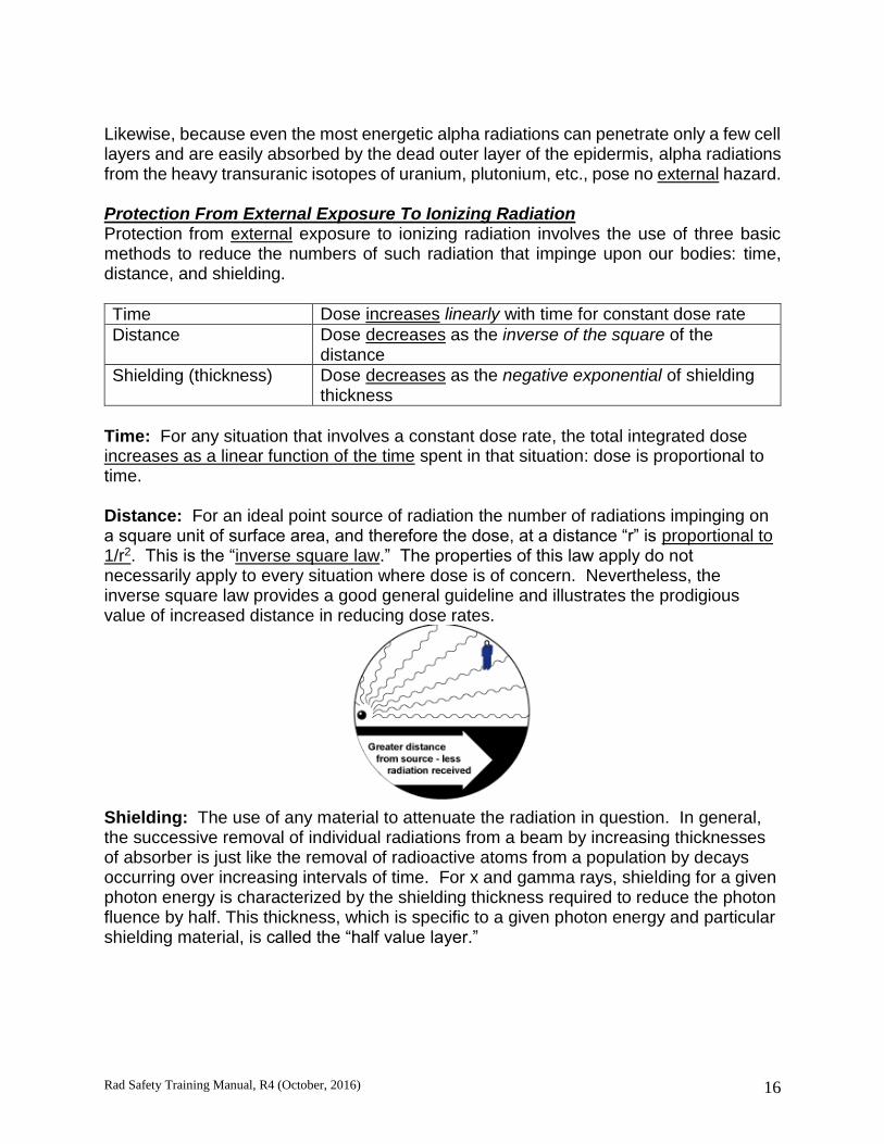
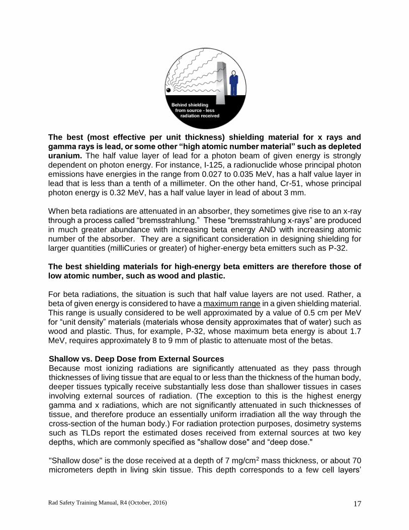
Likewise, because even the most energetic alpha radiations can penetrate only a few cell layers and are easily absorbed by the dead outer layer of the epidermis, alpha radiations from the heavy transuranic isotopes of uranium, plutonium, etc., pose no external hazard. Protection From External Exposure To Ionizing Radiation Protection from external exposure to ionizing radiation involves the use of three basic methods to reduce the numbers of such radiation that impinge upon our bodies: time, distance, and shielding.
Time Dose increases linearly with time for constant dose rate
Distance Dose decreases as the inverse of the square of the distance
Shielding (thickness) Dose decreases as the negative exponential of shielding thickness
Time: For any situation that involves a constant dose rate, the total integrated dose increases as a linear function of the time spent in that situation: dose is proportional to time.
Distance: For an ideal point source of radiation the number of radiations impinging on a square unit of surface area, and therefore the dose, at a distance “r” is proportional to 1/r2. This is the “inverse square law.” The properties of this law apply do not necessarily apply to every situation where dose is of concern. Nevertheless, the inverse square law provides a good general guideline and illustrates the prodigious value of increased distance in reducing dose rates.
Shielding: The use of any material to attenuate the radiation in question. In general, the successive removal of individual radiations from a beam by increasing thicknesses of absorber is just like the removal of radioactive atoms from a population by decays occurring over increasing intervals of time. For x and gamma rays, shielding for a given photon energy is characterized by the shielding thickness required to reduce the photon fluence by half. This thickness, which is specific to a given photon energy and particular shielding material, is called the “half value layer.”
Rad Safety Training Manual, R4 (October, 2016) 17
The best (most effective per unit thickness) shielding material for x rays and gamma rays is lead, or some other “high atomic number material” such as depleted uranium. The half value layer of lead for a photon beam of given energy is strongly dependent on photon energy. For instance, I-125, a radionuclide whose principal photon emissions have energies in the range from 0.027 to 0.035 MeV, has a half value layer in lead that is less than a tenth of a millimeter. On the other hand, Cr-51, whose principal photon energy is 0.32 MeV, has a half value layer in lead of about 3 mm. When beta radiations are attenuated in an absorber, they sometimes give rise to an x-ray through a process called “bremsstrahlung.” These “bremsstrahlung x-rays” are produced in much greater abundance with increasing beta energy AND with increasing atomic number of the absorber. They are a significant consideration in designing shielding for larger quantities (milliCuries or greater) of higher-energy beta emitters such as P-32. The best shielding materials for high-energy beta emitters are therefore those of low atomic number, such as wood and plastic. For beta radiations, the situation is such that half value layers are not used. Rather, a beta of given energy is considered to have a maximum range in a given shielding material. This range is usually considered to be well approximated by a value of 0.5 cm per MeV for “unit density” materials (materials whose density approximates that of water) such as wood and plastic. Thus, for example, P-32, whose maximum beta energy is about 1.7 MeV, requires approximately 8 to 9 mm of plastic to attenuate most of the betas. Shallow vs. Deep Dose from External Sources Because most ionizing radiations are significantly attenuated as they pass through thicknesses of living tissue that are equal to or less than the thickness of the human body, deeper tissues typically receive substantially less dose than shallower tissues in cases involving external sources of radiation. (The exception to this is the highest energy gamma and x radiations, which are not significantly attenuated in such thicknesses of tissue, and therefore produce an essentially uniform irradiation all the way through the cross-section of the human body.) For radiation protection purposes, dosimetry systems such as TLDs report the estimated doses received from external sources at two key depths, which are commonly specified as "shallow dose" and “deep dose." "Shallow dose" is the dose received at a depth of 7 mg/cm2 mass thickness, or about 70 micrometers depth in living skin tissue. This depth corresponds to a few cell layers’

Rad Safety Training Manual, R4 (October, 2016) 18
thickness and is intended to approximate the depth of the basal cells of the epidermis, which is taken to be the most radiosensitive depth in skin. In this context, the term "shallow dose" is equivalent to the term "skin dose." "Deep dose" is specified at a depth of one centimeter, and is used as a conservative indicator of the (maximum) dose that may be reaching the deeper and more radiosensitive tissues of the body, including the gonads and the blood forming organs. Thus, deep dose represents dose to the whole body in the sense of including all of the deeper tissues and organs, and is often called "whole body dose" in the context of external exposure to the penetrating photon radiations. "Eye dose" is sometimes also reported, and is specified at a depth intermediate between shallow and deep doses, at about 0.3 cm depth, corresponding to the lens of the eye. For the reasons just given, external exposure to high energy betas is virtually synonymous with skin dose, whereas whole body dose tends to be the overriding consideration in controlling external exposure to gamma rays and x rays. Eye dose, which is intermediate between skin and whole body dose in biological sensitivity and corresponding dose limits, can also be a serious consideration for either type of radiation, depending on the details involved. Internal Exposure Internal exposure to radiation occurs when radioactive material finds its way into the body via inhalation, ingestion, entry through a wound, or absorption through the skin. Thus, the radiations are emitted by atoms that reside within the tissues of the body. The relative hazard of a radioactive material with respect to internal exposure is called its radiotoxicity. Alpha radiations are by far the greatest hazard when internal radiation exposure is of concern. Because alpha particles deposit all of their energy in a very short path through tissue (microns or tens of microns), an alpha particle delivers all of its energy to a few cells. Therefore, alphas produce the most damage per atomic disintegration when the materials emitting them are in intimate contact with the living cells of the body. Beta radiations are intermediate in hazard as a source of internal radiation exposure. They are not as densely ionizing as alphas, and their energy is typically deposited in a larger number of cells than for alphas, but most of the energy from internally emitted betas is locally deposited - deposited within a few mm of the site of the radioactive atom emitting the beta. Gamma rays are the least hazardous radiation with respect to internal radiation exposure, for the same reason that they are so penetrating - they are very sparsely ionizing. Much of the energy of a gamma emitted from within the body’s tissues may escape the body altogether. When emitted from within the body, gammas generally create much less damage per atomic disintegration than alphas and betas.
Rad Safety Training Manual, R4 (October, 2016) 19
Protection From Internal Exposure To Ionizing Radiation Protection from internal exposure to ionizing radiations involves the use of methods to reduce the numbers of radioactive atoms that can enter the body. For the purposes of this discussion, protective methods will be categorized according to the routes of entry by which radioactive materials may enter the body. Inhalation: The first method that should be considered to minimize inhalation exposures is to minimize the amount of radioactive material that can become airborne in the first place. Precautions of this type include:
tightly capping or sealing materials that are being reacted, heated, centrifuged, sonicated, homogenized, etc.,
opening sealed vessels carefully with a cotton pledge or other absorbing material over the opening,
for vessels of stock radioiodine solutions, venting the air inside the vial through a charcoal-filled syringe to entrap the volatile radioiodine, and
careful planning to avoid spills, especially those involving explosion or implosion of reaction vessels, trapping flasks, etc.
A second method to avoid inhalation is to use a stream of air to entrain and evacuate any materials that are released, with the airstream being carried to a location outside the building and ejected forcefully away from the building at a location as far as possible from normal occupancy, in a way that causes it to be rapidly diluted into the atmosphere. This is called “local exhaust ventilation” and is a type of control measure that is called referred to as an “engineering control.” Fume hoods are a prime example of local exhaust ventilation. A third method of minimizing inhalation exposure is for the potentially exposed individual to wear some form of personal respiratory protection, such as a respirator. This method is generally not used in laboratory settings, for the reasons discussed in section 3.4.3.4.4 of the Radiation Safety Manual. Ingestion: Inexperienced persons may be unaware that ingestion of radioactive materials is a very real hazard in the laboratory.
Radioactive solutions must never be pipetted by mouth; mechanical pipetting devices must always be used.
Food and beverages must never be stored or consumed in areas where radioactive materials are used or stored. This includes walk-in freezers and cold rooms.

Rad Safety Training Manual, R4 (October, 2016) 20
Persons using radioactive materials must wear gloves and should always survey their hands and lab coats before leaving the area for breaks, lunch, or to go to other non-radioactive-materials areas. Failure to do so may result in the transfer of radioactive materials from the hands to beverages and foodstuffs, and a general spread of contamination.
Skin Absorption: Percutaneous absorption (absorption across intact skin) is a hazard that varies significantly with the chemical form of the radioactive material involved. Soluble forms such as radioiodine in the form of sodium iodide solution are notoriously hazardous in this way.
Gloves, lab coats, and eye protection are absolutely required when handling radioactive materials.
Self-surveys after each use of radioactive materials are very important, any suspected self-contamination must be reported immediately to Environmental Health and Safety at x4-0345.
Proper marking and regular surveys of laboratory surfaces are important tools in avoiding skin contamination.
Entry Via Wounds: A very important precaution in medical and biomedical research settings is to refrain from recapping syringe needles used with radioactive materials, for the same reason as in biosafety practice - to avoid puncture wounds (“needle sticks”), which may lead to self-injection of all or part of the syringe’s content. UNITS OF RADIATION DOSE AND DOSE EQUIVALENT Radiation Dosimetry Terminology During the early days of radiological experience there was no precise unit of radiation dose that was suitable either for radiation protection or for radiation therapy. Exposure and dose have inherent differences depending on the type and energy of radiation emitted. With research and time, it became necessary to distinguish between radiation exposure and absorbed dose. Exposure (X) Exposure is a measure of the ability of photons (X and gamma) to produce ionization in air. It is applicable only to the measure of x-ray or gamma radiation measured in dry air and is not a measure of damage to body tissues. Traditionally, the unit of exposure is the roentgen (R). There is no SI unit defined for exposure. Absorbed Dose (D) Units of dose measure the amount of radiation energy absorbed or deposited per unit of mass. The "energy deposited" by radiation is an expression for the "amount of ionization caused" and both expressions mean the same thing. The rad unit is useful as
Rad Safety Training Manual, R4 (October, 2016) 21
an indication of how much immediate damage radiation causes in a material such as body tissue: (1 rad = 100 ergs per gram); the SI unit is the Gray (Gy). (1 Gy = 100 rad) The Rad The old unit of absorbed dose is the rad, which is an acronym for Radiation Absorbed Dose. The unit rad can be applied to all types of radiation and is defined as the deposition by any radiation of 0.01 joules of energy in one kilogram of any material. The Gray (Gy) is the SI derived unit of absorbed dose, equivalent to the deposition of one joule of energy per kilogram (1 J/kg). Although the rad and gray are measures of ionization produced, they do not give any information about the biological effects of the radiation that is absorbed. It is meaningful to emphasize that the energy deposited by the radiation (as a result of the ionization) is the quantity that is actually measured in rad units. Thus, the amount of ionization produced in the detector of a radiation detection instrument can be related to the energy absorbed and expressed by the instrument meter in the unit rad or rad/hr. Quality Factor A quality factor is used to relate the absorbed dose of various types of radiation to the biological damage caused to the exposed tissue. A quality factor is necessary to relate the effects of radiation because the same amounts absorbed of different kinds of radiation cause different degrees of damage. The quality factor converts the absorbed dose to a unit of dose equivalence to a common scale that can be added with, and compared to, damage caused by any kind of radiation. The quality factor is a conversion factor used to derive the dose equivalent from the absorbed dose, expressed as: H = DQ Where: H = dose equivalent D = absorbed dose Q = quality factor
Table 3 Quality Factors
RADIATION TYPE QF
X-rays, Gamma rays, Beta particles 1
Alpha particles 20
Individual organs and tissues in the human body have different sensitivities to radiation. The table below shows the fractional contribution of each organ or tissue used to determine the total risk equivalent to whole body irradiation. The specific organs and tissues shown below here have a relatively high sensitivity to radiation and present an
Rad Safety Training Manual, R4 (October, 2016) 22
increased risk of developing cancer as compared to other less sensitive organs and tissues in the body such as skin, muscle and the brain.
ORGAN OR TISSUE WEIGHTING FACTOR Gonads 0.25 Breasts 0.15 Red bone marrow 0.12 Lungs 0.12 Thyroid 0.03 Bone surfaces 0.03 Remainder 0.30 TOTAL (Whole body) 1.00
The EDE represents the same risk expected from radiation to the whole body. Using the weighting factor from the chart above one can determine the relative risk from a 1REM dose as in the following example:
What is the risk of fatal cancer from a 1 REM (dose equivalent) to the gonads? EDE = Dose (Rem) X Weighting Factor = 1 Rem X 0.25
= 0.25 Rem or 250 mRem EDE
That is to say, risk of fatal cancer from 1 Rem dose to the gonads = Risk of fatal cancer from 0.25 Rem dose to the whole body.
CHARACTERISTICS AND PROPERTIES OF RADIONUCLIDES COMMONLY USED IN BIOMEDICAL RESEARCH Radiation workers in a biomedical research setting should be aware of properties among the typical isotopes used at this institution. Radiation workers should consider the following:
1. You should look at the table of usage limits requiring bioassay, given in section 3.4.3.4 of the Radiation Safety Manual, to develop a sense of the comparative radiotoxicities of the listed radionuclides, where the comparative radiotoxicity of a given radionuclide is inversely proportional to its limit (lower limits for more radiotoxic nuclides).
2. You should understand the following material concerning the physical and chemical forms of radioactive material used in biomedical research.
a. Almost all radioactive materials are purchased as liquid solutions of radio-labeled materials. There are a few radiolabeled materials that are themselves liquids (e.g., tritiated water) and they are a special concern because they can evaporate and produce radioactive vapors. Radioactive gases, such as radionuclides of the noble gases krypton and xenon, are rarely used at this institution, but they give rise to special exposure

Rad Safety Training Manual, R4 (October, 2016) 23
concerns. EHS should always be contacted prior to initiating any work with radioactive gases.
b. Most radiolabeled materials used in biomedical research are natural or synthetic organic compounds in which a stable atom of H, C, P, or S has been replaced by a radioactive atom of the same element.
c. In all of the above types of applications, we refer to the radioactive materials as "loose" or “unsealed” forms. When we use radioactive materials in unsealed form, we are using the radiations they emit to find out what is happening chemically and physically to the radiolabeled material (cells, proteins, etc).
d. “Sealed” sources contain the radioactive material throughout its use to produce radiations that are used for some other purpose. These “sealed” sources are used in situ to produce a specific type of radiation, typically gamma. Sealed sources can serve as “check” sources to calibrate Geiger counters, liquid scintillation counters and other survey equipment. Sealed sources are nearly indestructible and for someone to contact the radioactive materials enclosed within requires destruction or damage to the integrity of the source container.
3. You should definitely know the following information about these 5
radionuclides that are commonly used in biomedical research: H-3: you should know that H-3 is a very low energy (“soft”) beta emitter. It is very low in radiotoxicity, but is also very difficult to detect: it will not be seen at all by any Geiger-Mueller (GM) detector, and the only way that it can usually be detected is to prepare a sample presumably containing the H-3 that can be counted by liquid scintillation. C-14 and S-35: you should know that these are low energy (“soft”) beta emitters of low to moderate radiotoxicity, and that they can be seen with a GM detector if it has a thin window. P-32: you should know that P-32 is a high-energy beta emitter that needs to be carefully shielded with low atomic number materials, and that it is very radiotoxic if ingested because it tends to lodge in bone and irradiate the bone marrow. High-energy beta emitters are of concern also to the skin and eyes if appropriate precautions are not taken as they can penetrate several millimeters depth in tissue. I-125: you should know that I-125 is a low-energy gamma and x-ray emitter that is very easy to shield with thin lead, but is extremely radiotoxic because it tends to lodge in the thyroid with great concentration and tenacity if somehow internalized in the body. You should also know that it is very difficult to detect with a GM, for the same reasons as Cr-51, and is similarly much more easily detected with a proper scintillation detector. Finally, you should know that the unbound forms in which the radioactive iodine atom is not covalently bound to a non-volatile macromolecule are extremely hazardous with respect to inhalation and skin absorption.

Rad Safety Training Manual, R4 (October, 2016) 24
PERSONAL PROTECTIVE EQUIPMENT All workers handling radioactive materials must wear appropriate PPE to include as a minimum lab coats, gloves, protective eyewear, long pants and closed toed shoes. These minimum requirements help protect you from contamination while working with RAM. Gloves: The selection of proper gloves is an important aspect of protecting oneself when working with hazardous materials. Gloves should always be worn when working with radioisotopes. Consult EHS for information on appropriate protective fabrics when chemicals are being used in an experiment. Respirators: Respirators, regardless of type, are almost never specified by the CIR, for two compelling reasons:
engineering controls such as the ventilation controls described in the Radiation Safety Manual are generally considered preferable to respirators by safety professionals and standards-setting organizations, due to their lesser degree of reliance on an individual worker’s performance and other variable factors that affect the efficacy of respirators. Current Colorado regulations specifically require engineering controls such as fume hoods to be used whenever feasible, and
laboratories where radioactive materials are used are generally NOT classifiable as isolated and strictly controlled “restricted areas” where entry is restricted to selected, specially trained individuals wearing respiratory protection.
Individuals who chose to wear some form of individual respiratory protection for personal reasons must be aware that respirators require careful selection and maintenance, and usually require individual fit-testing to be effective. Respirators require medical qualification of the individual wearer because of the cardiopulmonary stresses of breathing through the respirator. Individual choices to use a respirator must never be relied upon to provide protection in a situation that would otherwise be deemed unsafe or inconsistent with the specific requirements of the PI’s authorizations for radioactive materials. Environmental Health and Safety must be consulted when considering the use of respirators. Environmental Health and Safety personnel will assist with training, fit-testing and the selection of appropriate respiratory protection. Use of Personal Monitoring Devices – Thermoluminescence Dosimeter (TLD): The TLD badge is a device that is used to evaluate your personal exposure to radiation over a period of time, typically per month or per quarter. It measures your external exposure to radiations arising from sources outside your body. It cannot generally be relied upon to provide an indication of any radiation dose that you might receive if the radioactive materials themselves were introduced into your body. TLD badges are issued for the exclusive use of one person and CANNOT be shared. TLD badges

Rad Safety Training Manual, R4 (October, 2016) 25
should be worn on the outside of your clothing on the front upper part of your body. When you leave for the day, TLD badges should be stored in a low background area such as your desk. The TLD works when ionizing radiation transfers energy to phosphors contained within the TLD. Energized electrons within the phosphor become detached from their atoms and then trapped by impurities added to the phosphor crystal. To determine the dose received by the wearer, the badges are returned to the vendor for processing after the monitoring period. TLDs are “read” by applying heat to the phosphors, which release the trapped electrons, returning them to the ground state. This annealing process produces energy in the form of light in which the intensity of the light flash is directly proportional to the dose received. Regulations dictate dosimetry monitoring is required for any individual who has the potential to exceed 100 mrem/yr in whole body exposure to penetrating radiation. This does not mean that you are expected to be exposed to significant amounts of radiation. In fact, most of the monitored persons do not work in situations in which they would normally be exposed to any level of radiation dose that significantly exceeds background. In order for EHS to fulfill its legal requirements in the personnel dosimetry program, we must obtain certain private information (e.g., birth date and employee number) on your application. We safeguard this information, along with your dosimetry results on our program, and release them outside the university only with your permission. You have a legal right to be informed of your monitoring results and you may request this information from us at any time. These records are maintained indefinitely. The results are reported by the vendor for shallow (i.e., skin) and deep (i.e., whole body) dose estimates, for the monitoring period in question, with cumulative totals for calendar quarter, year, and the entire time you have been on the system. Dose is reported in increments of 1 mrem, with ND indicating “none detected”, or <1 mrem. Because the system reports in 1 mrem increments, it is natural to expect that reports of 10 or even 20 mrem may occasionally occur due to statistical fluctuation alone. The average monthly background that people receive in Colorado is in the vicinity of 15 mrem. Reports of whole body dose over 125 mrem will be specifically brought to your attention by a notice from our As Low As Reasonably Achievable (ALARA) Program. We can provide a great deal of additional information about the personnel dosimetry program, and you should feel free to direct questions to us at any time. VENTILATION CONTROLS Ventilation controls are the usual means of protecting personnel against the hazard of inhaling airborne radioactive materials. Depending on the type and degree of hazard, a specific type of control will generally be specified during the CIR’s review of applications.

Rad Safety Training Manual, R4 (October, 2016) 26
General Ventilation: The general purpose ventilation that exists in a specific area, as opposed to completely stagnant air with no exchange to outside air, may sometimes be used in calculations to demonstrate that some potential releases are not sufficient to approach maximum permissible levels for members of the public. It is not the responsibility of the Principal Investigator to control general building ventilation, but it is prudent to contact campus Facilities Services when there appears to be a sudden lack of general ventilation in a radioactive materials laboratory. Fume Hoods: Fume hoods must be used exactly as specified in the PI’s radioactive materials authorizations. In general, fume hoods should always be used for the following types of operations:
opening sealed containers of liquid radioactive materials,
performing sonication, homogenization, or other forms of mechanical agitation with radioactive materials,
operations involving volatile forms of radioactive materials, most notably
1. radioiodine labeling reactions,
2. use of tritiated water or volatile tritiated organic compounds, including acetate,
3. use of sulfur-labeled methionine in some circumstances. Fume hoods may be used for operations that do not present a hazard of releasing radioactive materials into the air, in order to take advantage of the confinement that a fume hood cabinet can afford, for such things as splashes that may occur during liquids transfers. Fume hoods should not be used as storage areas for materials that do not require local exhaust ventilation and should be kept as free of obstructions as possible. Fume hoods must not be used for extended storage of plastic scintillation vials containing volatile solvents such as toluene and xylene. Fume hoods may be used for drying or other processing involving evaporation of water, if no volatile forms of radioactive material are present, but must not be used to dispose of volatile organic solvents by evaporation. Glove Boxes and Other Special Systems: Glove boxes or other special systems affording complete containment of airborne radioactive materials may be specified by the CIR for certain types of radioactive materials and operations involving a particularly high hazard of airborne radioactive materials, although such cases are rare in modern biomedical research.

Rad Safety Training Manual, R4 (October, 2016) 27
Section 2
RADIOACTIVITY, COUNT RATE, BACKGROUND, AND EFFICIENCY Review units of radioactivity as nuclear transformation events/time Because each unstable atom of a given description, i.e., a particular radionuclide as specified by
atomic number [# protons in nucleus],
atomic weight [total # of protons + neutrons in nucleus], and
nuclear energy state [e.g., a lower-case “m” after the atomic weight to denote metastable states]
gives rise to the same types and amounts of radiations with the same probabilities when it spontaneously decays, the natural unit for quantifying radioactivity is the number of such atoms that are decaying per unit time. The term “atomic/nuclear disintegration,” as used in the quantity “disintegrations per minute,” or “DPM,” refers to the spontaneous rearrangement of an unstable atomic nucleus accompanied by the emission of alpha and/or beta and/or gamma radiations. The term “nuclear transformation” is a slightly more general denotation that includes the energy-state transitions of metastable nuclei from the metastable state to the ground state, which do not include rearrangement of the nuclei, and are accompanied only by the emission of gamma rays. In common practice, the terms “DPM” and “DPS” (Disintegrations Per Second) are used to quantify all types of nuclear transformations. The SI unit for radioactivity is the Becquerel, which is one nuclear transformation per second. The terms Becquerel (Bq) and disintegration per second (DPS) are interchangeable. The Becquerel or DPS is a very small unit of radioactivity, and leads to the need to use very large numbers of Becquerel to describe practical amounts of radioactivity. Because counting periods are usually specified in minutes, and there is an important correspondence between the count rate of a sample in counts per minute (CPM) and the amount of radioactivity in the sample that gives rise to that count rate, the radioactivity present is often quantified in disintegrations per minute (DPM). The number of DPM is simply the number of DPS multiplied by 60 (60 seconds/minute).
Example: a sample containing 500 Bq (or 500 DPS) of the phosphorous radionuclide 32P contains 500 x 60 = 3,000 DPM.
The more traditional unit of radioactivity is the Curie, which is defined as 3.7 x 10¹⁰ nuclear transformations per second, or 3.7 x 10¹⁰ Becquerel.
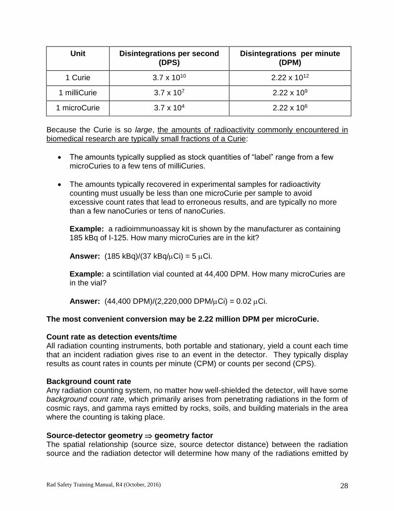
Rad Safety Training Manual, R4 (October, 2016) 28
Unit Disintegrations per second (DPS)
Disintegrations per minute (DPM)
1 Curie 3.7 x 10¹⁰ 2.22 x 10¹²
1 milliCurie 3.7 x 10⁷ 2.22 x 10⁹
1 microCurie 3.7 x 10⁴ 2.22 x 10⁶
Because the Curie is so large, the amounts of radioactivity commonly encountered in biomedical research are typically small fractions of a Curie:
The amounts typically supplied as stock quantities of “label” range from a few microCuries to a few tens of milliCuries.
The amounts typically recovered in experimental samples for radioactivity counting must usually be less than one microCurie per sample to avoid excessive count rates that lead to erroneous results, and are typically no more than a few nanoCuries or tens of nanoCuries.
Example: a radioimmunoassay kit is shown by the manufacturer as containing 185 kBq of I-125. How many microCuries are in the kit?
Answer: (185 kBq)/(37 kBq/Ci) = 5 Ci.
Example: a scintillation vial counted at 44,400 DPM. How many microCuries are in the vial?
Answer: (44,400 DPM)/(2,220,000 DPM/Ci) = 0.02 Ci. The most convenient conversion may be 2.22 million DPM per microCurie. Count rate as detection events/time All radiation counting instruments, both portable and stationary, yield a count each time that an incident radiation gives rise to an event in the detector. They typically display results as count rates in counts per minute (CPM) or counts per second (CPS). Background count rate Any radiation counting system, no matter how well-shielded the detector, will have some background count rate, which primarily arises from penetrating radiations in the form of cosmic rays, and gamma rays emitted by rocks, soils, and building materials in the area where the counting is taking place.
Source-detector geometry geometry factor The spatial relationship (source size, source detector distance) between the radiation source and the radiation detector will determine how many of the radiations emitted by

Rad Safety Training Manual, R4 (October, 2016) 29
the source will impinge upon the detector. This relationship is typically summarized in a geometry factor that may range from zero to one. Detection Efficiency Overall detecting efficiency for a given situation is typically defined in a very pragmatic way as count rate divided by nuclear transformation rate, e.g., CPM/DPM. This number is easy to measure for a calibrated source of a particular radionuclide of interest, but it must be borne in mind that the efficiency that is so defined applies only to
the specific radionuclide in the calibrated source, and
the geometry of the calibrated source (the size of the source and its position relative to the detector).
1. intrinsic efficiency
Any detector will have an intrinsic efficiency for radiations of a given type and energy, defined as the fraction of the radiations incident upon the detector that result in counts. This efficiency is usually not separately measured for the types of applications of interest here.
2. intrinsic efficiency * geometry factor overall efficiency
The overall counting efficiency is primarily a product of the intrinsic detection efficiency of the detector and the geometry factor, although it may be affected by other factors such as the average number of the radiations of interest that are emitted by decaying nuclei of the source radionuclide, and the absorption of the radiations of interest by any materials interposed between the source and the detector.
3. caveats regarding the use of a source other than the radionuclide of interest to
determine a counting efficiency - get some health physics help! 4. efficiency calibrations are provided by Environmental Health and Safety for low
and high energy betas Radioactivity present * overall efficiency = net count rate For a single source composed of one radionuclide, given an overall detection efficiency that is obtained in the manner described in paragraph (E.) above, the count rate attributable to that radioactive source is equal to the amount of radioactivity present in the source times the applicable overall detection efficiency. This product gives the net count rate in excess of background that is attributable to that source. Gross count rate The gross count rate is the count rate that is actually measured in the presence of background, and is equal to the sum of the background count rate and the net count rate from the radioactivity present.
Rad Safety Training Manual, R4 (October, 2016) 30
The Fundamental Calculation of Radioactivity: radioactivity = (gross count rate - background count rate) / (overall efficiency). To convert a count rate to units of true radioactivity,
first subtract the background count rate from the count rate of the sample,
then divide the net count rate by the applicable overall counting efficiency
Example: If the gross count rate for spot of contamination is 3,400 CPM, the background count rate is 70 CPM, and the applicable counting efficiency is 10%, a) how many DPM are present in the spot? b) how many microCuries is this? If the person who spilled the material was using 0.250 mCi of radioactive stock, c) what fraction of this stock quantity is present in the spot (assuming no consideration of decay)?
Answer a): 3,400 gross CPM - 70 background CPM = 3,330 net CPM
(3,330 CPM) / (0.10 efficiency) = 33,300 DPM
Answer b): (33,300 DPM) / (2,220,000 DPM/Ci) = 0.015 Ci
Answer c): (0.015 Ci) / (250 Ci) = 0.00006, then 1/x = 1/16,600 Statistical considerations Because radiation counting is a random process, statistics must be considered. Only the simplest rules of thumb are covered here, and the derivations based on counting statistics are not covered. The following discussion applies to situations in which numbers of counts are reported and recorded, and therefore will generally apply only to stationary counters such as liquid scintillation and gamma well counters. Additional discussion about the use of portable survey instruments near their limits of detection will be given in the section below on survey technique. 1. Determining whether a sample count rate differs from background
A proper determination whether a sample count rate differs from background involves determining whether a reasonable (e.g., 95%) confidence interval for the estimated net sample count rate includes zero.
When a blank and a sample are counted for equal times, a reasonable rule of thumb based on counting statistics is to consider the sample gross count S as being statistically significant in the sense of differing from the background as estimated by the blank count B if the estimated net sample count S - B is greater than two times its standard deviation, where the standard deviation of the estimated net sample count is the square root of the sum of the background count and the sample gross count, or
(S + B).

Rad Safety Training Manual, R4 (October, 2016) 31
That is, a significant result is declared if S - B > 2(S + B).
NOTE that this criterion uses the total counts, not the count rates. This is because the standard deviation of an estimated mean count rate depends on the length of the counting interval. To get the total count from a machine that displays count rate, simply multiply the count rate times the counting time Example: a sample gives a count rate of 37 CPM in a liquid scintillation counter, for the H-3 window (channel), whereas a blank used to determine the applicable background count rate was counted at 30 CPM in the H-3 window. Is this a significant result if the blank and the sample were counted for one minute each? What if they were counted for ten minutes each? For the one-minute counting interval, S = (37 CPM)(1 min) = 37 counts, B = (30 CPM)(1 min) = 30 counts, S - B = 7 counts;
2(S + B) = 2(37 + 30) = 267 = 16.4 counts, and the result is NOT significant because 7 < 16.4. For the ten-minute counting interval, S = (37 CPM)(10 min) = 370 counts, B = (30 CPM)(10 min) = 300 counts, S - B = 70 counts;
2(S + B) = 2(370 + 300) = 2670 = 51.8 counts, and the result IS significant because 70 > 51.8!
Because longer counting times yield more precise results, longer counting times allow lower limits of detection.
2. minimum detectable activity (MDA)
For any situation in which one specifies the counting instrument, its setup and operating parameters, and the radionuclide of interest, it is possible to calculate the minimum amount of radioactive material that must be present in order to produce, with some specified probability (e.g., > 0.95), a sample count rate that will be at least large enough to meet the criterion for a significant result as defined above. This quantity is
Rad Safety Training Manual, R4 (October, 2016) 32
called the Minimum Detectable Activity (MDA), and is determined by the overall counting efficiency, the background count rate, and the length of the counting interval. For most situations in which a liquid scintillation counter or gamma well counter is being used to count contamination swipes, a counting time of two to five minutes will produce a low enough MDA to meet regulatory requirements. Environmental Health and Safety can assist with such calculations and will assist in reviewing this issue during audits.
SELECTION AND USE OF PORTABLE RADIATION SURVEY INSTRUMENTS Instruments for contamination monitoring All instruments used for contamination monitoring are counting instruments that create and count individual detection events for the radiations that impinge upon the detector, as opposed to instruments that measure exposure rate or dose rate from such radiations. Geiger-Mueller tubes (GM) - gaseous ionization detection at high bias voltage gives a constant-size pulse output for all detection events due to avalanche. Each time a GM detector experiences an ionization in the sensitive volume of gas between its electrodes, it goes into electrical avalanche and produces a maximum pulse. This means that GM’s have no ability to provide different output pulses for different types and energies of radiations.
a) thin window (end window and pancake) can detect beta-emitting radionuclides with energies as low as those of C-14 and S-35, but cannot detect H-3.
the cylindrical type with the thin window on the end (“thin end window”), and
the disk-shaped type with a thin window on one of the circular faces, commonly called a “pancake” or “frisker” probe.
b) Beta emissions that can penetrate a GM detector are much more likely to create
a detection event than gamma and x rays. For radionuclides that emit gamma or x rays but have no beta or other electron emission that is sufficiently energetic to penetrate the detector window, GM detectors have very poor efficiencies (often considerably less than 1% overall efficiency). Radionuclides that have such extremely poor detection efficiencies for GM detectors include Cr-51 and I-125.
Scintillation detectors - scintillation detectors employ a solid inorganic/crystalline or organic/plastic medium that converts the energy from the absorption of an ionizing radiation into a flash (scintillation) of light in the visible and UV portions of the electromagnetic spectrum. A photomultiplier tube (PMT) is used to convert this light into an electronic pulse and provide a very large initial amplification. Scintillation detectors produce an output pulse whose size is proportional to the energy absorbed in the

Rad Safety Training Manual, R4 (October, 2016) 33
detection event. However, because of PMT’s and their associated electronics, scintillation detectors are fragile and sensitive to a number of interferences. Performing contamination surveys
1. Determine background, check the batteries and check instrument response with a check source
2. Survey technique
a. Hold detector close to, but not touching surface,
~ 1cm away from the surface.
b. Probe movement: slow and complete traversal of the surface. The probe must be moved slowly, not more than a few cm (or one probe width) per second, in order to avoid missing spots of contamination.
c. Audible response: the audible response of a survey meter (listening to the clicks) provides a major advantage in the time required to discern a “hot spot,” because the human ear is so good at distinguishing patterns. Once a hot spot is identified, hold the probe stationary over the spot to more accurately assess the amount of activity present.
d. Distinguish background from true counts. The instrument should be set slow response and held stationary at the suspect location until it is clear that the meter movement has stabilized. The resulting count rate is then compared to background.
3. Understand that items containing radioactivity will affect the instrument response.
Remove or shield the material to effectively determine surveyed areas are free from contamination. In the event this is not possible, an exhaustive swipe test survey should be performed to determine if removable contamination is present.
4. Distinguish fixed from removable contamination. Removable contamination on a
surface is that which is picked up on a proper swipe (e.g., a moistened piece of some appropriate absorbent material wiped over an area of 100 cm2). Direct measurements made with a portable survey instrument determines fixed plus removable contamination as it exists at the time of the measurement. To get a correct number for fixed contamination, one must assiduously decontaminate the surface until swipes indicate no remaining removable contamination.
Measuring dose rates - why to get help from Environmental Health and Safety Measuring gamma/x dose rates typically requires a special instrument with uniform response to different photon energies. Additionally, measuring beta skin dose rate requires a detailed knowledge of the applicable efficiency followed by a special calculation.
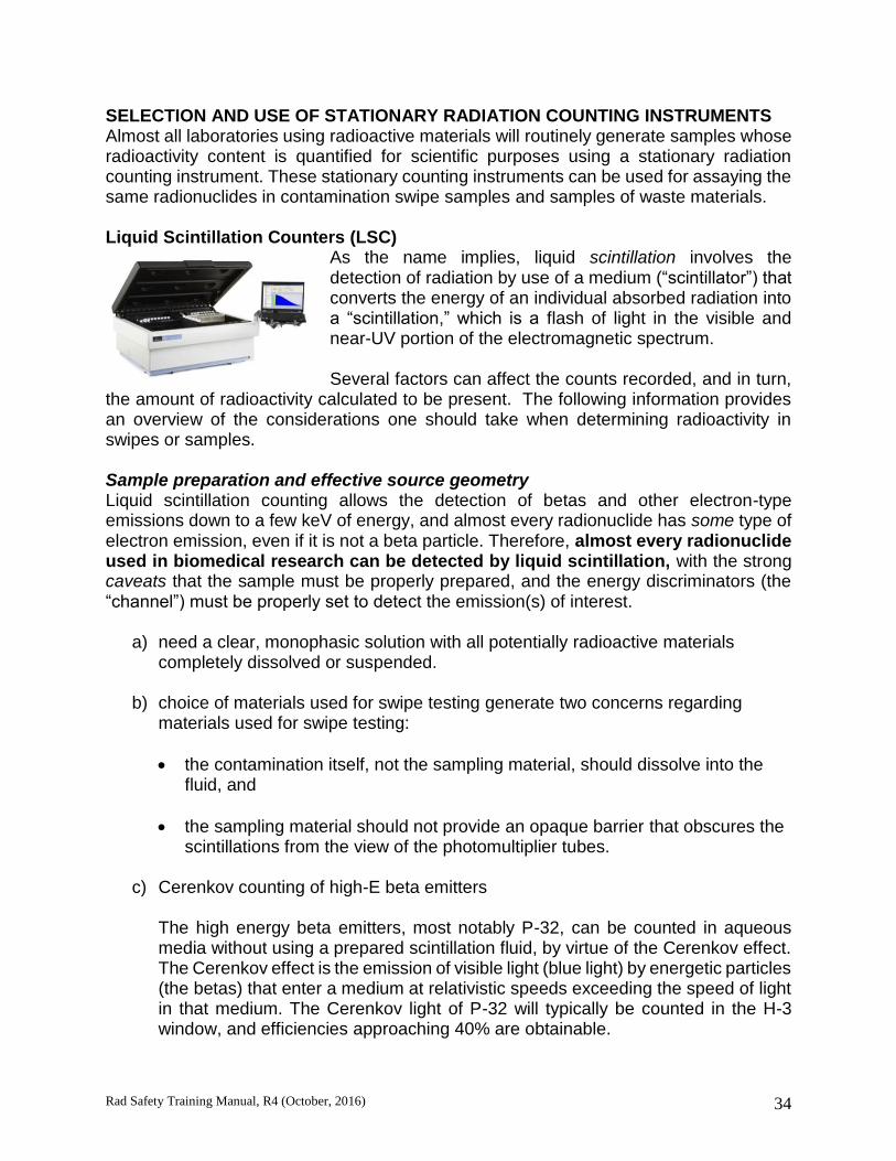
Rad Safety Training Manual, R4 (October, 2016) 34
SELECTION AND USE OF STATIONARY RADIATION COUNTING INSTRUMENTS Almost all laboratories using radioactive materials will routinely generate samples whose radioactivity content is quantified for scientific purposes using a stationary radiation counting instrument. These stationary counting instruments can be used for assaying the same radionuclides in contamination swipe samples and samples of waste materials. Liquid Scintillation Counters (LSC)
As the name implies, liquid scintillation involves the detection of radiation by use of a medium (“scintillator”) that converts the energy of an individual absorbed radiation into a “scintillation,” which is a flash of light in the visible and near-UV portion of the electromagnetic spectrum. Several factors can affect the counts recorded, and in turn,
the amount of radioactivity calculated to be present. The following information provides an overview of the considerations one should take when determining radioactivity in swipes or samples. Sample preparation and effective source geometry Liquid scintillation counting allows the detection of betas and other electron-type emissions down to a few keV of energy, and almost every radionuclide has some type of electron emission, even if it is not a beta particle. Therefore, almost every radionuclide used in biomedical research can be detected by liquid scintillation, with the strong caveats that the sample must be properly prepared, and the energy discriminators (the “channel”) must be properly set to detect the emission(s) of interest.
a) need a clear, monophasic solution with all potentially radioactive materials completely dissolved or suspended.
b) choice of materials used for swipe testing generate two concerns regarding
materials used for swipe testing:
the contamination itself, not the sampling material, should dissolve into the fluid, and
the sampling material should not provide an opaque barrier that obscures the scintillations from the view of the photomultiplier tubes.
c) Cerenkov counting of high-E beta emitters
The high energy beta emitters, most notably P-32, can be counted in aqueous media without using a prepared scintillation fluid, by virtue of the Cerenkov effect. The Cerenkov effect is the emission of visible light (blue light) by energetic particles (the betas) that enter a medium at relativistic speeds exceeding the speed of light in that medium. The Cerenkov light of P-32 will typically be counted in the H-3 window, and efficiencies approaching 40% are obtainable.
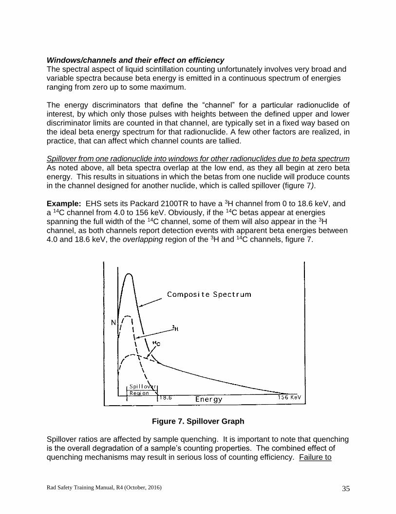
Rad Safety Training Manual, R4 (October, 2016) 35
Windows/channels and their effect on efficiency The spectral aspect of liquid scintillation counting unfortunately involves very broad and variable spectra because beta energy is emitted in a continuous spectrum of energies ranging from zero up to some maximum. The energy discriminators that define the “channel” for a particular radionuclide of interest, by which only those pulses with heights between the defined upper and lower discriminator limits are counted in that channel, are typically set in a fixed way based on the ideal beta energy spectrum for that radionuclide. A few other factors are realized, in practice, that can affect which channel counts are tallied. Spillover from one radionuclide into windows for other radionuclides due to beta spectrum As noted above, all beta spectra overlap at the low end, as they all begin at zero beta energy. This results in situations in which the betas from one nuclide will produce counts in the channel designed for another nuclide, which is called spillover (figure 7). Example: EHS sets its Packard 2100TR to have a 3H channel from 0 to 18.6 keV, and a 14C channel from 4.0 to 156 keV. Obviously, if the 14C betas appear at energies spanning the full width of the 14C channel, some of them will also appear in the 3H channel, as both channels report detection events with apparent beta energies between 4.0 and 18.6 keV, the overlapping region of the 3H and 14C channels, figure 7.
Figure 7. Spillover Graph Spillover ratios are affected by sample quenching. It is important to note that quenching is the overall degradation of a sample’s counting properties. The combined effect of quenching mechanisms may result in serious loss of counting efficiency. Failure to

Rad Safety Training Manual, R4 (October, 2016) 36
detect and correct for a loss of counting efficiency is a form of false negative error that may result in underestimating or completely failing to detect radioactivity that is actually present in the sample. For purposes of general swipe testing for removable contamination, a good practice is to set three channels: for H-3, C-14, and P-32. The ideal beta spectra of these radionuclides span a complete beta energy range from 0 to 1.7 MeV. Mechanisms of quenching Chemical quenching Any aspect of the chemistry of the sample that interferes with the transfer of a beta particle’s energy from the solvent to the fluor is a form of chemical quenching, including the dilution of the solvent/fluor system by higher loadings of sample per unit volume of scintillation fluid. Color quenching Any mechanism that keeps the emitted scintillation light from reaching the photomultiplier tube, by scattering or absorption, is a form of optical quenching. The common form is color quenching, used to describe the effect of a sample that has color in the sense that it contains some material that absorbs light in some portion of the wavelengths from blue visible light through near UV. Quenching and efficiency Whatever the index number of quenching that the manufacturer of the LSC has provided (“H#,” “SIE,” etc.), it is possible to use a set of quenched standards to plot this number vs. counting efficiency for a specified channel and corresponding radionuclide, in order to establish a quantitative relationship that allows the quench index to be used to predict counting efficiency. A quenched standards set must have scintillation vials that contain known amounts of some single radionuclide of interest, quenched to various levels.
The best kind of a quenched standards set is one that is custom made to resemble the chemistry of the same sample type, and is quenched by the same mechanism(s) that will be encountered in practice. However, manufactured quenched standards sets for both H-3 and C-14 are commonly available from the manufacturers of liquid scintillation counters and from other scientific suppliers.
Example: a manufactured quenched standards set containing H-3 was counted using a Packard Model 2100TR liquid scintillation counter. This machine uses a quench indicating index number called “tSIE,” which ranges from 1000 for a theoretically perfect unquenched sample down to zero for a totally quenched sample. The table below shows the H-3 counting efficiency for a channel set for H-3 from 0 to 18.6 keV, as a function of
the tSIE. Also displayed as a function of tSIE is the 3H14C spillover ratio (spillover of H-3 into the C-14 channel), as defined above. Note that, as the H-3 spectrum is shifted downward by increased quenching, proportionally fewer counts due to H-3 appear in the C-14 window, which is set from 4 to 156 keV.

Rad Safety Training Manual, R4 (October, 2016) 37
TABLE 6
Results for a Tritium Quenched-Standards Set
tSIE
H-3 Efficiency
(%)
(“C-14” CPM) /(H-3 CPM)
934 64 0.61
633 55 0.47
499 48 0.37
384 41 0.27
247 28 0.13
128 13 0.03
The Colorado Department of Public Health and Environment requires that those counting results on PI’s’ contamination surveys that exceed the limit for statistically significant results must be reported in units of true radioactivity, of which DPM is the obvious and most convenient unit. It should also be recognized that, because individual liquid scintillation vials are subject to being quenched to substantially different extents by various factors that are difficult to predict and reliably control, it is important for scientific reasons to use quench indicators to convert results to DPM. Artifacts producing spurious count rates Liquid scintillation counting is subject to a number of interferences that may produce spurious counts that appear to result from radioactivity in the sample, but actually arise from other sources. Chemiluminescence and phosphorescence Chemiluminescence is the production of light photons by chemical processes occurring in the sample, rather than by the absorption of ionizing radiations emitted by materials within the sample. Phosphorescence (photoluminescence) is the production of light photons by materials in the sample or the vial itself, due to their having been previously excited by UV radiation. At high intensities, both of these processes can deceive the scintillation counter’s electronics into believing that scintillations are being detected.
Phosphorescence tends to diminish rapidly, and can typically be eliminated by allowing the scintillation vial to sit in the dark for a half hour to an hour. Chemiluminescence may be much more persistent, and may require some experimentation with cooling the sample and/or allowing it to sit for extended periods of time.
Static electricity Static electricity can build up on scintillation vials, especially plastic ones, and especially in dry climates. This may cause numerous random discharges during counting, which cause emissions of light and which serve to add random, unpredictable numbers of counts. Some machines are equipped with an electrical device to remove static charge from each vial as it enters the counting chamber. Another option to reduce this problem

Rad Safety Training Manual, R4 (October, 2016) 38
is the use of anti-static sheets manufactured for home use in clothes dryers to wipe the vials before counting. Stray sources of gamma/x rays nearby Older liquid scintillation counters are heavily shielded with lead, whereas some newer models require less shielding because faster electronics and digital processing allow superior rejection of some types of detection events that are produced by natural background radiation. All liquid scintillation counters are subject to some interference, however, from stray gamma or x radiations arising from sources in the vicinity. Contamination of machine and/or samples Contamination of the exterior surfaces of scintillation vials may create spurious counts and may transfer to the sample-loading platform. This possibility is primarily of concern with the higher-energy beta emitters. Gamma well counters Gamma well counters utilize a solid scintillation detector with a hole called a “well,” into which the sample-containing end of a test tube is inserted. As the sample is almost
surrounded by the detector, the effective source-detector geometry is close to 4. In contrast to the scintillating medium in a liquid scintillation vial, the scintillator in a gamma well counter is a much larger and more massive detector of considerably higher effective atomic number, and therefore has a much greater detecting efficiency for gamma and x rays. Because the scintillator is physically separated from the sample being counted, the chemical composition and physical structure of the sample have virtually no effect on counting efficiency. The only relevant consideration is that the sample must be of small enough volume to fit in the bottom 1 to 2 cm of the test tube being used to contain it,
preserving the desirable (nearly 4) geometrical relationship with the detector. By virtue of their construction and operation, gamma well counters are not subject to the interferences of chemiluminescence or phosphorescence and static electricity. Gamma well counters are sensitive to stray radiations in the form of high-energy gamma and/or x rays arising from nearby sources. They are also, of course, subject to interferences from samples that carry external contamination. BASIC RADIOACTIVE DECAY CALCULATIONS Radioactive decay Radioactivity is the result of the process in which a parent radionuclide undergoes spontaneous disintegration. The decay fraction for any particular radionuclide is constant. Thus, if a particular radionuclide loses 25% of its atoms in a day, we will find it will lose 25% of its remaining atoms in the next day, and so on. Symbolic representation of radioactive decay is as follows:
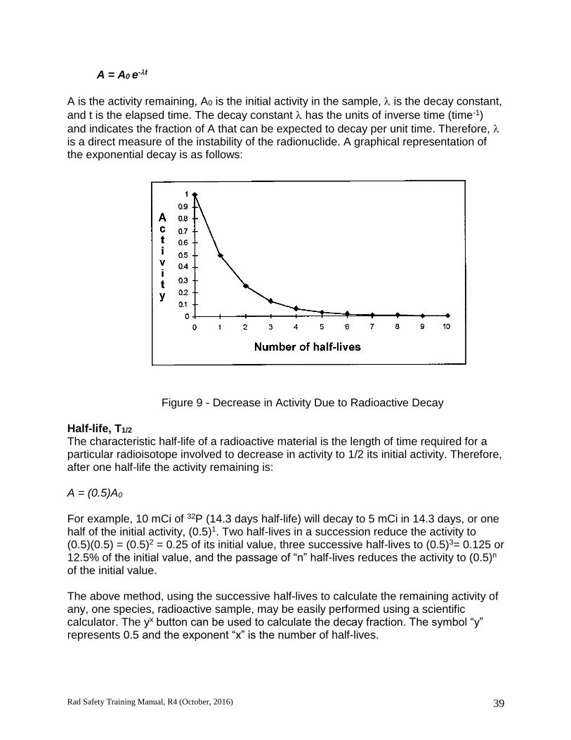
Rad Safety Training Manual, R4 (October, 2016) 39
A = A0 e-t
A is the activity remaining, A0 is the initial activity in the sample, is the decay constant,
and t is the elapsed time. The decay constant has the units of inverse time (time-1)
and indicates the fraction of A that can be expected to decay per unit time. Therefore, is a direct measure of the instability of the radionuclide. A graphical representation of the exponential decay is as follows:
Figure 9 - Decrease in Activity Due to Radioactive Decay
Half-life, T1/2 The characteristic half-life of a radioactive material is the length of time required for a particular radioisotope involved to decrease in activity to 1/2 its initial activity. Therefore, after one half-life the activity remaining is: A = (0.5)A0
For example, 10 mCi of 32P (14.3 days half-life) will decay to 5 mCi in 14.3 days, or one half of the initial activity, (0.5)1. Two half-lives in a succession reduce the activity to (0.5)(0.5) = (0.5)2 = 0.25 of its initial value, three successive half-lives to (0.5)3= 0.125 or 12.5% of the initial value, and the passage of “n” half-lives reduces the activity to (0.5)n of the initial value. The above method, using the successive half-lives to calculate the remaining activity of any, one species, radioactive sample, may be easily performed using a scientific calculator. The yx button can be used to calculate the decay fraction. The symbol “y” represents 0.5 and the exponent “x” is the number of half-lives.

Rad Safety Training Manual, R4 (October, 2016) 40
The same approach may be used to calculate the activity using the exponential decay formula. Scientific calculators have the constant “e”, the natural base for logarithms,
which can be used to calculate the decay fraction e-t.
References for Section 2
1. Shapiro, J.: Radiation Protection: A Guide for Scientists and Physicians, 3rd ed., Harvard University Press, 1990.
2. Kessler, M., ed., Liquid Scintillation Analysis: Science and Technology, Packard Instrument Co., Publication 169-3052. Rev G, Meriden, CT, 1989.
Rad Safety Training Manual, R4 (October, 2016) 41
Section 3
USING RADIOACTIVE MATERIAL IN A LABORATORY SETTING: RADIOACTIVE MATERIAL ACCOUNTABILITY, STORAGE AND WASTE, SURVEYS
The users of radioactive materials carry the responsibility for materials under your control. All radioactive material (RAM) must be tracked, documented and accounted for from the original purchase through the ultimate disposal. Information in this section will provide information regarding the accountability, control, storage and the considerations for disposing RAM. Radioactivity may not be procured, used, stored or disposed without an approved authorization from the Committee on Ionizing Radiation (CIR) issued to a designated, trained and qualified principal investigator. The RAM authorization specifically identifies the isotope, compound, and activity limits the named PI is allowed to use in the lab. Additionally, the RAM authorization is assigned a hazard category, lists the type of contamination surveys required and the frequency those surveys must be performed. Handling RAM must only be performed by trained, authorized radiation workers. Anyone who is not trained as a radiation worker under the specific RAM authorization is considered a member of the public and is not authorized access to RAM. Regulatory liability for exposure to members of the public is serious. Areas where RAM is used must be under constant surveillance by trained and qualified radiation workers to prevent inadvertent exposures to members of the public. All RAM must be secured from unauthorized access. Ordering radioactive material Pre-approval by EHS must be obtained prior to ordering any RAM from a vendor. Pre-approval verifies several items are in compliance:
The PI is authorized to use the compound, radionuclide and activity amount,
Training for all personnel is up-to-date,
Dosimetry, if applicable, is in place, and

Rad Safety Training Manual, R4 (October, 2016) 42
The current possession limit identified on the authorization document will not be exceeded
A “RAM Purchase Approval Request” form must be submitted prior to placing an order of RAM from a vendor. Upon review and approval of the request, EHS will issue a unique requisition number. After receiving the requisition number from EHS, you may place the order with the vendor. All radioactive material orders must be shipped to the EHS Support Facility, unless the Radiation Safety Officer approves an alternative location. This includes orders from vendors and other institutions. When speaking with the vendor about the shipping address, be specific that they include your EHS issued requisition number to the shipping label. Be sure to have RAM shipped to the following address: EHS Support Facility Requisition # 13178 E. 19th Avenue Aurora, CO 80045 If you receive radioactive materials at your lab directly from the shipper (e.g. FedEX, UPS), contact EHS immediately at 303-724-0345 so Radiation Safety may collect the package to properly receive the material. Contact EHS before shipping any radioactive materials from the university to other institutions. Receiving radioactive material Radioactive material arriving on campus receives a unique Radiation Safety Office number (RSO#) that is used to track the material from receipt through disposal. The RSO# helps identify the radionuclide, compound, activity, assay date, lot number, vendor and the PI. The Radiation Safety division will receive the package from the courier and confirm all DOT requirements have been met for the RAM package. A package inspection, survey and confirmation that the material received is the same as the material ordered is performed before delivering the package to the lab. After entering all the information into the EHS Assistant database, the RSO# is generated, as is an accounting sheet and a waste ticket sheet. The accounting sheet and waste tickets are delivered to the lab with the RAM package. A trained, authorized radiation worker must receive the package at the lab. EHS only surveys the exterior of the package prior to delivery. Once received at the lab, the radiation worker must perform a survey of the stock vial, pig or container holding the stock vial, and the interior of the package itself. Don the appropriate PPE and treat the package interior as potentially contaminated until proven otherwise. Include a

Rad Safety Training Manual, R4 (October, 2016) 43
background swipe (blank sample prepared in the same manner as other swipes) to compare your sample results to. After confirming there is no contamination present, place the stock vial and pig in your secured storage location. Attach the LSC printout for the receipt of your material to the accounting sheet for the identified RSO#. Sign the accounting sheet indicating that you completed the survey of the stock vial and packaging. Provided no contamination was identified on the packaging material, the shipping box may be discarded as non-radioactive waste in the routine lab waste. Any labeling on the package to indicate radioactive materials must be removed or defaced prior to disposing the shipping box. Be sure to document all of your survey results including proper annotation of which swipe belongs to which survey point. Securing and storing your radioactive material Your RAM must be stored in an approved location and be secured from unauthorized access. Typically, a refrigerator or freezer is designated to store stock vials. In the ideal world, there would be a dedicated storage device for the RAM received at the lab. In this scenario, the door to the storage device would be locked to prevent unauthorized access to the material. If the storage device must be shared with non-radioactive material, a lockable container must be installed inside the device that will deter unauthorized access to the RAM. In addition, the lockable container must also be secured within the storage device to prevent unauthorized removal of the lockable container itself. When you are ready to store your material, following the next few steps will help maintain positive accounting and keep the material from unauthorized workers:
Confirm the contents of your order amount and compound with the label on the stock vial or pig
Write the RSO# on the pig with a permanent marker
Place the pig inside another strong-type box, such as an acrylic box EHS conducts unannounced security spot checks to verify RAM storage is locked and secured from unauthorized access. This includes both storage of stock materials and waste cabinets. The following would be considered a violation of the PI’s RAM authorization and may warrant escalated action by the CIR:
Gaining access to any RAM without being stopped and asked for identification
Finding RAM unsecured from unauthorized access at any time

Rad Safety Training Manual, R4 (October, 2016) 44
RAM use at the lab A dedicated, locked and labelled procedure room provides the ideal location for working with radioactive materials. If this is not possible, the area where radioactivity is used must be under constant surveillance by a fully trained radiation worker to prevent inadvertent exposures to members of the public. Radiation workers have the responsibility of challenging those individuals in the lab that they do not recognize in order to provide protection from exposure from radioactive materials. Areas where RAM is used must be properly labelled to communicate the hazards present to others entering or passing through the area. Benchtops, equipment, cabinets, storage devices, drawers or any other item or location that may be used with or contain radioactive materials must be labelled. EHS will typically post approved locations for use, but radiation workers are responsible to demarcate benchtops and equipment used with radioactive materials. In general terms, most radioactivity for biomedical research comes in the form of a liquid compound in a stock vial. Briefly, some fraction of material is withdrawn from the stock vial, used in an experiment and through the experimental process, various RAM waste forms are generated. The entire life cycle of the radioactive material must be accounted for – see the example accounting sheet above and more detail about each section follows. The radiation worker is responsible to ensure documentation is maintained detailing how much radioactivity was used and where that radioactivity went. Accounting of radioactive material – aliquots from stock vials An accounting sheet is provided with each RAM package delivered to the lab. The lab is not required to utilize the accounting sheet provide, although it is strongly encouraged. The pre-printed accounting sheet includes specific information about the isotope, the RSO #, the activity amount ordered and provides space to record how much material is used on a given date.
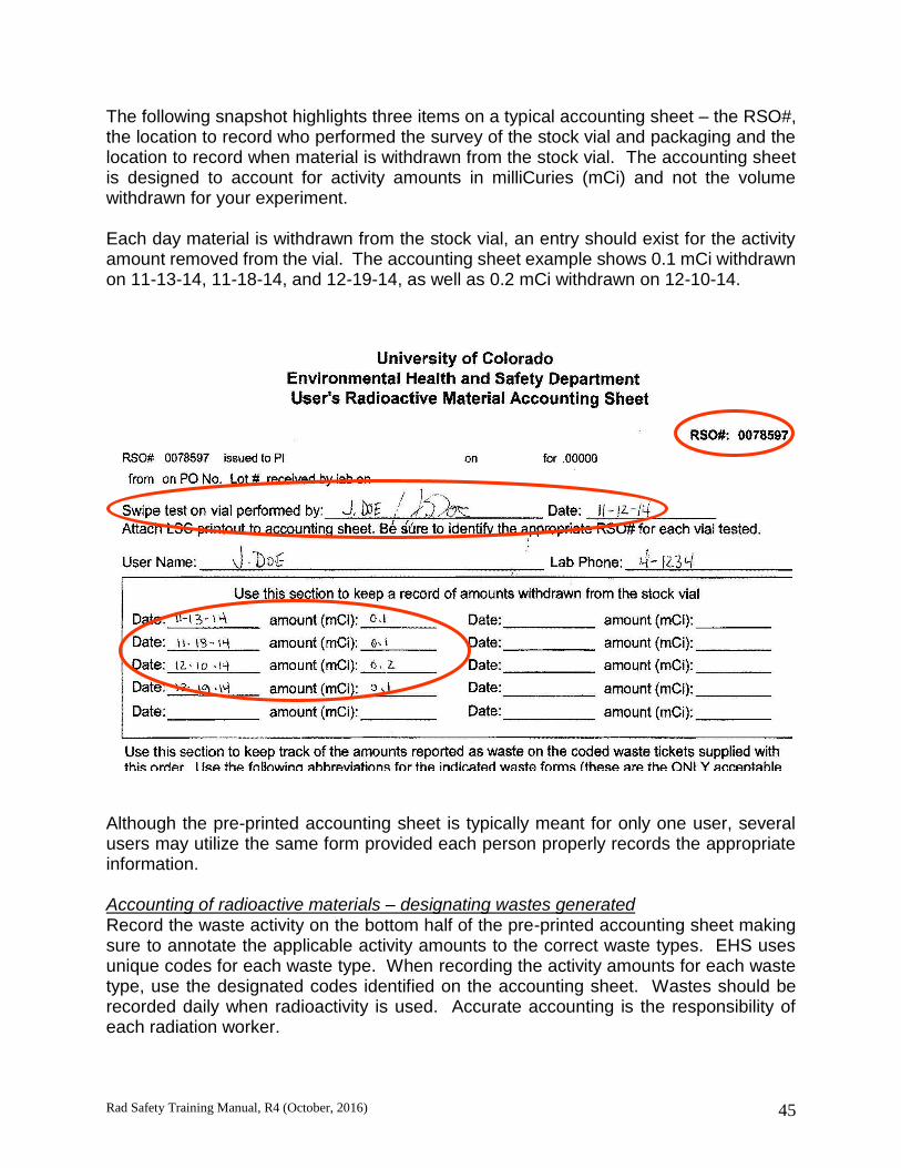
Rad Safety Training Manual, R4 (October, 2016) 45
The following snapshot highlights three items on a typical accounting sheet – the RSO#, the location to record who performed the survey of the stock vial and packaging and the location to record when material is withdrawn from the stock vial. The accounting sheet is designed to account for activity amounts in milliCuries (mCi) and not the volume withdrawn for your experiment. Each day material is withdrawn from the stock vial, an entry should exist for the activity amount removed from the vial. The accounting sheet example shows 0.1 mCi withdrawn on 11-13-14, 11-18-14, and 12-19-14, as well as 0.2 mCi withdrawn on 12-10-14.
Although the pre-printed accounting sheet is typically meant for only one user, several users may utilize the same form provided each person properly records the appropriate information. Accounting of radioactive materials – designating wastes generated Record the waste activity on the bottom half of the pre-printed accounting sheet making sure to annotate the applicable activity amounts to the correct waste types. EHS uses unique codes for each waste type. When recording the activity amounts for each waste type, use the designated codes identified on the accounting sheet. Wastes should be recorded daily when radioactivity is used. Accurate accounting is the responsibility of each radiation worker.

Rad Safety Training Manual, R4 (October, 2016) 46
Every time radioactive materials are used, some form of waste is being generated. Waste types and amounts are developed during the PI’s application process and represent an estimate of what percentage of the radioactivity originally used in the protocol is carried to each waste form. For example, the PI’s application states that the protocol will use 0.1 mCi of C-14 for each experiment. During the protocol steps, aqueous RAM waste, dry solid RAM waste and non-hazardous scintillation vials will be generated. Further, the PI estimates that 70% of the activity (0.07 mCi) will end up as aqueous waste, 29% (0.029 mCi) will go to dry solid RAM waste and the remaining 1% (0.001 mCi) will be in the non-hazardous scintillation vial waste. So, each day the experiment is performed from the beginning, the amounts recorded on the accounting sheet will be the same. The green highlight below represents how to record the activity and the waste types generated for the PI’s protocol given the numbers in the preceding paragraph.
Survey requirements, inspections, records, and dealing with emergencies The PI’s RAM authorization document provides useful information that should be covered during a radiation worker’s Radiation Safety On-The-Job training. Each authorization carries a hazard classification, which triggers a specific inspection frequency and survey frequency. These are outlined here:
For isotopes other than 3H, both portable instrument and swipe test surveys are required to be performed and documented, at the frequency dictated by the hazard classification.
Rad Safety Training Manual, R4 (October, 2016) 47
The surveys are required to be completed within the designated frequency when radioactivity is in use. The best way to ensure compliance with your survey frequency is to perform and document your surveys immediately following completion of the experiment. Inspectors will compare survey dates with the logged usage dates to determine whether the survey frequency has been met. Some important consideration for contamination surveys:
DO survey your work area before and after each experiment with a portable survey instrument (not required if only using 3H)
DO swipe surveys of stock vial and packaging upon receipt of material in the lab
DO scan your hands, clothing and shoes while you are handling RAM and perform a personal survey prior to leaving the area
DO survey hand held and stationary equipment used with RAM (e.g. pipettor, incubator, tweezers, etc.)
DO survey about 10 or more locations and include a background sample Immediately report any contamination found on skin, personal or protective clothing, or floors to EHS at 303-724-0345. Audits of radioactive materials use labs are performed by the Radiation Safety division every year or every two years, depending on the hazard classification. These are scheduled with the lab ahead of time and focus on assuring the lab is meeting the regulatory requirements imposed by the Colorado Department of Public Health and Environment through the university’s radioactive materials license. Most records generated to show compliance with radioactive materials use must be maintained for a minimum of 3 years. These include survey documents, instrument calibration records, inventory and waste accounting, and radiation worker training records, among others. The documentation provides the proof to regulators that all requirements for holding a radioactive materials license are being met. Be sure to maintain the following records:
Radiation Worker training certificates
Rad Safety OJT forms
Accounting Sheets
Portable instrument survey log
Swipe test records (LSC print out) and corresponding map
Waste pick-ups
RAM authorization and associated applications or approvals
Trained worker list Remember: “If it was not documented, it never happened” Dealing with emergencies Know who to call in case of emergency and be prepared to provide some basic information regarding the event. When waiting for EHS to arrive, keep people away from

Rad Safety Training Manual, R4 (October, 2016) 48
the affected area. If the area was evacuated, make sure no one leaves the assembly area until released by EHS. A life threatening scenario takes precedence over the contamination concerns.
During Business Hours M-F, 8am-5pm After Hours and Weekends
EHS Main Office University Police Department
303-724-0345 911 (from any campus phone)
303-724-4444 (from any phone)
You should be able to provide the following information:
Your name and names of other potentially affected individuals
The location of the incident
The type and amount of radioactive material involved
A description of the incident Managing radioactive waste Users of radioactive materials are also waste generators. Waste segregation is an important aspect of managing the various waste streams encountered while conducting research. It is equally important to segregate radioactive wastes into appropriate radioactive waste categories as it is to separate them from non-radioactive waste streams.
Radioactive waste containers must be identified with the standard radiation trefoil symbol. Use containers and liners purchased from EHS for collecting dry solid and aqueous waste and use separate containers for each isotope, when possible. Never overfill waste containers, keep them closed when not in use and remember to keep your waste secured from unauthorized access. In addition to accounting for wastes generated on the accounting sheet, a running inventory should be included on the waste container or the waste cabinet. Update the running waste inventory each time waste is placed into the container. There are several main types of radioactive wastes:
Dry Solids
Aqueous
Mixed Wastes
Scintillation Vials
Radioactive Biological
Stock Vials
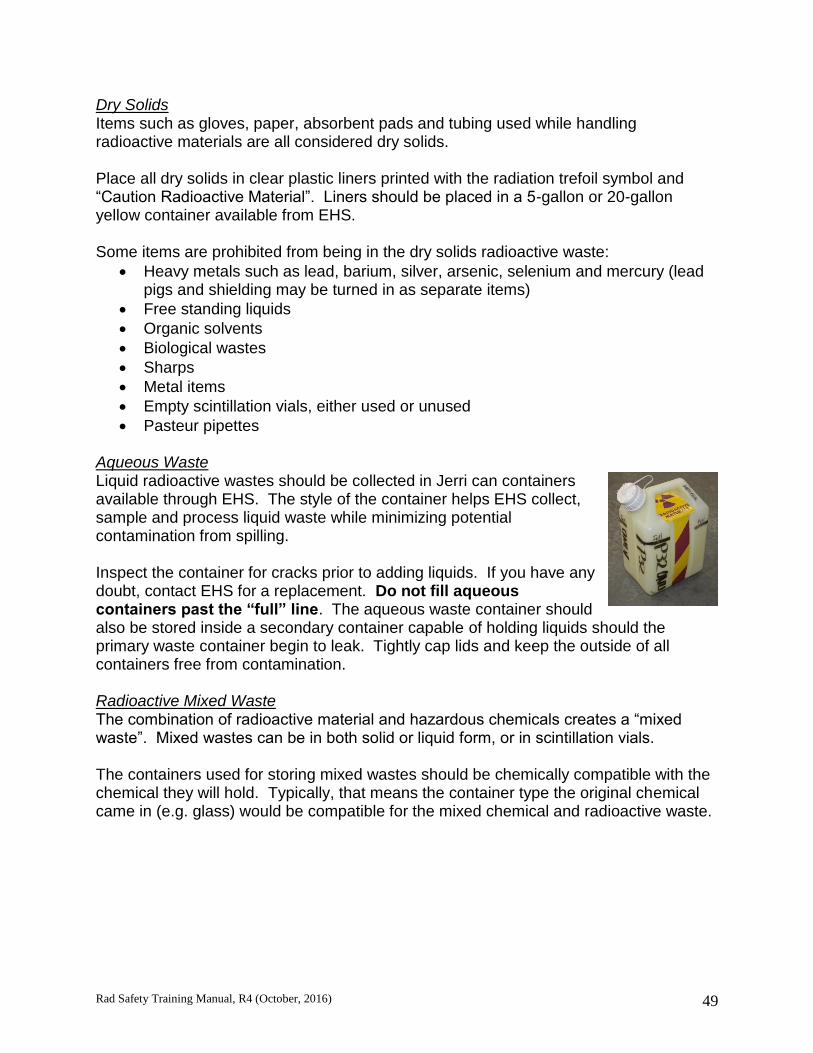
Rad Safety Training Manual, R4 (October, 2016) 49
Dry Solids Items such as gloves, paper, absorbent pads and tubing used while handling radioactive materials are all considered dry solids. Place all dry solids in clear plastic liners printed with the radiation trefoil symbol and “Caution Radioactive Material”. Liners should be placed in a 5-gallon or 20-gallon yellow container available from EHS. Some items are prohibited from being in the dry solids radioactive waste:
Heavy metals such as lead, barium, silver, arsenic, selenium and mercury (lead pigs and shielding may be turned in as separate items)
Free standing liquids
Organic solvents
Biological wastes
Sharps
Metal items
Empty scintillation vials, either used or unused
Pasteur pipettes Aqueous Waste Liquid radioactive wastes should be collected in Jerri can containers available through EHS. The style of the container helps EHS collect, sample and process liquid waste while minimizing potential contamination from spilling. Inspect the container for cracks prior to adding liquids. If you have any doubt, contact EHS for a replacement. Do not fill aqueous containers past the “full” line. The aqueous waste container should also be stored inside a secondary container capable of holding liquids should the primary waste container begin to leak. Tightly cap lids and keep the outside of all containers free from contamination. Radioactive Mixed Waste The combination of radioactive material and hazardous chemicals creates a “mixed waste”. Mixed wastes can be in both solid or liquid form, or in scintillation vials. The containers used for storing mixed wastes should be chemically compatible with the chemical they will hold. Typically, that means the container type the original chemical came in (e.g. glass) would be compatible for the mixed chemical and radioactive waste.

Rad Safety Training Manual, R4 (October, 2016) 50
Choose a reasonably sized container to collect mixed waste and avoid using the 4L container when a 1L or smaller container would suffice. Containers may be available from EHS. At the beginning of waste accumulation, an orange “mixed waste” label (provided by EHS) must be affixed. The container must be stored in a registered satellite accumulation area. When you are ready to dispose of the waste, complete a “RAM Mixed Waste Disposal” form prior to the waste pick up to give to EHS when collecting the waste.
Scintillation Vials Store scintillation vials in cardboard trays designed to hold the vials upright. Separate “cold” (non-radioactive) vials from others, and keep hazardous scintillation vials separate from non-hazardous vials. Hazardous scintillation vial trays must also be labeled with an orange hazardous scintillation vial label and properly stored. Cold vials are typically generated from swipe tests of areas that show no radioactivity is present. The Radiation Safety division will collect the cold scintillation vials to ensure proper disposal through the Hazardous Materials group. Scintillation vials generated as part of your experiment that do contain radioactivity should also be separated from non-radioactive vials. Separate vials with different isotopes. Empty scintillation vial trays are available through EHS and are provided free of charge. Stock Vials and pigs Keep stock vials and pigs as a separate pick-up item. Place the stock vial into the pig and complete one pre-coded waste ticket recording “STK” as the waste type. Tape the waste ticket to the pig for collection. Biologically Classed Materials Radioactive biological type waste is generally divided into “biological carcass” and “biological non-carcass” wastes. Further details on biologically classed materials are presented in the Radioactive Waste Disposal Manual. Infectious and potentially infectious type waste must first be disinfected and reclassified as “biological non-carcass” waste. Animal bedding, culture vessels, plastic tubing may all be considered “biological non-carcass waste”.

Rad Safety Training Manual, R4 (October, 2016) 51
Disinfect human cell lines regardless of whether they are primary cells or cell lines. Use an effective disinfectant for the type of material and the required contact time. Once disinfected, collect the dry and aqueous waste in separate containers from the non-biological radioactive wastes. Apart from the name designation, radioactive dry solids and dry solid biological non-carcass wastes are collected in the same manner: in a clear poly liner stenciled with the radiation trefoil symbol and the words “Caution Radioactive Material”. Carcass and tissue waste must remain frozen. Radioactive Waste Pick-Up Contact the Radiation Safety division to schedule a waste pick-up through email or phone. Complete the “Radioactive Waste Pick-Up Request” form, available on the EHS forms website, and submit the form to [email protected]. The form asks simple questions about quantity, activity, types of waste and what types of containers are needed from EHS. A Radiation Safety Specialist will contact the lab to schedule a waste pick-up. Waste pick-ups should be requested before the waste containers are completely full. The Radiation Safety staff strives to pick-up waste within 10 business days of the request. If you find the need for an emergency pick-up, please contact 303-724-0109 and leave a message. Waste tickets are a key component to removing RAM inventory from the lab’s record. Compile one waste ticket per RSO# for each type of waste that was generated by summing all the activity amounts of that waste type deposited into the container.
For example, the waste recorded on the pictured accounting sheet shows 4 entries for “DRY” waste from several different dates. Only one waste ticket showing “DRY” waste with the total activity amount of 0.147 mCi is necessary to turn in to EHS. Remember, each unique RSO# that is deposited into a container must have at least one waste ticket reporting the activity amount within the waste.

Rad Safety Training Manual, R4 (October, 2016) 52
All of the following should be available and complete prior to EHS arriving at the lab:
Waste tickets
Contamination survey
Mixed waste disposal form, if applicable
Accounting sheets are available for review

Rad Safety Training Manual, R4 (October, 2016) 53
Section 4
BIOLOGICAL EFFECTS OF RADIATION Introduction Within a year after Roentgen's discovery of X-rays in 1895, it was learned that exposure to ionizing radiation could lead to biological damage. Since that time, a tremendous amount of research has been done attempting to interpret the reactions which take place from the moment that radiation enters a living cell until some permanent damage is produced. From beginning to end, these initial reactions are probably completed in a millionth of a second, making them very difficult to study. For this reason, it is still not known which of the many chemical or biochemical reactions brought about by ionizing radiation are responsible for initiating biological damage. The DNA within our cells, indeed the entire human genome, is being mapped and will one-day lead to a framework in which to understand how DNA, genes, cells, the body, and the environment interact. Radiosensitivity and the Law of Bergonie and Tribondeau As early as 1906, an attempt was made to correlate the differences in sensitivity of various cells with differences in cellular physiology. These differences in sensitivity are stated in the Law of Bergonie and Tribondeau: “The radio-sensitivity of a tissue is inversely proportional to its degree of differentiation”. In other words, cells most active in reproducing themselves and cells not fully mature will be most harmed by radiation. This law is considered to be a rule-of-thumb and some cells and tissues demonstrate exceptions to the rule. Since the time that the Law of Bergonie and Tribondeau was formulated, it is generally accepted that cells tend to be radiosensitive if they are:
Cells that have a high rate of division;
Cells that have a high metabolic rate;
Cells that are of a non-specialized type and,
Cells that are well nourished. The law can be used to classify the following tissues as radiosensitive:
Germinal (reproductive) cells of the ovary and testis i.e., spermatogonia;
Hematopoietic (blood-forming) tissues: red bone marrow, spleen, lymph nodes, thymus;
Epithelium of the skin and,
Epithelium of the gastrointestinal tract (interstitial crypt cells) The law can be used to classify the following tissues as radioresistant:
Bone
Liver

Rad Safety Training Manual, R4 (October, 2016) 54
Kidney
Cartilage
Muscle
Nervous tissue Primary and Secondary Effects On Cells: Radiation passing through living cells may directly ionize or excite atoms and molecules in the cell structure. These changes affect the forces that bind the atoms together into molecules. The molecules may break up (dissociate) into fragments called free radicals and ions which are not chemically stable. Free radicals are electrically neutral structures having one unpaired electron. Because the cell has a high water content, the most important free radicals are those formed from water molecules in which the hydrogen radical H+ has an unpaired electron and the OH- radical will have nine electrons, one of which will be unpaired. Free radicals are very reactive chemically, and can recombine to produce hydrogen peroxide (H2O2), a chemical poison to the cell. The total harmful effect on cell processes is dependent upon the amount of absorbed radiation. Cellular processes are affected in varying degrees, and may include cell death. However, some damage to the cell may be repaired. Health risks A health risk is generally thought of as something that may endanger health. Scientists consider health risk to be the statistical probability or mathematical chance that personal injury, illness, or death may result from some action. Most people do not think about health risks in terms of mathematics. Instead, most of us consider the health risk of a particular action in terms of whether we believe that particular action will, or will not, cause us some harm. The intent of this section is to provide estimates of, and explain the basis for, the risk of injury, illness, or death from occupational radiation exposure. Risk can be quantified in terms of the probability of a health effect per unit of dose received. When x-rays, gamma rays, and ionizing particles interact with living materials such as our bodies, they may deposit enough energy to cause biological damage. Radiation can cause several different types of events such as the very small physical displacement of molecules, changing a molecule to a different form, or ionization, which is the removal of electrons from atoms and molecules. When the quantity of radiation energy deposited in living tissue is high enough, biological damage can occur as a result of chemical bonds being broken and cells being damaged or killed. These effects can result in observable clinical symptoms. Stochastic Effects: Stochastic effects are those in which the probability of an effect occurring within a population increases with dose, without threshold. Any dose, therefore, has a certain probability, however low, of causing the effect. Stochastic effects may result from injury to a single cell or a small number of cells. Carcinogenic (cancer) and genetic effects (capable of being inherited) are examples of stochastic effects. In these, once the effect is induced, the severity is already determined by the nature of the effect. Stochastic effects are assumed to have some chance of occurring no matter how low the dose.

Rad Safety Training Manual, R4 (October, 2016) 55
Although studies have not shown a consistent cause-and-effect relationship between current levels of occupational radiation exposure and biological effects, it is prudent from a worker protection perspective to assume that some effects may occur. At CU DENVER | ANSCHUTZ, established dose limits are intended to limit the probability of stochastic effects occurring to an acceptable level. But any exposure to radiation involves a risk, and no risk should be undertaken without the expectation of a net benefit. Non-Stochastic Effects: Non-stochastic, sometimes referred to as deterministic effects are those in which the severity of the effect varies with the dose. For these types of effects, a threshold dose may exist. That is, if the dose is kept below the threshold dose, the effect will not be observed. Non-stochastic effects are considered to result from the collective injury of a substantial number of cells in the tissue. Examples of such effects are cataracts, skin ulcerations or burns, depletion of blood-forming cells in bone marrow, and impairment of fertility. Radiation Risk Estimates From currently available data, the NRC has adopted a risk value for an occupational dose of 1 rem (0.01 Sv) Total Effective Dose Equivalent (TEDE) of 4 in 10,000 of developing a fatal cancer, or approximately 1 chance in 2,500 of fatal cancer per rem of TEDE received. The uncertainty associated with this risk estimate does not rule out the possibility of higher risk, or the possibility that the risk may even be zero at low occupational doses and dose rates. The radiation risk incurred by a worker depends on the amount of dose received. Under the linear model explained above, a worker who receives 5 rems (0.05 Sv) in a year incurs 10 times as much risk as another worker who receives only 0.5 rem (0.005 Sv). Only a very few workers U.S. Department of Energy workers receive doses near 5 rems (0.05 Sv) per year. According to the BEIR - V report and other national data, approximately one in five adults normally will die from cancer from all possible causes such as smoking, food, alcohol, drugs, air pollutants, natural background radiation, and inherited traits. Thus, in any group of 10,000 workers, we can estimate that about 2,000 (20 %) will die from cancer without any occupational radiation exposure. The normal chance of dying from cancer is about one in five for persons who have not received any occupational radiation dose. The additional chance of developing fatal cancer from an occupational exposure of 1 rem (0.01 Sv) is about the same as the chance of drawing any ace from a full deck of cards three times in a row. The additional chance of dying from cancer from an occupational exposure of 10 rem (0.1 Sv) is about equal to your chance of drawing two aces successively on the first two draws from a full deck of cards. These estimates are considered by the NRC staff to be the best available for the worker to use to make an informed decision concerning acceptance of the risks associated with exposure to radiation. A worker who decides to accept this risk should try to keep

Rad Safety Training Manual, R4 (October, 2016) 56
exposure-sure to radiation as low as is reasonably achievable (ALARA) to avoid unnecessary risk. Comparing the risk of cancer from radiation to other kinds of health risks: Several studies have compared the average days of life lost from exposure to radiation with the number of days lost as a result of being exposed to other health risks. The word "average" is important because an individual who gets cancer loses about 15 years of life expectancy, while his or her coworkers do not suffer any loss. For categories of NRC-regulated industries with larger doses, the average measurable occupational dose in 1993 was 0.31 rem (0.0031 Sv). A simple calculation based on the article by Cohen and Lee shows that 0.3 rem (0.003 Sv) per year from age 18 to 65 results in an average loss of 15 days. These estimates indicate that the health risks from occupational radiation exposure are smaller than the risks associated with many other events or activities we encounter and accept in normal day-to-day activities.
Table 6 Estimated Loss of Life Expectancy from Health Risks
Health Risk
Estimate of Life Expectancy Lost (Average)
Smoking 20 cigarettes a day 6 years
Overweight (by 15%) 2 years
Alcohol consumption (U.S. Average) 1 year
All accidents combined 1 year
Motor vehicle accidents 207 days
Home accidents 74 days
Drowning 24 days
All natural hazards (earthquake, lightning, flood, etc.) 7 days
Medical radiation 6 days
Occupational Exposure
0.3 rem/yr from age 18 to 65 15 days
1.0 rem/yr from age 18 to 65 51 days
It is also useful to compare the estimated average number of days of life lost from occupational exposure to radiation with the number of days lost as a result of working in several types of industries. Table 7 shows average days of life expectancy lost as a result of fatal work-related accidents. Table 7 does not include non-accident types of occupational risks such as occupational disease and stress because the data are not available.
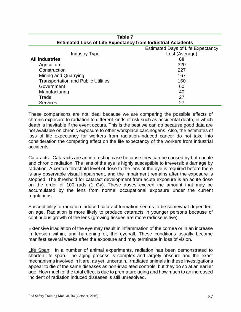
Rad Safety Training Manual, R4 (October, 2016) 57
Table 7 Estimated Loss of Life Expectancy from Industrial Accidents
Industry Type
Estimated Days of Life Expectancy Lost (Average)
All industries 60 Agriculture 320 Construction 227 Mining and Quarrying 167 Transportation and Public Utilities 160 Government 60 Manufacturing 40 Trade 27 Services 27
These comparisons are not ideal because we are comparing the possible effects of chronic exposure to radiation to different kinds of risk such as accidental death, in which death is inevitable if the event occurs. This is the best we can do because good data are not available on chronic exposure to other workplace carcinogens. Also, the estimates of loss of life expectancy for workers from radiation-induced cancer do not take into consideration the competing effect on the life expectancy of the workers from industrial accidents. Cataracts: Cataracts are an interesting case because they can be caused by both acute and chronic radiation. The lens of the eye is highly susceptible to irreversible damage by radiation. A certain threshold level of dose to the lens of the eye is required before there is any observable visual impairment, and the impairment remains after the exposure is stopped. The threshold for cataract development from acute exposure is an acute dose on the order of 100 rads (1 Gy). These doses exceed the amount that may be accumulated by the lens from normal occupational exposure under the current regulations. Susceptibility to radiation induced cataract formation seems to be somewhat dependent on age. Radiation is more likely to produce cataracts in younger persons because of continuous growth of the lens (growing tissues are more radiosensitive). Extensive irradiation of the eye may result in inflammation of the cornea or in an increase in tension within, and hardening of, the eyeball. These conditions usually become manifest several weeks after the exposure and may terminate in loss of vision. Life Span: In a number of animal experiments, radiation has been demonstrated to shorten life span. The aging process is complex and largely obscure and the exact mechanisms involved in it are, as yet, uncertain. Irradiated animals in these investigations appear to die of the same diseases as non-irradiated controls, but they do so at an earlier age. How much of the total effect is due to premature aging and how much to an increased incident of radiation induced diseases is still unresolved.
Rad Safety Training Manual, R4 (October, 2016) 58
The study of small amounts of exposure to radiation for beneficial purposes is termed radiation hormesis. One pioneer in the field, Dr. Luckey of the University of Missouri, Columbia, stated in a 1982 article, "Extensive literature indicates that minute doses of ionizing radiation benefit animal growth and development, fecundity (ability to produce offspring), health and longevity. Specific improvements appear in neurological function, growth rate and survival of young, wound healing, immune competence, and resistance to infection, radiation morbidity (radiation sickness), and tumor induction and growth." An extension of life and a lower incidence of cancer has been observed in rodents exposed to lower doses, (100 to 400 rads), over a lifetime. Radiation hormesis is a very controversial subject and there is no scientific consensus or broad-based support of Dr. Luckey’s work at this time. Radiation Dose Limits The NRC radiation dose limits in 10 CFR Part 20 were established by the NRC based on the recommendations of the ICRP and NCRP as endorsed in Federal radiation protection guidance developed by the EPA. The limits were recommended by the ICRP and NCRP with the objective of ensuring that working in a radiation-related industry was as safe as working in other comparable industries. The dose limits and the principle of ALARA should ensure that risks to workers are maintained indistinguishable from risks from background radiation. For adults, an annual limit that does not exceed
5 rems (0.05 Sv) for the total effective dose equivalent (TEDE), which is the sum of the deep dose equivalent (DDE) from external exposure to the whole body and the committed effective dose equivalent (CEDE) from intakes of radioactive material. The occupational dose limit for adult workers of 5 rems (0.05 Sv) TEDE is based on consideration of the potential for delayed biological effects. The 5 rem (0.05 Sv) limit, together with application of the concept of keeping occupational doses ALARA, provides a level of risk of delayed effects considered acceptable by the NRC. The limits for individual organs are below the dose levels at which early biological effects are observed in the individual organs
50 rems (0.5 Sv) for the total organ dose equivalent (TODE), which is the sum of the DDE from external exposure to the whole body and the committed dose equivalent (CDE) from intakes of radioactive material to any individual organ or tissue, other than the lens of the eye.
15 rems (0.15 Sv) for the lens dose equivalent (LDE), which is the external dose to the lens of the eye.
50 rems (0.5 Sv) for the shallow dose equivalent (SDE), which is the external dose to the skin or to any extremity.

Rad Safety Training Manual, R4 (October, 2016) 59
NOTE: For workers who are under the age of 18, the annual occupational dose limits are 10 percent of the dose limits for adult workers. For protection of the embryo/fetus of a declared pregnant woman, the dose limit is 0.5 rem (5 mSv) during the entire pregnancy. The dose limit for the embryo/fetus of a declared pregnant woman is based on a consideration of the possibility of greater sensitivity to radiation of the embryo/fetus and the involuntary nature of the exposure. Lethal Dose: Not only do various organisms vary in their sensitivity to radiation, but individuals of the same species also react differently. Because of this biological variability, the dose that is lethal to 50% of the individuals exposed is used. The concept used is LD 50/30. LD 50/30 is defined as the dose of radiation expected to cause death (Lethal Dose) within 30 days to 50% of those exposed, without medical treatment. The best estimate for the LD 50/30 for humans is between 300 and 500 rads, and is usually stated as 450 rad. Effects of Chronic Exposures to Ionizing Radiation Chronic radiation exposure effects involve a low dose over a relatively long period of time. The effects, if any occur, do not manifest themselves until many years after the exposure. Other than radiation sickness associated with acute exposure, there is no unique disease from radiation, but only a statistical increase in existing conditions. Cancer: The mechanisms of radiation-induced cancer are not completely understood. When radiation interacts with the cells of our bodies, a number of events can occur. The damaged cells can repair themselves and permanent damage is not caused. The cells can die, much like the large numbers of cells that die every day in our bodies, and be replaced through the normal biological processes. Or a change can occur in the cell's reproductive structure, the cells can mutate and subsequently be repaired without effect, or they can form pre-cancerous cells, which may become cancerous. Radiation is only one of many agents with the potential for causing cancer, and cancer caused by radiation cannot be distinguished from cancer attributable to any other cause. Radiobiologists have studied the relationship between large doses of radiation and cancer. These studies indicate that damage or change to genes in the cell nucleus is the main cause of radiation induced cancer. This damage may occur directly through the interaction of the ionizing radiation in the cell or indirectly through the actions of chemical products produced by radiation interactions within cells. Cells are able to repair most damage within hours; however, some cells may not be repaired properly. Such improperly repaired damage is thought to be the origin of cancer, but improper repair does not always cause cancer. Some cell changes are benign or the cell may die; these changes do not lead to cancer. Many factors such as age, general health, inherited traits, sex, as well as exposure to other cancer-causing agents such as cigarette smoke can affect susceptibility to the cancer-causing effects of radiation. Many diseases are caused by the interaction of several factors, and these interactions appear to increase the susceptibility to cancer.

Rad Safety Training Manual, R4 (October, 2016) 60
Acute Effects of Radiation Exposure Acute effects are classified as effects that occur shortly after a large exposure that is delivered within hours, to a few days or within 1-2 months of the exposure. Acute effects are observable after receiving a very large dose in a short period of time where the radiation dose is large enough to cause extensive biological damage to cells so that large numbers of cells are killed. For example, 300 rads (3 Gy) received within a few minutes to a few days would be a very large exposure. The same dose received over a long time period would not cause the same effect. Very high (hundreds of rads), short-term doses of radiation have been known to cause acute effects, such as vomiting and diarrhea, skin burns, cataracts, and even death. These symptoms are early indicators of what is referred to as the acute radiation syndrome, caused by high doses delivered over a short time period, which includes damage to the blood-forming organs such as bone marrow, damage to the gastrointestinal system, and, at very high doses, can include damage to the central nervous system. Our body's natural biological processes are constantly repairing damaged cells and replacing dead cells; if the cell damage is spread over time, our body is capable of repairing or replacing some of the damaged cells, reducing the observable adverse conditions. For example, a dose to the whole body of about 300-500 rads (3-5 Gy), more than 60 times the annual occupational dose limit, if received within a short time period (e.g., a few hours) will cause vomiting and diarrhea within a few hours; loss of hair, fever, and weight loss within a few weeks; and about a 50 percent chance of death if medical treatment is not provided. These effects would not occur if the same dose were accumulated gradually over many weeks or months. Thus, one of the justifications for establishing annual dose limits is to ensure that occupational dose is spread out in time. This definition of acute effects is somewhat arbitrary in view of the various factors that can affect the length of time between the exposure and the effect. Normally, acute effects are only observed if the dose is greater than 10 rads and delivered over a short time. Acute effects are not caused at the levels of radiation exposure allowed under the NRC's occupational limits or those expected from work performed at CU Denver | Anschutz. It is important to distinguish between whole body and partial body exposure. A localized dose to a small volume of the body would not produce the same effect as a whole body dose of the same magnitude. For example, if only the hand were exposed, the effect would mainly be limited to the skin and underlying tissue of the hand. An acute dose of 400 to 600 rads (4 - 6 Gy) to the hand would cause skin reddening; recovery would occur over the following months and no long-term damage would be expected. An acute dose of this magnitude to the whole body could cause death within a short time without medical treatment. Medical treatment would lessen the magnitude of the effects and the chance of death; however, it would not totally eliminate the effects or the chance of death Embryological Effects

Rad Safety Training Manual, R4 (October, 2016) 61
During certain stages of development, the embryo/ fetus is believed to be more sensitive to radiation damage than adults. Studies of atomic bomb survivors exposed to acute radiation doses exceeding 20 rads (0.2 Gy) during pregnancy show that children born after receiving these doses have a higher risk of mental retardation. Other studies suggest that an association exists between exposure to diagnostic x-rays before birth and carcinogenic effects in childhood and in adult life. Scientists are uncertain about the magnitude of the risk. Some studies show the embryo/fetus to be more sensitive to radiation-induced cancer than adults, but other studies do not. In recognition of the possibility of increased radiation sensitivity, and because dose to the embryo/fetus is involuntary on the part of the embryo/fetus, a more restrictive dose limit has been established for the embryo/fetus of a declared pregnant radiation worker. Heritable (Genetic) Effects Human body cells normally contain 46 chromosomes, made up of two similar (but not identical) sets of 23 chromosomes each. The 46 chromosomes of the human are believed to contain on the order of 105 genes, and it is these genes that, when passed on to the next generation, will determine the physical and psychological characteristics of the individual. If a mutation is produced in the germinal tissue of the reproductive organs, the damage is not confined to the immature germ cells then being formed, but all subsequent sperms or ova produced by the affected cell will carry the mutation. A mutation produced in a mature cell (sperm or ovum) is not so harmful, since this mutation will persist only if the actual cells involved give rise to a fertilized egg. Most of the mutations produced by ionizing radiation are recessive, so that the possibility of a change occurring in the first generation following exposure is slight. However, genetic damage is irreparable, and since a gene determines its own reproduction, the mutant gene will be reproduced and carried by the offspring. Mutated genes persist from generation to generation and accumulate in number until they are either eliminated by natural selection or are mated with identical genes and become expressed as changes in the inherited characteristics of individuals. Approximately 99% of all mutations are considered to be undesirable. Genetic damage in humans can result in a decrease in life expectancy, inability to produce offspring, an increased susceptibility to disease, or any number of changes of lesser or greater importance. Mutations of genetic material occur normally as a result of background radiation and ordinary physiological processes within the germ cells (called spontaneous mutations). It is generally believed that even the smallest amount of radiation will cause some increase in the normal mutation frequency, or, in other words, there is no threshold for genetic mutations resulting from exposure to ionizing radiations. If any genetic effects (i.e., effects in addition to the normal expected number) have been caused by radiation, the numbers are too small to have been observed in human populations exposed to radiation. For
Rad Safety Training Manual, R4 (October, 2016) 62
example, the atomic bomb survivors (from Hiroshima and Nagasaki) have not shown any significant radiation-related increases in genetic defects. THE “AS LOW AS REASONABLY ACHIEVABLE “ (ALARA) PROGRAM AT CU DENVER | ANSCHUTZ ALARA means "as low as is reasonably achievable." ALARA is not a dose unit, but a process which has the objective of achieving dose levels as far below applicable limits as reasonably achievable. In addition to providing an upper limit on an individual's permissible radiation dose, the NRC requires that its licensees establish radiation protection programs and use procedures and engineering controls to achieve occupational doses, and doses to the public, as far below the limits as is reasonably achievable. "Reasonably achievable" also means "to the extent practicable". What is practicable depends on the purpose of the job, the state of technology, the costs for averting doses, and the benefits. Although implementation of the ALARA principle is a required integral part of each licensee's radiation protection program, it does not mean that each radiation exposure must be kept to an absolute minimum, but rather that "reasonable" efforts must be made to avert dose. In practice, ALARA includes planning tasks involving radiation exposure so as to reduce dose to individual workers and the work group. There are several ways to control radiation doses, e.g., limiting the time in radiation areas, maintaining distance from sources of radiation, and providing shielding of radiation sources to reduce dose. The use of engineering controls, from the design of facilities and equipment to the actual set-up and conduct of work activities, is also an important element of the ALARA concept. An ALARA analysis should be used in determining whether the use of respiratory protection is advisable. In evaluating whether or not to use respirators, the goal should be to achieve the optimal sum of external and internal doses. For example, the use of respirators can lead to increased work time within radiation areas, which increases external dose. The advantage of using respirators to reduce internal exposure must be evaluated against the increased external exposure and related stresses caused by the use of respirators. Heat stress, reduced visibility, and reduced communication associated with the use of respirators could expose a worker to far greater risks than are associated with the internal dose avoided by use of the respirator. To the extent practical, engineering controls, such as containments and ventilation systems, should be used to reduce workplace airborne radioactive materials. Persons working with sources of ionizing radiations should be aware that the Colorado Maximum Permissible Doses (MPD) for occupational exposures to ionizing radiation are maximum legal limits and not generally practical working guidelines for acceptable levels of radiation exposure. The University of Colorado Denver is committed to maintaining employees' radiation exposures ALARA, under the MPD. University radiation workers are expected to take an active role in maintaining their exposures ALARA.
Rad Safety Training Manual, R4 (October, 2016) 63
The potential adverse health effects of low level radiation exposure, specifically, increased risk of carcinogenesis and/or genetic defects in future generations, are considered by the regulatory agencies to be non-threshold phenomena. The Maximum Permissible Dose does not provide a simple line of demarcation between “safe” and “unsafe” or “harmless” and “harmful” radiation doses. The risk of any radiation exposure, including those less than the Maximum Permissible Dose, decreases with the magnitude of the exposure. Doses less than the MPD carry risks that are considered for purposes of regulatory policy to be very small but NOT nonexistent. These considerations define the rationale for maintaining radiation exposures ALARA, so as to avoid any unnecessary risk, no matter how small. Biomedical research, in contrast to clinical radiology, rarely involves working with sources of penetrating radiations that are likely to deliver doses that exceed the ALARA limits. That is, the hazard of significant external doses from direct irradiation, which are the doses reported on film badges and other personnel dosimeters, is minimal. Correspondingly, very few such reports are seen, and dose reports substantially exceeding the ALARA limits are usually attributable to a misuse of the dosimeter or an artifact of the reporting process. Emphasis in the program is placed upon
thoroughly investigating those few dose reports that do exceed ALARA levels, and
promoting informational and educational communications to radioactive materials users in order to minimize the likelihood that a significant dose will be received by self-contamination (internal dose), which is the greater hazard in the biomedical research setting.
Radiation Safety Officer’s Review of Dosimetry and Bioassay Results New Maximum Permissible Dose (MPD) limits were published by the Colorado Department of Public Health and Environment, effective on the January 1, 1994, under RH 4.6, "Occupational Dose Limits for Adults", in the Colorado Rules and Regulations Pertaining to Radiation Control. These new limits are all in the form of annual, rather than quarterly, limits, and they require summation of dose from both internal and external sources.
Doses from external sources are taken directly from the reported doses on film badges and other personnel dosimeters.
Doses from internal sources are calculated when bioassay measurements indicate the presence of some measurable amount of a radio-nuclide in a worker's body.
The Radiation Safety Officer reviews each monthly or quarterly dosimetry badge report as it comes in from the vendor, in order to evaluate radiation exposure from external sources, and performs comparisons for the As Low As Reasonably Achievable (ALARA) Program simultaneously. Monthly dosimeters use comparisons based the cumulative quarterly totals that have been reported to date. That is, each time a monthly report is
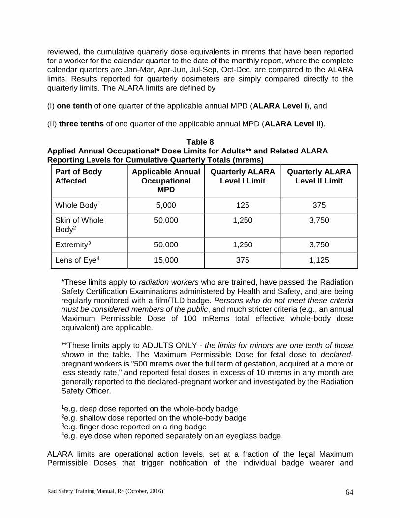
Rad Safety Training Manual, R4 (October, 2016) 64
reviewed, the cumulative quarterly dose equivalents in mrems that have been reported for a worker for the calendar quarter to the date of the monthly report, where the complete calendar quarters are Jan-Mar, Apr-Jun, Jul-Sep, Oct-Dec, are compared to the ALARA limits. Results reported for quarterly dosimeters are simply compared directly to the quarterly limits. The ALARA limits are defined by (I) one tenth of one quarter of the applicable annual MPD (ALARA Level I), and (II) three tenths of one quarter of the applicable annual MPD (ALARA Level II).
Table 8 Applied Annual Occupational* Dose Limits for Adults** and Related ALARA Reporting Levels for Cumulative Quarterly Totals (mrems)
Part of Body Affected
Applicable Annual Occupational
MPD
Quarterly ALARA Level I Limit
Quarterly ALARA Level II Limit
Whole Body1 5,000 125 375
Skin of Whole Body2
50,000 1,250 3,750
Extremity3 50,000 1,250 3,750
Lens of Eye4 15,000 375 1,125
*These limits apply to radiation workers who are trained, have passed the Radiation Safety Certification Examinations administered by Health and Safety, and are being regularly monitored with a film/TLD badge. Persons who do not meet these criteria must be considered members of the public, and much stricter criteria (e.g., an annual Maximum Permissible Dose of 100 mRems total effective whole-body dose equivalent) are applicable. **These limits apply to ADULTS ONLY - the limits for minors are one tenth of those shown in the table. The Maximum Permissible Dose for fetal dose to declared-pregnant workers is "500 mrems over the full term of gestation, acquired at a more or less steady rate," and reported fetal doses in excess of 10 mrems in any month are generally reported to the declared-pregnant worker and investigated by the Radiation Safety Officer. 1e.g, deep dose reported on the whole-body badge 2e.g. shallow dose reported on the whole-body badge 3e.g. finger dose reported on a ring badge 4e.g. eye dose when reported separately on an eyeglass badge
ALARA limits are operational action levels, set at a fraction of the legal Maximum Permissible Doses that trigger notification of the individual badge wearer and

Rad Safety Training Manual, R4 (October, 2016) 65
investigation by the Radiation Safety Officer. The use of quarterly ALARA limits allows more flexibility in accommodating month-to-month variations and small anomalies, than would be the case if each individual monthly dose were compared to one tenth of one twelfth of the annual limit for ALARA Level I, etc.; yet it still meets the ALARA Program requirements of the licensing agencies. The limits for reporting ALARA overexposures and initiating related correspondence from the Radiation Safety Officer to notify the affected individual are therefore as shown in the table above. All reports of doses exceeding ALARA limits, both Level I and Level II,
are reported to the affected individual in writing, along with explanatory information, and
are investigated by the Radiation Safety Officer, who seeks to determine their seriousness, depending on magnitude and cause.
For Level II dose reports, the affected individual is required to reply in writing to the Radiation Safety Officer regarding the circumstances that may have created the dose report. The Radiation Safety Officer may also seek to determine whether, in cases of concern, the worker's exposure could be reduced by reasonable and economical changes in procedures, facilities, or equipment, and may make corresponding recommendations to the Committee on Ionizing Radiation. Committees’ Quarterly Review of Dosimetry and Bioassay Results All reported doses exceeding ALARA levels are reported to the Committee on Ionizing Radiation by the Radiation Safety Officer, and are discussed at the CIR’s quarterly meetings. At its discretion, the CIR may choose to address problem situations individually and/or at the program level, by correspondence or other actions. Responsibility of Authorized Principal Investigators In addition to their responsibilities as defined elsewhere in the Radiation Safety Manual, authorized Principal Investigators are responsible for the implementation of any controls required by the CIR as a result of reported doses exceeding ALARA levels. Responsibility of All Persons Working With Radioactive Materials The individual working directly with radioactive materials bears a major responsibility in maintaining doses ALARA by practicing good radiological hygiene. This is especially so in the biomedical research setting, where most doses of concern are likely to result from incorrect actions of the affected individual(s), rather than being a necessary or likely concomitant of the activity being performed.

Rad Safety Training Manual, R4 (October, 2016) 66
Section 5
RADIATION WORK AND PREGNANCY: A GUIDE FOR PRINCIPAL INVESTIGATORS AND WORKERS AT CU DENVER | ANSCHUTZ Despite developments in the regulatory and legal arenas that have placed increased emphasis on the issues involved in pregnancy and work with sources of radiation, there is relatively little concern about actual radiation doses received in most cases of radiation workers in our biomedical research laboratories here at CU Denver | Anschutz. This is because the vast majority of “radiation workers” at the university have little potential exposure to ionizing radiation, if standard hygienic practices are followed to avoid self-contamination. However, because of the legal and health issues involved, and because of the great significance of the related emotional issues to individual workers, it is crucial that both workers and supervisors be well-informed. Furthermore, the university is required, by the Colorado Department of Public Health and the Environment, Radiation Control Division (who licenses the university for use of radioactive materials), to document that all radiation workers have been given this information. For that reason, the material contained in this chapter is now required reading for all radiation workers at CU Denver | Anschutz, and for all Principal Investigators authorized for use of radioactive materials or other sources of radiation. The following material is presented in a question-and-answer format to add interest. Additional questions should be directed to the Radiation Safety Officer, Environmental Health and Safety Department, Box F-484, 303-724-0345. Q. Who is a radiation worker, and who is not? A. A radiation worker is any person who works with any source of ionizing radiation apart from natural background radiation, during the course of his or her work activities, and whose work conditions are therefore controlled to minimize such exposure. This includes all persons who work directly with radioactive materials, even in very small quantities. Some additional personnel, such as persons using dental x-ray machines, are also considered radiation workers. Some, but not all radiation workers are required to wear personnel dosimeters (TLD [Thermoluminescent Dosimeter] badges) issued by EHS. (Workers who work only with soft beta emitters (H-3, C-l4, S-35) are not required to wear dosimeters.) Being a radiation worker implies that one is informed about the health issues related to radiation and the type and extent of potential radiation doses involved in one's work, and that one accepts these matters as conditions of employment. All radiation workers who work directly with radioactive materials are required to complete the radiation safety certification modules and examinations administered by EHS. Q. When is a radiation worker considered a pregnant radiation worker?
Rad Safety Training Manual, R4 (October, 2016) 67
A. This question is not nearly so foolish as it sounds! Due to legal precedents involving issues such as employment discrimination, the regulatory agencies with purview in this area have determined that a radiation worker is pregnant, for purposes of radiation safety regulations, only after she has declared her pregnancy in writing to her employer, along with the estimated date of conception. Until an employee makes said declaration, she is not considered pregnant, no matter how visibly apparent her condition may seem to others, including her supervisor. Furthermore, it is possible for a declared pregnant radiation worker to revoke her declaration of pregnancy, without regard to her actual condition. Supervisors must be sensitive to the fact that inquiry into an employee's pregnancy or fertility status, especially if they take on an intrusive or accusatory tone, may be legitimately protested by the employee as an invasion of privacy. Workers are encouraged to declare their pregnancy as soon as it becomes known to them, and to inform the Radiation Safety Officer in writing. Because so few workers at CU Denver | Anschutz have any potential exposure to significant levels of ionizing radiation, the pregnant worker and her supervisor can usually make decisions about work assignments without anticipating that she might, in any case, receive significant radiation doses in her work. But there are cases, discussed in more detail below, that warrant consideration of the radiation dose that may be received by the declared pregnant worker. Q. Wait a minute! What are you calling a "significant" radiation dose? A. All persons living in the Colorado area receive about 180 mrems per year, or fifteen millirems per month, of radiation dose to the whole body, from cosmic rays, from gamma rays given off by naturally-occurring radioactivity in soils, rocks, and building materials, and from other natural sources. In addition, all Colorado residents receive a dose to the lung epithelium, from the alpha particles given off by radon gas, primarily encountered in the home environment. This dose is typically equivalent to another 200 to 400 mrems of whole-body dose. Because each person's background dose varies with a number of lifestyle choices (e.g., place of residence, time spent in the mountains at higher elevations than Denver, time spent flying on jet aircraft, smoking habits, etc.), the individual background dose can easily vary by several tens of millirems per year or more. This provides a basis for one type of perspective on what levels of work-related dose might be considered significant, or even practical to control. Another and more legally compelling perspective comes from the regulations. State and federal regulations now (as of 1/1/94) require that no employer may expose a declared pregnant worker to more than 500 millirems of dose to the fetus over the full term of gestation, which equates to about 55 millirems per month. For the sake of conservatism, no supervisor would want to place a declared pregnant worker in a situation in which the anticipated dose would exceed a fraction of this amount

Rad Safety Training Manual, R4 (October, 2016) 68
By these criteria, anything more than ten millirems in a month, above background, should be considered potentially significant for a declared pregnant worker. Q. What happens if a pregnant worker decides not to declare her pregnancy? A. She would continue to be monitored as an adult worker. This means that the statutory Maximum Permissible Dose (MPD) that is applicable is 5 mrems (5,000 millirems) per year on the standard dosimeter badge, which is considered a "whole body" dosimeter. A non-declared pregnant worker would receive a letter from the Radiation Safety Officer, consistent with the ALARA Program that is always in place for all radiation workers at the university, if her whole-body dose in any calendar quarter (January through March, April through June, etc.) exceeds 125 millirems. (For exposure to penetrating radiations from external sources, we would normally assume fetal dose to be equal to whole body dose.) When a pregnant worker does not declare her pregnancy, the fetal dose is not separately monitored, tracked, and reported, and the 500-millirem limit for the nine-month term of pregnancy is not applied to fetal dose. In terms of the typical situations that apply at the university, the pregnant worker should be aware that, although she is unlikely to get a significant fetal dose in occupational situations that exist in the biomedical research laboratories, failure to declare her pregnancy means that the fetal dose will not be separately tracked and monitored, and her equivalent whole-body dose will not be scrutinized with as much rigor and timeliness, by the Radiation Safety Officer, as the fetal dose of a declared-pregnant worker. In the situations that exist at CU Denver | Anschutz, it is very unlikely that a female worker would be required to accept a new job assignment because she declared her pregnancy. Declaration of pregnancy is generally unlikely, for instance, to prevent an investigator from continuing her research. Female workers should consider the above implications carefully in deciding whether or not to declare their pregnancy. Q. How much radiation dose is a worker going to get by doing lab work? A. Typically, almost nothing. Let's talk first about external dose, arising from sources outside the body, so we are not talking about a situation in which the worker has contaminated herself with radioactive material and somehow gotten the material inside her body. Rather, we are talking only about the exposure that a person gets from being in the vicinity of some source of penetrating radiations that penetrate through the container in which the radioactive material is housed. Beta particles, even the higher-energy ones from P-32, are not sufficiently penetrating to reach the fetus, even if they are not shielded. For that reason, soft beta emitters (H-3, C-14, S-35, Ca-43) are not a potential source of external exposure. With regard to P-32, it must be noted that, although the high-energy betas themselves cannot penetrate and deliver fetal dose, a small fraction of those betas do generate bremsstrahlung x-rays as the betas are absorbed, and the x-rays are much more penetrating than the betas. When

Rad Safety Training Manual, R4 (October, 2016) 69
P-32 is present in quantities on the order of tens of milliCuries or more, the exposure rate from bremsstrahlung x-rays, within a meter or two of the P-32, may be significant enough to be of concern, if a worker is spending significant periods of time in that area. It is the radioisotopes that emit gamma rays and x-rays that are the principal concern in regard to external exposure. I-125 produces a dose rate of about 0.07 millirems per hour per milliCurie, at a distance of one meter, if it is totally unshielded. Cr-51, another commonly-used gamma emitter, only produces one photon for every ten atomic disintegrations, so it only produces a dose rate of about 0.016 millirems per hour per milliCurie, at a distance of one meter, totally unshielded. A few milliCuries of I-125, or five to ten milliCuries of Cr-51, should not produce exposure rates of concern, if they are shielded with a small amount of lead. Most shipping containers for these radionuclides currently provide adequate shielding for this purpose. I-131 is a more potent gamma emitter, producing a dose rate of about 0.22 millirems per hour per milliCurie, at a distance of one meter, totally unshielded. I-131 is also more difficult to shield. Thus, 1-131 requires substantially more consideration than I-125 and Cr-51. The Radiation Safety Officer should be consulted for more information about these and other gamma-emitting radionuclides, if they are present in quantities exceeding a few tens of microCuries. Another important note is that pregnant workers who may be using the large animal irradiator should consult the Radiation Safety Officer. This unit, with the source fully retracted into the shield, produces dose rates of less than 1 mrem per hour in the vicinity of the irradiator itself (inside the room, where the cells or other objects to be irradiated are placed into position), and does not produce exposure rates measurably in excess of background in the control console area. For these reasons, it is unlikely that a person using this unit, even several times per week could exceed one to two millirems of dose per month. However, the size and nature of this source, for which the room is posted as a High Radiation Area, require particular care in its use. Q. Is there any other way that a pregnant worker can be exposed to radiation? A. There certainly is. In fact, given the type and quantities of radioactive materials in use at the university, the most significant hazard is internal exposure to radioactive materials. This means getting the radioactive material itself inside your body. This can happen by accidental ingestion (e.g., by mouth-pipetting or by transferring contamination from your hands to food, etc.). It can also happen by skin absorption, if the contamination that you get on your skin is in a soluble form that can be absorbed across the skin into underlying tissues. And it can happen by inhalation, if you are working with radioactive material in volatile form, or if you disperse it into the air as an aerosol, and you are not working in a properly operating fume hood.
Rad Safety Training Manual, R4 (October, 2016) 70
Declared pregnant workers should not be performing radioiodination labeling experiments, if it can be avoided. It should be strongly emphasized that, in general, for biomedical research uses, the most important protective factor in most cases is good radiological hygiene. Q. What if a worker had already gotten some dose before she realized, and/or declared, her pregnancy? A. The dose limit is a little more complicated to apply in this case, but is set forth explicitly in the regulations. The Radiation Safety Officer should be consulted. Again, there is little likelihood that the worker will have received any dose of concern, if proper precautions that are appropriate for all workers are being followed, because of the limited types and quantities of radioactive materials that are in use at the university. Q. What happens to a baby that is exposed to radiation before birth? A. At the dose levels that are possible in routine biomedical research work, there is no certainty that anything will happen. In fact, even at dose levels of hundreds of millirems of acute fetal dose (dose received all at once), there is only a very small probability of any adverse effect. The potential health effects of concern are divided into three broad categories: carcinogenesis, mutagenesis, and teratogenesis. Carcinogenesis includes the induction of early childhood cancers and leukemia due to radiation dose received in utero. Mutagenesis includes the creation of defects in the genetic material of the parents, prior to conception, or possibly the creation of defects in the genetic material of the fetus as it exists in utero, that would result in heritable genetic defects being passed on to succeeding generations. Teratogenesis includes the induction of developmental defects in utero, resulting in the child being born with developmental abnormalities (birth defects). Such defects could include mental retardation, in addition to structural or metabolic deficiencies. All of these issues, including numerical risk estimates and a reference list, are addressed in detail in Appendices A and B of the U.S. Nuclear Regulatory Commission’s Regulatory Guide 8.13. Most of this document is incorporated into this Study Manual. For a complete copy contact EHS. In addition, further discussion of some of this health risk information may be warranted, in light of new information that has emerged since the publication of Regulatory Guide 8 13.

Rad Safety Training Manual, R4 (October, 2016) 71
With respect to induction of cancer or leukemia in early childhood, due to in utero exposure, a more recent review cites a risk factor of 2 x10 -4 excess cancer deaths per rem [Hoffman], as opposed to the factor of 6 x 10⁻⁶ per rem cited by the NRC publication. However, it should also be noted that the National Council on Radiation Protection and Measurements (NCRP) and the International Commission on Radiation Protection (ICRP) have estimated that in utero exposure leads to excess cancer risk later in life as an adult, with a risk coefficient two to three times that of exposure received as an adult [Meinhold]. This latter risk factor, for excess cancer deaths due to radiation dose received as an adult, is on the order of 2 x 10⁻⁴ to 4 x 10⁻⁴ per rem, depending on assumptions about dose rate factors [BEIR V]. With respect to induction of mental retardation, to which there is an apparent sensitivity in the period from about 8 to 15 weeks post concept-ion, it should be noted that there is substantial evidence for a threshold dose, below which this effect would not occur at all. This threshold has been estimated to be in the range of 10 to 20 rems. An alternative explanation for the existence of an apparent threshold derives from the view that this effect is at least predominantly a mass effect, also called a non-stochastic effect. This is an effect that occurs with certainty at all dose levels, but has a severity proportional to the radiation dose. The NCRP and ICRP have taken a position in recent years that supports the incidence of severe mental retardation as a manifestation of an overall IQ shift, which is greatest at the highest doses (on the order of 100 rems) received by atomic bomb survivors that were irradiated in utero [Meinhold]. The coefficient for IQ shift has been estimated to be in the range of 0.21 to 0.29 IQ points per rem of fetal dose. With respect to the production of heritable genetic defects in the genome of a fetus in utero, or, for that matter, in the genome of an exposed adult, there is very little discussion of this effect in the NRC Regulatory Guide. That is because the effect of mutagenesis has never been demonstrated in human study populations, even those as heavily irradiated as the atomic bomb survivors. The effect theoretically exists, based on the radiobiology of animals, such as the famous early studies of Drosophila, but is presumably only probable enough to be observed, even in a fraction of an exposed population, at very high doses. The most recent data support a calculated doubling dose (doubling the natural rate of mutations) in excess of 100 rems (BEIRV). There is some difficulty in specifying the existence of the effect as well, given that many mutations, such as recessive ones, would not be observable in individuals of the first few generations of an exposed population, and the fact that exposed populations would not be genetically isolated as they propagate. Despite these complications, the overall genetic risk is not considered to be a limiting factor in this situation, and the dose limits for the fetus are based primarily on the other potential health effects discussed above [NCRP] Q. Is there any reason for a male worker at CU Denver | Anschutz to be
concerned about radiation work, if he expects to have children? A. Probably not. Some concern was aroused a few years ago by a study in the UK, called the Gardner study or the Sellafield study. It demonstrated a statistically significant connection between a cluster of childhood leukemia cases and the occupational radiation

Rad Safety Training Manual, R4 (October, 2016) 72
exposure of the fathers prior to conception, which was on the order of several rems (several thousand millirems). This study has been seriously questioned as a valid indicator of paternal radiation dose as a cause of childhood leukemia, and this connection has not been supported by a recent Canadian study. The usual guidelines, including maintaining radiation doses ALARA, should be sufficient to protect male workers with respect to paternity, as well as for their own health. With reasonable care and appropriate procedures, no worker at the university should have to receive gonadal doses exceeding a few tens of mrems per month, and doses will typically be indistinguishable from background. This last statement is supported by many thousands of person-months' worth of monitoring reflected in the records maintained by EHS. Q. Does a pregnant radiation worker need a special dosimetry badge? A. Not absolutely, but it is recommended that declared pregnant workers take advantage of the fetal monitoring service provided by our dosimetry badge vendor, including the ordering of a separate fetal badge. A separate badge for fetal monitoring will be supplied at minimal expense, along with special dosimetry reports. The standard whole-body badge should be worn at waist level, on the front (anterior aspect) of the body. This provides a second valid estimate of fetal dose, so that two results are obtained for purposes of verification, the dosimeters should be exchanged promptly each month for the new badges provided by EHS. The RSO can be consulted at any time (ext. 4-0345) to obtain the most recent monitoring results. Q. If a worker declares her pregnancy, and she and her supervisor agree that there is no prospect of her receiving any significant radiation dose in connection with her normal work, is there any reason to consider changing her work conditions or work assignment? A. There may still be reason to consider changes. It should be borne in mind that a substantial fraction of all live births involve some defect. If the worker in question happens to be such a case, and especially if she was not comfortable with the radiation aspects of her work situation, the implications for all of the parties involved, worker, supervisor, and the university, are potentially problematic. On the other hand, there may be equally valid reasons to consider not changing the work conditions and assignment. For instance, in some cases, it may set an untenable precedent to excuse a worker from her normal duties on account of pregnancy, if the case involves a small department that must accomplish a certain workload, and dosimetric experience, along with health physics analysis, indicates that workers in the category in question are not receiving significant radiation doses. There is also a valid philosophical objection to creating or reinforcing the impression that radiation work is harmful or dangerous, even when proper controls are in place, materials are being used safely, and there is no prospect of a workers’ receiving a measurable radiation dose.
Rad Safety Training Manual, R4 (October, 2016) 73
Supervisors and/or workers are encouraged to request the Radiation Safety Officer to perform a health physics evaluation of a declared pregnant worker's assignments. If such an evaluation indicates that a given work assignment many be completed safely, and that no significant radiation dose is to be anticipated if standard precautions and hygienic practices are followed, the supervisor does have the right to require the declared pregnant worker to perform the assignment in question, if it is otherwise a normal part of her duties, and if the supervisor feels that the declared-pregnant worker’s performance of the assignment is necessary to the operation of the work unit. In cases where then is no reason to expect the declared-pregnant radiation worker to receive a significant radiation dose, the Radiation Safety Officer has no regulatory or legal basis to require, or even recommend, a specific decision about work conditions and assignments. In such a case, the decision rests solely with the worker and supervisor, who should consider the above factors, as well as any others that they think are valid. Additional questions should be directed to the Radiation Safety Officer at ext. 4-0345.

Rad Safety Training Manual, R4 (October, 2016) 74
TABLE 9 EFFECTS OF RISK FACTORS ON PREGNANCY OUTCOME
Effect # Occurring Risk Factor from Natural Causes
Excess Occurrences
from Risk Factors
Radiation Risks
Childhood Cancer
Cancer death in children
1.4 per 1,000 Radiation dose of 1,000 millirems received before birth
0.6 per 1,000
Abnormalities
Radiation Dose of 1,000 millirads received during specific periods after conception
Small head size 40 per 1,000 4-7 weeks after conception 5 per 1,000
Small head size 40 per 1,000 8-11 weeks after conception 9 per 1,000
Mental retardation
4 per 1,000 Radiation dose of 1,000 millirads received 8-15 weeks after conception
4 per 1,000
Non-Radiation Risks
Occupation Stillbirth or spontaneous abortion
200 per 1,000 Work in high-risk occupation 90 per 1,000
Alcohol Consumption Fetal alcohol syndrome
1 per 2,000 2-4 drinks per day 100 per 1,000
Fetal alcohol syndrome
1 per 2,000 More than 4 drinks per day 200 per 1,000
Fetal alcohol syndrome
1 per 2,000 Chronic alcoholic (more than 10 drinks per day)
350 per 1,000
Perinatal infant death
23 per 1,000 Chronic alcoholic (more than 10 drinks per day)
170 per 1,000
Smoking Perinatal infant death
23 per 1,000 Less than 1 pack per day 5 per 1,000
Perinatal infant death
23 per 1,000 One pack or more per day 10 per 1,000

Rad Safety Training Manual, R4 (October, 2016) 75

Rad Safety Training Manual, R4 (October, 2016) 76
Section 6
Principal Investigator Responsibilities Using Radioactive Materials Each PI requesting authorization to use RAM in their laboratory will complete an “open book” examination on material related to the process and procedures relevant to the authorization process for use of RAM. The examination is based on elements of the Radiation Safety Manual. The PI is encouraged to review the current version of the Radiation Safety Manual prior to arriving to take the exam so that within the one-hour test period, relatively specific questions of the topics below can be answered. A copy of the manual resides on the EHS website. Topics include but are not limited to: committee authorization process, acquisition, accounting and transfer of radioactive materials, authorization and decommissioning of laboratory areas, conditions on housekeeping and maintenance services.

Rad Safety Training Manual, R4 (October, 2016) 77
Glossary
Acute dose An acute dose means a person received a radiation dose over a short period of time. Alpha particle An alpha is a particle emitted from the nucleus of an atom, that contains two protons and two neutrons. It is identical to the nucleus of a Helium atom, without the electrons. Becquerel (Bq) The Becquerel is a unit used to measure a radioactivity. One Becquerel is that quantity of a radioactive material that will have 1 transformations in one second. Often radioactivity is expressed in larger units like: thousands (kBq), one millions (MBq) or even billions (GBq) of a becquerels. As a result of having one Becquerel being equal to one transformation per second, there are 3.7 x 10¹⁰ Bq in one curie. Beta particle A beta is a high speed particle, identical to an electron, that is emitted from the nucleus of an atom. Chronic dose A Chronic dose means a person received a radiation dose over a long period of time. Curie (Ci) The curie is a unit used to measure a radioactivity. One curie is that quantity of a radioactive material that will have 37,000,000,000 transformations in one second. Often radioactivity is expressed in smaller units like: thousandths (mCi), one millionths (uCi) or even billionths (nCi) of a curie. The relationship between becquerels and curies is: 3.7 x 10¹⁰ Bq in one curie. Dose In a general sense, Dose is a measure of the amount of energy from an ionizing radiation deposited in a mass of some material. Dose is affected by the type of radiation, the amount of radiation and the physical properties of the material itself. Specifically, we can talk about absorbed dose in tissue, or material like silicon. Other common doses are the effective and equivalent doses, which are adjusted to allow the comparison of different tissues or types of radiation. Absorbed doses are normally measured in units of rads (grays), and effective and equivalent doses in rems (or sieverts). Gamma Rays Gamma rays are electromagnetic waves or photons emitted from the nucleus (center) of an atom. Genetic effects Genetic effects are effects from some agent, which are seen in the offspring of the individual who received the agent. The agent must be encountered pre-conception.
Rad Safety Training Manual, R4 (October, 2016) 78
Gray (Gy) The gray is a unit used to measure a quantity called absorbed dose. This relates to the amount of energy actually absorbed in some material, and is used for any type of radiation and any material. One gray is equal to one joule of energy deposited in one kg of a material. The unit gray can be used for any type of radiation, but it does not describe the biological effects of the different radiations. Absorbed dose is often expressed in terms of hundredths of a gray, or centi-grays. One gray is equivalent to 100 rads. Health Physics Health Physics is an interdisciplinary science and its application, for the radiation protection of humans and the environment. Health Physics combines the elements of physics, biology, chemistry, statistics and electronic instrumentation to provide information that can be used to protect individuals from the effects of radiation. Ionizing radiation Ionizing radiation is radiation with enough energy so that during an interaction with an atom, it can remove tightly bound electrons from their orbits, causing the atom to become charged or ionized. Examples are gamma rays and neutrons. Radiation is measured in many ways, and commonly expressed in units of roentgens. Neutrons Neutrons are neutral particles that are normally contained in the nucleus of all atoms and may be removed by various interactions or processes like collision and fission. Non-ionizing radiation Non-ionizing radiation is radiation without enough energy to remove tightly bound electrons from their orbits around atoms. Examples are microwaves and visible light. Non-stochastic effect Non-stochastic effects are effects that can be related directly to the dose received. The effect is more severe with a higher dose, i.e., the burn gets worse as dose increases. It typically has a threshold, below which the effect will not occur. A skin burn from radiation is a non-stochastic effect. Rad (radiation absorbed dose): The rad is a unit used to measure a quantity called absorbed dose. This relates to the amount of energy actually absorbed in some material, and is used for any type of radiation and any material. One rad is defined as the absorption of 100 ergs per gram of material. The unit rad can be used for any type of radiation, but it does not describe the biological effects of the different radiations. Radioactive Contamination Radioactive contamination is radioactive material distributed over some area, equipment or person. It tends to be unwanted in the location where it is, and has to be cleaned up or decontaminated.

Rad Safety Training Manual, R4 (October, 2016) 79
Radioactive Material Radioactive Material is any material that contains radioactive atoms. Radiation Radiation is energy in transit in the form of high speed particles and electro-magnetic waves. We encounter electromagnetic waves every day. They make up our visible light, radio and television waves, ultra violet (UV), and microwaves with a spectrum of energies. These examples of electromagnetic waves do not cause ionizations of atoms because they do not carry enough energy to separate molecules or remove electrons from atoms. Radioactivity Radioactivity is the spontaneous transformation of an unstable atom and often results in the emission of radiation. This process is referred to as a transform-ation, decay or disintegrations of an atom. Rem (roentgen equivalent man) The rem is a unit used to derive a quantity called equivalent dose. This relates the absorbed dose in human tissue to the effective biological damage of the radiation. Not all radiation has the same biological effect, even for the same amount of absorbed dose. Equivalent dose is often expressed in terms of thousandths of a rem, or mrem. To determine equivalent dose (rem), you multiply absorbed dose (rad) by a quality factor (Q) that is unique to the type of incident radiation. Roentgen (R) The roentgen is a unit used to measure a quantity called exposure. This can only be used to describe an amount of gamma and X-rays, and only in air. One roentgen is equal depositing to 2.58 x 10⁻⁴ coulombs per kg of dry air. It is a measure of the ionizations of the molecules in a mass of air. The main advantage of this unit is that it is easy to measure directly, but it is limited because it is only for deposition in air, and only for gamma and x rays. Sievert (Sv) The sievert is a unit used to derive a quantity called equivalent dose. This relates the absorbed dose in human tissue to the effective biological damage of the radiation. Not all radiation has the same biological effect, even for the same amount of absorbed dose. Equivalent dose is often expressed in terms of millionths of a sievert, or micro-sievert. To determine equivalent dose (Sv), you multiply absorbed dose (Gy) by a quality factor (Q) that is unique to the type of incident radiation. One sievert is equivalent to 100 rem. SI Prefixes Many units are broken down into smaller units or expressed as multiples, using standard metric prefixes. As examples, a kilobecquerel (kBq) in 1000 Becquerels, a millirad (mrad) is 10-3 rad, a microrem (µrem) is 10-6 rem, a nanogram is 10-9 grams, and

Rad Safety Training Manual, R4 (October, 2016) 80
a picocurie is a 10-12 curies.
SI Prefixes
Factor Prefix Symbols Factor Prefix Symbols
1018 exa E 10-18 atto a
1015 peta P 10-15 femto f
1012 tera T 10-12 pico p
109 giga G 10-9 nano n
106 mega M 10-6 micro µ
103 kilo k 10-3 milli m
102 hecto h 10-2 centi c
101 deka da 10-1 deci d
Somatic effects Somatic effects are effects from some agent, like radiation that are seen in the individual who receives the agent. Stochastic effects Stochastic effects are effects that occur on a random basis with its effect being independent of the size of dose. The effect typically has no threshold and is based on probabilities, with the chances of seeing the effect increasing with dose. Cancer is a stochastic effect. Teratogenic effects Teratogenic effects are effects from some agent that are seen in the offspring of the individual who received the agent. The agent must be encountered during the gestation period. X-rays X Rays are electromagnetic waves or photons not emitted from the nucleus, but normally emitted by energy changes in electrons. These energy changes are either in electron orbital shells that surround an atom or in the process of slowing down such as in an X-ray machine.