![[Slides] Four Steps Brands Can Take to Design Internet of Things Experiences](https://static.fdocuments.in/doc/165x107/55a889711a28abdb288b47a3/slides-four-steps-brands-can-take-to-design-internet-of-things-experiences.jpg)
[Slides] Four Steps Brands Can Take to Design Internet of Things Experiences

Glomerular pathology in systemic disease

Lecture outline
• Lupus nephritis
• Diabetic nephropathy
• Glomerulonephritis Associated with Bacterial Endocarditis and Other Systemic Infections
• Henoch-Schonlein Purpura
Lupus nephritis
• Kidney involvement is one of the most important clinical features of SLE
…renal failure is the most common cause of death
• Mainly glomerular changes…interstitial and tubular changes are also seen
• Pathogenesis: deposition of DNA–anti-DNA complexes within the glomeruli
• Although the kidney appears normal by light microscopy in 25% to 30% of cases, almost all cases of SLE show some renal abnormality if examined by immunofluorescence and electron microscopy
6 patterns of glomerular disease in SLE …none of which is specific to the disease
• Minimal mesangial lupus nephritis (class I)
• Mesangial proliferative lupus nephritis (class II)
• Focal lupus nephritis (class III)
• Diffuse lupus nephritis (class IV)
• Membranous lupus nephritis (class V)
• Advanced sclerosing lupus nephritis (class VI)
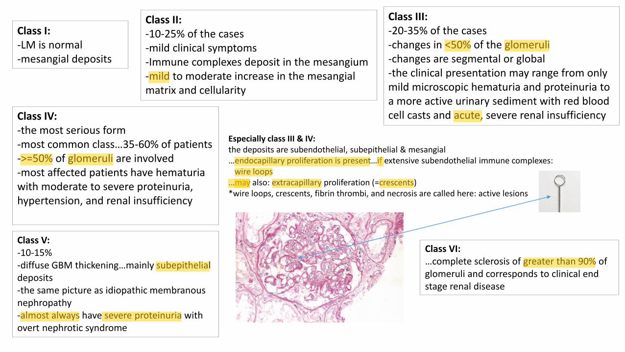
Class I: -LM is normal -mesangial deposits
Class II: -10-25% of the cases -mild clinical symptoms -Immune complexes deposit in the mesangium -mild to moderate increase in the mesangial matrix and cellularity
Class III: -20-35% of the cases -changes in <50% of the glomeruli -changes are segmental or global -the clinical presentation may range from only mild microscopic hematuria and proteinuria to a more active urinary sediment with red blood cell casts and acute, severe renal insufficiency Class IV:
-the most serious form -most common class…35-60% of patients ->=50% of glomeruli are involved -most affected patients have hematuria with moderate to severe proteinuria, hypertension, and renal insufficiency
Especially class III & IV: the deposits are subendothelial, subepithelial & mesangial …endocapillary proliferation is present…if extensive subendothelial immune complexes: wire loops …may also: extracapillary proliferation (=crescents) *wire loops, crescents, fibrin thrombi, and necrosis are called here: active lesions
Class V: -10-15% -diffuse GBM thickening…mainly subepithelial deposits -the same picture as idiopathic membranous nephropathy -almost always have severe proteinuria with overt nephrotic syndrome
Class VI: …complete sclerosis of greater than 90% of glomeruli and corresponds to clinical end stage renal disease
Diabetic nephropathy
• Renal failure is second only to myocardial infarction as a cause of death from this disease
• 3 major lesions:
1-Glomerular lesions
2-Renal vascular lesions, especially: arteriolosclerosis
3-Pyelonephritis, including necrotizing papillitis
Diabetic nephropathy, glomerular lesions
• Capillary basement membrane thickening
…can be detected by electron microscopy within a few years of the
onset of diabetes, sometimes without any associated change in
renal function
• Diffuse mesangial sclerosis
• Nodular glomerulosclerosis
Diabetic nephropathy, diffuse mesangial sclerosis
• Diffuse increase in mesangial matrix along with mesangial cell proliferation
…always associated with basement membrane thickening
…found in most individuals with disease of more than 10 years’
duration
…if become marked: nephrotic syndrome
Diabetic nephropathy, nodular glomerulosclerosis
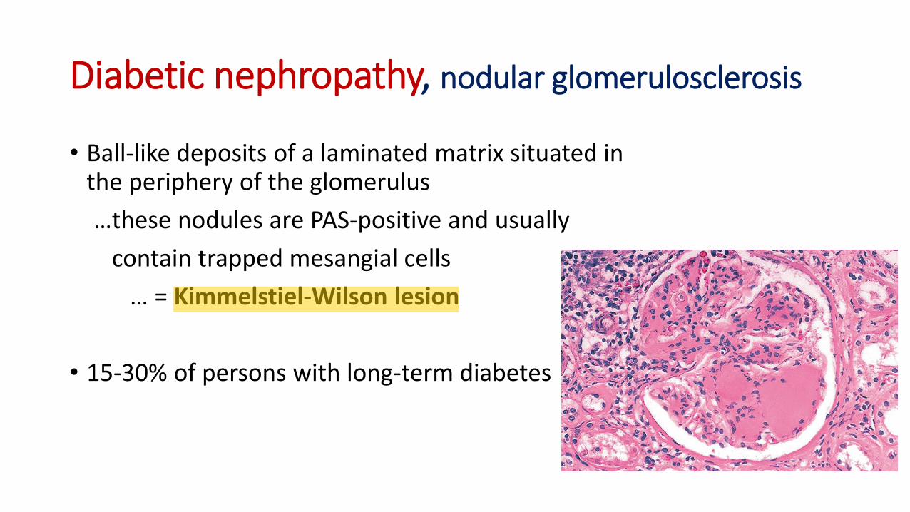
• Ball-like deposits of a laminated matrix situated in the periphery of the glomerulus
…these nodules are PAS-positive and usually
contain trapped mesangial cells
… = Kimmelstiel-Wilson lesion
• 15-30% of persons with long-term diabetes
Important note for differential diagnosis
• Diffuse mesangial sclerosis also may be seen in association with old age and hypertension
…but: the nodular form of glomerulosclerosis, once certain unusual
forms of nephropathies have been excluded is essentially
pathognomonic of diabetes
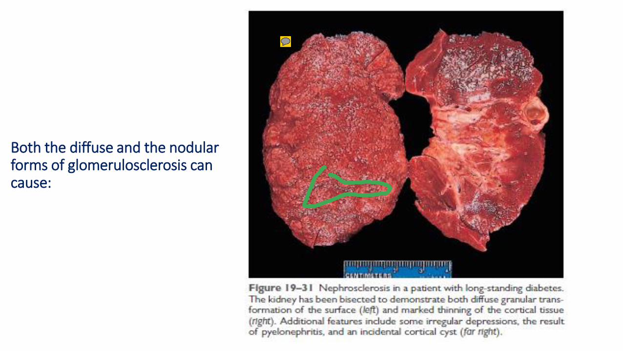
Both the diffuse and the nodular forms of glomerulosclerosis can cause:
Diabetic nephropathy, renal atherosclerosis and arteriolosclerosis
• These are macrovascular manifestations of the disease
• Hyaline arteriolosclerosis of afferent & efferent arterioles
important to differentiate diabetic cause from other causes
Diabetic nephropathy, pyelonephritis
• More common & more severe in diabetics than in nondiabetics
• One special pattern of acute pyelonephritis, necrotizing papillitis (or papillary necrosis), is much more prevalent in diabetics than in nondiabetics
Glomerulonephritis associated with bacterial endocarditis and other systemic infections
• Immune complex nephritis initiated by complexes of bacterial antigen and antibody
• Hematuria and proteinuria of various degrees
• Acute nephritic presentation is not uncommon
• RPGN may occur in rare instances
• The histologic features may vary from a focal and segmental necrotizing glomerulonephritis to a diffuse and more global exudative and proliferative glomerulonephritis, which may have a MPGN pattern
• The rapidly progressive forms show large numbers of crescents
Henoch-Schonlein purpura
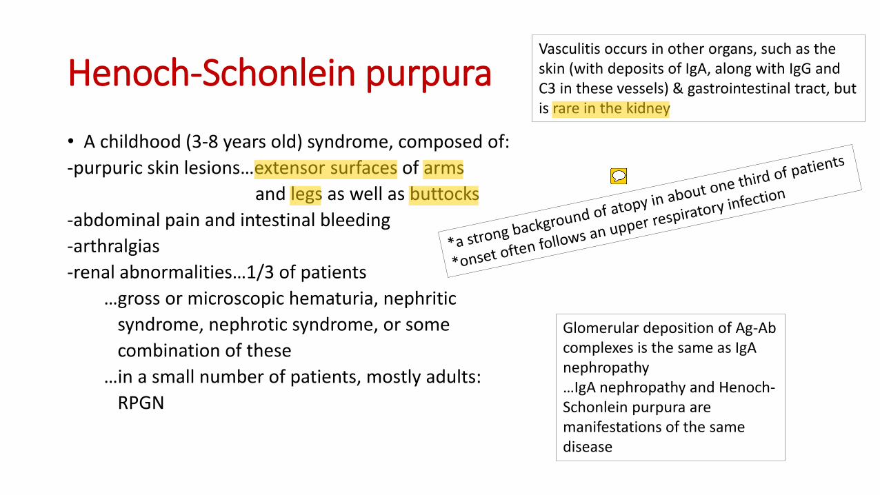
• A childhood (3-8 years old) syndrome, composed of:
-purpuric skin lesions…extensor surfaces of arms
and legs as well as buttocks
-abdominal pain and intestinal bleeding
-arthralgias
-renal abnormalities…1/3 of patients
…gross or microscopic hematuria, nephritic
syndrome, nephrotic syndrome, or some
combination of these
…in a small number of patients, mostly adults:
RPGN
Glomerular deposition of Ag-Ab complexes is the same as IgA nephropathy …IgA nephropathy and Henoch-Schonlein purpura are manifestations of the same disease
Vasculitis occurs in other organs, such as the skin (with deposits of IgA, along with IgG and C3 in these vessels) & gastrointestinal tract, but is rare in the kidney

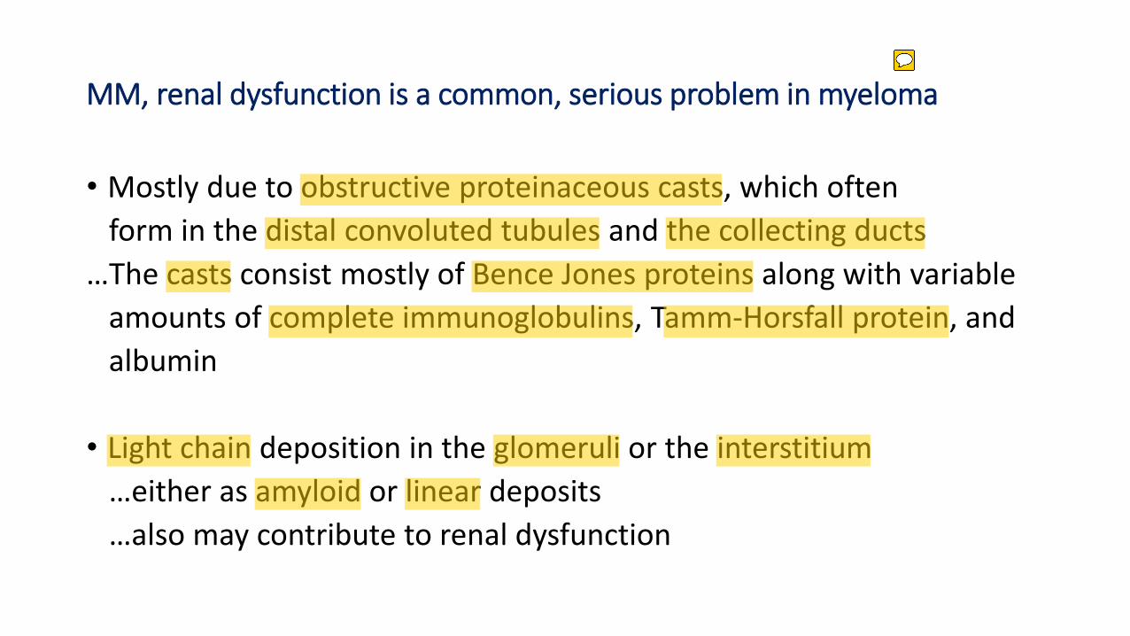
MM, renal dysfunction is a common, serious problem in myeloma
• Mostly due to obstructive proteinaceous casts, which often
form in the distal convoluted tubules and the collecting ducts
…The casts consist mostly of Bence Jones proteins along with variable
amounts of complete immunoglobulins, Tamm-Horsfall protein, and
albumin
• Light chain deposition in the glomeruli or the interstitium
…either as amyloid or linear deposits
…also may contribute to renal dysfunction
MM, renal dysfunction, cont’d
• Hypercalcemia, which may lead to dehydration and renal stones
• Frequent bouts of bacterial pyelonephritis, which stem in part from
the hypogammaglobulinemia
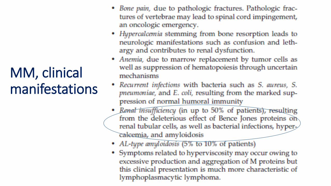
MM, clinical manifestations