Quantitative Proteomics Reveals the Defense Response...
15
1 SCIENTIFIC REPORTS | 6:34261 | DOI: 10.1038/srep34261 www.nature.com/scientificreports Quantitative Proteomics Reveals the Defense Response of Wheat against Puccinia striiformis f. sp. tritici Yuheng Yang 1,2 , Yang Yu 1 , Chaowei Bi 1 & Zhensheng Kang 2 Wheat stripe rust, caused by Puccinia striiformis f. sp. tritici (Pst), is considered one of the most aggressive diseases to wheat production. In this study, we used an iTRAQ-based approach for the quantitative proteomic comparison of the incompatible Pst race CYR23 in infected and non-infected leaves of the wheat cultivar Suwon11. A total of 3,475 unique proteins were identified from three key stages of interaction (12, 24, and 48 h post-inoculation) and control groups. Quantitative analysis showed that 530 proteins were differentially accumulated by Pst infection (fold changes >1.5, p < 0.05). Among these proteins, 10.54% was classified as involved in the immune system process and stimulus response. Intriguingly, bioinformatics analysis revealed that a set of reactive oxygen species metabolism-related proteins, peptidyl–prolyl cis–trans isomerases (PPIases), RNA-binding proteins (RBPs), and chaperonins was involved in the response to Pst infection. Our results were the first to show that PPIases, RBPs, and chaperonins participated in the regulation of the immune response in wheat and even in plants. This study aimed to provide novel routes to reveal wheat gene functionality and better understand the early events in wheat–Pst incompatible interactions. Rust fungi are a monophyletic group of obligate biotrophic parasites that invade and cause diseases in economi- cally important plants 1,2 . Species of rusts have evolved such that the pathogens are highly specific to the plant spe- cies they can infect, colonize, and reproduce 3 . A well-known representative of rust fungus is Puccinia striiformis f. sp. tritici (Pst), the causal agent of the wheat stripe rust disease, which is considered one of the most aggres- sive diseases in wheat production 4 . As an economically important pathogen, remarkable progress in stripe rust resistance genes and molecular perspectives has been made in the interaction between Pst and wheat 4 . However, the frequent virulence variation of Pst races oſten overcome the race-specific resistance of hosts and negate the breeders’ efforts, which has been a prominent question for durable disease control. Moreover, gene expression is regulated at different levels by multifactorial and system-level approaches; consequently, the DNA sequence is insufficient to elucidate sophisticated immune interaction characteristics 5 . erefore, the development of novel approaches is necessary to further understand the resistance mechanisms of wheat response against Pst. Proteomics has emerged as complementary to genomics and transcriptomics because it focuses on gene products, thereby providing a more direct view of cellular immunological processes than genomics or tran- scriptomics 6,7 . Proteomics offers the possibility of simultaneously studying protein localization, protein–protein interactions, enzymatic complexes, or post-translational modifications that are essential to better understand plant–pathogen interactions 8 . To date, proteomics based on mass spectrometry (MS) has matured and become a powerful “hypothesis-generating engine” that provides a framework for translating large data sets to understand complex biological processes 9–11 . Extensive quantitative proteomic studies with high-throughput proteome research techniques have been con- ducted on plant–pathogen interactions, such as Arabidopsis–Pseudomonas syringae 12 , tomato–P. syringae 13 , pota- to–Phytophthora infestans 14 , and Zantedeschia aethiopica–Pectobacterium carotovorum 15 . However, quantitative 1 College of Plant Protection, Southwest University, Beibei, Chongqing, 400715, P. R. China. 2 Key Laboratory of Plant Protection Resources and Pest Management of Ministry of Education, State Key Laboratory of Crop Stress Biology for Arid Areas and College of Plant Protection, Northwest A&F University, Yangling, Shaanxi, 712100, P. R. China. Correspondence and requests for materials should be addressed to Y.H.Y. (email: [email protected]) or Z.K. (email: [email protected]) Received: 25 November 2015 Accepted: 12 September 2016 Published: 28 September 2016 OPEN
-
Upload
truongkiet -
Category
Documents
-
view
216 -
download
0
Transcript of Quantitative Proteomics Reveals the Defense Response...
1Scientific RepoRts | 6:34261 | DOI: 10.1038/srep34261
www.nature.com/scientificreports
Quantitative Proteomics Reveals the Defense Response of Wheat against Puccinia striiformis f. sp. triticiYuheng Yang1,2, Yang Yu1, Chaowei Bi1 & Zhensheng Kang2
Wheat stripe rust, caused by Puccinia striiformis f. sp. tritici (Pst), is considered one of the most aggressive diseases to wheat production. In this study, we used an iTRAQ-based approach for the quantitative proteomic comparison of the incompatible Pst race CYR23 in infected and non-infected leaves of the wheat cultivar Suwon11. A total of 3,475 unique proteins were identified from three key stages of interaction (12, 24, and 48 h post-inoculation) and control groups. Quantitative analysis showed that 530 proteins were differentially accumulated by Pst infection (fold changes >1.5, p < 0.05). Among these proteins, 10.54% was classified as involved in the immune system process and stimulus response. Intriguingly, bioinformatics analysis revealed that a set of reactive oxygen species metabolism-related proteins, peptidyl–prolyl cis–trans isomerases (PPIases), RNA-binding proteins (RBPs), and chaperonins was involved in the response to Pst infection. Our results were the first to show that PPIases, RBPs, and chaperonins participated in the regulation of the immune response in wheat and even in plants. This study aimed to provide novel routes to reveal wheat gene functionality and better understand the early events in wheat–Pst incompatible interactions.
Rust fungi are a monophyletic group of obligate biotrophic parasites that invade and cause diseases in economi-cally important plants1,2. Species of rusts have evolved such that the pathogens are highly specific to the plant spe-cies they can infect, colonize, and reproduce3. A well-known representative of rust fungus is Puccinia striiformis f. sp. tritici (Pst), the causal agent of the wheat stripe rust disease, which is considered one of the most aggres-sive diseases in wheat production4. As an economically important pathogen, remarkable progress in stripe rust resistance genes and molecular perspectives has been made in the interaction between Pst and wheat4. However, the frequent virulence variation of Pst races often overcome the race-specific resistance of hosts and negate the breeders’ efforts, which has been a prominent question for durable disease control. Moreover, gene expression is regulated at different levels by multifactorial and system-level approaches; consequently, the DNA sequence is insufficient to elucidate sophisticated immune interaction characteristics5. Therefore, the development of novel approaches is necessary to further understand the resistance mechanisms of wheat response against Pst.
Proteomics has emerged as complementary to genomics and transcriptomics because it focuses on gene products, thereby providing a more direct view of cellular immunological processes than genomics or tran-scriptomics6,7. Proteomics offers the possibility of simultaneously studying protein localization, protein–protein interactions, enzymatic complexes, or post-translational modifications that are essential to better understand plant–pathogen interactions8. To date, proteomics based on mass spectrometry (MS) has matured and become a powerful “hypothesis-generating engine” that provides a framework for translating large data sets to understand complex biological processes9–11.
Extensive quantitative proteomic studies with high-throughput proteome research techniques have been con-ducted on plant–pathogen interactions, such as Arabidopsis–Pseudomonas syringae12, tomato–P. syringae13, pota-to–Phytophthora infestans14, and Zantedeschia aethiopica–Pectobacterium carotovorum15. However, quantitative
1College of Plant Protection, Southwest University, Beibei, Chongqing, 400715, P. R. China. 2Key Laboratory of Plant Protection Resources and Pest Management of Ministry of Education, State Key Laboratory of Crop Stress Biology for Arid Areas and College of Plant Protection, Northwest A&F University, Yangling, Shaanxi, 712100, P. R. China. Correspondence and requests for materials should be addressed to Y.H.Y. (email: [email protected]) or Z.K. (email: [email protected])
Received: 25 November 2015
accepted: 12 September 2016
Published: 28 September 2016
OPEN

www.nature.com/scientificreports/
2Scientific RepoRts | 6:34261 | DOI: 10.1038/srep34261
proteomic research has rarely examined the changes in the wheat proteome in response to biotrophic fungi (espe-cially to rust fungi); only a few studies based on gel electrophoresis methods are available16,17. The key issue for this shortcoming is that the allohexaploid wheat genome consists of three closely related sub-genomes (A, B and D)18, which greatly influenced the research on wheat functional genomics and proteomics.
To further clarify the resistance mechanism of wheat against Pst at the proteomic level in the current study, the quantitative proteome of the wheat cultivar Suwon11 (Su11) was compared between plants inoculated or uninoc-ulated with the avirulent Pst race CYR23 in planta. Results showed that 530 proteins were differentially expressed by Pst infection. Notably, a set of reactive oxygen species (ROS) metabolism-related proteins, peptidyl–prolyl cis–trans isomerases (PPIases), RNA-binding proteins (RBPs), and chaperonins was revealed to be involved in the response to Pst infection for the first time. This study provides a novel route and a theoretical basis to further clarify the molecular mechanism and defense network of wheat response against Pst.
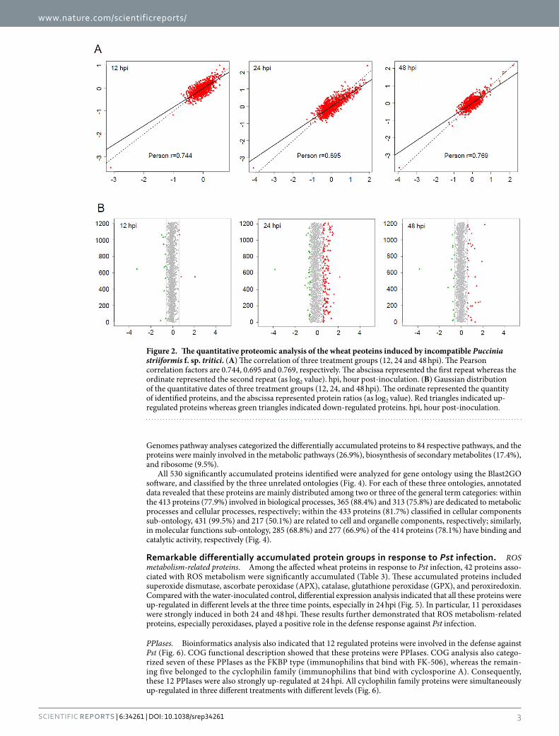
ResultsOverview of the proteome identification of wheat–Pst incompatible interaction. Wheat leaf samples from 12, 24, and 48 h post-inoculation (hpi) treatments or water-inoculated controls were collected and analyzed by iTRAQ. After labeling, SCX fractionation, and LC-ESI-MS/MS analysis, a total of 3475 proteins were identified from the wheat–Pst incompatible interaction. Among these proteins, 1774, 338, and 1363 wheat proteins were obtained from three separate genome sequence databases of Aegilops tauschii (the wheat D genome progenitor), Triticum urartu (the wheat A genome progenitor), and the hexaploid Triticum aestivum cultivar Chinese Spring, respectively (Table 1 and S1). The mass distribution of each identified protein spanned across a wide range of molecular weights higher than 10 kDa (Fig. 1). A data correlation analysis of the 3475 proteins showed the Pearson correlation coefficient values of 0.744, 0.834 and 0.769, respectively among the replicates of three treatments compared to control (Fig. 2A). All the identified wheat proteins were analyzed by gene ontology (GO) and classified by the three ontologies (cellular component, biological process, and molecular function). After excluding the GO entries without a corresponding protein, all the identified proteins were associated with 49 GO categories (Figure S1).
Bioinformatics analysis of the regulated proteins. To reveal the proteins with a putative regulatory function in the wheat–Pst incompatible interaction, the Gaussian distributions of the quantitative ratio (as log2 value) were performed (Fig. 2B). According to the means and the standard deviation values of the Gaussian distribution, there were a combined total of 530 wheat proteins with significantly altered expression in the 12, 24, and 48 hpi treatments compared with the controls by a fold-change > 1.5 (P < 0.05) (Fig. 3). Among these regulated proteins, 279 proteins were identified from Ae. tauschii, 56 proteins were identified from T. urartu, and 195 proteins were identified from T. aestivum (Table S2, Figure S2). Compared with control group, there were 171, 29 and 122 up-regulated proteins and 58, 17 and 35 down-regulated proteins in the 24 hpi treatment between Ae. tauschii, T. urartu, and T. aestivum, respectively (Table 2). The subsequent Kyoto Encyclopedia of Genes and
GroupTotal
spectra SpectraUnique spectra Peptide
Unique peptide Protein
Aegilops tauschii
288,769
55,806 50,519 4,729 4,373 1,774
Triticum urartu 15,502 11,325 953 915 338
Triticum aestivum 52,473 45,248 3,996 3,683 1,363
Table 1. Summary of identified proteins regulated by avirulent Puccinia striiformis f. sp. tritici.
Figure 1. Molecular mass distribution of the wheat proteins identified from the iTRAQ analysis spanned across a wide range of molecular weights, which were induced by incompatible Puccinia striiformis f. sp. tritici. The abscissa represented the molecular weight of identified proteins (kDa), and the ordinate represented the number of identified proteins.
www.nature.com/scientificreports/
3Scientific RepoRts | 6:34261 | DOI: 10.1038/srep34261
Genomes pathway analyses categorized the differentially accumulated proteins to 84 respective pathways, and the proteins were mainly involved in the metabolic pathways (26.9%), biosynthesis of secondary metabolites (17.4%), and ribosome (9.5%).
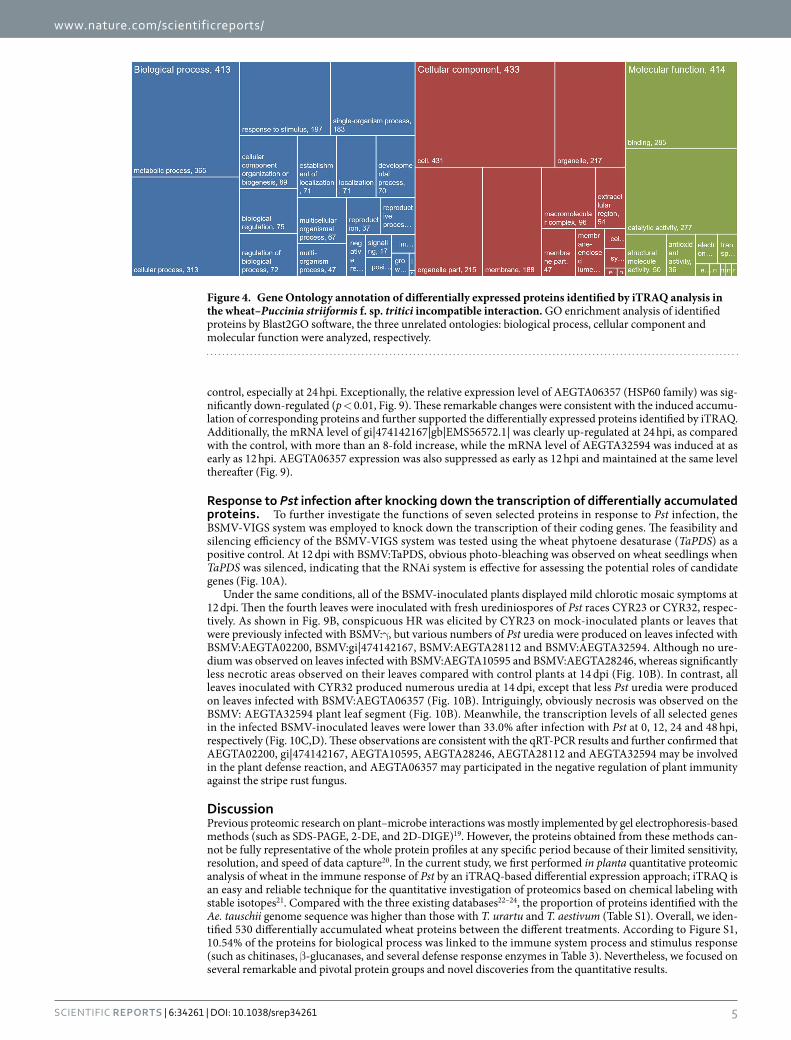
All 530 significantly accumulated proteins identified were analyzed for gene ontology using the Blast2GO software, and classified by the three unrelated ontologies (Fig. 4). For each of these three ontologies, annotated data revealed that these proteins are mainly distributed among two or three of the general term categories: within the 413 proteins (77.9%) involved in biological processes, 365 (88.4%) and 313 (75.8%) are dedicated to metabolic processes and cellular processes, respectively; within the 433 proteins (81.7%) classified in cellular components sub-ontology, 431 (99.5%) and 217 (50.1%) are related to cell and organelle components, respectively; similarly, in molecular functions sub-ontology, 285 (68.8%) and 277 (66.9%) of the 414 proteins (78.1%) have binding and catalytic activity, respectively (Fig. 4).
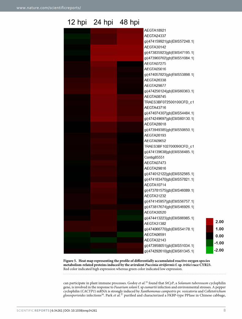
Remarkable differentially accumulated protein groups in response to Pst infection. ROS metabolism-related proteins. Among the affected wheat proteins in response to Pst infection, 42 proteins asso-ciated with ROS metabolism were significantly accumulated (Table 3). These accumulated proteins included superoxide dismutase, ascorbate peroxidase (APX), catalase, glutathione peroxidase (GPX), and peroxiredoxin. Compared with the water-inoculated control, differential expression analysis indicated that all these proteins were up-regulated in different levels at the three time points, especially in 24 hpi (Fig. 5). In particular, 11 peroxidases were strongly induced in both 24 and 48 hpi. These results further demonstrated that ROS metabolism-related proteins, especially peroxidases, played a positive role in the defense response against Pst infection.
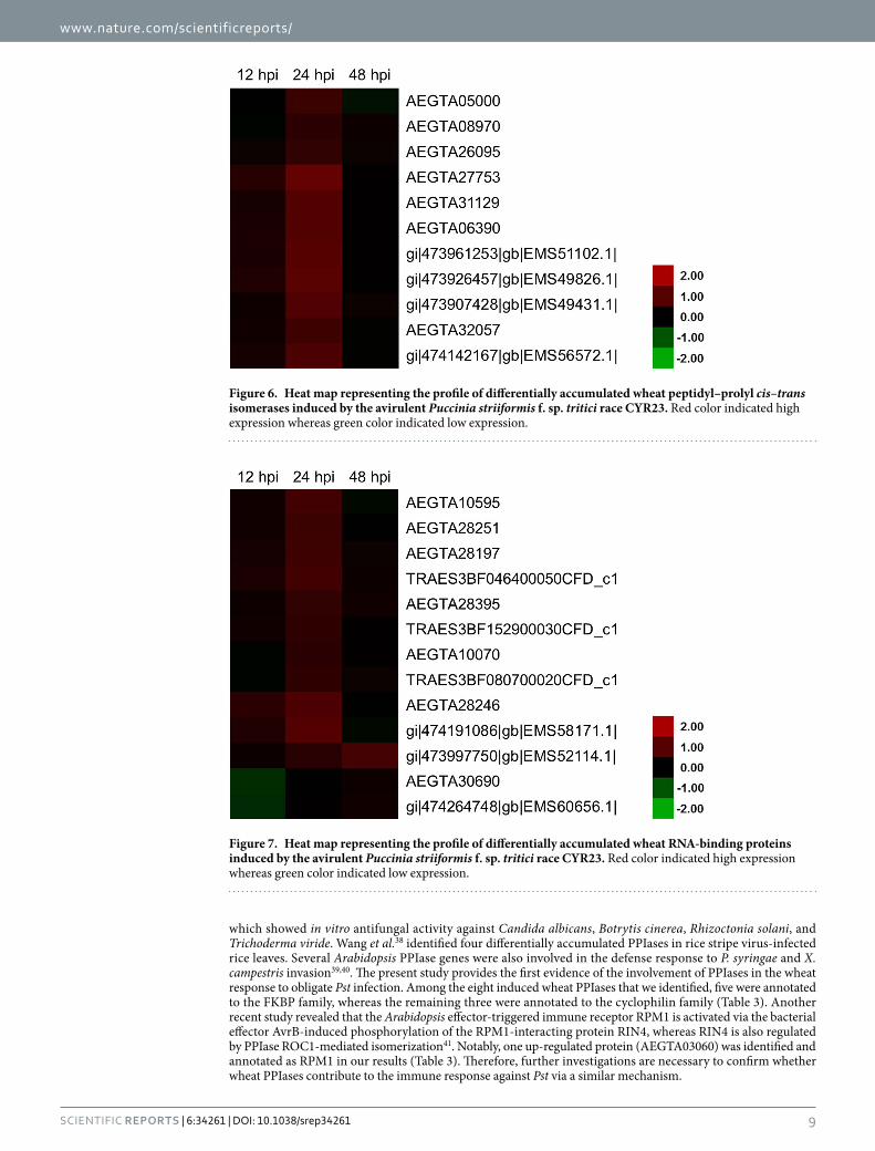
PPIases. Bioinformatics analysis also indicated that 12 regulated proteins were involved in the defense against Pst (Fig. 6). COG functional description showed that these proteins were PPIases. COG analysis also catego-rized seven of these PPIases as the FKBP type (immunophilins that bind with FK-506), whereas the remain-ing five belonged to the cyclophilin family (immunophilins that bind with cyclosporine A). Consequently, these 12 PPIases were also strongly up-regulated at 24 hpi. All cyclophilin family proteins were simultaneously up-regulated in three different treatments with different levels (Fig. 6).
Figure 2. The quantitative proteomic analysis of the wheat peoteins induced by incompatible Puccinia striiformis f. sp. tritici. (A) The correlation of three treatment groups (12, 24 and 48 hpi). The Pearson correlation factors are 0.744, 0.695 and 0.769, respectively. The abscissa represented the first repeat whereas the ordinate represented the second repeat (as log2 value). hpi, hour post-inoculation. (B) Gaussian distribution of the quantitative dates of three treatment groups (12, 24, and 48 hpi). The ordinate represented the quantity of identified proteins, and the abscissa represented protein ratios (as log2 value). Red triangles indicated up-regulated proteins whereas green triangles indicated down-regulated proteins. hpi, hour post-inoculation.

www.nature.com/scientificreports/
4Scientific RepoRts | 6:34261 | DOI: 10.1038/srep34261
RBPs. As shown in Fig. 7, 13 proteins were categorized as RBPs and significantly up-regulated during the incompatible interaction. Therefore, these RBPs were associated with the stress response of Pst infec-tion. These RBPs included one alternative splicing regulator (AEGTA28251), one arginine/serine-rich splicing factor (TRAES3BF080700020CFD_c1), two predicted glycine-rich RBPs (AEGTA28395 and TRAES3BF152900030CFD_c1), two eukaryotic translation initiation factor (AEGTA30690 and gi|474264748|g-b|EMS60656.1|), and the remaining seven RBPs were described as predicted proteins containing RNA recog-nition motifs. Except for AEGTA30690, all the selected proteins had a significantly altered level of response in 24 hpi (Fig. 7).
Chaperonins. According to the COG functional description, seven proteins were identified and anno-tated as chaperonins based on their expression profiles after Pst inoculation (Fig. 8). Among the chaperon-ins, six (AEGTA27057, AEGTA28112, AEGTA28933, AEGTA32594, gi|474209261|gb|EMS58795.1|, and gi|474407512|gb|EMS66632.1|) were obviously up-regulated at 24 hpi, but one of the chaperonins (AEGTA06357) was down-regulated at the three different time points (Fig. 8). These results were consistent with the COG func-tional description, which categorized the six up-regulated proteins into the same type as the co-chaperonin GroES (HSP10), and the down-regulated one as the chaperonin GroEL (HSP60 family). These results suggested that the co-chaperonin GroES (HSP10) was likely to play crucial roles in the defense response of rust fungus.
Validation of differentially accumulated proteins by qRT-PCR. To further confirm the expres-sion patterns of coding genes after incompatible Pst infection, qRT-PCR analysis was performed. Due to that ROS plays a vital role in plant immunity as well-known, seven coding genes of identified PPIases, RBPs and Chaperonins mentioned above were selected. As revealed in Fig. 9, the mRNA levels of six proteins of them exhibited significant rises (p < 0.01, fold− change > 3) in at least one sampling time point compared with the
Figure 3. Venn diagram representing the overlap among differentially expressed proteins identified by iTRAQ analysis of three treatment groups (12, 24, and 48 hpi) of wheat–Puccinia striiformis f. sp. tritici incompatible interaction. hpi, hour post-inoculation.
Group
Up-regulated Down-regulated
12 hpi 24 hpi 48 hpi 12 hpi 24 hpi 48 hpi
Aegilops tauschii 7 171 38 28 58 29
Triticum urartu 1 29 8 7 17 6
Triticum aestivum 7 122 30 19 35 22
Table 2. The numbers of differentially regulated wheat proteins in three treatment groups (12, 24, and 48 hpi)by avirulent Puccinia striiformis f. sp. tritici. hpi, hour post-inoculation.
www.nature.com/scientificreports/
5Scientific RepoRts | 6:34261 | DOI: 10.1038/srep34261
control, especially at 24 hpi. Exceptionally, the relative expression level of AEGTA06357 (HSP60 family) was sig-nificantly down-regulated (p < 0.01, Fig. 9). These remarkable changes were consistent with the induced accumu-lation of corresponding proteins and further supported the differentially expressed proteins identified by iTRAQ. Additionally, the mRNA level of gi|474142167|gb|EMS56572.1| was clearly up-regulated at 24 hpi, as compared with the control, with more than an 8-fold increase, while the mRNA level of AEGTA32594 was induced at as early as 12 hpi. AEGTA06357 expression was also suppressed as early as 12 hpi and maintained at the same level thereafter (Fig. 9).
Response to Pst infection after knocking down the transcription of differentially accumulated proteins. To further investigate the functions of seven selected proteins in response to Pst infection, the BSMV-VIGS system was employed to knock down the transcription of their coding genes. The feasibility and silencing efficiency of the BSMV-VIGS system was tested using the wheat phytoene desaturase (TaPDS) as a positive control. At 12 dpi with BSMV:TaPDS, obvious photo-bleaching was observed on wheat seedlings when TaPDS was silenced, indicating that the RNAi system is effective for assessing the potential roles of candidate genes (Fig. 10A).
Under the same conditions, all of the BSMV-inoculated plants displayed mild chlorotic mosaic symptoms at 12 dpi. Then the fourth leaves were inoculated with fresh urediniospores of Pst races CYR23 or CYR32, respec-tively. As shown in Fig. 9B, conspicuous HR was elicited by CYR23 on mock-inoculated plants or leaves that were previously infected with BSMV:γ , but various numbers of Pst uredia were produced on leaves infected with BSMV:AEGTA02200, BSMV:gi|474142167, BSMV:AEGTA28112 and BSMV:AEGTA32594. Although no ure-dium was observed on leaves infected with BSMV:AEGTA10595 and BSMV:AEGTA28246, whereas significantly less necrotic areas observed on their leaves compared with control plants at 14 dpi (Fig. 10B). In contrast, all leaves inoculated with CYR32 produced numerous uredia at 14 dpi, except that less Pst uredia were produced on leaves infected with BSMV:AEGTA06357 (Fig. 10B). Intriguingly, obviously necrosis was observed on the BSMV: AEGTA32594 plant leaf segment (Fig. 10B). Meanwhile, the transcription levels of all selected genes in the infected BSMV-inoculated leaves were lower than 33.0% after infection with Pst at 0, 12, 24 and 48 hpi, respectively (Fig. 10C,D). These observations are consistent with the qRT-PCR results and further confirmed that AEGTA02200, gi|474142167, AEGTA10595, AEGTA28246, AEGTA28112 and AEGTA32594 may be involved in the plant defense reaction, and AEGTA06357 may participated in the negative regulation of plant immunity against the stripe rust fungus.
DiscussionPrevious proteomic research on plant–microbe interactions was mostly implemented by gel electrophoresis-based methods (such as SDS-PAGE, 2-DE, and 2D-DIGE)19. However, the proteins obtained from these methods can-not be fully representative of the whole protein profiles at any specific period because of their limited sensitivity, resolution, and speed of data capture20. In the current study, we first performed in planta quantitative proteomic analysis of wheat in the immune response of Pst by an iTRAQ-based differential expression approach; iTRAQ is an easy and reliable technique for the quantitative investigation of proteomics based on chemical labeling with stable isotopes21. Compared with the three existing databases22–24, the proportion of proteins identified with the Ae. tauschii genome sequence was higher than those with T. urartu and T. aestivum (Table S1). Overall, we iden-tified 530 differentially accumulated wheat proteins between the different treatments. According to Figure S1, 10.54% of the proteins for biological process was linked to the immune system process and stimulus response (such as chitinases, β -glucanases, and several defense response enzymes in Table 3). Nevertheless, we focused on several remarkable and pivotal protein groups and novel discoveries from the quantitative results.
Figure 4. Gene Ontology annotation of differentially expressed proteins identified by iTRAQ analysis in the wheat–Puccinia striiformis f. sp. tritici incompatible interaction. GO enrichment analysis of identified proteins by Blast2GO software, the three unrelated ontologies: biological process, cellular component and molecular function were analyzed, respectively.

www.nature.com/scientificreports/
6Scientific RepoRts | 6:34261 | DOI: 10.1038/srep34261
Accession No. Database Description
ROS metabolism
AEGTA00133 Aegilops tauschii cytosolic Cu/Zn superoxide dismutase
AEGTA05616 Ae. tauschii glutathione peroxidase 1
AEGTA06591 Ae. tauschii peroxidase
AEGTA07275 Ae. tauschii NADPH2:quinone reductase
AEGTA07473 Ae. tauschii peroxidase
AEGTA08745 Ae. tauschii class III peroxidase
AEGTA09652 Ae. tauschii peroxidase
AEGTA10714 Ae. tauschii peroxidase
AEGTA18921 Ae. tauschii peroxidase
AEGTA24337 Ae. tauschii class III peroxidase
AEGTA26193 Ae. tauschii glutathione peroxidase
AEGTA26338 Ae. tauschii phospholipid hydroperoxide glutathione peroxidase-like protein
AEGTA28018 Ae. tauschii peroxidase
AEGTA29816 Ae. tauschii L-ascorbate peroxidase
AEGTA29877 Ae. tauschii Cu/Zn superoxide dismutase
AEGTA30142 Ae. tauschii peroxidase 4
AEGTA30520 Ae. tauschii peroxidase
AEGTA31232 Ae. tauschii thioredoxin-dependent peroxidase
AEGTA31382 Ae. tauschii NADPH2:quinone reductase
AEGTA32143 Ae. tauschii PREDICTED: 2-Cys peroxiredoxin BAS1, chloroplastic-like
AEGTA43716 Ae. tauschii peroxidase 8
Contig85551 Ae. tauschii plastid thylakoid-bound ascorbate peroxidase, partial
TRAES3BF072500100CFD_c1 Triticum aestivum peroxidase 8
TRAES3BF102700090CFD_c1 T. aestivum PREDICTED: peroxiredoxin-2F, mitochondrial-like
gi|473781575|gb|EMS46089.1| T. urartu L-ascorbate peroxidase
gi|473817674|gb|EMS46926.1| T. urartu plastid thylakoid-bound ascorbate peroxidase, partial
gi|473835923|gb|EMS47195.1| T. urartu peroxidase
gi|473949385|gb|EMS50650.1| T. urartu peroxidase 6
gi|473958051|gb|EMS51034.1| T. urartu catalase
gi|473960762|gb|EMS51084.1| T. urartu peroxidase 5
gi|474012122|gb|EMS52565.1| T. urartu PREDICTED: peroxidase 54-like [Brachypodium distachyon]
gi|474057823|gb|EMS53898.1| T. urartu glutathione peroxidase, partial
gi|474066770|gb|EMS54178.1| T. urartu quinone reductase
gi|474074307|gb|EMS54484.1| T. urartu peroxidase 8
gi|474139638|gb|EMS56485.1| T. urartu PREDICTED: peroxiredoxin-2F, mitochondrial-like
gi|474145957|gb|EMS56757.1| T. urartu thioredoxin-dependent peroxidase
gi|474159921|gb|EMS57248.1| T. urartu peroxidase 3
gi|474183470|gb|EMS57821.1| T. urartu predicted protein
gi|474249697|gb|EMS60130.1| T. urartu PREDICTED: peroxidase 5-like
gi|474256124|gb|EMS60363.1| T. urartu Superoxide dismutase 2
gi|474292610|gb|EMS61345.1| T. urartu putative Td650 protein
gi|474413223|gb|EMS66985.1| T. urartu predicted protein
Peptidyl-prolyl cis-trans isomerases
AEGTA02200 Ae. tauschii FKBP-type peptidyl-prolyl cis-trans isomerases 1 PPIases
AEGTA05000 Ae. tauschii FKBP-type peptidyl-prolyl cis-trans isomerases 1 PPIases
AEGTA06390 Ae. tauschii Peptidyl-prolyl cis-trans isomerase (rotamase) - cyclophilin family
AEGTA08970 Ae. tauschii FKBP-type peptidyl-prolyl cis-trans isomerases 1 PPIases
AEGTA26095 Ae. tauschii Peptidyl-prolyl cis-trans isomerase (rotamase) - cyclophilin family
Continued

www.nature.com/scientificreports/
7Scientific RepoRts | 6:34261 | DOI: 10.1038/srep34261
ROS-related proteins. In plants, ROS production is one of the earliest cellular responses following suc-cessful pathogen recognition. ROS act as executioners of pathogens, as well as signaling molecules involved in triggering the hypersensitive response (HR) and activating signal transduction processes to stop pathogen growth25–27. Previously, ROS generation was associated with hypersensitive cell death responses in the incom-patible interaction between wheat and avirulent Pst races28. ROS are commonly generated by NADPH oxidases (also known as the respiratory burst oxidases) and peroxidases in plant cells25,29. NADPH oxidases were initially described in mammalian neutrophils and are located in the plasma membrane; these proteins correspond to one of the most studied systems that participate in ROS production to defend cells from invasion25,30. Notably, none of the NADPH oxidases were detected in our current results, but 16 peroxidases were identified and showed noticeably higher expression during infection than in the controls (including class III peroxidase, GPX, APX, and thioredoxin-dependent peroxidase; Table 3). These results suggested that peroxidases but not NADPH oxidases might play more important roles in the oxidative response of wheat to Pst invasion. Consistent with our results, Dmochowska-Boguta et al.31 proved that the induction of peroxidases is more pronounced than that of NADPH oxidases in wheat–P. triticina interactions, and postulated that class III peroxidases play a leading role in the for-mation of ROS molecules during the response of wheat to P. triticina infection. Moreover, a recent report showed that the wheat stripe rust resistance protein WKS1 is targeted to the thylakoid-associated ascorbate peroxidase to detoxify ROS32. All the aforementioned results revealed that the production of ROS in wheat against rust infec-tion might be more dependent on peroxidases.
PPIases. In prokaryotic and eukaryotic cells, PPIases form a superfamily of proteins for facilitating the cis–trans isomerization of N-terminal peptide bonds to proline residues within polypeptide chains33. These proteins were first identified in mammals as receptors of the immune-suppressing drug cyclosporine A. PPIases have been classified into three distinct families: the cyclosporin-binding cyclophilins (CyP), FK506-binding proteins (FKBP), and FK506- and cyclosporin-binding protein (FCBP)34. Several studies have demonstrated that PPIases
Accession No. Database Description
AEGTA27753 Ae. tauschii FKBP-type peptidyl-prolyl cis-trans isomerases 1 PPIases
AEGTA31129 Ae. tauschii Peptidyl-prolyl cis-trans isomerase (rotamase) - cyclophilin family
AEGTA32057 Ae. tauschii FKBP-type peptidyl-prolyl cis-trans isomerases 1
gi|473907428|gb|EMS49431.1| T. urartu FKBP-type peptidyl-prolyl cis-trans isomerases 1
gi|473926457|gb|EMS49826.1| T. urartu Peptidyl-prolyl cis-trans isomerase (rotamase) - cyclophilin family
gi|473961253|gb|EMS51102.1| T. urartu Peptidyl-prolyl cis-trans isomerase (rotamase) - cyclophilin family
gi|474142167|gb|EMS56572.1| T. urartu FKBP-type peptidyl-prolyl cis-trans isomerases 1
RNA-binding proteins
AEGTA10070 Ae. tauschii RNA-binding proteins (RRM domain)
AEGTA10595 Ae. tauschii RNA-binding proteins (RRM domain)
AEGTA28197 Ae. tauschii RNA-binding proteins (RRM domain)
AEGTA28246 Ae. tauschii RNA-binding proteins (RRM domain)
AEGTA28251 Ae. tauschii RNA-binding proteins (RRM domain)
AEGTA28395 Ae. tauschii RNA-binding proteins (RRM domain)
AEGTA30690 Ae. tauschii RNA-binding proteins (RRM domain)
TRAES3BF046400050CFD_c1 T. aestivum RNA-binding proteins (RRM domain)
TRAES3BF080700020CFD_c1 T. aestivum RNA-binding proteins (RRM domain)
TRAES3BF152900030CFD_c1 T. aestivum RNA-binding proteins (RRM domain)
gi|473997750|gb|EMS52114.1| T. urartu RNA-binding proteins (RRM domain)
gi|474191086|gb|EMS58171.1| T. urartu RNA-binding proteins (RRM domain)
gi|474264748|gb|EMS60656.1| T. urartu RNA-binding proteins (RRM domain)
Chaperonins
AEGTA27057 Ae. tauschii Co-chaperonin GroES (HSP10)
AEGTA28112 Ae. tauschii Co-chaperonin GroES (HSP10)
AEGTA28933 Ae. tauschii Co-chaperonin GroES (HSP10)
AEGTA32594 Ae. tauschii Co-chaperonin GroES (HSP10)
AEGTA06357 Ae. tauschii Chaperonin GroEL (HSP60 family)
gi|474209261|gb|EMS58795.1| T. urartu Co-chaperonin GroES (HSP10)
gi|474407512|gb|EMS66632.1| T. urartu Co-chaperonin GroES (HSP10)
Table 3. Protein groups with remarkable differential accumulation in response to avirulent Puccinia striiformis f. sp. tritici infection.
www.nature.com/scientificreports/
8Scientific RepoRts | 6:34261 | DOI: 10.1038/srep34261
can participate in plant immune processes. Godoy et al.35 found that StCyP, a Solanum tuberosusm cyclophilin gene, is involved in the response to Fusarium solani f. sp eumartii infection and environmental stresses. A pepper cyclophilin (CACYP1) mRNA is strongly induced by Xanthomonas campestris pv. vesicatoria and Colletotrichum gloeosporioides infections36. Park et al.37 purified and characterized a FKBP-type PPIase in Chinese cabbage,
Figure 5. Heat map representing the profile of differentially accumulated reactive oxygen species metabolism-related proteins induced by the avirulent Puccinia striiformis f. sp. tritici race CYR23. Red color indicated high expression whereas green color indicated low expression.
www.nature.com/scientificreports/
9Scientific RepoRts | 6:34261 | DOI: 10.1038/srep34261
which showed in vitro antifungal activity against Candida albicans, Botrytis cinerea, Rhizoctonia solani, and Trichoderma viride. Wang et al.38 identified four differentially accumulated PPIases in rice stripe virus-infected rice leaves. Several Arabidopsis PPIase genes were also involved in the defense response to P. syringae and X. campestris invasion39,40. The present study provides the first evidence of the involvement of PPIases in the wheat response to obligate Pst infection. Among the eight induced wheat PPIases that we identified, five were annotated to the FKBP family, whereas the remaining three were annotated to the cyclophilin family (Table 3). Another recent study revealed that the Arabidopsis effector-triggered immune receptor RPM1 is activated via the bacterial effector AvrB-induced phosphorylation of the RPM1-interacting protein RIN4, whereas RIN4 is also regulated by PPIase ROC1-mediated isomerization41. Notably, one up-regulated protein (AEGTA03060) was identified and annotated as RPM1 in our results (Table 3). Therefore, further investigations are necessary to confirm whether wheat PPIases contribute to the immune response against Pst via a similar mechanism.
Figure 6. Heat map representing the profile of differentially accumulated wheat peptidyl–prolyl cis–trans isomerases induced by the avirulent Puccinia striiformis f. sp. tritici race CYR23. Red color indicated high expression whereas green color indicated low expression.
Figure 7. Heat map representing the profile of differentially accumulated wheat RNA-binding proteins induced by the avirulent Puccinia striiformis f. sp. tritici race CYR23. Red color indicated high expression whereas green color indicated low expression.

www.nature.com/scientificreports/
1 0Scientific RepoRts | 6:34261 | DOI: 10.1038/srep34261
RBPs. RBPs are proteins that bind to RNA molecules in cells and coordinate RNA processing and post-transcriptional gene regulation42. In mammals, sequence-specific RBPs play critical roles in the immune response to modulate the gene expression of target mRNAs43. Based on the latest evidence, RBPs are important regulators of plant immunity at each level of RNA processing44. In Arabidopsis, several RBPs have been implicated in the defense against viral and bacterial pathogens via RNA-specific binding45–50. In pepper, the RNA-binding protein 1 gene (CaRBP1) was identified as essential for HR and defense signaling in the cytoplasm51. However, plant RBPs that participate in the regulation of the defense response against fungal pathogen infection, especially in monocots, have not been reported prior to this work. This study is the first to identify wheat RBPs involved in host immunity during pathogen infection. Subsequent research is required to determine the functions of these RBPs.
Chaperonins. Chaperones are the most prominent class of proteins that promote substrate protein fold-ing; these proteins are usually classified according to their molecular weight52. Group I chaperonins are impor-tant components of chaperones known as GroEL-GroES in Escherichia coli and the heat shock proteins (HSPs) HSP60-HSP10 in eukaryotes53. In mammals, HSPs link the innate and adaptive immune systems; in circulation, these proteins serve as intercellular signals to the host54. In particular, HSP60 and HSP10 appear to be related to
Figure 8. Heat map representing the profile of differentially accumulated wheat chaperonins induced by the avirulent Puccinia striiformis f. sp. tritici race CYR23. Red color indicated high expression whereas green color indicated low expression.
Figure 9. Expression patterns of differentially expressed proteins induced by the avirulent Puccinia striiformis f. sp. tritici race CYR23. hpi, hour post-inoculation. Leaf tissues were sampled for both inoculated and mock-inoculated plants at 0, 12, 24, 48, 72 and 120 hpi. The relative expression levels of these genes were calculated using the comparative threshold (2−ΔΔC
T) method. The mean value and standard deviation of gene expression were calculated from three independent biological replications. ANOVA was conducted to determine the differences between each time point. Superscripts with the same letter indicate that values are not significantly different at p < 0.01.

www.nature.com/scientificreports/
1 1Scientific RepoRts | 6:34261 | DOI: 10.1038/srep34261
pregnancy, cancer, and autoimmune inhibition in association with each other55,56. Mycobacterium tuberculosis HSPl0 increases the rate of apoptosis in the mouse P19 teratocarcinoma cell line57. Evidence also suggested that human HSPl0 might be considered a pathogen-associated molecular pattern and damage-associated molecular pattern molecule to trigger Toll-like receptor signaling55. Combined with the results of the present study (Figs 7 and 8), plant HSP10 might play an important role in the positive regulation of the immune response as signaling
Figure 10. Functional characterization of differentially expressed proteins by BSMV-VIGS system. (A) Mild chlorotic mosaic symptoms were observed on the fourth leaves inoculated with BSMV:TaPDS at 12 dpi; uninoculated control, wheat leaves treated with full-strength inoculation buffer. (B) Disease symptoms were observed at 14 dpi on the fourth leaves of wheat plants that were inoculated with the avirulent pathogen CYR23 and virulent pathogen CYR32, respectively. HR, hypersensitive response; U, uredium. (C) Relative transcript levels of differentially expressed proteins assayed in knocked-down wheat leaves inoculation with CYR23. (D) Relative transcript levels of differentially expressed proteins assayed in knocked-down wheat leaves inoculation with CYR32. The mean value and standard deviation of gene expression were calculated from three independent biological replications. ANOVA was conducted to determine the differences between each time point. Superscripts with the same letter indicate that values are not significantly different at p < 0.01.

www.nature.com/scientificreports/
1 2Scientific RepoRts | 6:34261 | DOI: 10.1038/srep34261
molecules, whereas HSP60 might negatively regulate the defense reaction against pathogen infection. Further studies on the relationship of HSP10 and HSP60 for wheat defense response should be conducted in the future.
In addition, more than 80% spectra could not match the unique peptides or proteins (Table 3), whereas more than 200 regulated proteins were described as predicted proteins (Table S1), even when three database releases were used for protein identification22–24. This result was attributed to the huge and complex 17 Gbp hexaploid genome of wheat24, which has delayed wheat functional genomic research. This limitation also explains why research on wheat proteomics is rare.
MethodsPlant and pathogen. Wheat cultivar Su11 and Pst race CYR23 were used for incompatible reaction in this study. Su11 displays a typical HR upon infection with CYR23. The Pst isolate was maintained and propagated on a susceptible Chinese wheat cultivar, Mingxian 169. For biological stress treatments, the plants were grown, inoculated and maintained as previously described58. The Pst-inoculated leaves were sampled at 12, 24 and 48 hpi, and stored at − 80 °C. Control plants were treated with sterile water.
Protein preparation. For protein quantification, approximately 0.5 g fresh leaf tissue per sample was used to extract leaf total protein using the trichloroacetic acid-acetone precipitation method59. The cells were sus-pended in the Lysis buffer (7 mol/L Urea, 2 mol/L Thiourea, 4% CHAPS, 40 mmol/L Tris-HCl, pH 8.5, 1 mM PMSF, 2 mmol/L EDTA) and sonicated in ice. The proteins were reduced with 10 mmol/L DTT (final concen-tration) at 56 °C for 1 h and then alkylated by 55 mmol/L IAM (final concentration) in the darkroom for 1 h. The reduced and alkylated protein mixtures were precipitated by adding 4 × volume of chilled acetone at − 20 °C for 2 hours. After centrifugation at 4 °C, 30,000 g, the pellet was dissolved in 0.5 mol/L TEAB (Applied Biosystems, Milan, Italy) and sonicated in ice. After centrifuging at 30,000 g at 4 °C, the supernatant was transferred to a new tube and quantified by using Bradford’s reagent (Sigma–Aldrich, St. Louis, MO, USA)60, and then kept at − 80 °C for further analysis.
iTRAQ Labeling and SCX fractionation. iTRAQ analysis was implemented at Beijing Genomics Institute (BGI, Shenzhen, China). Total protein (100 μ g) was taken out of each sample solution and then the protein was digested with Trypsin Gold (Promega, Madison, WI, USA) with the ratio of protein: trypsin = 30: 1 at 37 °C for 16 h. After trypsin digestion, peptides were dried by vacuum centrifugation, and then reconstituted in 0.5 mol/L tetraethyl-ammonium bromide and processed according to the manufacture’s protocol for 8-plex iTRAQ rea-gent (Applied Biosystems). Briefly, one unit of iTRAQ reagent was thawed and reconstituted in 24 μ L isopro-panol. Samples were labeled with the iTRAQ tags as follow: Control-1 (113 tag), Su11-CYR23-12 hpi-1 (114 tag), Su11-CYR23-24 hpi-1 (115 tag), Su11-CYR23-48 hpi-1 (116 tag), Control-2 (117 tag), Su11-CYR23-12 hpi-2 (118 tag), Su11-CYR23-24 hpi-2 (119 tag), Su11-CYR23-48 hpi-2 (121 tag), respectively. The peptides were labeled with the isobaric tags, incubated at room temperature for 2 h. The labeled peptide mixtures were then pooled and dried by vacuum centrifugation.
SCX chromatography was performed with a LC-20AB HPLC Pump system (Shimadzu, Kyoto, Japan). The iTRAQ-labeled peptide mixtures were reconstituted with 4 mL buffer A (25 mmol/L NaH2PO4 in 25% ACN, pH 2.7) and loaded onto a 4.6 × 250 mm Ultremex SCX column containing 5-μ m particles (Phenomenex). The peptides were eluted at a flow rate of 1 mL/min with a gradient of buffer A for 10 min, 5–60% buffer B (25 mmol/L NaH2PO4, 1 mol/L KCl in 25% ACN, pH 2.7) for 27 min, 60–100% buffer B for 1 min. The eluted peptides were pooled into 20 fractions, desalted with a Strata X C18 column (Phenomenex) and vacuum-dried.
LC-ESI-MS/MS analysis. Each fraction was resuspended in buffer A (5% ACN, 0.1%FA) and centrifuged at 20,000 g for 10 min, the final concentration of peptide was about 0.5 μ g/μ L on average. 10 μ L supernatant was loaded on a LC-20AD nanoHPLC (Shimadzu, Kyoto, Japan) by the autosampler onto a 2 cm C18 trap column. After that, the peptides were eluted onto a 10 cm analytical C18 column (inner diameter 75 μ m) packed in-house. The samples were loaded at 8 μ L/min for 4 min, then the 35 min gradient was run at 300 nL/min starting from 2 to 35% B (95% ACN, 0.1% FA), followed by 5 min linear gradient to 60%, then, followed by 2 min linear gradient to 80%, and maintenance at 80% B for 4 min, and finally return to 5% in 1 min.
The peptides were subjected to nanoelectrospray ionization followed by tandem mass spectrometry (MS/MS) in an Q EXACTIVE (Thermo Fisher Scientific, San Jose, CA) coupled online to the HPLC. Intact peptides were detected in the Orbitrap at a resolution of 70,000. Peptides were selected for MS/MS using high-energy colli-sion dissociation (HCD) operating mode with a normalized collision energy setting of 27.0; ion fragments were detected in the Orbitrap at a resolution of 17,500. A data-dependent procedure that alternated between one MS scan followed by 15 MS/MS scans was applied for the 15 most abundant precursor ions above a threshold ion count of 20,000 in the MS survey scan with a following Dynamic Exclusion duration of 15 s. The electrospray voltage applied was 1.6 kV. For MS scans, the m/z scan range was 350 to 2,000 Da. For MS2 scans, the m/z scan range was 100–1,800. To account for biological variation and ensure only reproducible responses to treatments were selected, two independent biological replicate experiments were performed.
Proteomic data analysis. Raw data files acquired from the Orbitrap were converted into MGF files using Proteome Discoverer 1.2 (PD 1.2, Thermo). Proteins identification was performed by using Mascot search engine (Matrix Science, London, UK; version 2.3.02). For protein identification, a mass tolerance of 20 ppm was permit-ted for intact peptide masses and 0.05 Da for fragmented ions, with allowance for one missed cleavages in the trypsin digests. The charge states of peptides were set to + 2 and + 3. To reduce the probability of false peptide identification, only peptides with significance scores ( ≥20) at the 99% confidence interval by a Mascot probabil-ity analysis greater than “identity” were counted as identified. And each confident protein identification involves at least one unique peptide.

www.nature.com/scientificreports/
13Scientific RepoRts | 6:34261 | DOI: 10.1038/srep34261
For protein quantitation, it was required that a protein contains at least two unique peptides. The quanti-tative protein ratios were weighted and normalized by the median ratio in Mascot. We only used ratios with p-values < 0.05, and only fold changes of > 1.5 were considered as significant.
Bioinformatics analysis. Functional annotations of the proteins were conducted using Blast2GO program against the non-redundant protein database (NR; NCBI) and three public wheat genome database, independently (http://gigadb.org/search/index/keyword/Triticum+ aestivum/%20yt0/Search/file_page/22#result_files; http://www.ncbi.nlm.nih.gov/bioproject/PRJNA182347; http://wheat-urgi.versailles.inra.fr/Seq-Repository/). The KEGG database (http://www.genome.jp/kegg/) and the COG database (http://www.ncbi.nlm.nih.gov/COG/) were used to classify and group these identified proteins.
RNA extraction, cDNA synthesis and qRT-PCR assay. Total RNA was extracted using Trizol Reagent (Life Technologies, Grand Island, NY, USA) according to the manufacturer’s instruction. Genomic DNA con-taminants were removed by DNase I treatment. First-strand cDNA was synthesized using the M-MLV reverse transcriptase (Promega, Shenzhen, China) with an oligo-(dT18) primer. qRT-PCR was performed using a CFX96 real-time PCR detection system (Bio-Rad, Hercules, CA, USA). The primers for qRT-PCR are listed in Table S3. Relative gene quantification was performed as described in detail previously58 and normalized using the corre-sponding expression of the wheat elongation factor gene TaEF-1a (Genbank accession No. Q03033). All reactions were performed in triplicate, including three controls without the template.
BSMV-mediated gene silencing. Seven special gene fragments were used to silence the transcription of selected proteins (Table S3). Linearized plasmids containing the tripartite BSMV genome were transcribed to RNA. Seven BSMV viruses were inoculated individually on the second leaf of the wheat seedlings at the two-leaf stage, as described previously58. After inoculation for 24 h in the dark, all treated seedlings were placed in a growth chamber at 25 ± 2 °C and then examined for symptoms. BSMV:TaPDS was used as a neg-ative control61. Control plants were treated with full-strength inoculation buffer. The fourth leaf of each plant was then inoculated with fresh urediniospores of CYR23 or CYR32 at 12 days after the viral inoculation, and these leaves sampled at 0, 12, 24 and 48 hpi for RNA isolation to evaluates the silencing efficiencies of selected genes using qRT-PCR. The infection types of stripe rust were examined at 14 dpi. The experiment was repeated three times.
Statistical analysis. Analysis of variance (ANOVA) was performed to determine the significant differences between each treatment using SAS (version 8.12; SAS Institute Inc., Cary, NC, USA). Duncan’s multiple range tests were used for multiple comparison tests.
References1. Hovmoller, M. S. et al. Diversity of Puccinia striiformis on Cereals and Grasses. Annu Rev Phytopathol 49, 197–217 (2011).2. Dean, R. et al. The Top 10 fungal pathogens in molecular plant pathology. Mol Plant Pathol 13, 414–430 (2012).3. Kolmer, J. A., Ordonez, M. E. & Groth, J. V. The Rust Fungi. In: eLS (ed^(eds). John Wiley & Sons, Ltd (2009).4. Chen, W. Q. et al. Wheat stripe (yellow) rust caused by Puccinia striiformis f. sp. tritici. Mol Plant Pathol 15, 433–446 (2014).5. Quirino, B. F. et al. Proteomic approaches to study plant-pathogen interactions. Phytochemistry 71, 351–362 (2010).6. Pandey, A. & Mann, M. Proteomics to study genes and genomes. Nature 405, 837–846 (2000).7. Delaunois, B. et al. Uncovering plant-pathogen crosstalk through apoplastic proteomic studies. Front Plant Sci 5 (2014).8. Yates, J. R., Ruse, C. I. & Nakorchevsky, A. Proteomics by Mass Spectrometry: Approaches, Advances, and Applications. Annu Rev
Biomed Eng 11, 49–79 (2009).9. Cravatt, B. F., Simon, G. M. & Yates, J. R. The biological impact of mass-spectrometry-based proteomics. Nature 450, 991–1000
(2007).10. Altelaar, A. F. M., Munoz, J. & Heck, A. J. R. Next-generation proteomics: towards an integrative view of proteome dynamics. Nat
Rev Genet 14, 35–48 (2013).11. Meissner, F. & Mann, M. Quantitative shotgun proteomics: considerations for a high-quality workflow in immunology. Nat Immunol
15, 112–117 (2014).12. Kaffarnik, F. A. R. et al. Effector proteins of the bacterial pathogen Pseudomonas syringae alter the extracellular proteome of the host
plant, Arabidopsis thaliana. Mol Cell Proteomics 8, 145–156 (2009).13. Parker, J. et al. Quantitative proteomics of tomato defense against Pseudomonas syringae infection. Proteomics 13, 1934–1946 (2013).14. Lim, S. et al. Proteomics analysis suggests broad functional changes in potato leaves triggered by phosphites and a complex indirect
mode of action against Phytophthora infestans. Journal of proteomics 93, 207–223 (2013).15. Luzzatto-Knaan, T. et al. Priming of protein expression in the defence response of Zantedeschia aethiopica to Pectobacterium
carotovorum. Mol Plant Pathol 15, 364–378 (2014).16. Maytalman, D. et al. Proteomic analysis of early responsive resistance proteins of wheat (Triticum aestivum) to yellow rust (Puccinia
striiformis f. sp. tritici) using ProteomeLab PF2D. Plant Omics 6, 24–35 (2013).17. Rampitsch, C. et al. Analysis of the wheat and Puccinia triticina (leaf rust) proteomes during a susceptible host - pathogen
interaction. Proteomics 6, 1897–1907 (2006).18. Marcussen, T. et al. Ancient hybridizations among the ancestral genomes of bread wheat. Science 345 (2014).19. Gupta, R. et al. Understanding the plant-pathogen interactions in the context of proteomics-generated apoplastic proteins inventory.
Front Plant Sci 6 (2015).20. Jorrin-Novo, J. V. et al. Plant proteomics update (2007–2008): Second-generation proteomic techniques, an appropriate experimental
design, and data analysis to fulfill MIAPE standards, increase plant proteome coverage and expand biological knowledge. Journal of proteomics 72, 285–314 (2009).
21. Schmidt, C. et al. Mass spectrometry-based relative quantification of proteins in precatalytic and catalytically active spliceosomes by metabolic labeling (SILAC), chemical labeling (iTRAQ), and label-free spectral count. Rna 20, 406–420 (2014).
22. Jia, J. Z. et al. Aegilops tauschii draft genome sequence reveals a gene repertoire for wheat adaptation. Nature 496, 91–95 (2013).23. Ling, H. Q. et al. Draft genome of the wheat A-genome progenitor Triticum urartu. Nature 496, 87–90 (2013).24. Mayer, K. F. et al. A chromosome-based draft sequence of the hexaploid bread wheat (Triticum aestivum) genome. Science 345,
1251788 (2014).25. Torres, M. A., Jones, J. D. G. & Dangl, J. L. Reactive oxygen species signaling in response to pathogens. Plant Physiol 141, 373–378 (2006).

www.nature.com/scientificreports/
1 4Scientific RepoRts | 6:34261 | DOI: 10.1038/srep34261
26. Coll, N. S., Epple, P. & Dangl, J. L. Programmed cell death in the plant immune system. Cell Death Differ 18, 1247–1256 (2011).27. Nanda, A. K. et al. Reactive oxygen species during plant-microorganism early interactions. J Integr Plant Biol 52, 195–204 (2010).28. Wang, C. F. et al. Histochemical studies on the accumulation of reactive oxygen species (O2
− and H2O2) in the incompatible and compatible interaction of wheat - Puccinia striiformis f. sp. tritici. Physiol Mol Plant P 71, 230–239 (2007).
29. Tripathy, B. C. & Oelmuller, R. Reactive oxygen species generation and signaling in plants. Plant signaling & behavior 7, 1621–1633 (2012).
30. Suzuki, N. et al. Respiratory burst oxidases: the engines of ROS signaling. Curr Opin Plant Biol 14, 691–699 (2011).31. Dmochowska-Boguta, M., Nadolska-Orczyk, A. & Orczyk, W. Roles of peroxidases and NADPH oxidases in the oxidative response
of wheat (Triticum aestivum) to brown rust (Puccinia triticina) infection. Plant Pathol 62, 993–1002 (2013).32. Gou, J. Y. et al. Wheat stripe rust resistance protein WKS1 reduces the ability of the thylakoid-associated ascorbate peroxidase to
detoxify reactive oxygen species. Plant Cell 27, 1755–1770 (2015).33. Shaw, P. E. Peptidyl-prolyl isomerases: a new twist to transcription. Embo Rep 3, 521–526 (2002).34. Adams, B. et al. A novel class of dual-family immunophilins. J Biol Chem 280, 24308–24314 (2005).35. Godoy, A. V. et al. Expression of a Solanum tuberosum cyclophilin gene is regulated by fungal infection and abiotic stress conditions.
Plant Sci 152, 123–134 (2000).36. Kong, H. Y., Lee, S. C. & Hwang, B. K. Expression of pepper cyclophilin gene is differentially regulated during the pathogen infection
and abiotic stress conditions. Physiol Mol Plant P 59, 189–199 (2001).37. Park, S. C. et al. Purification and characterization of an antifungal protein, C-FKBP, from chinese cabbage. J Agr Food Chem 55,
5277–5281 (2007).38. Wang, B. A. et al. iTRAQ-based quantitative proteomics analysis of rice leaves infected by Rice stripe virus reveals several proteins
involved in symptom formation. Virol J 12 (2015).39. Pogorelko, G. V. et al. Characterization of three Arabidopsis thaliana immunophilin genes involved in the plant defense response
against Pseudomonas syringae. Gene 538, 12–22 (2014).40. Mokryakova, M. V. et al. The role of peptidyl-prolyl cis/trans isomerase genes of Arabidopsis thaliana in plant defense during the
course of xanthomonas campestris infection. Russ J Genet+ 50, 140–148 (2014).41. Li, M. et al. Proline Isomerization Of The Immune Receptor-Interacting Protein RIN4 by a cyclophilin inhibits effector-triggered
immunity in Arabidopsis. Cell Host Microbe 16, 473–483 (2014).42. Gerstberger, S., Hafner, M. & Tuschl, T. A census of human RNA-binding proteins. Nat Rev Genet 15, 829–845 (2014).43. Newman, R. & Institute, B. RNA binding proteins as regulators of immune cell biology. Clinical & Experimental Immunology, 183,
37–49 (2016).44. Woloshen, V., Huang, S. & Li, X. RNA-binding proteins in plant immunity. Journal of pathogens 2011, 278697 (2011).45. Fu, Z. Q. et al. A type III effector ADP-ribosylates RNA-binding proteins and quells plant immunity. Nature 447, 284–288 (2007).46. Fujisaki, K. & Ishikawa, M. Identification of an Arabidopsis thaliana protein that binds to tomato mosaic virus genomic RNA and
inhibits its multiplication. Virology 380, 402–411 (2008).47. Jakubiec, A., Yang, S. W. & Chua, N. H. Arabidopsis DRB4 protein in antiviral defense against Turnip yellow mosaic virus infection.
Plant J 69, 14–25 (2012).48. Huh, S. U., Kim, M. J. & Paek, K. H. Arabidopsis Pumilio protein APUM5 suppresses Cucumber mosaic virus infection via direct
binding of viral RNAs. P Natl Acad Sci USA 110, 779–784 (2013).49. Lim, G. H. et al. Double-stranded RNA-binding protein 4 is required for resistance signaling against viral and bacterial pathogens.
Phytopathology 104, 69–69 (2014).50. Huh, S. U. & Paek, K.-H. Plant RNA binding proteins for control of RNA virus infection. Frontiers in Physiology 4, 397 (2013).51. Lee, D. H., Kim, D. S. & Hwang, B. K. The pepper RNA-binding protein CaRBP1 functions in hypersensitive cell death and defense
signaling in the cytoplasm. Plant J 72, 235–248 (2012).52. Hartl, F. U., Bracher, A. & Hayer-Hartl, M. Molecular chaperones in protein folding and proteostasis. Nature 475, 324–332 (2011).53. Gruber, A. V. et al. The complexity of chloroplast chaperonins. Trends Plant Sci 18, 688–694 (2013).54. Tsan, M. F. & Gao, B. Heat shock proteins and immune system. J Leukocyte Biol 85, 905–910 (2009).55. Jia, H. et al. Heat shock protein 10 (Hsp10) in immune-related diseases: one coin, two sides. International journal of biochemistry and
molecular biology 2, 47–57 (2011).56. Grundtman, C. et al. Heat shock protein 60 and immune inflammatory responses in Atherosclerosis. Arterioscl Throm Vas 31,
960–U938 (2011).57. Galli, G. et al. Mycobacterium tuberculosis heat shock protein 10 increases both proliferation and death in mouse P19 teratocarcinoma
cells. In Vitro Cell Dev-An 32, 446–450 (1996).58. Yang, Y. H. et al. Glycerol-3-Phosphate metabolism in wheat contributes to systemic acquired resistance against Puccinia striiformis
f. sp. tritici. Plos One 8, e81756 (2013).59. Zhao, J., Yang, Y. H. & Kang, Z. S. Proteomic analysis of rice nonhost resistance to Puccinia striiformis f. sp. tritici using two-
dimensional electrophoresis. Int J Mol Sci 15, 21644–21659 (2014).60. Kruger, N. J. The Bradford method for protein quantitation. Methods in molecular biology 32, 9–15 (1994).61. Holzberg, S. et al. Barley stripe mosaic virus‐induced gene silencing in a monocot plant. The Plant Journal 30, 315–327 (2002).
AcknowledgementsThis work was supported by the Fundamental Research Funds for the Central Universities (XDJK2016A020, XDJK2015C060, SWU114046, 2362015xk04), the Fundamental and Advanced Research Projects of Chongqing City (cstc2016jcyjA0316), the Open Project Program of State Key Laboratory of Crop Stress Biology for Arid Areas (CSBAA2015009), and the Visiting Scholar Funds of Key Laboratory of Plant Protection Resources and Pest Management of Ministry of Education. We thank anonymous reviewers for their kind suggestions.
Author ContributionsY.H.Y., Y.Y. and Z.K. designed the research. Y.H.Y., Y.Y. and C.B. performed the experiments. Y.H.Y., Y.Y. and C.B. analyzed the data and wrote the manuscript.
Additional InformationSupplementary information accompanies this paper at http://www.nature.com/srepCompeting financial interests: The authors declare no competing financial interests.How to cite this article: Yang, Y. et al. Quantitative Proteomics Reveals the Defense Response of Wheat against Puccinia striiformis f. sp. tritici. Sci. Rep. 6, 34261; doi: 10.1038/srep34261 (2016).

www.nature.com/scientificreports/
1 5Scientific RepoRts | 6:34261 | DOI: 10.1038/srep34261
This work is licensed under a Creative Commons Attribution 4.0 International License. The images or other third party material in this article are included in the article’s Creative Commons license,
unless indicated otherwise in the credit line; if the material is not included under the Creative Commons license, users will need to obtain permission from the license holder to reproduce the material. To view a copy of this license, visit http://creativecommons.org/licenses/by/4.0/ © The Author(s) 2016