Quantitative light microscopy of dense suspensions ...
15
Quantitative light microscopy of dense suspensions: Colloid science at the next decimal place Brian D. Leahy a , b , 1 , Neil Y.C. Lin a , b , 1 , Itai Cohen a , ⁎ Since the days of Perrin (1908) [ 1], microscopy methods have played an important role in the study of colloidal suspensions. Along with the continued development of new imaging techniques, colloid scientists have also implemented a sophisticated range of computational analyses. These analysis techniques are often the unsung heroes that hold the promise of unlocking scientific mysteries at the next decimal place of colloid science. They now enable precision measurements of particle location and size (Bierbaum et al., 2017; Kurita et al., 2012) as well as measurements of local stresses and forces (Lin et al., 2016). Here, we spotlight these exciting advances focusing on the analysis of simple brightfield and confocal microscope images of dense colloidal suspensions as well as the scientific mysteries they may unravel. Address a Department of Physics, Cornell University, Ithaca, NY 14850, United States b School of Engineering and Applied Sciences, Harvard University, Cambridge, MA 02138, United States ⁎ Corresponding author. ([email protected]) Keywords: Colloidal suspensions Microscopy Imaging Forces Measurement science Current Opinion in Colloid & Interface Science (2018) 34, 32–46 This review comes from a themed issue on Microscopy Methods For a complete overview see the issue and the Editorial Article History: Received 10 December 2017 Received in revised form 12 March 2018 Accepted 13 March 2018 Available online 23 March 2018 https://doi.org/10.1016/j.cocis.2018.03.002 1359-0294/© 2018 Elsevier Ltd. All rights reserved. 1. Introduction “Why should one wish to make measurements with ever increasing precision? Because the whole history of physics proves that a new discovery is likely to be found lurking in the next decimal place.” [(Floyd K. Richtmyer, 1931) [5]] Look through a colloid scientist's microscope and you'll find an amazing array of phenomena that scientists and engineers investigate with colloidal suspensions. Particles entropically assemble into a zoo of crystal structures [6–11]. Aided by patterned substrates [12,13], gel coatings [14], depletion interactions [15–20], charge induced interactions [21–23], magnetic interactions [24–26] and DNA programmable inter- actions [27–30] the variety of accessible structures that suspensions form is breath taking. Active colloidal particles jet around, providing fertile testing grounds for far-from- equilibrium physics [31–36]. The behaviors of liquids [37,38], interfaces [39], gels [17,40,41] and glasses [42–47] are resolved with fine detail. Confocal rheoscopes illuminate the structural underpinnings of rheological behaviors from thixotropy [45,48,49] to shear thickening under confinement [50]. And the particles are no longer just spheres [51]; particle shapes have blossomed into a beautiful set of rods [52], clusters [24,53], dimers [54], lock-and-key particles [55], and even designed shapes from pentagons to letters [56–59], with simulations promising new materials from many of these shapes [60]. Simple microscopy methods such as brightfield and confocal are a large reason for the continued excitement in exploring such colloidal phenomena. The particles' large, micron-scale size and slow, millisecond- to second-scale dynamics allow key behaviors to be easily investigated by microscopy [19,44,46,61–65]. A modern microscope can image thousands of particles in a blink of an eye, and simple automated software routines can locate these particles to within 30 nm, while missing only a few percent of the Current Opinion in Colloid & Interface Science (2018) 34, 32–46 www.sciencedirect.com Available online at www.sciencedirect.com ScienceDirect
Transcript of Quantitative light microscopy of dense suspensions ...
Ava i l ab l e on l i ne a t www.sc i enced i r ec t . com
ScienceDirect
Quantitative light microscopy of dense suspensions:
Colloid science at the next decimal place Brian D. Leahy a,b,1, Neil Y.C. Lin a,b,1, Itai Cohena,⁎Since the days of Perrin (1908) [ 1], microscopy methods have played an
important role in the study of colloidal suspensions. Along with thecontinued development of new imaging techniques, colloid scientistshave also implemented a sophisticated range of computationalanalyses. These analysis techniques are often the unsung heroes thathold the promise of unlocking scientific mysteries at the next decimalplace of colloid science. They now enable precision measurements ofparticle location and size (Bierbaum et al., 2017; Kurita et al., 2012) aswell as measurements of local stresses and forces (Lin et al., 2016).Here, we spotlight these exciting advances focusing on the analysis ofsimple brightfield and confocal microscope images of dense colloidalsuspensions as well as the scientific mysteries they may unravel.Addressa Department of Physics, Cornell University, Ithaca, NY 14850,United Statesb School of Engineering and Applied Sciences, Harvard University,Cambridge, MA 02138, United States
⁎ Corresponding author.([email protected])
Keywords:Colloidal suspensionsMicroscopyImagingForcesMeasurement science
Current Opinion in Colloid & Interface Science (2018) 34, 32–46This review comes from a themed issue on Microscopy MethodsFor a complete overview see the issue and the Editorial
Article History:Received 10 December 2017Received in revised form 12 March 2018Accepted 13 March 2018Available online 23 March 2018
https://doi.org/10.1016/j.cocis.2018.03.0021359-0294/© 2018 Elsevier Ltd. All rights reserved.
Current Opinion in Colloid & Interface Science (2018) 34, 32–46
1. Introduction
“Why should one wish to make measurements with everincreasing precision? Because the whole history of physics provesthat a new discovery is likely to be found lurking in the nextdecimal place.”
[(Floyd K. Richtmyer, 1931) [5]]
Look through a colloid scientist's microscope and you'll findan amazing array of phenomena that scientists and engineersinvestigate with colloidal suspensions. Particles entropicallyassemble into a zoo of crystal structures [6–11]. Aided bypatterned substrates [12,13], gel coatings [14], depletioninteractions [15–20], charge induced interactions [21–23],magnetic interactions [24–26] and DNA programmable inter-actions [27–30] the variety of accessible structures thatsuspensions form is breath taking. Active colloidal particlesjet around, providing fertile testing grounds for far-from-equilibrium physics [31–36]. The behaviors of liquids [37,38],interfaces [39], gels [17,40,41] and glasses [42–47] areresolved with fine detail. Confocal rheoscopes illuminatethe structural underpinnings of rheological behaviors fromthixotropy [45,48,49] to shear thickening under confinement[50]. And the particles are no longer just spheres [51];particle shapes have blossomed into a beautiful set of rods[52], clusters [24,53], dimers [54], lock-and-key particles[55], and even designed shapes from pentagons to letters[56–59], with simulations promising new materials from manyof these shapes [60].
Simple microscopy methods such as brightfield andconfocal are a large reason for the continued excitement inexploring such colloidal phenomena. The particles' large,micron-scale size and slow, millisecond- to second-scaledynamics allow key behaviors to be easily investigated bymicroscopy [19,44,46,61–65]. A modern microscope canimage thousands of particles in a blink of an eye, and simpleautomated software routines can locate these particles towithin 30 nm, while missing only a few percent of the
www.sciencedirect.com

33 Quantitative light microscopy of dense suspensions
particles [66–68••]. Importantly, microscopy allows for localmeasurements of a suspension. Since particle interactionsare often short ranged, the suspensions are frequentlyheterogeneous, and it is difficult to apply mean fieldconcepts to understand their bulk behavior. Thus, the abilityto image the local suspension structure has catalyzed agrowth in the field, as theories that link local structures andtheir dynamics to the bulk suspension behavior can be testedon a per-particle level with statistically robust and well-controlled samples.
Yet some of the most interesting physical phenomena incolloidal science are not testable with traditional micros-copy techniques. In typical experimental configurations,colloidal particles only deviate from hard-sphere interac-tions at nanometer separations. Numerous pioneeringstudies have elucidated how particle pairs interact at shortdistances [69–72]. However, measuring the strength ofthese interactions in dense suspensions, is difficult withthe 30-nm localization accuracies of typical microscopymethods. For particles interacting via longer-range deple-tion forces, which occur on the 50–100 nm range, the bondenergy can change greatly over the 30 nm uncertainty oftypical approaches. Accurate localization is even moreimportant for advancing studies of non-equilibrium colloidalmatter, such as glasses and gels. Here, small changes ininterparticle spacing can have huge effects on the macro-scopic physics – a slightly tighter cage in a glass will have amuch longer relaxation time, two particles slightly fartherapart in a gel might no longer be bonded together. Worse,these systems are fundamentally heterogeneous, so there islittle hope of inferring local properties of the material fromscattering and bulk measurements. Moreover, since even themost monodisperse suspensions are not perfectly homoge-neous [3], there is no guarantee that forces, stresses, orviscosities are homogeneous. Bulk methods such asrheometry cannot measure these local variations, whiletraditional microscopy cannot measure these mechanicalproperties at all. Finally, as particles with more complicatedstructure start to be synthesized more readily, it willbecome increasingly important to determine how theselocal properties depend on particle shape and size.
But over the past decade the situation has changed. With theadvent ofmodern imaging techniques, faster computers to analyzethe data, and the synergistic combination of the two, newtechniques have been developed that are paving the way for arenaissance in the study of colloid suspensions. Today, researchgroups regularly acquire three-dimensional images of colloidalsuspensions in real time. Coupled with careful analysis, theseimages provide a vast amount of information, yielding everythingfrom nanometer-accurate position and size measurements [2••],shape information and orientation about non-spherical particles[52,73,74••], to even super-resolution images of individual particlemicrostructure [75•,76]. Just as exciting, researchers have foundways to usemicroscopes tomeasure stresses [4••], forces [77], andmoduli [4••,78••]. Such techniques promise to fundamentally alterthe toolbox available to scientists investigating suspensions.
In this article we highlight some of the more recentadvances in the analysis of confocal and light microscopy
Current Opinion in Colloid & Interface Science (2018) 34, 32–46
images of dense colloidal suspensions. Such analyses have hadan enormous impact on colloid science but have received lessattention than developments in microscopic imaging. Therehave, of course, been many exciting developments inmicroscopy over the past decade. For instance, there havebeen amazing developments in super-resolution imagingtechniques including photoactivated localization microscopy(PALM) [79], stochastic optical reconstruction microscopy(STORM) [80], and stimulated emission depletion (STED) [81],as well as the continued development of ever more powerfultechniques ranging from structured illumination [82] tolattice-light sheet [83] and Bessel Beam microscopy [84].Another new set of techniques to keep an eye out for isholographic microscopy. While the idea of holography is old[85,86], recent improvements in the analysis of digitalholograms have catapulted holography to the cutting edge ofcolloidal microscopy methods. For dilute suspensions, holog-raphy provides particle position, size, and even refractiveindex and shape with nanometer accuracy, from imagescaptured with millisecond time resolution. Holographic mi-croscopy excels for imaging single spheres [87–92•], clusters ofspheres [93–95], rodlike colloidal particles [96,97], and evenindividual tracer particles in an otherwise un-imaged densesuspension [98•]. It can even be done on the cheap [99].However, current methods are not yet able to accuratelyanalyze holographic images of dense suspensions. As such, theapplication of holography to analyzing dense colloidal suspen-sions awaits further developments. Our focus on image analysisis based partly on practical criterion of space, but it is alsobased on a philosophical underpinning: New microscopeimaging techniques can be expensive, whereas computers arecheap. Microscopes are costly to duplicate and requireextensive maintenance, whereas software is easy to transferfrommachine to machine and is practically maintenance-free.Why spend money buying new microscopes and complicatedoptics when you can improve the data you already have withbetter inference from the same images and analysis techniquesthat enable new science?
2. Better Inference
Since the landmark work by Crocker and Grier in the 1990s[66], colloidal science has relied heavily on automatedparticle tracking. The original Crocker-Grier routines in IDLcan readily locate thousands of particles in microscopeimages and link the particles' trajectories through time, allin a few seconds. The ability to measure thousands ofindividual particle positions and to track those particlesthrough time enables the testing of detailed microscopictheories, which has had a profound effect on colloidalscience. It is because of particle tracking that we canvisualize how crystals form and melt [62], understand howglasses rearrange and relax [44], and investigate thepatterns formed in coffee rings as they dry [100]. Just asimportant, these particle tracking routines are easy to use,requiring no equipment other than a computer and anordinary microscope, and requiring little extra training.
www.sciencedirect.com

BD Leahy et al. 34
2.1. Centroid identification and related methods
The original particle location algorithm of Crocker and Grier[103] is now more than two decades old and still findsmassive usage. Its permanence and effectiveness is due in alarge part to its simplicity: it identifies particle positions asthe intensity centroid of bright regions in an image. And dueto its extensive usage, the original routine has beenimproved greatly by many people over the past two decades.Researchers have layed out how to take the best images forparticle tracking [67,104]. Sources of bias, such as fromanisotropic preprocessing kernels [105] or pixel bias[106–108•], have been identified and diminished. Extensionsto the algorithm can identify nearly all the particles in afield of view [68••]. And, inspired by the original method,other algorithms have sprung up that can rapidly identifyparticle centers with little bias [109] or even for identifyingparticles that are not spheres, with shapes from rods totriangles being located [52,73,74••,110,111].
However, the original Crocker-Grier method struggles withmeasuring particle sizes, and it has difficulty in measuringparticle positions in size-polydisperse suspensions. Mostparticle location algorithms, including the method of Crockerand Grier, start by smoothing the image with a Gaussian blurto accentuate the particles (blob detection). But the optimalsmoothing kernel size is tricky to find – use too small of akernel and the image will be too noisy, use too big of a kerneland the particles will be blurred out into nothingness. Evenworse, for samples with significant size polydispersity, anoptimal kernel size might not exist. Leocmach and Tanaka[101] circumvent this problem of finding the correct kernelsize by filtering the image repeatedly on many scales, atechnique from computer vision known as a Scale InvariantFeature Transform or SIFT. Processing the image on a range ofscales both enables particle identification in suspensions withpolydisperse sizes and provides a way to measure the particleradii – the particle radius is the size at which the filterproduces the maximal response (Fig. 1a–b). By using thistechnique to measure particle size in a size-polydispersesuspension, Leocmach and Tanaka [101] were able to measurethe coupling between structural heterogeneity and particlesize in super-cooled liquids (Fig. 1c), and they were able toshow that smaller particles are expelled out of growingcrystallites. However, while the method provides goodrelative sizing, it needs external calibration to give absoluteparticle radii measurements.
A conceptually similar approach is employed by Brujic et al.[112] to measure particle size. Rather than using SIFT byconvolving the image with a Gaussian blob over a range ofscales, Brujic et al. use deconvolution, by repeatedlydeconvolving the image with the kernel of a sphere over arange of sphere radii. Since an image of a sphere can beviewed as a delta-function at the sphere's position convolvedwith a spherical kernel, the deconvolution of the image with aspherical kernel produces a series of spikes at the spheres'locations. This method gives the sphere's radii as well –the sphere's radii can be identified with the kernel whoseradius produces the strongest response. Using this approach,
Current Opinion in Colloid & Interface Science (2018) 34, 32–46
researchers have been able to characterize force networksand distributions [112] and the number of excess contacts[113] in jammed emulsion droplets.
But the most exciting new improvements to inferencefrom images go beyond treating the particles as fuzzy brightblobs on the camera. Instead, they use additional informa-tion that the experimentalist knows about the image and thesample to greatly improve measurements of the recordedcolloidal particles.
2.2. Inferring radii from positions
One such approach is to assume something about the relationbetween images over time. Combining fast but comparativelyuninformative analysis methods, such as centroid-finding forparticle positions, with a secondary analysis can not onlyincrease precision, but can even provide new information.
A prime example of this idea is using only the measuredparticle positions to infer the particle radii [3,114]. Sinceapproximate particle positions can be measured quickly,easily, and robustly, this inference technique provides arapid way to back out individual particle radii. The algorithminfers the particle radii by assuming that the time-averagesurface-to-surface separation of one particle is the same asthe time-average surface-to-surface separation in the entiresample. By tracking the center-to-center separation distanceof all the particles in the image over time, and using the knownsample-average surface-to-surface separation, Kurita andWeeks [3] can infer the individual particle radii from a timeseries (Fig. 1d). While it is not clear how good the assumptionof equal separations is in general, Kurita et al. [3] have shownthat the algorithm produces accurate results when fedsimulated positions, such as positions of particles in a roughlymonodisperse suspension, particles in a roughly bidispersesuspension, randomly-close-packed particles, and even ofparticles in a colloidal gel. In addition, they show that theexperimentally measured radii distributions determined fromtheir inference conform to a directly-measured size distribu-tion from holography (Fig. 1e). From simulation data, theyshow that their indirect inference method can measureparticle radii with an accuracy of either 0.5% of the particle'sradius or 15% of the radii polydispersity, whichever is greater.Using these and related radii inference methods, researchershave shown that nucleation in 3D colloidal crystals tends tohappen in regions of exceptionally high monodispersity [3],large particles in colloidal glasses have slower dynamics[115•], and jammed suspensions of hard [114] or soft [102•]spheres are hyperuniform (Fig. 1f).
While inferring particle radii solely from measuredpositions is rapid and easy to apply to existing datasets, itsapplicability is limited to systems where the assumption ofequal average separation is valid. The original version of thealgorithm assumes that the experimentalist knows theneighbor-averaged surface-to-surface separation, and thatthe average separation is the same for all particles on thetime scale of the time series. In addition, the inferred radiicould be biased if the errors in particle positions are
www.sciencedirect.com

Fig. 1 Additional inference from images. Multiscale method: The multiscale method finds particle radii by the response to blobdetection over a series of scale (a), which allows direct measurement of particle sizes (b). By looking at particle size distributions in asupercooled liquid (panel c), Leocmach and Tanaka showed that local icosahedral ordering (purple particles) centers around smaller(green) particles, and that crystal-like regions (red particles) do not occur near smaller particles. Adapted from ref. [101]. InferredRadii: An alternative method to finding particle radii is to infer them from particle separations over a time series of images, assumingthe average particle separation is the same throughout the sample (d). This provides easy access to particle radii, as shown by theagreement in particle size distributions compared to holography (e). By using this and similar methods, Dreyfus et al. were able toaccurately measure hyperuniformity in jammed suspensions of size-bidisperse soft spheres (panel f, small and large particles as redand blue markers, respectively). Adapted from refs [3,102•]. PERI: PERI measures particle properties by generating a completereconstruction of the entire image (panel g, raw data on left and reconstruction on right). The fitted parameters in the reconstructionprovide accurate particles measurements, including particle radii to 3 nm (h). The accuracy is good enough to measure nm-scaleinteraction potentials (i).
35 Quantitative light microscopy of dense suspensions
correlated with the particle separation, which is the case forsimple methods such as centroid-finding that do not accountfor blurring of the point-spread function. As such, inferringparticle radii from the positions is best suited for long timeseries of homogeneous suspensions.
Current Opinion in Colloid & Interface Science (2018) 34, 32–46
2.3. All the information
When extremely accurate measurements of particle positionsand radii are needed for individual images, the most exacttechnique is PERI, or Parameter Extraction fromReconstrucing
www.sciencedirect.com

BD Leahy et al. 36
Images [2••]. PERI operates by fitting the image to a detailed,physically accurate, in silico model of the sample and theoptical train, which creates a reconstruction of the entireimage (Fig. 1g). Once the model, which includes everythingfrom particle positions and sizes to aberrations in themicroscope lens, accurately fits the experiment image, thefitted parameters are read out to get highly accurateinference on particle positions and sizes. In fact, when thephysical model is complete and correct, PERI extracts all theuseful information from the image: the unexplained portion ofthe data is only photon shot noise and the electronic noise ofthe detector. PERI can readily analyze three-dimensionalimages of thousands of particles, locating each and everyparticle's position and radius with accuracies of 1–3 nm intypical imaging conditions (Fig. 1h). By correctly accountingfor optical diffraction and uneven illumination in the image,PERI can analyze images of dense suspensions without thesignificant bias present in other analysis techniques. The dataare clean enough to measure interparticle interactions with a10 nm range, even in a size-polydisperse suspension of spheres(Fig. 1i) tebierbaum2017light. While the principle of PERI isgeneral, it is currently only implemented for confocal imagesof colloidal particles, regardless of volume fraction.
The power of PERI comes at a price in computer time anduser knowledge. While traditional methods for identifyingparticles, such as centroiding, can analyze a 3D image in a fewseconds, PERI can take from 1 to 24 h to fit a complete image.Analyzing a time series of a few thousand separate imagesquickly becomes impractical on a single desktop computer.Fortunately, since the images are analyzed independently,this process is easily undertaken for each image in parallel on acluster or supercomputer. With the advent of cheap cloudcomputing platforms such as Amazon Web Services, aresearcher can spawn hundreds of processes at pennies onthe compute-hour, allowing rapid and easy analysis ofseparate time series. Additionally, PERI works best when themicroscope's optical train is accurately described. It remainsto be seen how robust a given description of the optical train isacross different microscopes and imaging variations. If themicroscope's optical train is not well described by PERI, thenthe user will have to spend a considerable amount of time
Table 1 Comparison of inference methods for microscopy images
Centroiding [103,116,117] Multiscale det
Analysis time 1 s 10 sParallel computing? Yes YesPositional Accuracy 10–30 nm ∼30 nmRadii accuracy n/a ∼0.1RAssumptions Qualitative Qualitative
Strengths Simple and rapid Simple relativmeasurement
Drawbacks Inaccurate for densesuspensions
Calibration neabsolute radii
Open source? Yes Yes
Current Opinion in Colloid & Interface Science (2018) 34, 32–46
implementing a detailed model of the microscope. Hopefully,as PERI matures these questions will be answered.
We provide a comparison of the aforementioned infer-ence techniques in Table 1. While this list of particleposition inference methods is by no means complete, ithighlights some of the exciting developments and the rangeof analysis capabilities that are now readily accessible.
3. Using light microscopy to measure forces &stresses
This next decimal of precision in image analyses is alsoopening the door to measuring forces at the single particlescale. This capability is, in turn, enabling profound newscientific discoveries in colloid science. As a community weare now poised to understand in unprecedented detail howcolloid based materials respond and evolve microscopicallywhen they flow, yield, crack, creep, or even fracture.
Until recently, measurements of force and stress distribu-tions were inaccessible in light or confocal microscopy ofdense suspensions. Instead, microscopy has mainly been usedto characterize the local suspension structure using, forexample, pair correlation functions and various order param-eters (Ψ6, Ψ4, etc.) as well as particle motions ranging fromsimple diffusion to cage hopping [44]. While these pioneeringmeasurements have provided fruitful information aboutcolloidal materials in liquid, crystal, glass, and gel phases,many scientific questions and debates remain because of thelack of information related to force distributions in thesesuspensions. Recently, however, new avenues for measuringsuch force and stress distributions have become available,through techniques such as local strain measurements,boundary stresses measurements, Stress Assessment fromLocal Structure Anisotropy (SALSA), phonon modes analyses,and microrheology. The underlying methodology of thesetechniques can be roughly divided into two categories:analysis based on elastic linear response (strain measurementsand boundary stress microscopy) and statistical mechanicsapproaches (SALSA, phonon mode, and microrheology).Because of their different working mechanisms, these
.
ection [101,118] Inferred radii [3] PERI [2••,119]
Not listed 1–24 hNo Yesn/a 1–3 nm≲10 nm 3 nmLong-time imaging,radii don't change
Optical trainaccurately described
e radii Fast R Highly accurate x, y,z, R measurement
eded formeasurement
Radii & positionalerrors coupled
Long analysis time
No Yes
www.sciencedirect.com

37 Quantitative light microscopy of dense suspensions
methods can also be used in combination to back out forcesdue to Brownian, hydrodynamic and contact interactions, aswell as suspension viscosities [48,120•], and moduli [4••,78••].
3.1. Strain measurements
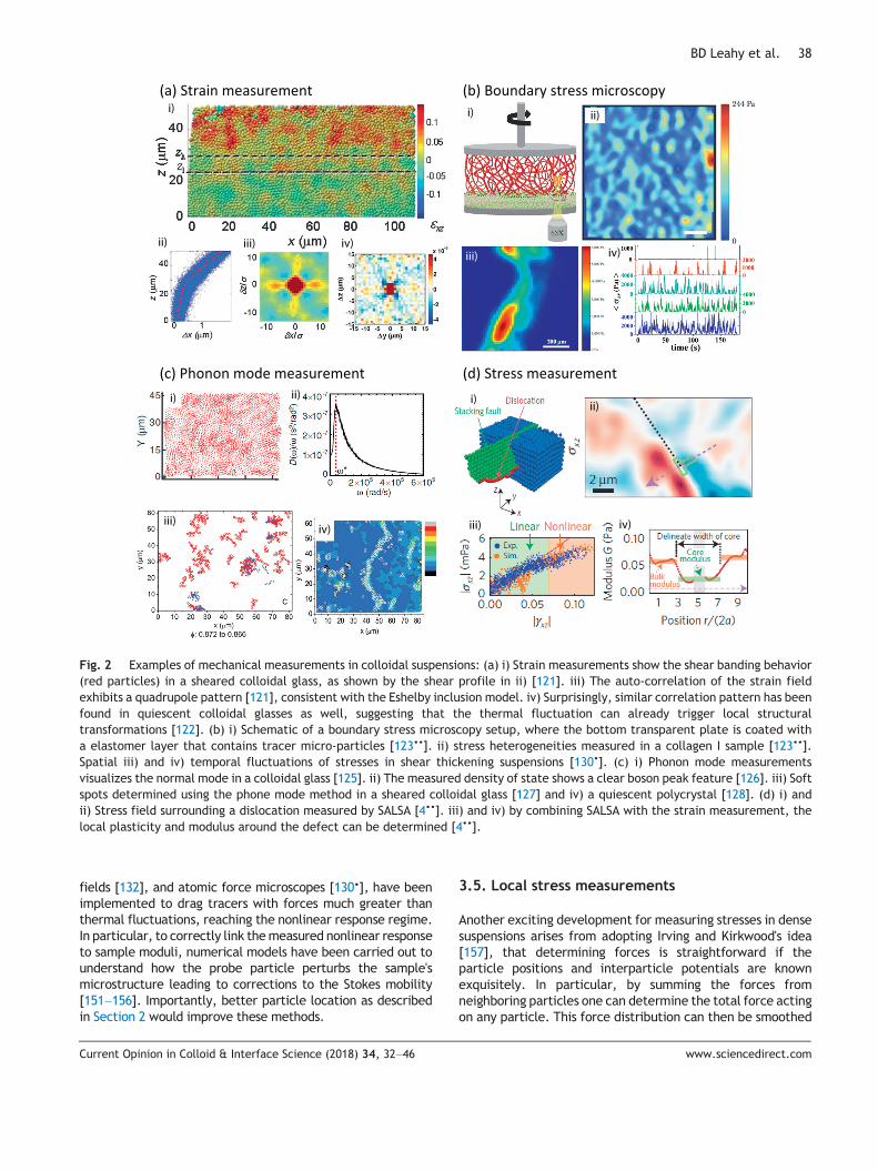
All solids display highly heterogeneous deformation fieldswhen they yield. Therefore, understanding the local deforma-tion or strain tensor and how it evolves under external loads isimportant to elucidating the nonlinear response of materials.The strain measurement technique calculates the local strainat a central particle from the neighboring particle displace-ments [5,47,63,121,122,133,134•,135–137]. The determinedstrain can then further be separated into affine and non-affinedeformations. The non-affine part of the strain is criticallyrelevant to the nonlinear response in yielded materials. Thestrain measurement technique has been used to study thedefect nucleation dynamics in colloidal crystals [63], and theyielding mechanisms in glasses with specific focuses on theshear transformation zone [47,122,135] and Eshelby inclusions[121,122] (Fig. 2a). Importantly, however, the absolute strainrelative to an undeformed lattice or configuration can only bedetermined for a crystalline suspension. Moreover, only theaffine response can be related to the stress via ameasurementof the modulus. In liquid or glassy suspensions the situation isfurther complicated by the fact that only relative strains arecalculated. Whether these relax or further concentrate localstresses is difficult to interpret from experimental measure-ments of the strains alone.
3.2. Boundary stress microscopy
The mechanical response of soft materials can be highlyheterogeneous. Measuring such spatial fluctuations of forcescan provide valuable insights into the mechanisms that giverise to the extraordinary strengths of amorphous systems.Unfortunately, conventional microscopic flow measurementsare only able to report the response that is averaged over alarge area, glossing over many important microscopic fea-tures. Boundary stress microscopy however, visualizes theseintriguing force patterns at the sample boundary [123••,124••](Fig. 2b). This method shares the same principles as tractionforce microscopy but is applied to dense suspensions. Here, athin layer of elastomer with embedded tracer particles isattached to the plate of a shear device (e.g. a rheometer orcustom-made shear cell). When the loaded sample is sheared,its shear stress is then transmitted to the boundary anddeforms the elastomer. By measuring the displacement of thetracer particles and the elastomer storage modulus, one candetermine the spatial variation of the stress on the boundary.The main difference in philosophy with traction forcemicroscopy is the focus on large surface regions andcharacterization of the inhomogeneous stress distributions.
Because the elastomer usually has excellent responseproperties, boundary stress microscopy is particularly usefulfor observing local stresses that can vary over many orders ofmagnitudes. For instance, it has been used to show that in a
Current Opinion in Colloid & Interface Science (2018) 34, 32–46
collagen I gel, the local stress can exceed average stressesby an order of magnitude, with a length scale that is muchlarger than the fiber mesh [123••]. Furthermore, boundarystress microscopy has revealed dramatic spatial and tempo-ral stress fluctuations in shear thickening suspensions. Thisevidence suggests that a shear thickening sample spontane-ously and intermittently separates into different fluid phaseswith significantly different properties [124••].
3.3. Phonon mode measurements
While vibrational modes in colloidal crystals can be heavilydamped by the surrounding solvent [138–141], it has beenrecently proposed that suspensions can still be used as a“shadow system” for calculating the properties of glassysystems. The idea is to start with the particle positions and tocalculate the vibrational modes as though the particles were ina simple harmonic potential [125,126,129]. Using a normalmode analysis, the displacement covariance matrices of theparticles are diagonalized to obtain the normalmodes (Fig. 2c).Some numerical and experimental studies have extended theprincipal of density of state measurement to correlate the softspots – regions with concentrated low-frequency vibrationalmodes – to the shear transformation zones and defects inglasses [127,142,143] and crystals [128,144], respectively. Thisanalysis technique is particularly interesting since it representsa hybrid approach that combines experiments and simulations.
3.4. Microrheology
Mechanical rheometry is a long-standing standard methodused to characterize the flow behavior of complex materials.However, conventional rheological measurements have sev-eral critical limitations, such as the need for relatively largesample volumes (on the order of a milliliter), the inability tocharacterize local mechanical heterogeneity, and low probingfrequency due to instrument inertia. In the last few decades,microrheology has been developed and become a popular toolto overcome these challenges. In the original papers ofmicrorheology [145,146], Mason and Weitz tracked thetrajectories of the tracer particles in various of complexfluids, determined their mean square displacement (MSD),and, via the Stokes-Sutherland-Einstein relation, determinedthe material's complex modulus. While in principal, anymethods that measure the particle MSD (e.g. Diffusing wavespectroscopy, dynamic light scattering, and fluorescencecorrelation spectroscopy) can be used for conductingmicrorheology, direct imaging has the advantage of providingvaluable information about the spatial distribution of me-chanical heterogeneity. Modern implementations of directimaging include both measurement of tracer particle motionsand differential dynamic microscopy [147•,148••,149].
Over time,many important improvements have beenmade.For example, two-point microrheology has been introduced tomitigate the artifacts arising from the tracer shape and sizedependencies, as well as the coupling between the tracer andits embedded medium [150]. Optical tweezers [131], magnetic
www.sciencedirect.com
i)
ii) iii) iv)
i) ii)
iii)
i)
iv)
ii)
iii) iv)
i) ii)
iii) iv)
Fig. 2 Examples of mechanical measurements in colloidal suspensions: (a) i) Strain measurements show the shear banding behavior(red particles) in a sheared colloidal glass, as shown by the shear profile in ii) [121]. iii) The auto-correlation of the strain fieldexhibits a quadrupole pattern [121], consistent with the Eshelby inclusion model. iv) Surprisingly, similar correlation pattern has beenfound in quiescent colloidal glasses as well, suggesting that the thermal fluctuation can already trigger local structuraltransformations [122]. (b) i) Schematic of a boundary stress microscopy setup, where the bottom transparent plate is coated witha elastomer layer that contains tracer micro-particles [123••]. ii) stress heterogeneities measured in a collagen I sample [123••].Spatial iii) and iv) temporal fluctuations of stresses in shear thickening suspensions [130•]. (c) i) Phonon mode measurementsvisualizes the normal mode in a colloidal glass [125]. ii) The measured density of state shows a clear boson peak feature [126]. iii) Softspots determined using the phone mode method in a sheared colloidal glass [127] and iv) a quiescent polycrystal [128]. (d) i) andii) Stress field surrounding a dislocation measured by SALSA [4••]. iii) and iv) by combining SALSA with the strain measurement, thelocal plasticity and modulus around the defect can be determined [4••].
BD Leahy et al. 38
fields [132], and atomic force microscopes [130•], have beenimplemented to drag tracers with forces much greater thanthermal fluctuations, reaching the nonlinear response regime.In particular, to correctly link themeasured nonlinear responseto sample moduli, numerical models have been carried out tounderstand how the probe particle perturbs the sample'smicrostructure leading to corrections to the Stokes mobility[151–156]. Importantly, better particle location as describedin Section 2 would improve these methods.
Current Opinion in Colloid & Interface Science (2018) 34, 32–46
3.5. Local stress measurements
Another exciting development for measuring stresses in densesuspensions arises from adopting Irving and Kirkwood's idea[157], that determining forces is straightforward if theparticle positions and interparticle potentials are knownexquisitely. In particular, by summing the forces fromneighboring particles one can determine the total force actingon any particle. This force distribution can then be smoothed
www.sciencedirect.com

39 Quantitative light microscopy of dense suspensions
to determine the local stress. The situation is a bit morecomplicated for hard spheres since they only interact whenparticles collide with one another. Brady theoretically deriveda formula to predict stresses in hard sphere suspensions basedon calculation of the system averaged pair correlationfunction gð r!Þ [158]. The calculation relates the anisotropicparticle distribution as measured by gð r!Þ to the suspensionstress tensor. In 2010, Gao et al. [49] partially adopted thisapproach to experiments by measuring the pair correlationfunctions for flowing suspensions and comparing them toStokesian Dynamics simulations. In 2011, Cheng et al. [48]implemented Brady's approach in full and showed that withexperiments alone it is possible to extract the bulk stress fromthe pair correlation functions. This technique has gained rapidacceptance in the colloid community and is now implementedroutinely by numerous groups [38,159,160].
In 2016, Lin et al. showed this macroscopic approach, canbe generalized to the particle level. In particular, StressAssessment from Local Structural Anisotropy (SALSA)measuresthe orientation distribution and probability of pairwisecollisions between a particle and its nearest neighbors todetermine the stresses arising from thermal collisions [4••].SALSA has already been used to visualize the stress distribu-tions surrounding crystal defects [4••] (See Fig. 2d) and theevolution of stresses in crystals under compression [78••].Furthermore, SALSA has also been used to determine theviscosity of quiescent colloidal liquids by simply measuringthermally induced fluctuations of the shear stress [120•].While current implementations have been used to investigatesuspensions in the Brownian or low Péclet regime, it is likelythat such measurements will also inform measurements ofstress distributions under higher shear rates.
In Table 2 we provide a comparison of the range oftechniques available for measuring forces. Once again,rather than a complete list, this table is meant to highlightthe range of exciting techniques available for studying theheterogeneous mechanics in dense suspensions.
4. Application of analysis techniques to addresslong standing challenges in colloid science
The most exciting aspect of these novel analyses is theavenues that they open for investigating some of the mostdifficult problems in the science of suspension behaviors.Here, we sketch approaches for moving forward on a few ofthe science questions relating to crystals, glasses, gels, andshear thickening suspensions.
4.1. Crystals
Because of the relatively slow time scales associated withthe particle motions, colloidal suspensions have been avaluable system for investigating a range of crystallizationphenomena. In entropic crystals alone, these phenomenarange from epitaxial growth [12,18] to defect nucleation andgrowth [13,63,161]. As colloidal systems become moresophisticated with different particle shapes [54,162] and
Current Opinion in Colloid & Interface Science (2018) 34, 32–46
tunable interparticle potentials [19–30] understanding howthe energetic driving forces alter rates of various processeswill become increasingly important for making progress.Already, techniques for inferring local strains are makingvaluable contributions related to the role of substratecurvature to crystallization [3,163,164]. Local stress mea-surements have already shown the importance of nonlinearstresses surrounding defects [4••] and the role that squeezeflows induced by increased confinement have on the stressdistribution in colloidal crystals [78••]. It is entirelyconceivable that such measurements can also be used todetermine the role of nonlinear stresses in driving defectdynamics, an exciting prospect for scientists hoping todevelop design principles for guiding defect nucleation,propagation, and coalescence.
The ability to measure variations in particle radii presentsanother fascinating direction for studying the phase separa-tion or crystallization of one particle species in the presence ofanother impurity or secondary phase. Already, it has beenshown that for a nearly monodisperse suspensions, evenslightly smaller particles end up accumulating at the bound-aries of crystal grains [3] and that a low concentration ofimpurities can substantially alter crystallization [165]. Theability to determine the 3D position and size of a large numberof secondary particles opens the door to investigations of howa secondary phase and the stresses it generates altercrystallization pathways and resulting kinetics, an increasinglyimportant problem for which relatively few model systemsexist [166].
4.2. Glasses
Colloids have been a cornerstone model system for investi-gating glassy phenomena. They have been instrumental inverifying concepts ranging from dynamic heterogeneity andcage hopping [42,44], to identifying soft spots associated withlow order normal modes [127,142,143] and long range straincorrelations associated with yielding [121,122,136]. Movingforward, there are many avenues for improving measurementtechniques to usher in an understanding of glasses at “the nextdecimal place.” For example, most of the studies in colloidalglasses have been conducted with density-quenched mono-disperse particles. This choice of system has been primarilydictated by experimental necessity – until recently it has beendifficult to extract particle locations and sizes from images ofdense polydisperse suspensions. Butmonodisperse suspensionslocally crystallize, which makes them far from ideal forstudying glassy behavior. The new inference techniquesdescribed in Table 1 now make it possible to easily investigatephenomena in bidisperse [3,46,167] and polydisperse suspen-sions [2••,3,101,112], determine volume fractions to muchgreater precision [2••], and even determine inhomogeneitiesin cage structure, which could elucidate whether the liquid toglass transition in colloids has the form of a Gardner transitionwith an increasingly fractal energy basin [168].
Additionally, much of the dynamic analysis of yielding inglassy suspensions has focused on measuring the magnitude
www.sciencedirect.com

Table 2 Image-based methods for measuring mechanical properties of soft materials.
Strain Boundary stress Phonon modes Microrheology Stressmeasurements(SALSA)
Mechanisms Particledisplacements
Tracers embedded inelastomer
Particle positions forcalculating vibrationmodes
Passive: tracer's meansquare displacement;Active: tracer's dragforce
Inter-particlecollision rate
Instrumentmodification
No Elastomer coated onbottom substrate
No Passive: no; Active:external field
No
Analysis challenges Cannot infernonlinear stress
Only shows stresseson the sampleboundary; resolutionbounded by tracerdensity
Requires long-timeaverage todetermineequilibriumpositions; systematicerrors from finitestatistics [128]
Active: requirestheoretical models foraccuratelyinterpreting nonlinearresponses
Particle potentialand collisioncriteria needcareful validation
Sample limitation Colloidalsuspensions; onlycrystals allowforce inferencesrelative to anundeformedlattice
None None Passive and active:tracer size must betaken into account
Inter-particlepotential must beconsidered
Scientific findingexamples
Sheartransformationzones in colloidalglasses [47];colloidal crystaldefect nucleationdynamics [63]
Boundary stressinhomogeneities inshear thickeningsuspensions [124••]and bio-gels [123••]
Boson peaks and softspots in colloidalglasses [125,127,129]
Passive and active:alternative method torheometry for testingsamples with smallvolume [130•,131,132]
Visualizingnonlinear stressessurroundingcolloidal crystaldefects [4••]
BD Leahy et al. 40
of and correlations in local relative strains [133]. Since thereis no underlying lattice, it is not possible to define a strain indisordered suspensions. As such it is also not possible todetermine whether the observed relative strains arerelaxing or concentrating stresses. However, it should bepossible to use techniques like boundary stress microscopyand SALSA to measure inhomogeneous stress distributionsin sheared glasses and determine how they changeunder yielding. There are, of course, hurdles that remain.For example, the biggest difficulty with SALSA in a size-polydisperse suspension is to measure the particle posi-tions and sizes with sufficient precision to accuratelydetermine particle collisions probabilities. However, thisparticular hurdle can now be overcome by techniques likePERI. With such hurdles resolved, it should now be possibleto observe how local stresses rearrange and percolateto enable the observed shear banding in yielding glasses[47].
4.3. Gels
Many of these improvements in measurement can also beapplied to investigations of colloidal gels. In particular,
Current Opinion in Colloid & Interface Science (2018) 34, 32–46
analyses in Table 1 can be used to track gels made out ofpolydisperse particles. Additionally, PERI can be used inconjunction with simulations to extract nm scale interparticlepotentials [2••]. This enables the use of SALSA for measuringthe individual particle stresses in colloidal gel networks withdepletion or DLVO interparticle potentials. For the first time,we will be able to image and statistically catalog thepercentage of structurally necessary force chains versusredundant internal force loops [169]. In combination withboundary stress microscopy it should be possible to determinehow the structurally necessary boundary forces distributewithin the bulk of the gel. This combination of measurementswould garner a substantially deeper understanding of howthe local structure of gels dictates their loading, yielding, andflow response.
Recently, excitement surrounding colloidal gel behavior hasfocused on the ability to embed a system-wide memory usingoscillatory shearing protocols. By applying a given oscillatorystrain, one can repeatedly break and reform the gel networkuntil it can withstand a particular strain [170•,171]. Measure-ments of local force inhomogeneities would also enable us togo much further in differentiating the properties associatedwith different regions of the gel responsible for maintainingrigidity versus those that enable plasticity and breakage. A
www.sciencedirect.com

• of special interest.•• of outstanding interest.
41 Quantitative light microscopy of dense suspensions
better understanding of these mechanisms will enable theformation of gels with properties that can be designed throughvarious shear protocols.
4.4. Shear thickening
Colloidal suspensions also serve as an important model systemfor investigating non-Newtonian flows. These rheologicalphenomena include shear thickening at high strain rates,shear jamming of high suspension concentrations under rapidextensions, and solidification under rapid compression. Thereare many challenges in elucidating the microscopic origin forthese behaviors. In shear thickening alone, there has been avigorous and healthy debate relating to whether hydrodynamicor effective contact interactions between particles generatesuch behaviors [50,172–176•,177•]. Here too, the analyses wehave described could play an important role in resolving suchcontroversies. For example, using SALSA in combination withforce-sensitive dyes [77] it may be possible to determine thenet force on a particle due to contact interactions. Alterna-tively, PERI could be used to determine interactions due tocharge repulsion enabling measurement of effective contactsbetween particles. These contact and effective contactinteractions are thought by many to determine shear thicken-ing behavior in suspensions. The analyses described here wouldenable a more rigorous testing of these ideas. On larger lengthscales boundary stress microscopy is being used to identifyphase inhomogeneities in shear-thickened suspensions [124••].As the shear rate increases, the suspension forms regions ofthickened material that grow in size, coalesce, and eventuallytake over the entire suspension. Such measurements illustratethe rich physics that must be understood on intermediatelength scales in order to achieve sufficient understanding ofthese behaviors.
4.5. “… a new discovery is likely to be found lurkingin the next decimal place”
The scientific problems and methodologies we havesketched here to address them are only a small taste of thenew mysteries and scientific tool box developments tocome. As new analyses are developed to complement therapid expansion of microscope capabilities, it is likely thatnew science will be discovered, and with it new problems toaddress. One final idea that we would like to put forth is thatfuture techniques will most likely make even more use ofcomputation – not just to analyze images, but as anadditional tool for experiments. For instance, in SALSAcomputation is used to infer stresses based on estimation ofinterparticle collision probabilities. In phonon mode mea-surements, the colloidal suspension merely serves as astarting point for generating a computational model of a“shadow system.” And cheap computation can be used notonly to generate model fits to images, as in PERI, but also tointerpret the experimental data, such as fitting experimen-tal data with a complete simulation to extract interparticlepotentials [2••]. Such trends foreshadow an increasingly rich
Current Opinion in Colloid & Interface Science (2018) 34, 32–46
range of creative approaches for making discoveries at “thenext decimal place” of colloid science.
We thank Eric Weeks, Vinothan Manoharan, Roseanna Zia,and Mathieu Leocmach for helpful conversations in preparingthis manuscript. This work was supported in part by NationalScience Foundation (NSF) Grant No. DMR-1507607 (I. C.),NSF Grant No. CBET-1509308 (N. L.), and ACS Grant No. PRF56046-ND7 (B. L.).
References and recommended reading•,••
[1] Perrin J. La loi de stokes et le mouvement brownien. ComptRendus 1908;147:475–6.
••[2] Bierbaum M, Leahy BD, Alemi AA, Cohen I, Sethna JP. Light
microscopy at maximal precision. Phys Rev X 2017;7(4):041007.Parameter Estimation from Reconstruction of Images: Showedthat it is possible to extract colloid particle position and sizefrom an image at the information theoretic limit.
[3] Kurita R, Ruffner DB, Weeks ER. Measuring the size ofindividual particles from three-dimensional imaging experi-ments. Nat Commun 2012;3 [ncomms2114].
••[4] Lin NY, Bierbaum M, Schall P, Sethna JP, Cohen I. Measuring
nonlinear stresses generated by defects in 3d colloidal crystals.Nat Mater 2016;15(11):1172–6. SALSA: Showed how to imageforces at the single particle scale.
[5] Richtmyer F. The romance of the next decimal place. Science1932;75(1931):1–5.
[6] Pieranski P, Strzelecki L, Pansu B. Thin colloidal crystals. PhysRev Lett 1983;50(12):900.
[7] Pieranski P. Colloidal crystals. Contemp Phys 1983;24(1):25–73.
[8] Pieranski P. Two-dimensional interfacial colloidal crystals.Phys Rev Lett 1980;45(7):569.
[9] Pusey PN, Van Megen W. Phase behaviour of concentratedsuspensions of nearly hard colloidal spheres. Nature 1986;320(6060):340–2.
[10] Sirota E, Ou-Yang H, Sinha S, Chaikin P, Axe J, Fujii Y.Complete phase diagram of a charged colloidal system: asynchro-tron X-ray scattering study. Phys Rev Lett 1989;62(13):1524.
[11] Murray CB, Kagan C, Bawendi M. Synthesis and characterizationof monodisperse nanocrystals and close-packed nanocrystalassemblies. Annu Rev Mater Sci 2000;30(1):545–610.
[12] Van Blaaderen A, Ruel R, Wiltzius P, et al. Template-directedcolloidal crystallization. Nature 1997;385(6614):321–4.
[13] Schall P, Cohen I, Weitz DA, Spaepen F. Visualization ofdislocation dynamics in colloidal crystals. Science 2004;305(5692):1944–8.
[14] Yethiraj A, van Blaaderen A. A colloidal model system with aninteraction tunable from hard sphere to soft and dipolar.Nature 2003;421(6922):513–7.
[15] Asakura S, Oosawa F. On interaction between two bodiesimmersed in a solution of macromolecules. J Chem Phys1954;22(7):1255–6.
[16] Asakura S, Oosawa F. Interaction between particles suspendedin solutions of macromolecules. J Polym Sci A Polym Chem1958;33(126):183–92.
www.sciencedirect.com

BD Leahy et al. 42
[17] Lekkerkerker H, Poon W-K, Pusey P, Stroobants A, Warren P.Phase behaviour of colloid+ polymer mixtures. Europhys Lett1992;20(6):559.
[18] Lin K-h, Crocker JC, Prasad V, Schofield A, Weitz DA, LubenskyT, Yodh A. Entropically driven colloidal crystallization onpatterned surfaces. Phys Rev Lett 2000;85(8):1770.
[19] Ganapathy R, Buckley MR, Gerbode SJ, Cohen I. Directmeasurements of island growth and step-edge barriers incolloidal epitaxy. Science 2010;327(5964):445–8.
[20] Wang Y, Wang Y, Breed DR, Manoharan VN, Feng L,Hollingsworth AD, Weck M, Pine DJ. Colloids with valence andspecific directional bonding. Nature 2012;491(7422):51–5.
[21] Leunissen ME, Christova CG, Hynninen A-P, Royall CP,Campbell AI, Imhof A, Dijkstra M, Van Roij R, VanBlaaderen A. Ionic colloidal crystals of oppositely chargedparticles. Nature 2005;437(7056):235–40.
[22] Shevchenko EV, Talapin DV, Kotov NA, O'brien S, Murray CB.Structural diversity in binary nanoparticle superlattices.Nature 2006;439(7072):55–9.
[23] Kalsin AM, Fialkowski M, Paszewski M, Smoukov SK, BishopKJ, Grzybowski BA. Electrostatic self-assembly of binarynanoparticle crystals with a diamond-like lattice. Science2006;312(5772):420–4.
[24] Erb RM, Son HS, Samanta B, Rotello VM, Yellen BB. Magneticassembly of colloidal superstructures with multipole sym-metry. Nature 2009;457(7232):999–1002.
[25] Yellen BB, Hovorka O, Friedman G. Arranging matter bymagnetic nanoparticle assemblers. Proc Natl Acad Sci U S A2005;102(25):8860–4.
[26] Bacri J-C, Perzynski R, Cabuil V, Massart R. Phase diagram ofan ionic magnetic colloid: experimental study of the effectof ionic strength. J Colloid Interface Sci 1989;132(1):43–53.
[27] Mirkin CA, Letsinger RL, Mucic RC, Storhoff JJ. A DNA-basedmethod for rationally assembling nanoparticles into macro-scopic materials. Nature 1996;382(6592):607–9.
[28] Biancaniello PL, Kim AJ, Crocker JC. Colloidal interactionsand self-assembly using DNA hybridization. Phys Rev Lett2005;94(5):058302.
[29] Rogers WB, Shih WM, Manoharan VN. Using DNA to programthe self-assembly of colloidal nanoparticles and microparti-cles. Nat Rev Mater 2016;1:16008.
[30] Wang Y, Jenkins IC, McGinley JT, Sinno T, Crocker JC.Colloidal crystals with diamond symmetry at opticallengthscales. Nat Commun 2017;8:14173.
[31] Paxton WF, Kistler KC, Olmeda CC, Sen A, Angelo SKSt, Cao Y,Mallouk TE, Lammert PE, Crespi VH. Catalytic nanomotors:autonomous movement of striped nanorods. J Am Chem Soc2004;126(41):13424–31.
[32] Wang W, Duan W, Ahmed S, Mallouk TE, Sen A. Small power:autonomous nano-and micromotors propelled by self-generated gradients. Nano Today 2013;8(5):531–54.
[33] Palacci J, Sacanna S, Steinberg AP, Pine DJ, Chaikin PM.Living crystals of light-activated colloidal surfers. Science2013;339(6122):936–40.
[34] Palacci J, Sacanna S, Kim S-H, Yi G-R, Pine D, Chaikin P.Light-activated self-propelled colloids. Phil Trans R Soc A2014;372(2029):20130372.
[35] Ginot F, Theurkauff I, Levis D, Ybert C, Bocquet L, Berthier L,Cottin-Bizonne C. Nonequilibrium equation of state in suspensionsof active colloids. Phys Rev X 2015;5(1):011004.
[36] Niu R, Palberg T, Speck T, et al. Self-assembly of colloidalmolecules due to self-generated flow. Phys Rev Lett 2017;119(2):028001.
Current Opinion in Colloid & Interface Science (2018) 34, 32–46
[37] Aarts DG, Lekkerkerker HN, Guo H, Wegdam GH, Bonn D.Hydrodynamics of droplet coalescence. Phys Rev Lett 2005;95(16):164503.
[38] Lin NY, Goyal S, Cheng X, Zia RN, Escobedo FA, Cohen I. Far-from-equilibrium sheared colloidal liquids: disentanglingrelaxation, advection, and shear-induced diffusion. PhysRev E 2013;88(6):062309.
[39] Aarts DG, Schmidt M, Lekkerkerker HN. Direct visual observa-tion of thermal capillary waves. Science 2004;304(5672):847–50.
[40] Lu PJ, Conrad JC, Wyss HM, Schofield AB, Weitz DA. Fluids ofclusters in attractive colloids. Phys Rev Lett 2006;96(2):028306.
[41] Dibble CJ, Kogan M, Solomon MJ. Structure and dynamics ofcolloidal depletion gels: coincidence of transitions andheterogeneity. Phys Rev E 2006;74(4):041403.
[42] Kegel WK, van Blaaderen A. Direct observation of dynamicalheterogeneities in colloidal hard-sphere suspensions.Science 2000;287(5451):290–3.
[43] van Blaaderen A, Wiltzius P. Real-space structure of colloidalhard-sphere glasses. Science 1995;270(5239):1177–9.
[44] Weeks ER, Crocker JC, Levitt AC, Schofield A, Weitz DA.Three-dimensional direct imaging of structural relaxationnear the colloidal glass transition. Science 2000;287(5453):627–31.
[45] Pham K, Petekidis G, Vlassopoulos D, Egelhaaf S, Poon W,Pusey P. Yielding behavior of repulsion-and attraction-dominated colloidal glasses. J Rheol 2008;52(2):649–76.
[46] Yunker P, Zhang Z, Aptowicz KB, Yodh AG. Irreversiblerearrangements, correlated domains, and local structure inaging glasses. Phys Rev Lett 2009;103(11):115701.
[47] Schall P, Weitz DA, Spaepen F. Structural rearrangements thatgovern flow in colloidal glasses. Science 2007;318(5858):1895–9.
[48] Cheng X, McCoy JH, Israelachvili JN, Cohen I. Imaging themicroscopic structure of shear thinning and thickeningcolloidal suspensions. Science 2010;333:1276.
[49] Gao C, Kulkarni S, Morris J, Gilchrist J. Direct investigationof anisotropic suspension structure in pressure-driven flow.Phys Rev E 2010;81(4):041403.
[50] Ramaswamy M, Lin NY, Leahy BD, Ness C, Fiore AM, SwanJW, Cohen I. How confinement-induced structures alter thecontribution of hydrodynamic and short-ranged repulsionforces to the viscosity of colloidal suspensions. Phys Rev X2017;7(4):041005.
[51] Glotzer SC, Solomon MJ. Anisotropy of building blocks andtheir assembly into complex structures. Nat Mater 2007;6:557.
[52] Mohraz A, Solomon MJ. Direct visualization of colloidal rodassembly by confocal microscopy. Langmuir 2005;21(12):5298–306.
[53] Kraft DJ, Groenewold J, Kegel WK. Colloidal molecules withwell-controlled bond angles. Soft Matter 2009;5(20):3823–6.
[54] Lee SH, Gerbode SJ, John BS, Wolfgang AK, Escobedo FA,Cohen I, Liddell CM. Synthesis and assembly of nonsphericalhollow silica colloids under confinement. J Mater Chem2008;18:4912.
[55] Sacanna S, Irvine W, Chaikin PM, Pine DJ. Lock and keycolloids. Nature 2010;464(7288):575–8.
[56] Hernandez CJ, Mason TG. Colloidal alphabet soup: monodis-perse dispersions of shape-designed lithoparticles. J PhysChem C 2007;111(12):4477–80.
www.sciencedirect.com

43 Quantitative light microscopy of dense suspensions
[57] Zhao K, Mason TG. Frustrated rotator crystals and glasses ofbrownian pentagons. Phys Rev Lett 2009;103(20):208302.
[58] Badaire S, Cottin-Bizonne C, Woody JW, Yang A, Stroock AD.Shape selectivity in the assembly of lithographically de-signed colloidal particles. J Am Chem Soc 2007;129(1):40–1.
[59] Rolland JP, Maynor BW, Euliss LE, Exner AE, Denison GM,DeSimone JM. Direct fabrication and harvesting of monodis-perse, shape-specific nanobiomaterials. J Am Chem Soc 2005;127(28):10096–100.
[60] Damasceno PF, Engel M, Glotzer SC. Predictive self-assemblyof polyhedra into complex structures. Science 2012;337(6093):453–7.
[61] Praad V, Semwogerere D, Weeks ER. Confocal microscopy ofcolloids. J Phys Condens Matter 2007;19:113102.
[62] Gasser U, Weeks ER, Schofield A, Pusey P, Weitz D. Real-space imaging of nucleation and growth in colloidalcrystallization. Science 2001;292(5515):258–62.
[63] Schall P, Cohen I, Weitz DA, Spaepen F. Visualizingdislocation nucleation by indenting colloidal crystals. Nature2006;440(7082):319–23.
[64] Savage J, Dinsmore A. Experimental evidence for two-stepnucleation in colloidal crystallization. Phys Rev Lett 2009;102(19):198302.
[65] Savage JR, Hopp SF, Ganapathy R, Gerbode SJ, Heuer A,Cohen I. Entropy-driven crystal formation on highly strainedsubstrates. Proc Natl Acad Sci 2013;110(23):9301–4.
[66] Crocker JC, Grier DG. Methods of digital video microscopyfor colloidal studies. J Colloid Interface Sci 1995;179:298.
[67] Jenkins MC, Egelhaaf SU. Confocal microscopy of colloidalparticles: towards reliable, optimum coordinates. Adv ColloidInterface Sci 2008;136(1):65–92.
••[68] Jensen KE, Nakamura N. Note: an iterative algorithm to
improve colloidal particle locating. Rev Sci Instrum 2016;87(6):066103. Showed how to get all particles using Crocker-Grier algorithm.
[69] Israelachvili J, Tabor D. The measurement of van der Waalsdispersion forces in the range 1.5 to 130 nm 331; 1972;19–38.
[70] Crocker JC, Grier DG. Microscopic measurement of the pairinteraction potential of charge-stabilized colloid. Phys RevLett 1994;73(2):352.
[71] Trefalt G, Behrens SH, Borkovec M. Charge regulation in theelectrical double layer: ion adsorption and surface interac-tions. Langmuir 2015;32(2):380–400.
[72] Crocker JC, Matteo J, Dinsmore A, Yodh A. Entropicattraction and repulsion in binary colloids probed with aline optical tweezer. Phys Rev Lett 1999;82(21):4352.
[73] Mukhija D, Solomon MJ. Translational and rotational dynam-ics of colloidal rods by direct visualization with confocalmicroscopy. J Colloid Interface Sci 2007;314:98–106.
••[74] Besseling TH, Hermes M, Kuijk A, de Nijs B, Deng T-S, Dijkstra
M, Imhof A, van Blaaderen A. Determination of the positionsand orientations of concentrated rod-like colloids from 3dmicroscopy data. J Phys Condens Matter 2015;27:194109.Showed how to feature rods.
•[75] Conley GM, Nöjd S, Braibanti M, Schurtenberger P, Scheffold
F. Superresolution microscopy of the volume phase transitionof pnipam microgels. Colloids Surf A Physicochem Eng Asp2016;499:18–23. Use of PALM to image NIPA microparticles.
[76] Conley GM, Aebischer P, Nöjd S, Schurtenberger P, ScheffoldF. Jamming and overpacking fuzzy microgels: Deformation,interpenetration, and compression. Sci Adv 2017;3(10):e1700969.
Current Opinion in Colloid & Interface Science (2018) 34, 32–46
[77] Suhina T, Weber B, Carpentier CE, Lorincz K, Schall P, BonnD, Brouwer AM. Fluorescence microscopy visualization ofcontacts between objects. Angew Chem Int Ed 2015;54(12):3688–91.
••[78] Lin NY, Cohen I. Relating microstructure and particle-level
stress in colloidal crystals under increased confinement. SoftMatter 2016;12(44):9058–67. Application of SALSA to under-stand stresses induced by applying squeeze flow to a crystal.
[79] Betzig E, et al. Imaging intracellular fluorescent proteins atnanometer resolution. Science 2006;313:1642–5.
[80] Rust MJ, Bates M, Zhuang X. Sub-diffraction-limit imaging bystochastic optical reconstruction microscopy (storm). NatMethods 2006;3:793.
[81] Hell SW, Wichmann J. Breaking the diffraction resolutionlimit by stimulated emission: stimulated-emission-depletionfluorescence microscopy. Opt Lett 1994;19:780.
[82] Kner P, Chhun BB, Griffis ER, Winoto L, Gustafsson MGL.Super-resolution video microscopy of live cells by structuredillumination. Nat Methods 2009;6:339–42.
[83] Chen B-C, LegantWR,Wang K, Shao L, Milkie DE, DavidsonMW,Janetopoulos C, Wu XS, Hammer JA, Liu Z, et al. Lattice light-sheet microscopy: imaging molecules to embryos at highspatiotemporal resolution. Science 2014;346(6208):1257998.
[84] Snoeyink C, Wereley S. A novel 3d3c particle trackingmethod suitable for microfluidic flow measurements. ExpFluids 2013;54(1):1453.
[85] Gabor D. A new microscopic principle. Nature 1948;161:777–8.[86] Gabor D, et al. Microscopy by reconstructed wave-fronts 197
(1051); 1949; 454–87.[87] Sheng J, Malkiel E, Katz J. Digital holographic microscope for
measuring three-dimensional particle distributions andmotions. Appl Optics 2006;45(16):3893–901.
[88] Lee S-H, Grier DG. Holographic microscopy of holographi-cally trapped three-dimensional structures. Opt Express2007;15(4):1505–12.
[89] Lee S-H, Roichman Y, Yi G-R, Kim S-H, Yang S-M, VanBlaaderen A, Van Oostrum P, Grier DG. Characterizing andtracking single colloidal particles with video holographicmicroscopy. Opt Express 2007;15(26):18275–82.
[90] Cheong FC, Krishnatreya BJ, Grier DG. Strategies for three-dimensional particle tracking with holographic video micros-copy. Opt Express 2010;18(13):13563–73.
[91] Dixon L, Cheong FC, Grier DG. Holographic deconvolutionmicroscopy for high-resolution particle tracking. Opt Express2011;19(17):16410–7.
•[92] Dimiduk TG, Manoharan VN. Bayesian approach to analyzing
holograms of colloidal particles. Opt Express 2016;24(21):24045–60. Applications of Bayesian techniques to analysis ofsuspension hologram images.
[93] Fung J, Martin KE, Perry RW, Kaz DM, McGorty R, ManoharanVN. Measuring translational, rotational, and vibrationaldynamics in colloids with digital holographic microscopy.Opt Express 2011;19(9):8051–65.
[94] Fung J, Perry RW, Dimiduk TG, Manoharan VN. Imagingmultiple colloidal particles by fitting electromagneticscattering solutions to digital holograms. J Quant SpectroscRadiat Transf 2012;113(18):2482–9.
[95] Fung J, Manoharan VN. Holographic measurements ofanisotropic three-dimensional diffusion of colloidal clusters.Phys Rev E 2013;88(2):020302.
[96] Cheong FC, Grier DG. Rotational and translational diffusionof copper oxide nanorods measured with holographic videomicroscopy. Opt Express 2010;18(7):6555–62.
www.sciencedirect.com

BD Leahy et al. 44
[97] Wang A, Dimiduk TG, Fung J, Razavi S, Kretzschmar I,Chaudhary K, Manoharan VN. Using the discrete dipoleapproximation and holographic microscopy to measurerotational dynamics of non-spherical colloidal particles. JQuant Spectrosc Radiat Transf 2014;146:499–509.
•[98] Cheong FC, Kasimbeg P, Ruffner DB, Hlaing EH, Blusewicz JM,
Philips LA, Grier DG. Holographic characterization of colloidalparticles in turbid media. Appl Phys Lett 2017;111(15):153702.Application of holographic imaging in turbid suspensions.
[99] Dimiduk TG, Kosheleva EA, Kaz D, McGorty R, Gardel EJ,Manoharan VN. A simple, inexpensive holographic micro-scope. Biomedical Optics, Optical Society of America; 2010.p. JMA38.
[100] Deegan RD, Bakajin O, Dupont TF, Huber G, Nagel SR, WittenTA. Capillary flow as the cause of ring stains from driedliquid drops. Nature 1997;389(6653):827–9.
[101] Leocmach M, Tanaka H. A novel particle tracking methodwith individual particle size measurement and its applicationto ordering in glassy hard sphere colloids. Soft Matter 2013;9(5):1447–57.
•[102] Dreyfus R, Xu Y, Still T, Hough LA, Yodh A, Torquato S.
Diagnosing hyperuniformity in two-dimensional, disordered,jammed packings of soft spheres. Phys Rev E 2015;91(1):012302. Analysis of hyperuniformity in 2D suspensions.
[103] Crocker JC, Grier DG. Methods of digital video microscopy forcolloidal studies. J Colloid Interface Sci 1996;179(1):298–310.
[104] Elliot MS, Poon WC. Conventional optical microscopy ofcolloidal suspensions. Adv Colloid Interface Sci 2001;92(1):133–94.
[105] Lu PJ, Shutman M, Sloutskin E, Butenko AV. Locatingparticles accurately in microscope images requires image-processing kernels to be rotationally symmetric. Opt Express2013;21(25):30755–63.
[106] Gao Y, Kilfoil ML. Accurate detection and complete trackingof large populations of features in three dimensions. OptExpress 2009;17(6):4685–704.
[107] Berglund AJ, McMahon MD, McClelland JJ, Liddle JA. Fast,bias-free algorithm for tracking single particles with variablesize and shape. Opt Express 2008;16(18):14064–75.
•[108] Burov S, Figliozzi P, Lin B, Rice SA, Scherer NF, Dinner AR.
Single-pixel interior filling function approach for detectingand correcting errors in particle tracking. Proc Natl Acad Sci2017;114(2):221–6. Demonstrated a approach for detectingand correcting errors in particle tracking.
[109] Pathasarathy R. Rapid, accurate particle tracking by calcula-tion of radial symmetry centers. Nat Methods 2012;9:724–6.
[110] Mayoral K, Kennair TP, Zhu X, Milazzo J, Ngo K, Fryd MM,Mason TG. Rotational fourier tracking of diffusing polygons.Phys Rev E 2011;84(5):051405.
[111] Leahy B, Cheng X, Ong DC, Liddell-Watson C, Cohen I.Enhancing rotational diffusion using oscillatory shear. PhysRev Lett 2013;110:228301.
[112] Brujić J, Edwards SF, Grinev DV, Hopkinson I, Brujić D, MakseHA. 3d bulk measurements of the force distribution in acompressed emulsion system. Faraday Discuss 2003;123:207–20.
[113] Jorjadze I, Pontani L-L, Brujic J. Microscopic approach to thenonlinear elasticity of compressed emulsions. Phys Rev Lett2013;110(4):048302.
[114] Kurita R, Weeks ER. Incompressibility of polydisperse random-close-packed colloidal particles. Phys Rev E 2011;84(3):030401.
•[115] Heckendorf D, Mutch K, Egelhaaf S, Laurati M. Size-
dependent localization in polydisperse colloidal glasses.
Current Opinion in Colloid & Interface Science (2018) 34, 32–46
Phys Rev Lett 2017;119(4):048003. Observed size-dependentlocalization in polydisperse glasses.
[116] D. B. Allan, T. A. Caswell, N. C. Keim, Trackpy v0.2 (May2014). https://doi.org/10.5281/zenodo.9971. URL github.com/soft-matter/trackpy
[117] [link]. URL http://www.physics.emory.edu/faculty/weeks//idl/.
[118] https://doi.org/10.5281/zenodo.31286, [link]. URL https://doi.org/10.5281/zenodo.31286
[119] [link]. URL https://github.com/peri-source/peri.
•[120] Lin NY, Bierbaum M, Cohen I. Determining quiescent colloidal
suspension viscosities using the green-kubo relation andimage-based stress measurements. Phys Rev Lett 2017;119(13):138001. Analyzed thermal fluctuations in a quiescentsuspension to determine the Brownian viscosity as a functionof volume fraction.
[121] Chikkadi V, Wegdam G, Bonn D, Nienhuis B, Schall P. Long-range strain correlations in sheared colloidal glasses. PhysRev Lett 2011;107(19):198303.
[122] Jensen K, Weitz DA, Spaepen F. Local shear transformationsin deformed and quiescent hard-sphere colloidal glasses.Phys Rev E 2014;90(4):042305.
••[123] Arevalo RC, Kumar P, Urbach JS, Blair DL. Stress heterogene-
ities in sheared type-i collagen networks revealed by boundarystress microscopy. PLoS One 2015;10(3):e0118021. Usedboundary stress to measure heterogeneities in sheared type-Icollagen networks.
••[124] Rathee V, Blair DL, Urbach JS. Localized stress fluctuations drive
shear thickening in dense suspensions. Proc Natl Acad Sci 2017;114(33):8740–5 [arXiv: http://www.pnas.org/content/114/33/8740.full.pdf, https://doi.org/10.1073/pnas.1703871114. URLhttp://www.pnas.org/content/114/33/8740.abstract]. Usedboundary stress to measure heterogeneities in thickeningsuspension. Established a meso-scale phase separation duringtransition to thickened state.
[125] Kaya D, Green N, Maloney C, Islam M. Normal modes anddensity of states of disordered colloidal solids. Science 2010;329(5992):656–8.
[126] Chen K, Ellenbroek WG, Zhang Z, Chen DT, Yunker PJ,Henkes S, Brito C, Dauchot O, Van Saarloos W, Liu AJ, et al.Low-frequency vibrations of soft colloidal glasses. Phys RevLett 2010;105(2):025501.
[127] Chen K, Manning M, Yunker PJ, Ellenbroek WG, Zhang Z, LiuAJ, Yodh AG. Measurement of correlations between low-frequency vibrational modes and particle rearrangements inquasi-two-dimensional colloidal glasses. Phys Rev Lett 2011;107(10):108301.
[128] Chen K, Still T, Schoenholz S, Aptowicz KB, Schindler M,Maggs A, Liu AJ, Yodh A. Phonons in two-dimensional softcolloidal crystals. Phys Rev E 2013;88(2):022315.
[129] Ghosh A, Chikkadi VK, Schall P, Kurchan J, Bonn D. Density ofstates of colloidal glasses. Phys Rev Lett 2010;104(24):248305.
•[130] Rigato A, Miyagi A, Scheuring S, Rico F. High-frequency
microrheology reveals cytoskeleton dynamics in living cells.Nat Phys, 13; 2017. p. 771–5. Application of microrheology tobiopolymer-membrane complexes.
[131] Helfer E, Harlepp S, Bourdieu L, Robert J, MacKintosh F,Chatenay D. Microrheology of biopolymer-membrane com-plexes. Phys Rev Lett 2000;85(2):457.
[132] Amblard F, Maggs AC, Yurke B, Pargellis AN, Leibler S.Subdiffusion and anomalous local viscoelasticity in actinnetworks. Phys Rev Lett 1996;77(21):4470.
www.sciencedirect.com
45 Quantitative light microscopy of dense suspensions
[133] Falk ML, Langer JS. Dynamics of viscoplastic deformation inamorphous solids. Phys Rev E 1998;57(6):7192.
•[134] Sussman DM, Schoenholz SS, Xu Y, Still T, Yodh A, Liu AJ.
Strain fluctuations and elastic moduli in disordered solids.Phys Rev E 2015;92(2):022307. Analyzed a colloidal suspen-sion as a shadow system to determine strain fluctuations andelastic moduli in disordered solids.
[135] Rahmani Y, Koopman R, Denisov D, Schall P. Visualizing thestrain evolution during the indentation of colloidal glasses.Phys Rev E 2014;89(1):012304.
[136] Chikkadi V, Miedema D, Dang M, Nienhuis B, Schall P. Shearbanding of colloidal glasses: observation of a dynamic first-order transition. Phys Rev Lett 2014;113(20):208301.
[137] Illing B, Fritschi S, Hajnal D, Klix C, Keim P, Fuchs M. Strainpattern in supercooled liquids. Phys Rev Lett 2016;117(20):208002.
[138] Cheng Z, Zhu J, Russel WB, Chaikin P. Phonons in an entropiccrystal. Phys Rev Lett 2000;85(7):1460.
[139] Keim P, Maret G, Herz U, von Grünberg H-H. Harmoniclattice behavior of two-dimensional colloidal crystals. PhysRev Lett 2004;92(21):215504.
[140] Baumgartl J, Dietrich J, Dobnikar J, Bechinger C, vonGrünberg HH. Phonon dispersion curves of two-dimensionalcolloidal crystals: the wavelength-dependence of friction.Soft Matter 2008;4(11):2199–206.
[141] Ohshima YN, Nishio I. Colloidal crystal: bead–spring latticeimmersed in viscous media. J Chem Phys 2001;114(19):8649–58.
[142] Ding J, Patinet S, Falk ML, Cheng Y, Ma E. Soft spots and theirstructural signature in a metallic glass. Proc Natl Acad Sci2014;111(39):14052–6.
[143] Manning ML, Liu AJ. Vibrational modes identify soft spots in asheared disordered packing. Phys Rev Lett 2011;107(10):108302.
[144] Rottler J, Schoenholz SS, Liu AJ. Predicting plasticity withsoft vibrational modes: from dislocations to glasses. Phys RevE 2014;89(4):042304.
[145] Mason TG, Weitz D. Optical measurements of frequency-dependent linear viscoelastic moduli of complex fluids. PhysRev Lett 1995;74(7):1250.
[146] Mason T, Weitz D. Linear viscoelasticity of colloidal hardsphere suspensions near the glass transition. Phys Rev Lett1995;75(14):2770.
•[147] Bayles AV, Squires TM, Helgeson ME. Dark-field differential
dynamic microscopy. Soft Matter 2016;12(8):2440–52.Showed how to apply dark-field imaging ddm to extractdisplacements and relate these to stresses viamicrorheology.
••[148] Edera P, Bergamini D, Trappe V, Giavazzi F, Cerbino R, et al.
Illustration of ddm approach to microrheology. Phys Rev Mater2017;1(7):073804.
[149] Cerbino R, Trappe V. Differential dynamic microscopy:probing wave vector dependent dynamics with a micro-scope. Phys Rev Lett 2008;100(18):188102.
[150] Crocker JC, Valentine MT, Weeks ER, Gisler T, KaplanPD, Yodh AG, Weitz DA. Two-point microrheology ofinhomogeneous soft materials. Phys Rev Lett 2000;85(4):888.
[151] Zia RN. Active and passive microrheology: theory andsimulation. Annu Rev Fluid Mech 2018;150:371–405.
[152] Khair AS, Brady JF. Single particle motion in colloidaldispersions: a simple model for active and nonlinearmicrorheology. J Fluid Mech 2006;557:73–117.
Current Opinion in Colloid & Interface Science (2018) 34, 32–46
[153] Squires TM, Brady JF. A simple paradigm for active andnonlinear microrheology. Phys Fluids 2005;17(7):073101.
[154] Squires TM. Nonlinear microrheology: bulk stresses versusdirect interactions. Langmuir 2008;24(4):1147–59.
[155] Zia RN, Brady JF. Single-particle motion in colloids: force-induced diffusion. J Fluid Mech 2010;658:188–210.
[156] Swan JW, Zia RN, Brady JF. Large amplitude oscillatorymicrorheology. J Rheol 2014;58(1):1–41.
[157] Irving J, Kirkwood JG. The statistical mechanical theoryof transport processes. iv. The equations of hydrodynamics.J Chem Phys 1950;18(6):817–29.
[158] Brady JF. The rheological behavior of concentrated colloidaldispersions. J Chem Phys 1993;99:567.
[159] Xu B, Gilchrist JF. Microstructure of sheared monosizedcolloidal suspensions resulting from hydrodynamic and elec-trostatic interactions. J Chem Phys 2014;140(20):204903.
[160] Koumakis N, Laurati M, Jacob A, Mutch K, Abdellali A,Schofield A, Egelhaaf S, Brady J, Petekidis G. Start-up shearof concentrated colloidal hard spheres: stresses, dynamics,and structure. J Rheol 2016;60(4):603–23.
[161] Gerbode SJ, Lee SH, Liddell CM, Cohen I. Restricteddislocation motion in crystals of colloidal dimer particles.Phys Rev Lett 2008;101(5):058302.
[162] Rossi L, Sacanna S, Irvine WT, Chaikin PM, Pine DJ, PhilipseAP. Cubic crystals from cubic colloids. Soft Matter 2011;7(9):4139–42.
[163] Irvine WT, Vitelli V, Chaikin PM. Pleats in crystals on curvedsurfaces. Nature 2010;468(7326):947–51.
[164] Meng G, Paulose J, Nelson DR, Manoharan VN. Elasticinstability of a crystal growing on a curved surface. Science2014;343(6171):634–7.
[165] de Villeneuve VW, Dullens RP, Aarts DG, Groeneveld E,Scherff JH, Kegel WK, Lekkerkerker HN. Colloidal hard-sphere crystal growth frustrated by large spherical impuri-ties. Science 2005;309(5738):1231–3.
[166] De Yoreo JJ, Gilbert PU, Sommerdijk NA, Penn RL,Whitelam S,Joester D, Zhang H, Rimer JD, Navrotsky A, Banfield JF, et al.Crystallization by particle attachment in synthetic, biogenic,and geologic environments. Science 2015;349(6247):aaa6760.
[167] Nugent CR, Edmond KV, Patel HN, Weeks ER. Colloidal glasstransition observed in confinement. Phys Rev Lett 2007;99(2):025702.
[168] Parisi G, Charbonneau P, Zamponi F, Urbani P, Kurchan J.Fractal free energy landscapes in structural glasses. NatCommun 2014;5(5):3725.
[169] Maxwell JC. L. on the calculation of the equilibrium andstiffness of frames. London Edinb Dublin Philos Mag J Sci1864;27(182):294–9.
•[170] Moghimi E, Jacob AR, Koumakis N, Petekidis G. Colloidal gels
tuned by oscillatory shear. Soft Matter 2017;13(12):2371–83.Showed that the mechanical properties of colloidal gels canbe tuned through application of oscillatory shear.
[171] Schwen E, Ramaswamay M, Jan L, Cheng C-M, Cohen I.Embedding memories in colloidal gels though oscillatoryshear. Bull Am Phys Soc 2017;62.
[172] Wagner NJ, Brady JF. Shear thickening in colloidal disper-sions. Phys Today 2009;62(10):27–32.
[173] Lootens D, Van Damme H, Hémar Y, Hébraud P. Dilatant flowof concentrated suspensions of rough particles. Phys RevLett 2005;95(26):268302.
[174] Seto R, Mari R, Morris JF, Denn MM. Discontinuous shearthickening of frictional hard-sphere suspensions. Phys RevLett 2013;111(21):218301.
www.sciencedirect.com
BD Leahy et al. 46
[175] Brown E, Jaeger HM. Shear thickening in concentratedsuspensions: phenomenology, mechanisms and relations tojamming. Rep Prog Phys 2014;77(4):046602.
•[176] Lin NY, Guy BM, Hermes M, Ness C, Sun J, Poon WC, Cohen I.
Hydrodynamic and contact contributions to continuous shearthickening in colloidal suspensions. Phys Rev Lett 2015;115(22):228304. Determined that contact or effective contact
Current Opinion in Colloid & Interface Science (2018) 34, 32–46
forces dominate shear thickening in silica and PMMA colloidalsuspensions.
•[177] Lin NY, Ness C, Cates ME, Sun J, Cohen I. Tunable shear
thickening in suspensions. Proc Natl Acad Sci 2016;113(39):10774–8 [201608348]. Showed how to dethicken a suspensionthrough application of orthogonal shear flows.
www.sciencedirect.com