Pulmonary Metastasis from Intraspinal Meningioma - ajnr.org · Pulmonary Metastasis from...
2
483 Pulmonary Metastasis from Intraspinal Meningioma K. J. Dastur\ M. Reza Raji\ and W. I. Smith, Jr. 2 Most intracranial and intraspinal meningiomas are benign. Aggressive "malignant" behavior of intracranial meningiomas, with distant metastases usually to the lungs, has been well described [1-4]. We report a rare case of intraspinal menin- gioma with distant metastases to the lungs. Our review of the literature revealed only one other case of intraspinal meningioma with metastases to the lung [5]. Case Report A 67-year-old woman had back pain radiating down the right leg . Review of systems was negative. On neurologic examination pain was elicited during straight leg raising on the right. No motor or sensory deficits were noted. The probable clinical neurologic impres- sion was right L5 root compression. Metrizamide myelography re- vealed a well marginated intradural defect at the T12-L 1 level situated predominantly to the right of the midline (fig. 1 A) . A computed tomographic (CT) scan at the T12-L 1 level showed a high-density intradural lesion with CT numbers suggestive of calcification. An admission chest film revealed a rounded nodular density measuring about 2.5 cm situated in the left lower lobe (fig. 1 B) . Chest CT revealed the lesions to be uncalcified and compatible with a lung neoplasm. At spinal surgery, an intradural tumor was removed. Histology revea led a solid tumor of sheets and whorls of ce ll s with indistinct borders characteristic of meningioma (fig. 1 C) . Four weeks later the lung lesion was removed at thoracotomy. Histologic exami- nation revealed a solid tumor composed of sheets of cells with increased mitoses but not pathologically mali gnant (fig. 1 D) . Electron microscopic studies revealed complex interdigitating surface mem- branes reminiscent of meningeal cells (fig. 1 E) . Discussion Metastases from intracranial and intraspinal meningioma are uncommon. Of 61 cases of intracranial meningiomas that metastasized, 60% had metastasized to the lungs, 34% to the abdominal organs, 11 % to the vertebral bodies, and 11 % to the long bones, pelvis , or sk ull. In contrast to metastasis from intracranial meningiomas, metastases from intraspinal meningiomas are less well documented. Allen et al. [5] re- ported the only case to our knowledge of an intraspinal meningioma with distant metastases, a 14-year-old girl with a cervical spine meningioma who died 1 month after surgery. At autopsy there were metastases to the lu ngs, pleura, and parabronchiallymph nodes. The definition of malignancy in meningioma is difficult , because in most cases the local invasion of brain parenchyma arid calvarium has been interpreted as an aggressive pattern . Shuangshoti et al. [2] reported that histologic demonstration of malignant features , for example, mitotic activity, is often not distinctive. In their series, 15% of intracranial meningiomas with distant metastasis were of the angioblastic variety, but all cellular types were seen. Incriminated in the pathway of extracranial spread of meningiomas is vascular invasion of venous structures with the venous return of blood subse- quently leading to tumor deposits in the lungs. At surgery, tumor cells may enter lacerated blood vessels. However, extracranial dissemination of meningioma has been noted in patients who did not undergo a craniotomy. In our case there was no myelographic evidence of an aggressive or malignant lesion. The discovery of the nodular lesion in the chest was an incidental finding. Crucial to this case is the documentation that the primary neoplasm was a meningioma and that it did indeed metastasize. The spinal neoplasm had histologic characteristics of an intraspinal me- ningioma (fig. 1 C) . More difficult is the diagnostic problem of the lung lesion. The surgical and gross pathologic findings favored a metastatic tumor over a primary lung tumor. The tumor was well circumscribed and seemed to be compressing the adjacent lung, rather than infiltrating as primary lung tumors may do. Histologically the tumor was very vascular. In addition, the finding of interdigitating membranes by elec- tron microscopy supported the diagnosis of meningioma. In conclusion , although meningiomas metastasize rarely, a pulmonary nodule in the presence of a central nervous system meningioma should raise the suspicion of lung metastases. ACKNOWLEDGMENTS We thank George H. Gray, Jr ., and Ronald V. Pell egrini , the attending physicians, and their associates; Lilia D' Ami co for secretar- ial assi stance; and Glenn Hangard for photographic assistan ce. Received January 12 , 1983; accepted after revision May 27, 1983. 1 Department of Radiology, Section of Neuroradiolog y, Mercy Hospi tal, Pride and Locust Sts ., Pittsburgh, PA 152 19. Address reprin t requests to K. J. Dastur. 2 Department of Pathology, Mercy Hospital , Pittsburgh , PA 15219. AJNR 5:483-484, July/August 1984 0195- 6108/84/0504-0483 $2 .00 © Ameri can Roentgen Ray Society
Transcript of Pulmonary Metastasis from Intraspinal Meningioma - ajnr.org · Pulmonary Metastasis from...
483
Pulmonary Metastasis from Intraspinal Meningioma K. J. Dastur\ M. Reza Raji\ and W. I. Smith, Jr.2
Most intracranial and intraspinal meningiomas are benign. Aggressive "malignant" behavior of intracranial meningiomas, with distant metastases usually to the lungs, has been well described [1-4]. We report a rare case of intraspinal meningioma with distant metastases to the lungs. Our review of the literature revealed only one other case of intraspinal meningioma with metastases to the lung [5].
Case Report
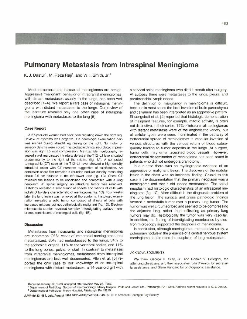
A 67-year-old woman had back pain radiating down the right leg. Review of systems was negative. On neurologic examination pain was elicited during straight leg raising on the right. No motor or sensory deficits were noted . The probable clinical neurologic impression was right L5 root compression . Metrizamide myelography revealed a well marginated intradural defect at the T12-L 1 level situated predominantly to the right of the midline (fig. 1 A) . A computed tomographic (CT) scan at the T12-L 1 level showed a high-density intradural lesion with CT numbers suggestive of calcification . An admission chest film revealed a rounded nodular density measuring about 2.5 cm situated in the left lower lobe (fig . 1 B) . Chest CT revealed the lesions to be uncalcified and compatible with a lung neoplasm. At spinal surgery , an intradural tumor was removed. Histology revealed a solid tumor of sheets and whorls of cells with indistinct borders characteristic of meningioma (fig . 1 C). Four weeks later the lung lesion was removed at thoracotomy. Histologic examination revealed a solid tumor composed of sheets of cells with increased mitoses but not pathologically malignant (fig . 1 D) . Electron microscopic studies revealed complex interdigitating surface membranes reminiscent of meningeal cells (fig . 1 E).
Discussion
Metastases from intracranial and intraspinal meningioma are uncommon. Of 61 cases of intracranial meningiomas that metastasized, 60% had metastasized to the lungs, 34% to the abdominal organs, 11 % to the vertebral bodies, and 11 % to the long bones, pelvis , or skull. In contrast to metastasis from intracranial meningiomas, metastases from intraspinal meningiomas are less well documented. Allen et al. [5] reported the only case to our knowledge of an intraspinal meningioma with distant metastases , a 14-year-old girl with
a cervical spine meningioma who died 1 month after surgery. At autopsy there were metastases to the lungs , pleura, and parabronchiallymph nodes.
The definition of malignancy in meningioma is difficult , because in most cases the local invasion of brain parenchyma arid calvarium has been interpreted as an aggressive pattern . Shuangshoti et al. [2] reported that histologic demonstration of malignant features , for example, mitotic activity, is often not distinctive. In their series, 15% of intracranial meningiomas with distant metastasis were of the angioblastic variety, but all cellular types were seen . Incriminated in the pathway of extracranial spread of meningiomas is vascular invasion of venous structures with the venous return of blood subsequently leading to tumor deposits in the lungs. At surgery, tumor cells may enter lacerated blood vessels. However, extracranial dissemination of meningioma has been noted in patients who did not undergo a craniotomy.
In our case there was no myelographic evidence of an aggressive or malignant lesion . The discovery of the nodular lesion in the chest was an incidental finding. Crucial to this case is the documentation that the primary neoplasm was a meningioma and that it did indeed metastasize. The spinal neoplasm had histologic characteristics of an intraspinal meningioma (fig . 1 C). More difficult is the diagnostic problem of the lung lesion. The surgical and gross pathologic findings favored a metastatic tumor over a primary lung tumor. The tumor was well circumscribed and seemed to be compressing the adjacent lung, rather than infiltrating as primary lung tumors may do. Histologically the tumor was very vascular. In addition, the finding of interdigitating membranes by electron microscopy supported the diagnosis of meningioma.
In conclusion , although meningiomas metastasize rarely , a pulmonary nodule in the presence of a central nervous system meningioma should raise the suspicion of lung metastases.
ACKNOWLEDGMENTS
We thank George H. Gray, Jr., and Ronald V. Pellegrini , the attending physicians, and their associates; Lilia D'Amico for secretarial assistance; and Glenn Hangard for photographic assistance.
Received January 12, 1983; accepted after revision May 27, 1983. 1 Department of Radiology, Section of Neuroradiology, Mercy Hospital, Pride and Locust Sts., Pittsburgh, PA 15219. Address reprint requests to K. J. Dastur. 2 Department of Pathology, Mercy Hospital , Pittsburgh , PA 15219.
AJNR 5:483-484, July/August 1984 0195- 6108/84/0504-0483 $2.00 © American Roentgen Ray Society

484 DASTUR ET AL. AJNR :5. July/August 1984
A B
c o
REFERENCES
1. Karasick JL. Mullan SF. A survey of metastatic meningioma. J Neurosurg 1974;39 :206-211
2, Shuangshoti S. Hongsaprabhas C, Netsky MG. Metastasizing meningioma. Cancer 1970;26 :832- 840
3. Cushing H. Eisenhardt L, Meningiomas: their classification, re -
E
Fig. 1.-A. Metrizamide myelogram. Well marginated. right-sided intradural defect at T12-L1 level. B. Admission film . Nodular mass in left lower lobe. C, Intraspinal meningioma. Whorls and sheets of cells with vesicular nuclei , indistinct cell borders , and occasional psammoma bodies. (Original magnification, x 25.) D, Metastatic meningioma to lung. Solid tumor is highly vascular. Cytologically , nuclei are vesicular and cell borders are indistinct. (Original magnification . x 1 00.) E, Electron microscopy of metastatic tumor. Cell membranes are interdigitating as in meningioma. Tumor lacks basement membrane seen in vascular tumors. (Original magnification. x 8000 .)
gional behavior, life history, and surgical end results. Springfield , IL: Thomas, 1938
4. Gordon A, Maloney AFJ . A case of metastasizing meningioma. J Neural Neurasurg Psychiatry 1965;28: 159-162
5. Allen IV, McClure J, McCormick D, Gleadhill CA. LDH isoenzyme pattern in a meningioma with pulmonary metastases. J Pathol 1977;123':187-191