Pseudomonas aeruginosa endocarditis in drug addicts
9
Pseudomonas aeruginosa endocarditis in drug addicts Gordon Archer, M.D. F. Robert Fekety, M.D. Ronald Supena, M.D. Ann Arbor and Eloise, Mich. Bacterial endocarditis is a serious medical com- plication of heroin addiction and should be sus- pected in any addict with a fever and positive blood cultures. However, gram-negative bacteria, and particularly Pseudomonas aeruginosa, are uncommon causes of bacterial endocarditis and their presence in a blood culture may be dis- missed as a contaminant or as originating from another site. Therefore, to call attention to the problem and its clinical features, we are report- ing six recently observed cases of P aeruginosa endocarditis in heroin addicts. Materials and methods All patients were seen at one of the University of Michigan Affiliated Hospitals between Nov. 1970 and April 1972. The criteria for the diagno- sis of Pseudomonas endocarditis were: (1) multi- ple blood cultures positive for the organism, (21 absence of another source of bacteremia, and (3) either a significant cardiac murmur or a speci- men of the valve which harbored the organism as determined by culture or microscopic examina- tion. P. aeruginosa was identified from blood cul- tures using standard laboratory procedures.’ In vitro antibiotic susceptibility studies were per- formed with standard Kirby-Bauer disc tech- niques using a 50 microgram carbenicillin disc.2 Serial broth dilution susceptibility studies were performed in Mueller-Hinton broth using a microtiter plate method.3 From the Department of Internal Medicine, Section of Infection Dis- eases, University of Michigan Medical Center, Ann Arbor, and the Wayne County General Hospital, Eloise, Mich. Received for publication Feb. 6,1974. Reprint requests to: Dr. Gordon Archer, Veterans Administration Hospital, Ann Arbor, Mich. 48105. General characteristics. All of the patients were heroin addicts. There were five men and one woman. The mean age was 35.7 years, and all were black. Clinical features. The duration of symptoms before the onset of therapy varied from several days to six months. All of the patients were feb- rile, and five of the six had splenomegaly. In five patients, the infection involved the right side of the heart, the sixth patient having endocarditis of a prosthetic aortic valve. Four of the infections involved previously normal valves and two were on prosthetic valves. Four of the five patients with tricuspid valve endocarditis had pulmonary emboli. All four patients were symptomatic with chest pain and shortness of breath, and three had audible pleural rubs. A murmur referable to the involved valve was heard in every instance, but in two patients a faint systolic murmur sug- gestive of tricuspid insufficiency was appreciated only in restrospect after the diagnosis had been firmly established. Evidence of peripheral emboli such as petechiae, splinter hemorrhages, Roth spots, and hematuria was found in only two of the six cases (Table I). One of the two was a patient with aortic valve involvement, and the other had concomitant bacteremia with Staphylococcus aureus. Three patients remained remarkably well clinically during several months of con- tinued Pseudomonas bacteremia. None of the patients developed hypotension during their Pseudomonas sepsis. Two patients were found to be using intravenous drugs while in the hospital, and in two others this was suspected Results Blood cultures. Multiple blood cultures were positive in all cases, but in three instances en- 570 November, 1974, Vol. 88, No. 5, pp. 570-578
Transcript of Pseudomonas aeruginosa endocarditis in drug addicts

Pseudomonas aeruginosa endocarditis in drug addicts
Gordon Archer, M.D. F. Robert Fekety, M.D. Ronald Supena, M.D. Ann Arbor and Eloise, Mich.
Bacterial endocarditis is a serious medical com- plication of heroin addiction and should be sus- pected in any addict with a fever and positive blood cultures. However, gram-negative bacteria, and particularly Pseudomonas aeruginosa, are uncommon causes of bacterial endocarditis and their presence in a blood culture may be dis- missed as a contaminant or as originating from another site. Therefore, to call attention to the problem and its clinical features, we are report- ing six recently observed cases of P aeruginosa endocarditis in heroin addicts.
Materials and methods
All patients were seen at one of the University of Michigan Affiliated Hospitals between Nov. 1970 and April 1972. The criteria for the diagno- sis of Pseudomonas endocarditis were: (1) multi- ple blood cultures positive for the organism, (21 absence of another source of bacteremia, and (3) either a significant cardiac murmur or a speci- men of the valve which harbored the organism as determined by culture or microscopic examina- tion. P. aeruginosa was identified from blood cul- tures using standard laboratory procedures.’ In vitro antibiotic susceptibility studies were per- formed with standard Kirby-Bauer disc tech- niques using a 50 microgram carbenicillin disc.2 Serial broth dilution susceptibility studies were performed in Mueller-Hinton broth using a microtiter plate method.3
From the Department of Internal Medicine, Section of Infection Dis- eases, University of Michigan Medical Center, Ann Arbor, and the Wayne County General Hospital, Eloise, Mich.
Received for publication Feb. 6,1974.
Reprint requests to: Dr. Gordon Archer, Veterans Administration Hospital, Ann Arbor, Mich. 48105.
General characteristics. All of the patients were heroin addicts. There were five men and one woman. The mean age was 35.7 years, and all were black.
Clinical features. The duration of symptoms before the onset of therapy varied from several days to six months. All of the patients were feb- rile, and five of the six had splenomegaly. In five patients, the infection involved the right side of the heart, the sixth patient having endocarditis of a prosthetic aortic valve. Four of the infections involved previously normal valves and two were on prosthetic valves. Four of the five patients with tricuspid valve endocarditis had pulmonary emboli. All four patients were symptomatic with chest pain and shortness of breath, and three had audible pleural rubs. A murmur referable to the involved valve was heard in every instance, but in two patients a faint systolic murmur sug- gestive of tricuspid insufficiency was appreciated only in restrospect after the diagnosis had been firmly established. Evidence of peripheral emboli such as petechiae, splinter hemorrhages, Roth spots, and hematuria was found in only two of the six cases (Table I). One of the two was a patient with aortic valve involvement, and the other had concomitant bacteremia with Staphylococcus aureus. Three patients remained remarkably well clinically during several months of con- tinued Pseudomonas bacteremia. None of the patients developed hypotension during their Pseudomonas sepsis. Two patients were found to be using intravenous drugs while in the hospital, and in two others this was suspected
Results
Blood cultures. Multiple blood cultures were positive in all cases, but in three instances en-
570 November, 1974, Vol. 88, No. 5, pp. 570-578

P. aeruginosa endocarditis
Table I. Clinical features of six patients with Pseudomonas endocarditis -
Skin stigmata of Radiological
Splinter intravenous evidence of Splena- hemor- drug Pleural cavitating
Patient Fever Murmur megaly -rhages Clubbing abuse rub Hematuria Initial WRC infiltrates
1 + + f + - + - + 17,400 -
left shift. 2 + + + - - + + - 14,000 +
left shift 3 + + + - - + + - 9,400 +
normal differential
4 + + f - + + - - 9,800 + normal
differential 5 + + + - + + - - 7,200 -
left shift 6 + + - - - + + + 10,200 +
left shift Total 6/6 6/6 516 l/6 2/6 6/6 316 216 216 high 416
Table II. Blood cultures in six patients with Pseudomonas endocarditis
Patient Organisms at admission No. cultures positive
1
2
P. aeruginosa
P. aeruginosa
6/8 on Day 1* (Pseudo) 414 over next 2 weeks (Pseudo) 6/6 on Day 1 (Pseudo) All negative during therapy
Group A beta-hemolytic streptococcus
S. fecalis
2113 after therapy, before discharge (Pseudo) 414 on Day 1 (Gr A Strep) 818 on Day 5 (Pseudo) All positive until surgery (Pseudo) Negative afterwards 414 on Day 16’. fecalis) 114 on Day 13 (Pseudo)
5 P. aeruginosa and S. viridans All positive over next 8 months (Pseudo) 414 on Day 1 (Pseudo and S. viridans) 515 on Day 12 (Pseudo)
6 S. aureus and P. aeruginosa
*Hospital day when positive blood cultures were drawn.
Negative for 3 months with relapse (Pseudo) 6/6 Day 1 LS. aureus and Pseudo)
docarditis was not initially suspected as the source of bacteremia. In only one patient was Pseudomonas the first and only organism isolated from blood cultures. Four patients had other organisms grown from blood culture on admis- sion to the hospital, and Pseudomonas was even- tually recovered and persisted after the initially recognized pathogens were treated successfully (Table II).
Antibiotic therapy. Five out of six patients were treated with gentamicin for practically
their entire course of medical therapy. The mean duration of therapy in these five patients was 94.4 days, (range: 36 to 171 days). The usual dosage was 4.5 mg. per kilogram per day in divided doses in patients with normal renal func- tion. There was no good evidence of ototoxicity or renal toxicity from gentamicin in any of the pa- tients. All five patients also received carbenicillin for most of the course of treatment in addition to gentamicin. The usual dosage of carbenicillin was 30 to 40 Gm. per day. Two patients received
American Heart Journal 571
Archer, Fekety, and S~WUI
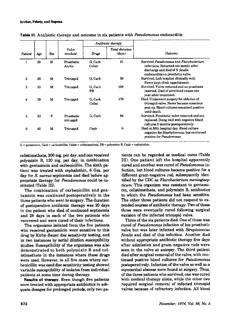
Table 111. Antibiotic therapy and outcome in six patients with Pseudomonas endocarditis
Antibiotic therapy
Patient Age Sex Valve
involved Drugs Total duration
hiuys) Outcome
1 29 M Proethetic G, Garb 21 Survived Pseudomonas and Flav~bacterium Aortic Colist infections. Returned one month after
discharge and died of S. fecalia endocarditis on prosthetic valve.
2 39 M Tricuspid G, Carb 39 Survived. Left hospital clinically well. Never kept clinic appointment.
3 23 M Tricuspid G, Carb 158 Survived. Valve removed and no prosthesis PB inserted Died of unrelated causes one
year after treatment. 4 39 M Tricuspid G, Carb 178 Died Underwent surgery for ablation of
Colist tricuspid valve. Never became conscious post-op. Blood cultures remained positive until death.
5 33 F Prosthetic G, Carb 94 Survived Prosthetic valve removed and not tricuspid replaced. Doing well with negative blood
cultures 3 months postoperatively. 6 46 M Tricuspid Ceph 6 Died on fifth hospital day. Blood culture
negative for Staphylococcus, but continued positive for Pseudomonas.
G = gentamicin, Carb = carbenicillin, Cdist = colietimethate, PB = polymixin B, Ceph = cephalothin.
colistimethate, 300 mg. per day. and one received polymixin B, 120 mg. per day, in combination with gentamicin and carbenicillin. The sixth pa- tient was treated with cephalothin, 8 Gm. per day for S. aureus septicemia and died before ap- propriate therapy for Pseudomonas could be in- stituted (Table III).
The combination of carbenicillin and gen- tamicin was continued postoperatively in the three patients who went to surgery. The duration of postoperative antibiotic therapy was 20 days in the patient who died of continued septicemia and 28 days in each of the two patients who recovered and were cured of their infections.
The organisms isolated from the five patients who received gentamicin were sensitive to this drug by Kirby-Bauer disc sensitivity testing, and in two instances by serial dilution susceptibility studies. Susceptibility of the organisms was also demonstrated to both polymixin B and col- istimethate in the instances where these drugs were used. However, in all five cases where car- benicillin was used disc sensitivity testing showed variable susceptibility of isolates from individual patients at some time during therapy.
Results of therapy. Even though five patients were treated with appropriate antibiotics in ade- quate dosages for prolonged periods, only two pa-
tients can be regarded as medical cures (Table III). One patient left the hospital apparently cured and another was cured of Pseudomonas in- fection, but blood cultures became positive for a different gram-negative rod, subsequently iden- tified by the CDC as Fluvobacterium meningkep- ticum. This organism was resistant to gentami- tin, colistimethate, and polymixin B, antibiotics to which the Pseudomonas had been sensitive. The other three patients did not respond to ex- tended courses of antibiotic therapy. Two of these three were eventually cured following surgical excision of the infected tricuspid valve.
Three of the six patients died One of these was cured of Pseudomonas infection of his prosthetic valve but was later infected with Streptococcus fecalis and died of this infection. Another died without appropriate antibiotic therapy five days after admission and gram-neg.ative rods were seen in the valve at autopsy. The third patient died after surgical removal of the valve, with con- tinued positive blood cultures for Pseudomonas postoperatively. Infection of the valve as well as a myocardial abscess were found at surgery. Thus, of the three patients who survived, one was cured with medical therapy alone, while the other two required surgical removal of infected tricuspid valves because of refractory infection. All blood
572 November, 1974, Vol. 88, No. 5

P. aeruginosa endocarditis
Table IV. Pathologic findings on valves at surgery or autopsy in six patients with Pseudomonas endocarditis
Patient Valve involved Microscopic
-. Culture
Prosthetic aortic
Tricuspid Tricuspid
4 Tricuspid
5
6
Prosthetic tricuspid
Tricuspid
Vegetations on Gram-positive struts cocci
No specimen No specimen Valve friable Chronic intlammation
and thickened No organisms seen
Leaflets sclerotic and thickened with firm vegetations
Vegetations on struts
Acute valvulitis
Covered with fibrinous exudate. No organisms seen
Not done
Necrotizing arteritis and myocoarditis; gram-negative rods.
S. fecalis --
No specimen Valve negative, but left
atria1 blood culture positive for P. aeruginosa
P. aeruginosa
P. aeruginosa
Not done
cultures remained negative after surgery in the latter two patients who were continued on anti- biotics. These two patients were free from infec- tion, had minimal signs of congestive heart failure, and had returned to work when seen three months postoperatively. They have been lost to follow-up since that time.
The mean duration of hospitalization was 3.8 months, with the shortest seven days and the longest eight months. None of the three patients who survived has returned for more than one out-patient follow-up visit.
Pathologic data. There were five patients from whom tissue from the involved valve was ob- tained for examination (Table IV). P aeruginoscc was cultured from two patients, and gram-nega- tive rods were seen in the valve of a third patient whose postmortem heart blood culture grew the organism. A fourth patient presumably had his valve sterilized of Pseudomonas by antibiotic therapy, and died of endocarditis due to S. fecalis. A fifth patient ceased having positive blood cul- tures after the removal of a chronically inflamed valve, although the section of valve cultured was sterile.
Illustrative cases
Patient No. 1, a !29-year-old male, was a known heroin addict who was admitted to the Wayne County General Hospital on Oct. 30, 1971. A di- agnosis of acute Staphylococcal endocarditis of the aortic valve was made and antibiotic therapy was instituted. A ruptured aortic valve cusp ne- cessitated urgent replacement of the valve with a
Starr-Edwards prosthesis. It was strongly sus- pected that he continued the intravenous use of narcotics while in the hospital. He was dis- charged in Jan. 1972. On April 15, 1972, he re- turned with fever, chills, and night sweats, and admitted to continued heroin abuse. He had loud systolic murmurs over his entire precordium, splenomegaly, splinter hemorrhages, and gross hematuria. Blood cultures were positive for P. aeruginosa, and therapy was begun with gen- tamicin and carbenicillin. Eventually, colis- timethate was added, but he continued to be feb- rile with positive blood cultures. He had an inter- mittent diastolic murmur that changed with respiration and bounding jugular venous pulses, and it was uncertain whether his endocarditis in- volved the tricuspid valve, the aortic prosthesis, or both. Cardiac catheterization with cinean- giography was performed, and revealed no dys- function of the tricuspid valve. Quantitative blood cultures obtained from both the right and left side of the heart at the time of catheteriza- tion suggested that the prosthetic valve was the focus of infection. There were 35 colony-forming units per milliliter of blood in the ascending aor- ta, but only 6 per milliliter in right ventricular blood. The organism isolated during this pro- cedure was found later not to be the original I? aeruginosa, and was identified by the CDC as F. meningkepticum, resistant to all antibiotics but clindamycin, rifampicin, and vancomycin. He refused surgical replacement of his prosthetic valve and therapy with these three antibiotics was begun. After three months of treatment, his
American Heart Journal 573

Archer, Fekety, and Supems
blood cultures became negative and he was dis- charged clinically well on Sept. 9, 1972. He re- turned on Oct. 21,1972, with blood cultures posi- tive for S. fecalis and died soon thereafter of cerebral emboli. Cultures of posthetic valve vegetations at autopsy were positive only for S. fecalis. The tricuspid valve appeared normal.
Comment. This remarkable patient had en- docarditis with four different pathogens in ten months. He survived the first three and died of the fourth, caused by S. fecalis. The last three in- fections were all on his prosthetic valve. This il- lustrates the futility of inserting prosthetic valves into drug addicts who are continuing to use intravenous narcotics, and also suggests the usefulness of quantitative blood cultures in con- junction with cardiac catheterization as a means of localizing the infected valve. This is of particu- lar importance with Pseudomonas endocarditis because the infection is often refractory to anti- biotic therapy, and surgical removal of the valve may be the only means of eradicating the site of infection. Since addicts can infect one or several valves not known to have been previously de- formed, clinical localization prior to surgery is often difficult.
Patient No. 2, a 36year-old male, was a chronic narcotics abuser, including intravenous injection of heroin, paregoric, and cocaine. Two months prior to admission he noted the onset of fever, night sweats, bilateral pleuritic chest pain, anorexia, fatigue, and dyspnea. He presented on April 19, 1972, with a temperature of 103” F., tachypnea, disabling chest pain, and a 20-pound weight loss. Physical examination revealed a characteristic murmur of tricuspid insufficiency, splenomegaly, and scars of intravenous drug ad- ministration on his forearms. Blood gases revealed severe hypoxemia and a chest x-ray showed multiple scattered infiltrates. Multiple blood cultures grew P. ueruginosu, and therapy was begun with intravenous carbenicillin and gentamicin. The presumptive diagnosis was tri- cuspid valvulitis and septic pulmonary emboliza- tion. Five days after therapy was begun he had sudden severe chest pain, dyspnea, and a pleural rub. He gradually improved and became afebrile on May 3,1972. Blood cultures on four occasions during therapy were negative. Infiltrates on the chest x-ray cavitated and slowly resolved. Anti- biotics were discontinued on May 30, 1972, after six weeks of treatment, and the patient was clini-
tally well. Two of six blood cultures taken when antibiotics were discontinued were positive for Pseudomonas, but seven subsequent blood cul- tures obtained over the next-week were negative. He was discharged on June 13,1972. He was not suspected of using narcotics while in the hospital. He never kept follow-up appointments. One of his friends reported to us that he was alive in early 1973, but his medical status is otherwise unknown.
Comment. This patient probably represents a cure with antibiotics alone. It is possible that his two positive blood cultures after treatment were the result of surreptitious intravenous drug use.
Patient No. 3, a 23-year-old man, was an addict who admitted to almost daily intravenous injec- tions. He presented on Nov. 17,1970, with a one- week history of hemoptysis, malaise, fever, chills, anorexia, and left-sided pleuritic chest pain. There were &es, decreased breath sounds, and a friction rub at the left base. Thrombophlebitis and cellulitis of the right arm and obvious needle marks were present. Splenomegaly, splinter hemorrhages, petechiae, hematuria, and cardiac murmurs were not found. Sputum culture grew Diplococcus pneumoniue, and chest x-ray showed a left lower lobe infiltrate with effusion. Cultures from the right arm and blood grew Group A beta- hemolytic Streptococci. Therapy was begun with intravenous penicillin, four million units per day. Blood cultures repeated on Nov. 22 were reported on Nov. 24 as growing I? ueruginosu in all four bottles, and intravenous carbenicillin was added to penicillin. He became afebrile and he felt well. On Dec. 5, after eleven days of carbenicillin and eighteen days of intravenous penicillin, all anti- biotics were discontinued. A cardiac focus of Pseudomonas septicemia was not suspected. Ad- ditional blood cultures were obtained on Dec. 8, but he was discharged, receiving oral phenox- ymethylpenicillin. On Dec. 16, one of two blood cultures was growing I? ueruginosu, but attempts to recall him for further treatment were unsuc- cessful.
On Feb. 24, 1971, he presented to the hospital with several weeks of weakness, shortness of breath, chills, fever, pleuritic chest pain, and hemoptysis. On physical examination he was feb- rile and tachypneic with r&lea and a pleural rub at the right base. A loud murmur characteristic of tricuspid insufficiency was heard at the lower sternal border. Jugular veins were distended and
574 November, 1974, Vol. 88, No. 5

P. aeruginosa endocarditis
bounding and splenomegaly was present, but there were no splinters or petechiae. Chest x-ray revealed patchy bilateral infiltrates and a cavity in the right midlung field. All eight blood cultures from admission grew I? aeruginosa, and therapy was begun with intravenous cephalothin and in- tramuscular gentamicin. He continued to be feb- rile, blood cultures remained positive, and pleuritic chest pain became more severe. Car- benicillin was substituted for cephalothin when the nature of the organism was known, but car- benicillin and gentamicin, carbenicillin alone, and carbenicillin with gentamicin and polymixin B failed to decrease symptoms or sterilize the blood. After four months of failure of intensive medical therapy, he was taken to the operating room on June 23, 1971. A very friable and thickened tricuspid valve was removed, and no prosthesis was inserted. Culture of the left atria1 blood at surgery yielded P. aeruginosa, but the portion of valve cultured was sterile. He did well postoperatively, with no recurrence of chest pain, and with increased appetite, weight gain, and negative blood cultures. He was discharged and was doing well one month later when he re- turned for his first follow-up visit. He was lost to follow-up for nine months and died on July 22, 1972, of a gunshot wound to the abdomen.
Comment. One possible explanation for failure of therapy in this man was late treatment because of the failure to diagnose endocarditis when it first presented. The bacteremia was not felt to originate from the heart, in spite of lack of good evidence to support other sites, and he was treated with only a short course of carbenicillin. By the time definitive therapy was instituted, en- docarditis was well established. Since four months of bactericidal antibiotic therapy did not cure the infection, surgery was required and ap- peared curative.
Discussion
Bacterial endocarditis caused by P. aeruginosa was an extremely rare disease prior to 1960, with only 32 reported cases. 4*5 These consisted of nine patients who had infection of presumably normal valves, eleven patients with previous valve damage secondary to rheumatic heart disease, and seven patients with infection of an intracar- diac foreign body following cardiac surgery. Since 1960, wht spectrum of disease has changed so that virtually all of the cases have been seen
either following cardiac surge@+ or in drug ad- dicts. The organism is now recognized as a com- mon contaminant of hospital equipment and a potential source of infection of intracardiac foreign bodies.‘O However, only recently has Pseudomonas endocarditis in the narcotic user reached serious proportions. Reyes and co- workersI’ recently reported 21 cases of Pseudomonus endocarditis in heroin addicts seen at the Detroit General Hospital. Our findings confirm teeirs and support their conclusions. Their cases, together with our six patients, are in contrast to only 13 cases of Pseudomonas en- docarditis out of a total of 273 cases of endocar- ditis in drug users gleaned from the literature.4a 12-18 All of our patients, and those patients re- ported by Reyes, have been seen since 1969, and are drawn from the same geographic area.
Several features in our patients are similar to those reported by Reyes. The tricuspid valve was involved in five of six 033 per cent) of our patients and in 16 of 21 (76 per cent) of Reyes’ patients. The increased frequency of tricuspid endocar- ditis among addicts as compared to specifically matched controls among the nonaddict endocar- ditis population has been noted by Cherubin and co-workers.‘” However, they found no organism predilection for a particular valve in addicts as compared with control subjects.
Two (33 per cent) of our patients were cured by medical therapy alone, which is similar to Reyes’ experience (24 per cent). All of the patients in our series (one) and five out of six patients in Reyes’ series with infection of the left side of the heart died. Seven out of 21 (33 per cent of the com- bined series of tricuspid valve infections re- sponded to intensive antibiotic treatment. Thus, although infection of the tricuspid valve seemed to offer a better prognosis than left-sided en- docarditis, two-thirds of the patients with tri- cuspid endocarditis did not respond to antibiotic therapy alone.
Surgery was eventually required for removal of the infected focus in three of our six patients (50 per cent), and in 12 out of 21 of Reyes’ pa- tients (57 per cent). All had failed to respond to prolonged courses of antibiotics. Three of Reyes’ patients underwent surgery with insertion of a prosthesis, and another had an attempted exci- sion of a left atria1 focus of infection. These four patients died of continued infection. All three of our surgical patients and the remaining eight of
American Heart Journal 575

Archer, Fekety, and Supena
Reyes’ patients had removal of the tricuspid valve without insertion of a prosthesis. Eight (73 per cent) of these patients survived and were free of infection when last seen. We treated our pa- tients for one month with appropriate antibiotics postoperatively.
There is little to offer the addict with left-sided Pseudomonas endocarditis who does not respond to medical therapy. Reinfection of inserted prostheses has been very frequent am ng
i drug
users, as observed in Reyes’ patients an in ours. However, total ablation of the tricuspid valve is well tolerated and offers an excellent opportunity for salvaging the patient with an infected tri- cuspid valve who has not responded to conven- tional therapy.l’
Antibiotic therapy with a combination of gen- tamicin and carbenicillin was used in five out of six (83 per cent) of our patients and in 17 out of 21 (81 per cent) of Reyes’ patients. In neither series could the success of medical therapy alone be predicted on the basis of drug dosage, duration of therapy, or in vitro antibiotic susceptibility. In our study, variable resistance and sensitivity of the same organism to carbenicillin, as tested by the disc sensitivity method, was noted in all pa- tients. There is evidence that some organisms found resistant by use of the standard 50 microgram carbenicillin disc currently recom- mended by the Food and Drug Administration (FDA) may be found to be susceptible by the serial broth dilution method (susceptibility defined as a minimal inhibitory concentration of equal to or less than 100 micrograms per milli- liter). The use of a 100 microgram disc is reported to markedly reduce the discrepancies between the two susceptibility testing methods.le In Reyes’ series, organisms from five out of six pa- tients who responded to medical therapy alone had a minimal inhibitory concentration of 100 micrograms per milliliter or greater as deter- mined by the serial broth dilution method. These organisms would have been determined resistant by the disc diffusion method, using even the 100 microgram disc. Thus, the correlation between in vitro testing and in the vivo efficacy of car- benicillin in treating Pseudomonas endocarditis is unclear. There may be in vivo synergism be- tween carbenicillin and gentamicin to explain the apparent efficacy of this combination in treating some of the patients.ls
The clinical presentation, physical findings,
and course of the illnesses were similar in the majority of the cases in both series. The typical patient was a black.male heroin addict under for- ty years of age who presented with a history of several weeks or months of malaise, anorexia, and weight loss. The symptoms were more sug- gestive of subacute than acute bacterial endocar- ditis. Chest pain or chills were usually responsi- ble for the patient seeking medical help. On physical examination, fever was invariably present but vital signs were otherwise normal. A murmur of tricuspid insufficiency was usually present, but meticulous auscultation was re- quired because these murmurs were soft and changeable. Banks, Fletcher, and Nayabl’ have recently reported on 50 patients with heroin as- sociated endocarditis, 42 of whom had tricuspid involvement. In 16 of these 42 patients with tri- cuspid regurgitation, the tricuspid murmur was not present or not recognized at the time of ad- mission. In two patients, an atria1 diastolic gallop, a ventricular diastolic gallop, or a systolic click, heard over the left lower sternal border and external jugular vein, preceded the late ap- pearance of typical murmurs of tricuspid insuffi- ciency.
Cardiac catheterization may be necessary to delineate valvular dysfunction in some patients with minimal auscultatory signs, particularly if cardiac surgery is contemplated. If catheterixa- tion is performed, quantitative blood cultures from both the right and left sides of the heart may be an additional method of localizing the site of infection to a particular valve. This is ex- emplified by one of our patients (Patient No. 1) in whom quantitative blood cultures suggested that the prosthetic aortic valve and not the tricuspid valve was infected. This was confirmed at autop- W*
Splenomegaly was common in our series (Table I) but uncommon in Reyes’ series. Peripheral manifestations of endocarditis, such as pete- chiae, Roth’s spots, and hematuria were rarely seen. Scars produced by multiple intravenous in- fections were usually found when diligently sought. The white blood cell count was not often helpful. Although it was often above 10,000 per cubic millimeter, there was frequently no in- crease in the percentage of neutrophils or band forms. Chest x-rays showed multiple infiltrates, frequently but not always cavitary, which repre- sented septic emboli. These were not present
576 November, 1974, Vol. 88, No. 5

P. aeruginosa endocarditk
when the left side of the heart alone was in- volved. There was usually no radiologic evidence of cardiomegaly or congestive heart failure. If the patient had received no prior antibiotic therapy, all blood cultures usually grew the organism. Cultures could remain positive for pro- longed periods during treatment before eventual bacteriologic conversion. It was remarkable how well the patient felt while continuously bac- teremic. Variations in the typical presentation of Pseudomonas endocarditis occurred where there was a mixed infection with S. aureus. In such cases, the patient often presented with fulminant acute bacterial endocarditis. There was nothing about the clinical presentation of Pseudomonas endocarditis that distinguished it from endocar- ditis due to other pathogens of intermediate virulence. However, it is important to recognize that the recovery of P. aeruginosa from blood cul- tures in a drug user who does not appear very ill might represent endocarditis.
Two of our patients had infection of prosthetic valves. These valves had been inserted because of valve destruction secondary to bacterial endocar- ditis. Presumably, the organisms were originally introduced with intravenous narcotic injection. The subsequent infection of the prosthesis was felt likely to be due to organisms injected during intravenous drug use and not to contamination at the time of surgery, because blood cultures became positive four and six months after sur- gery and both patients admitted heavy use of drugs in the interim.
The pathogenesis of Pseudomonas endocarditis remains obscure. Infection of heart valves with gram-negative organisms is unusual and infec- tion of undamaged valves by them is exceedingly rare. The organisms are undoubtedly injected in- travenously as contaminants of the narcotic preparation, syringe, or diluent, although at- tempts to recover the organism from these sources have not been very rewarding.” P. ueruginosu is a hardy organism, surviving in such common moist locations as sinks, drains, taps, and soaps.lO This is particularly true in hospitals, where the incidence of nosocomial infections with this organism is high. The increasing inci- dence of endocarditis with this organism suggests that medical facilities may be a source of the im- plicated syringes, fillers, or diluents. However, frequent or continuous bacteremia with Pseudomonas in severely burned patients is com-
mon but rarely results in endocarditis.20*21 Thus, there seems to be some predisposing factor in narcotic addicts which makes them more suscep- tible to endocarditis with this organism. Such things as changes in hemodynamics in the right side of the heart resulting in turbulent flow or the quantity and frequency of injection of the bacteria are possible contributory factors.
Both Angrist and Oka2’ and Durack and Beeson have proposed that infection of heart valves with organisms of low virulence is depen- dent on previous disruption of the endothelial surface of the valve. A sterile vegetation or roughened valve surface is produced and is then colonized by bacteria at the time of bacteremia. In the narcotic addict there are several possible mechanisms of such valve damage. Some materials used to cut the narcotic, such as talc or starch, are particulate and could cause damage to the surface of the tricuspid valve. Another possibility is that bacteremia with more virulent organisms could damage the valve. Such organ- isms may co-exist with Pseudomonas in a polymicrobial infection. If a virulent organism, such as S. uureus, is not present in large numbers it may be supplanted on the valve surface by Pseudomonas. This is particularly true if anti- biotics are administered by the patient or a local physician, suppressing the original pathogen and allowing Pseudomonas to flourish. Rabin and co- workersz3 reported three cases of Pseudomonas endocarditis in burned patients following Pseudomonas septicemia. In all three cases, the Pseudomonas septicemia had followed prolonged Stuphylococcal septicemia, or was coincident with it. In one case, both P. aeruginosu and S. uureus were recovered from an aortic vegetation at postmortem examination. In four of the 21 narcotics users in Reyes’ series, both P, aeruginosa and S. aureus were recovered from the blood at some time during hospitalization. In three of our patients, organisms known to cause damage to normal heart valves were isolated from the blood on admission to t,he hospital. Group A beta-hemolytic Streptococcus, S. fecalis, and S. aureus were grown from the blood one to thirteen days before the first culture became positive for Pseudomonas. Since two other pa- tients in our series had their infection on pros- thetic valves, there remains only one patient with normal valves who had Pseudomonas as the sole organism isolated from blood cultures.
American Heart Journal 577

Archer, F&&y, and Supena
The financial drain on society that the addict represents is exemplified by our patients. The stubborn and refractory illness leading to pro- longed hospitalization in five of the six patients produced an average hospital bill of $31,000, with a range of $7,000 to $42,000.
Summary
Six intravenous heroin users with I? endocar- &tk were seen between 1970 and 1972. Five out of six (83 per cent) had tricuspid valvulitis. Fever and a significant murmur were found in all cases, a subacute presentation and splenomegaly were found in 83 per cent, and chest pain with cavitat- ing infiltrates on x-ray was observed in 67 per cent. Three patients died of infection. Three pa- tients survived; one was cured with medical therapy alone, and two required surgical removal of the infected valve as well. The success of surgi- cal ablation of the tricuspid valve in these pa- tients is encouraging, particularly in view of their poor response before surgery to combina- tions of antibiotics with proved in vitro efficacy. Prior bacteremia with more virulent organisms may be important in the pathogenesis of en- docarditis with Pseudomonas.
REFERENCES
1.
2.
3.
4.
5.
6.
7.
Bailey, W. R., and Scott, E. G.: Diagnostic microbiology, Ed. 3, St. Louis, 1970, The C. V. Mosby Company, p. 161. Bauer, A. W., Kirby, W. M. M., Sherris, J. C., and Turck, M.: Antibiotic susceptibility testing by a standardized single disc method, Am. J. Clin. Pathol. 45493, 1966. Harwick, H. J., Weiss, P., and Fekety, F. R.: Application of microtitration techniques to bacteriostatic and bac- tericidal antibiotic susceptibility testing, J. Lab. Clin. Med. 72:511, 1968. Saroff, A. L., Armstrong, D., and Johnson, W. D.: Pseudomonas endocarditis, Am. J. Cardiol. 32:234, 1973. Hills, H. F.: A case of puerperal septicemia due to Pseudomonas pyocyanea with a short review of the liter- ature, J. Ob&t. Gynaecol. Br. Emp. 55860, 1949. Littlefield. J. B.. Muller. W. H.. and Dammann, J. F.: Successful treatment of Pseudomonas oerugino& sep- ticemia following total aortic valve replacement, Cir- culation (Suppl. 1) 315, 1965. Yeh, T. J., Anabtaw, I. N. Cornet& V. E., White, A.,
8.
9.
10.
11.
12.
13.
14.
15.
16.
17.
18.
19.
20.
21.
22.
23.
24.
25.
Stern, W. H., and Ellison, R. G.: Bacterial endocarditis following open-heart surgery, Ann. Thorac. Surg. 329, 1967. Rror, W. B.: Infection following open-heart surgery with special reference to the role of prophylactic anti- biotics, J. Thorac. Cardiovasc. Surg. 53:371, 1967. Block, P. C., DeSanctis, R. W. Weinberg, A. N., and Austen, W. G.: Prosthetic valve endocarditis, J. Thorac. Cardiovasc. Surg. 60:540,1970. Whitby, J. L., and Rampling, A.: Pseudomonas oeruginosa contamination in domestic and hospital en- vironments, Lancet 1:15, 1972. Reyes, M. P., Palutke, W. A., Wylin, R. F., andLemer, A. M.: Pseudomonas endocarditis in the Detroit Medical Center, 1969-1972, Medicine 52:173, 1973. Ramsey, R. G., Gunnar, P. M., and Tobin, J. R.: En- docarditis in the drug addict, Am. J. Cardiol. 25:608, 1970. Dreyer, N. P., and Fields, B. N.: Heroin-associated infec- tive endocarditis, a report of 28 cases, Ann. Intern. Med. 78:699, 1973. - Banks, T., Fletcher R., and Nayab, A.: Infective en- docarditis in heroin addicts. Am. J. Med. 55~444, 1973. Menda, K. B., and Gorbach; S. L: Favorable experience with bacterial endocarditis in heroin addicts, Ann. In- tern. Med. 78:25, 1973. Cherubin, C. E., Baden, M., Kavalier, F., Lerner, S., and Cline, W.: Infective endocarditis in narcotic addicts, Ann. Intern. Med. 69:1091, 1968. Graham, D. Y., Reul, G. J., Martin, R., Morton, J., and Kennedy, J. H.: Infective endocarditis in drug addicts: experiences with medical and surgical treatment, Cir- culation 47 and 48@uppl. IIIl:III, 1973. Carrathers, M. M., and Kanokvechayant, R.: Pseu- domonas oeruginosa endocarditis: report of a case, with review of the literature, Am. J. Med. 55:811,1973. Arbulu, A., Thomas, N. W., and Wilson, R. F.: Valvulec- tomy without prosthetic replacement, a livesaving operation for tricuspid Pseudomonas endocarditis, J. Thorac. Cardiovasc. Surg. 64~103, 1972. Washington, J. A., Hall, M. M., Fausch, C. J., and Brackin, K. L.: In vitro activity of carbenicillin against Pseudomonas using the disc diffusion method, Mayo Clin. Proc. 45:718, 1973. Andriole, V. T.: Synergy of carbenicillin and gentamicin in experimental infection with Pseudomonas, J. Infect. Dis. 124(Suppl.):S46, 1971. Stone, H. H.: A review of Pseudomonas sepsis in ther- mal burns. Verdoglobin determination of gentamicin therapy. Ann. Surg. 163:297, 1966. Rabin, E., Garber, C. D., Vogel, E. H., Finkelstein, R. A., and Tumbusch, W. A.: Fatal Pseudomonas infection in burned patients, N. Engl. J. Med. 265:1225,1961. Angrist, A., and Oka, M.: Pathogenesis of bacterial en- docarditis, J. A. M. A. 183:249,1963. Durack, D. T., and Beeson, P. B.: Experimental bacte- rial endocarditis. I. Colonization of sterile vegetation, Br. J. Exp. Pathol. 53:44, 1972.
578 November, 1974, Vol. 88, No. 5