Endocarditis review
75
Endocarditis Endocarditis
-
Upload
scott-homann -
Category
Documents
-
view
59 -
download
0
Transcript of Endocarditis review
EndocarditisEndocarditis

EpidemiologyEpidemiology
An estimated 10,000 to 15,000 new cases of infective endocarditis (IE) are diagnosed in the United States each year. Men predominate in most case series, with male-to-female ratios ranging from 2:1 to 9:1.
The importance of underlying heart disease in children with IE varies with age: 50 to 70 percent of children less than 2 years old have no obvious underlying cardiac disease , while most older children have coexistent congenital heart disease.
A number of factors predispose to the development of infective endocarditis. These include IVDA, prosthetic heart valves, and structural heart disease.

Newer trends show endocarditis has increasingly become a disease of the elderly. This trend is probably due to the decline in the incidence and importance of rheumatic heart disease as a risk factor for IE and the increasing proportion of elderly subjects in the general population.
Also the proportion of cases that present with an indolent course and the proportion due to streptococci has declined recently, while simultaneous increases have occurred in the proportion of cases due to Staphylococcus aureus, fungi, and gram negative bacilli.
The rising incidence of injection drug use (IVDA) has had an impact on the overall incidence of endocarditis, as well as it’s geographic distribution. Drug addiction as a cause of IE is most prominent in patients less than 40 years old.

Intravenous Drug Abuse (IVDA)Intravenous Drug Abuse (IVDA)
In addition to the injection itself, specific drugs of abuse may also be risk factors for the development of endocarditis which often involves the right side of the heart. One large study found that those who used cocaine were significantly more likely to have endocarditis than those using other drugs. Ann Int Med 1987. 106:833.
The most significant risk factor for right-sided IE is IVDA; however, left-sided disease may be more common in addicts.
The most common infecting organism is S. aureus, particularly in right-sided infection with prevalence up to 82 percent. The vegetations are usually > or =1 cm in diameter, and there is a marked increase in mortality associated with vegetations greater than 2 cm (33 versus 1.3 percent for smaller vegetations). Ann Int Med 1992.
117;560.

Prosthetic Heart ValveProsthetic Heart Valve
Prosthetic valve endocarditis develops in 1 to 4 percent of valve recipients during the first year following valve replacement, and in approximately 0.5-1 percent per year thereafter.
The type of prosthetic valve (bio vs mech) does not have an impact on the development of IE.

Structural Heart DiseaseStructural Heart DiseaseApproximately 3/4 of all patients with IE have a preexisting structural cardiac abnormality at the time that endocarditis begins.
The risk of IE in patients with mitral valve prolapse and associated regurgitation is estimated to be 5 to 8 times higher than that in the normal population. However, MVP without MR is a more common abnormality that is associated with only a small risk of endocarditis. NEJM 1982. 307;776.
The most common predisposing congenital heart lesions are bicuspid aortic valves, patent ductus arteriosus, ventricular septal defect, coarctation of the aorta, and tetralogy of Fallot.
Prevalence of rheumatic valve disease is on the decline.

The risk of IE appears to be dependent upon the specific congenital or acquired cardiac lesion. One study, for example, evaluated 2401 patients with congenital heart lesions who were followed prospectively for 40,000 days. Circulation 1993. 87; 121
• The overall incidence of endocarditis was 135 cases/100,000 person years. • The highest rates of endocarditis occurred in patients with aortic stenosis (271)and ventricular septal defects (145 cases/100,000 person yrs). •Among patients with AS, the higher the peak gradient, the higher the risk of developing endocarditis.• The risk of IE with AS was approximately twice as high as with aortic regurgitation. • The lowest rate of endocarditis occurred with pulmonic stenosis.•The risk of IE also appears to be very low in adults with congenital heart disease, inherently normal pulmonic and tricuspid valves, and pulmonary or tricuspid regurgitation due to pulmonary hypertension.

Other Risk factorsOther Risk factorsA prior history of endocarditis is an important predisposing cause for IE. Recurrent endocarditis occurred in 4.5 percent of one large cohort of non-addicts who survived their initial episode. Ann Int Med 1992 117;567.
A number of cases of IE have been reported in patients with HIV infection with some valves have been infected with unusual organisms such as Salmonella and Listeria. It has been suggested that HIV infection is an independent risk factor for IE in injection drug abusers but, this has not been confirmed in other reports.
Other less common predisposing factors for IE are pregnancy, arteriovenous fistulas used for hemodialysis, central venous and pulmonary artery catheters, peritoneovenous shunts for the control of intractable ascites and ventriculoatrial shunts for the management of hydrocephalus.
Patients with ulcerative lesions of the colon due to carcinoma or inflammatory bowel disease have a poorly understood predilection to develop endocarditis secondary to Streptococcus bovis.
IE has also been reported in patients undergoing liver, heart, and heart-lung transplantation.
PathogenesisPathogenesisThe endothelial lining of the heart and its valves is normally resistant to infection with bacteria and fungi.
Although a few highly virulent organisms such as Staphylococcus aureus are capable of infecting normal human heart valves, the initial step in the establishment of a vegetation is injury to the endocardium, followed by focal adherence of platelets and fibrin.
These injuries can probably occur naturally in bacteremic humans with congenital or acquired cardiac lesions that induce continuous endocardial trauma via regurgitant flow or high pressure jets of blood through stenotic lesions.
The initially sterile platelet-fibrin nidus then becomes infected by microorganisms circulating in the bloodstream, following colonization then microbial growth results in the secondary accumulation of more platelets and fibrin.
Microbial adherence is a crucial early event in the pathogenesis of endocarditis. As an example, bacteria that are typically found in patients with endocarditis, such as Staphylococcus aureus, viridans streptococci, enterococci and P. aeruginosa, adhere more avidly to excised canine heart valves in vitro than species which rarely cause endocarditis (such as Escherichia coli).
Dextran in the cell wall of gram positive organisms is one factor.
Degree of intrinsic binding affinity to fibronectin is important in the virulence of S. aureus species.

Components found in damaged endothelium and/or platelet-fibrin aggregates that may mediate adherence include fibrinogen, laminin, and type 4 collagen.
Animals treated with aspirin had reduction in the weight of valvular vegetations, vegetation growth, density of bacteria in the kidneys and in vegetations, and a decrease in embolic events compared to controls.
This interference with platelet-fibrin aggregates suggests that addition of low dose aspirin can reduce risk of embolization during antibiotic treatment, or risk of IE recurrence.
A small clinical study of 9 pts suggested ASA to be protective. J Int Med 1992; 231:543
However a more recent randomized trial showed despite promising experimental data, there was no clinical benefit to ASA.
115 pts in Can&US were doubly blinded to receive ASA or placebo.Followed for 4wks for primary outcome of clinical embolism, secondary outcome of CT shown subclinical CVA, hemorrhage or death.
No sig difference in embolization. (ASA 28% vs placebo 20%)Non-sig trend for combined major&minor hemorrhage in ASA grp.Total of 70% had CT head done, with significant lesions in 39.5% ASA group vs 29.3% in placebo.Total of 42% had TEE, which showed similar rate of vegetation decrease or valvular regurgitation.
JACC 2003; 42:775

Physics of VegetationsPhysics of VegetationsIf nebulized Serratia marcescens, are injected into an air stream passing through an agar-coated Venturi tube, the highest concentration of bacteria is found in the low pressure area immediately distal to the narrowing. Circulation 1963. 27;18
Vegetations in patients with preexisting valvular lesions are usually located on the atrial surface of incompetent atrioventricular valves, or the ventricular surfaces of incompetent semilunar valves.
Patients with ventricular septal defects tend to develop vegetations on the orifice of the defect, on the right ventricular side of the opening, and secondarily on the tricuspid and pulmonic valves.
Vegetations may occasionally localize on the chordae tendineae of the anterior leaflet of the mitral valve in patients with aortic insufficiency.
Patients with mitral regurgitation may develop vegetations (MacCallum's patch) on the wall of the left atrium where the regurgitant jet strikes the atrial wall and results in endocardial thickening.

Clinical SequelaeClinical Sequelae
Vegetations often prevent proper valvular leaflet or cusp coaptation, thereby causing valvular incompetence.
Congestive heart failure may result from worsening valvular incompetence or leaflet perforation secondary to vegetation growth.
Patients with mitral or tricuspid valve vegetations may develop chordal rupture.
Infection may also extend into surrounding structures such as the valve ring, the adjacent myocardium, the cardiac conduction system, or the mitral-aortic intravalvular fibrosa.
Rarely, cavitation of periaortic abscesses occurs into the adjacent aortic wall, resulting in the formation of a diverticulum or aneurysm.
Even more rarely, such aneurysms perforate into surrounding structures, resulting in aortic-atrial or aortic-pericardial fistulae.

Etiology (Native Valve)Etiology (Native Valve)Although any bacteria can cause IE, Streptococci, Enterococci and Staphylococci account for the majority of the cases.
Streptococci
Streptococci account for about 55% of non-IVDA cases.
Viridans streptococci account for ~75% of these cases. Most common of the group are Streptococcus mutans, S.
mitis, S. Milleri.S. Bovis account for ~20%.Other Streptococci are ~5%.Viridans strep are normal pharynx inhabitants and are generally very
susceptible to PCN.80% of S. Bovis cases occurs in elderly (>60yrs). More than a third
have a malignant or pre-malignant colonic lesion.

Enterococci
Cause about 6% of native valve endocarditis and are normal flora of GI tract, anterior urethra and occasionally the mouth.
They are relatively resistant to PCN-G and require the addition of an aminoglycoside to achieve bactericidal effect.
More common among males who develop infection around 60 yrs of age.
Many have history of GU tract manipulation, trauma or disease.Cystoscopy, Foley catheter, prostatectomy.Caesarian section, abortion, pregnancy.

Staphylococci
Staph cause about 30% of native valve IE cases, with S. aureus being 5 to 10 times more frequent than S. epidermidis.
S. Aureus can rapidly attack normal or damaged valves. It has a fulminant course with death from bacteremia within days, or from heart failure within weeks. Abscesses are common at multiple sites.
S. Epidermidis usually infects prosthetic valves.

HACEK organisms
HaemophilusActinobacillusCardiobacteriumEikenellaKingella
They are part of the oropharyngeal flora and produce large vegetations with a subacute presentation.
They are difficult to isolate from blood and grow slowly in blood culture media. Incubation for 7 to 14 days may be required to detect their growth. This delayed growth makes standard antibiotic susceptibility testing difficult.

Other Bacteria
Almost any bacteria can be an occasional cause of acute or subacute endocarditis.
Streptococcus pneumoniaeNeisseria gonorrhoeaeEnteric gram –ve bacilliPseudomonasSalmonellaStreptobacillusSerratia marcescensBacteroidesBrucellaMycobacteriumNeisseria meningitidisListeriaLegionellaCorynebacterium

Fungi
Rarely cause native valve IE. Subacute course with large vegetations and large emboli.
Candida and Aspergillus species can occur in persons with intravascular catheters who are frequently on glucocorticoids, broad spectrum Abx, cytotoxic drugs.
Grave prognosis due to the relatively poor activity of antifungal; agents.
Other Organisms
Rare causes can include Spirochetes, Rickettsiae (Coxiella burnetti), Chlamydiae (C. pneumoniae, C. trachomatis, C. psittaci).

Etiology (IVDA)Etiology (IVDA)
Frequently occurs in young males with the skin being the most common source of organisms. Contamination of drugs is less common.
More than 50% is by S. aureus. ~20% by Streptococci and Enterococci. ~6% Fungi, mainly candida. ~6% gram –ve bacilli, usually Pseudomonas species.
Onset is usually acute, with only 20% of addicts having previously damaged valves.
Half the cases are involving the tricuspid valve. Aorta is involved in 25% of cases. Mitral valve is involved 20% of the time. Multiple valves less than 5%.
In TV endocarditis, murmurs are usually absent and pneumoniae from septic emboli is frequently the presenting picture.

Etiology (Prosthetic Valve)Etiology (Prosthetic Valve)
With the aging population, prosthetic valve endocarditis now accounts for 10 to 20% of cases.
Intravascular sutures, pacemaker wires and Teflon-Silastic tubes are all foci for infection.
Most patients are males over 60 and IE occurs in 1-2% in the first year after surgery , and 0.5% a year thereafter.
AV prosthesis are more likely to be involved than MV, and infection is usually at the suture line.
Early onset (within 60days) is usually as a result of valve contamination or peri-operative bacteremia.

Almost half of early onset, and third of late onset endocarditis are due to Staphylococci. S. epidermidis is more frequently involved than S. aureus.
Streptococci are the most common cause (40%) of late onset endocarditis, but are rare in early onset.

Clinical ManifestationsClinical ManifestationsSymptoms generally start within two weeks of precipitating event. With low pathogenicity organism (ie viridans)the onset is usually slow with low fevers. With fulminant disease (ie S. aureus) the onset is rapid with high fevers.
Fever is almost always present. Arthralgias, myalgias and low back pain are common.
Cardiac murmurs are almost always present, except in acute early IE and in IVDA with TV lesions.
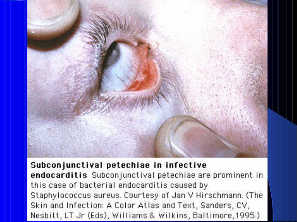
Splenomegaly and petechia are usually found in disease of long duration.
Petechiae are more common on conjunctiva, palate, buccal mucosa and upper extremities.

Splinter hemorrhages are subungual, linear dark red streaks that may appear in IE, but can also result from trauma.
Roth’s spots are oval retinal hemorrhages with a clear pale center. But it can occur in connective tissue diseases and sever anemia.
Osler’s nodes are small tender nodules in the pads of fingers and toes.
Janeway’s lesions are small circular hemorrhagic lesions, that are nodular and occur on palms and soles of feet.
Clubbing has been reported in patients with long standing disease.
Embolic episodes can occur during or after therapy. Large emboli are generaly fungal.

Neurologic manifestations are more common in left sided disease, and in S. aureus infection.
Heart failure can occur long after it’s cure. Causes include valvular damage, myocarditis, abscess formation and coronary artery embolization with infarction.
Conduction defects occur due to septal invasion.
Renal disease exists in most patients due to renal emboli, or immune complex mediated glomerulonephritis.


Diagnostic WorkupDiagnostic Workup
At least three blood cultures should be obtained from separate sites over a time period ranging from a few hours to one to two days depending upon the severity of illness and urgency of the need for treatment.
The additional diagnostic yield of more than three cultures is minimal in patients who have not recently received antimicrobial therapy.

A TTE should initially be obtained in patients with native heart valves, while those with prosthetic valves should undergo TEE.
Detection of a vegetation is a positive test. However, this procedure has relatively low sensitivity in IE. Thus, a negative study does not preclude the diagnosis
TTE should be followed by TEE, which has higher spatial resolution, when there is an intermediate or high suspicion of IE and valvular abnormalities were found on TTE.
In general the sensitivity of TTE in detecting vegetations is ~65-80%. The addition of TEE increases the sensitivity to 95%.
In prosthetic valves TTE is has poor diagnostic value. However, TEE carries a 90% sensitivity in this setting. JACC 1991;18
A meticulous clinical examination should be performed looking for clinical evidence of small and large emboli with special attention to the fundi, conjunctivae, skin, and digits.
In addition, a careful cardiac examination may reveal signs of new regurgitant murmurs and signs of congestive heart failure, and neurologic evaluation may detect evidence of focal neurologic impairment and can be used to document a baseline should such abnormalities appear later.

Additional clues to the presence of IE, such as microscopic hematuria, leukocytosis, and evidence of renal impairment should be sought with laboratory testing.
All patients with suspected IE should have an electrocardiogram to determine whether there is evidence of heart block or a conduction delay and to establish a baseline should such a complication develop later.
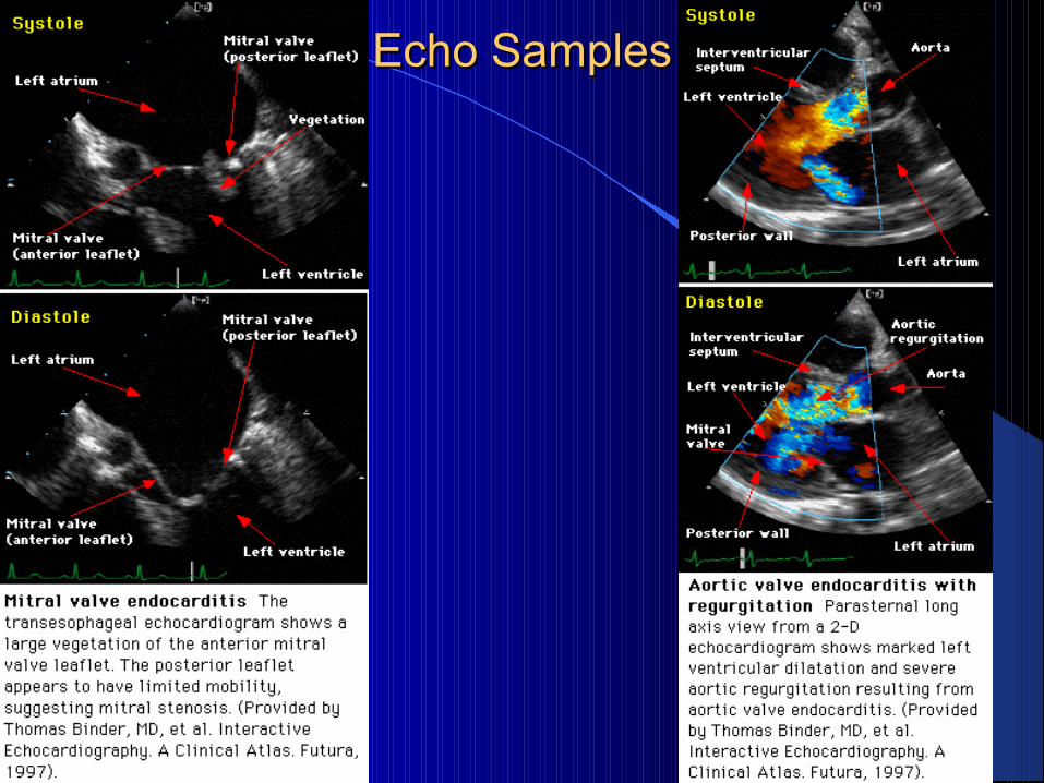
Echo SamplesEcho Samples

Duke ClassificationDuke Classification
In 1994 investigators from Duke University modified the von Reyn criteria to include the role of echocardiography in diagnosis. They also expanded the category of predisposing heart conditions to include injection drug use.
This was later modified further to include the role of TEE as well as the agent of Q fever (Coxiella brunetti).
The Duke classification relies upon major and minor criteria in a manner similar to the Jones criteria for rheumatic fever.

Major Criteria•Isolation of causative organism by two separate blood culture’s at least 12hrs apart.•Endocardial involvement evidence by echo. Oscillating mass, prosthetic valve dehiscence, abscess, new regurgitation.
Minor criteria•Predisposing lesion or IVDA•Fever >38C•Signs of embolization: Janeway lesion, Intracran hem.•Immunologic phenomena: Glomerulonephritis, Oslers nodes, Rheumatoid factor, Roths spots.•Positive blood culture not meeting major criteria.•Echo finding, but not meeting major criteria.
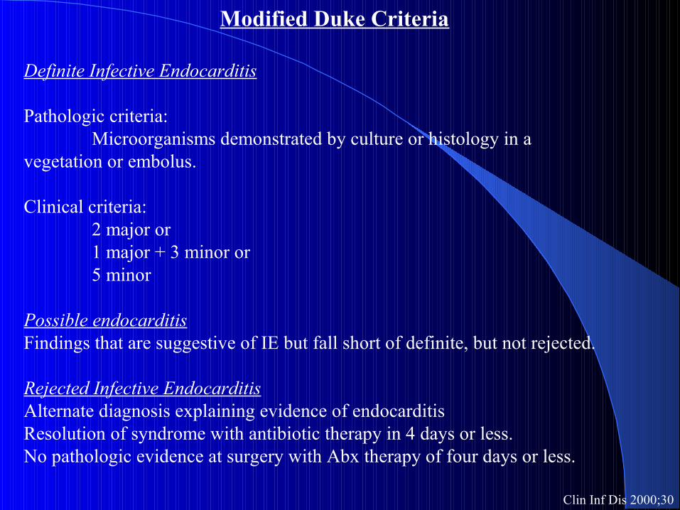
Modified Duke Criteria
Definite Infective Endocarditis
Pathologic criteria:Microorganisms demonstrated by culture or histology in a
vegetation or embolus.
Clinical criteria:2 major or1 major + 3 minor or5 minor
Possible endocarditisFindings that are suggestive of IE but fall short of definite, but not rejected.
Rejected Infective EndocarditisAlternate diagnosis explaining evidence of endocarditisResolution of syndrome with antibiotic therapy in 4 days or less.No pathologic evidence at surgery with Abx therapy of four days or less.
Clin Inf Dis 2000;30
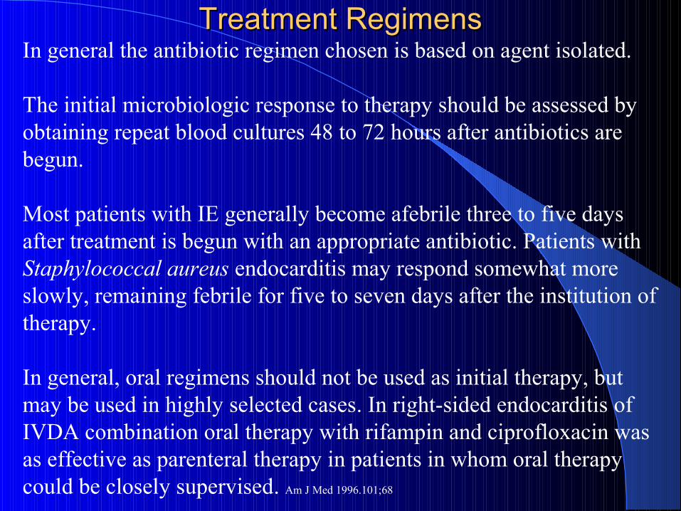
Treatment RegimensTreatment RegimensIn general the antibiotic regimen chosen is based on agent isolated.
The initial microbiologic response to therapy should be assessed by obtaining repeat blood cultures 48 to 72 hours after antibiotics are begun.
Most patients with IE generally become afebrile three to five days after treatment is begun with an appropriate antibiotic. Patients with Staphylococcal aureus endocarditis may respond somewhat more slowly, remaining febrile for five to seven days after the institution of therapy.
In general, oral regimens should not be used as initial therapy, but may be used in highly selected cases. In right-sided endocarditis of IVDA combination oral therapy with rifampin and ciprofloxacin was as effective as parenteral therapy in patients in whom oral therapy could be closely supervised. Am J Med 1996.101;68

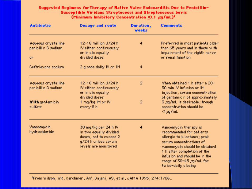
Treatment regimen summaryNative valve
Strep Viridans or Strep Bovis -PCN sensitivePCN-G or Ceftriaxone 4wksIf allergic, Vancomycin 4wks
Strep Viridans or Strep Bovis - PCN resistantPCN 4wks+Gent 2wkVancomycin 4wks
EnterococcusPCN/AMP + Gent 6wksVanco + Gent 6wks
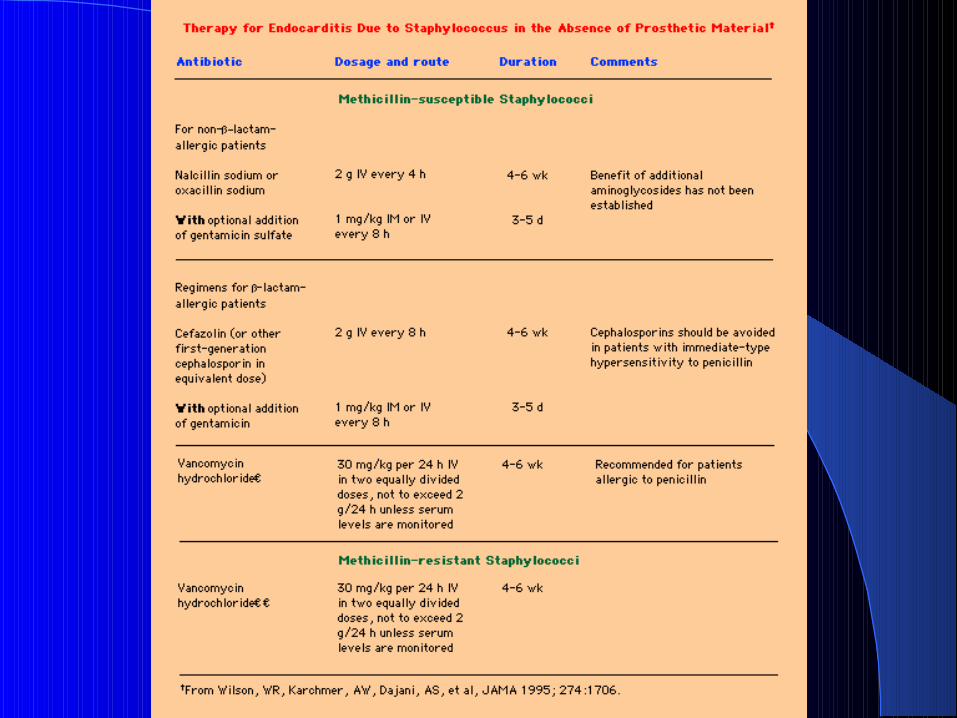
Staph infectionCefazolin 6wks +/- Gent 5daysVanco 6wks
HACEK OrganismsCeftriaxone 4wks

Prosthetic valve
Streptococcus-PCN sensistiveCeftriaxone 6wks + Gent 2wks Vanco 6wks
Streptococcus-PCN resistant StrepVanco 6wks
EnterococciPCN or Amp 6wks + Gent 6wksVanco+Gent 6wks
Methicllin Susceptible StaphNafcillin 6wks + Gent 2wks + Rifampin 6wks
MRSAVanco 8wks + Gent 2wks + rifampin 8wks
HACEKCeftriaxone 6wks
DiptheroidesPCN +Gent 6wksVanco 6wks

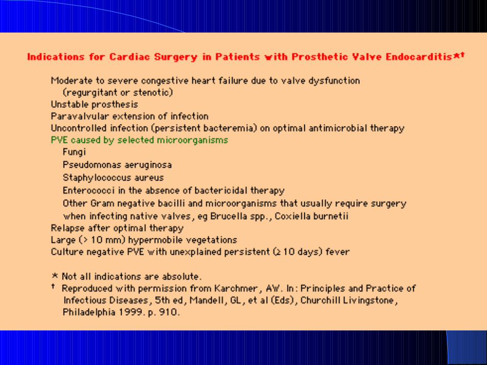
Surgical InterventionSurgical Intervention
The outcome of surgery is better when the infection is partially treated or healed. So, patients with severe valve dysfunction who respond to antibiotic therapy and who manifest evidence of only mild and nonprogressive heart failure should complete a full course of antibiotics before surgery is undertaken to minimize morbidity and mortality.
However, surgery should not be delayed to complete antimicrobial therapy in patients with progressive heart failure or evidence of other complications.
Two general classes of valves exist, bioprosthetic and mechanical.

Prosthetic ValvesProsthetic Valves

Class I indication for native valve surgery
Acute AR or MR with heart failureAcute AR with tachycardia or early closure or MV on echoFungal endocarditisAortic or annular abscess, or aortic aneurysmValve dysfunction or infection after 10 days of Abx therapy
Class II indication for native valve surgery
Recurrent emboli after >24hrs Abx therapyMobile vegetations >10mm

Recommendation for Bioprosthesis placement
Pt cannot take warfarin>65yrs who do not need warfarin for another cause
Recommendation for mechanical valve placement
Expected long life spanMechanical valve in another positionPt with renal failure or hypercalcemiaRequire warfarin for another cause

Non-bacterial Thrombotic Non-bacterial Thrombotic EndocarditisEndocarditis
Combination of endothelial injury and hypercoagulable state can lead to platelet-thrombin deposition.
These deposits are found on the atrial side of MV & TV, and the ventricular side of AV & PV; similar to location of infective vegetations.
Most common associated syndrome with this condition is the anti-phospholipid syndrome.
The atypical verroucous Libman-Sacks vegetations are usually difficult to see on TTE.
They usually accumulate on the distal tip of the MV.

Metastatic tumours can also involve cardiac valves and produce lesions similar to IE.
These marantic endocarditis lesions occur more frequently in Hodgkin and adenocarcinoma of the lung, stomach and colon.
All non bacterial thrombotic lesion can become infected if exposed to bacteremia.

Antibiotic ProphylaxisAntibiotic Prophylaxis
Bacteria colonize and adhere to platelet-fibrin aggregates during bacteremia.
Although many bacteria enter the blood stream, only those suited to adhere to surfaces can become infective.
Certain predisposing valvular lesions are also required to increase your risk of colonization.
This has led to identifying patients at risk during surgical procedures and the use of prophylactic antibiotics.

Prophylaxis recommended
Dental procedures, including scaling and cleaningTonsillectomy & adenoidectomySurgery involving GI or upper respiratory mucosaEsophageal varices sclerotherapyERCPCholecystectomyCystoscopy, urethral dilatationGU surgeryIncision and drainage of infected tissue

Prophylaxis not recommended
Dental procedures not likely to produce bleeding, i.e. filling above gum line, orthodontic adjustments.Endotracheal intubationTEEFlexible bronchoscope +/- biopsyAngiography +/- PTCAPacemaker implantationGI Endoscopy +/- biopsyC-section
In absence of infection: uncomplicated vaginal delivery, therapeutic abortion, IUD, laparoscopy, circumcision.

High Risk Patients
Prosthetic valvePrior IECyanotic congenital heart diseasePDAARASMRMS + MRVSDCoarctation of AoSurgically constructed systemic-pulmonary shuntsSurgically corrected cardiac lesion with residual hemodynamic abn or prosthesis.

Intermediate Risk Patients
MV prolapse or thickening with murmurPure MSTV diseasePSAsymmetrical septal hypertrophyBicuspid AV with minimal hemodynamic changesAo sclerosis with minimal hemodynamic changesSurgically repaired intracardiac lesions, less than six mos postop, with minimal hemodynamic changes.

Low Risk Patients
MV prolapse without murmurIsolated secundum ASDImplanted devicesCABGPrior Kawasaki’s or Rheumatic disease without valvular dysfunction

Antibiotic Prophylaxis SummaryAntibiotic Prophylaxis SummaryGI/GU regimenHigh risk Amp 2g & Gent 1.5mg/kg. Mod risk Amox 2g or Amp.
PCN allergy = Substitute Vanco 1g
Oral/Esoph/Resp regimenAmox PO or Amp IV
PCN allergy = Substitute Clinda 600mg PO/IV Cefazolin 1g Azith/Clarith PO 500mg
Abx should be given prior to procedure, 1hr for oral 1/2hr for parenteral


Questions?Questions?


UTD Abx TablesUTD Abx Tables


UTD Surgery tablesUTD Surgery tables