Protocol for optimization of histological, histochemical...
8
Protocol for optimization of histological, histochemical and immunohistochemical analyses of larval tissues: application in histopathology of honey bee E. C. M. Silva-Zacarin 1,* , M. P. Chauzat 2 , S. Zeggane 2 , P. Drajnudel 2 , F. Schurr 2 , J. P. Faucon 2 , O. Malaspina 3 , J. A. Engler 4 1 Laboratory of Functional and Structural Biology (LABEF), Federal University of São Carlos (UFSCar), Campus Sorocaba, Rodovia João Leme dos Santos, Km 110 - SP-264, CEP 18052-780, Sorocaba, SP, Brazil 2 Unit of Honey bee Pathology, ANSES (French agency for food, environmental and occupational health safety), 105, route des Chappes, BP 111, F-06902 Sophia Antipolis, France 3 Center for the Study of Social Insects (CEIS), Departement of Biology, Institute of Biosciences, UNESP University, Rio Claro, SP, Brazil 4 UMR/ISA Institute Sophia Agrobiotech, Institut National de la Recherche Agronomique, 400 route des Chappes, F- 06903 Sophia Antipolis, France * [email protected] The honey bee Apis mellifera is a pollinator insect constantly exposed to pesticides and pathogens in the field. In order to discriminate the sublethal effects of these stressors on bees, histopathological analysis is important as an additional tool for diagnosis. This chapter describes a protocol for optimization of histological, histochemical and imunohistochemical analyses in honey bee organs, which joined the best tissue morphology with the optima immunostaining for DNA fragmentation, which is indicative of cell death, and heat shock protein (HSP), which is indicative of cell response to stress. With this aim, two dehydration protocols were tested: the conventional (Protocol I) and the low temperature slow- dehydration (Protocol II). Our data showed that the same fixative solution (4% paraformaldehyde in PBS buffer) associated with the Protocol II (low temperature slow-dehydration) can be applied for various technical approaches in order to optimize the histopathological analysis of honey bee. Keywords: dehydration procedure, histology, cell markers. 1. Introduction The honey bees (Apis mellifera L.) are pollinator insects in agro-ecosystems. In addition to its economic value in agriculture, the honey bee is a model organism for the study of communication, learning and memory formation and provides the best-studied social insect system [1-2]. In this model, genotype, social environment, social history, behavior, social tasks, nutrition, physiology, and health can be controlled therefore allowing the identification and understanding of mechanisms that affect life-history [1-4] and aging [5]. Additionally, studies about honey bee are in focus worldwide due to the Colony Collapse Disorder (CCD) that has threatened the apiaries in the USA and Europe (for review see [6-7]). In this scenario, this domesticated pollinator is constantly exposed to stress conditions in the field, such as pesticides [8-10] and pathogens [11]. Analysis of morphological alterations of bee organs, named histopathology, associated to immunodetection of cell markers can be used as additional tools for diagnosis of side-effects induced by pesticide or pathogen exposure on honey bee [12-15]. In this way, it is important to choose a reliable and reproducible protocol for application in histopathology of bees that joins the high quality as in preservation of tissue morphology as in its chemical compounds, which provides the possibility of performing immunohistochemical labeling to evaluate cell markers of exposure and toxicity to pesticides and pathogens. In order to obtain a good resolution of bee organs for histopathological analysis, it is necessary to perform sample fixation, dehydration and embedding methods that allow the best tissue preservation. Compared to adult honey bees, larvae are more difficult to perform histological studies and, for these reason, they has been chosen to this study. Many larval organs are particularly fragile and not accessible through dissection due to their small size and softness of the tissues. Therefore, it is necessary to process the whole larvae, what is also advantageous because it allows the analysis of various organs of honey bee larvae, such as the nervous system, midgut, Malpighi tubules, fat body, silk glands, since that these organs are important to study sublethal effects of pesticides and/or pathogens [12-13, 15-20]. Histological methods for fixation and processing of animal tissue and organs has been described in literature there are many decades ago [21-23] and they are also applicable to honey bees or other insects. These histological methods have provided good results until now, since that these procedures have been carried out with caution to obtain optimal tissue preservation. Therefore, aiming to optimize the histophatological analysis of honey bee, the goal of the present work was to apply procedures for processing animal tissues in order to choose just one protocol that provides high resolution and accuracy for morphological and histochemical analysis of bee organs, rendering epitopes accessible to antibody recognition, which allows immunohistochemical analysis in the same samples. Current Microscopy Contributions to Advances in Science and Technology (A. Méndez-Vilas, Ed.) © 2012 FORMATEX 696
Transcript of Protocol for optimization of histological, histochemical...

Protocol for optimization of histological, histochemical and immunohistochemical analyses of larval tissues: application in histopathology of honey bee
E. C. M. Silva-Zacarin1,*, M. P. Chauzat2, S. Zeggane2, P. Drajnudel2, F. Schurr2, J. P. Faucon2, O. Malaspina3, J. A. Engler4 1Laboratory of Functional and Structural Biology (LABEF), Federal University of São Carlos (UFSCar), Campus
Sorocaba, Rodovia João Leme dos Santos, Km 110 - SP-264, CEP 18052-780, Sorocaba, SP, Brazil 2 Unit of Honey bee Pathology, ANSES (French agency for food, environmental and occupational health safety), 105,
route des Chappes, BP 111, F-06902 Sophia Antipolis, France 3Center for the Study of Social Insects (CEIS), Departement of Biology, Institute of Biosciences, UNESP University, Rio
Claro, SP, Brazil 4 UMR/ISA Institute Sophia Agrobiotech, Institut National de la Recherche Agronomique, 400 route des Chappes, F-
06903 Sophia Antipolis, France * [email protected]
The honey bee Apis mellifera is a pollinator insect constantly exposed to pesticides and pathogens in the field. In order to discriminate the sublethal effects of these stressors on bees, histopathological analysis is important as an additional tool for diagnosis. This chapter describes a protocol for optimization of histological, histochemical and imunohistochemical analyses in honey bee organs, which joined the best tissue morphology with the optima immunostaining for DNA fragmentation, which is indicative of cell death, and heat shock protein (HSP), which is indicative of cell response to stress. With this aim, two dehydration protocols were tested: the conventional (Protocol I) and the low temperature slow-dehydration (Protocol II). Our data showed that the same fixative solution (4% paraformaldehyde in PBS buffer) associated with the Protocol II (low temperature slow-dehydration) can be applied for various technical approaches in order to optimize the histopathological analysis of honey bee.
Keywords: dehydration procedure, histology, cell markers.
1. Introduction
The honey bees (Apis mellifera L.) are pollinator insects in agro-ecosystems. In addition to its economic value in agriculture, the honey bee is a model organism for the study of communication, learning and memory formation and provides the best-studied social insect system [1-2]. In this model, genotype, social environment, social history, behavior, social tasks, nutrition, physiology, and health can be controlled therefore allowing the identification and understanding of mechanisms that affect life-history [1-4] and aging [5]. Additionally, studies about honey bee are in focus worldwide due to the Colony Collapse Disorder (CCD) that has threatened the apiaries in the USA and Europe (for review see [6-7]). In this scenario, this domesticated pollinator is constantly exposed to stress conditions in the field, such as pesticides [8-10] and pathogens [11]. Analysis of morphological alterations of bee organs, named histopathology, associated to immunodetection of cell markers can be used as additional tools for diagnosis of side-effects induced by pesticide or pathogen exposure on honey bee [12-15]. In this way, it is important to choose a reliable and reproducible protocol for application in histopathology of bees that joins the high quality as in preservation of tissue morphology as in its chemical compounds, which provides the possibility of performing immunohistochemical labeling to evaluate cell markers of exposure and toxicity to pesticides and pathogens. In order to obtain a good resolution of bee organs for histopathological analysis, it is necessary to perform sample fixation, dehydration and embedding methods that allow the best tissue preservation. Compared to adult honey bees, larvae are more difficult to perform histological studies and, for these reason, they has been chosen to this study. Many larval organs are particularly fragile and not accessible through dissection due to their small size and softness of the tissues. Therefore, it is necessary to process the whole larvae, what is also advantageous because it allows the analysis of various organs of honey bee larvae, such as the nervous system, midgut, Malpighi tubules, fat body, silk glands, since that these organs are important to study sublethal effects of pesticides and/or pathogens [12-13, 15-20]. Histological methods for fixation and processing of animal tissue and organs has been described in literature there are many decades ago [21-23] and they are also applicable to honey bees or other insects. These histological methods have provided good results until now, since that these procedures have been carried out with caution to obtain optimal tissue preservation. Therefore, aiming to optimize the histophatological analysis of honey bee, the goal of the present work was to apply procedures for processing animal tissues in order to choose just one protocol that provides high resolution and accuracy for morphological and histochemical analysis of bee organs, rendering epitopes accessible to antibody recognition, which allows immunohistochemical analysis in the same samples.
Current Microscopy Contributions to Advances in Science and Technology (A. Méndez-Vilas, Ed.)
© 2012 FORMATEX 696
2. Materials and Methods
2.1 Fixation of honey bee larvae
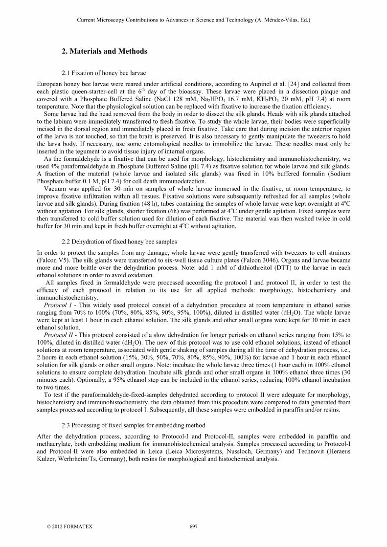
European honey bee larvae were reared under artificial conditions, according to Aupinel et al. [24] and collected from each plastic queen-starter-cell at the 6th day of the bioassay. These larvae were placed in a dissection plaque and covered with a Phosphate Buffered Saline (NaCl 128 mM, Na2HPO4 16.7 mM, KH2PO4 20 mM, pH 7.4) at room temperature. Note that the physiological solution can be replaced with fixative to increase the fixation efficiency. Some larvae had the head removed from the body in order to dissect the silk glands. Heads with silk glands attached to the labium were immediately transferred to fresh fixative. To study the whole larvae, their bodies were superficially incised in the dorsal region and immediately placed in fresh fixative. Take care that during incision the anterior region of the larva is not touched, so that the brain is preserved. It is also necessary to gently manipulate the tweezers to hold the larva body. If necessary, use some entomological needles to immobilize the larvae. These needles must only be inserted in the tegument to avoid tissue injury of internal organs. As the formaldehyde is a fixative that can be used for morphology, histochemistry and immunohistochemistry, we used 4% paraformaldehyde in Phosphate Buffered Saline (pH 7.4) as fixative solution for whole larvae and silk glands. A fraction of the material (whole larvae and isolated silk glands) was fixed in 10% buffered formalin (Sodium Phosphate buffer 0.1 M, pH 7.4) for cell death immunodetection. Vacuum was applied for 30 min on samples of whole larvae immersed in the fixative, at room temperature, to improve fixative infiltration within all tissues. Fixative solutions were subsequently refreshed for all samples (whole larvae and silk glands). During fixation (48 h), tubes containing the samples of whole larvae were kept overnight at 4oC without agitation. For silk glands, shorter fixation (6h) was performed at 4oC under gentle agitation. Fixed samples were then transferred to cold buffer solution used for dilution of each fixative. The material was then washed twice in cold buffer for 30 min and kept in fresh buffer overnight at 4oC without agitation.
2.2 Dehydration of fixed honey bee samples
In order to protect the samples from any damage, whole larvae were gently transferred with tweezers to cell strainers (Falcon V5). The silk glands were transferred to six-well tissue culture plates (Falcon 3046). Organs and larvae became more and more brittle over the dehydration process. Note: add 1 mM of dithiothreitol (DTT) to the larvae in each ethanol solutions in order to avoid oxidation. All samples fixed in formaldehyde were processed according the protocol I and protocol II, in order to test the efficacy of each protocol in relation to its use for all applied methods: morphology, histochemistry and immunohistochemistry. Protocol I - This widely used protocol consist of a dehydration procedure at room temperature in ethanol series ranging from 70% to 100% (70%, 80%, 85%, 90%, 95%, 100%), diluted in distilled water (dH2O). The whole larvae were kept at least 1 hour in each ethanol solution. The silk glands and other small organs were kept for 30 min in each ethanol solution. Protocol II - This protocol consisted of a slow dehydration for longer periods on ethanol series ranging from 15% to 100%, diluted in distilled water (dH2O). The new of this protocol was to use cold ethanol solutions, instead of ethanol solutions at room temperature, associated with gentle shaking of samples during all the time of dehydration process, i.e., 2 hours in each ethanol solution (15%, 30%, 50%, 70%, 80%, 85%, 90%, 100%) for larvae and 1 hour in each ethanol solution for silk glands or other small organs. Note: incubate the whole larvae three times (1 hour each) in 100% ethanol solutions to ensure complete dehydration. Incubate silk glands and other small organs in 100% ethanol three times (30 minutes each). Optionally, a 95% ethanol step can be included in the ethanol series, reducing 100% ethanol incubation to two times. To test if the paraformaldehyde-fixed-samples dehydrated according to protocol II were adequate for morphology, histochemistry and immunohistochemistry, the data obtained from this procedure were compared to data generated from samples processed according to protocol I. Subsequently, all these samples were embedded in paraffin and/or resins.
2.3 Processing of fixed samples for embedding method
After the dehydration process, according to Protocol-I and Protocol-II, samples were embedded in paraffin and methacrylate, both embedding medium for immunohistochemical analysis. Samples processed according to Protocol-I and Protocol-II were also embedded in Leica (Leica Microsystems, Nussloch, Germany) and Technovit (Heraeus Kulzer, Werhrheim/Ts, Germany), both resins for morphological and histochemical analysis.
Current Microscopy Contributions to Advances in Science and Technology (A. Méndez-Vilas, Ed.)
© 2012 FORMATEX 697

2.3.1 Technovit and Leica embedding methods
Dehydrated-samples were placed in 50% ethanol-Technovit mixture and kept at 4oC with occasional gentle agitation for at least 2 h for silk glands or overnight for the whole larvae. This solution was subsequently changed to 100% Technovit (containing 1g/100ml of Hardener I) and incubated at 4oC overnight or longer. Organs or larvae were oriented into embedding molds containing the inclusion medium 100% Technovit (containing 1ml/15ml of Hardener II) and they were left for polymerization around 2 hour, at room temperature. The remaining samples were placed in Leica resin and were kept overnight at 4oC. Organs and whole larvae were oriented into embedding molds containing the inclusion medium (100% Leica resin containing 1ml/15ml of Hardener) and they were left for polymerization during 1 hour at 37oC or overnight at room temperature.
2.3.2 Metacrylate embedding method
Dehydrated-samples were placed in 50% ethanol-BM-methacrylate mixture (4:1 butylmethacrylate: methylmethacrylate) and were kept in this solution at 4oC, at least for 2 h for silk glands, with occasional gentle agitation, or overnight for the whole larvae. Note: add 1 mM DTT if necessary to avoid tissue oxidation. Material should be properly dehydrated because the methacrylate mixture is not miscible with water. Samples were subsequently placed in 100% BM-methacrylate mixture containing 1mM DTT and were kept overnight at 4oC, preferentially with gentle agitation. The methacrylate mixture was then refreshed with the same mixture medium containing 0.5% benzoin ethyl ether and samples were kept on 4oC. Note: samples can stay a few days in either of these solutions before polymerization. Samples were oriented into capsules containing the embedding medium (100% methacrylate mixture and 0.5% benzoin ethyl ether) and polymerization was allowed for 6 hours at 4oC under UV light.
2.3.3 Paraffin embedding method
After fixation and dehydration, samples were gently transferred into glass small containers. Samples dehydrated according to Protocol I or Protocol II were placed into a solution containing 50% xylene:50% ethanol for 1h, with occasional shaking at room temperature. This solution was subsequently replaced by a 75% xylene:25% ethanol solution and incubated for 1 h. This solution was then replaced with 100% xylene three times for at least 1 hour each at room temperature, in order to remove completely the ethanol from all tissues. Paraplast chips were added gradually in the glass containers containing the samples and finally placed in an oven at 60oC for 2 hours until overnight. Molten paraplast was then replaced three times for at least 8 hours each time to obtain optimal infiltration of paraffin into the whole larvae. Note: samples can stay few days in paraplast before polymerization. The embedding molds containing molten paraplast were placed on a hot plate (60oC) and the larvae and silk glands were oriented in these molds with warmed tweezers. After paraplast polymerization, paraffin blocks were stored at 4oC until use.
2.4 Sectioning embedded material
2.4.1Technovit and Leica embedded material
Polymerized samples were removed from embedding molds and attached to a holder with glue (Technovit 3040 kit) and samples were sectioned to 2 μm on a microtome. Tissue sections were placed on drops of dH2O on poly(L-lysine)-coated slides, dried on a hot plate at 45oC and subsequently stored in dry boxes until use.
2.4.2 Methacrylate-embedded material
Polymerized samples were removed from embedding capsules with a razor blade and samples were sectioned on a microtome. Note: thicker sections are made for immunohistochemical analysis (5 µm) in order to have more proteins available for detection by the antibody. Tissue sections were placed on drops of ddH2O on coated slides and dried on a hot plate at 60oC. The slides were incubated overnight at 4oC for an adequate adherence of sections on the slides. These slides are stored in dry boxes until use.
2.4.3 Paraffin-embedded material
The paraffin-embedded tissues were mounted onto wooden blocks and the edges shaped with a razor blade to form a pyramid. Ribbons of 8 μm using a metal knife were performed on a microtome and placed on ddH2O-flooded (coated) slides with the ribbon shiny side toward the water surface. Excess of water was removed and slides were incubated overnight at 45oC to promote an optimal tissue adhesion to the slide. Slides are stored in dry boxes until use.
Current Microscopy Contributions to Advances in Science and Technology (A. Méndez-Vilas, Ed.)
© 2012 FORMATEX 698

2.4.4 Removal of embedding medium
This procedure is only necessary for paraffin and methacrylate-embedded material. For paraffin-embedded sections, the slides were inserted into slide racks and then placed into staining dishes containing 100% xylene (or Histoclear) and gently stirred twice for 15 min each one. Slides were removed from xylene and rinsed twice in 100% ethanol. Slides were then gradually rehydrated into ethanol series (100% to 30%) and rinsed in dH2O three times dipping up and down until any visible “streaks” disappeared. For methacrylate embedded sections, the slides were placed into 100% acetone, gently stirred for 30 min and rinsed twice in 100% ethanol solution. The same procedure described above was applied for rehydration of sections.
2.5 Histology, histochemistry and immunohistrochemistry
Samples embedded in Technovit or Leica resins were submitted to Hematoxylin-Eosin staining for morphological analysis and to the following stainings for histochemical analyses: Alcian blue for acid glycoconjugates detection [21], Periodic-Acid-Schiff (PAS) for neutral glycoconjugates detection [25], Bromophenol blue [26] and Xylidine Ponceau [27] for proteins detection, Sudam black [26] for lipid detection and the simultaneous staining: Bromophenol blue-PAS [28] for simultaneous detection of glycoconjugates and proteins; Alcian Blue and PAS [29] for simultaneous detection of acid and neutral glycoconjugates. Sections embedded in both paraffin and methacrylate were submitted to immunohistochemical methods. For cell death detection we have applied the Apoptag Plus Kit according the protocol described by the manufacturer (MP Biomedicals Brazil, São Paulo, Brazil). For immunohistochemical labeling of HSP, we used monoclonal antibody HSP70 produced in mouse and peroxidase conjugated-secondary antibody produced in rabbit (both from Sigma-Aldrich Brazil Ltda. São Paulo, Brazil). The detailed procedure has been described by Silva-Zacarin et al [18].
3. Results and Discussion
In general, samples fixed in 4% paraformaldehyde and processed according Protocol II (low temperature slow-dehydration) gave better results than those processed with Protocol I (usual procedure for dehydration at room temperature) for morphological, histochemical and immunohistochemical analysis. Table 1 showed the compilation of results generated by dehydration according to Protocol I and Protocol II. When whole larvae were processed according the Protocol II, it was possible to observe a higher morphological resolution of the larval organs in comparison to whole larvae that were processed according to Protocol I. For isolated silk glands, although both protocols provided the same high definition for morphology, differences in accuracy was observed in histochemical analyses when the Protocol II was applied.
Table 1 Analysis of honey bee organs dehydrated according to Protocols I and II and fixed with in different solutions for morphological (all organs listed) and histochemical analysis (fat body and silk glands). High definition or labeling = A; Medium definition or labeling = B; Poor definition or labeling = C; Presence of artefacts = +; Absence of artefacts = - ; 0 = method not performed. Protocol I= usual fast dehydration at room temperature; Protocol II= slow dehydration at low temperature (4oC).
Protocol I Protocol II
Paraformaldehyde Paraformaldehyde
Cell
compounds Fat
body Silk
glands Mid-gut
Malpighi tubs
Brain Fat body
Silk glands
Mid-gut
Malpighi tubs
Brain
proteins B B 0 0 0 A A 0 0 0
glycol-conjugate
C C 0 0 0 A A 0 0 0 Histo-
chemistry
lipids B C 0 0 0 A A 0 0 0 Morpho-
logical and chemical
preservation
Secretion in lumen or in cytoplasm granules
B B 0 0 0 A A 0 0 0
cytoplasm B A B B B A A A A A Morphology
nucleus B A B B B A A A A A Artifact + - - - - - - - - -
High morphological definition of the fat body was observed when whole larvae were processed according to Protocol II in comparison with those processed according to Protocol-I (Fig. 1A compared to Fig. 1B), in which some artifacts are generally observed in trophocytes.
Current Microscopy Contributions to Advances in Science and Technology (A. Méndez-Vilas, Ed.)
© 2012 FORMATEX 699

Fig. 1 Fat body of Apis mellifera larvae fixed in 4% paraformaldehyde and embedded in resin. Histological sections stained with Hematoxilin-Eosin. (A) Larvae processed according to Protocol I. (B) Larvae processed according to Protocol II. Note that the artifacts (ar), which are observed in (A), are not present in (B). For Protocol I and II: see details in the text. e= oenocyte; t= trophocyte; gr= cytoplasm granules; n= nucleus; ar=artifact; black arrow= cytoplasm around the nucleus; white arrow= big granule. Bars= 50 μm.
The fixation with paraformaldehyde associated to dehydration according to Protocol II also provided the best resolution for morphological analysis of the midgut (Fig. 2A), because microvilli of digestive cells and the regenerative cells presented high definition, as well as the Malpighi tubules (Fig. 2B) and nervous tissue (Fig. 2C).
Fig. 2 Organs of Apis mellifera larvae fixed in 4% paraformaldehyde, processed according to Protocol II and embedded in resin. Histological sections were stained with Hematoxilin-Eosin. (A) Midgut. dc= digestive cells; as= apocrine secretion; n= nucleus; cl= cell liberation; white arrow= brush border (microvilli); lu= lumen; black arrow= regenerative cells; Bar= 30μm. (B) Malpighi tubules showing brush border (microvilli= write arrow) and secretion (black arrow) in the lumen (lu). n= nucleus of the excretory cell. Bar= 20μm. (C) Nervous tissue (nt) in the brain; n= nucleus of nervous cell. Bar= 20μm. In relation to histochemical analysis, glycoconjugates were notably better preserved in larval organs (silk gland and fat body) when Protocol II was applied (Fig. 3). In silk glands, the details of the secretion present in the lumen were observed with high definition only when Protocol II was applied (Fig. 3A compared to Fig. 3B). When Protocol I was used (Fig. 3A), the resolution of gland secretion, which is rich in neutral and acid glycoconjugates, was significantly less accurate than those detected by the Protocol II (Fig. 3B). Fat body processed according to protocol II, showed fine granulation PAS-positive with very specific location in the cytoplasm (Fig. 3C), as well as PAS-positive big granules
Current Microscopy Contributions to Advances in Science and Technology (A. Méndez-Vilas, Ed.)
© 2012 FORMATEX 700

(Fig. 3C). Then, the low temperature slow-dehydration allowed better resolution of the neutral and acid glyconjugates in the silk gland, which was not observed in our previous study using conventional protocols [31]. Although Sudam black has originally been proposed to cryosections to detect lipids, in our study we showed that fixation with 4% paraformaldehyde followed by slow dehydration in cold ethanol solutions (protocol II) was optimal to preserve lipids inside the granules of trophocyte, presenting a very specific stainning (Fig. 3D).
Fig. 3 A) and B) Silk glands isolated from Apis mellifera larvae and fixed in 4% paraformaldehyde, processed according to Protocol I (A) or Protocol II (B), and embedded in resin. Histological sections were submitted to PAS/ Alcian blue double staining. Note the high definition in secretion presented in the gland lumen only in (B). Sc= secretory cell; gs= gland secretion in the lumen; n= nucleus; white arrow= heterogeneous secretion positive for PAS and Alcian Blue; Bars= 50μm. C) and D) Fat body from Apis mellifera larvae fixed in 4% paraformaldehyde, processed according to Protocol II, and embedded in resin. C) Histological sections of fat body submitted to histochemical method for neutral glycoconjugate detection (PAS). gr= granules; n= nucleus; black arrow= PAS-positive fine granulation in cytoplasm; white arrow= PAS-positive granule. Bar= 50μm. D) Histological sections of fat body submitted to histochemical method for lipid detection (Sudam Black). Note positive granules (black arrow) inside the cytoplasm of trophocyte. n= nucleus; Bar= 20μm. Leica or Technovit resins resulted in similar high-quality morphology preservation, similar to methacrylate, but they do not enable the immunohistochemistry application. Then, immunolabelling methods need to be performed in samples embedded in paraffin or in methacrylate, since that both embedding medium can be dissolved in order to enable the antibody incubation for specific protein recognition on tissue sections. The present work also focused on the obtention of high resolution in morphology combined with optima immunostaining for detection of DNA fragmentation, which is indicative of cell death, and heat shock protein (HSP), which is indicative of cell stress [15]. Paraphormaldehyde-fixed tissues submitted to the low temperature slow-dehydration (Protocol II) kept the immunoreactivity in tissues, as well as in samples processed according to the Protocol-I. As a good preservation of tissue morphology is a key factor for localization and interpretation of data generated from immunolabeling, the use of methacrylate resin provided the best results. Although the organ morphology preservation was significantly better when larvae were embedded in methacrylate (Fig. 4C, 4D), compared to those embedded in paraffin (Fig. 4A, 4B), there are some advantages of using paraffin. As different thickness of sections also can have an influence on definition of labeling, paraffin section around 8 µm could have more intensity of immunolabeling in comparison to methacrylate sections that are thinner, reaching the maximum of 5-6 µm. Then, for immunodetection of scarce proteins in tissues, the embedding of the samples in paraffin could be better than in methacrylate. Methacrylate sections of larvae processed according to Protocol II and submitted to immunodetection of HSP70 (Fig. 5) gave optimal results considering the superior morphology of tissues and, also, optimal immunolabeling. The successful use of formaldehyde for immunohistochemistry is well known (for review see [31]). In addition, it is also recognized that the major artifact induced by aldehyde fixations is the masking of tissue antigens due to cross-linking of protein amino acid residues. The protocol present herein showed immunoreactivity without undesirable background. In this study, we tried for the first time the methacrylate embedding in honey bees for immunohistochemical detection of HSP 70. Previous reports have used paraffin-embedded honey bee organs [16, 18, 32] that also presented good resolution of the immunolabelling. These results are of great importance since it sustains the use of the same fixation, dehydration and embedding conditions for processing honey bee tissues for the application of various techniques. Analysis of morphological, histochemical and immunohistochemical alterations on honey bee organs might improve the understanding of stress
Current Microscopy Contributions to Advances in Science and Technology (A. Méndez-Vilas, Ed.)
© 2012 FORMATEX 701

effects induced by pesticides and pathogens [12, 15] in this pollinator insect, representing an additional tool for diagnosis.
Fig. 4 Immunohistochemical detection of cell death in larval tissues of Apis mellifera, by means of the Apoptag kit. DAB was used as substrate of the immunoreaction. Positive reactions are indicated by white arrows. Negative nuclei are indicated by black arrows. (A) Fat body of whole larvae fixed in 10% buffered formalin (pattern fixative for apoptosis kit) and embedded in paraffin; Protocol I. (B) Fat body of whole larvae fixed in 4% paraformaldehyde and embedded in paraffin; Protocol II. (C) and (D) Organs of whole larvae fixed in 4% paraformaldehyde and embedded in methacrylate; Protocol II. MG= midgut; mv= brush border (microvilli); t= trophocyte; MT= Malpighi tubules. Bars: A = 20μm; B, C, D= 50μm.
Fig. 5 (A) and (B) Organs of Apis mellifera larvae fixed in 4% paraformaldehyde, processed according Protocol II and embedded in methacrylate. Immunostaining using the monoclonal antibody anti-HSP70, as primary antibody, and secondary antibody conjugated with peroxidase. DAB was used as substrate of the immunoreaction. Arrows indicate positive reaction in the cytoplasm of fat body (CG), nucleus of Malpighi tubules (TM) and cytoplasm of digestive cells of midgut (md). Bars: A-B= 50μm. In summary, the present study reinforces the importance of the procedure for tissue dehydration that when is performed gradually on low temperature, improve the tissue preservation. Although specific fixatives have been advised for each histochemical staining [21, 26], especially for glycoconjugate and lipid preservation, our results showed that fixation with paraformaldehyde associated with Protocol II fulfills the requirements of morphological diagnostics and preserved sufficiently the cellular macromolecules for the application of different histochemical methods. In addition, this optimized protocol preserves proteins allowing immunohistochemical application. In this way, time and money are saved for the researcher because the same protocol could be used for different technical approaches.
Current Microscopy Contributions to Advances in Science and Technology (A. Méndez-Vilas, Ed.)
© 2012 FORMATEX 702

Acknowledgments Dr. Elaine C.M. Silva-Zacarin thanks FAPESP-Brazil for financial support (2008/50813 and 2008/51473). We thank Dr. Fábio C. Abdalla for laboratorial support for immunohistochemistry (LABEF, UFSCar, Brazil) and Rafael Ferreira (CEIS, UNESP, Brazil) for his help with the histochemical methods.
References
[1] Robinson GE, Grozinger CM, Whitfield CW. Sociogenomics: Social life in molecular terms. Nat Rev Genet. 2005; 6:257–270. [2] Muench D, Amdam GV, Wolschin F. Physiological and cognitive aging in honey bees: robustness, plasticity and molecular
regulation. Func Ecol. 2008; 22:407–421. [3] Toth AL, Robinson GE. Worker nutrition and division of labour in honeybees. Anim Behav. 2005; 69:427–435. [4] Weinstock GM, Robinson GE, Gibbs RA, Weinstock GM, Weinstock GM. Insights into social insects from the genome of the
honeybee Apis mellifera. Nature. 2006; 443:931–949. [5] Münch D, Amdam GV. The curious case of aging plasticity in honey bees. BS Lett. 2010; 584:2496-2503. [6] Ellis JD, Evans JD, Pettis J. Colony losses, managed colony population decline, and Colony Collapse Disorder in the United
States. J Apic Res. 2010; 49:134-136. [7] Neumann P, Carreck NL. Honey bee colony losses. J Apic Res. 2010; 49:1-6. [8] Chauzat MP, Faucon JP, Martel AC, Lachaize J, Cougoule N, Aubert M. A survey on pesticide residues in pollen loads collected
by honey-bees (Apis mellifera) in France. J Econ Entomol. 2006; 99:253-262. [9] Wu JY, Anelli CM, Sheppard S. Sub-lethal effects of pesticides residues in brood comb on worker honey bee (Apis mellifera)
development and longevity. PLoS ONE. 2011; 6:1-11. [10] Schneider CW, Tautz J, Grunewald B, Fuchs S. RFID Tracking of sublethal effects of two neonicotinoid insecticides on the
foraging behavior of Apis mellifera. PLoS ONE. 2012; 7:1-9. [11] Faucon JP, Mathieu L, Ribière M, Martel AC, Drajnudel P, Zeggane S, Aurières C, Aubert M. Honey bee winter mortality in
France in 1999 and 2000. Bee World. 2002; 83:14-23. [12] Malaspina O, Silva-Zacarin ECM. Cell markers for ecotoxicological studies in target organs of bees. Braz J Morphol Sci. 2006;
23:303-309. [13] Cruz A, Silva-Zacarin ECM, Bueno OC, Malaspina O. Morphological alterations induced by boric acid and fipronil in the
midgut of worker honey bee (Apis mellifera L.) larvae. Cell Biol Toxicol. 2010; 26:165-176. [14] Gregorc A, Ellis J. Cell death localization in situ in laboratory reared honey bee (Apis mellifera L.) larvae treated with
pesticides. Pestic Biochem Physiol. 2011; 99: 200-207. [15] Gregorc A, Silva-Zacarin ECM, Nocelli RCF. Cellular response in honey bees to non-pathogenic effects of pesticides. In:
Sammataro D, Yoder JA, eds. Honey Bee Colony Health: Challenges & Sustainable Solutions. Taylor & Francis Group, New York; 2012: 161-180.
[16] Gregorc A, Bowen ID. In situ localization of heat-shock and histone proteins in honeybee (Apis mellifera L.) larvae infected with Paenibacillus larvae. Cell Biol Int. 1999; 23:211–218.
[17] Gregorc A, Bowen ID. Histochemical characterization of cell death in honeybee larvae midgut after treatment with Paenibacillus larvae, amitraz and oxytetracycline. Cell Biol Int. 2000; 24:319-324.
[18] Silva-Zacarin ECM, Gregorc A, Moraes RLMS. In situ localization of heat-shock proteins and cell death labelling in the salivary gland of acaricide-treated honeybee larvae. Apidologie. 2006, 37: 507-516.
[19] Nocelli RCF, Roat TC, Silva-Zacarin ECM, Palma MS, Malaspina O. Social Insects - morphophysiology of the nervous system. In: Stewart EM, ed. Social Insects - Structure, Function, and Behavior. Nova Publishers, Hauppauge, New York; 2011: 105-120.
[20] Silva-Zacarin, E.C.M., Costa-Ferreira, R.A., Nocelli, R.C.F., Roat, T.C., Palma, M.S., Malaspina, O. Structure and function of the intestine an Malpighian tubules - from bee biology to cell marker development for toxicological analysis. In: Stewart EM, ed. Social Insects - Structure, Function, and Behavior. Nova Publishers, Hauppauge, New York; 2011: 121-142.
[21] Lison L. Histochemie et Cytochemie Animales - Principles et Methodes. 2v, Gauthier-Villars, Paris; 1960. [22] Lillie RD, Fullmer HM. Histopathologic technic and practical histochemistry. 4th ed. Mc Graw-Hill, New York; 1976. [23] Brancoft JD, Stevens A. Theory and Practice of Histological Techniques. 2nd ed. New York: Churchill Livingstone; 1982. [24] Aupinel P, Fortini D, Dufour H, Tasei JN, Michaud B, Odoux JF, Pham-Delègue MH. Improvement of artificial feeding in a
standard in vitro method for rearing Apis mellifera larvae. B Insectol. 2005; 58:107-111. [25] McManus JFA. Histological demonstration of mucin after periodic acid. Nature. 1946; 158:202. [26] Pearse AGE. Histochemistry Theoretical and Applied. Jet. Churchill Ltda, London; 1960. [27] Vidal BC. Dichroism on collagen bundles stained with Xylidine ponceau 2R. Ann Histochim. 1970; 15:289-296. [28] Coello S. A new staining schedule for formalin-fixed, glycol methacrylate-embedded fish ovaries. J Fish Biol. 1989; 34:329-
330. [29] Mowry RW. Alcian blue technics for the histochemical study of acidic carbohydrates. J Histochem Cytochem. 1956; 4:407-408. [30] Silva-Zacarin E C M, Silva de Moraes RLM, Taboga SR. Silk formation mechanism in the larval salivary glands of Apis
mellifera (Hymenoptera: Apidae). J Bioscience. 2003; 28:753-764. [31] Grizzle WE. Special symposium: fixation and tissue processing models. Biotech Histochem. 2009; 84:185-93. [32] Smodis-Skerl MI, Gregorc A. Heat shock proteins and cell death in situ localization in hypopharyngeal glands of honeybee
(Apis mellifera carnica) workers after imidacloprid or coumaphos treatment. Apidologie. 2010; 41:73-86.
Current Microscopy Contributions to Advances in Science and Technology (A. Méndez-Vilas, Ed.)
© 2012 FORMATEX 703