Histological and Histochemical Studies on the Nervous Influence on
12
Histological and Histochemical Studies on the Nervous Influence on Minced Muscle Regeneration of Triceps Surae of the Rat'-2 FRANZ S. F. MONG 3.4 Department of Anatomy, University ofMichigan, Medical School, Ann Arbor, Michigan 481 04 ABSTRACT The degree of minced rat muscle regeneration in the absence of nerve fibers was compared with that of normal regenerates between one and 270 days postoperatively. Up to around 30 days, the number of muscle fibers and their morphology were comparable in both normal innervated and denervated regener- ates; both showed clear cross striations and peripherally located nuclei. Histochemically, SDH and inyofibrillar ATPase (pH = 9.4) reactions were positive, but there were no typical signs of fiber types in either case of regeneration. The only consistent difference in the early period was the smaller fiber cross sectional areas in denervated regenerates than in innervated ones. Starting about 40 days, the muscle fibers in innervated regenerates became differentiated into different fiber types (fast-twitch-oxidative-glycolytic, FOG. , fast-twitch-glycolytic, FG., slow-twitch-oxidative, SO.) but there were no such activities in denervated regen- erates, although their SDH and myofibrillar ATPase reactions remained positive for a long time. Degenerating muscle fibers could no longer be identified in innervated regenerates. In the denervated regenerates, however, muscle fibers underwent atrophic or degenerative changes and were replaced by connective tissue. The complete disappearance of muscle fibers varied with individual regenerates. In some cases, it occurred about 90 days and in others, traces of muscle fibers could still be seen as late as 150 days postoperatively. Thus, nerves seem to be important primarily in the late phase of regeneration; namely, differentiation of fiber types and maintenance of the structural integrity of muscle fibers. That muscle can regenerate after trauma and disease has been known for more than a century, but the problem has been inten- sively studied only during the last two or three decades (Hudgson and Field, '73, for review). The most traumatic injury used to cause muscle to regenerate is mincing (Studitsky et al., '63; Carlson, '68, '70) in which one removes a muscle (triceps surae [gastrocnemius, soleus, and plantaris] in the present study), minces it into fragments of one mm3 in size and replaces them in the original muscle bed. The implanted frag- ments first degenerate and, from these de- enerating muscles, myogenic elements ? myoblasts) emerge. These myoblasts then fuse to form multinucleated structures called myotubes which eventually differen- J. MRPH., 151: 451-462. tiate into mature muscle fibers. Carlson and Gutmann ('72) found that the mature re- generates proved to be functional upon di- rect stimulation and behaved like fast con- tracting muscle; however, histochemical studies of specific muscle fiber types were not made. One question frequently asked in the study of muscle regeneration is whether nerves are necessary for muscles to regen- erate. Studitsky et al. ('63) first stated that Supported in part by NIH predoctoral training grant and a grant from Muscular Dystrophy Association of America to Dr. B. M. Carlson. Reported earlier in abstract form (Mong, '75). 3Th& material was taken in part from a thesis for the degree of Doctor of Philosophy, University of Michigan. Present address: Department of Anatomy, Medical Col- lege of Virginia, Virginia Commonwealth University, Rich- mond, Virginia 23298. 45 1
Transcript of Histological and Histochemical Studies on the Nervous Influence on

Histological and Histochemical Studies on the Nervous Influence on Minced Muscle Regeneration of Triceps Surae of the Rat'-2
FRANZ S. F. MONG 3.4 Department of Anatomy, University ofMichigan, Medical School, Ann Arbor, Michigan 481 04
ABSTRACT The degree of minced rat muscle regeneration in the absence of nerve fibers was compared with that of normal regenerates between one and 270 days postoperatively. Up to around 30 days, the number of muscle fibers and their morphology were comparable in both normal innervated and denervated regener- ates; both showed clear cross striations and peripherally located nuclei. Histochemically, SDH and inyofibrillar ATPase (pH = 9.4) reactions were positive, but there were no typical signs of fiber types in either case of regeneration. The only consistent difference in the early period was the smaller fiber cross sectional areas in denervated regenerates than in innervated ones. Starting about 40 days, the muscle fibers in innervated regenerates became differentiated into different fiber types (fast-twitch-oxidative-glycolytic, FOG. , fast-twitch-glycolytic, FG., slow-twitch-oxidative, SO.) but there were no such activities in denervated regen- erates, although their SDH and myofibrillar ATPase reactions remained positive for a long time. Degenerating muscle fibers could no longer be identified in innervated regenerates. In the denervated regenerates, however, muscle fibers underwent atrophic or degenerative changes and were replaced by connective tissue. The complete disappearance of muscle fibers varied with individual regenerates. In some cases, it occurred about 90 days and in others, traces of muscle fibers could still be seen as late as 150 days postoperatively. Thus, nerves seem to be important primarily in the late phase of regeneration; namely, differentiation of fiber types and maintenance of the structural integrity of muscle fibers.
That muscle can regenerate after trauma and disease has been known for more than a century, but the problem has been inten- sively studied only during the last two or three decades (Hudgson and Field, '73, for review). The most traumatic injury used to cause muscle to regenerate is mincing (Studitsky et al., '63; Carlson, '68, '70) in which one removes a muscle (triceps surae [gastrocnemius, soleus, and plantaris] in the present study), minces it into fragments of one mm3 in size and replaces them in the original muscle bed. The implanted frag- ments first degenerate and, from these de- enerating muscles, myogenic elements ? myoblasts) emerge. These myoblasts then
fuse to form multinucleated structures called myotubes which eventually differen-
J. M R P H . , 151: 451-462.
tiate into mature muscle fibers. Carlson and Gutmann ('72) found that the mature re- generates proved to be functional upon di- rect stimulation and behaved like fast con- tracting muscle; however, histochemical studies of specific muscle fiber types were not made.
One question frequently asked in the study of muscle regeneration is whether nerves are necessary for muscles to regen- erate. Studitsky et al. ('63) first stated that
Supported in part by NIH predoctoral training grant and a grant from Muscular Dystrophy Association of America to Dr. B. M. Carlson.
Reported earlier in abstract form (Mong, '75). 3Th& material was taken in part from a thesis for the
degree of Doctor of Philosophy, University of Michigan. Present address: Department of Anatomy, Medical Col-
lege of Virginia, Virginia Commonwealth University, Rich- mond, Virginia 23298.
45 1

452 FRANZ S. F. MONG
fusion of myoblasts to form myotubes re- quired the presence of nerves and that de- nervation would stop muscle regeneration. Zheneskaya ('62) maintained that, in the absence of nerves, myoblasts and myotubes developed intensively. Only further differ- entiation of muscle fibers was retarded. Both of these studies involved mincing pro- cedures. In the post-ischemic muscle re- generation of rabbits, however, Allbrook and Aitkin ('51) found few histological differences between normal and dener- vated regenerates. Hsu ('71) studied minced muscle regeneration of frogs and observed, in the denervated regenerates, muscle fibers with cross-striation and pe- ripheral nuclei. These muscle fibers, how- ever, eventually degenerated. Neither in the rabbit (Allbrook and Aitkin, '51) nor in the frog (Hsu, '71) was the degree of mus- cle fiber maturity determined in terms of histochemical fiber types. Thus, the pur- pose of the present study is two-fold: (1) to determine whether and when distinct fiber types would return in normal innervated muscle regenerates and (2) to investigate whether or not nerve is necessary for re- generation of minced muscle and, if so, the extent to which muscle fiber types, size (cross-sectional area), and morphology de- pend on the presence of nerve fibers.
MATERIALS AND METHODS
Male Sprague Dawley rats, 70-100 gm in weight, were operated upon accordin to the technique used by Studitsky et al. f63) and Carlson ('68, '70). The experiment was divided into three series:
I. Normal control: Normal innervated muscle regeneration
11. Experimental: Denervated muscle regeneration
111. Quantitative comparison between normal and denervated muscle re- generation.
Series I: Normal, innervated muscle regenera tion
In this series, all the nerves were left in situ. Attention was focused on (1) morpho- logical development, (2) return of nerve
fibers, (3) return of motor end plates (MEP's), and (4) muscle fiber type differen- tiation.
In all, 80 animals were used and the re- generates were examined from day 1 to day 270 after the operation. Of the 80 ani- mals, 23 were used to study the return of nerve fibers, with the modified Palmgren's (Hsu, '71) silver stain method (9-pm thick longitudinal paraffin sections), 14 were used to study the return of MEP's by Gomori's acetylthiocholinesterase method (Pearse, '72, 10-pm thick frozen section), and the rest (43 animals) were used to check the histochemical differentiation of muscle fiber types by the SDH reaction (Nachlas et al., '57) and the myofibrillar ATPase (pH = 9.4) reaction of Padykula and Herman ('55). The classification of three fiber ty es: FOG (fast-twitch-oxida- tive-glycolyti3 , FG (fast-twitch-glycolytic) and SO (slow-twitch oxidative) is according to the terminology of Peter et al. ('72). Frozen sections 10-Fm thick were used for both reactions.
Control sections for the histochemical reactions were prepared in the same fashion except that the substrates (acetyl- thio-choline, sodium succinate, and ATP) for AChE, SDH, and myofibrillar ATPase respectively, were eliminated from the incubation media. Routine hematoxylin and eosin staining was used for morphological examinations in both cross and longitudinal sections.
Series 11: Denervated minced muscle regeneration
The sciatic nerve of 43 animals was cut high in the gluteal region above its bifurca- tion before the actual muscle mincing. The nerve stumps distal to the cut were com- pletely removed down to the level of the ankle joint. The nerve stump proximal to the cut was tied and placed between the skin and gluteal muscles to prevent nerve regrowth to the regenerates. Attention was focused on (1) morphological development and (2) histochemical fiber type differen- tiation. The staining methods used were the same as those used in Series I. Random

NEHVOUS INFLUENCE ON MINCED MUSCLE REGENERATION 453
longitudinal sections were stained with modified Palmgren's method to make cer- tain that there were no nerve fibers in the denervated regenerates.
Series II I : Quantitative comparisons between normal and denervated
muscle regeneration As degenerating muscle fibers and cen-
trally located nuclei are encountered in both normal and denervated regenerates, quantitative studies were carried out to compare the two. The parameters are the following:
(1) Cross-sectional area of muscle fibers (2) Total number of muscle fibers (3 ) Percentage of muscle fibers with pe-
ripheral nuclei per 100 nucleated muscle fibers counted
(4) Percentage of normal muscle fibers (as opposed to degenerating muscle fibers) per 50 fibers counted.
Before paraffin embedding, the distal tendon was discarded and each regenerate was divided into three e ual segments (proximal, middle, and distal 3 . About thirty serial cross sections from the distal end of each segment were taken and mounted. The paraffin blocks were then trimmed so that longitudinal sections could be cut. All three segments of each regenerate were serially sectioned and the ribbons of each segment were divided into six equal por- tions. Thirty to thirty-five sections from ad- jacent portions of the ribbons were selected so that five slides could be made (No. 1 to No. 5). The purpose of this maneuver was to make certain that a ran- dom sample could be selected throughout the regenerate.
The cross-sectional areas of muscle fibers was measured by projecting the cross sec- tions of regenerates under high power ( x 40) to a paper screen which contained a rectangular outline (20 X 25 cm) in the center. The muscle fibers falling into this rectangle were traced individually along the contour of the fibers and measured with a planimeter. The fibers in one rec- tangle were considered as one sample. The mean cross-sectional area (pm2) of each re-
generate was eventually calculated from four samples.
For estimation of total muscle fiber numers, a grid (10 X 10, 100 divisions) was inserted into the eyepiece. Under high power ( X 40), the grid was moved step- wise and successively throughout the entire cross section. Fibers included in the grid were counted and added together.
For the estimation of the percentage of muscle fibers with peripheral nuclei per 100 fibers counted, a line was set up to divide the field of the microscope into two halves. Under high power ( X 40) the line swept across the entire section, passing through its center. Muscle fibers touched by the line were observed and those with nuclei were tabulated as central or periph- eral until one sample (one sample = 100 nucleated muscle fibers) was collected. In total, six samples were collected from each segment of the regenerated. For the esti- mation of the percentage of normal muscle fibers per 50 fibers counted, longitudinal sections were used and the line (in the eye- piece) swept through the distal end of the sections of each segment. Muscle fibers touched by the line were counted and tab- ulated as normal or degenerating mucle fibers until one sample (one sample = 50 fibers) was collected. Again six samples were collected from each segment of the regenerate.
Statistical analyses (Three-Way Analysis of Variance: Winer, '71) were carried out on parameters (3) and (4). Regenerates of age 10,20,30 and 40 days were tested and two normal and two denervated regener- ates were used for each date.
RESULTS
Series I : Normal, innervated muscle regeneration
The time and events of normal, inner- vated minced muscle regeneration have been reported on in detail (Carlson, '68, '70).
The nerve fibers began sprouting from the nerve trunk into the regenerates about six or seven days after the operation. How- ever, the nerves did not come in contact

454 FRANZ S. F. MONG
with the regenerating muscle fibers until a week later. Figure 1 shows nerve twigs spreading among muscle fibers of a 30-day- old regenerate. Motor end plates (MEP’s) also returned to the regenerates. Starting
from the middle of the second week, posi- tive AChE could be seen in the periphery of the regenerates, but the reaction prod- ucts were distributed in a diffuse manner along the regenerating muscle fibers. The

NERVOUS INFLUENCE ON MINCED MUSCLE REGENEHATION 455
first sign of localized AChE reaction ap- peared around 20 days. Figure 2 shows the MEP’s of a 45-day-old regenerate.
Fiber type differentiation also occurred in the normal regenerates. In the early stage (10-30 days), there was no sign of typical fiber type differentiation, although the reactions of SDH and myofibrillar ATPase were both positive. The first sign of typical fiber type differentiation oc- curred around 40 days. Light and dark fibers could be seen both in SDH and in myofibrillar ATPase reactions (figs. 3, 4). The three fiber types (FOG, FG and SO) of normal gastrocnemius and normal regener- ates are shown in figures 5 and 6, according to the terminology of Peter et al. (’72).
Series 11: Denervated minced muscle regeneration
The time and events of the denervated regeneration were surprisingly similar to those of normal innervated regenerates. Myoblasts, myotubes and three zones of
Thirty-day-old regenerate. Sections stained with Hsu’s modified Palmgren silver method. It shows nerve fibers (arrows) spreading in among the regener- ating muscle fibers. 9-pm thick section. x 65.
Forty-five-day-old regenerate. Section was incubated to show AChE (MEP) according to Gomori’s method. The reaction is localized and typi- cal MEP’s (arrows) can he seen. 10-pm frozen section. X 260.
Forty-day-old normal regenerate with SDH reaction. Dark staining fibers are in the lower left cor- ner, while light staining fibers are in the upper right corner. Thus, fiber type differentiation can be seen. Note: Muscle fibers of the same type tend to group together. 10-pm frozen section. X 65.
Forty-day-old normal regenerate with myo- fibrillar ATPase reaction. Lightly stained fibers (dark arrows) and darkly stained fibers (white arrow) can be seen clearly at this stage. 10-pm frozen section. x 65.
Normal gastrocnemius serial sections show- ing three fiber types. SDH reaction is on the left and myofihrillar ATPase reaction is on the right. FOG, fast-twitch-oxidative-glycolytic fibers; FG, fast- twitch-glycolytic fibers; SO, slow-twitch-oxidative fibers. (10-pm thick frozen section. x 65.)
Serial sections of normal regenerate show- ing three fiber types. SDH reaction is on the left and myofibrillar ATPase reaction is on the right. FOG, fast-twitch-oxidative-glycolytic fibers; FG, fast- twitch-glycolytic fibers; SO, slow-twitch-oxidative fibers. 10 pm-thick frozen section. x 260.
Fig. 1
Fig. 2
Fig. 3
Fig. 4
Fig. 5
Fig. 6
differential activity could be seen in the denervated regenerates, as in normal ones. Muscle fibers with cross striations and pe- ripherally located nuclei could also be seen (fig. 7). The denervated regenerates also established connections with calaneal (Achilles) tendons.
The close similarities between normal and denervated regenerates persisted until about 40 days. Then, the degenerating muscle fibers (fig. 8) in denervated re- generates could be seen more readily than those in early regenerates, whether normal or denervated. In normal regenerates, the muscle fibers became mature and degener-
TABLE 1
Cross-sectional area &mz) of muscle fibers in normal and denervated regenerates
AW Denervated hormal
10 days 46 k 1.35 80+ 3.37 20 days 8822.79 208-t 4.19 30 days 161 k3.20 299% 8.70 40 days 127t2.64 5152 20.29 65 days 84 2 2.46 801-t- 32.01 90 days 3521.48 1238% 76.02
150 days - 1462-t 98.99 270 days - 1523*103.10
Samples were collected only from proximal parts of the re- generates. For each scheduled date two animals were used except for days 90, 150, and 270 in which only one animal was used. Each numher is a mean t S.E.
TABLE 2
Total number of musclefibers in normal and deneruated regenerates
Age Normal Denervated
10 days N 1 =4763 D 1 =4870 N 2 =4148 D 2 =4572
20 days N 1 =6921 D 1 =7848 N 2 =5961 D 2 =7608
30 days N 1 =8709 D 1 =2653 N 2 =3993 D2=5118
40 days N 1 =9018 D 1 =3456 N 2 =9333 D 2 =6876
65 days N 1 =6451 D 1 =2623 N 2 =3476 D 2 =3231
Fibers were counted only from the cross sections of the prox- imal parts of regenerates. N. indicates normal regenerates; D, indicates denervated regenerates. 1 and 2, represent first and second animal.

456 FRANZ S. F. MONG
ating muscle fibers gradually disappeared. ple reduction in fiber size. No detectable In denervated regenerates, degeneration signs of abnormality in sarcoplasm and nu- set in gradually. There were two major clei were observed. The fibers simply types of fiber degeneration. The first and underwent gradual atrophy. Regardless of most rapid type was fiber swelling with the type of degeneration, connective tissue deeply stained sarcoplasm (fig. 8). The nu- and adipose tissue gradually replaced the clei were pycnotic. A second type was sim- muscle fibers (figs. 9, 10). The time for

KERVOUS INFLUENCE ON MINCED MUSCLE REGENERATION 457
TABLE 3
Number of musclefibers with peripheral nuclei per 1 Oofibers counted (P%, mean f S E.) and number of normal musclefibers per 50fibet-s counted (NM/50, mean f S. E.)
of normal and deneruated regenerates from 10 to 40 days postoperatively
P% NM/50
Normal Denervated Normal Denervated
10 days 75.17k2.24 74.5021.45 36.08 f 2.52 36.17f 1.58 20 days 90.92 k0.96 85.1720.84 47.17-CO.74 46.17: 1.13 30 days 92.08k0.67 88.92t1.22 47.83rO.44 45.17 -C0.65
25.50 -t 2.99 40 days 97.4220.51 81.25 f 1.29 50.00 CO.00
In P% of the left column, each number is a percentage and in NMI50 of the right column, each number is a ratio out of 50. Data are only from proximal parts of regenerates and are shown in figures 15 and 16.
complete disappearance of muscle fibers varied with individual regenerates. In some cases, it occurred around 90 days and, in others, traces of fibers could still be seen as late as 150 days postoperatively.
The course of histochemical fiber type differentiation in the denervated regener- ates was almost identical to that of normal ones in the early stages (10-30 days). Both SDH and myofibrillar ATPase reactions were positive. By 35 to 40 days, dener- vated regenerates remained in an undiffer- ~ ~~ ~ ~~
Fig. 7 Longitudinal sections of 25 day-old dener- vated regenerate. Cross striations and peripherally located nuclei can be seen clearly. 1-pm thick paraffin section, H&E staining. X 65.
Degenerating muscle fibers (DM) of a 25- day denervated regenerate. Their sarcoplasms are deeply stained and swollen (white arrow) ; occasional- ly a retraction cap and clot (dark arrow) can be seen. The nuclei are usually shrunken and pyknotic (white arrow-head). NM, normal muscle fibers. 9-pm thick section, H&E staining. x 65.
Fig. 9 Cross-section of 90-day-old denervated re- generate, low power view ( x 7). Connective tissue (C) and adipose tissue (A) become the main consti- tuents of the regenerate. In the central area (encir- cled by arrows), however, a number of small degen- erating fibers can still be seen. 9-pm paraffin section, H&E staining.
Fig. 10 Cross section of 90-day-old denervated regenerate, high power view ( x 265) showing the de- generating muscle fibers in the central area of figure 9. F, muscle fibers; C, connective tissue. 9-pm paraffin section, H&E staining.
Fig. 11 Forty-day-old denervated regenerate with SDH reaction. No indication of fiber type differentiation can be seen. The reaction remains posi- tive. 10-pm frozen section. X 65.
Fig. 12 Forty-day-old denervated regenerate with myofibrillar ATPase. The reaction remains posi- tive, but fiber type differentiation has not occurred. 10-pm frozen section. X 42.
Fig. 8
entiated primitive stage (figs. 11,12), while normal ones had begun to differentiate (figs. 3, 4). As time went on, the enzymatic activity of denervated regenerating fibers became progressively weaker. There was no indication throughout the study of heterogeneous fiber type differentiation.
Series IIZ: Quantitative comparisons between normal and denervated
regeneration (1) The mean cross sectional areas of re-
generating muscle fibers are shown in table 1 and figure 13. They show that muscle fibers increased in cross sectional areas in normal and, in the early stages, also in the denervated regenerates. The major in- crease in the normal regenerates took place between 30 and 150 days. In the de- nervated regenerates, the mean cross sec- tional areas increased to a maximum at 30 days and then decreased gradually. The frequency distribution of different muscle fiber cross sectional areas is shown in figure 14. It shows that the percentages of differ- ent fibers cross sectional area are compara- ble between normal and denervated re- generates up to 30 days. After 30 days, most fibers in normal regenerates moved to large fiber range. On the contrary, most of the muscle fibers in denervated regener- ates became reduced in cross sectional areas and fell into small fiber range.
( 2 ) Table 2 shows the number of muscle fibers in normal and denervated regener- ates. It indicates:
(a) The number of muscle fibers varied considerably among the regener-
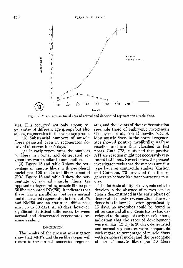
458
15
14
13-
12-
11
10
9
8
7
6
5
4
3
2
1
FRANZ S. F. MONG
-
. N O R M A L
0 D E N E R V A l E D
10 20 30 40 6 5 90 150 270
D A Y S
Fig. 13 Mean cross-sectional area of normal and denervated regenerating muscle fibers.
ates. This occurred not only among re- generates of different age groups but also among regenerates in the same age group.
(b) Substantial numbers of muscle fibers persisted even in regenerates de- prived of nerves for 65 days.
(c) In early regenerates, the numbers of fibers in normal and denervated re- generates were similar to one another.
(3 ) Figure 15 and table 3 show the per- centage of muscle fibers with peripheral nuclei per 100 nucleated fibers counted (P%). Figure 16 and table 3 show the per- centage of normal muscle fibers (as opposed to degenerating muscle fibers) per 50 fibers counted (NM/50). It indicates that there was a parallelism between normal and denervated regenerates in terms of P% and NM/50 and no statistical differences exist up to 30 days. At 40 days, however, significant statistical differences between normal and denervated regenerates be- come evident.
DISCUSSION
The results of the present investigation show that MEP's and three fiber types will return to the normal innervated regener-
ates, and the events of their differentiation resemble those of embryonic myogenesis (Tennyson et al., '73; Dubowitz, '65a,b). Most muscle fibers in the normal regener- ates showed positive myofibrillar ATPase reaction and are thus classified as fast fibers. Guth ('73) cautioned that positive ATPase reaction might not necessarily rep- resent fast fibers. Nevertheless, the present investigator feels that these fibers are fast type because contractile studies (Carlson and Gutmann, '72) revealed that the re- generates behave like fast contracting mus- cles.
The intrinsic ability of myogenic cells to develop in the absence of nerves can be clearly demonstrated in the early phases of denervated muscle regeneration. The evi- dence is as follows: (1) After approximately 15 days, no myotubes could be found in either case and all myogenic tissues had de- veloped to the stage of early muscle fibers, indicating that the rates of development were similar. (2) Up to 30 days, denervated and normal regenerates were comparable with regard to percentage of muscle fibers with peripheral nuclei and the percentage of normal muscle fibers per 50 fibers

NERVOUS INFLUENCE ON MINCED MUSCLE REGENERATION
0 0
$1 I D “ ”
P
459

FRANZ S. F. MONG
2.0 3'0 4'0 mo[ l8 DAYS
Fig. 15 Number of muscle fibers with peripheral nuclei per 100 fibers counted of normal and denervated regenerates. Data are taken from the proximal part of the regenerates only. No significant differences exist from 10 to 30 days between normal and denervated regenerates. At 40 days, how- ever, the difference becomes apparent.
normal
3.0 40 2 0 DAYS
Fig. 16 Number of normal muscle fibers per 50 fibers counted of normal and denervated regener- ates. Data are taken from proximal part of the regenerates only. No significant differences exist from 10 to 30 days between normal and denervated regenerates. On 40 days, however, the difference be- comes apparent.

NERVOUS INFLUENCE ON MINCED MUSCLE REGENERATION 46 1
counted. (3) The two types of regenerates were also comparable in the total numbers of muscle fibers. (4) The regenerated mus- cle fibers in both cases showed clear cross striations. (5) Both SDH and myofibrillar ATPase reactions were positive and primi- tive in denervated regenerates and they could not be distinguished from those of normal ones up to 30 days postoperatively. This intrinsic ability of myogenic tissues to differentiate has also been observed in other regeneration studies (Allbrook and Aitkin, '51; Hall-Craggs and Seyan, '75), and in the studies on muscle ontogeny (Zelena, '62; Engel and Karpati, '68; Shafiq et al., '72).
The only consistent difference between normal and denervated regenerates in the early stages was the smaller fiber cross sec- tional areas in the denervated regenerates. First, the small cross sectional area might not necessarily imply abnormality. Second, it could well be due to the lack of use of the denervated legs rather than lack of nerve fibers, since it is well known that use and disuse of muscle affects muscle fiber diam- eters (Edgerton et al., '72; Maier et al., '73; Tomanek and Lund, '74). In the case of denervated regenerates, the leg was per- mantly flexed and not used. Thus the small fiber cross sectional area could be due to the lack of exercise.
Starting around 40 days, differences be- tween normal and denervated regenerates became evident. Histochemically, the nor- mal regenerates were in the last stage of development - histochemical fiber type differentiation. In the denervated regener- ates, however, there were no indications of such activity, although both SDH and myofibrillar ATPase remained positive for a long time. Morphologically, degenerating muscle fibers could be easily seen. Thus, nerves seem to be important in the late phase of regeneration, namely the differen- tiation of fiber types and maintenance of structural integrity of muscle fibers. This view is also shared by Hall-Craggs and Seyan ('75) and Hsu ('71) in regeneration studies, Shafiq et al., ('72) and Engel and Karpati ('68) in ontogenic studies and
Askanas et al. ('72) and Crain ('70) in tissue culture studies.
Contractile properties of the denervated regenerates were not studied in the pres- ent investigation. Carlson and Gutmann ('72) observed contractile activities of innervated regenerates upon direct stimu- lation as early as eight days postoperative- ly. At this early date, functional contact be- tween nerves and muscle fibers has not developed yet. In recent studies with de- nervated sliced muscle transplantation ex- periments, Carlson and Gutmann (personal communication) also observed contractile activity in the denervated transplants. Thus, one would speculate that the dener- vated minced muscle regenerates would also be contractile upon direct stimulation.
ACKNOWLEDGMENT
The author would like to thank Dr. Bruce M. Carlson for his patience and care- ful guidance in carrying out this research and Mr. Peter P. S. Ruey for his assistance in statistical analysis.
LITERATURE CITED Allbrook, D. B., and J. T. Aitkin 1951 Reinnervation
of skeletal muscle after acute ischemia. J. Anat. (London), 85: 376-390.
1972 Histochemistry of cultured aneural chick muscle: morphological maturation without differentiation of fiber types. Exp. Neurol., 37: 218-230.
Carlson, B. M. 1968 Regeneration of the completely excised gastrocnemius muscle in the frog and rat from minced muscle fragments. J. Morph., 125: 447- 471.
1970 The regeneration of entire muscle from minced fragments. In: Regeneration of Striated Muscle and Myogenesis. A. Mauro, S. A. Shafiq and A. T. Milhorat, eds. Excerpta Medica, Amsterdam,
Carlson, M. B., and E. Gutmann 1972 Development of contractile properties of minced muscle regener- ates in the rat. Exp. Neurol., 36: 239-249.
Crain, S. M. 1970 Bioelectric interactions between cultured fetal rodent spinal cord and skeletal mus- cle after innervation in oitro. J. Exp. Zool., 173:
Dubowitz, V. 1965a Enzyme histochemistry of skele- tal muscle, Part I (developing animal muscle). J. Neurol. Neurosurg. Psychiat., 28: 516-519.
1965b Enzyme histochemistry of skeletal muscle, Part I1 (developing human muscle). J. Neu- rol. Neurosurg. Psychiat., 28: 519-524.
Askanas, V., S . A. Shafiq and A.T. Milhorat
pp. 194-211.
353-370.

462 FRANZ S. F. MONG
Edgerton, V. R., R. J. Barnard, J. B. Peter, C.A. Gillespie and D. R. Simpson 1972 Overloaded skeletal muscles of a nonhuman primate (Galago senegalinsis). Exp. Neurol., 37: 322-339.
Engel, W. K., and G. Karpati 1968 Impaired skeletal muscle maturation following neonatal neurectomy. Devel. Biol., 17: 713-723.
Guth, L. 1973 Fact and artifact in histochemical pro- cedure for myofibrillar ATPase. Exp. Neurol., 41:
Hall-Craggs, E. C. B., and H. S. Seyan 1975 Histo- chemical changes in innervated and denervated skeletal muscle fibers following treatment with Bupivacaine (Marcain). Exp. Neurol., 46: 345-354.
Hsu, L. 1971 The role of nerves in the regeneration of minced skeletal muscle in adult anurans. Ph.D. Diss., Univ. of Michigan.
Hudgson, P., and E. J. Field 1973 Regeneration of muscle. In: The Structure and Function of Muscle. Vol. 11. G. H. Bourne, ed. Academic Press Inc., New York, pp. 312-363.
Maier, A., E. Eldred and V. R. Edgerton 1973 The effects on spindles of muscle atrophy and hypertro- phy. Exp. Neurol., 37: 100-123.
Mong, F. S. F. 1975 Nervous influence on minced muscle regeneration in rats. Anat. Rec., 181: 429.
Nachlas, M. M., K. C. Tsou, E. de Souza, C. S. Cheng and A. M. Seligman 1957 Cytochemical demon- stration of succinic dehydrogenase by the use of new p-nitrophenyl substituted ditetrazole. J. Histochem. and Cytochem., 5: 420-436.
Padykula, H. A., and E. Herman 1955 Factors affecting the activity of adenosine triphosphatase and other phosphatases as measured by histochemi-
440-450.
cal techniques. J. Histochem. and Cytochem., 3:
Pearse, A. G. E. 1972 Histochemistry. Vol2. Third ed. Churchill Livingstone, London.
Peter, J., R. J. Barnard, V. R. Edgerton, C. Y. Gillespie and K. E. Stempel 1972 Metabolic profiles of three fiber types of skeletal muscle in guinea pigs and rabbits. Biochemistry, 11; 2627-2633.
Shafiq, S. A., S. A. Asiedu and A. T. Milhorat 1972 Effect of neonatal neurectomy on differentiation of fiber types in rat skeletal muscles. Exp. Neurol., 35:
Studitsky, A. N . , R. P. Zhenevskaya and O.N. Rumyantseva 1963 The role of neurotrophic influences upon the restitution of structures and function of regenerating muscles. In: The Effect of Use and Disuse in Neuromuscular Functions. E. Gutmann and P. Hnik, eds. Publ. House. Czechoslovak Acad. Sci., Prague, pp. 71-81.
Tennyson, V. M., M. Brzin and L. Kremzner 1973 Acetylcholinesterase activity in the myotube and muscle satellite cell of the fetal rabbit. J. Histochem. Cytochem., 21: 634-652.
Tomanek, R. J., and R. D. Lund 1974 Degeneration of different types of skeletal muscle fibers. 11. Immobilization. J. Anat., 118: 531-541.
Winer, B. J. 1971 Statistical Principle in Experimen- tal Design. Second ed. McGraw Hill Co., New York.
Zelena, J. 1962 The effect of denervation on muscle development. in: The Denervated Muscle. E. Gut- mann, ed. Publishing House of Czech. Acad. Sci., Prague, pp. 103-126.
Zhenevskaya, R. P. 1962 Experimental histologic in- vestigation of skeletal muscle tissue. Rev. Canad. Biol., 21; 457-470.
16 1 - 169.
529-540.