Proteomic Analysis of Ocular Surface Components by use of ... · Johannes Gutenberg-Universität in...
265
Proteomic Analysis of Ocular Surface Components by use of HPLC based Mass Spectrometric Strategies Dissertation zur Erlangung des Grades „Doktor der Naturwissenschaften“ am Fachbereich Biologie der Johannes Gutenberg-Universität in Mainz von Sebastian Funke geboren in Iserlohn Mainz, 15.09.2014
Transcript of Proteomic Analysis of Ocular Surface Components by use of ... · Johannes Gutenberg-Universität in...
Proteomic Analysis
of Ocular Surface Components
by use of HPLC based
Mass Spectrometric Strategies
Dissertation
zur Erlangung des Grades
„Doktor der Naturwissenschaften“
am Fachbereich Biologie der
Johannes Gutenberg-Universität
in Mainz
von
Sebastian Funke
geboren in Iserlohn
Mainz, 15.09.2014

Dekan:
1. Berichterstatter:
2. Berichterstatter:
Tag der mündlichen Prüfung: 20.04.2015

I
Annotation
Parts of the thesis have been published in international journals and/or presented on
international conferences
Publications related to the thesis
Funke, S., Azimi, D., Wolers, D, Grus, F. H, Pfeiffer, N. 2012. Longitudinal analysis of taurine
induced effects on the tear proteome of contact lens wearers and dry eye patients using a
RP-RP-Capillary HPLC-MALDI TOF/TOF MS approach. Journal of Proteomics 75: 3177-
3190
Bell, K., Funke, S., Pfeiffer, N., and Grus, F.H. 2012. Serum and antibodies of glaucoma
patients lead to changes in the proteome, especially cell regulatory proteins, in retinal cells.
PLoS One 7(10): e46910
Bell, K., Gramlich, O. W., Von Thun Und Hohenstein-Blaul, N., Beck, S., Funke, S., Wilding,
C.; Pfeiffer, N., Grus, F. H. 2013. Does autoimmunity play a part in the pathogenesis of
glaucoma? Progress in Retinal and Eye Research 36: 199-216
Von Thun Und Hohenstein-Blaul, N., Funke, S., Grus, F.H. 2013. Tears as a source of
biomarkers for ocular and systemic diseases. Experimental Eye Research. 117: 126-137
Boehm, N., Funke, S., Wiegand, M., Wehrwein, N., Pfeiffer, N., and Grus, F.H. 2013.
Alterations in the tear proteome of dry eye patients--a matter of the clinical phenotype.
Investigative Ophthalmolology & Vision Science 54(3): 2385-2392
Perumal, N., Funke, S., Pfeiffer, N., Grus, F. H. 2014. Characterization of lacrimal proline-
rich protein 4 (PRR4) in human tear proteome. Proteomics 00: 1-12
Bell, K., Wilding, C., Funke, S., Pfeiffer, N., Grus, F. H. 2014. Protective effect of 14-3-3
antibodies on stressed neuroretinal cells via the mitochondrial pathway. BMC Neuroscience,
submitted
Perumal, N., Funke, S., Wolters, D., Pfeiffer, N., Grus, F. H. 2014. Upregulation of lacrimal
proline-rich protein 4 (PRR4) in human reflex tear proteome. Proteomics, submitted
Funke, S., Beck, S., Lorenz, K., Kotterer, M., Wolters, D., Pfeiffer, N., Grus, F. H. 2014.
Analysis of the effects of preservative-free tafluprost on the tear proteome. Opthalmology,
submitted
II
Publications related to the thesis (in preparation)
Funke, S., Bell, K., Wilding, C., Wolters, D., Pfeiffer, N., Grus, F. H. Taurine effects on the
conjunctival cell proteome.
Funke, S., Bechter, L., Perumal, N., Wolters, D., Pfeiffer, N., Grus, F. H. Proteomic analysis
of glaucomatous human trabecular meshwork.
Funke, S., Perumal, N., Gabelt-Scheurich, S., Gramlich, O., Beck, S., Boehm, N., Wolters,
D., Pfeiffer, N., Grus, F. H. Proteomic mass spectrometric analysis of the human
glaucomatous retina.
Funke, S., Perumal, N., Scieranski, M., Beck, S., Pfeiffer, N., Grus, F. H. Proteomic
investigation on the tear proteome of glaucoma patients with dry eye sensation.
Funke, S., Perumal, N., Gabelt-Scheurich, S., Mwiiri, F. K., Wolters, D, Gerbig, C., Weiler, L.,
Pfeiffer, N., Grus, F. H. In depth mass spectrometric analyis of the porcine retina proteome.
Perumal, N., Funke, S., Wolters, D., Pfeiffer, N., Grus, F. H. Identification of human tear fluid
biomarkers in different dry eye disease subgroups using label-free quantitative proteomics.
Poster presentations on international conferences
Funke, S., Azimi, D., Pfeiffer, N., Grus F. H. 2011. Taurine induced effects on the tear
proteome of dry eye patients and contact lens wearers using a RP-RP-2D-capillary-LC-
MALDI-TOF/TOF MS approach. 6680-D1167. Fort Lauderdale, USA. ARVO Visionary
Genomics World Conference 2011
Perumal, N., Funke, S., Boehm, N., Peiffer, N., Grus, F. H. 2011. Defining proline rich
proteins (PRPs) distribution in tear proteome. 6681-D1168. Fort Lauderdale, USA. ARVO
Visionary Genomics World Conference 2011
Wolters, D., Funke, S., Grus, F. H., Pfeiffer, N. 2012. Post transcriptional modifications in
mass spectrometry based proteomics data. P8. Obergurgl, Austria. ARVO Optic Nerve
Degeneration and Ageing Conference 2012.
Wilding, C., Bell, K., Funke, S., Pfeiffer, N., Grus, F. H. 2012. Gamma synuclein antibody
effects on mitochondrial apoptosis pathways. P2. Obergurgl, Austria. ARVO Optic Nerve
Degeneration and Ageing Conference 2012.
III
Wilding, C., Bell, K., Funke, S., Beck, S., Peiffer, N., Grus, F. H. 2013. Protective effect of
GFAP antibody on RGC5 cells via actin cytoskeleton pathways. 432-A0163. Seattle, USA.
ARVO Life Changing Research World Conference 2013
Funke, S., Wolters, D., Weiler, L., Mwiiri, F. K., Bell, K., Wilding, C., Peiffer, N., Grus, F. H.
2013. In depth analysis of retinal proteins using a combinatory HPLC based mass
spectrometric platform. 698-D0255. Seattle, USA. ARVO Life Changing Research World
Conference 2013
Bell, K., Wilding, C., Funke, S., Peiffer, N., Grus, F. H. Changes in antibodies from glaucoma
patients lead to changes in apoptosis pathways of neuroretinal cells. 1605-D0032. Seattle,
USA. ARVO Life Changing Research World Conference 2013
Perumal, N., Funke, S., Pfeiffer, N., Grus, F. H. Characterization and post-translational
modifications of proline-rich protein 4 (PRP4) in basal and reflex tear proteome of human.
2453-D0058. Seattle, USA. ARVO Life Changing Research World Conference 2013
Perumal, N., Funke, S., Pfeiffer, N., Grus, F. H. 2013. Significant up-regulation of proline-rich
protein 4 (PRR4) in human reflex tear proteome. St Malo, France. EuPA Conference 2013
Funke, S., Lorenz, K., Kotterer, M., Beck, S., Wolters, D., Pfeiffer, N., Grus, F. H. 2014.
Analysis of the effects of Taflotan® sine on tear protein profiles. 1474-C0113. Orlando, USA.
ARVO Leading Eye and Vision Research World Conference 2014
Perumal, N., Funke, S., Wolters, D., Pfeiffer, N., Grus, F. H. 2014. In depth protein profiling
and identification of tear fluid biomarkers in different subgroups of dry eye disaese: Proline-
rich protein 4 (PRR4) as apotential biomarker for aqueous-deficient dry eye syndrome. 2002-
B0301. Orlando, USA. ARVO Leading Eye and Vision Research World Conference 2014
Wilding, C., Bell, K., Funke, S., Pfeiffer, N., Grus, F. H. 2014. Participation of ERP57 in the
pro-survival potential of GFAP antibodies on RGC5. 2438-B0137. Orlando, USA. ARVO
Leading Eye and Vision Research World Conference 2014
Perumal, N. , Funke, S., Pfeiffer, N., Grus, F. H. 2014. Identification of human tear fluid
biomarkers in different dry eye disease subgroups using label-free quantitative proteomics.
Madrid, Spain. 13th Human Proteome Organization World Congress, accepted
IV
Oral papers on international conferences
Funke, S., Beck, S., Boehm, N., Pfeiffer, N., Grus, F. H. 2012. Retinal biomarkers in
glaucoma. O063. Berlin, Germany. ISER XX Biennial Meeting of the International Society for
Eye Research Conference 2012
Funke, S., Wolters, D., Bell, K., Wilding, C., Pfeiffer, N., Grus, F. H. 2012. Protein
modifications and ageing. Obergurgl, Austria. ARVO Optic Nerve Degeneration and Ageing
Conference 2012.
Funke, S., Bell, K., Wilding, C., Perumal, N., Pfeiffer, N, Grus, F. H. 2013. Mass
spectrometric based strategies to recover and characterize proteins from retinal sample
species. Obergurgl, Austria. Optic Nerve Conference 2013
Wilding, C., Bell, K., Beck, S., Funke, S., Pfeiffer, N., Grus, F. H. 2013. GFAP antibodies
interact with ERP57 on the cell membrane of RGC5 cells and have prosurvival potential
against oxidative stress. Obergurgl, Austria. Optic Nerve Conference 2013
Research prices
Sicca-Förderpreis des Ressorts Trockenes Auge und Oberflächenerkrankungen im
Berufsverband der Augenärzte Deutschlands 2013. Taurine induced effects on tear film and
ocular surface revealed by mass spectrometric based proteomics. Berlin, Germany. 111th
DOG Excellent Vision-Vision of Excellence Conference 2013

I
Aknowledgements
First, I would like to thank my supervisor for the opportunity to realize the thesis in the
Experimental Ophthalmology lab of the University Medical Center Mainz and for his help,
support and scientific experience he shared with me. I also thank my co-supervisor for his
support.
Furthermore, I would like to thank my collegues for their contribution to the realization of this
work.
Special thanks go to my wife and families for their help and support over the past years.
Table of Content
I
Table of Content
Table of Content I
1 Purpose 1
2 Introduction 2
2.1 Mass spectrometry (MS) based proteomics ............................................................... 2
2.2 Bottom Up (BU) vs. Top Down (TD) ........................................................................... 4
2.3 MALDI-TOF-(TOF) MS (/MS) ..................................................................................... 6
2.4 LTQ-Orbitrap XL MS .................................................................................................. 8
2.5 HPLC in mass spectrometry ......................................................................................10
2.6 Ocular surface proteomics ........................................................................................12
2.7 Dry Eye: Causes and consequences ........................................................................15
2.8 Therapeutic management with special focus on Taflotan® sine and taurine ..............18
3 Material & Methods 23
3.1 Training samples .......................................................................................................23
3.2 Study samples ..........................................................................................................24
3.2.1 Tear fluid ..............................................................................................................24
3.3 Sample preparation ...................................................................................................25
3.3.1 Intact sample processing ......................................................................................25
3.3.2 SDS-PAGE ...........................................................................................................25
3.3.3 In-gel digestion .....................................................................................................26
3.3.4 Solid phase extraction (SPE) ................................................................................26
3.3.5 Microbore-RP-HPLC for MALDI Top Down (TD) analysis .....................................27
3.3.6 Capillary-RP-RP-HPLC (for MALDI) .....................................................................28
3.3.7 Capillary-RP-HPLC (for ESI) ................................................................................31
3.3.8 MALDI-TOF/TOF MS/MS .....................................................................................33
3.3.9 ESI LTQ Orbitrap XL MS ......................................................................................34
3.3.10 Data processing & Protein identification ...............................................................36
3.3.11 Protein quantification and statistics .......................................................................38

Table of Content
II
3.3.12 Protein functional annotation analysis...................................................................38
3.3.13 Ocular surface studies ..........................................................................................39
4 Results 46
4.1 Microbore-RP-HPLC (for MALDI) ..............................................................................46
4.2 Capillary-RP-RP-HPLC (for MALDI) ..........................................................................53
4.3 Capillary-RP-HPLC (for ESI) .....................................................................................57
4.4 Proteomic characterization of ocular surface sample species ...................................64
4.4.1 Capillary tears ......................................................................................................64
4.4.2 Schirmer strip eluted tears ....................................................................................68
4.4.3 General tear results ..............................................................................................73
4.4.4 Conjunctival cells ..................................................................................................75
4.5 Influence of Taflotan® sine on the ocular surface of POAG patients ..........................79
4.6 Influence of taurine on the ocular surface ..................................................................89
4.6.1 Taurine effects on the tear proteome of contact lens wearers and sicca patients .89
4.6.2 Taurine effects on the conjunctival proteome........................................................92
5 Discussion 102
5.1 Top Down (TD) vs. Bottom Up (BU) ........................................................................ 102
5.2 Bottom-up (BU) analysis: LC-MALDI vs LC-ESI workflow ....................................... 104
5.3 Characterization of the ocular surface proteome ..................................................... 106
5.4 Effects of Taflotan® sine on the ocular surface proteome ........................................ 110
5.5 Taurine effects on the tear and cell proteome ......................................................... 114
5.6 Deviated strategies facing dry eye and ocular surface complications ...................... 119
6 Conclusion 121
7 Summary 122
8 Abbreviations 123
9 Instruments/Technical equipment 125
10 Software 127
11 Chemicals 128
12 References 130
Table of Content
III
13 Appendices 179
14 Eidesstattliche Erklärung 253
Purpose
1
1 Purpose
The ocular surface is a complex functional unit, not completely understood, ecspecially
focusing on the proteome. Dry eye disease as a multifactorial disorder directly impacts the
status of the ocular surface encircling tear film and cellular components, reflected by
quantitative and qualitative changes in the proteome. Therefore, in this work, an intensive
proteomic characterization of ocular surface components with special focus on tear film and
ocular surface epithelial cells should be realized. Beside explorative analysis of the ocular
surface proteome, two therapeutic strategies should be determined, encircling the influence
of taurine eye drops on the tear film proteome of dry eye patients and contact lens wearers
as well as the influence on the ocular surface of primary open angle glaucoma (POAG)
patients switching from a common topical anti-glaucoma medication (Xalatan®) to a
preservative-free formulation (Taflotan® sine) in terms of ocular surface complications.
Accordingly, for “in depth” proteomic analysis of ocular surface components, HPLC based
mass spectrometric workflows should be developed, optimized and compared. Results
should provide a detailed proteomic view to the ocular surface and uncover underlying
mechanism and potentials in the two focused therapeutic interventions, taurine and Taflotan®
sine appliance.
Introduction
2
2 Introduction
2.1 Mass spectrometry (MS) based proteomics
Proteomics first drafted by the groups of Wilkins and Wasinger (Wasinger et al., 1995,
Wilkins et al., 1996a) referring to “PROTein complement of the genOME” (Apweiler et al.,
2009, Kraj and Silberring, 2008, Wilkins et al., 1996b) describes the analysis of all proteins in
living systems (Guerrera and Kleiner, 2005). Displaying high dynamics, (Kettman et al.,
2001) (Fig.1) a highlighted aim in proteomic science is the illumination of molecular changes
in the course of a disease with the aim to find specific disease-related proteins indicating
early diagnosis, molecular targeting or monitoring of therapeutic success (Etzioni et al., 2003,
Kraj and Silberring, 2008, Lescuyer et al., 2007, Rifai et al., 2006). MS evolved as the
proteomic method of choice starting up from the 1980s demonstrating sensitivity and
accuracy towards proteomic investigations (Mann et al., 2001b). Generally, the first step of a
proteomic workflow is to extract proteins or peptides from their sample source using various
homogenization and extraction techniques (Chertov et al., 2004, Cox and Emili, 2006, Ferro
et al., 2000, Goklen and Hatton, 1987, Holmquist and Carlson, 1977, Kushnirov, 2000,
Molloy et al., 1999, Naoe et al., 1999, Naoe et al., 1998, Selber et al., 2004). Thus, for MS
prior to protein/peptide ionization complexity reduction is mandatory to avoid masking effects
by high abundant components (Granger et al., 2005) and to reduce ion suppression
provoked by salts, detergents and coextracted contaminants (Annesley, 2003, Hirabayashi et
al., 2007, Jessome and Volmer, 2006, Piwowar et al., 2009, Remane et al., 2010b, Wang et
al., 2003). Therefore, MS compatible fractionation (Issaq, 2001, Issaq et al., 2005) or
depletion (Bjorhall et al., 2005, Magagnotti et al., 2010) strategies have been developed.
After sample processing, components are ionized and introduced to MS instruments showing
a common fundamental design consisting of ion source, optics, mass analyzer and data
processing electronics (Guerrera and Kleiner, 2005, Yates et al., 2009). In case of
proteomics soft ionization with low internal intensities leading to low ion fragmentation is
usually coupled to mass analyzers (Daniel et al., 2002, Pappin et al., 1993).The two main
soft ionization techniques are matrix assisted laser desorption ionization (MALDI) developed
by Karas and Hillenkamp (Karas and Hillenkamp, 1988), in parallel by Tanaka and collegues
(Tanaka et al., 1988) and electrospray ionization (ESI) invented by Fenn and collegues
(Fenn et al., 1989, 1990) (Fig 2). MALDI features analyte co-crystallization with UV-
absorbent small organic acids (Bird et al., 2002, Canas et al., 2007) and high vacuum pulsed
laser evaporation leading to matrix and analyte desorption (Bahr et al., 1992, Erra-Balsells
and Nonami, 2003, Koubenakis et al., 2004). The matrix transfers and receives protons and
is believed to absorb energy as well as to isolate analytes from each other (Canas et al.,
2007). Usually, MALDI generates singly charged ions in the form [M+H]+ or [M-H]+ (Karas et
Introduction
3
al., 2000b, Yates et al., 2009). In contrast ESI features ionization by application of high
voltage (2-6kV) to a soluble analyte passing a narrow capillary which leads to the formation
of an electrically charged spray, Taylor cone (Yates et al., 2009) resulting in micro-drop
evaporation and release of multiply charged analyte ions (Fenn et al., 1990, Whitehouse et
al., 1985, Yates et al., 2009). However, ESI and MALDI have different properties, ionization
characteristics, sensitivity values and compatibilities. Importantly, MALDI is used in the off-
line mode decopling sample processing and MS analysis (Baessmann et al., 2005, Mitulovic
and Mechtler, 2006, Murray, 1997, Rejtar et al., 2002, Stevenson and Loo, 1998) with
respect to some exceptions (Brivio et al., 2002, Musyimi et al., 2005, Musyimi et al., 2004,
Preisler et al., 1998). In contrast ESI is an on-line technique (He, 2000, Wang et al., 2006,
Xiang et al., 1999). Pulsed MALDI sources are usually combined with time-of-flight (TOF)
analyzers discriminating ions based on their time of flight in a field-free tube (Guerrera and
Kleiner, 2005, Yates, 2000). In contrast continuous ESI sources are commonly coupled to ion
traps (Guerrera and Kleiner, 2005). Due to their complementary character, MALDI and ESI
are proposed to be used in combination (Bodnar et al., 2003, Mo and Karger, 2002, Stapels
and Barofsky, 2004, Stapels et al., 2004). Regarding mass analyzers two fundamental
instrument types exist: scanning/ion beam instruments [TOF, quadrupol (Q)] and traps [ion
trap (IT), Orbitrap, Fourier transform ion cyclotron resonance (FT-ICR)] (Yates et al. 2009).
Accordingly ion separation corresponds to time of flight, quadrupol electric field selection,
selective ion ejection from a three-dimensional trapping field (Mann et al., 2001a) and orbital
ion trapping (Scigelova and Makarov, 2006). Generally two types of information are obtained
by MS, accurate mass and amino acid sequence (Yates, 2000). To achieve information on
the amino acid sequence, parent ions entering the mass analyzer can be further fragmented
using different techniques, e.g. post source decay (PSD), collision induced decay (CID), high
energy C-trap dissociation (HCD), electron capture dissociation (ECD) (Mo and Karger,
2002, Olsen et al., 2007, Suckau et al., 2003) or electron transfer dissociation (ETD) (Mikesh
et al., 2006, Syka et al., 2004). The fragments can be measured with higher resolution e.g. in
TOF/TOF analyzers (Medzihradszky et al., 2000) or in hybrid instruments like QTOF or
Orbitrap analyzers (Hardman and Makarov, 2003, Hu et al., 2005). Method selection strongly
depends on the technical features of a mass analyzer. PSD is a common technique in
MALDI TOF/TOF MS/MS workflows (Medzihradszky et al., 2000) featuring the metastable
fragmentation of peptides by single or double cleavage of the peptide backbone (Suckau et
al., 2003). CID commonly features trap instruments inducing similar fragmentation at the N-O
peptide bond leading to the generation of b- and y-ions with respect to amino acid side
chains (Papayannopoulos, 1995, Schlosser and Lehmann, 2000) by collision with gas
molecules such as argon or helium (Hoteling et al., 2003). However, beside the
heterogeneity of mass analyzers and fragmentation workflows, in this work special focus is

Introduction
4
laid on two instrument types: a MALDI-TOF/TOF and an ESI LTQ Orbitrap hybrid instrument.
The application of both approaches towards the ocular surface proteome in association to
dry eye related phenomena and potential therapeutic approaches were addressed in
particular. Advantages, limitations and also the complementary character of both techniques
were also examined.
Fig 1 Hierarchical principle of proteomics science linked to mass spectrometry (illustrated according to Kraj and
Silberring 2008).
Fig 2 Soft ionization principles for MS based proteomics; A. MALDI: After analyte/matrix cocrystallization analyte
molecules are ionized and vacuum evaporated by laser beam and enter the mass analyzer. B: ESI: Analyte solution passes an electric field leading to Taylor cone formation, charged droplet release and multiply charged analyte ion transfer to the mass analyzer (illustrated according to Kraj and Silberring 2008; www.intechopen.com).
2.2 Bottom Up (BU) vs. Top Down (TD)
Two major MS proteomic categories coexist: “Top Down” (TD) and “Bottom Up” (BU). TD
means the MS introduction of sample ions derived from intact non-digested components
(Reid and McLuckey, 2002), whereas in BU proteins are first enzymatic digested followed by
MS analysis. Accordingly, the categoric definition refers to the entities introduced to an
Introduction
5
analyzer, which is in agreement with the majority of researchers (Capriotti et al., 2011, Reid
and McLuckey, 2002) based on the concept of McLafferty and collegues (Kelleher et al.,
1999). However, there is growing confidence that a combination of both approaches will be
most effective (Mo and Karger, 2002). Regarding TD a clear benefit of MALDI is the high
analysis speed and contaminant robustness playing a predominant role in fast and direct
experiments of crude sample materials and the possibility of storage and reanalysis (Frohlich
and Arnold, 2006, Mukhopadhyay, 2005). In both approaches, there is a certain need of
sample precleaning and purification to increase sensitivity, especially regarding low abundant
components (Kraj and Silberring, 2008) desirable for their potential to predict health status or
treatment success (Chertov et al., 2005, Veenstra et al., 2005). Regarding fractionation and
separation methods applicable to recover low abundant intact peptides/proteins there is still a
lack of development in the proteomics research field (Capriotti et al., 2011). Therefore,
several complexity reducing TD approaches have been addressed, e.g. solvent precipitation
(Chertov et al., 2004, Chertov et al., 2005), filter size exclusion (Berven et al., 2007, Wang et
al., 2003), subcellular fractionation (Cox and Emili, 2006, Huber et al., 2003, Jiang et al.,
2004, Stasyk and Huber, 2004), liquid-liquid extraction (Du et al., 2007, Kjellstrom and
Jensen, 2003) or depletion of predominant proteins (Chromy et al., 2004, Fratantoni et al.,
2010, Fu et al., 2005, Yocum et al., 2005). Regarding TD the special focus of the clinical
proteomic biomarker research in the last decade was on solid phase extraction (SPE)
methods, e.g. agarose/polyacrylamide bead based affinity chromatography (Cuatrecasas,
1970a, b, Nishikawa and Bailon, 1975), magnetic beads (Ketterlinus et al., 2005, Rehm,
2006, van Hagen, 2008), which were successfully used to determine the human proteomic
profiles of body fluids like serum (Baumann et al., 2005, Jimenez et al., 2007a), plasma
(Zhang et al., 2004b), saliva (Wu et al., 2009b), urine (Fiedler et al., 2007), cerebrospinal
fluid (Jimenez et al., 2007b) and tear fluid (Sekiyama et al., 2008). A common TD strategy is
surface enhanced laser desorption ionization (SELDI) ProteinChip® technology providing
component enrichment on surface modified metal chips (Hutchens and Yip, 1993, Issaq et
al., 2002, Tang et al., 2004). In spite of its promising use for proteomic analysis of body fluids
(Issaq et al., 2003, Jr et al., 1999, Paweletz et al., 2001, Petricoin et al., 2004) SELDI was
contrary discussed regarding reproducibility (Baggerly et al., 2004, Bons et al., 2006),
robustness (Gast et al., 2009) and sensitivity (Neuhoff et al., 2004). Beyond technical
aspects, high costs regarding single-use chips are disadvantageous (Rehm, 2006). In
contrast, Callesen and colleagues reviewed the MALDI and SELDI platform regarding m/z
peak biomarker candidate establishment in ovarian cancer and reported high reproducibility
across different research groups and pre-fractionation methods (Callesen et al., 2012,
Callesen et al., 2008) which gives new input to the debate of SELDI application to clinical TD
proteomics. However, sensitivity and reproducibility of the mentioned approaches is very low
Introduction
6
compared to the abilities of high pressure liquid chromatography (HPLC). The use of LC
MALDI for TD proteomic profiling is limited to a few publications. Wall et al. described a TD
LC MALDI approach for the proteomic mapping of whole E.coli cell lysates whereby they first
collected LC fractions in solution followed by a transfer to MALDI tragets (Wall et al., 1999).
Koomen and colleagues investigated on the intact low molecular weight human serum
proteome <5500 m/z by use of a LC MALDI TOF approach (Koomen et al., 2005). Hu et al.
used LC MALDI for oral cancer biomarker screening of saliva samples (Hu et al., 2007).
Thus, in contrast to bead or chip based approaches regarding ocular sample species there is
still a lack of publications highlighting TD LC MALDI proteomics. Furthermore, most of the
reported studies refer to in-solution fraction collection and direct on target collection
approaches automated by robotic units have been attempted (Mukhopadhyay, 2005). Beside
TD approaches to get a deeper view to the proteome BU workflows are a promising toolbox
for clinical research. BU experiments focus on complex enzymatic peptide mixtures (Duncan
et al., 2010) challenging for MS performance due to contaminants, ion suppression and
masking effects (Annesley, 2003, Gumerov et al., 2002, Gustavsson et al., 2001, Remane et
al., 2010a, Sjodahl et al., 2005). Therefore, like in TD prefractionation, methods have to be
integrated at protein level, at peptide level or at both stages. One usual strategy is to
fractionate proteins in the first dimension by one-dimensional (1D) or two-dimensional (2D)
gel electrophoresis followed by targeted digestion of molecular weight areas, gel lanes or
single spots (Fountoulakis and Langen, 1997, Rosenfeld et al., 1992). In contrast,
multidimensional protein identification technology (Mudpit) highlights the gel-free digestion
and fractionation of complex protein extracts by use of multidimensional HPLC systems
(Chen et al., 2006, Kislinger et al., 2005, McDonald et al., 2002). In fact, peptides generally
cope better with chromatography and MS than proteins regarding solubility, ionization and
fragmentation (Duncan et al., 2010). Despite its lab intensive character, a certain advantage
of gel-based systems is the effective removal of contaminants through electrophoresis (Lu
and Zhu, 2005) taking advantage of the reproducibility of precast gels (Berkelman et al.,
2010, Herbert et al., 2001). Furthermore, the gel technology itself allows quantification, for
example realized by scanning densitometry (Claeys et al., 1995, Ng et al., 2000b) as well as
control steps for method development. In summary, strategy selection, BU or TD strongly
depends on research questions and sample type. The evaluation of both strategies for the
analysis of ocular components is featured in this work.
2.3 MALDI-TOF-(TOF) MS (/MS)
MALDI-TOF MS provides ion mass-to-charge ratio recording flight time in a field-free vacuum
adjusted metal tubing of specific length (Domon and Aebersold, 2006) after source ionization
considering faster travel of low m/z than high m/z molecules (Rehm, 2006). Resolution of
Introduction
7
TOF analyzers is limited but can be increased with tubing elongation (Schiller et al., 2007).
By coupling two TOF analyzers in tandem, PSD or CID fragments can be interpreted with
higher resolution (Medzihradszky et al., 2000). In these approaches a whole sample MS
spectrum is generated in the first run, whereas in a follow-up run parent peaks corresponding
to a mass list of the MS spectrum as well as their fragments are selected by a voltage
dependent ion gating occurring in the precursor ion selector (PCIS) due to their flight time
(Rehm, 2006, UltraflexTOF/TOFOperatorManual, 2001). Resolution is additionally increased
by an “electrostatic mirror”, the reflector (Schiller et al., 2007), which turns on ions
compensating for small differences in their initial kinetic energies due to adsorbed initial laser
energy (Aebersold and Mann, 2003, Rehm, 2006). This consequently results in higher
resolution generating Gaussian shaped peak pattern allowing exact mass determination of
isotopic peaks in MS/MS spectra (Suckau et al., 2003). MS spectra can be used for simple
peptide mass fingerprinting (PMF) via data bases, whereas MS/MS spectra give more
additional information on the amino acid sequence in more complex samples (Kraj and
Silberring, 2008, van Hagen, 2008). However, since the reflector works only for ions within a
specific energy potential range, the analytic performance is usually limited to small
components <4000 Da (Rehm, 2006) (UltraflexTOF/TOFOperatorManual, 2001). Decreased
ion transmission to the detector in the reflector mode (Hortin, 2006) is the main reason for
the analysis of intact peptides or proteins in the instruments linear mode without reflector.
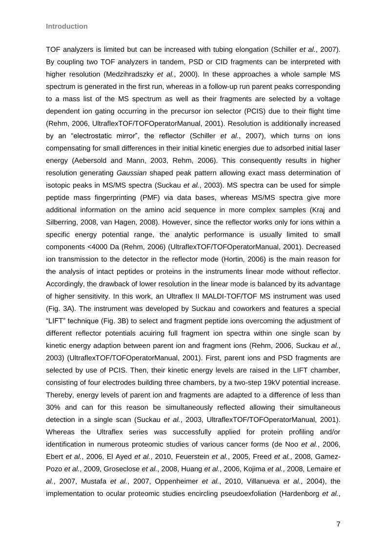
Accordingly, the drawback of lower resolution in the linear mode is balanced by its advantage
of higher sensitivity. In this work, an Ultraflex II MALDI-TOF/TOF MS instrument was used
(Fig. 3A). The instrument was developed by Suckau and coworkers and features a special
“LIFT” technique (Fig. 3B) to select and fragment peptide ions overcoming the adjustment of
different reflector potentials acuiring full fragment ion spectra within one single scan by
kinetic energy adaption between parent ion and fragment ions (Rehm, 2006, Suckau et al.,
2003) (UltraflexTOF/TOFOperatorManual, 2001). First, parent ions and PSD fragments are
selected by use of PCIS. Then, their kinetic energy levels are raised in the LIFT chamber,
consisting of four electrodes building three chambers, by a two-step 19kV potential increase.
Thereby, energy levels of parent ion and fragments are adapted to a difference of less than
30% and can for this reason be simultaneously reflected allowing their simultaneous
detection in a single scan (Suckau et al., 2003, UltraflexTOF/TOFOperatorManual, 2001).
Whereas the Ultraflex series was successfully applied for protein profiling and/or
identification in numerous proteomic studies of various cancer forms (de Noo et al., 2006,
Ebert et al., 2006, El Ayed et al., 2010, Feuerstein et al., 2005, Freed et al., 2008, Gamez-
Pozo et al., 2009, Groseclose et al., 2008, Huang et al., 2006, Kojima et al., 2008, Lemaire et
al., 2007, Mustafa et al., 2007, Oppenheimer et al., 2010, Villanueva et al., 2004), the
implementation to ocular proteomic studies encircling pseudoexfoliation (Hardenborg et al.,
Introduction
8
2009, Joachim et al., 2007a), glaucoma (Brust et al., 2008, Grus et al., 2008, Joachim et al.,
2007b, Joachim et al., 2009, Joachim et al., 2008, Joachim et al., 2010) or eye surface
diseases (Acera et al., 2011, Kramann et al., 2011) is still expandable. In this work the
instrument is used as for TD as well as for BU analysis.
Fig 3 Principle of the Ultraflex II MALDI TOF-TOF MS instrument; A: Panorama view of the instruments interieur
containing MALDI source at the left side and flight tube on the right side of the machine; B: Ions passing the flight tube are compensated for kinetic enegy differences by the reflector leading to increased resolution. Kinectic energy differences of precursor masses and corresponding fragment ions are approximated to each other in a LIFT chamber allowing their simoultan detection.
2.4 LTQ-Orbitrap XL MS
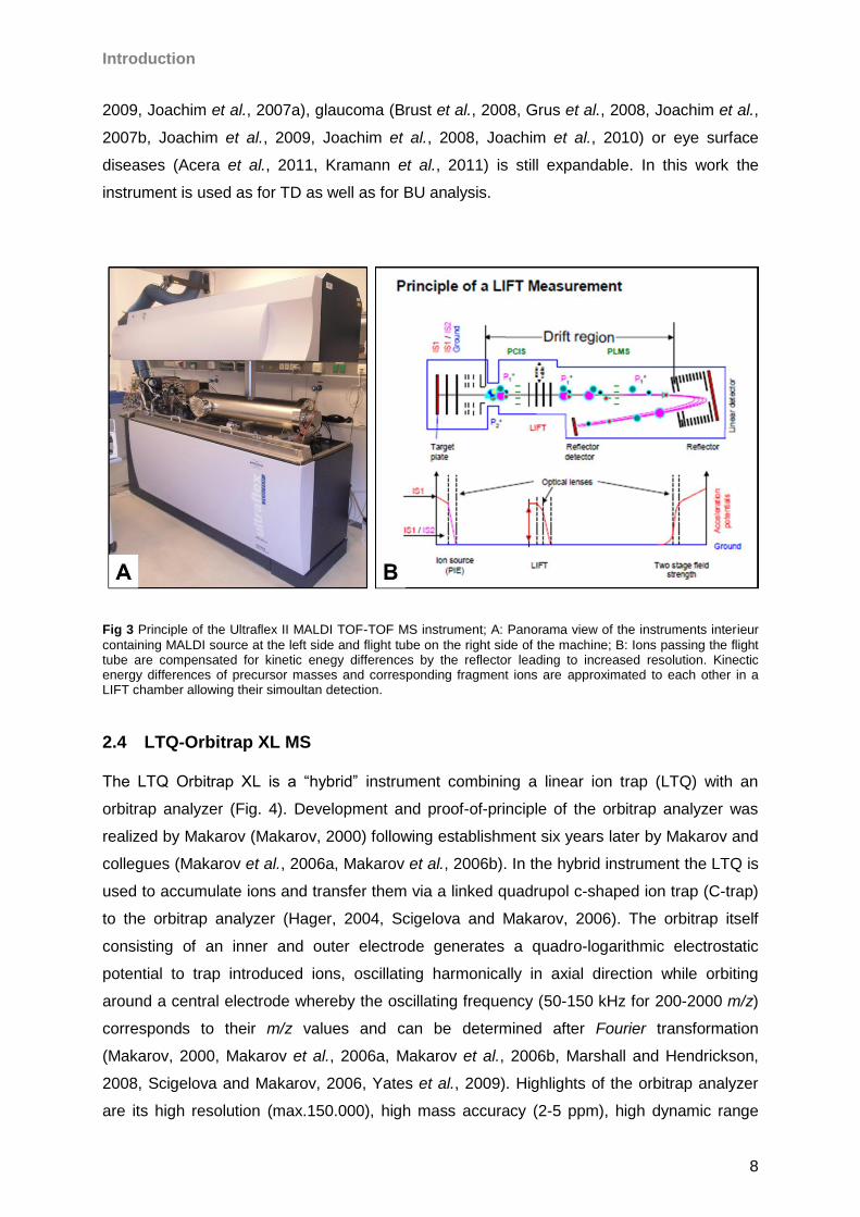
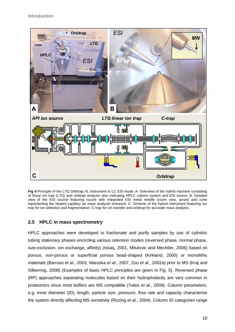
The LTQ Orbitrap XL is a “hybrid” instrument combining a linear ion trap (LTQ) with an
orbitrap analyzer (Fig. 4). Development and proof-of-principle of the orbitrap analyzer was
realized by Makarov (Makarov, 2000) following establishment six years later by Makarov and
collegues (Makarov et al., 2006a, Makarov et al., 2006b). In the hybrid instrument the LTQ is
used to accumulate ions and transfer them via a linked quadrupol c-shaped ion trap (C-trap)
to the orbitrap analyzer (Hager, 2004, Scigelova and Makarov, 2006). The orbitrap itself
consisting of an inner and outer electrode generates a quadro-logarithmic electrostatic
potential to trap introduced ions, oscillating harmonically in axial direction while orbiting
around a central electrode whereby the oscillating frequency (50-150 kHz for 200-2000 m/z)
corresponds to their m/z values and can be determined after Fourier transformation
(Makarov, 2000, Makarov et al., 2006a, Makarov et al., 2006b, Marshall and Hendrickson,
2008, Scigelova and Makarov, 2006, Yates et al., 2009). Highlights of the orbitrap analyzer
are its high resolution (max.150.000), high mass accuracy (2-5 ppm), high dynamic range
Introduction
9
(>103) and a m/z range of 6000 (Scigelova and Makarov, 2006, Yates et al., 2009). Actually,
instrumental mass deviation can be decreased to <1 ppm by the introduction of “lock-mass”
background signals for internal calibration (Olsen et al., 2005, Scheltema et al., 2008).
Usually, the LTQ Orbitrap workflow features ion fragmentation in the LTQ ion trap allowing
multiple fragmentation levels (MSn) and generates full MS scans in the orbitrap taking
advantage of the speed (3-5 spectra/s) and sensitivity of the ion trap considering the mass
accuracy and resolution of the orbitrap (Scigelova and Makarov, 2006, Yates et al., 2009).
Moreover, ion trap and orbitrap work in parallel since the initial orbitrap high resolution
spectrum is used to select precursors for fragmentation in the ion trap while high resolution
orbitrap measurement is still in progress (Scigelova and Makarov, 2006). For fragmentation
CID, HCD, ECD or in modified machines ETD can be choosen and combined in experimental
designs. Numerous studies in different proteomic fields, especially cancer proteomics (Kim et
al., 2012, Kosanam et al., 2011, Lu et al., 2009, Saratsis et al., 2012, Whelan et al., 2009,
Zeng et al., 2011) have been done since its commercial introduction in 2005. Nevertheless,
the number of opthalmological studies run on the LTQ Orbitrap XL is still limited (Bennett et
al., 2011, de Souza et al., 2006, Galiacy et al., 2011, Srinivasan et al., 2012). In this work the
instrument is used for a BU workflow for the analysis of the ocular surface.
Introduction
10
Fig 4 Principle of the LTQ Orbitrap XL instrument in LC ESI mode; A: Overview of the hybrid machine consisting
of linear ion trap (LTQ) and orbitrap analyzer also indicating HPLC column system and ESI source; B: Detailed view of the ESI source featuring nozzle with integrated ESI metal needle (zoom view, arrow) and cone representing the heated capillary as mass analyzer entrance; C: Scheme of the hybrid instrument featuring ion trap for ion selection and fragmentation, C-trap for ion transfer and orbitrap for accurate mass analysis.
2.5 HPLC in mass spectrometry
HPLC approaches were developed to fractionate and purify samples by use of cylindric
tubing stationary phases encircling various retention modes (reversed phase, normal phase,
size-exclusion, ion exchange, affinity) (Issaq, 2001, Mitulovic and Mechtler, 2006) based on
porous, non-porous or superficial porous bead-shaped (Kirkland, 2000) or monolithic
materials (Barroso et al., 2003, Maruska et al., 2007, Zou et al., 2002a) prior to MS (Kraj and
Silberring, 2008) (Examples of basic HPLC principles are given in Fig. 5). Reversed phase
(RP) approaches separating molecules based on their hydrophobicity are very common in
proteomics since most buffers are MS compatible (Yates et al., 2009). Column parameters,
e.g. inner diameter (ID), length, particle size, pressure, flow rate and capacity characterize
the system directly affecting MS sensitivity (Rozing et al., 2004). Column ID categories range
Introduction
11
from “preparative” (> 10 mm) over “narrow(small)-bore” (4 mm ≥ ID ≥ 2.1 mm) to “open
tubular” (< 25 µm) (Rozing, 2003). However, in proteomics “microbore” (2.1 mm ≥ ID ≥1 mm),
“capillary” (1 mm ≥ ID.≥ 100 µm) and “nanobore” (100 µm ≥ ID ≥ 25 µm) are of common
interest (Rozing, 2003) due to sensitivity increase by ID and flow rate scale-down (Frohlich
and Arnold, 2006). Accordingly, microbore columns demonstrate a higher performance than
“semi-preparative” columns (Bhown et al., 1986, Bowermaster and Mcnair, 1983) and were
early proposed for direct MS coupling (Garcia and Barcelo, 1993) allowing appropriate flow
rates between 50 and 1000 µl/min (Rozing, 2003). Nevertheless, in MALDI experiments ID
and corresponding flow rates are not as crucial as in ESI workflows since fractions are
collected off-line allowing complete solvent evaporation and analyte concentration on the
target plate. In contrast, in on-line ESI approaches, column ID and flow rate will directly affect
sample concentration introduced to the analyzer favoring down-scaled flow rates regarding
ion formation (Karas et al., 2000a). Despite increased sensitivity of nanobore systems
(Mitulovic and Mechtler, 2006), disadvantages on the pratical side addressing risk of column
blocking, overloading, decreased column life times, consequent need for advanced sample
pre-cleaning steps, extended analysis times due to nano flow rates, challenging pressure
and dead volumes, advanced nano pump systems or split systems and higher costs have to
be mentioned (Noga et al., 2007). The use of HPLC techniques combined with gel-based or
orthogonal chromatographic approaches linked to MS allowed the proteomic analysis of
several ophthalmological relevant sample species like e.g. lens (Grey and Schey, 2009,
Hoehenwarter et al., 2005, Shearer et al., 2008, Ueda et al., 2002, Wilmarth et al., 2009),
optic nerve tissue (Bhattacharya et al., 2006), optic nerve head astrocytes (Rogers et al.,
2012), microglia (Glanzer et al., 2007), vitreous humour (Gao et al., 2008, Kim et al., 2007b,
Ouchi et al., 2005, Yu et al., 2008), aqueous humor (Chowdhury et al., 2010, Duan et al.,
2008, Funding et al., 2005), sclera (Frost and Norton, 2007, 2012) and cornea (Dyrlund et
al., 2012, Galiacy et al., 2011, Karring et al., 2006, Karring et al., 2005, Thompson et al.,
2003).Despite overall growing research attemps in ocular proteomics, there is still a high
degree of necessity to use HPLC based methods for a more detailed basic appreciation of
the eye surface realized in the present work. Accordingly, a brief introduction to
achievements and limitations in ocular surface proteomics is given in the following chapters.
Introduction
12
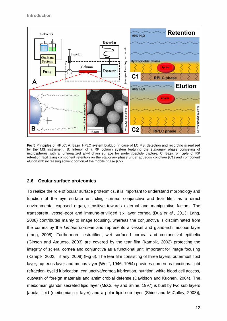
Fig 5 Principles of HPLC; A: Basic HPLC system buildup, in case of LC MS; detection and recording is realized
by the MS instrument; B: Interior of a RP column system featuring the stationary phase consisting of microspheres with a funtionalized alkyl chain surface for protein/peptide capture; C: Basic principle of RP retention facilitating component retention on the stationary phase under aqueous condition (C1) and component elution with increasing solvent portion of the mobile phase (C2).
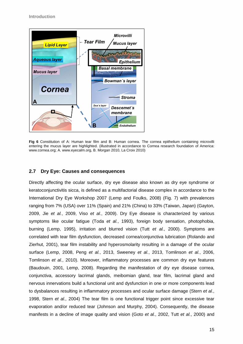
2.6 Ocular surface proteomics
To realize the role of ocular surface proteomics, it is important to understand morphology and
function of the eye surface encircling cornea, conjunctiva and tear film, as a direct
environmental exposed organ, sensitive towards external and manipulative factors. The
transparent, vessel-poor and immune-priviliged six layer cornea (Dua et al., 2013, Lang,
2008) contributes mainly to image focusing, whereas the conjunctiva is discriminated from
the cornea by the Limbus corneae and represents a vessel and gland-rich mucous layer
(Lang, 2008). Furthermore, estratified, wet surfaced corneal and conjunctival epithelia
(Gipson and Argueso, 2003) are covered by the tear film (Kampik, 2002) protecting the
integrity of sclera, cornea and conjunctiva as a functional unit, important for image focusing
(Kampik, 2002, Tiffany, 2008) (Fig 6). The tear film consisting of three layers, outermost lipid
layer, aqueous layer and mucus layer (Wolff, 1946, 1954) provides numerous functions: light
refraction, eyelid lubrication, conjunctiva/cornea lubrication, nutrition, white blood cell access,
outwash of foreign materials and antimicrobial defense (Davidson and Kuonen, 2004). The
meibomian glands’ secreted lipid layer (McCulley and Shine, 1997) is built by two sub layers
[apolar lipid (meibomian oil layer) and a polar lipid sub layer (Shine and McCulley, 2003)],
Introduction
13
which are both associated to lipophil proteins like lipocalins (Glasgow et al., 1995). Tear
lipids are bound to proteins and protein-lipid interactions providing the functional integrity to
the lipid layer protecting against fluid evaporation from the ocular surface (Bron et al., 2004,
Craig and Tomlinson, 1997). Tear mucins and also non-mucin proteins are known to lower
tear surface tension assisting to spread the precorneal tear film (Lemp, 1973) for viscosity
and stability control (Gouveia and Tiffany, 2005, Schoenwald et al., 1997). The aqueous
phase secreted by the lacrimal glands (Tiffany, 2008) includes electrolytes, amino acids,
sugars, peptides and proteins (Dunson, 1970) highlighting gel-building mucins and
glycoproteins (Holly and Lemp, 1977), which provide an optimal consistence of the tear fluid
(Davidson and Kuonen, 2004). Beside pathogen defense by antimicrobial peptides (Haynes
et al., 1999), corneal nutrition is the predominant function of the aqueous layer (Chen et al.,
1997, Iwata, 1973). The glycocalyx consists mainly of glycoproteins and sol-building mucins,
which are secreted by corneal epithelia while some mucin groups are membrane associated
(Gipson, 1994, Gipson and Inatomi, 1998). Ocular glands, tear fluid and protein composition
have been studied in several species like dog (Barrera et al., 1992, Berger and King, 1998,
de Freitas Campos et al., 2008), cat (Petznick et al., 2012, Petznick et al., 2011), horse
(Deeg et al., 2006, Martin et al., 1997, Parma et al., 1987), camel (Chen et al., 2011b,
Shamsi et al., 2011), rabbit (Zhou et al., 2007), dolphin (Tarpley and Ridgway, 1991, Young
and Dawson, 1992), crocodile (Dunson, 1970, Rehorek et al., 2005, Schmidtnielsen and
Fange, 1958) and frog (Nowack and Wohrmann-Repenning, 2010). However, regarding the
tear proteome the most intensive studies were applied on humans. Using MS, de Souza and
collegues reported 491 proteins encircling numerous proteases and protease-inhibitors (de
Souza et al., 2006), for example cystatin S demonstrated to inactivate cysteine proteinases
(Barka et al., 1991, Ghiso et al., 1988). Zhou et al. could increase sensitivity, identifying 1543
tear proteins using a LC ESI Triple TOF 5600 system (Zhou et al., 2012). Also, by targeted
proteomic analysis of meibomian gland fluid 90 proteins have been revealed (Evans et al.,
2003, Tsai et al., 2006). In fact, tear fluid is predominated by a small subset of high abundant
proteins providing 70-80% of the whole protein content (Azzarolo et al., 2004) including
lysozyme, lactoferrin and lipocalin (Sack et al., 2001). Furthermore, ceruloplasmin, Zinc-α-2-
glycoprotein, secretory IgA, IgG, albumin and glycoproteins are detectable in moderate
concentrations (Davidson and Kuonen, 2004, Green-Church and Nichols, 2008, Tiffany,
2008). In addition, tear proline-rich proteins have been characterized (Fung et al., 2004).
Most of the abundant tear proteins display antimicrobial activity like lysozyme (Jensen and
Gluud, 1985, Selinger et al., 1979), lipocalin (Fluckinger et al., 2004), cystatine (Stoka et al.,
1995), secretory phospholipase A2 (Qu and Lehrer, 1998) and lactoferrin (Broekhuyse, 1974,
Flanagan and Willcox, 2009, Jensen and Gluud, 1985, Selinger et al., 1979). Deeper
functions have been proposed for certain proteins, e.g. lipocalin including immunomodulation
Introduction
14
(Flower, 1996). A correlation between tear proteomic composition and the health status
(Antoine et al., 2010) has been demonstrated for ocular disorders, especially dry eye (Grus
and Augustin, 1999, Grus et al., 2005b, Li et al., 2005, Versura et al., 2010, Zhou et al.,
2009c), meibomian gland disease (Tong et al., 2011), pterygium (Zhou et al., 2009a) and
glaucoma (Aho et al., 2002, Pieragostino et al., 2013, Pieragostino et al., 2012, Roedl et al.,
2007, Roedl et al., 2008), ocular affected Sjögren´s syndrome (Caffery et al., 2008,
Tomosugi et al., 2005b) or systemic diseases like cancer (Evans et al., 2001, Lebrecht et al.,
2009, Streckfus et al., 2006). There are many external factors that can influence the ocular
surface including anti-glaucoma medication (Malvitte et al., 2007, Wong et al., 2011), use of
contact lenses (Glasson et al., 2006, Li et al., 2005) and lens care solutions inducing
proteomic changes (Green-Church and Nichols, 2008, Grus et al., 2005a, Zhao et al., 2009).
In accordance, the tear proteome represents a rich source of markers to evaluate the ocular
health status (von Thun Und Hohenstein-Blaul et al., 2013) with special focus on cornea and
conjunctiva (Macri and Pflugfelder, 2000, Pisella et al., 2000). In terms of dry eye disease,
several proteins are already proposed as “biomarkers”. Despite numerous descriptions of the
term (Hakansson, 2007, Strimbu and Tavel, 2010), a biomarker is commonly defined as “a
characteristic that is objectively measured and evaluated as an indicator of normal biological
processes, pathogenic processes, or pharmacologic responses to a therapeutic intervention”
(BiomarkersDefinitionWorkingGroup, 2001). Accordingly, biomarker proteins can be used in
targeted monitoring to evaluate the ocular surface status (de Paiva et al., 2013) encircling
conjunctival impression cytology, immunocytological procedures, tear film glycomics,
lipidomics and proteomics (Deschamps and Baudouin, 2013). In fact, tear proteins have
been proposed as dry eye biomarkers including S 100A8, S100A9, lipocalin-1, secretory
phospholipase A2, metalloproteinase 9, proline-rich protein 4, mammaglobin B and mucins
as well as several cytokines and chemokines (Aluru et al., 2012, Boehm et al., 2013,
Corrales et al., 2011, Enriquez-de-Salamanca et al., 2012, Kramann et al., 2005, Zhou et al.,
2009b). However, there is still need for more investigation to explain tear proteomic changes
related to ocular surface complications and to obtain a deeper view to the ocular surface
proteome, especially under therapeutic condition. Therefore, in this work a HPLC based MS
platform should be developed allowing in depth analysis of the ocular surface with special
focus of topical appliance of taurine and Taflotan® sine.
Introduction
15
Fig 6 Constitution of A: Human tear film and B: Human cornea. The cornea epithelium containing microvilli
entering the mucus layer are highlighted. (illustrated in accordance to Cornea research foundation of America: www.cornea.org; A. www.eyecalm.org, B. Morgan 2010, La Croix 2010)
2.7 Dry Eye: Causes and consequences
Directly affecting the ocular surface, dry eye disease also known as dry eye syndrome or
keratoconjunctivitis sicca, is defined as a multifactorial disease complex in accordance to the
International Dry Eye Workshop 2007 (Lemp and Foulks, 2008) (Fig. 7) with prevalences
ranging from 7% (USA) over 11% (Spain) and 21% (China) to 33% (Taiwan, Japan) (Gayton,
2009, Jie et al., 2009, Viso et al., 2009). Dry Eye disease is characterized by various
symptoms like ocular fatigue (Toda et al., 1993), foreign body sensation, photophobia,
burning (Lemp, 1995), irritation and blurred vision (Tutt et al., 2000). Symptoms are
correlated with tear film dysfunction, decreased cornea/conjunctiva lubrication (Rolando and
Zierhut, 2001), tear film instability and hyperosmolarity resulting in a damage of the ocular
surface (Lemp, 2008, Peng et al., 2013, Sweeney et al., 2013, Tomlinson et al., 2006,
Tomlinson et al., 2010). Moreover, inflammatory processes are common dry eye features
(Baudouin, 2001, Lemp, 2008). Regarding the manifestation of dry eye disease cornea,
conjunctiva, accessory lacrimal glands, meibomian gland, tear film, lacrimal gland and
nervous innervations build a functional unit and dysfunction in one or more components lead
to dysbalances resulting in inflammatory processes and ocular surface damage (Stern et al.,
1998, Stern et al., 2004) The tear film is one functional trigger point since excessive tear
evaporation and/or reduced tear (Johnson and Murphy, 2004). Consequently, the disease
manifests in a decline of image quality and vision (Goto et al., 2002, Tutt et al., 2000) and
Introduction
16
affects patients daily life quality (Deschamps et al., 2013, Kim et al., 2011, Li et al., 2012,
Miljanovic et al., 2007, Miljanovic et al., 2004, Nelson et al., 2000, van Landingham et al.,
2013). Several risk factors have been documented, e.g. arthritis (McCarty et al., 1998, Moss
et al., 2000), diabetis (Barcaroli et al., 1997, Seifart and Strempel, 1994) (Barcaroli et al.,
1997, Kaiserman et al., 2005), caffeine consumption, high-density lipoprotein cholesterol
ratio (Moss et al., 2000), smoking (Thomas et al., 2012), use of video display terminals
(Blehm et al., 2005, Schlote et al., 2004, Tsubota and Nakamori, 1993, Uchino et al., 2013,
Von Stroh, 1993) and general diet (Miljanovic et al., 2005). Thereby, female gender,
progressed age (Schaumberg et al., 2003) as well as an ethnic bias have been reported as
prevalences (Lee et al., 2002) (Foong et al., 2007, Lavanya et al., 2009) (Guo et al., 2010,
Tran et al., 2013). Furthermore, contact lens wearing was documented to induce dry eye with
controverse findings regarding gender prevalence (Chalmers and Begley, 2006, Fonn et al.,
1995, Hikichi et al., 1995, Mackie, 1985, Nichols and Sinnott, 2006, Young et al., 2011).
Hormonal changes (Rocha et al., 2013, Sullivan, 2004, Sullivan et al., 2002) and viral
infections (Alves et al., 2013, Kamoi and Mochizuki, 2012, Lemp, 2008, Lestari et al., 2013)
are validated as further risk factors. Moreover, clinical surgery like LASIK (laser in situ
keratomileusis) (Donnenfeld et al., 2003, Huang et al., 2012, Toda, 2007, Toda et al., 2001)
can lead to dry eye and other eye diseases like macular edema or diabetic retinopathy
(Manaviat et al., 2008, Najafi et al., 2013) were proposed for prevalence. Regarding ocular
disorders topical medications represent risk factors for ocular surface complications since
ophthalmic formulations commonly contain antimicrobial preservatives (Barkman et al., 1969,
Messmer, 2012, Olson and White, 1990, Tu et al., 2013), also able to realize intraocular
penetration (Messmer, 2012). Various preservatives are in use (Herrero Vanrell, 2007)
displaying toxic qualities (Gasset et al., 1974). Former used preservatives like neoplycine,
cocaine and phospholine iodid lead to morphological changes of the ocular surface indicated
by loss of peripheral microvilli and complete corneal layers (Pfister and Burstein, 1976).
Quaternary ammoniums (Lapiere and Thunus, 1967), displaying a wide range of
antimicrobial properties (Caillier et al., 2009) are common in ophthalmic solutions (Herrero
Vanrell, 2007). Among these chemicals, benzalkonium chloride (BAC) (Fig. 8) is widely
distributed in eye drop formulations (Messmer, 2012). Especially, topical application of BAC
containing intraocular pressure (IOP) reducing agents in anti-glaucoma therapy (Fig. 8) is
known to elevate the risk of ocular surface complications in POAG patients (Arici et al., 2000,
Dejong et al., 1994, Erb et al., 2008, Jandrokovic et al., 2013, Noecker, 2001, Nordmann et
al., 2003, Steven and Cursiefen, 2013) with a prevalence of 42% (Stewart et al., 2011)
leading to “medication induced dry eye” (Fraunfelder et al., 2012). De facto, long-term BAC
utilization had been reported to alter the ocular surface (Arici et al., 2000, Cho et al., 2011,
Yalvac et al., 1995) supported by dose-dependent BAC toxicity on epithelia demonstrated by
Introduction
17
in-vitro and in-vivo studies (Baudouin et al., 2007, Becquet et al., 1998, Liang et al., 2008,
Whitson et al., 2006, Yee et al., 2006). In fact, BAC was found to lead to membrane damage
of cornea epithelia while also rupturing tight junctions (Pfister and Burstein, 1976) and
increasing corneal permeability (Ramselaar et al., 1988). Tripathi and coworkers could
demonstrate that 0.01% BAC caused cessation of cell movement, cytokinesis and mitosis
followed by complete cell degeneration within 2-8 h (Tripathi et al., 1992). Apoptotic
processes were supported for lower BAC concentrations, whereas higher concentrations
were reported to induce necrosis (Debbasch et al., 2001). In accordance, BAC associated
morphological and anti-proliferative changes on cells have been reported for glaucoma
therapeutics (Seibold et al., 2013). Moreover, in vivo studies showed negative BAC induced
effects on tear fluid production (Kuppens et al., 1995) and stability (Baudouin and de
Lunardo, 1998, Wilson et al., 1975). Most toxic effects are due to the chemical nature of BAC
displaying detergent-like features, binding and altering the cellular membrane, inducing toxic
processes in cells, e.g. through interaction with cellular proteins and precipitation of enzymes
(Furrer et al., 2002, Grant and Acosta, 1996, Herrero Vanrell, 2007). BAC induced declined
membrane integrity leads to decreased viability in rabbit corneal cells (Georgiev, 2011).
Strong BAC lipid interactions could be monitored and also perturbation of the tear film lipid
layer has been observed (Georgiev, 2011). Besides, BAC was found to display inflammatory
potential beyond toxicity, reflected by tolerance breakdown (Galletti et al., 2013) and
sensitizer and histamine releaser activity in allergic reactions (Weston and Assem, 1994).
Accordingly, latanoprost containing BAC caused increase in eosinophiles in glaucoma
patients (Costagliola et al., 2001). In confidence inflammation was found to occur on
conjunctival epithelial cells administred in the course of topical antiglaucoma monotherapy
using latanoprost, betaxolol and timolol (Cvenkel and Ihan, 2002). However, there is still a
need to investigate on proteomic ocular surface changes in the course of glaucoma
medication. Monitoring of proteomic changes of the tear film can help to objectively estimate
the health status of the ocular surface in POAG sicca patients building a fundament for new
therapeutic strategies. Tear proteomic changes in POAG sicca, non-glaucoma sicca and
contact lens wearers should be focused on in this work. Therefore, as a toolbox HPLC based
MS methods should be developed for the detailed investigations on the ocular surface
proteome.
Introduction
18
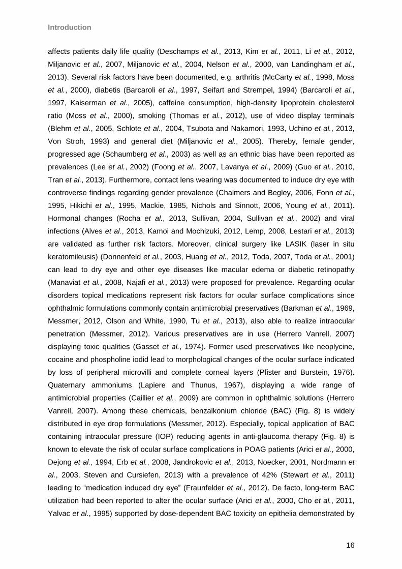
Fig 7 Classification of dry eye syndrome pathologies based on the concept of the 2007 Dry Eye Workshop
(DEWS) (Lemp 2007). The arrow highlighted dry eye forms are especially focused on in the present work.
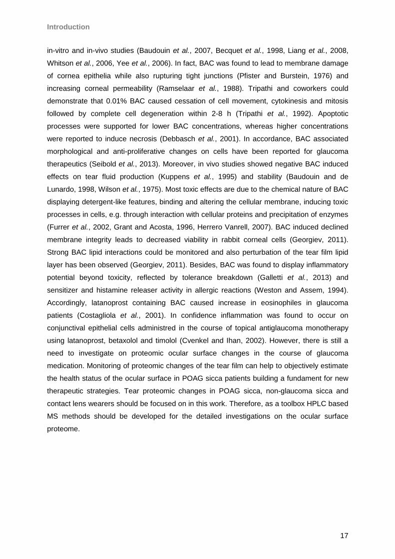
Fig 8 Prostaglandine related IOP reducing agents and commercial formulations used in POAG glaucoma therapy;
A. Latanoprost; B: Tafluprost; C: Xalatan® (Pfizer) containing 0.005% latanoprost and 0.02% BAC; D:
Preservative-free Taflotan® sine (Santen) containing 0.0015% tafluprost.
2.8 Therapeutic management with special focus on Taflotan® sine and taurine
According to the complex character of dry eye disease, new therapeutic strategies have to
be addressed. Thereby, special focus is on “medication induced” dry eye (Fraunfelder et al.,
2012) in POAG therapy. Consequently, therapeutic key features should address
preservative, especially BAC reduction, in topical formulation. Horsley & Kahook reported
beneficial effects on the ocular surface in glaucoma patients switching from latanoprost
(0.02% BAC) to sofZIATM medication indicated by an amendment in ocular surface disease
Introduction
19
index (OSDI), corneal staining and tear break-up time (TBUT) (Horsley and Kahook, 2009).
Because BAC displays toxic potential in very low concentrations (Furrer et al., 2002), a
complete avoidance of this preservative most likely is the aim for long term application.
Nevertheless, common prostaglandine analogue formulations contain preservatives, e.g.
latanoprost, travoprost or bimatoprost (Liu and Mao, 2013). In contrast tafluprost (AFP-168),
the latest prostaglandin F2α derivate (Takagi et al., 2004), as the first IOP lowering agent
available in a preservative-free formulation, evidenced promising results regarding effectivity
and tolerability (Hamacher et al., 2008, Hommer et al., 2010, Hos et al., 2013). In
comparison to preservative-containing formulations tafluprost exhibited very low or no
apoptotic/necrotic/oxidative effects revealed in IOBA-NHC cells (Brasnu et al., 2008). Local
tolerability was evaluated to improve up to 86% after a medical switch from preservative-
based medication to tafluprost in a large patient cohort of 2123 patients (Erb et al., 2011).
Uusitalo and collegues reported that tafluprost compared to latanoprost showed comparable
results revealed from a study encircling 533 glaucoma patients (Uusitalo et al., 2010). In
confidence Janulevičienè and coworkers showed an equal IOP lowering effect but a higher
ocular tolerability indicated by lower tear osmolarity levels for tafluprost compared with BAC
containing latanoprost (Januleviciene et al., 2012). Tafluprost available under the trade name
Taflotan® sine (Santen Pharmaceutical Co., Ltd.), is in use in glaucoma therapy, but objective
indicators for the effect of Taflotan® sine on the ocular surface are still missing and no
detailed proteomic examination has been realized so far. Despite the preservative focused
strategy in “medication induced dry eye” additional therapeutic strategies generally adressing
dry eye had been proposed including tear secretagogues (Koh et al., 2013, Nakamura et al.,
2012, Nichols et al., 2004), anti-inflammatory topical therapeutics (McCabe and Narayanan,
2009, Mrukwa-Kominek et al., 2007, Pflugfelder, 2003, 2004) like cyclosporine, tetracycline,
doxycycline, corticosteroids, vitamin A topical utilization (Calonge, 2001, De Paiva et al.,
2006, Kim et al., 2009a, Kobayashi et al., 1997, Perry et al., 2008, Yang et al., 2006),
cytokines (Pflugfelder et al., 2013), anakinra (Vijmasi et al., 2013), coumarin 18 (Govek et al.,
2010) and the utilization of artificial tears (Sindt and Foulks, 2013). However, there is still
need to discover new drugs and to obtain a deeper understanding of proposed agents in dry
eye therapy (Aquavella, 2013, Dogru et al., 2013, Gadaria-Rathod et al., 2013, Skalicky et
al., 2013). Thereby, topical or oral appliance of small receptor interacting molecules like
CF101 (Avni et al., 2010), essential fatty acids (Kokke et al., 2008, Rashid et al., 2008),
peptides (Li et al., 2013, Peral et al., 2008, Sosne et al., 2012) amino acids (Aragona et al.,
2013, Peral et al., 2008) are among the future components for dry eye management.
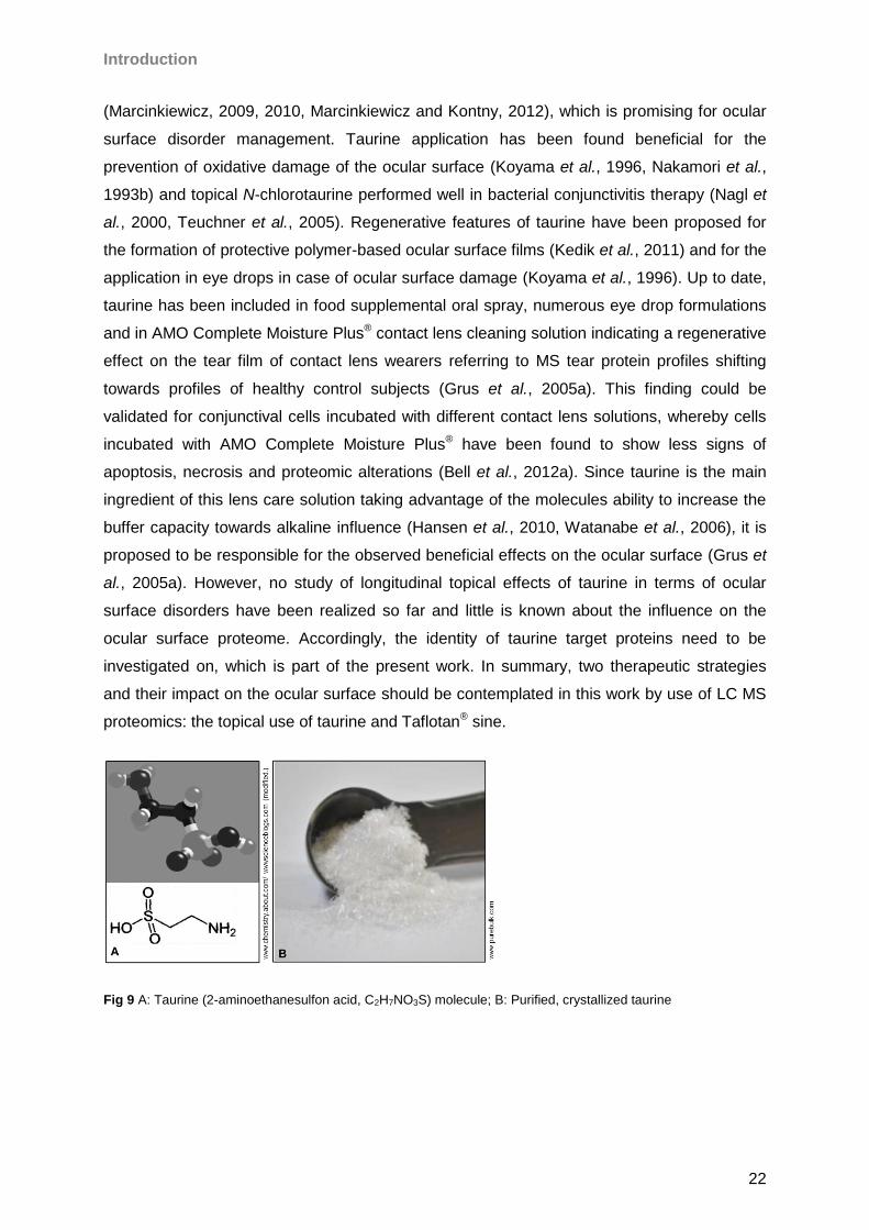
Regarding amino acids special focus is on taurine (Fig. 9), which was proposed as a
promising agent in preventive medicine (Birdsall, 1998, Chesney, 1985, Kendler, 1989) and
is still under investigation for its therapeutic potential facing ocular disorders (Ripps and
Introduction
20
Shen, 2012). Taurine is a multifunctional zwitterionic, sulfonated, non-proteinaceous ß-amino
acid (Huxtable, 1992, Redmond et al., 1998, Wright et al., 1986) derived from methionine
and cysteine metabolism (Brosnan and Brosnan, 2006, Redmond et al., 1998) and is present
in high concentration in tissues of algae and animals (Jacobsen and Smith, 1968). Also, in
mammalian species taurine has been documented for various tissues (Awapara et al., 1950),
with high concentrations in ocular tissues (Heinamaki et al., 1986) including lens, iris ciliar
body (Gupta and Mathur, 1983, Heinamaki et al., 1986, Reddy, 1967, Reddy, 1970) and
retina (Lake and Verdonesmith, 1989, Orr et al., 1976, Pow et al., 2002, Rassin et al., 1978,
Sturman et al., 1978, Voaden et al., 1977). Especially, on the ocular surface, high taurine
abundance had been reported encircling cornea, conjunctiva and tear film (Nakatsukasa et
al., 2011, Pescosolido et al., 2009, Reddy, 1967). Beyond metabolic functions including
nutrition (Tesseraud et al., 2009, Wu, 2009), more specialized features are associated to
taurine, e.g. vitamin transport (Petrosian and Haroutounian, 2000) and osmoregulation
(Gupta and Mathur, 1983, Lange, 1963, Thurston et al., 1980, Uchida et al., 1991). De facto,
taurine was found to display anti-toxic cytoprotective features in host tissues (Dawson et al.,
2002, Lerdweeraphon et al., 2013, Pasantes-Morales and Cruz, 1985, Saad and Al-Rikabi,
2002, Timbrell et al., 1995, Venkatesan et al., 1997, Waterfield et al., 1994, Wettstein and
Haussinger, 1997). Most of these effects are due to the ability of taurine to prevent oxidative
stress and peroxidative damage (Cozzi et al., 1995, Endo et al., 2002, Mahalakshmi et al.,
2003, Nakamori et al., 1993a, Pasantes-Morales and Cruz, 1984) by its membrane
interacting and stabilizing features (Koyama et al., 1996, Lopez-Colome and Pasantes-
Morales, 1981, Pasantes-Morales and Cruz, 1985). Confidently, the amino acid has been
demonstrated to protect plasma/mitochondrial and ER membranes under stress condition
(Ye et al., 2013) facilitating the interaction with lipids and proteins (Sebring and Huxtable,
1986), stabilizing proteins, especially membrane proteins from denaturation (Arakawa and
Timasheff, 1985, Nandhini and Anuradha, 2003, Ye et al., 2013). Interestingly, osmolytes like
taurine have been lately proven to protect lysozyme as a major tear protein representative
(Bruzdziak et al., 2013). Antioxidative effects of taurine have been intensively studied
(Nakamori et al., 1993a), whereby protective effects exceed effectivity of vitamin C and E
(Rodriguez-Martinez et al., 2004). Regulation of Ca2+ uptake, Ca2+ATPase, generation of
peroxide and superoxide anions in mitochondria (Chang et al., 2004) as well as inhibition of
consequent oxidative damage could be demonstrated for various tissues and organisms, e.g.
for rat spermatozoa (Alvarez and Storey, 1983), rat liver (Kerai et al., 1998), rat mesangial
cells (Trachtman et al., 1993), rat liver cells (Kerai et al., 1998), plasma, liver and aorta of
rabbits (Balkan et al., 2002), hamster bronchioles (Gordon et al., 1986) and human
lymphoblastoid cells (Pasantesmorales et al., 1984). Regarding ocular tissues, taurine was
documented to prevent lipid peroxidation in rod outer segments (Pasantes-Morales, 1982,
Introduction
21
Pasantes-Morales and Cruz, 1985), photoreceptors (Keys and Zimmerman, 1999) and
diabetic precataractous lens (Kilic et al., 1999, Mitton et al., 1999, Obrosova and Stevens,
1999). Beside the ability of stabilizing proteins and membranes, taurine prevents
uncontrolled protein modification, e.g. nonenzymatic glycosylation which plays a role for a
proteomic functional decrease in aging and disease (Rattan, 1996, Soskic et al., 2008).
Szwergold could demonstrate that taurine has the ability to prevent generation of a sugar-
protein “Schiffbase”, which represents the initial step in glycation (Szwergold, 2005, 2006).
Confidently, taurine was demonstrated to inhibit glycation of lens proteins (Devamanoharan
et al., 1997), to counterstrike glucose-induced apoptosis (Di Wu et al., 1999), to prevent
sugar induced cataracts in rabbits (Malone et al., 1993) and retinal glial cell apoptosis in rats
(Zeng et al., 2010). In fact, anti-apoptotic effects of taurine have been intensively studied
(Verzola et al., 2002) evidencing taurine to inhibit the formation of the Apaf-1/Caspase-9
apoptosome (Takatani et al., 2004), to inhibit the m-calpain and caspase-3 mediated
cascade (Sun and Xu, 2008) and to reduce Bcl-2 (Wu et al., 2009a). Furthermore, taurine
can modulate cellular pathways by modulation of protein phosphorylation (Lombardini, 1985,
Lombardini et al., 1996, Mollerup and Lambert, 1996) and receptor binding, e.g. triggering
GABA (Bureau and Olsen, 1991, Louzada et al., 2004) and insulin receptors (Maturo and
Kulakowski, 1988) reflecting the wide range of molecular taurine trigger points. Moreover,
immunomodulative functions (Kim, 2006, Marcinkiewicz and Kontny, 2012, Redmond et al.,
1998, Schuller-Levis and Park, 2003) due to chloramine generation (Marcinkiewicz et al.,
1995, Verdrengh and Tarkowski, 2005) have been reported for the molecule. In fact, taurine
chloramine release by activated granulocytes and monocytes during oxidative burst (Arnitz et
al., 2006) has been widely demonstrated (Schuller-Levis and Park, 2004). Taurine levels
decrease in ocular tissues in the course of aging (Baskin et al., 1977) explaining a correlation
between aging, oxidative damage, taurine avaibility and disease (Malone et al., 1990,
Militante and Lombardini, 2004, Yildirim et al., 2007). In confidence with these findings
protective effects of taurine are known for various disorders including arterial calcionosis
(Yamauchitakihara et al., 1986), pancreatitis (Akay et al., 2013), closed head injury (Sun et
al., 2013), diabetes (Kim et al., 2007a), heart diseases (Franconi et al., 1985, Wojcik et al.,
2010), renal injuries (Chiba et al., 2002, Wang et al., 2008), including bladder and kidney
injuries (Sener et al., 2005) and neuronal injuries like Alzheimer´s disease (Louzada et al.,
2004). Special interest was on ocular disorders indicating taurine´s regenerative potential in
goldfish retina (Lima et al., 1993), its role in retinal ganglion cell survival in glaucoma models
(Froger et al., 2012, Gaucher et al., 2012) and its beneficial performance regarding diabetic
cataracts (Hsu et al., 2012, Kilic et al., 1999). Actually taurine showed high performance in
epithelial woundhealing (Degim et al., 2002, Dincer et al., 1996) and derivates like taurine
bromamine have been found effective for the management of epithelial inflammation
Introduction
22
(Marcinkiewicz, 2009, 2010, Marcinkiewicz and Kontny, 2012), which is promising for ocular
surface disorder management. Taurine application has been found beneficial for the
prevention of oxidative damage of the ocular surface (Koyama et al., 1996, Nakamori et al.,
1993b) and topical N-chlorotaurine performed well in bacterial conjunctivitis therapy (Nagl et
al., 2000, Teuchner et al., 2005). Regenerative features of taurine have been proposed for
the formation of protective polymer-based ocular surface films (Kedik et al., 2011) and for the
application in eye drops in case of ocular surface damage (Koyama et al., 1996). Up to date,
taurine has been included in food supplemental oral spray, numerous eye drop formulations
and in AMO Complete Moisture Plus® contact lens cleaning solution indicating a regenerative
effect on the tear film of contact lens wearers referring to MS tear protein profiles shifting
towards profiles of healthy control subjects (Grus et al., 2005a). This finding could be
validated for conjunctival cells incubated with different contact lens solutions, whereby cells
incubated with AMO Complete Moisture Plus® have been found to show less signs of
apoptosis, necrosis and proteomic alterations (Bell et al., 2012a). Since taurine is the main
ingredient of this lens care solution taking advantage of the molecules ability to increase the
buffer capacity towards alkaline influence (Hansen et al., 2010, Watanabe et al., 2006), it is
proposed to be responsible for the observed beneficial effects on the ocular surface (Grus et
al., 2005a). However, no study of longitudinal topical effects of taurine in terms of ocular
surface disorders have been realized so far and little is known about the influence on the
ocular surface proteome. Accordingly, the identity of taurine target proteins need to be
investigated on, which is part of the present work. In summary, two therapeutic strategies
and their impact on the ocular surface should be contemplated in this work by use of LC MS
proteomics: the topical use of taurine and Taflotan® sine.
Fig 9 A: Taurine (2-aminoethanesulfon acid, C2H7NO3S) molecule; B: Purified, crystallized taurine
Material & Methods
23
3 Material & Methods
3.1 Training samples
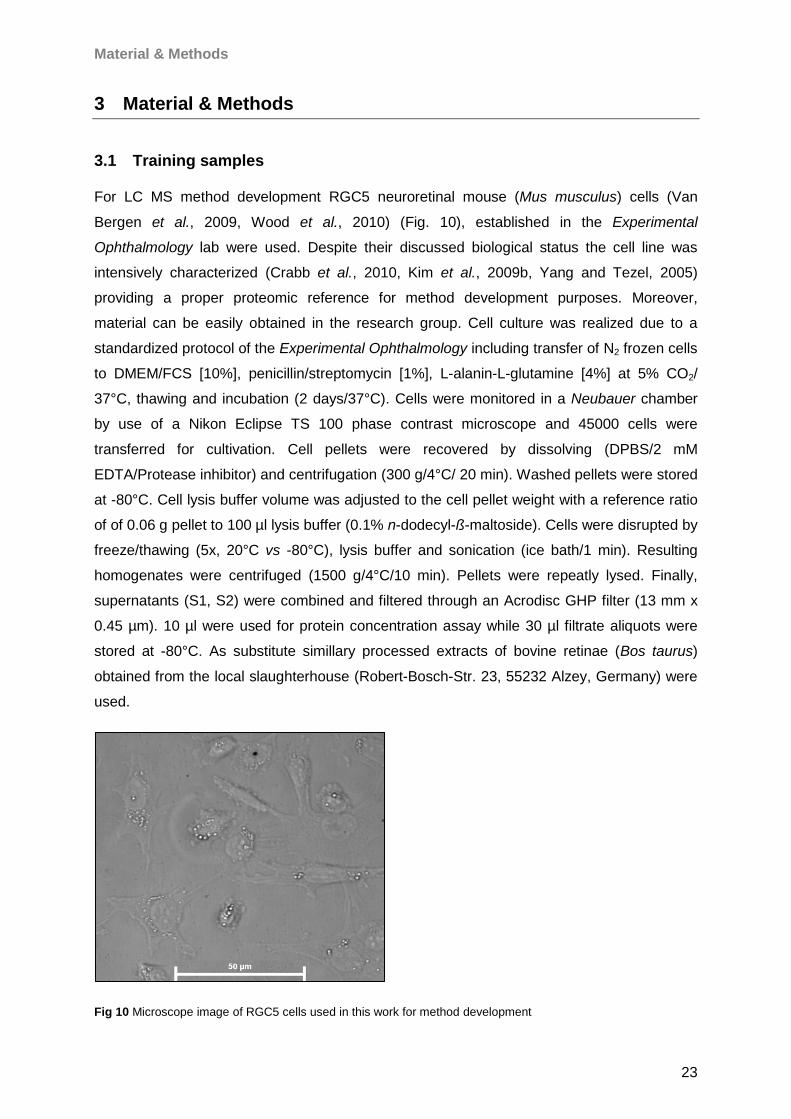
For LC MS method development RGC5 neuroretinal mouse (Mus musculus) cells (Van
Bergen et al., 2009, Wood et al., 2010) (Fig. 10), established in the Experimental
Ophthalmology lab were used. Despite their discussed biological status the cell line was
intensively characterized (Crabb et al., 2010, Kim et al., 2009b, Yang and Tezel, 2005)
providing a proper proteomic reference for method development purposes. Moreover,
material can be easily obtained in the research group. Cell culture was realized due to a
standardized protocol of the Experimental Ophthalmology including transfer of N2 frozen cells
to DMEM/FCS [10%], penicillin/streptomycin [1%], L-alanin-L-glutamine [4%] at 5% CO2/
37°C, thawing and incubation (2 days/37°C). Cells were monitored in a Neubauer chamber
by use of a Nikon Eclipse TS 100 phase contrast microscope and 45000 cells were
transferred for cultivation. Cell pellets were recovered by dissolving (DPBS/2 mM
EDTA/Protease inhibitor) and centrifugation (300 g/4°C/ 20 min). Washed pellets were stored
at -80°C. Cell lysis buffer volume was adjusted to the cell pellet weight with a reference ratio
of of 0.06 g pellet to 100 µl lysis buffer (0.1% n-dodecyl-ß-maltoside). Cells were disrupted by
freeze/thawing (5x, 20°C vs -80°C), lysis buffer and sonication (ice bath/1 min). Resulting
homogenates were centrifuged (1500 g/4°C/10 min). Pellets were repeatly lysed. Finally,
supernatants (S1, S2) were combined and filtered through an Acrodisc GHP filter (13 mm x
0.45 µm). 10 µl were used for protein concentration assay while 30 µl filtrate aliquots were
stored at -80°C. As substitute simillary processed extracts of bovine retinae (Bos taurus)
obtained from the local slaughterhouse (Robert-Bosch-Str. 23, 55232 Alzey, Germany) were
used.
Fig 10 Microscope image of RGC5 cells used in this work for method development
Material & Methods
24
3.2 Study samples
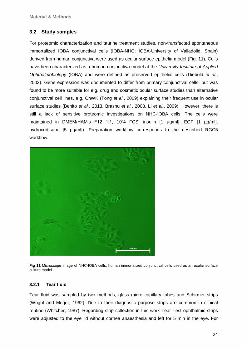
For proteomic characterization and taurine treatment studies, non-transfected spontaneous
immortalized IOBA conjunctival cells (IOBA-NHC; IOBA-University of Valladolid, Spain)
derived from human conjunctiva were used as ocular surface epithelia model (Fig. 11). Cells
have been characterized as a human conjunctiva model at the University Institute of Applied
Ophthalmobiology (IOBA) and were defined as preserved epithelial cells (Diebold et al.,
2003). Gene expression was documented to differ from primary conjunctival cells, but was
found to be more suitable for e.g. drug and cosmetic ocular surface studies than alternative
conjunctival cell lines, e.g. ChWK (Tong et al., 2009) explaining their frequent use in ocular
surface studies (Benito et al., 2013, Brasnu et al., 2008, Li et al., 2009). However, there is
still a lack of sensitive proteomic investigations on NHC-IOBA cells. The cells were
maintained in DMEM/HAM’s F12 1:1, 10% FCS, insulin [1 µg/ml], EGF [1 µg/ml],
hydrocortisone [5 µg/ml]). Preparation workflow corresponds to the described RGC5
workflow.
Fig 11 Microscope image of NHC-IOBA cells, human immortalized conjunctival cells used as an ocular surface
culture model.
3.2.1 Tear fluid
Tear fluid was sampled by two methods, glass micro capillary tubes and Schirmer strips
(Wright and Meger, 1962). Due to their diagnostic purpose strips are common in clinical
routine (Whitcher, 1987). Regarding strip collection in this work Tear Test ophthalmic strips
were adjusted to the eye lid without cornea anaesthesia and left for 5 min in the eye. For
Material & Methods
25
sample analysis, each strip was soaked in 300 µl DPBS and tear proteins were extracted
overnight on a KS250 basic Intellimixer at 4°C. Regarding capillary tears, tear fluid was
collected by use of Ringcaps® Duran® disposable micro pipettes with ring mark. In both
methods resulting samples were stored at -80°C until use. Prior to use, samples had been
adjusted to -20°C and then to 4°C to allow a gentle adjustment of proteins to avoid
freeze/thawing protein loss effects as much as possible. Taflotan® sine study sample
collection and taurine study sample collection were supported by members of the lab staff.
3.3 Sample preparation
3.3.1 Intact sample processing
Protein amount estimation was realized by use of the Pierce® BCA Protein assay and a
Multiscan Ascent scanner according to the protocol of the manufactor. Freshly thawed
sample aliquots for TD LC MALDI analysis were adjusted to 3 µg/µl final concentration due to
15 µg maximum column capacity and 5 µl injection volume (3 µg/µl x 5 µl = 15 µg) with
running buffer A (4% ACN, 0.1% TFA). 40 µl sample volume were prepared for each LC run.
The samples were transferred to V-bottom 96 well plates for automated sample injection.
3.3.2 SDS-PAGE
Since a direct MS analysis is crucial in BU proteomic approaches due to high complexity, 1D
SDS PAGE invented by Lämmli (Laemmli, 1970) was used to fractionate proteins
corresponding to their molecular weight prior to LC MS analysis. Usually, form, charge and
mass influence migration of a protein through a polyacrylamide gel network (Schagger and
von Jagow, 1987). Anionic SDS detergent binds to hydrophobic regions (Löffler, 2003)
provides negative charges and faciliates denaturation. Consequently, electronical field
migration through the gel meshwork is directly proportional to the protein mass, whereby
smaller proteins migrate faster than higher mass proteins (Löffler, 2003). However, the
qualitative output depends on various factors, e.g. the maximum gel loading capacity (van
Hagen, 2008). Therefore, protein load should not impede migration, but consider MS
sensitivity. Based on RGC5 extracts, a protein load of 80 µg was estimated optimal for ESI
and MALDI experiments. Above 80 µg protein lanes clearly smeared due to overloading
effects. In contrast, if available, at minimum 20 µg protein should be separated per lane to fit
the MS dynamic range. Samples encircling RGC5 or IOBA-NHC lysates and tear fluid were
mixed with NuPAGE LDS sample buffer (+/- reducing agent). Corresponding samples were
heated (10 min/90°C) and run on NuPage 12% Bis-Tris, 10-well prepared minigels (100 V/10
min - 150 V) in the XCell SureLockTM system using MES or MOPs buffers. SeabluePlus 2
prestained protein standard was used as molecular weight reference. Reproducibility was
Material & Methods
26
tested using replicate aliquots of test samples. Gels were fixed and stained overnight using
the Novex Colloidal Blue staining kit according to the manual of the manufactor. For
documentation gels were recorded by use of a DCP-9042 CDN benchtop scanner with a
resolution of 1200 x 1200 dpi as tif-data assisted by Photoshop C53 extended version.
3.3.3 In-gel digestion
The procedure was based on a modified protocol of Shevchenkov et al. (Shevchenko et al.,
2006) considering ESI/MALDI optimizations. In case of LC MALDI experiments, a gel lane of
interest was divided into 15 slices, which allows one lane to be positioned on a 386 spot
target. In case of LC ESI a gel lane was divided into 17 slices. The lane division was decided
corresponding to the CBB protein signal abundance. Mass area slices were cut into equally
small pieces, destained (50% ACN, 50% 100 mM Ambi, 10 min) and dehydrated (100%
ACN). Dried gel pieces were saturated with trypsin solution (200 µl reaction buffer, 1300 µl
20% ACN/10 mM Ambi) ([15 ng/µl], 60-100 µl/slice) and incubated over night (37°C).
Digestion was quenched by a pH shift by addition of 10 µl 0.1% TFA. Supernatants were
transferred to fresh 0.5 ml tubes and peptides were extracted with appropriate amount of
extraction buffers on a Unimax 1010 shaker at 700 rpm. In case of LC MALDI, prior to
analysis, combined supernatants and peptide extracts of each slice were filtered through
GHP Acrodisc 13 mm x 0.45 µm pre-HPLC syringe filters followed by vacuum concentration
(30°C) in a Concentrator 5301 instrument. LC MALDI destinated samples were equilibrated
with 20 µl running buffer A (1% MeOH, 1% ACN 0.05% TFA), whereas LC ESI samples were
equilibrated with 20 µl 0.1% TFA. In case of LC ESI analysis, sonication resolubilized
peptides were SPE purified once again to reduce background signals due to the less
contaminant robust character of ESI systems (Kraj and Silberring, 2008).
3.3.4 Solid phase extraction (SPE)
Because SPE tips had been demonstrated to generate more accurate results than magnetic
beads (Tiss et al. 2007) ZIPTIP® C18 pipette tips (15 µm, 200 Å) were used. 10 µl peptides
were bound by 20 binding cycles on the C18 resin followed by a 0.1% TFA wash step
followed by 10 µl duplicate elution (50% ACN, 0.1% TFA). The tip was once again rinsed with
pure ACN, equilibrated with 0.1% TFA and binding cycles were repeated. Resulting aliquots
were combined leading to a 40 µl peptide sample corresponding to each slice digest.
Duplicate ZIPTIP® purification was used to exceed the resin saturation limit of 5 µg protein
(UserGuideforRPZipTipPipetteTipsforSamplePreparation, 2007) since approximately 4-10 µg
protein load (1 lane = 80 µg/17 slices = 4.7 µg/slice; ~4-10 µg/slice) was provided per gel
slice with respect to protein distribution pattern. Lyophilized peptides were stored at -80°C
until use. For LC ESI analysis peptides were resolubilized (20 µl 0.1% TFA, ice
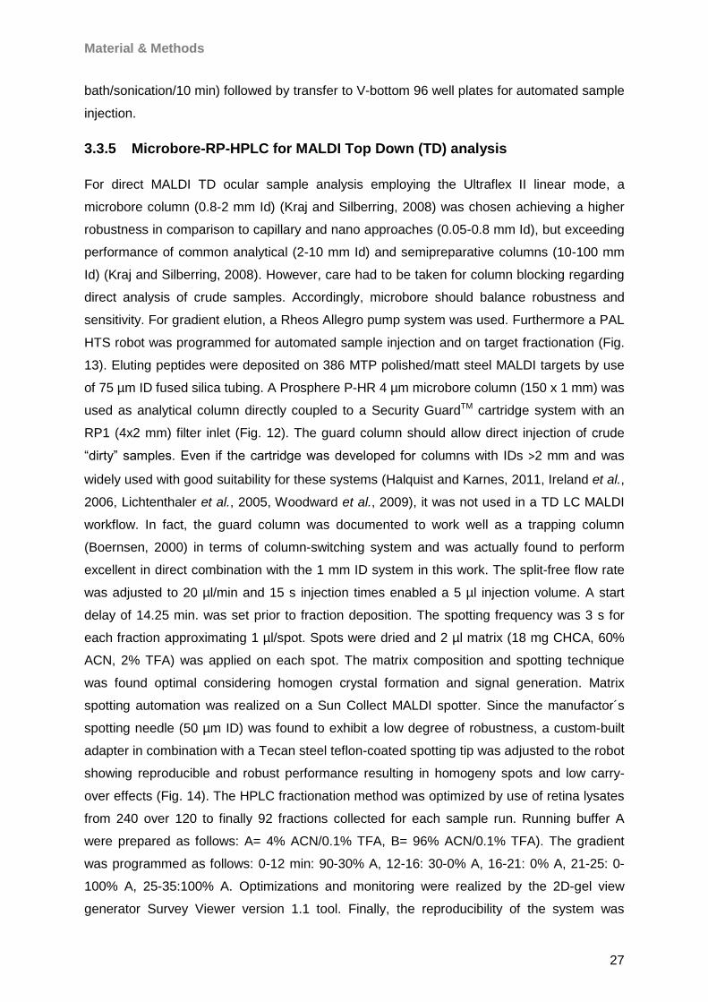
Material & Methods
27
bath/sonication/10 min) followed by transfer to V-bottom 96 well plates for automated sample
injection.
3.3.5 Microbore-RP-HPLC for MALDI Top Down (TD) analysis
For direct MALDI TD ocular sample analysis employing the Ultraflex II linear mode, a
microbore column (0.8-2 mm Id) (Kraj and Silberring, 2008) was chosen achieving a higher
robustness in comparison to capillary and nano approaches (0.05-0.8 mm Id), but exceeding
performance of common analytical (2-10 mm Id) and semipreparative columns (10-100 mm
Id) (Kraj and Silberring, 2008). However, care had to be taken for column blocking regarding
direct analysis of crude samples. Accordingly, microbore should balance robustness and
sensitivity. For gradient elution, a Rheos Allegro pump system was used. Furthermore a PAL
HTS robot was programmed for automated sample injection and on target fractionation (Fig.
13). Eluting peptides were deposited on 386 MTP polished/matt steel MALDI targets by use
of 75 µm ID fused silica tubing. A Prosphere P-HR 4 µm microbore column (150 x 1 mm) was
used as analytical column directly coupled to a Security GuardTM cartridge system with an
RP1 (4x2 mm) filter inlet (Fig. 12). The guard column should allow direct injection of crude
“dirty” samples. Even if the cartridge was developed for columns with IDs >2 mm and was
widely used with good suitability for these systems (Halquist and Karnes, 2011, Ireland et al.,
2006, Lichtenthaler et al., 2005, Woodward et al., 2009), it was not used in a TD LC MALDI
workflow. In fact, the guard column was documented to work well as a trapping column
(Boernsen, 2000) in terms of column-switching system and was actually found to perform
excellent in direct combination with the 1 mm ID system in this work. The split-free flow rate
was adjusted to 20 µl/min and 15 s injection times enabled a 5 µl injection volume. A start
delay of 14.25 min. was set prior to fraction deposition. The spotting frequency was 3 s for
each fraction approximating 1 µl/spot. Spots were dried and 2 µl matrix (18 mg CHCA, 60%
ACN, 2% TFA) was applied on each spot. The matrix composition and spotting technique
was found optimal considering homogen crystal formation and signal generation. Matrix
spotting automation was realized on a Sun Collect MALDI spotter. Since the manufactor´s
spotting needle (50 µm ID) was found to exhibit a low degree of robustness, a custom-built
adapter in combination with a Tecan steel teflon-coated spotting tip was adjusted to the robot
showing reproducible and robust performance resulting in homogeny spots and low carry-
over effects (Fig. 14). The HPLC fractionation method was optimized by use of retina lysates
from 240 over 120 to finally 92 fractions collected for each sample run. Running buffer A
were prepared as follows: A= 4% ACN/0.1% TFA, B= 96% ACN/0.1% TFA). The gradient
was programmed as follows: 0-12 min: 90-30% A, 12-16: 30-0% A, 16-21: 0% A, 21-25: 0-
100% A, 25-35:100% A. Optimizations and monitoring were realized by the 2D-gel view
generator Survey Viewer version 1.1 tool. Finally, the reproducibility of the system was
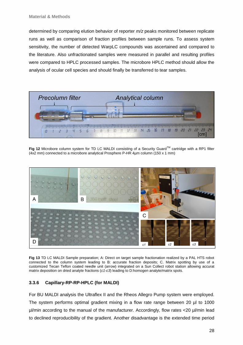
Material & Methods
28
determined by comparing elution behavior of reporter m/z peaks monitored between replicate
runs as well as comparison of fraction profiles between sample runs. To assess system
sensitivity, the number of detected WarpLC compounds was ascertained and compared to
the literature. Also unfractionated samples were measured in parallel and resulting profiles
were compared to HPLC processed samples. The microbore HPLC method should allow the
analysis of ocular cell species and should finally be transferred to tear samples.
Fig 12 Microbore column system for TD LC MALDI consisting of a Security GuardTM
cartridge with a RP1 filter
(4x2 mm) connected to a microbore analytical Prosphere P-HR 4µm column (150 x 1 mm)
Fig 13 TD LC MALDI Sample preparation; A: Direct on target sample fractionation realized by a PAL HTS robot
connected to the column system leading to B: accurate fraction deposits; C. Matrix spotting by use of a customized Tecan Teflon coated needle unit (arrow) integrated on a Sun Collect robot station allowing accurat matrix deposition on dried analyte fractions (c1-c3) leading to D:homogen analyte/matrix spots.
3.3.6 Capillary-RP-RP-HPLC (for MALDI)
For BU MALDI analysis the Ultraflex II and the Rheos Allegro Pump system were employed.
The system performs optimal gradient mixing in a flow rate range between 20 µl to 1000
µl/min according to the manual of the manufacturer. Accordingly, flow rates <20 µl/min lead
to declined reproducibility of the gradient. Another disadvantage is the extended time period

Material & Methods
29
the gradient needs to reach the column head, probably causing diffusion effects and
decreased performance. For optimal conditions, a pre-column flow splitting strategy was
established. Thereby, the flow split should consider the pump´s optimal gradient range and
should leave the analytical column with low flow rate. As mechanical split system an M-472
graduated micro-split valve was used. The micro-split valve was selected due to a pressure
resistance >200 bar. The challenge in flow splitting refers to the column system, which
creates high pressure. Consequently, without appropriate waste port backpressure all
solvent will flow out of the waste port failing to pass the split port and without entering the
column. Therefore, appropriate backpressure adjustment is a challenging step. Accordingly,
for optimal pump flow splitting different splitter graduation settings and pump flow rates have
been tested. In consequence, the adjustment of the split flow rate exclusively by the splitter
graduation was found to be unsuitable for accurate fine regulation of the split stream. Hence,
reproducible adjustment was successfully achieved only in a small setting window confident
with manual splitter inspection, which supports the splitter to work preliminary as a shut-off
valve with distinct limitation in split ratio adjustment. Consequently, as a counteracting
splitting strategy, peek tubings with varying IDs were connected to the waste port to generate
appropriate backpressure at a fixed graduation setting which was also documented for
comparable splitter models (Kozerski et al., 2003, Rogatsky et al., 2009). The use of peek
tubings for backpressure adjustment is an economic strategy to down-scale common HPLC
systems to capillary or nano dimension (Aguilar, 2004, Moritz, 2007, Profrock, 2010, Yi et al.,
2003, Zhang et al., 2004a). Therefore, variations of peek tubing length and pump flow rates
were combinatorial determined for backpressure genearation and capillary flow rate setting.
A combination of a 1/16” OD x 0.10” ID x 5 ft PEEKTM tubing under a pump flow rate of 300
µl/min was found to work optimal resulting in a split flowrate of 7.8 µl/min, matching the
capillary range. Hence, 300 µl/min pump flow had been documented for most systems to
guarantee suitable pump function and solvent portioning (Chervet et al., 1996). Split flow rate
reproducibility was tested by time assays monitoring different mobile phase compositions
passing through a 20 µl defined capillary tube. The splitting system was connected to the
injection valve by combination of 1/16” OD x 0.0025” ID and 1/16” OD x 0.007” PEEKTM
tubings connected by use of a stainless steel NanoTightTM union (10-32 to 10-32, 0.007” thru-
hole) to allow an easy reinstallation of the microbore system or other systems (Fig. 14). All
tubings and connectors were capillary compatible to avoid dead volumes in the system.
Accordingly, the 6-port valve outlet was connected to the column system by use of a
1/16”OD x 0.005 ID PEEKTM tubing. Tubings were connected by use of 10-32 PEEKTM
fingertight fittings. Since the present instrumentation did not allow column switching and
peptide trapping, the design of cationic/anionic exchange and reversed phase as common
2D-HPLC strategies for BU analysis (Link, 2002) was not possible. To counteract this
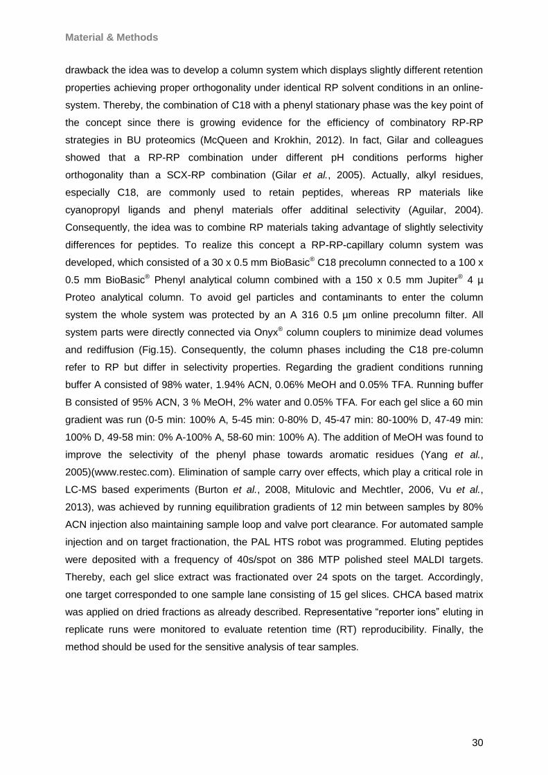
Material & Methods
30
drawback the idea was to develop a column system which displays slightly different retention
properties achieving proper orthogonality under identical RP solvent conditions in an online-
system. Thereby, the combination of C18 with a phenyl stationary phase was the key point of
the concept since there is growing evidence for the efficiency of combinatory RP-RP
strategies in BU proteomics (McQueen and Krokhin, 2012). In fact, Gilar and colleagues
showed that a RP-RP combination under different pH conditions performs higher
orthogonality than a SCX-RP combination (Gilar et al., 2005). Actually, alkyl residues,
especially C18, are commonly used to retain peptides, whereas RP materials like
cyanopropyl ligands and phenyl materials offer additinal selectivity (Aguilar, 2004).
Consequently, the idea was to combine RP materials taking advantage of slightly selectivity
differences for peptides. To realize this concept a RP-RP-capillary column system was
developed, which consisted of a 30 x 0.5 mm BioBasic® C18 precolumn connected to a 100 x
0.5 mm BioBasic® Phenyl analytical column combined with a 150 x 0.5 mm Jupiter® 4 µ
Proteo analytical column. To avoid gel particles and contaminants to enter the column
system the whole system was protected by an A 316 0.5 µm online precolumn filter. All
system parts were directly connected via Onyx® column couplers to minimize dead volumes
and rediffusion (Fig.15). Consequently, the column phases including the C18 pre-column
refer to RP but differ in selectivity properties. Regarding the gradient conditions running
buffer A consisted of 98% water, 1.94% ACN, 0.06% MeOH and 0.05% TFA. Running buffer
B consisted of 95% ACN, 3 % MeOH, 2% water and 0.05% TFA. For each gel slice a 60 min
gradient was run (0-5 min: 100% A, 5-45 min: 0-80% D, 45-47 min: 80-100% D, 47-49 min:
100% D, 49-58 min: 0% A-100% A, 58-60 min: 100% A). The addition of MeOH was found to
improve the selectivity of the phenyl phase towards aromatic residues (Yang et al.,
2005)(www.restec.com). Elimination of sample carry over effects, which play a critical role in
LC-MS based experiments (Burton et al., 2008, Mitulovic and Mechtler, 2006, Vu et al.,
2013), was achieved by running equilibration gradients of 12 min between samples by 80%
ACN injection also maintaining sample loop and valve port clearance. For automated sample
injection and on target fractionation, the PAL HTS robot was programmed. Eluting peptides
were deposited with a frequency of 40s/spot on 386 MTP polished steel MALDI targets.
Thereby, each gel slice extract was fractionated over 24 spots on the target. Accordingly,
one target corresponded to one sample lane consisting of 15 gel slices. CHCA based matrix
was applied on dried fractions as already described. Representative “reporter ions” eluting in
replicate runs were monitored to evaluate retention time (RT) reproducibility. Finally, the
method should be used for the sensitive analysis of tear samples.
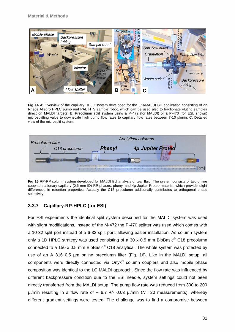
Material & Methods
31
Fig 14 A: Overview of the capillary HPLC system developed for the ESI/MALDI BU application consisting of an
Rheos Allegro HPLC pump and PAL HTS sample robot, which can be used also to fractionate eluting samples direct on MALDI targets; B: Precolumn split system using a M-472 (for MALDI) or a P-470 (for ESI, shown) microsplitting valve to downscale high pump flow rates to capillary flow rates between 7-10 µl/min; C: Detailed view of the microsplit system.
Fig 15 RP-RP column system developed for MALDI BU analysis of tear fluid. The system consists of two online
coupled stationary capillary (0.5 mm ID) RP phases, phenyl and 4µ Jupiter Proteo material, which provide slight differences in retention properties. Actually the C18 precolumn additionally contributes to orthogonal phase selectivity.
3.3.7 Capillary-RP-HPLC (for ESI)
For ESI experiments the identical split system described for the MALDI system was used
with slight modifications, instead of the M-472 the P-470 splitter was used which comes with
a 10-32 split port instead of a 6-32 split port, allowing easier installation. As column system
only a 1D HPLC strategy was used consisting of a 30 x 0.5 mm BioBasic® C18 precolumn
connected to a 150 x 0.5 mm BioBasic® C18 analytical. The whole system was protected by
use of an A 316 0.5 µm online precolumn filter (Fig. 16). Like in the MALDI setup, all
components were directly connected via Onyx® column couplers and also mobile phase
composition was identical to the LC MALDI approach. Since the flow rate was influenced by
different backpressure condition due to the ESI needle, system settings could not been
directly transferred from the MALDI setup. The pump flow rate was reduced from 300 to 200
µl/min resulting in a flow rate of ~ 6.7 +/- 0.03 µl/min (N= 20 measurements), whereby
different gradient settings were tested. The challenge was to find a compromise between
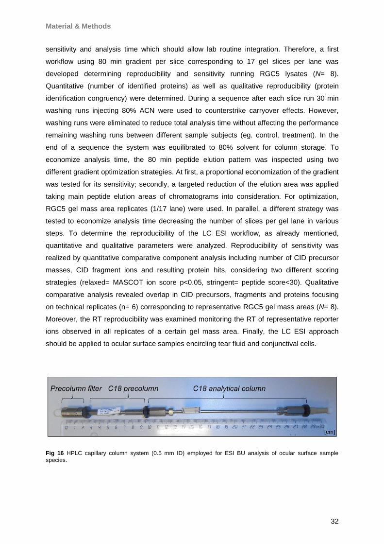
Material & Methods
32
sensitivity and analysis time which should allow lab routine integration. Therefore, a first
workflow using 80 min gradient per slice corresponding to 17 gel slices per lane was
developed determining reproducibility and sensitivity running RGC5 lysates (N= 8).
Quantitative (number of identified proteins) as well as qualitative reproducibility (protein
identification congruency) were determined. During a sequence after each slice run 30 min
washing runs injecting 80% ACN were used to counterstrike carryover effects. However,
washing runs were eliminated to reduce total analysis time without affecting the performance
remaining washing runs between different sample subjects (eg. control, treatment). In the
end of a sequence the system was equilibrated to 80% solvent for column storage. To
economize analysis time, the 80 min peptide elution pattern was inspected using two
different gradient optimization strategies. At first, a proportional economization of the gradient
was tested for its sensitivity; secondly, a targeted reduction of the elution area was applied
taking main peptide elution areas of chromatograms into consideration. For optimization,
RGC5 gel mass area replicates (1/17 lane) were used. In parallel, a different strategy was
tested to economize analysis time decreasing the number of slices per gel lane in various
steps. To determine the reproducibility of the LC ESI workflow, as already mentioned,
quantitative and qualitative parameters were analyzed. Reproducibility of sensitivity was
realized by quantitative comparative component analysis including number of CID precursor
masses, CID fragment ions and resulting protein hits, considering two different scoring
strategies (relaxed= MASCOT ion score p<0.05, stringent= peptide score<30). Qualitative
comparative analysis revealed overlap in CID precursors, fragments and proteins focusing
on technical replicates (n= 6) corresponding to representative RGC5 gel mass areas (N= 8).
Moreover, the RT reproducibility was examined monitoring the RT of representative reporter
ions observed in all replicates of a certain gel mass area. Finally, the LC ESI approach
should be applied to ocular surface samples encircling tear fluid and conjunctival cells.
Fig 16 HPLC capillary column system (0.5 mm ID) employed for ESI BU analysis of ocular surface sample
species.
Material & Methods
33
3.3.8 MALDI-TOF/TOF MS/MS
3.3.8.1 Linear mode for Top Down (TD) MS (microbore HPLC)
For TD LC MALDI analysis, the Ultraflex II instrument was used. The instrument was
equipped with a nitrogen laser under control of MarathonControl software. Samples were
measured in the MS linear mode controlled by Flex Control/Flex Analysis under priority of
WARP-LC software. For Flex Control adjustment, the pulsed ion extraction was set to 100 ns
and the linear detector gain was adjusted 38x with 0.10 sample rate and highest electronic
gain. Matrix clusters were suppressed up to 1000 Da implementing a blast of 10 shots with
75% laser power (LP) for background suppression. For signal recording 600 shots in 60 shot
steps with 25% LP were found to give the best results balancing intensity and resolution.
Laser shots were fired without fuzzy logic on each spot using a hexagon raster focusing on
masses between 1000 and 17000 Da (S/N>3; resolution>100). The system was calibrated
using the protein calibration standard I for reference.
3.3.8.2 Reflector mode for Bottom Up (BU) MS (capillary RP-RP HPLC)
For fraction analyis the Ultraflex II instrument was used taking advantage of the instrument´s
TOF/TOF MS/MS concept using the described control software platform. The instrument was
run scanning a range of 900-3700 m/z to record precursor masses. Thereby, 500 shots in 50
shot steps from ten different spot positions were applied for each fraction without fuzzy logic.
MS peaks were detected using the SNAP (Sophisticated Numerical Annotation Procedure)
algorithm, which detects monoisotopic masses in raw reflector MS spectra recognizing a
component´s isotopic distribution by a non-linear fit function
(FlexAnalysis2.4OperatorManual, 2005). Based on detected peaks a WARP-LC compound
list was generated. Thereby, the WARP-LC algorithm represents an intelligent concept
linking MS to chromatography (WARP-LC1.1UserManual, 2006), whereby peptides that had
once been fragmented are excluded for further fragmentation, which increases the probability
of low abundant peaks to be selected for analysis. The strategy economizes shots, sample
and time while providing higher sensitivity. It is important to note that each laser shot during
fragmentation physically eliminates sample components, which means that multiple
fragmentation cycles corresponding to a particular peptide reduce the chance to identify low
abundant coeluting peptides in a fraction. Confidently, chromatographic effects like peak
shifting and smearing are taken into consideration (Berger, 1993, Castells and Castells,
1998, Conder, 1982, Fraga et al., 2001). Furthermore, compound list settings are used as
MS/MS precursor selection filter. To account for chromatographic blur, peaks of similar m/z
value considering 100 ppm mass tolerance were defined as identical compound if they were
separated by less than 3 fractions (S/N>10, intensity>700), which provided best results for
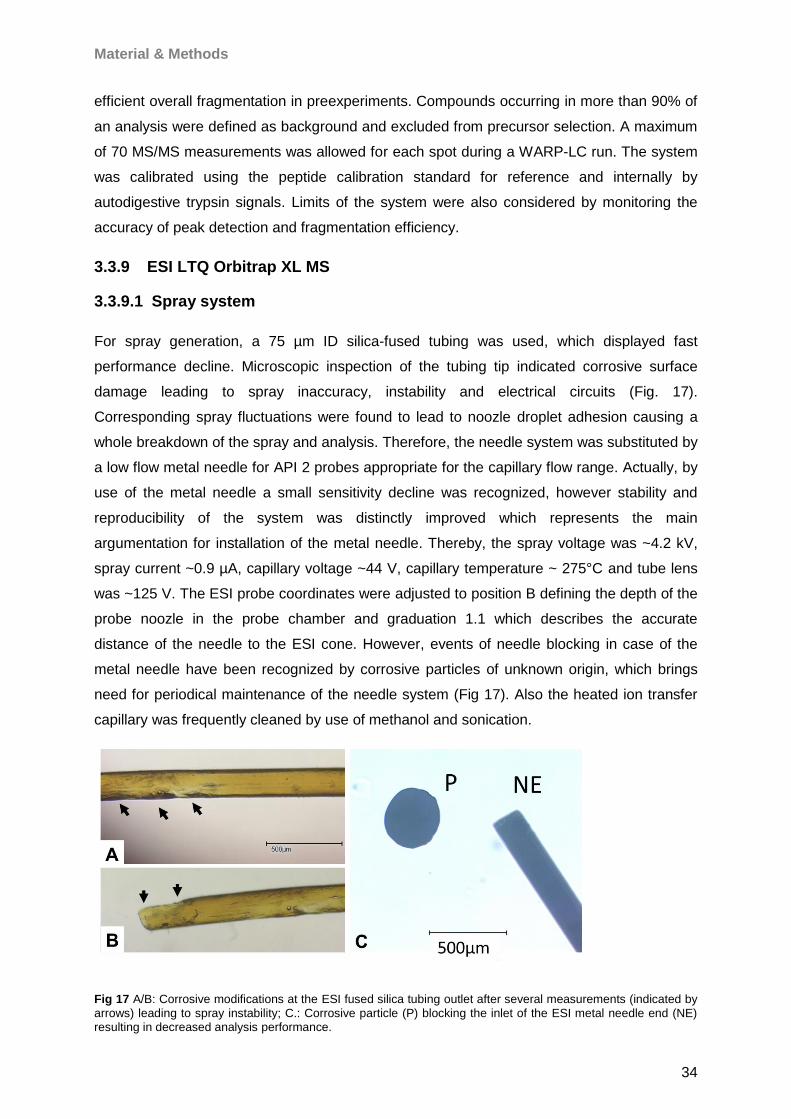
Material & Methods
34
efficient overall fragmentation in preexperiments. Compounds occurring in more than 90% of
an analysis were defined as background and excluded from precursor selection. A maximum
of 70 MS/MS measurements was allowed for each spot during a WARP-LC run. The system
was calibrated using the peptide calibration standard for reference and internally by
autodigestive trypsin signals. Limits of the system were also considered by monitoring the
accuracy of peak detection and fragmentation efficiency.
3.3.9 ESI LTQ Orbitrap XL MS
3.3.9.1 Spray system
For spray generation, a 75 µm ID silica-fused tubing was used, which displayed fast
performance decline. Microscopic inspection of the tubing tip indicated corrosive surface
damage leading to spray inaccuracy, instability and electrical circuits (Fig. 17).
Corresponding spray fluctuations were found to lead to noozle droplet adhesion causing a
whole breakdown of the spray and analysis. Therefore, the needle system was substituted by
a low flow metal needle for API 2 probes appropriate for the capillary flow range. Actually, by
use of the metal needle a small sensitivity decline was recognized, however stability and
reproducibility of the system was distinctly improved which represents the main
argumentation for installation of the metal needle. Thereby, the spray voltage was ~4.2 kV,
spray current ~0.9 µA, capillary voltage ~44 V, capillary temperature ~ 275°C and tube lens
was ~125 V. The ESI probe coordinates were adjusted to position B defining the depth of the
probe noozle in the probe chamber and graduation 1.1 which describes the accurate
distance of the needle to the ESI cone. However, events of needle blocking in case of the
metal needle have been recognized by corrosive particles of unknown origin, which brings
need for periodical maintenance of the needle system (Fig 17). Also the heated ion transfer
capillary was frequently cleaned by use of methanol and sonication.
Fig 17 A/B: Corrosive modifications at the ESI fused silica tubing outlet after several measurements (indicated by
arrows) leading to spray instability; C.: Corrosive particle (P) blocking the inlet of the ESI metal needle end (NE) resulting in decreased analysis performance.
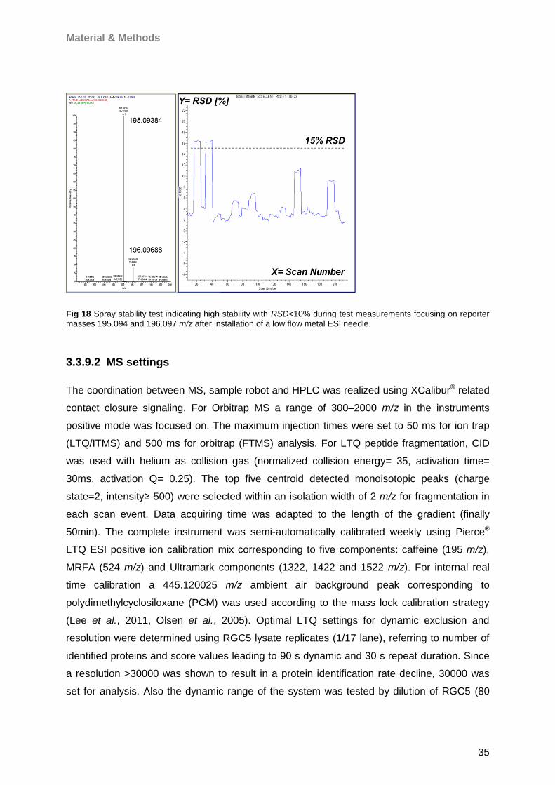
Material & Methods
35
Fig 18 Spray stability test indicating high stability with RSD<10% during test measurements focusing on reporter masses 195.094 and 196.097 m/z after installation of a low flow metal ESI needle.
3.3.9.2 MS settings
The coordination between MS, sample robot and HPLC was realized using XCalibur® related
contact closure signaling. For Orbitrap MS a range of 300–2000 m/z in the instruments
positive mode was focused on. The maximum injection times were set to 50 ms for ion trap
(LTQ/ITMS) and 500 ms for orbitrap (FTMS) analysis. For LTQ peptide fragmentation, CID
was used with helium as collision gas (normalized collision energy= 35, activation time=
30ms, activation Q= 0.25). The top five centroid detected monoisotopic peaks (charge
state=2, intensity≥ 500) were selected within an isolation width of 2 m/z for fragmentation in
each scan event. Data acquiring time was adapted to the length of the gradient (finally
50min). The complete instrument was semi-automatically calibrated weekly using Pierce®
LTQ ESI positive ion calibration mix corresponding to five components: caffeine (195 m/z),
MRFA (524 m/z) and Ultramark components (1322, 1422 and 1522 m/z). For internal real
time calibration a 445.120025 m/z ambient air background peak corresponding to
polydimethylcyclosiloxane (PCM) was used according to the mass lock calibration strategy
(Lee et al., 2011, Olsen et al., 2005). Optimal LTQ settings for dynamic exclusion and
resolution were determined using RGC5 lysate replicates (1/17 lane), referring to number of
identified proteins and score values leading to 90 s dynamic and 30 s repeat duration. Since
a resolution >30000 was shown to result in a protein identification rate decline, 30000 was
set for analysis. Also the dynamic range of the system was tested by dilution of RGC5 (80
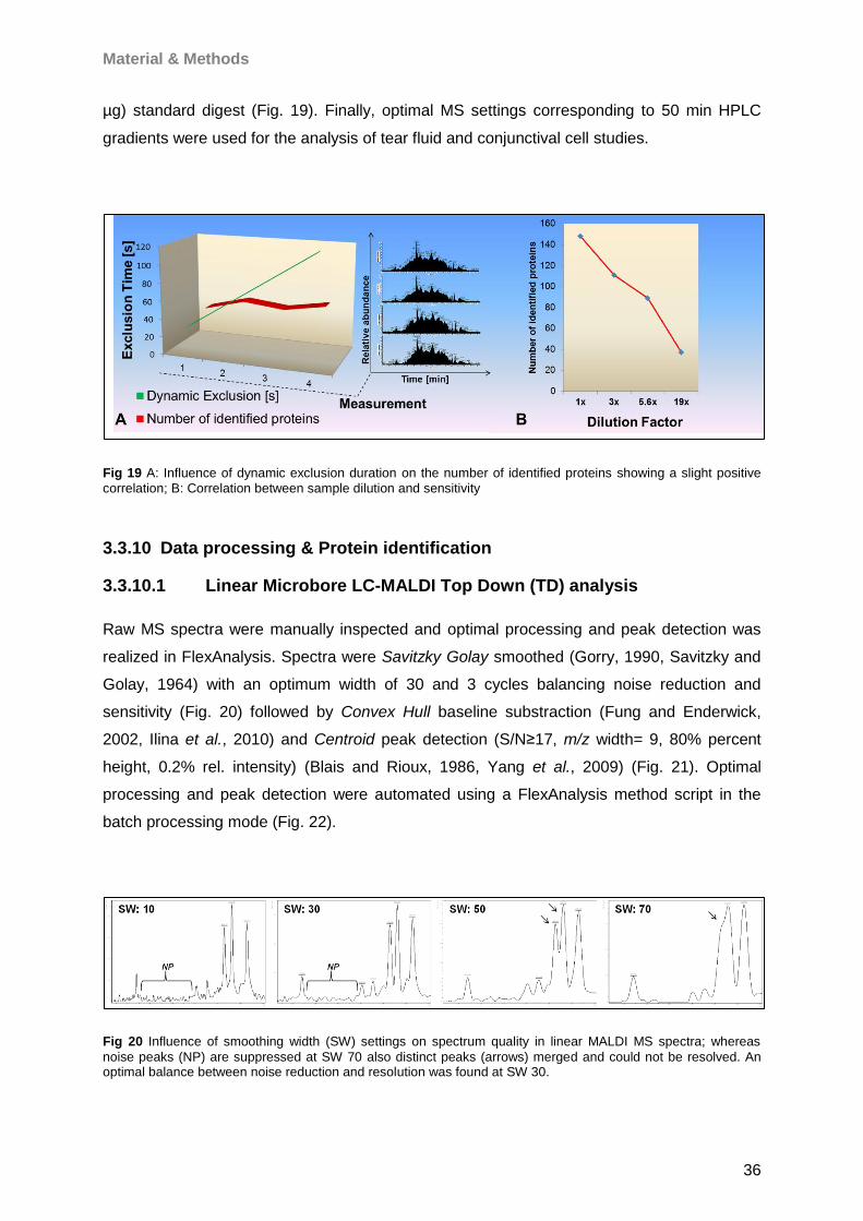
Material & Methods
36
µg) standard digest (Fig. 19). Finally, optimal MS settings corresponding to 50 min HPLC
gradients were used for the analysis of tear fluid and conjunctival cell studies.
Fig 19 A: Influence of dynamic exclusion duration on the number of identified proteins showing a slight positive
correlation; B: Correlation between sample dilution and sensitivity
3.3.10 Data processing & Protein identification
3.3.10.1 Linear Microbore LC-MALDI Top Down (TD) analysis
Raw MS spectra were manually inspected and optimal processing and peak detection was
realized in FlexAnalysis. Spectra were Savitzky Golay smoothed (Gorry, 1990, Savitzky and
Golay, 1964) with an optimum width of 30 and 3 cycles balancing noise reduction and
sensitivity (Fig. 20) followed by Convex Hull baseline substraction (Fung and Enderwick,
2002, Ilina et al., 2010) and Centroid peak detection (S/N≥17, m/z width= 9, 80% percent
height, 0.2% rel. intensity) (Blais and Rioux, 1986, Yang et al., 2009) (Fig. 21). Optimal
processing and peak detection were automated using a FlexAnalysis method script in the
batch processing mode (Fig. 22).
Fig 20 Influence of smoothing width (SW) settings on spectrum quality in linear MALDI MS spectra; whereas
noise peaks (NP) are suppressed at SW 70 also distinct peaks (arrows) merged and could not be resolved. An optimal balance between noise reduction and resolution was found at SW 30.
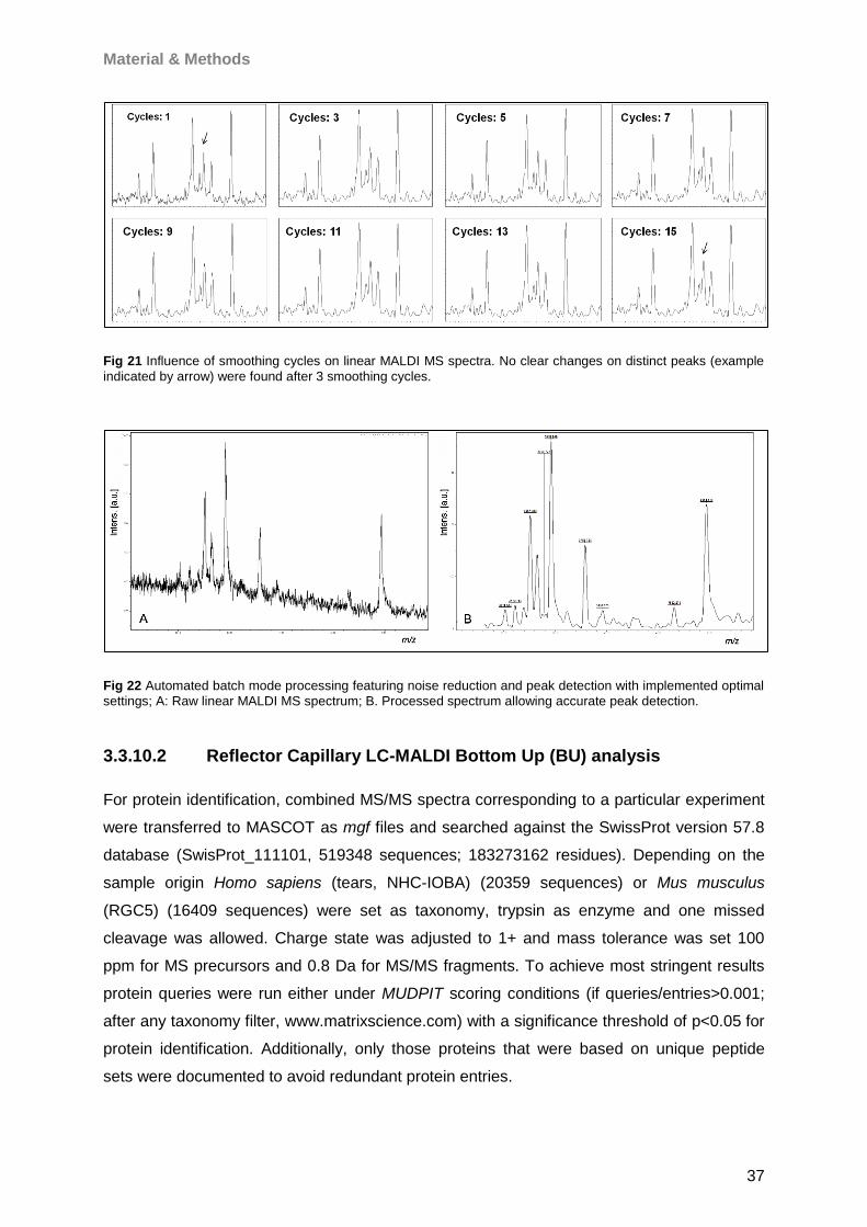
Material & Methods
37
Fig 21 Influence of smoothing cycles on linear MALDI MS spectra. No clear changes on distinct peaks (example
indicated by arrow) were found after 3 smoothing cycles.
Fig 22 Automated batch mode processing featuring noise reduction and peak detection with implemented optimal
settings; A: Raw linear MALDI MS spectrum; B. Processed spectrum allowing accurate peak detection.
3.3.10.2 Reflector Capillary LC-MALDI Bottom Up (BU) analysis
For protein identification, combined MS/MS spectra corresponding to a particular experiment
were transferred to MASCOT as mgf files and searched against the SwissProt version 57.8
database (SwisProt_111101, 519348 sequences; 183273162 residues). Depending on the
sample origin Homo sapiens (tears, NHC-IOBA) (20359 sequences) or Mus musculus
(RGC5) (16409 sequences) were set as taxonomy, trypsin as enzyme and one missed
cleavage was allowed. Charge state was adjusted to 1+ and mass tolerance was set 100
ppm for MS precursors and 0.8 Da for MS/MS fragments. To achieve most stringent results
protein queries were run either under MUDPIT scoring conditions (if queries/entries>0.001;
after any taxonomy filter, www.matrixscience.com) with a significance threshold of p<0.05 for
protein identification. Additionally, only those proteins that were based on unique peptide
sets were documented to avoid redundant protein entries.

Material & Methods
38
3.3.10.3 Capillary LC-ESI Bottom Up (BU) analysis
All MS/MS spectra of a particular experiment were combined by use of the Proteome
Discoverer® version 1.1. The resulting msf file containing all experimental data was MASCOT
searched against the SwissProt as described for MALDI. ESI protein identification settings
were realized in a Proteome Discoverer® script featuring a precursor mass range of 150 to
2000 Da (charge= 1-4, S/N ≥6), trypsin as enzyme (1 allowed missed cleavage), 20 ppm
mass tolerance for precursors and 0.8 Da for fragments. Moreover, ammonia-loss (N-term
C), oxidation (H/W, M) and phosphorylation (S/T, Y) were selected for variable modifications
and carbamidomethyl (C) was set as fixed modification.
3.3.11 Protein quantification and statistics
In case of LC MALDI BU analysis semi quantitative protein level estimation was carried out
by use of a modified version of the in-house developed P2M software platform (Tschoerner,
2008), whereby MS and MS/MS spectra were imported directly from Flex Analysis following
normalization of MS spectra according to the mean total ion current (TIC) (IntensityMS x
meanTICMS/TICMS). Spectra with a minimum factor of 0.1 or maximum factor of 10 were
categorized as “low quality” and excluded from analysis. To allow comparison of protein
levels between all samples, a protein that failed to be identified in some of the samples was
extrapolated regarding the intensity value at the corresponding m/z position with a tolerance
of 0.3 Da. Since proteins can smear over several gel slices, a “Blur” function considering
drifting proteins in gels was implicated with a tolerance of +/- 1 slice. All sub experiment files
were merged in one Excel csv summary file and transferred to Statistica version 8/10.
Moreover all normalized peptide levels, which contributed to the identification of a particular
protein, were summarized for the protein. Protein levels were then used for statistical protein
level determination. In case of LC ESI experiments, a further optimized P2M workflow was
established (Wolters, 2011, Wolters, 2012) featuring raw files and Proteome Discoverer®
result mgf file containing protein and peptide information for TIC normalization. Thereby,
each particular peptide corresponding to an identified protein is searched with a tolerance
between 0.02-0.05 Da considering experimental quality in all samples of the experiment that
should be compared. Moreover a RT shift algorithm had been implemented in the software,
which scans spectra for peptides within the RT range, where the pepide was primarily
identified. Normalized peptides were summarized for each particular identified protein and
transferred to a final Excel csv file for statistical analysis of protein levels in Statistica.
3.3.12 Protein functional annotation analysis
To determine origin, function and implementation of ocular surface proteins two different
software solutions were used. Cytoscape version 2.8.3 with integrated BINGO 2.44 plugin
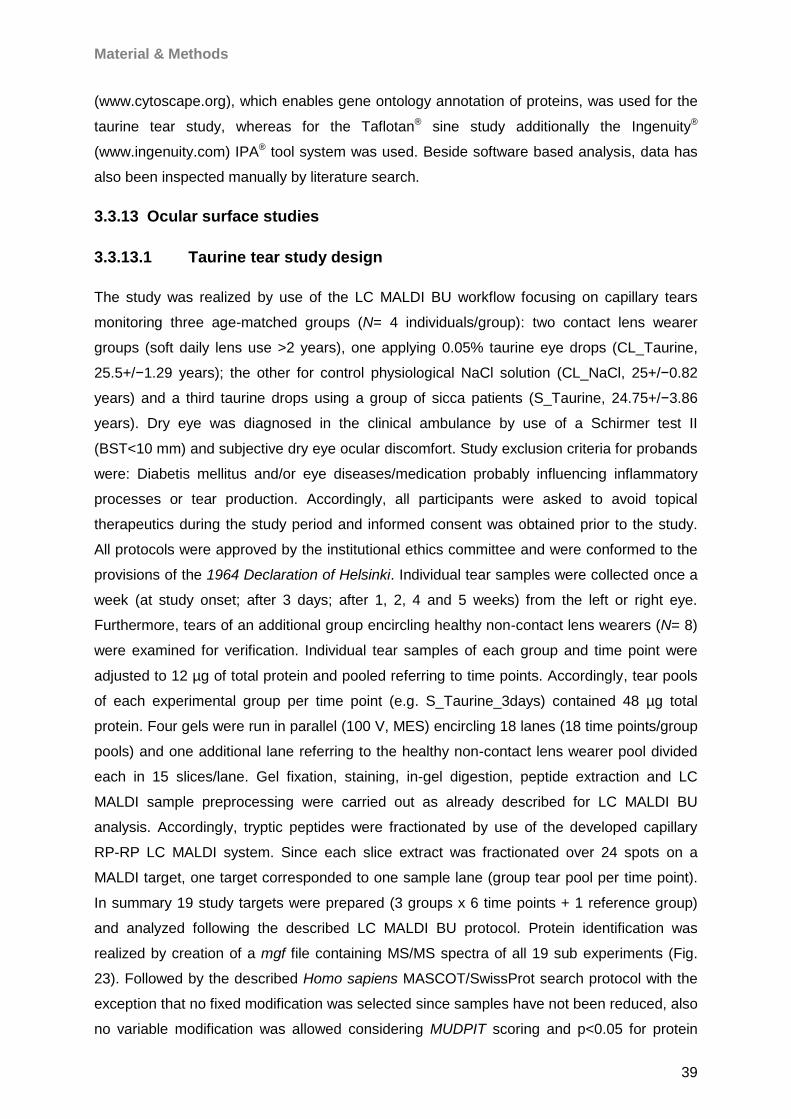
Material & Methods
39
(www.cytoscape.org), which enables gene ontology annotation of proteins, was used for the
taurine tear study, whereas for the Taflotan® sine study additionally the Ingenuity®
(www.ingenuity.com) IPA® tool system was used. Beside software based analysis, data has
also been inspected manually by literature search.
3.3.13 Ocular surface studies
3.3.13.1 Taurine tear study design
The study was realized by use of the LC MALDI BU workflow focusing on capillary tears
monitoring three age-matched groups (N= 4 individuals/group): two contact lens wearer
groups (soft daily lens use >2 years), one applying 0.05% taurine eye drops (CL_Taurine,
25.5+/−1.29 years); the other for control physiological NaCl solution (CL_NaCl, 25+/−0.82
years) and a third taurine drops using a group of sicca patients (S_Taurine, 24.75+/−3.86
years). Dry eye was diagnosed in the clinical ambulance by use of a Schirmer test II
(BST<10 mm) and subjective dry eye ocular discomfort. Study exclusion criteria for probands
were: Diabetis mellitus and/or eye diseases/medication probably influencing inflammatory
processes or tear production. Accordingly, all participants were asked to avoid topical
therapeutics during the study period and informed consent was obtained prior to the study.
All protocols were approved by the institutional ethics committee and were conformed to the
provisions of the 1964 Declaration of Helsinki. Individual tear samples were collected once a
week (at study onset; after 3 days; after 1, 2, 4 and 5 weeks) from the left or right eye.
Furthermore, tears of an additional group encircling healthy non-contact lens wearers (N= 8)
were examined for verification. Individual tear samples of each group and time point were
adjusted to 12 µg of total protein and pooled referring to time points. Accordingly, tear pools
of each experimental group per time point (e.g. S_Taurine_3days) contained 48 µg total
protein. Four gels were run in parallel (100 V, MES) encircling 18 lanes (18 time points/group
pools) and one additional lane referring to the healthy non-contact lens wearer pool divided
each in 15 slices/lane. Gel fixation, staining, in-gel digestion, peptide extraction and LC
MALDI sample preprocessing were carried out as already described for LC MALDI BU
analysis. Accordingly, tryptic peptides were fractionated by use of the developed capillary
RP-RP LC MALDI system. Since each slice extract was fractionated over 24 spots on a
MALDI target, one target corresponded to one sample lane (group tear pool per time point).
In summary 19 study targets were prepared (3 groups x 6 time points + 1 reference group)
and analyzed following the described LC MALDI BU protocol. Protein identification was
realized by creation of a mgf file containing MS/MS spectra of all 19 sub experiments (Fig.
23). Followed by the described Homo sapiens MASCOT/SwissProt search protocol with the
exception that no fixed modification was selected since samples have not been reduced, also
no variable modification was allowed considering MUDPIT scoring and p<0.05 for protein
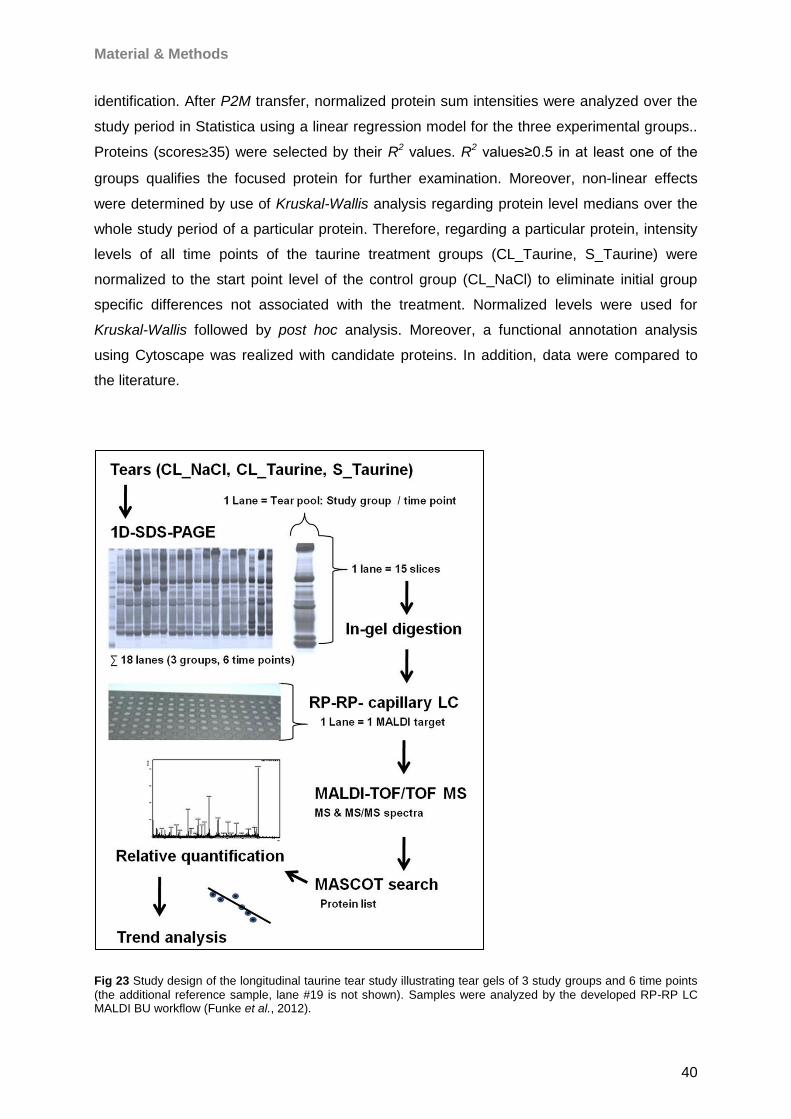
Material & Methods
40
identification. After P2M transfer, normalized protein sum intensities were analyzed over the
study period in Statistica using a linear regression model for the three experimental groups..
Proteins (scores≥35) were selected by their R2 values. R2 values≥0.5 in at least one of the
groups qualifies the focused protein for further examination. Moreover, non-linear effects
were determined by use of Kruskal-Wallis analysis regarding protein level medians over the
whole study period of a particular protein. Therefore, regarding a particular protein, intensity
levels of all time points of the taurine treatment groups (CL_Taurine, S_Taurine) were
normalized to the start point level of the control group (CL_NaCl) to eliminate initial group
specific differences not associated with the treatment. Normalized levels were used for
Kruskal-Wallis followed by post hoc analysis. Moreover, a functional annotation analysis
using Cytoscape was realized with candidate proteins. In addition, data were compared to
the literature.
Fig 23 Study design of the longitudinal taurine tear study illustrating tear gels of 3 study groups and 6 time points
(the additional reference sample, lane #19 is not shown). Samples were analyzed by the developed RP-RP LC MALDI BU workflow (Funke et al., 2012).

Material & Methods
41
3.3.13.2 Taurine conjunctival cell study design
To monitor taurine effects on stressed conjunctival cells (IOBA-NHC), cells were prepared in
24well plates for two stress conditions: staurosporine (30000 cells, 0.25 µM/5 h) and H2O2
(40000 cells, 20 mM/1 h). Cell viability was examined by crystal violet assay and reactive
oxygen species (ROS) level by DCFH-DA ROS assay, both performed in duplet for each
taurine concentration. Moreover, each experiment was repeated four times. For LC ESI MS
analysis 50000 cells/dish were seeded to pool for proper protein amount (taurine=
3xdishes/concentration; control= 3xdishes). Taurine solutions were freshly prepared in cell
buffer featuring 3h cell incubation for each concentration: 100, 50, 20, 4, 2, 1, 0.5, 0.2, 0.05
mM inferred from the literature (Das et al., 2011, Franconi et al., 1985, Jong et al., 2012,
Muhling et al., 2002, Pan et al., 2010, Pan et al., 2012, Pasantes-Morales, 1982, Weiss et
al., 1982, Wu et al., 1999) followed by cell harvest, lysis and protein extraction, BCA assay,
SDS PAGE and LC ESI MS. For the first experiment 6 gel lanes (50, 20, 4 mM; 3x control)
were run, for the second experiment two duplicate lanes (2x 0.05 mM, 2x control) were run in
parallel to monitor gel reproducibility. For the first experiment all lanes (N= 6), for the second
experiment one taurine (0.05mM) and one control lane were prepared for LC ESI MS. Also,
all samples were group randomized and measured ( N= 6) in the first experiment.
Corresponding raw files of three samples (4 mM, 50 mM taurine; control) were combined for
quantification. For the second experiment, each sample lane was measured in duplicate to
additionally counteract technical bias, whereby raw files of all runs were combined.
SwissProt corresponding normalized proteins were quantified using the described
P2M/Statistica workflow. Statistical analysis was realized using a minimum 2fold level
alteration threshold in confidence with the literature (Blagoev et al., 2004, Choe et al., 2005,
Friedman et al., 2004, Leal et al., 2012, Mann and Kelleher, 2008, Molloy et al., 2003,
Ohlmeier et al., 2004, Old et al., 2005, Wilkins, 2006) for potential taurine responding protein
candidates. Conjunctival proteome characterization was done by Cytoscape and literature
screening. The complete workflow is depicted below (Fig. 24).
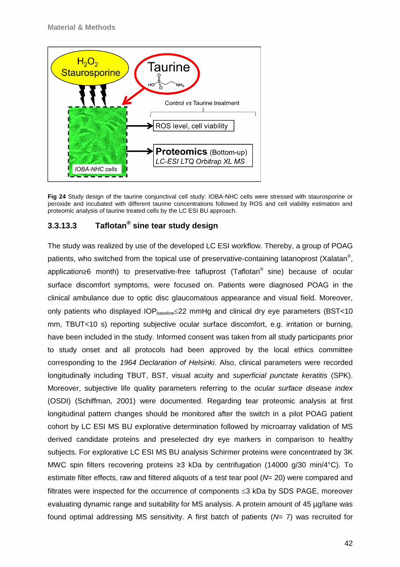
Material & Methods
42
Fig 24 Study design of the taurine conjunctival cell study: IOBA-NHC cells were stressed with staurosporine or
peroxide and incubated with different taurine concentrations followed by ROS and cell viability estimation and proteomic analysis of taurine treated cells by the LC ESI BU approach.
3.3.13.3 Taflotan® sine tear study design
The study was realized by use of the developed LC ESI workflow. Thereby, a group of POAG
patients, who switched from the topical use of preservative-containing latanoprost (Xalatan®,
application≥6 month) to preservative-free tafluprost (Taflotan® sine) because of ocular
surface discomfort symptoms, were focused on. Patients were diagnosed POAG in the
clinical ambulance due to optic disc glaucomatous appearance and visual field. Moreover,
only patients who displayed IOPbaseline22 mmHg and clinical dry eye parameters (BST<10
mm, TBUT<10 s) reporting subjective ocular surface discomfort, e.g. irritation or burning,
have been included in the study. Informed consent was taken from all study participants prior
to study onset and all protocols had been approved by the local ethics committee
corresponding to the 1964 Declaration of Helsinki. Also, clinical parameters were recorded
longitudinally including TBUT, BST, visual acuity and superficial punctate keratitis (SPK).
Moreover, subjective life quality parameters referring to the ocular surface disease index
(OSDI) (Schiffman, 2001) were documented. Regarding tear proteomic analysis at first
longitudinal pattern changes should be monitored after the switch in a pilot POAG patient
cohort by LC ESI MS BU explorative determination followed by microarray validation of MS
derived candidate proteins and preselected dry eye markers in comparison to healthy
subjects. For explorative LC ESI MS BU analysis Schirmer proteins were concentrated by 3K
MWC spin filters recovering proteins ≥3 kDa by centrifugation (14000 g/30 min/4°C). To
estimate filter effects, raw and filtered aliquots of a test tear pool (N= 20) were compared and
filtrates were inspected for the occurrence of components 3 kDa by SDS PAGE, moreover
evaluating dynamic range and suitability for MS analysis. A protein amount of 45 µg/lane was
found optimal addressing MS sensitivity. A first batch of patients (N= 7) was recruited for
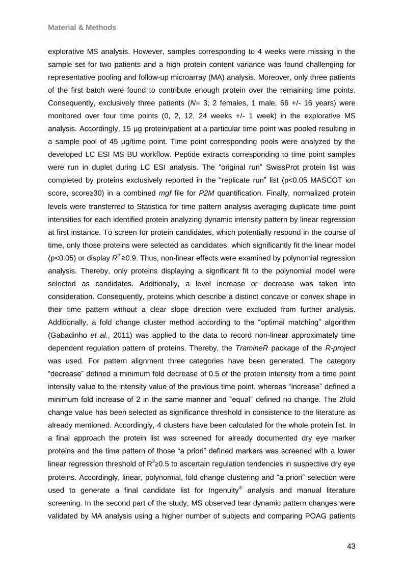
Material & Methods
43
explorative MS analysis. However, samples corresponding to 4 weeks were missing in the
sample set for two patients and a high protein content variance was found challenging for
representative pooling and follow-up microarray (MA) analysis. Moreover, only three patients
of the first batch were found to contribute enough protein over the remaining time points.
Consequently, exclusively three patients (N= 3; 2 females, 1 male, 66 +/- 16 years) were
monitored over four time points (0, 2, 12, 24 weeks +/- 1 week) in the explorative MS
analysis. Accordingly, 15 µg protein/patient at a particular time point was pooled resulting in
a sample pool of 45 µg/time point. Time point corresponding pools were analyzed by the
developed LC ESI MS BU workflow. Peptide extracts corresponding to time point samples
were run in duplet during LC ESI analysis. The “original run” SwissProt protein list was
completed by proteins exclusively reported in the “replicate run” list (p<0.05 MASCOT ion
score, score≥30) in a combined mgf file for P2M quantification. Finally, normalized protein
levels were transferred to Statistica for time pattern analysis averaging duplicate time point
intensities for each identified protein analyzing dynamic intensity pattern by linear regression
at first instance. To screen for protein candidates, which potentially respond in the course of
time, only those proteins were selected as candidates, which significantly fit the linear model
(p<0.05) or display R2 ≥0.9. Thus, non-linear effects were examined by polynomial regression
analysis. Thereby, only proteins displaying a significant fit to the polynomial model were
selected as candidates. Additionally, a level increase or decrease was taken into
consideration. Consequently, proteins which describe a distinct concave or convex shape in
their time pattern without a clear slope direction were excluded from further analysis.
Additionally, a fold change cluster method according to the “optimal matching” algorithm
(Gabadinho et al., 2011) was applied to the data to record non-linear approximately time
dependent regulation pattern of proteins. Thereby, the TramineR package of the R-project
was used. For pattern alignment three categories have been generated. The category
“decrease” defined a minimum fold decrease of 0.5 of the protein intensity from a time point
intensity value to the intensity value of the previous time point, whereas “increase” defined a
minimum fold increase of 2 in the same manner and “equal” defined no change. The 2fold
change value has been selected as significance threshold in consistence to the literature as
already mentioned. Accordingly, 4 clusters have been calculated for the whole protein list. In
a final approach the protein list was screened for already documented dry eye marker
proteins and the time pattern of those “a priori” defined markers was screened with a lower
linear regression threshold of R2≥0.5 to ascertain regulation tendencies in suspective dry eye
proteins. Accordingly, linear, polynomial, fold change clustering and “a priori” selection were
used to generate a final candidate list for Ingenuity® analysis and manual literature
screening. In the second part of the study, MS observed tear dynamic pattern changes were
validated by MA analysis using a higher number of subjects and comparing POAG patients

Material & Methods
44
(N= 16, 70 +/-10 years, 11 females, 5 males) with age-matched healthy controls without any
ocular disorders (N= 15, 11 females, 4 males, 51 +/- 10 years) over five time points (0, 2, 4,
12, 24 weeks +/- 1week) for POAG and over three time points (0, 4, 24 weeks/+/-1 week) for
healthy subjects. Thereby, commercial available antibodies against selected representative
MS derived candidate proteins, dry eye associated preselected markers and cytokines were
immobilized on nitrocellulose slices and incubated with fluorescent labeled longitudinal tear
protein samples. For analysis, a sciFLEXARRAYER MA spotter equipped with a piezo driven
spotting unit (100-500 pl/drop) and a 428TM array laser scanner equipped with a green and
red laser for fluorophor excitation (532/635 nm) were employed. MA preparation followed a
laboratory-established workflow (Boehm et al. 2011, 2012; Bohm et al.2011). Accordingly, 1
nl commercial antibodies was applied per spot on Oncyte Avid 6.5mm x 6.5mm nitrocellulose
slides in triplicate and incubated with 7.5 µg of fluorescent labeled (Cy5 Alexa Fluor 647) tear
protein of each individual and time point to generate a longitudinal intensity pattern for each
subject. Proteins, which could not been spotted properly, were excluded from further
analysis. Spots were scanned in preview with 20 and 50 µm and for quantification with 10 µm
resolution. Quantification was realized by use of ImaGene® version 5.5 exporting median
spot and background signals to Statistica and difference was calculated between both signal
median values. Negative difference values were excluded from the list, which contained
triplicate antibody response intensities for each patient. Mean intensities encircling all group
individuals (POAG vs Control) were calculated for each time point and each candidate. For
regression analysis, only tendencies were focused on. To obtain a more accurate
impression, if the mean intensity level of a certain protein converges from POAG to control, a
time point comparison was operated. For that purpose, the mean of all time point intensities
of the Control group was compared to the mean intensity of the initial and final time point of
the POAG group estimating the confidential degree of the “POAG to Control level
approximation” by t-test. A highly confidential approximation should therefore show
significant intensity differences between the initial time point of the POAG group compared to
the mean value encircling all time points of the Control group, but displaying non-significant
differences between the final time points of the POAG group compared to the overall control
group level. A brief overview on the study workflow is illustrated in Fig. 25.
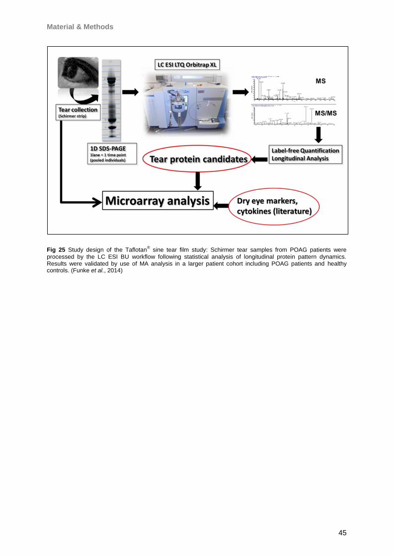
Material & Methods
45
Fig 25 Study design of the Taflotan® sine tear film study: Schirmer tear samples from POAG patients were
processed by the LC ESI BU workflow following statistical analysis of longitudinal protein pattern dynamics. Results were validated by use of MA analysis in a larger patient cohort including POAG patients and healthy controls. (Funke et al., 2014)
Results
46
4 Results
4.1 Microbore-RP-HPLC (for MALDI)
The developed column system was found appropriate for direct fractionation of crude
samples without further sample pretreatment indicated by succesfull retaining of contaminant
particles by the Security GuardTM cartridge (Fig. 26). Defined isolated fractions could be
deposited on MALDI targets displaying low sample carry-over and homogene crystallization
(Fig. 27). Regarding MS output, the method generates “high quality” linear MS spectra
regarding m/z peak pattern of selected LC fractions (Fig. 28). Furthermore, high separation
efficiency of the LC system could be achieved (Fig. 29). Compared to non-fractionated
samples, an elaborative purification benefit could be registered for the HPLC prepared
samples which resulted in a distinct increase of peak intensities (Fig. 30). Regarding training
samples, the method showed a high level of run-to-run reproducibility (Fig. 31, 32). Retention
deviation was estimated <12 s on average for a 3 s fraction deposition frequency monitored
for selected reporter peaks 2481, 4962 and 6275 m/z based on first component detection
(Fig. 33). The spotting time could be adjusted to a high degree of speed counting 4 min 36 s
for each LC run. During training set runs, the HPLC running time for each run was 35 min
and the complete HPLC running time of 24 samples including equilibration runs and
automated matrix spotting could be recorded (92x24x35= 7782 min + 24 x 10 Dummy runs=
240 min + 92 x 24 x 10 s matrix spotting/spot= 368 min) <6 days (139.8 h). The linear MS
measuring rate using 50 hz laser frequency was set to approximately 44 s/spot. Accordingly,
the study encircling analysis of all 6 study targets could be finished in approximately 27 h. In
summary, the total analysis time including HPLC fractionation, matrix spotting and MALDI
TOF MS did not exceed 7 days for 24 samples. Therefore, high quality MS pattern could be
generated and the overall sensitivity of the approach prior to normalization was estimated by
856 RGC5 WARP-LC compounds corresponding to a mean number of 960 peaks detected
in each LC run (N= 24). However, identification of exemplary reporter components 3789,
4279, 6648, 7208 and 8567 m/z detected in all samples was challenging. Direct fractionation
of masses >4000 Da is critical in MALDI MS (Nilsen et al., 2011) and even direct
fractionation of peaks below this threshold was found problematic due to decreased reflector
sensitivity. Only 3789 m/z could be detected by reflector analysis (Fig. 34), however, failing
to reach threshold abundance for successful fragmentation. For this reason, an alternative
strategy in reference to a comparable workflow (Boehm et al., 2013) was chosen to clarify
protein identities including ZIPTIP® C18 fractionation (2-40% ACN in 2% steps) of intact
lysates, 1D SDS PAGE, intact protein extraction from the gel mass range of interest, MALDI
linear reanalysis and in-gel digestion of the corresponding gel mass range for MALDI/LC ESI
MS/MS analysis (Fig. 35, 36A). Despite reporters 3279, 4279 and 8567 m/z could be
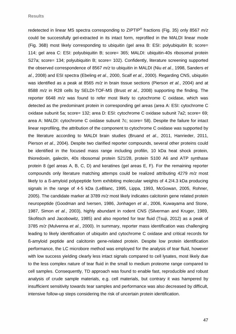
Results
47
redetected in linear MS spectra corresponding to ZIPTIP® fractions (Fig. 35) only 8567 m/z
could be successfully gel-extracted in its intact form, reprofiled in the MALDI linear mode
(Fig. 36B) most likely corresponding to ubiquitin (gel area B: ESI: polyubiquitin B; score=
114; gel area C: ESI: polyubiquitin B; score= 365; MALDI: ubiquitin-40s ribosomal protein
S27a; score= 134; polyubiquitin B; score= 102). Confidently, literature screening supported
the observed correspondence of 8567 m/z to ubiquitin in MALDI (Niu et al., 1998, Sanders et
al., 2008) and ESI spectra (Ebeling et al., 2000, Scalf et al., 2000). Regarding CNS, ubiquitin
was identified as a peak at 8565 m/z in brain tissue sections (Pierson et al., 2004) and at
8588 m/z in R28 cells by SELDI-TOF-MS (Brust et al., 2008) supporting the finding. The
reporter 6648 m/z was found to refer most likely to cytochrome C oxidase, which was
detected as the predominant protein in corresponding gel areas (area A: ESI: cytochrome C
oxidase subunit 5a; score= 132; area D: ESI: cytochrome C oxidase subunit 7a2; score= 69;
area A: MALDI: cytochrome C oxidase subunit 7c; score= 58). Despite the failure for intact
linear reprofiling, the attribution of the component to cytochrome C oxidase was supported by
the literature according to MALDI brain studies (Bruand et al., 2011, Hanrieder, 2011,
Pierson et al., 2004). Despite two clarified reporter compounds, several other proteins could
be identified in the focused mass range including profilin, 10 kDa heat shock protein,
thioredoxin, galectin, 40s ribosomal protein S21/28, protein S100 A6 and ATP synthase
protein 8 (gel areas A, B, C, D) and keratines (gel areas E, F). For the remaining reporter
compounds only literature matching attemps could be realized attributing 4279 m/z most
likely to a ß-amyloid polypeptide form exhibiting molecular weights of 4.2/4.3 kDa producing
signals in the range of 4-5 kDa (LeBlanc, 1995, Lippa, 1993, McGowan, 2005, Rohner,
2005). The candidate marker at 3789 m/z most likely indicates calcitonin gene related protein
neuropeptide (Goodman and Iversen, 1986, Jonhagen et al., 2006, Kuwayama and Stone,
1987, Simon et al., 2003), highly abundant in rodent CNS (Silverman and Kruger, 1989,
Skofitsch and Jacobowitz, 1985) and also reported for tear fluid (Tsuji, 2012) as a peak of
3785 m/z (Mulvenna et al., 2000). In summary, reporter mass identification was challenging
leading to likely identification of ubiquitin and cytochrome C oxidase and critical records for
ß-amyloid peptide and calcitonin gene-related protein. Despite low protein identification
performance, the LC microbore method was employed for the analysis of tear fluid, however
with low success yielding clearly less intact signals compared to cell lysates, most likely due
to the less complex nature of tear fluid in the small to medium proteome range compared to
cell samples. Consequently, TD approach was found to enable fast, reproducible and robust
analysis of crude sample materials, e.g. cell materials, but contrary it was hampered by
insufficient sensitivity towards tear samples and performance was also decreased by difficult,
intensive follow-up steps considering the risk of uncertain protein identification.
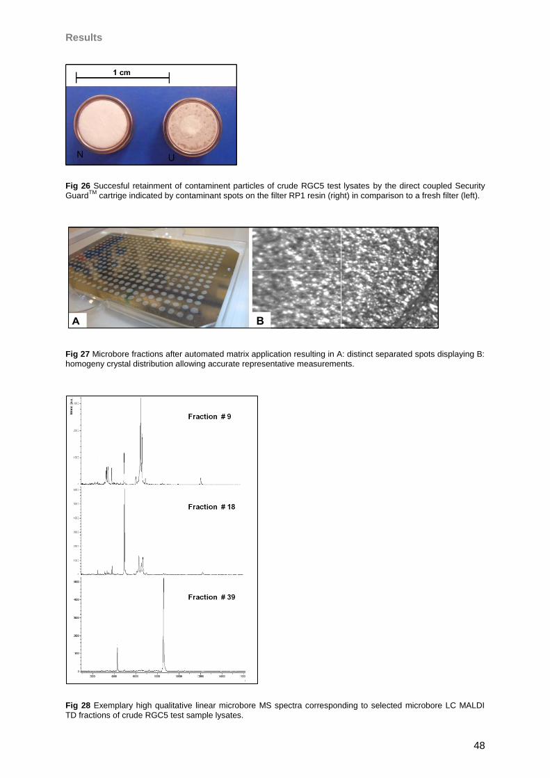
Results
48
Fig 26 Succesful retainment of contaminent particles of crude RGC5 test lysates by the direct coupled Security
GuardTM
cartrige indicated by contaminant spots on the filter RP1 resin (right) in comparison to a fresh filter (left).
Fig 27 Microbore fractions after automated matrix application resulting in A: distinct separated spots displaying B:
homogeny crystal distribution allowing accurate representative measurements.
Fig 28 Exemplary high qualitative linear microbore MS spectra corresponding to selected microbore LC MALDI
TD fractions of crude RGC5 test sample lysates.
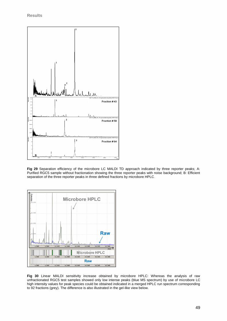
Results
49
Fig 29 Separation efficiency of the microbore LC MALDI TD approach indicated by three reporter peaks; A:
Purified RGC5 sample without fractionation showing the three reporter peaks with noise background; B: Efficient separation of the three reporter peaks in three defined fractions by microbore HPLC.
Fig 30 Linear MALDI sensitivity increase obtained by microbore HPLC: Whereas the analysis of raw
unfractionated RGC5 test samples showed only low intense peaks (blue MS spectrum) by use of microbore LC high intensity values for peak species could be obtained indicated in a merged HPLC run spectrum corresponding to 92 fractions (grey). The difference is also illustrated in the gel-like view below.
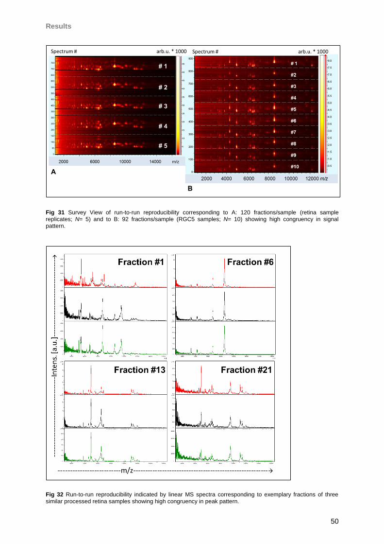
Results
50
Fig 31 Survey View of run-to-run reproducibility corresponding to A: 120 fractions/sample (retina sample replicates; N= 5) and to B: 92 fractions/sample (RGC5 samples; N= 10) showing high congruency in signal pattern.
Fig 32 Run-to-run reproducibility indicated by linear MS spectra corresponding to exemplary fractions of three
similar processed retina samples showing high congruency in peak pattern.
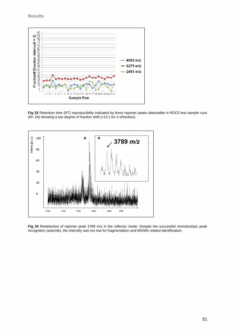
Results
51
Fig 33 Retention time (RT) reproducibility indicated by three reporter peaks detectable in RGC5 test sample runs
(N= 24) showing a low degree of fraction shift (<12 s for 3 s/fraction).
Fig 34 Redetection of reporter peak 3789 m/z in the reflector mode: Despite the successful monoisotopic peak
recognition (asterisk), the intensity was too low for fragmentation and MS/MS related identification.
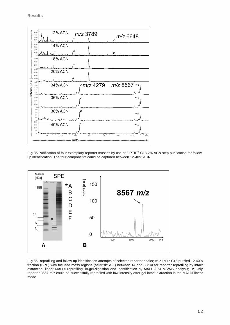
Results
52
Fig 35 Purification of four exemplary reporter masses by use of ZIPTIP® C18 2% ACN step purification for follow-
up identification. The four components could be captured between 12-40% ACN.
Fig 36 Reprofiling and follow-up identification attempts of selected reporter peaks; A: ZIPTIP C18 purified 12-40%
fraction (SPE) with focused mass regions (asterisk: A-F) between 14 and 3 kDa for reporter reprofiling by intact extraction, linear MALDI reprofiling, in-gel-digestion and identification by MALDI/ESI MS/MS analysis; B: Only reporter 8567 m/z could be successfully reprofiled with low intensity after gel intact extraction in the MALDI linear mode.
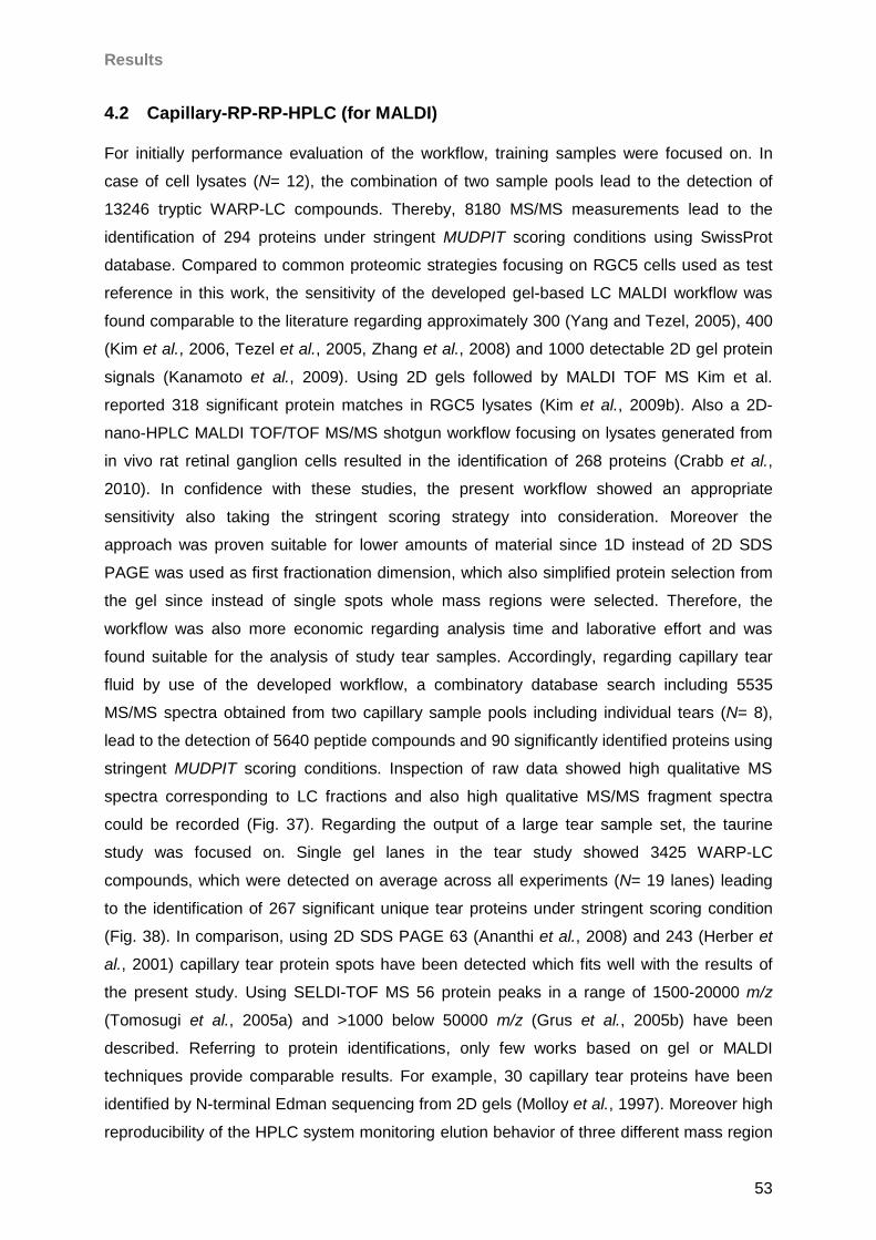
Results
53
4.2 Capillary-RP-RP-HPLC (for MALDI)
For initially performance evaluation of the workflow, training samples were focused on. In
case of cell lysates (N= 12), the combination of two sample pools lead to the detection of
13246 tryptic WARP-LC compounds. Thereby, 8180 MS/MS measurements lead to the
identification of 294 proteins under stringent MUDPIT scoring conditions using SwissProt
database. Compared to common proteomic strategies focusing on RGC5 cells used as test
reference in this work, the sensitivity of the developed gel-based LC MALDI workflow was
found comparable to the literature regarding approximately 300 (Yang and Tezel, 2005), 400
(Kim et al., 2006, Tezel et al., 2005, Zhang et al., 2008) and 1000 detectable 2D gel protein
signals (Kanamoto et al., 2009). Using 2D gels followed by MALDI TOF MS Kim et al.
reported 318 significant protein matches in RGC5 lysates (Kim et al., 2009b). Also a 2D-
nano-HPLC MALDI TOF/TOF MS/MS shotgun workflow focusing on lysates generated from
in vivo rat retinal ganglion cells resulted in the identification of 268 proteins (Crabb et al.,
2010). In confidence with these studies, the present workflow showed an appropriate
sensitivity also taking the stringent scoring strategy into consideration. Moreover the
approach was proven suitable for lower amounts of material since 1D instead of 2D SDS
PAGE was used as first fractionation dimension, which also simplified protein selection from
the gel since instead of single spots whole mass regions were selected. Therefore, the
workflow was also more economic regarding analysis time and laborative effort and was
found suitable for the analysis of study tear samples. Accordingly, regarding capillary tear
fluid by use of the developed workflow, a combinatory database search including 5535
MS/MS spectra obtained from two capillary sample pools including individual tears (N= 8),
lead to the detection of 5640 peptide compounds and 90 significantly identified proteins using
stringent MUDPIT scoring conditions. Inspection of raw data showed high qualitative MS
spectra corresponding to LC fractions and also high qualitative MS/MS fragment spectra
could be recorded (Fig. 37). Regarding the output of a large tear sample set, the taurine
study was focused on. Single gel lanes in the tear study showed 3425 WARP-LC
compounds, which were detected on average across all experiments (N= 19 lanes) leading
to the identification of 267 significant unique tear proteins under stringent scoring condition
(Fig. 38). In comparison, using 2D SDS PAGE 63 (Ananthi et al., 2008) and 243 (Herber et
al., 2001) capillary tear protein spots have been detected which fits well with the results of
the present study. Using SELDI-TOF MS 56 protein peaks in a range of 1500-20000 m/z
(Tomosugi et al., 2005a) and >1000 below 50000 m/z (Grus et al., 2005b) have been
described. Referring to protein identifications, only few works based on gel or MALDI
techniques provide comparable results. For example, 30 capillary tear proteins have been
identified by N-terminal Edman sequencing from 2D gels (Molloy et al., 1997). Moreover high
reproducibility of the HPLC system monitoring elution behavior of three different mass region
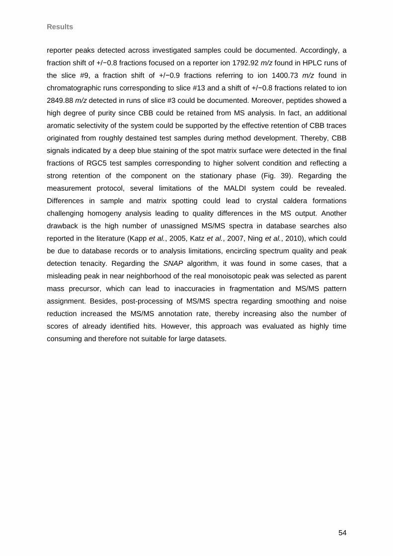
Results
54
reporter peaks detected across investigated samples could be documented. Accordingly, a
fraction shift of +/−0.8 fractions focused on a reporter ion 1792.92 m/z found in HPLC runs of
the slice #9, a fraction shift of +/−0.9 fractions referring to ion 1400.73 m/z found in
chromatographic runs corresponding to slice #13 and a shift of +/−0.8 fractions related to ion
2849.88 m/z detected in runs of slice #3 could be documented. Moreover, peptides showed a
high degree of purity since CBB could be retained from MS analysis. In fact, an additional
aromatic selectivity of the system could be supported by the effective retention of CBB traces
originated from roughly destained test samples during method development. Thereby, CBB
signals indicated by a deep blue staining of the spot matrix surface were detected in the final
fractions of RGC5 test samples corresponding to higher solvent condition and reflecting a
strong retention of the component on the stationary phase (Fig. 39). Regarding the
measurement protocol, several limitations of the MALDI system could be revealed.
Differences in sample and matrix spotting could lead to crystal caldera formations
challenging homogeny analysis leading to quality differences in the MS output. Another
drawback is the high number of unassigned MS/MS spectra in database searches also
reported in the literature (Kapp et al., 2005, Katz et al., 2007, Ning et al., 2010), which could
be due to database records or to analysis limitations, encircling spectrum quality and peak
detection tenacity. Regarding the SNAP algorithm, it was found in some cases, that a
misleading peak in near neighborhood of the real monoisotopic peak was selected as parent
mass precursor, which can lead to inaccuracies in fragmentation and MS/MS pattern
assignment. Besides, post-processing of MS/MS spectra regarding smoothing and noise
reduction increased the MS/MS annotation rate, thereby increasing also the number of
scores of already identified hits. However, this approach was evaluated as highly time
consuming and therefore not suitable for large datasets.
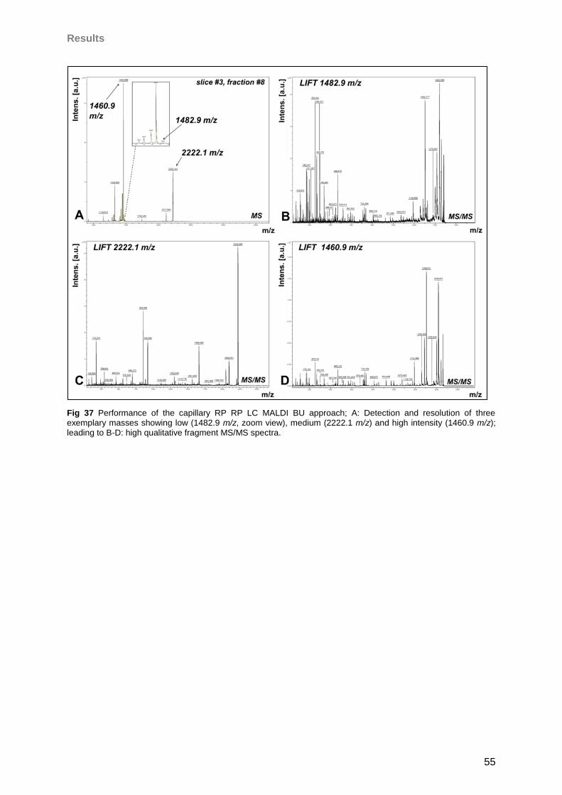
Results
55
Fig 37 Performance of the capillary RP RP LC MALDI BU approach; A: Detection and resolution of three exemplary masses showing low (1482.9 m/z, zoom view), medium (2222.1 m/z) and high intensity (1460.9 m/z); leading to B-D: high qualitative fragment MS/MS spectra.
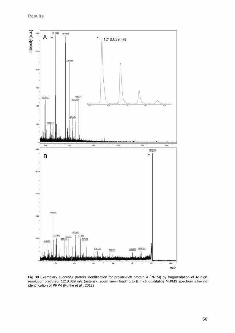
Results
56
Fig 38 Exemplary succesful protein identification for proline-rich protein 4 (PRP4) by fragmentation of A: high
resolution precursor 1210.639 m/z (asterisk, zoom view) leading to B: high qualitative MS/MS spectrum allowing identification of PRP4 (Funke et al., 2012)
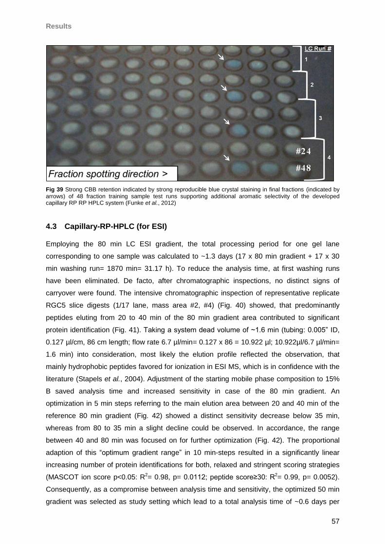
Results
57
Fig 39 Strong CBB retention indicated by strong reproducible blue crystal staining in final fractions (indicated by
arrows) of 48 fraction training sample test runs supporting additional aromatic selectivity of the developed capillary RP RP HPLC system (Funke et al., 2012)
4.3 Capillary-RP-HPLC (for ESI)
Employing the 80 min LC ESI gradient, the total processing period for one gel lane
corresponding to one sample was calculated to ~1.3 days (17 x 80 min gradient + 17 x 30
min washing run= 1870 min= 31.17 h). To reduce the analysis time, at first washing runs
have been eliminated. De facto, after chromatographic inspections, no distinct signs of
carryover were found. The intensive chromatographic inspection of representative replicate
RGC5 slice digests (1/17 lane, mass area #2, #4) (Fig. 40) showed, that predominantly
peptides eluting from 20 to 40 min of the 80 min gradient area contributed to significant
protein identification (Fig. 41). Taking a system dead volume of ~1.6 min (tubing: 0.005” ID,
0.127 µl/cm, 86 cm length; flow rate 6.7 µl/min= 0.127 x 86 = 10.922 µl; 10.922µl/6.7 µl/min=
1.6 min) into consideration, most likely the elution profile reflected the observation, that
mainly hydrophobic peptides favored for ionization in ESI MS, which is in confidence with the
literature (Stapels et al., 2004). Adjustment of the starting mobile phase composition to 15%
B saved analysis time and increased sensitivity in case of the 80 min gradient. An
optimization in 5 min steps referring to the main elution area between 20 and 40 min of the
reference 80 min gradient (Fig. 42) showed a distinct sensitivity decrease below 35 min,
whereas from 80 to 35 min a slight decline could be observed. In accordance, the range
between 40 and 80 min was focused on for further optimization (Fig. 42). The proportional
adaption of this “optimum gradient range” in 10 min-steps resulted in a significantly linear
increasing number of protein identifications for both, relaxed and stringent scoring strategies
(MASCOT ion score p<0.05: R2= 0.98, p= 0.0112; peptide score≥30: R2= 0.99, p= 0.0052).
Consequently, as a compromise between analysis time and sensitivity, the optimized 50 min
gradient was selected as study setting which lead to a total analysis time of ~0.6 days per
Results
58
sample (lane) (17x50 min [analytical gradient] + 1x 30 min [washing run] = 880 min= 14.67 h)
representing a 54% reduction of analysis time. The final optimized 50 min LC ESI workflow
showed a reproducible efficient peptide elution pattern with the majority of masses observed
between 10 and 40 min RT (Fig. 44) and a high chromatographic congruency of technical
replicates of corresponding gel mass areas indicated by inspection of TIC chromatograms
(Fig. 45). A high RT reproducibility inferred from time monitoring of reporter ions (n= 20)
observable in all technical replicates (N= 6) of investigated gel mass areas (N= 8, M1-8) (Fig.
43) could be demonstrated. Thereby, the mean CV was 0.62% (SD= 0.57%) with a minimum
CV value of 0.15% for reporter peak 1067.00 m/z observed at 34.02 min mean RT in all
replicates of gel mass area M6 (Fig. 46A) and a maximum CV value of 2.37% for reporter
peak 544.33 m/z observed at 16.65 min mean RT in all replicates of gel mass area M2 (Fig.
46B). The reproducibility values of the system fits well with observations reported by the
literature for comparable systems reporting a range of 0.17-4.5% RSD (Duan et al. 2009)
and 0.1-2.6% RSD (Dunn et al., 2008). Furthermore, high analysis accuracy observing most
of high scoring masses with an error of approximately 2-4 ppm (Fig. 47) could be achieved.
Also, a reproducibility of quantitative sensitivity was determined with a CV of 1.94%
regarding the number of CID fragmented precursor ions, which means that in replicate runs
nearly identical numbers of peptides have been detected. Thereby, the qualitative overlap of
CID precursors was 21% in all replicates, after outlier extraction increasing to 57% in 4/6 of
the samples. The analysis of CID fragment congruency lead to a heterogenic image of
fragmentation since the overlap of investigated precursors showed a mean value of 22% with
a SD of 28% with a maximum value of 64% and minimum value of 0% depending on
estimated precursor ions (Fig. 48). These observations conclude a relatively high congruency
in precursor detection and selection but a lower degree of precursor fragmentation control.
Resulting consequences for protein characterization were investigated on in the next steps.
The sensitivity of the initial 80 min workflow towards RGC5 samples (N= 8) was estimated
with a mean of 1521 +/- 128 proteins (MASCOT ion score, p<0.05) and 633 +/- 72 proteins
using the more stringent scoring strategy (peptide ion score >30) decreasing only slightly in
the optimized 50 min gradient workflow (Fig. 49, 50). Regarding the qualitative reproducibility
analysis, a congruency of 33% in all samples to 76% corresponding to half of the sample set
was observed in cell samples under relaxed scoring condition. Additionally, a congruency of
49% in all samples to 88% in half of the sample set using more stringent condition could be
determined regarding the different cell samples run on different days on different gels
focusing on the 80 min gradient method. The optimized 50 min gradient workflow resulted in
a mean overlap of 52% in all technical replicates (N= 6) to 76% overlap in 4/6 of replicates
encircling all investigated gel mass areas (N= 8) under relaxed scoring condition. By use of
more stringent scoring the overlap was increased to 63% in all replicates and 88%
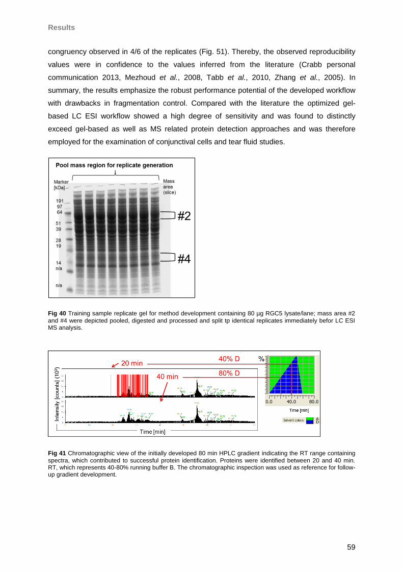
Results
59
congruency observed in 4/6 of the replicates (Fig. 51). Thereby, the observed reproducibility
values were in confidence to the values inferred from the literature (Crabb personal
communication 2013, Mezhoud et al., 2008, Tabb et al., 2010, Zhang et al., 2005). In
summary, the results emphasize the robust performance potential of the developed workflow
with drawbacks in fragmentation control. Compared with the literature the optimized gel-
based LC ESI workflow showed a high degree of sensitivity and was found to distinctly
exceed gel-based as well as MS related protein detection approaches and was therefore
employed for the examination of conjunctival cells and tear fluid studies.
Fig 40 Training sample replicate gel for method development containing 80 µg RGC5 lysate/lane; mass area #2
and #4 were depicted pooled, digested and processed and split tp identical replicates immediately befor LC ESI MS analysis.
Fig 41 Chromatographic view of the initially developed 80 min HPLC gradient indicating the RT range containing
spectra, which contributed to successful protein identification. Proteins were identified between 20 and 40 min. RT, which represents 40-80% running buffer B. The chromatographic inspection was used as reference for follow-up gradient development.
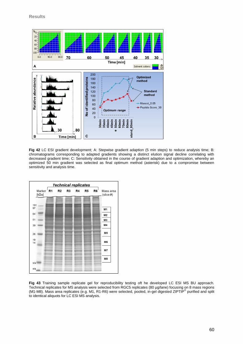
Results
60
Fig 42 LC ESI gradient development; A: Stepwise gradient adaption (5 min steps) to reduce analysis time; B:
chromatograms corresponding to adapted gradients showing a distinct elution signal decline correlating with decreased gradient time; C: Sensitivity obtained in the course of gradient adaption and optimization, whereby an optimized 50 min gradient was selected as final optimum method (asterisk) due to a compromise between sensitivity and analysis time.
Fig 43 Training sample replicate gel for reproducibility testing oft he developed LC ESI MS BU approach.
Technical replicates for MS analysis were selected from RGC5 replicates (80 µg/lane) focusing on 8 mass regions (M1-M8). Mass area replicates (e.g. M1, R1-R6) were selected, pooled, in-gel digested ZIPTIP
® purified and split
to identical aliquots for LC ESI MS analysis.
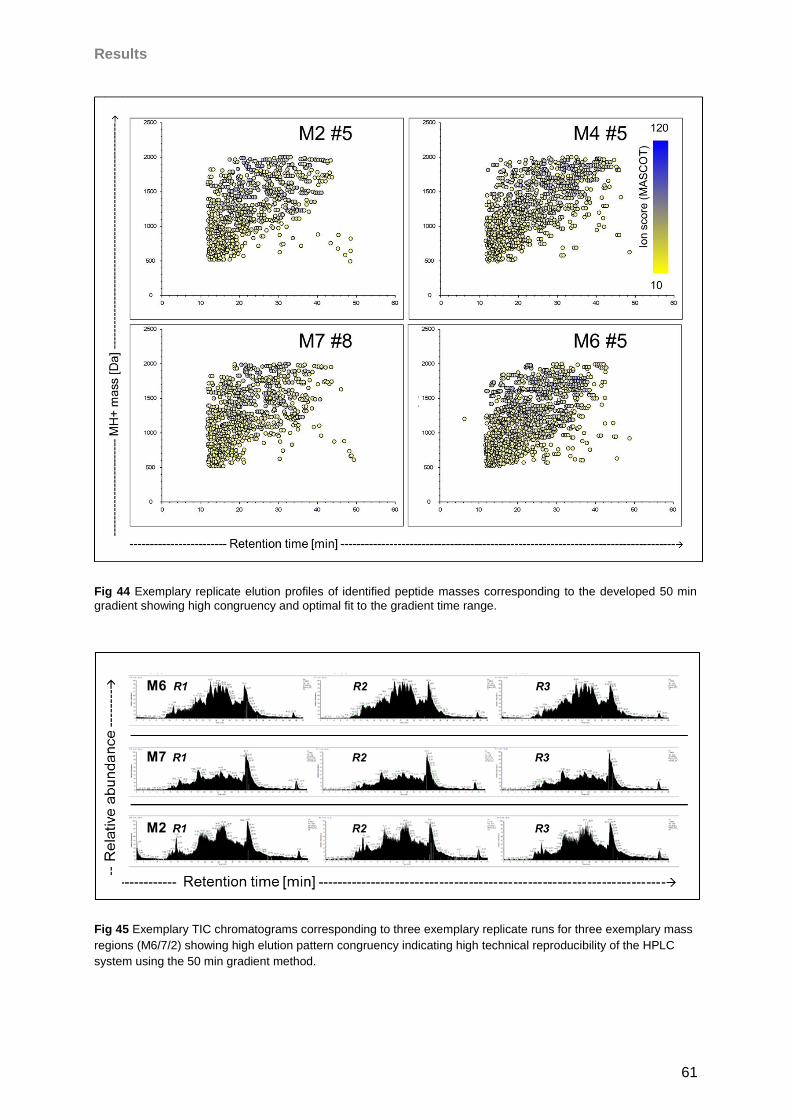
Results
61
Fig 44 Exemplary replicate elution profiles of identified peptide masses corresponding to the developed 50 min
gradient showing high congruency and optimal fit to the gradient time range.
Fig 45 Exemplary TIC chromatograms corresponding to three exemplary replicate runs for three exemplary mass
regions (M6/7/2) showing high elution pattern congruency indicating high technical reproducibility of the HPLC
system using the 50 min gradient method.
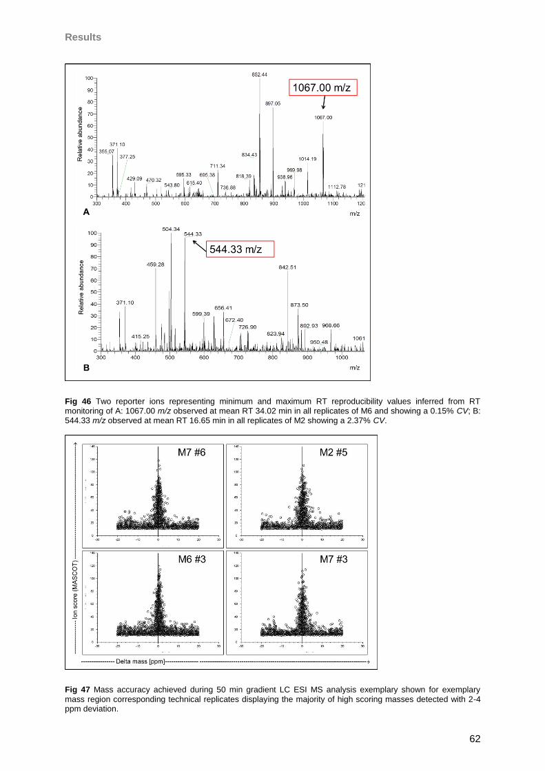
Results
62
Fig 46 Two reporter ions representing minimum and maximum RT reproducibility values inferred from RT monitoring of A: 1067.00 m/z observed at mean RT 34.02 min in all replicates of M6 and showing a 0.15% CV; B: 544.33 m/z observed at mean RT 16.65 min in all replicates of M2 showing a 2.37% CV.
Fig 47 Mass accuracy achieved during 50 min gradient LC ESI MS analysis exemplary shown for exemplary
mass region corresponding technical replicates displaying the majority of high scoring masses detected with 2-4 ppm deviation.
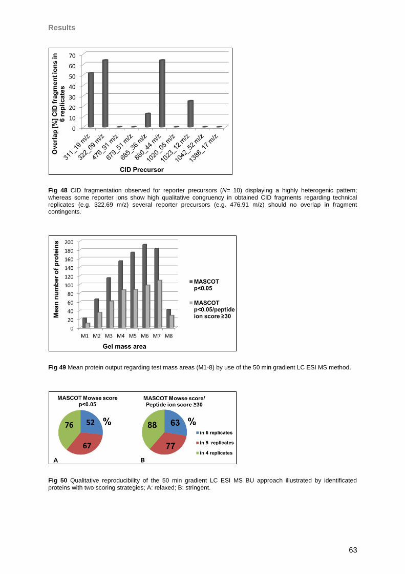
Results
63
Fig 48 CID fragmentation observed for reporter precursors (N= 10) displaying a highly heterogenic pattern;
whereas some reporter ions show high qualitative congruency in obtained CID fragments regarding technical replicates (e.g. 322.69 m/z) several reporter precursors (e.g. 476.91 m/z) should no overlap in fragment contingents.
Fig 49 Mean protein output regarding test mass areas (M1-8) by use of the 50 min gradient LC ESI MS method.
Fig 50 Qualitative reproducibility of the 50 min gradient LC ESI MS BU approach illustrated by identificated
proteins with two scoring strategies; A: relaxed; B: stringent.
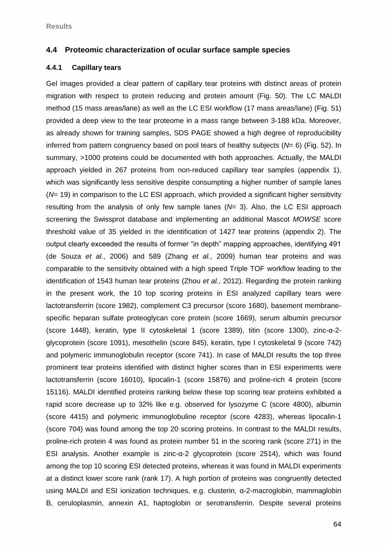
Results
64
4.4 Proteomic characterization of ocular surface sample species
4.4.1 Capillary tears
Gel images provided a clear pattern of capillary tear proteins with distinct areas of protein
migration with respect to protein reducing and protein amount (Fig. 50). The LC MALDI
method (15 mass areas/lane) as well as the LC ESI workflow (17 mass areas/lane) (Fig. 51)
provided a deep view to the tear proteome in a mass range between 3-188 kDa. Moreover,
as already shown for training samples, SDS PAGE showed a high degree of reproducibility
inferred from pattern congruency based on pool tears of healthy subjects (N= 6) (Fig. 52). In
summary, >1000 proteins could be documented with both approaches. Actually, the MALDI
approach yielded in 267 proteins from non-reduced capillary tear samples (appendix 1),
which was significantly less sensitive despite consumpting a higher number of sample lanes
(N= 19) in comparison to the LC ESI approach, which provided a significant higher sensitivity
resulting from the analysis of only few sample lanes (N= 3). Also, the LC ESI approach
screening the Swissprot database and implementing an additional Mascot MOWSE score
threshold value of 35 yielded in the identification of 1427 tear proteins (appendix 2). The
output clearly exceeded the results of former “in depth” mapping approaches, identifying 491
(de Souza et al., 2006) and 589 (Zhang et al., 2009) human tear proteins and was
comparable to the sensitivity obtained with a high speed Triple TOF workflow leading to the
identification of 1543 human tear proteins (Zhou et al., 2012). Regarding the protein ranking
in the present work, the 10 top scoring proteins in ESI analyzed capillary tears were
lactotransferrin (score 1982), complement C3 precursor (score 1680), basement membrane-
specific heparan sulfate proteoglycan core protein (score 1669), serum albumin precursor
(score 1448), keratin, type II cytoskeletal 1 (score 1389), titin (score 1300), zinc-α-2-
glycoprotein (score 1091), mesothelin (score 845), keratin, type I cytoskeletal 9 (score 742)
and polymeric immunoglobulin receptor (score 741). In case of MALDI results the top three
prominent tear proteins identified with distinct higher scores than in ESI experiments were
lactotransferrin (score 16010), lipocalin-1 (score 15876) and proline-rich 4 protein (score
15116). MALDI identified proteins ranking below these top scoring tear proteins exhibited a
rapid score decrease up to 32% like e.g. observed for lysozyme C (score 4800), albumin
(score 4415) and polymeric immunoglobuline receptor (score 4283), whereas lipocalin-1
(score 704) was found among the top 20 scoring proteins. In contrast to the MALDI results,
proline-rich protein 4 was found as protein number 51 in the scoring rank (score 271) in the
ESI analysis. Another example is zinc-α-2 glycoprotein (score 2514), which was found
among the top 10 scoring ESI detected proteins, whereas it was found in MALDI experiments
at a distinct lower score rank (rank 17). A high portion of proteins was congruently detected
using MALDI and ESI ionization techniques, e.g. clusterin, α-2-macroglobin, mammaglobin
B, ceruloplasmin, annexin A1, haptoglobin or serotransferrin. Despite several proteins
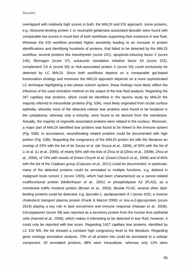
Results
65
overlapped with relatively high scores in both, the MALDI and ESI approach, some proteins,
e.g. ribosome-binding protein 1 or neutrophil gelatinase-associated lipocalin were found with
comparable low scores in result lists of both workflows supporting their existence in tear fluid.
Whereas the ESI workflow provided higher sensitivity leading to an increase in protein
identifications and identifying hundreds of proteins, that failed to be detected by the MALDI
workflow, several proteins like transthyretin (score 241), apoptosis-inducing factor 2 (score
145), fibrinogen (score 37), eukaryotic translation initiation factor 2A (score 152),
complement C4 A (score 50) or Nck-associated protein 5 (score 53) could exclusively be
detected by LC MALDI. Since both workflows depend on a comparable gel-based
fractionation strategy and moreover the MALDI approach depends on a more sophisticated
LC technique highlighting a two phase column system, these findings most likely reflect the
influence of the used ionization method on the output of the tear fluid analysis. Regarding the
267 capillary tear proteins, which could be identified in the taurine study by MALDI the
majority referred to intracellular proteins (Fig. 53A), most likely originated from ocular surface
epithelia, whereby most of the detected cellular tear proteins were found to be localized in
the cytoplasma, whereas only a minority, were found to be derived from the membrane.
Actually, the majority of organelle associated proteins were related to the nucleus. Moreover,
a major part of MALDI identified tear proteins was found to be linked to the immune system
(Fig. 53B). In accordance, woundhealing related proteins could be documented with high
portion (Fig. 53B). Regarding the congruency of the MALDI protein list with the literature an
overlap of 23% with the list of de Souza et al. (de Souza et al., 2006), of 50% with the list of
Li et al. (Li et al., 2005), of nearly 50% with the lists of Zhou et al (Zhou et al., 2009b, Zhou et
al., 2006), of 74% with results of Green-Church et al. (Green-Church et al., 2008) and of 84%
with the list of the Cojakaru group (Cojocaru et al., 2011) could be documented. In particular,
many of the detected proteins could be annotated to multiple functions, e.g. deleted in
malignant brain tumors 1 (score 1305), which had been characterized as a cancer-related
multifunctional protein (Mollenhauer et al., 2001) or phospholipase A2 (PLA2), as a
membrane traffic involved protein (Brown et al., 2003). Beside PLA2, several other lipid-
binding proteins could be detected, e.g. lipocalin-1, apolipoprotein A I (score 425), a reverse
cholesterol transport plasma protein (Frank & Marcel 2000) or zinc-α-2-glycoprotein (score
2514) playing a key role in lipid recruitment and immune response (Hassan et al., 2008).
Ceruloplasmin (score 39) was reported as a secretory protein from the human lens epithelial
cells (Harned et al., 2006), which makes it interesting to be detected in tear fluid, however, it
could only be reported with low score. Regarding 1427 capillary tear proteins, identified by
LC ESI MS, the list showed a constant high congruency level to the literature. Regarding
gene ontology annotation analysis, 73% of all protein hits could be annotated to a cellular
component. Of annotated proteins, 88% were intracellular, whereas only 12% were
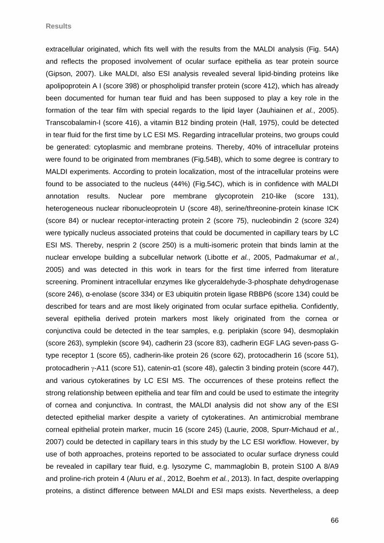
Results
66
extracellular originated, which fits well with the results from the MALDI analysis (Fig. 54A)
and reflects the proposed involvement of ocular surface epithelia as tear protein source
(Gipson, 2007). Like MALDI, also ESI analysis revealed several lipid-binding proteins like
apolipoprotein A I (score 398) or phospholipid transfer protein (score 412), which has already
been documented for human tear fluid and has been supposed to play a key role in the
formation of the tear film with special regards to the lipid layer (Jauhiainen et al., 2005).
Transcobalamin-I (score 416), a vitamin B12 binding protein (Hall, 1975), could be detected
in tear fluid for the first time by LC ESI MS. Regarding intracellular proteins, two groups could
be generated: cytoplasmic and membrane proteins. Thereby, 40% of intracellular proteins
were found to be originated from membranes (Fig.54B), which to some degree is contrary to
MALDI experiments. According to protein localization, most of the intracellular proteins were
found to be associated to the nucleus (44%) (Fig.54C), which is in confidence with MALDI
annotation results. Nuclear pore membrane glycoprotein 210-like (score 131),
heterogeneous nuclear ribonucleoprotein U (score 48), serine/threonine-protein kinase ICK
(score 84) or nuclear receptor-interacting protein 2 (score 75), nucleobindin 2 (score 324)
were typically nucleus associated proteins that could be documented in capillary tears by LC
ESI MS. Thereby, nesprin 2 (score 250) is a multi-isomeric protein that binds lamin at the
nuclear envelope building a subcellular network (Libotte et al., 2005, Padmakumar et al.,
2005) and was detected in this work in tears for the first time inferred from literature
screening. Prominent intracellular enzymes like glyceraldehyde-3-phosphate dehydrogenase
(score 246), α-enolase (score 334) or E3 ubiquitin protein ligase RBBP6 (score 134) could be
described for tears and are most likely originated from ocular surface epithelia. Confidently,
several epithelia derived protein markers most likely originated from the cornea or
conjunctiva could be detected in the tear samples, e.g. periplakin (score 94), desmoplakin
(score 263), symplekin (score 94), cadherin 23 (score 83), cadherin EGF LAG seven-pass G-
type receptor 1 (score 65), cadherin-like protein 26 (score 62), protocadherin 16 (score 51),
protocadherin -A11 (score 51), catenin-α1 (score 48), galectin 3 binding protein (score 447),
and various cytokeratines by LC ESI MS. The occurrences of these proteins reflect the
strong relationship between epithelia and tear film and could be used to estimate the integrity
of cornea and conjunctiva. In contrast, the MALDI analysis did not show any of the ESI
detected epithelial marker despite a variety of cytokeratines. An antimicrobial membrane
corneal epithelial protein marker, mucin 16 (score 245) (Laurie, 2008, Spurr-Michaud et al.,
2007) could be detected in capillary tears in this study by the LC ESI workflow. However, by
use of both approaches, proteins reported to be associated to ocular surface dryness could
be revealed in capillary tear fluid, e.g. lysozyme C, mammaglobin B, protein S100 A 8/A9
and proline-rich protein 4 (Aluru et al., 2012, Boehm et al., 2013). In fact, despite overlapping
proteins, a distinct difference between MALDI and ESI maps exists. Nevertheless, a deep
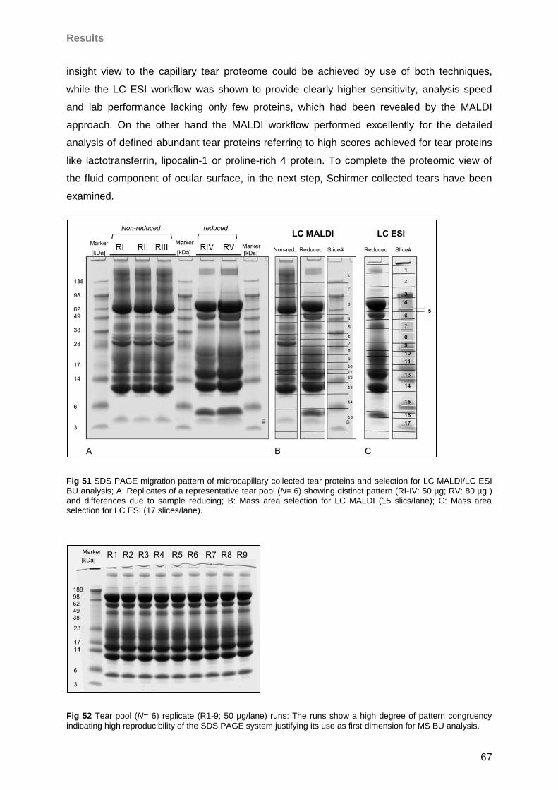
Results
67
insight view to the capillary tear proteome could be achieved by use of both techniques,
while the LC ESI workflow was shown to provide clearly higher sensitivity, analysis speed
and lab performance lacking only few proteins, which had been revealed by the MALDI
approach. On the other hand the MALDI workflow performed excellently for the detailed
analysis of defined abundant tear proteins referring to high scores achieved for tear proteins
like lactotransferrin, lipocalin-1 or proline-rich 4 protein. To complete the proteomic view of
the fluid component of ocular surface, in the next step, Schirmer collected tears have been
examined.
Fig 51 SDS PAGE migration pattern of microcapillary collected tear proteins and selection for LC MALDI/LC ESI
BU analysis; A: Replicates of a representative tear pool (N= 6) showing distinct pattern (RI-IV: 50 µg; RV: 80 µg ) and differences due to sample reducing; B: Mass area selection for LC MALDI (15 slics/lane); C: Mass area selection for LC ESI (17 slices/lane).
Fig 52 Tear pool (N= 6) replicate (R1-9; 50 µg/lane) runs: The runs show a high degree of pattern congruency
indicating high reproducibility of the SDS PAGE system justifying its use as first dimension for MS BU analysis.
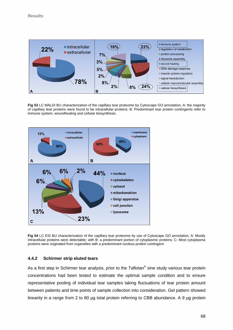
Results
68
Fig 53 LC MALDI BU characterization of the capillary tear proteome by Cytoscape GO annotation; A: the majority
of capillary tear proteins were found to be intracellular proteins; B: Predominant tear protein contingents refer to immune system, woundhealing and cellular biosynthesis.
Fig 54 LC ESI BU characterization of the capillary tear proteome by use of Cytoscape GO annotation; A: Mostly
intracellular proteins were detectable; with B: a predominant portion of cytoplasmic proteins; C: Most cytoplasma proteins were originated from organelles with a predominant nucleus protein contingent.
4.4.2 Schirmer strip eluted tears
As a first step in Schirmer tear analysis, prior to the Taflotan® sine study various tear protein
concentrations had been tested to estimate the optimal sample condition and to ensure
representative pooling of individual tear samples taking fluctuations of tear protein amount
between patients and time points of sample collection into consideration. Gel pattern showed
linearity in a range from 2 to 80 µg total protein referring to CBB abundance. A 9 µg protein
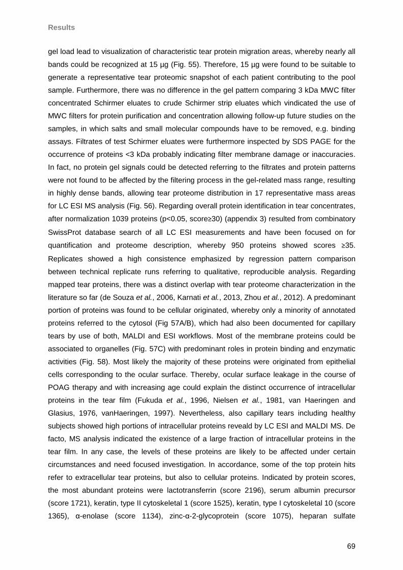
Results
69
gel load lead to visualization of characteristic tear protein migration areas, whereby nearly all
bands could be recognized at 15 µg (Fig. 55). Therefore, 15 µg were found to be suitable to
generate a representative tear proteomic snapshot of each patient contributing to the pool
sample. Furthermore, there was no difference in the gel pattern comparing 3 kDa MWC filter
concentrated Schirmer eluates to crude Schirmer strip eluates which vindicated the use of
MWC filters for protein purification and concentration allowing follow-up future studies on the
samples, in which salts and small molecular compounds have to be removed, e.g. binding
assays. Filtrates of test Schirmer eluates were furthermore inspected by SDS PAGE for the
occurrence of proteins <3 kDa probably indicating filter membrane damage or inaccuracies.
In fact, no protein gel signals could be detected referring to the filtrates and protein patterns
were not found to be affected by the filtering process in the gel-related mass range, resulting
in highly dense bands, allowing tear proteome distribution in 17 representative mass areas
for LC ESI MS analysis (Fig. 56). Regarding overall protein identification in tear concentrates,
after normalization 1039 proteins (p<0.05, score≥30) (appendix 3) resulted from combinatory
SwissProt database search of all LC ESI measurements and have been focused on for
quantification and proteome description, whereby 950 proteins showed scores ≥35.
Replicates showed a high consistence emphasized by regression pattern comparison
between technical replicate runs referring to qualitative, reproducible analysis. Regarding
mapped tear proteins, there was a distinct overlap with tear proteome characterization in the
literature so far (de Souza et al., 2006, Karnati et al., 2013, Zhou et al., 2012). A predominant
portion of proteins was found to be cellular originated, whereby only a minority of annotated
proteins referred to the cytosol (Fig 57A/B), which had also been documented for capillary
tears by use of both, MALDI and ESI workflows. Most of the membrane proteins could be
associated to organelles (Fig. 57C) with predominant roles in protein binding and enzymatic
activities (Fig. 58). Most likely the majority of these proteins were originated from epithelial
cells corresponding to the ocular surface. Thereby, ocular surface leakage in the course of
POAG therapy and with increasing age could explain the distinct occurrence of intracellular
proteins in the tear film (Fukuda et al., 1996, Nielsen et al., 1981, van Haeringen and
Glasius, 1976, vanHaeringen, 1997). Nevertheless, also capillary tears including healthy
subjects showed high portions of intracellular proteins reveald by LC ESI and MALDI MS. De
facto, MS analysis indicated the existence of a large fraction of intracellular proteins in the
tear film. In any case, the levels of these proteins are likely to be affected under certain
circumstances and need focused investigation. In accordance, some of the top protein hits
refer to extracellular tear proteins, but also to cellular proteins. Indicated by protein scores,
the most abundant proteins were lactotransferrin (score 2196), serum albumin precursor
(score 1721), keratin, type II cytoskeletal 1 (score 1525), keratin, type I cytoskeletal 10 (score
1365), α-enolase (score 1134), zinc-α-2-glycoprotein (score 1075), heparan sulfate
Results
70
proteoglycan core protein (score 1073) and retinal dehydrogenase 1 (score 1029). Also anti-
α-trypsin (score 886) and pyruvate kinase isozymes M1/M2 (score 666) were detected
among the top 30 protein hits (Fig. 59), which was comparable to the capillary tear results of
prior LC ESI/MALDI experiments. Proline-rich protein 4, which was found among the most
prominent proteins, revealed by MALDI MS, was also detected in ESI analysis of capillary
tears, only in the medium score rank (score 167, rank 98), probably reflecting its chemical
preverence for MALDI ionization. Like in capillary tears several immunoglobuline fractions
could be detected with high scores, e.g. Ig α-1 chain C region (score 588), Ig α-2 chain C
region (score 525) or Ig ĸ chain V-III region WOL (score 381). Also, several retina
characteristic proteins could be documented with high scores including retinal
dehydrogenase 1 (score 1029), peroxiredoxin 1 (score 654) and heat shock protein ß 1
(score 646), which indicates distant ocular regions as additional tear protein source. Actually,
scores of detected proteins decline dramatically at a value of 251, whereby after this value
also the correlation between score and number of identified peptides for each protein
fluctuates to a certain degree. Most peptides were identified for lactotransferrin (7530
peptides), lipocalin-1 (3987 peptides), lysozyme C (3675 peptides) and proline-rich protein 4
(1671 peptides), which were also documented among the predominant MALDI detected
proteins. Mammaglobin B (1021 peptides) and secretoglobin family 1D member 1 (550
peptides), both reported as dry eye markers (Boehm et al., 2013, Kramann et al., 2011),
could also be characterized with high peptide numbers. Like in capillary tears, also epithelial
proteins, most likely derived from conjunctiva or cornea, e.g. desmoplakin (score 273),
desmoglein 2 (score 69), desmoglein 4 (score 47), dermatan-sulfate epimerase-like protein
(score 47), multiple epidermal growth factor-like domains protein 8 (score 38), protocadherin-
9 (score 35) and -A12 (score 38), cadherin-5 (score 79), plectin (score 231) as well as
various cytokeratine species, also documented for capillary tear fluid, could be identified. In
summary, even consumpting a smaller protein amount than in capillary tear analysis the LC
ESI MS strategy provided a detailed image of the tear proteome emphasizing the high
sensitivity of the developed ESI approach. Moreover, a high degree of congruency between
capillary and Schirmer tear proteome could be documented with 95% of Schirmer eluted
proteins also detected in capillary tears by the developed LC ESI MS workflow, which most
likely shows that different results are unlikely to be a matter of the tear sample species, but
are more likely to depend on the sensitivity of the method. In addition, retinal proteins; retinal
dehydrogenase 1 (score 1029) and retinal guanylyl cyclase 1 (score 48) could be exclusively
detected in the Schirmer eluted tear samples. Also, other highlighting proteins like leukocyte
elastastase inhibitor (score 747), aldehyde dehydrogenase (dimeric NADP-preferring) (score
540), 14-3-3 protein (score 373) or alcohol dehydrogenase (score 343) could be
exclusively identified with relatively high scores in Schirmer tears. In summary, an in-depth
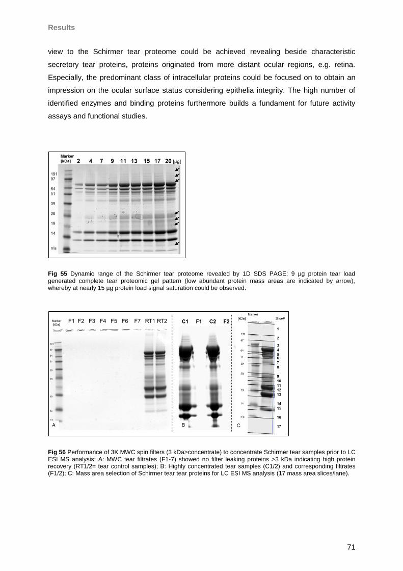
Results
71
view to the Schirmer tear proteome could be achieved revealing beside characteristic
secretory tear proteins, proteins originated from more distant ocular regions, e.g. retina.
Especially, the predominant class of intracellular proteins could be focused on to obtain an
impression on the ocular surface status considering epithelia integrity. The high number of
identified enzymes and binding proteins furthermore builds a fundament for future activity
assays and functional studies.
Fig 55 Dynamic range of the Schirmer tear proteome revealed by 1D SDS PAGE: 9 µg protein tear load
generated complete tear proteomic gel pattern (low abundant protein mass areas are indicated by arrow), whereby at nearly 15 µg protein load signal saturation could be observed.
Fig 56 Performance of 3K MWC spin filters (3 kDa>concentrate) to concentrate Schirmer tear samples prior to LC
ESI MS analysis; A: MWC tear filtrates (F1-7) showed no filter leaking proteins >3 kDa indicating high protein recovery (RT1/2= tear control samples); B: Highly concentrated tear samples (C1/2) and corresponding filtrates (F1/2); C: Mass area selection of Schirmer tear tear proteins for LC ESI MS analysis (17 mass area slices/lane).
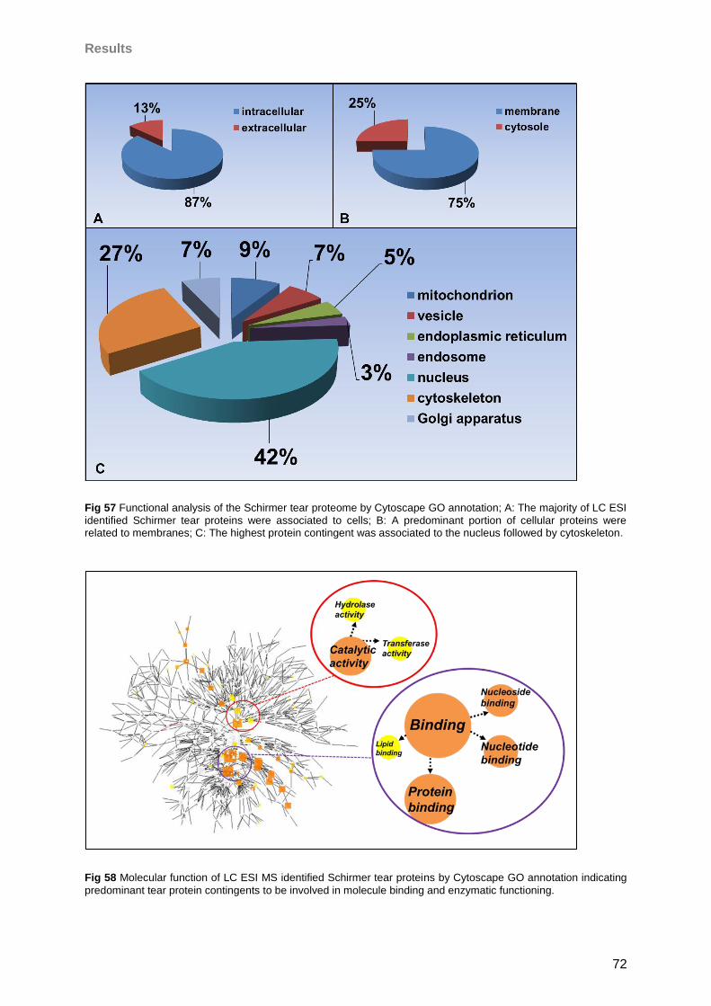
Results
72
Fig 57 Functional analysis of the Schirmer tear proteome by Cytoscape GO annotation; A: The majority of LC ESI
identified Schirmer tear proteins were associated to cells; B: A predominant portion of cellular proteins were related to membranes; C: The highest protein contingent was associated to the nucleus followed by cytoskeleton.
Fig 58 Molecular function of LC ESI MS identified Schirmer tear proteins by Cytoscape GO annotation indicating
predominant tear protein contingents to be involved in molecule binding and enzymatic functioning.
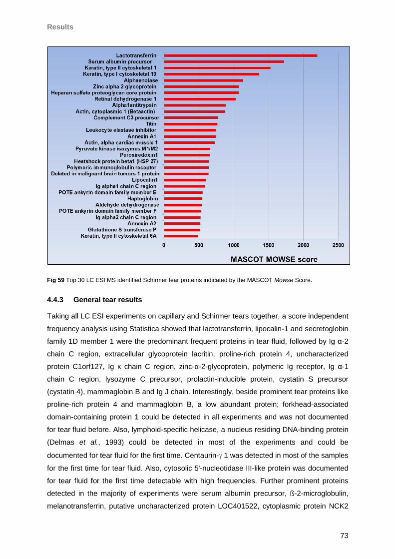
Results
73
Fig 59 Top 30 LC ESI MS identified Schirmer tear proteins indicated by the MASCOT Mowse Score.
4.4.3 General tear results
Taking all LC ESI experiments on capillary and Schirmer tears together, a score independent
frequency analysis using Statistica showed that lactotransferrin, lipocalin-1 and secretoglobin
family 1D member 1 were the predominant frequent proteins in tear fluid, followed by Ig α-2
chain C region, extracellular glycoprotein lacritin, proline-rich protein 4, uncharacterized
protein C1orf127, Ig ĸ chain C region, zinc-α-2-glycoprotein, polymeric Ig receptor, Ig α-1
chain C region, lysozyme C precursor, prolactin-inducible protein, cystatin S precursor
(cystatin 4), mammaglobin B and Ig J chain. Interestingly, beside prominent tear proteins like
proline-rich protein 4 and mammaglobin B, a low abundant protein; forkhead-associated
domain-containing protein 1 could be detected in all experiments and was not documented
for tear fluid before. Also, lymphoid-specific helicase, a nucleus residing DNA-binding protein
(Delmas et al., 1993) could be detected in most of the experiments and could be
documented for tear fluid for the first time. Centaurin- 1 was detected in most of the samples
for the first time for tear fluid. Also, cytosolic 5'-nucleotidase III-like protein was documented
for tear fluid for the first time detectable with high frequencies. Further prominent proteins
detected in the majority of experiments were serum albumin precursor, ß-2-microglobulin,
melanotransferrin, putative uncharacterized protein LOC401522, cytoplasmic protein NCK2
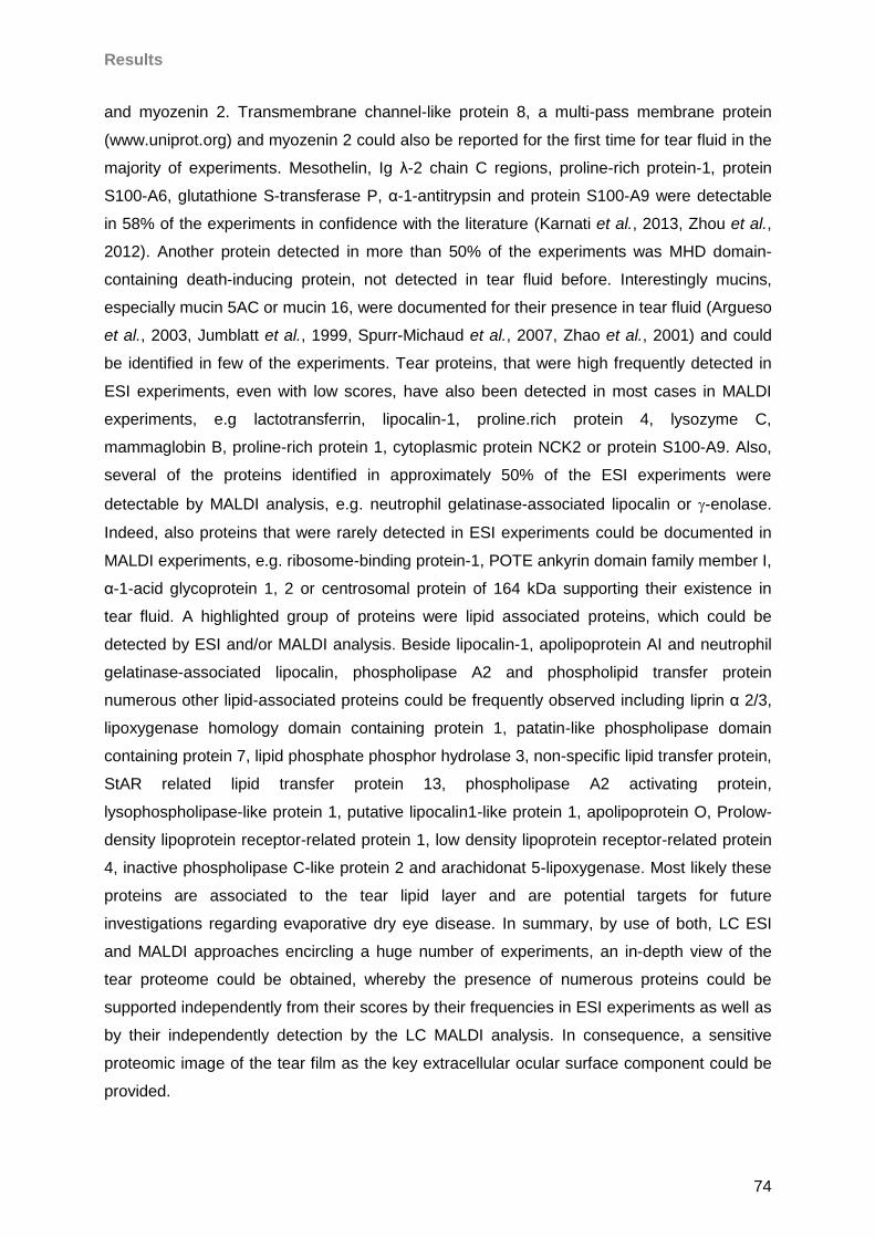
Results
74
and myozenin 2. Transmembrane channel-like protein 8, a multi-pass membrane protein
(www.uniprot.org) and myozenin 2 could also be reported for the first time for tear fluid in the
majority of experiments. Mesothelin, Ig λ-2 chain C regions, proline-rich protein-1, protein
S100-A6, glutathione S-transferase P, α-1-antitrypsin and protein S100-A9 were detectable
in 58% of the experiments in confidence with the literature (Karnati et al., 2013, Zhou et al.,
2012). Another protein detected in more than 50% of the experiments was MHD domain-
containing death-inducing protein, not detected in tear fluid before. Interestingly mucins,
especially mucin 5AC or mucin 16, were documented for their presence in tear fluid (Argueso
et al., 2003, Jumblatt et al., 1999, Spurr-Michaud et al., 2007, Zhao et al., 2001) and could
be identified in few of the experiments. Tear proteins, that were high frequently detected in
ESI experiments, even with low scores, have also been detected in most cases in MALDI
experiments, e.g lactotransferrin, lipocalin-1, proline.rich protein 4, lysozyme C,
mammaglobin B, proline-rich protein 1, cytoplasmic protein NCK2 or protein S100-A9. Also,
several of the proteins identified in approximately 50% of the ESI experiments were
detectable by MALDI analysis, e.g. neutrophil gelatinase-associated lipocalin or -enolase.
Indeed, also proteins that were rarely detected in ESI experiments could be documented in
MALDI experiments, e.g. ribosome-binding protein-1, POTE ankyrin domain family member I,
α-1-acid glycoprotein 1, 2 or centrosomal protein of 164 kDa supporting their existence in
tear fluid. A highlighted group of proteins were lipid associated proteins, which could be
detected by ESI and/or MALDI analysis. Beside lipocalin-1, apolipoprotein AI and neutrophil
gelatinase-associated lipocalin, phospholipase A2 and phospholipid transfer protein
numerous other lipid-associated proteins could be frequently observed including liprin α 2/3,
lipoxygenase homology domain containing protein 1, patatin-like phospholipase domain
containing protein 7, lipid phosphate phosphor hydrolase 3, non-specific lipid transfer protein,
StAR related lipid transfer protein 13, phospholipase A2 activating protein,
lysophospholipase-like protein 1, putative lipocalin1-like protein 1, apolipoprotein O, Prolow-
density lipoprotein receptor-related protein 1, low density lipoprotein receptor-related protein
4, inactive phospholipase C-like protein 2 and arachidonat 5-lipoxygenase. Most likely these
proteins are associated to the tear lipid layer and are potential targets for future
investigations regarding evaporative dry eye disease. In summary, by use of both, LC ESI
and MALDI approaches encircling a huge number of experiments, an in-depth view of the
tear proteome could be obtained, whereby the presence of numerous proteins could be
supported independently from their scores by their frequencies in ESI experiments as well as
by their independently detection by the LC MALDI analysis. In consequence, a sensitive
proteomic image of the tear film as the key extracellular ocular surface component could be
provided.
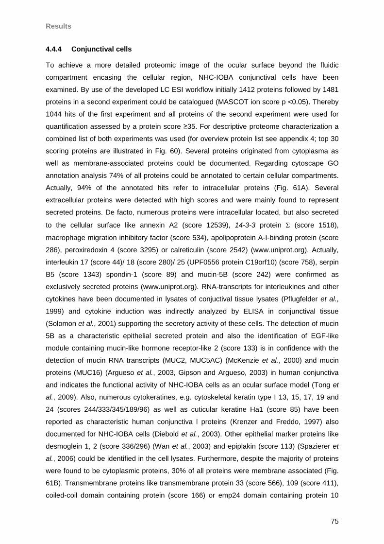
Results
75
4.4.4 Conjunctival cells
To achieve a more detailed proteomic image of the ocular surface beyond the fluidic
compartment encasing the cellular region, NHC-IOBA conjunctival cells have been
examined. By use of the developed LC ESI workflow initially 1412 proteins followed by 1481
proteins in a second experiment could be catalogued (MASCOT ion score p <0.05). Thereby
1044 hits of the first experiment and all proteins of the second experiment were used for
quantification assessed by a protein score ≥35. For descriptive proteome characterization a
combined list of both experiments was used (for overview protein list see appendix 4; top 30
scoring proteins are illustrated in Fig. 60). Several proteins originated from cytoplasma as
well as membrane-associated proteins could be documented. Regarding cytoscape GO
annotation analysis 74% of all proteins could be annotated to certain cellular compartments.
Actually, 94% of the annotated hits refer to intracellular proteins (Fig. 61A). Several
extracellular proteins were detected with high scores and were mainly found to represent
secreted proteins. De facto, numerous proteins were intracellular located, but also secreted
to the cellular surface like annexin A2 (score 12539), 14-3-3 protein (score 1518),
macrophage migration inhibitory factor (score 534), apolipoprotein A-I-binding protein (score
286), peroxiredoxin 4 (score 3295) or calreticulin (score 2542) (www.uniprot.org). Actually,
interleukin 17 (score 44)/ 18 (score 280)/ 25 (UPF0556 protein C19orf10) (score 758), serpin
B5 (score 1343) spondin-1 (score 89) and mucin-5B (score 242) were confirmed as
exclusively secreted proteins (www.uniprot.org). RNA-transcripts for interleukines and other
cytokines have been documented in lysates of conjuctival tissue lysates (Pflugfelder et al.,
1999) and cytokine induction was indirectly analyzed by ELISA in conjunctival tissue
(Solomon et al., 2001) supporting the secretory activity of these cells. The detection of mucin
5B as a characteristic epithelial secreted protein and also the identification of EGF-like
module containing mucin-like hormone receptor-like 2 (score 133) is in confidence with the
detection of mucin RNA transcripts (MUC2, MUC5AC) (McKenzie et al., 2000) and mucin
proteins (MUC16) (Argueso et al., 2003, Gipson and Argueso, 2003) in human conjunctiva
and indicates the functional activity of NHC-IOBA cells as an ocular surface model (Tong et
al., 2009). Also, numerous cytokeratines, e.g. cytoskeletal keratin type I 13, 15, 17, 19 and
24 (scores 244/333/345/189/96) as well as cuticular keratine Ha1 (score 85) have been
reported as characteristic human conjunctiva l proteins (Krenzer and Freddo, 1997) also
documented for NHC-IOBA cells (Diebold et al., 2003). Other epithelial marker proteins like
desmoglein 1, 2 (score 336/296) (Wan et al., 2003) and epiplakin (score 113) (Spazierer et
al., 2006) could be identified in the cell lysates. Furthermore, despite the majority of proteins
were found to be cytoplasmic proteins, 30% of all proteins were membrane associated (Fig.
61B). Transmembrane proteins like transmembrane protein 33 (score 566), 109 (score 411),
coiled-coil domain containing protein (score 166) or emp24 domain containing protein 10
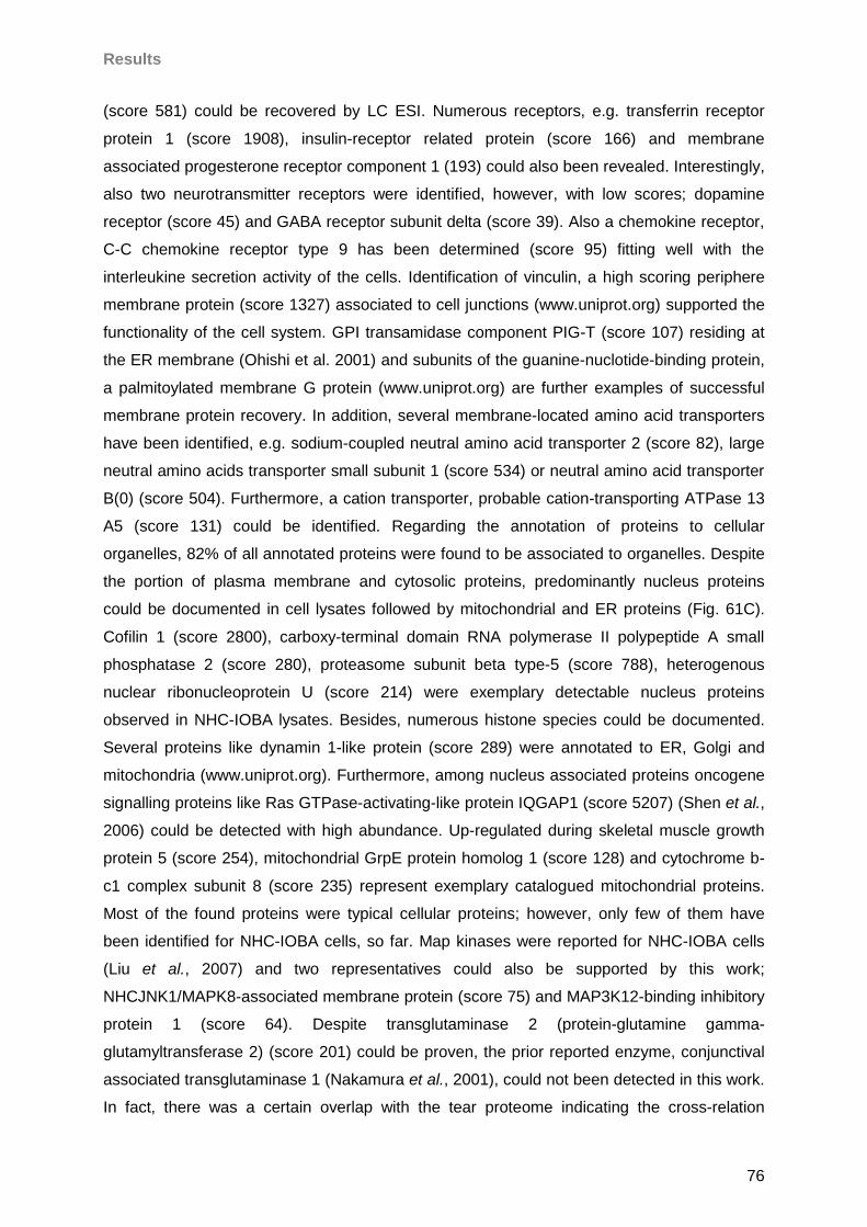
Results
76
(score 581) could be recovered by LC ESI. Numerous receptors, e.g. transferrin receptor
protein 1 (score 1908), insulin-receptor related protein (score 166) and membrane
associated progesterone receptor component 1 (193) could also been revealed. Interestingly,
also two neurotransmitter receptors were identified, however, with low scores; dopamine
receptor (score 45) and GABA receptor subunit delta (score 39). Also a chemokine receptor,
C-C chemokine receptor type 9 has been determined (score 95) fitting well with the
interleukine secretion activity of the cells. Identification of vinculin, a high scoring periphere
membrane protein (score 1327) associated to cell junctions (www.uniprot.org) supported the
functionality of the cell system. GPI transamidase component PIG-T (score 107) residing at
the ER membrane (Ohishi et al. 2001) and subunits of the guanine-nuclotide-binding protein,
a palmitoylated membrane G protein (www.uniprot.org) are further examples of successful
membrane protein recovery. In addition, several membrane-located amino acid transporters
have been identified, e.g. sodium-coupled neutral amino acid transporter 2 (score 82), large
neutral amino acids transporter small subunit 1 (score 534) or neutral amino acid transporter
B(0) (score 504). Furthermore, a cation transporter, probable cation-transporting ATPase 13
A5 (score 131) could be identified. Regarding the annotation of proteins to cellular
organelles, 82% of all annotated proteins were found to be associated to organelles. Despite
the portion of plasma membrane and cytosolic proteins, predominantly nucleus proteins
could be documented in cell lysates followed by mitochondrial and ER proteins (Fig. 61C).
Cofilin 1 (score 2800), carboxy-terminal domain RNA polymerase II polypeptide A small
phosphatase 2 (score 280), proteasome subunit beta type-5 (score 788), heterogenous
nuclear ribonucleoprotein U (score 214) were exemplary detectable nucleus proteins
observed in NHC-IOBA lysates. Besides, numerous histone species could be documented.
Several proteins like dynamin 1-like protein (score 289) were annotated to ER, Golgi and
mitochondria (www.uniprot.org). Furthermore, among nucleus associated proteins oncogene
signalling proteins like Ras GTPase-activating-like protein IQGAP1 (score 5207) (Shen et al.,
2006) could be detected with high abundance. Up-regulated during skeletal muscle growth
protein 5 (score 254), mitochondrial GrpE protein homolog 1 (score 128) and cytochrome b-
c1 complex subunit 8 (score 235) represent exemplary catalogued mitochondrial proteins.
Most of the found proteins were typical cellular proteins; however, only few of them have
been identified for NHC-IOBA cells, so far. Map kinases were reported for NHC-IOBA cells
(Liu et al., 2007) and two representatives could also be supported by this work;
NHCJNK1/MAPK8-associated membrane protein (score 75) and MAP3K12-binding inhibitory
protein 1 (score 64). Despite transglutaminase 2 (protein-glutamine gamma-
glutamyltransferase 2) (score 201) could be proven, the prior reported enzyme, conjunctival
associated transglutaminase 1 (Nakamura et al., 2001), could not been detected in this work.
In fact, there was a certain overlap with the tear proteome indicating the cross-relation
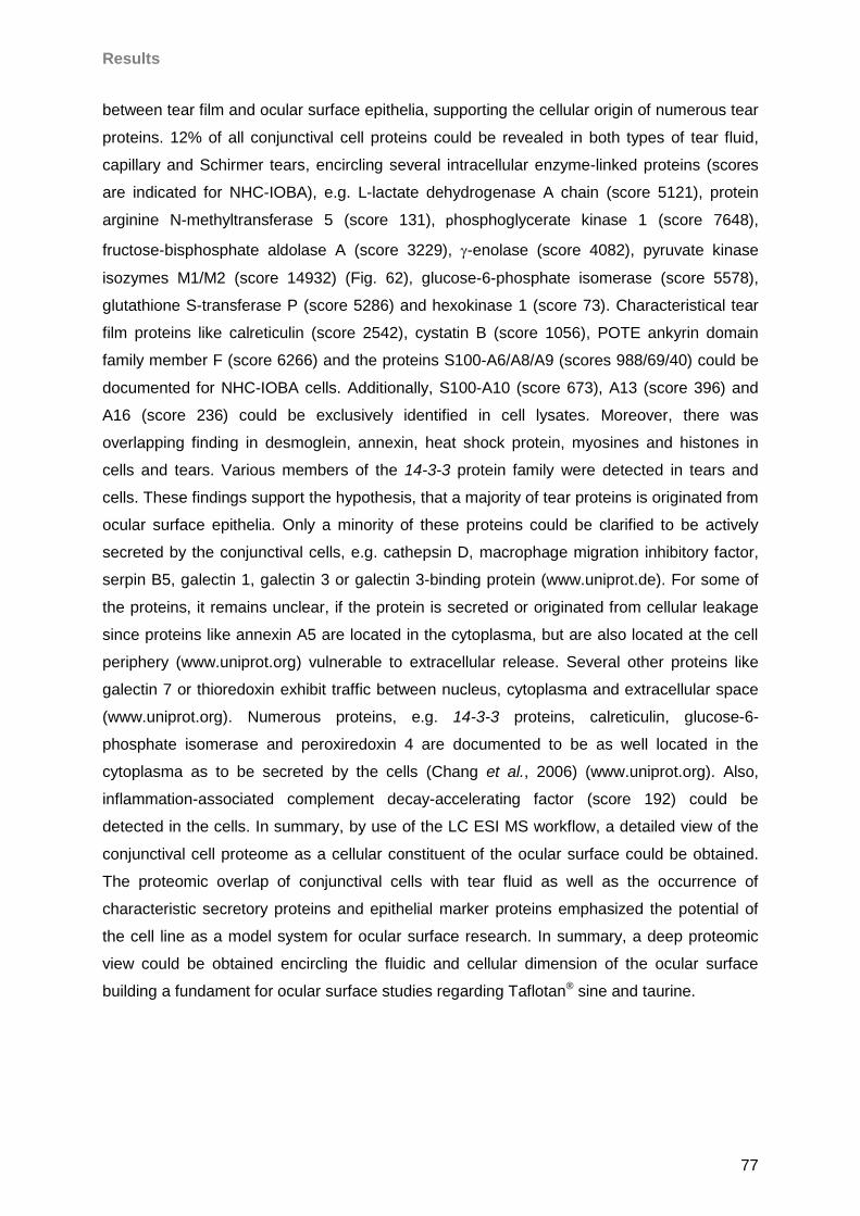
Results
77
between tear film and ocular surface epithelia, supporting the cellular origin of numerous tear
proteins. 12% of all conjunctival cell proteins could be revealed in both types of tear fluid,
capillary and Schirmer tears, encircling several intracellular enzyme-linked proteins (scores
are indicated for NHC-IOBA), e.g. L-lactate dehydrogenase A chain (score 5121), protein
arginine N-methyltransferase 5 (score 131), phosphoglycerate kinase 1 (score 7648),
fructose-bisphosphate aldolase A (score 3229), -enolase (score 4082), pyruvate kinase
isozymes M1/M2 (score 14932) (Fig. 62), glucose-6-phosphate isomerase (score 5578),
glutathione S-transferase P (score 5286) and hexokinase 1 (score 73). Characteristical tear
film proteins like calreticulin (score 2542), cystatin B (score 1056), POTE ankyrin domain
family member F (score 6266) and the proteins S100-A6/A8/A9 (scores 988/69/40) could be
documented for NHC-IOBA cells. Additionally, S100-A10 (score 673), A13 (score 396) and
A16 (score 236) could be exclusively identified in cell lysates. Moreover, there was
overlapping finding in desmoglein, annexin, heat shock protein, myosines and histones in
cells and tears. Various members of the 14-3-3 protein family were detected in tears and
cells. These findings support the hypothesis, that a majority of tear proteins is originated from
ocular surface epithelia. Only a minority of these proteins could be clarified to be actively
secreted by the conjunctival cells, e.g. cathepsin D, macrophage migration inhibitory factor,
serpin B5, galectin 1, galectin 3 or galectin 3-binding protein (www.uniprot.de). For some of
the proteins, it remains unclear, if the protein is secreted or originated from cellular leakage
since proteins like annexin A5 are located in the cytoplasma, but are also located at the cell
periphery (www.uniprot.org) vulnerable to extracellular release. Several other proteins like
galectin 7 or thioredoxin exhibit traffic between nucleus, cytoplasma and extracellular space
(www.uniprot.org). Numerous proteins, e.g. 14-3-3 proteins, calreticulin, glucose-6-
phosphate isomerase and peroxiredoxin 4 are documented to be as well located in the
cytoplasma as to be secreted by the cells (Chang et al., 2006) (www.uniprot.org). Also,
inflammation-associated complement decay-accelerating factor (score 192) could be
detected in the cells. In summary, by use of the LC ESI MS workflow, a detailed view of the
conjunctival cell proteome as a cellular constituent of the ocular surface could be obtained.
The proteomic overlap of conjunctival cells with tear fluid as well as the occurrence of
characteristic secretory proteins and epithelial marker proteins emphasized the potential of
the cell line as a model system for ocular surface research. In summary, a deep proteomic
view could be obtained encircling the fluidic and cellular dimension of the ocular surface
building a fundament for ocular surface studies regarding Taflotan® sine and taurine.
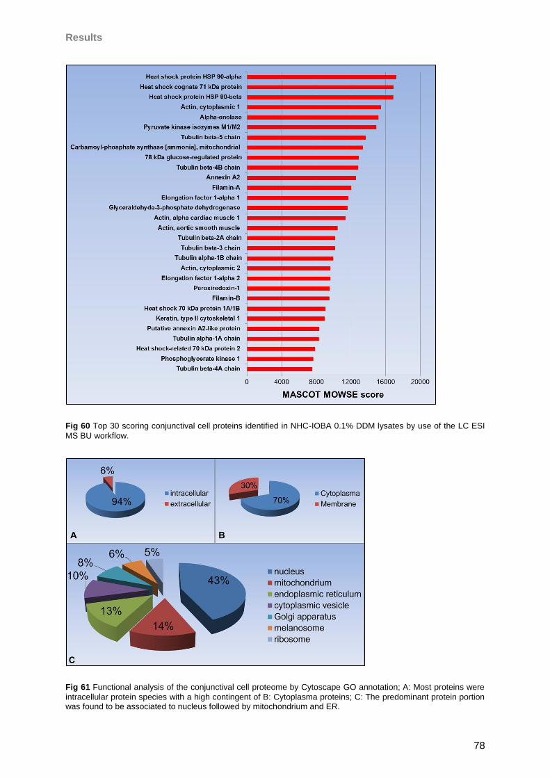
Results
78
Fig 60 Top 30 scoring conjunctival cell proteins identified in NHC-IOBA 0.1% DDM lysates by use of the LC ESI
MS BU workflow.
Fig 61 Functional analysis of the conjunctival cell proteome by Cytoscape GO annotation; A: Most proteins were
intracellular protein species with a high contingent of B: Cytoplasma proteins; C: The predominant protein portion was found to be associated to nucleus followed by mitochondrium and ER.
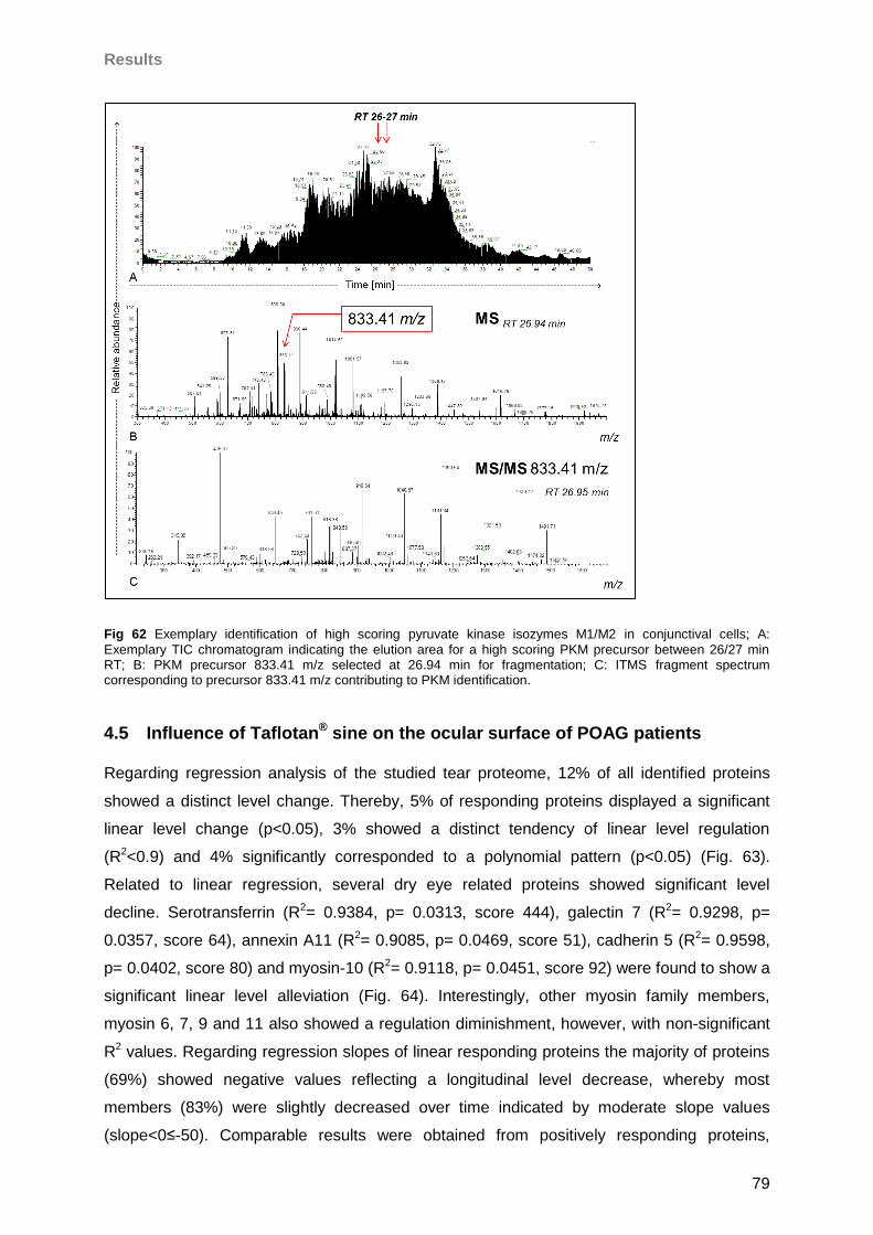
Results
79
Fig 62 Exemplary identification of high scoring pyruvate kinase isozymes M1/M2 in conjunctival cells; A:
Exemplary TIC chromatogram indicating the elution area for a high scoring PKM precursor between 26/27 min RT; B: PKM precursor 833.41 m/z selected at 26.94 min for fragmentation; C: ITMS fragment spectrum corresponding to precursor 833.41 m/z contributing to PKM identification.
4.5 Influence of Taflotan® sine on the ocular surface of POAG patients
Regarding regression analysis of the studied tear proteome, 12% of all identified proteins
showed a distinct level change. Thereby, 5% of responding proteins displayed a significant
linear level change (p<0.05), 3% showed a distinct tendency of linear level regulation
(R2<0.9) and 4% significantly corresponded to a polynomial pattern (p<0.05) (Fig. 63).
Related to linear regression, several dry eye related proteins showed significant level
decline. Serotransferrin (R2= 0.9384, p= 0.0313, score 444), galectin 7 (R2= 0.9298, p=
0.0357, score 64), annexin A11 (R2= 0.9085, p= 0.0469, score 51), cadherin 5 (R2= 0.9598,
p= 0.0402, score 80) and myosin-10 (R2= 0.9118, p= 0.0451, score 92) were found to show a
significant linear level alleviation (Fig. 64). Interestingly, other myosin family members,
myosin 6, 7, 9 and 11 also showed a regulation diminishment, however, with non-significant
R2 values. Regarding regression slopes of linear responding proteins the majority of proteins
(69%) showed negative values reflecting a longitudinal level decrease, whereby most
members (83%) were slightly decreased over time indicated by moderate slope values
(slope<0≤-50). Comparable results were obtained from positively responding proteins,
Results
80
whereby also the majority of proteins (78%) showed slight level increase (slope>0≤50) (Fig.
65). Regarding non-linear time progression, several dry eye related proteins diplayed a
significant polynomial decrease over time like e.g. low density lipoprotein receptor-related
protein 4 (p= 0.0207, score 59) or manifested a significant increase like ankyrin-3 (p= 0.0432,
score 54). Beside polynomial regression fit, the fold change “optimal match” cluster analysis
revealed a group of non-linear responding candidate proteins. The analysis calculated four
clusters, whereby 56% of all proteins were categorized as non-regulated, showing an
“equal”, “equal”, “equal”-level alteration time pattern grouped in cluster #1. Despite proteins
that show high fluctuating time pattern in cluster #4 encircling 14% of all proteins, 68% of
cluster members showed a distinct fold drop over time (“equal”, “decrease”, “equal”). 45% of
the proteins in cluster #3 including 17% of all proteins showed a level increase pattern over
time (“equal”, “increase”, “equal”). Moreover, related to all identified proteins the portion of
proteins displaying a longitudinal fold decline was 21% higher than the increasing protein
contingent. Furthermore, manual screening for longitudinal protein responses focusing on
proteins documented as dry eye marker proteins, provided most proteins showing a linear
descending tendency (R2≥0.5) like secretoglobin 1 family member D1 (R2= 0.5680, score
245), lactotransferrin (R2= 0.6940, score 2196), heat shock related 70 kDa protein 2 (R2=
0.6415, score 46) or heat shock cognate 71 kDa protein (R2= 0.5842, score 60), whereas
some proteins exhibit tendencies of linear ascending like e.g. α-1-anti-trypsin (R2= 0.8007,
score 886), interleukine-12-receptor ß-2-chain precursor (R2= 0.7400, score 41) or tumor
necrosis factor α induced protein 3 (R2= 0.5951). However, known dry eye marker proteins
like S100A4, 6, 8 and 9 failed for response. Also proline-rich protein 4 (score 167) and
proline-rich protein 1 (score 261) were found unaffected. Among the proline-rich proteins only
the large proline-rich protein BAT2 showed a slight tendency of linear decrease (R2= 0.6256,
score 78). Ingenuity® protein allocation analysis classified only 10% of the selected candidate
proteins as extracellular proteins, whereas the remaining 90% were depicted to be originated
from cellular compartments. Most of these proteins appertain to cytoplasm followed by
nucleus related proteins (Fig. 66). The functional process analysis revealed 18 functional
process hits above the adjusted protein allocation threshold of 25. Thereby processes like
tissue development, cellular development as well as cell survival and cell death including
apoptosis were highlighted (Fig. 67). In summary, a high number of cellular proteins could be
detected in the tear film of POAG patients most likely reflecting ocular surface epithelial cell
leakage events. It could be observed, that in first instance overall cellular processes were
influenced by the medical switch since predominantly intracellular proteins most likely
originated from ocular surface epithelial cells were found in the tear film and showed a
decline over the investigative period after the medical switch. Several marker proteins also
showed alterations in their regulation pattern, which indicates a changing health status of the
Results
81
ocular surface after switching. MA analysis of selected marker candidates emphasized the
level decrease of several characteristic proteins highlighted by MS analysis. “A priori” dry eye
associated MA tested proteins were: mucin 6, aquaporine 5, proline-rich protein 4,
lactotransferrin, lipocalin, hemopexin, mammaglobin B, α-2-macroglobin, cystatin, S100A8,
S100A9, lysozyme C, anti-trypsin, complement C3b and cytokines: TGF-ß1, TGF-ß2, TNF-α,
IL1a, IL1b, IL2, IL5, IL8, IL9, IL10, IL12, IL13, IL17a, IL23a, mcp1/ccl2, MIP1a/ccl3. For
proteomic result validation commercial antibodies against the following marker candidates
have been selected: serotransferrin, pyruvate kinase isozymes M1/M2, plectin, annexin A5,
annexin A11, myosin 10, galectin 7, tight junction protein ZO1 and cadherin 5. 46% of all
tested proteins showed distinct tendencies of linear approximation of the POAG group to the
control group in the course of the study (Fig. 68). Thereby, the following “a priori” dry eye
associated marker proteins were found to be affected by the Taflotan® sine appliance:
aquaporin 5, mammaglobin B, anti-trypsin and complement factor C3/C3b. Interestingly
numerous cytokines were found to shift to the control level including the interleukines, IL1a,
IL2, IL2b, IL13, IL17, IL23a, and the chemokines MIP1a/ccl3 and TGFB1 (Fig. 69). This
finding clearly reflects a diminishment of the inflammatory status of the ocular surface in the
course of the study. Focusing on MS derived marker candidates, several proteins showed
tendencies of linear level diminution in direction to the healthy control level and furthermore a
significant approximation response revealed by t-test (p-values are denoted in brackets; p1=
POAGtimepoint1 compared to Controlmean level, p2= POAGtimepoint5 compared to Controlmean level).
Thereby, pyruvate kinase isozymes M1/M2 (p1= 0.0012, p2= 0.0740), annexin A11 (p1=
0.0009, p2= 0.2026), cadherin 5 (p1= 0.0084, p2= 0.0712), serotransferrin (p1= 0.0013, p2=
0.0700) and kinectin (p1= 0.0007, p2= 0.1320) were found to converge significantly to the
healthy control level in the course of Taflotan® sine exertion since at the start of the study
protein levels for the highlighted proteins were significantly different between both groups. At
the end of the study, no significant level difference could be observed anymore predicting an
alignment of protein levels between the POAG and the healthy control group (examples are
illustrated in Fig. 70). Beside MS derived candidates, this was also demonstrated for “a priori”
MA selected dry eye associated proteins lipocalin (p1= 0.0005, p2= 0.0641) and complement
factor c3/c3b (p1= 0.0001, p2= 0.0846). Moreover, the level decline of cytokines IL1b (p1=
0.0009, p2= 0.2794), IL2 (p1= 0.0003, p2= 0.1620) and IL23a (p1= 0.0021, p2= 0.0804)
could be evaluated as a significant drawing to the healthy stage. Accordingly, microarray
results clearly supported a beneficial effect of Taflotan® sine application reflected by
cytokines and MS derived protein candidates. Moreover, objective clinical parameters
represented by TBUT and BST strengthen beneficial effects of therapeutic switching since
both parameters exhibited significant alterations revealed by t-test “start to endpoint”
comparison (Fig. 71). In summary, a strong evidence for ocular surface regeneration in the
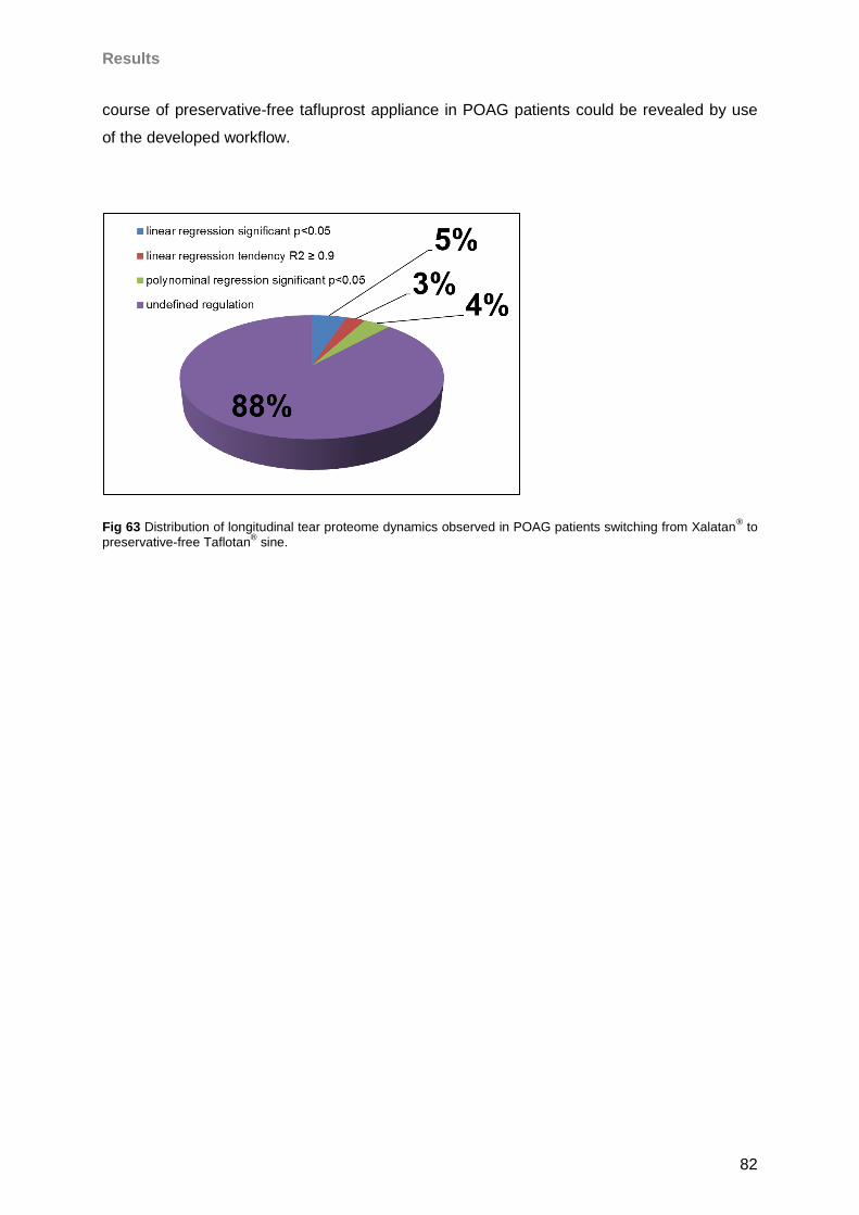
Results
82
course of preservative-free tafluprost appliance in POAG patients could be revealed by use
of the developed workflow.
Fig 63 Distribution of longitudinal tear proteome dynamics observed in POAG patients switching from Xalatan® to
preservative-free Taflotan® sine.
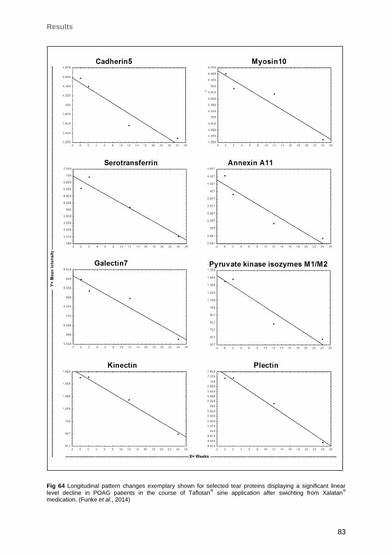
Results
83
Fig 64 Longitudinal pattern changes exemplary shown for selected tear proteins displaying a significant linear
level decline in POAG patients in the course of Taflotan® sine application after swichting from Xalatan
®
medication. (Funke et al., 2014)
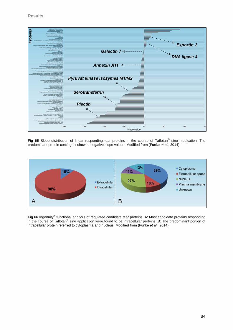
Results
84
Fig 65 Slope distribution of linear responding tear proteins in the course of Taflotan® sine medication: The
predominant protein contingent showed negative slope values. Modified from (Funke et al., 2014)
Fig 66 Ingenuity® functional analysis of regulated candidate tear proteins; A: Most candidate proteins responding
in the course of Taflotan® sine application were found to be intracellular proteins; B: The predominant portion of
intracellular protein referred to cytoplasma and nucleus. Modified from (Funke et al., 2014)

Results
85
Fig 67 Ingenuity® functional analysis of candidate tear proteins responding after the medicative switch: Numerous
candidates were found to be involved in cell maintenance, survival and death; interestingly also several candidates could be annotated to neurological disease considering glaucoma.
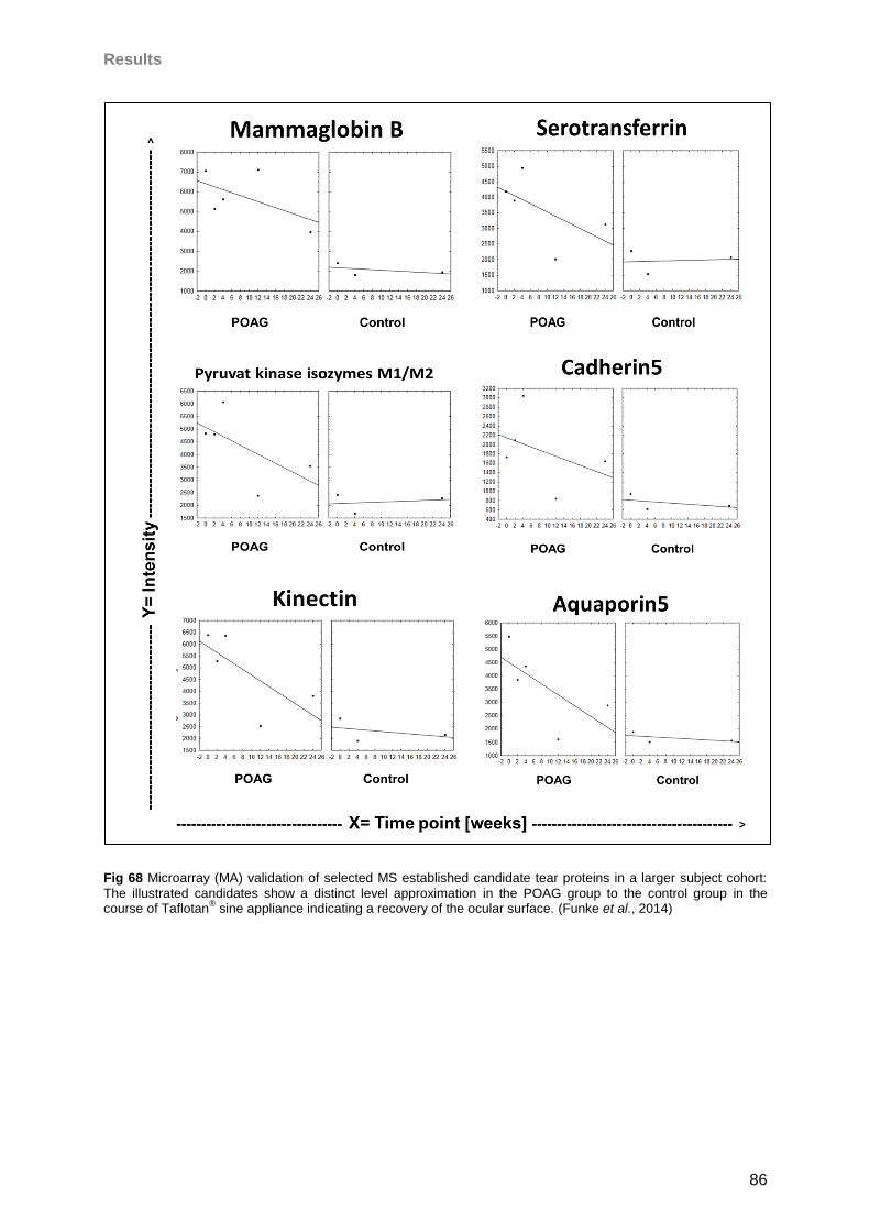
Results
86
Fig 68 Microarray (MA) validation of selected MS established candidate tear proteins in a larger subject cohort:
The illustrated candidates show a distinct level approximation in the POAG group to the control group in the course of Taflotan
® sine appliance indicating a recovery of the ocular surface. (Funke et al., 2014)
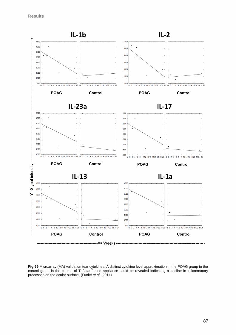
Results
87
Fig 69 Microarray (MA) validation tear cytokines: A distinct cytokine level approximation in the POAG group to the
control group in the course of Taflotan® sine appliance could be revealed indicating a decline in inflammatory
processes on the ocular surface. (Funke et al., 2014)
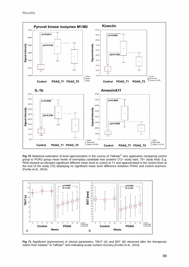
Results
88
Fig 70 Statistical estimation of level approximation in the course of Taflotan® sine application comparing control
group to POAG group mean levels of exemplary candidate tear proteins (T1= study start, T5= study end): E.g. PKM showed an elevated significant different mean level to control at T1 and approximated to the control level at the end of the study (T5) displaying no significant mean level difference between POAG and control anymore. (Funke et al., 2014)
Fig 71 Significant improvement of clinical parameters, TBUT (A) and BST (B) observed after the therapeutic switch from Xalatan
® to Taflotan
® sine indicating ocular surface recovery.(Funke et al., 2014)
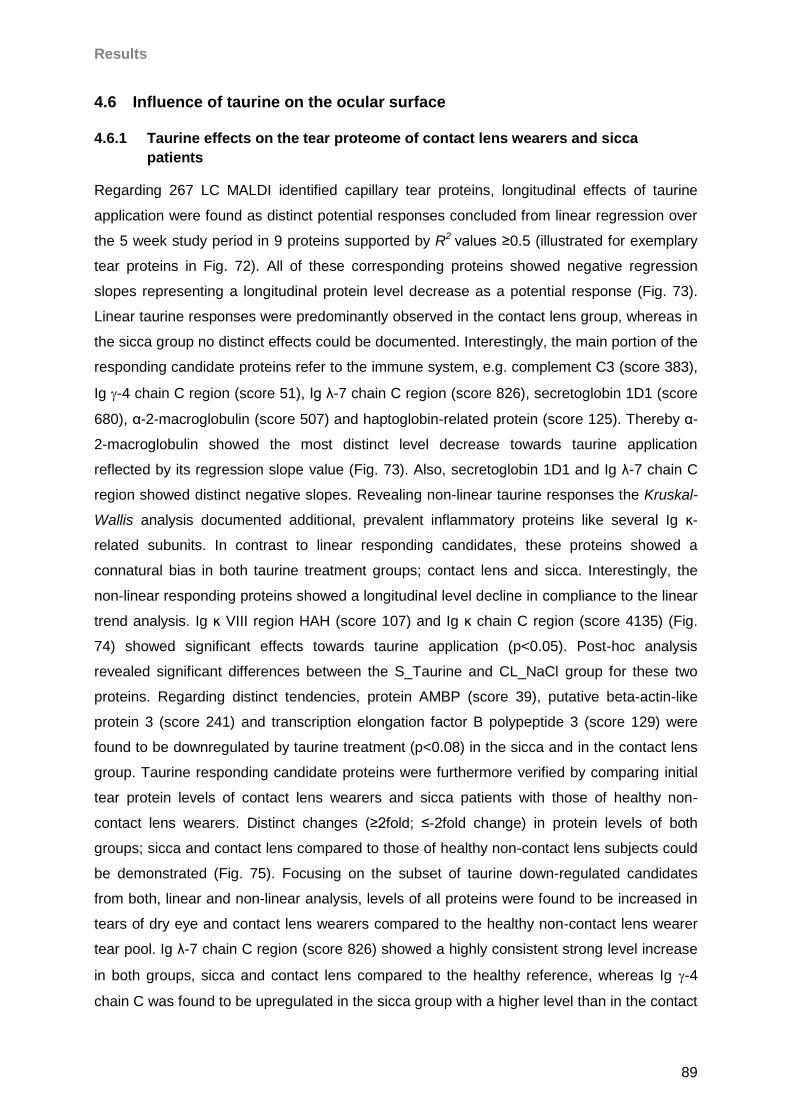
Results
89
4.6 Influence of taurine on the ocular surface
4.6.1 Taurine effects on the tear proteome of contact lens wearers and sicca
patients
Regarding 267 LC MALDI identified capillary tear proteins, longitudinal effects of taurine
application were found as distinct potential responses concluded from linear regression over
the 5 week study period in 9 proteins supported by R2 values ≥0.5 (illustrated for exemplary
tear proteins in Fig. 72). All of these corresponding proteins showed negative regression
slopes representing a longitudinal protein level decrease as a potential response (Fig. 73).
Linear taurine responses were predominantly observed in the contact lens group, whereas in
the sicca group no distinct effects could be documented. Interestingly, the main portion of the
responding candidate proteins refer to the immune system, e.g. complement C3 (score 383),
Ig -4 chain C region (score 51), Ig λ-7 chain C region (score 826), secretoglobin 1D1 (score
680), α-2-macroglobulin (score 507) and haptoglobin-related protein (score 125). Thereby α-
2-macroglobulin showed the most distinct level decrease towards taurine application
reflected by its regression slope value (Fig. 73). Also, secretoglobin 1D1 and Ig λ-7 chain C
region showed distinct negative slopes. Revealing non-linear taurine responses the Kruskal-
Wallis analysis documented additional, prevalent inflammatory proteins like several Ig ĸ-
related subunits. In contrast to linear responding candidates, these proteins showed a
connatural bias in both taurine treatment groups; contact lens and sicca. Interestingly, the
non-linear responding proteins showed a longitudinal level decline in compliance to the linear
trend analysis. Ig ĸ VIII region HAH (score 107) and Ig ĸ chain C region (score 4135) (Fig.
74) showed significant effects towards taurine application (p<0.05). Post-hoc analysis
revealed significant differences between the S_Taurine and CL_NaCl group for these two
proteins. Regarding distinct tendencies, protein AMBP (score 39), putative beta-actin-like
protein 3 (score 241) and transcription elongation factor B polypeptide 3 (score 129) were
found to be downregulated by taurine treatment (p<0.08) in the sicca and in the contact lens
group. Taurine responding candidate proteins were furthermore verified by comparing initial
tear protein levels of contact lens wearers and sicca patients with those of healthy non-
contact lens wearers. Distinct changes (≥2fold; ≤-2fold change) in protein levels of both
groups; sicca and contact lens compared to those of healthy non-contact lens subjects could
be demonstrated (Fig. 75). Focusing on the subset of taurine down-regulated candidates
from both, linear and non-linear analysis, levels of all proteins were found to be increased in
tears of dry eye and contact lens wearers compared to the healthy non-contact lens wearer
tear pool. Ig λ-7 chain C region (score 826) showed a highly consistent strong level increase
in both groups, sicca and contact lens compared to the healthy reference, whereas Ig -4
chain C was found to be upregulated in the sicca group with a higher level than in the contact
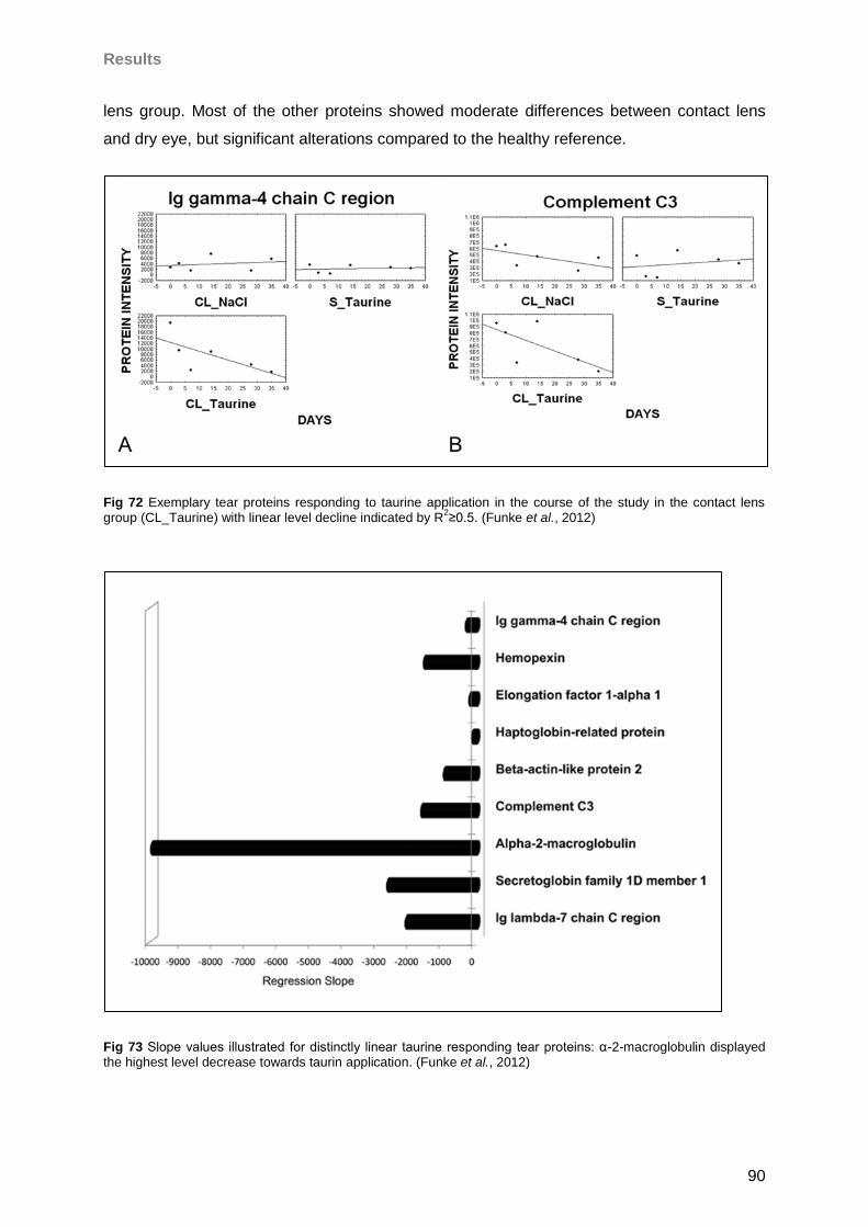
Results
90
lens group. Most of the other proteins showed moderate differences between contact lens
and dry eye, but significant alterations compared to the healthy reference.
Fig 72 Exemplary tear proteins responding to taurine application in the course of the study in the contact lens group (CL_Taurine) with linear level decline indicated by R
2≥0.5. (Funke et al., 2012)
Fig 73 Slope values illustrated for distinctly linear taurine responding tear proteins: α-2-macroglobulin displayed the highest level decrease towards taurin application. (Funke et al., 2012)
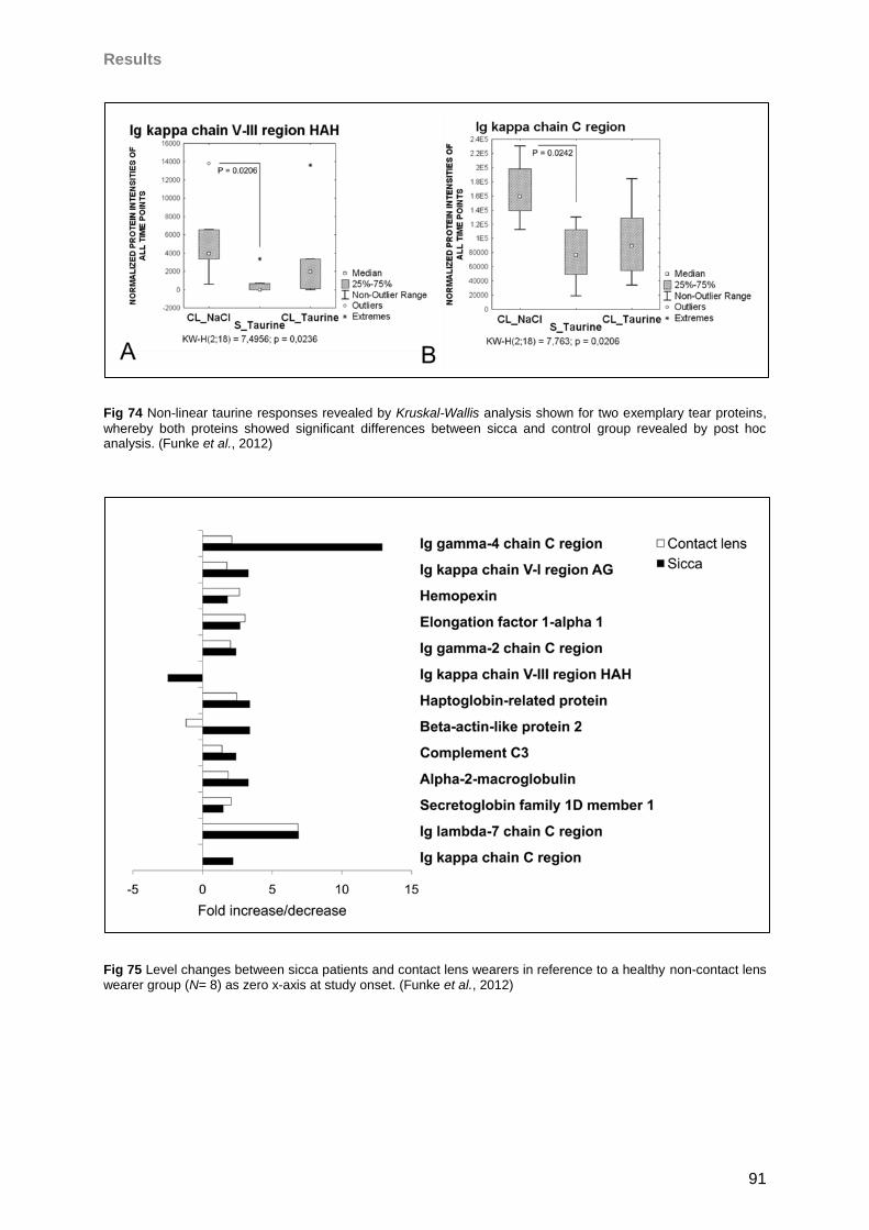
Results
91
Fig 74 Non-linear taurine responses revealed by Kruskal-Wallis analysis shown for two exemplary tear proteins,
whereby both proteins showed significant differences between sicca and control group revealed by post hoc analysis. (Funke et al., 2012)
Fig 75 Level changes between sicca patients and contact lens wearers in reference to a healthy non-contact lens wearer group (N= 8) as zero x-axis at study onset. (Funke et al., 2012)
Results
92
4.6.2 Taurine effects on the conjunctival proteome
Regarding the oxidative stress level of H2O2 stressed NHC-IOBA cells, it could be
demonstrated that incubation with tested taurine concentrations (0.05, 0.2, 0.5, 1, 2 mM) lead
to a distinct decline in ROS production compared to stressed control cells, which were not
taurine treated. Accordingly, application of 0.05 mM taurine lead to a ROS reduction of 26%
(p= 0.0356), incubation with 0.2 mM to a decrease of 29% (p= 0.0153) and treatment with 1
mM taurine showed a decline of 32% (p= 0.0016). Also, the other tested concentrations
showed distinct ROS decrease responses, however, not significant reactions (Fig. 76A).
Despite the obvious effects on ROS production, no significant effects were observed for
taurine treatment of H2O2 stressed cells regarding cell viability. Furthermore, staurosporine
stressed cells also failed to show significant viability effects and showed only weak
tendencies of increased viability, reflected by a viability increase of 7% in cells incubated with
0.05 mM taurine. In contrast incubation of unstressed cells with taurine lead to a significant
increase in viability observed for 0.05 mM (26%, p= 0.0055) and 0.2 mM (19%, p= 0.0181).
Whereas also 0.5 mM taurine showed tendency of increased viability (15%, p= 0.0681),
concentrations above this value showed no effect or negative responses of cell viability
towards taurine treatment (Fig. 76B). Incubation of cells with taurine in a 20-100 mM range
concluded in a significant loss of cells up to a percentage of 20% (100 mM, p= 0.0010) (Fig.
77B). Accordingly, in the presence of staurosporine for these critical concentrations additive
negative effects could be observed with a maximum of 32% viability decline at 100 mM
taurine (p= 0.0001). Moreover, ROS levels were found to be increased significantly from
taurine concentrations above 20 mM with a maximum of 102% supplement of ROS
production at 100 mM taurine (p= 0.0005). Thereby, incubation of stressed cells with taurine
showed no significant counteraction towards ROS production displaying a maximum ROS
level of 82% at 100 mM taurine (p= 0.000002). Contrary to ROS results of low taurine
concentration, slight ROS elevations could also been observed in the initial concentrations,
but were not revealed as significant and clearly differing from higher concentrations above 20
mM (Fig. 77A). In summary, these findings give evidence of taurine dose dependent effects
regarding cell viability and ROS production. Whereas taurine incubation performed
beneficially on cell viability and ROS generation in a 0.05-0.5 mM range, concentrations >0.5
mM displayed negative effects. For BU proteomic analysis on cellular taurine effects cell
lysates obtained from taurine treated unstressed NHC-IOBA cells displayed high qualitative
SDS PAGE migration pattern for both study parts corresponding to “elevated” and to “low”
taurine concentrations (Fig. 78). Focusing on proteomic results, LC ESI analysis of
unstressed conjunctival cells incubated with critical “low” taurine concentrations (4, 20, 50
mM) revealed several taurine target protein candidates. Thereby, taurine affected cellular
proteins could be revealed. Interestingly, exclusively significant elevations reflected by a
Results
93
minimum 2fold level increase in comparison to the control mean level could be observed. 30
proteins showed significantly increased levels in minimum one of the taurine concentration
groups (4, 20, 50 mM) and a tendency of increase in the remaining groups (Fig. 79). Among
these proteins, phosphoglycerate kinase 2 (score 3317), protein SET (score 2713), ubiquitin
60s ribosomal protein L40 (score 2235), protein S100 P (score 927), enoyl-COA delta
isomerase 1 (score 561) were found as the top 5 scoring proteins. Moreover, several of the
taurine responding proteins revealed by fold change displayed a significant linear response
according to an increasing taurine concentration, which makes them highly suspicious for
direct or indirect taurine interaction, e.g. glutamate dehydrogenase 2 (R2= 0.978, p= 0.011)
and glutaredoxin 1 (R2= 0.959, p= 0.021). Next to the affected glutaredoxin 1 (score 214),
which is indicative for cellular stress (Anathy et al., 2009, Enoksson et al., 2005, Murata et
al., 2003, Voehringer, 1999), numerous further altered proteins were found to be involved in
cellular stress and apoptotic processes, e.g. cytochrome C oxidase subunit 2 (score 69)
(Buron et al., 2006, Green and Reed, 1998, Kadenbach et al., 2004), which showed the
highest increase level of all affected proteins (12.4 fold) or septin 2 (score 185) (Garcia-
Fernandez et al., 2010, Kinoshita, 2003, Longtine et al., 1996). Also, an interaction with
thimet oligopeptidase (score 143), a cytoplasma located metalloprotease (www.uniprot.org)
could be proposed since taurine was also demonstrated to interact with matrix
metalloproteinase 9 (Park et al., 2000). Also sodium/nucleoside cotransporter 2 (score 37),
nevertheless detected with a low score, could be proposed to interact with taurine since
taurine was found to be released from cells by treatment with adenosine transport inhibitors
(Hada et al., 1996). In summary, the level increases of the mentioned proteins most likely
indicate a direct cellular response towards elevated taurine concentrations which fits well
with the observed negative taurine effects for these concentrations, demonstrated by ROS
and cell viability assays. Additionally, the detected apoptotic and stress response proteins
reflect the declined viability of the cells incubated with critical high taurine concentrations. In
accordance, these findings support the functionality of the LC ESI MS workflow since
apoptitic events on the cellular level could be reflected on a proteomic level. Thus, a huge
contingent of responding proteins was not associated to apoptosis, conclusively highly
interesting to be modulated by taurine in alternative pathways. Encircling both apoptic
associated and non-associated proteins, among the responding proteins several candidates
were found to interact in a direct or indirect manner with taurine inferred from the literature,
e.g. phosphoglycerate kinase (Midwinter et al., 2004, Nandhini et al., 2005, Peskin and
Winterbourn, 2006), cytochrome C oxidase (Millett et al., 1982), protein S100 P (Reymond et
al., 1996) and glutamate dehydrogenase 2 (Schaffer et al., 2000). Another interesting protein
that was found upregulated by taurine treatment was sepiapterin reductase (score 97), which
was suggested to play a crucial role in lipid metabolism (de Roos et al., 2005). Upregulation
Results
94
of proteolipid protein 2 (score 235), also gave evidence for taurine interaction. Since
hydrophobic surfactant proteins had been already detected for tear fluid and/or the lacrimal
gland system and had been proposed to play a key role for tear film stability (Brauer et al.,
2007a, Brauer et al., 2007b) it is likely to suggest a comparable function for proteolipid 2 and
moreover worth to mention, that it responds to taurine treatment. Since the membrane
modifying potential of taurine is well documented (Huxtable, 1990, Lleu et al., 1992)
proteolipid 2 increase could be a consequence of taurine-membrane interactions.
Confidently, transmembrane protein 109 (Mitsugumin 23) (score 411), an ER/nucleus
membrane protein (Venturi et al., 2011), detected for the first time for the ocular surface in
this study, was found to display a level increase by taurine treatment, which emphasizes the
influence of taurine on membrane systems. Regarding response reactions towards taurine
treatment, several proteins showed no distinct fold changes, but 87 proteins were found to
show a significant linear response correlating with taurine concentration increase, probably
indicating a weak taurine response. Among them, the top 5 scoring candidates were
heterogeneous carbamoyl-phosphate synthase (score 13385), elongation factor 1-α 2 (score
9574), neuroblast differentiation-associated protein (score 6396), L-lactate dehydrogenase B
chain (score 6193) and multifunctional protein ADE2 (score 1718). Candidate related
proteins displaying a clear fold change were depicted by the linear analysis. For example,
importin-9 (score 131) was found to be significantly elevated, whereas importin-5 (score 815)
was found to show a linear increase (slope: 3405, R2= 0.959, p= 0.021) below the fold
change threshold. De facto, 47% of potential apoptosis marker proteins, that have been
detected in a study of hepatic stellate cells incubated with taurine could also been detected in
this study with relatively high scores, e.g. annexin A1 (score 6596), peroxiredoxin 2 (score
4946) or protein DJ-1 (PARK7, Parkinson disease protein 7) (score 633) (Deng et al., 2010).
Protein DJ-1 is known to directly interact with EF-hand calcium-binding domain-containg
protein 6 (score 150) (www.uniprot.org), a nucleus protein, which was found to be
significantly upregulated by taurine treatment in the present study. Since taurine is well
documented for its role in calcium translocation and homeostasis (Dolara et al., 1978,
Pasantes-Morales, 1982), it is very likely that it interacts with calcium-binding proteins.
However, only translationally-controlled tumor protein (score 698), as one of these proteins,
showed a significant linear level increase towards taurine treatment in confidence with the
results of the hepatic stellate cell study. Interestingly, several proteins associated to oxidative
stress response have been also found to respond to taurine treatment in a linear manner,
e.g. thioredoxin 1 (score 121) (Holmgren, 2000, Yildirim et al., 2007). In summary, 87
proteins were found to show a linear pattern change with increasing taurine concentration
encircling protein up-and downregulations. However, in most cases intensities were below
the fold change threshold, but are likely to indicate a regulation prognosis. Regarding
Results
95
potential taurine protein targets obtained from LC ESI experiments, focusing on cells
incubated with 0.05 mM taurine as preselected “beneficial” concentration, numerous
responses could be recorded. Accordingly, 41% of the conjunctival cell proteins were found
to respond to taurine treatment demonstrated by a minimum 2fold intensity change, whereby
a small portion of proteins showed high level alterations towards taurine. Analysis of
molecular function showed that the majority of the 2fold regulated proteins are involved in
catalytic and molecule binding processes, whereby most of the proteins mediate protein-
protein interactions (Fig. 80). In addition, whereas among the group of 2 to 5fold regulated
proteins up and downregulation could be documented, the group of “highly regulated”
proteins (>10fold) displayed exclusively downregulations, whereby like documented for the
“mild regulated” proteins the majority of the “high regulated” proteins were involved in protein
and nucleic acid binding (an overview of regulation distribution is given in Fig. 81). Thereby
calmodulin, the taurine transporter control major player (Huang et al., 2001, Ramamoorthy et
al., 1994), was found to be extremely decreased (24fold). Also, calmodulin-like protein 3
(score 95) was found to be extremely diminished (6fold). Additionally, another calmodulin
related protein, calcium/calmodulin-dependent protein kinase type 1 (score 37) showed a
slight decrease (2.3fold). ER calcium Atpase 2 (score 64) another calcium depending protein
was also found to be weakly downregulated (2fold). These findings support a response of the
calcium/calmodulin apparatus towards taurine treatment (Fig. 82). Furthermore a complete
class of proteins, the 14-3-3 proteins (α, ß, , , , , ) (scores: 1207-1435) exhibited slight
level diminishments (2.2-2.5fold). Beside intracellular proteins, 7% of candidate proteins
were found to be associated to the extracellular milieu, whereby the secretory protein
galectin-1 (score 203) was slightly decreased (2.5fold) and galectin 3-binding protein (score
94), was found to be highly diminished (14fold). Also, a signalling protein, interleukin
enhancer-binding factor 2 showed a distinct decline (10fold). The response of secretory
proteins indicates the impact of taurine on cellular interactions with the extracellular
environment. Focusing on “highly regulated” candidates, a predominance of downregulated
histone species could be observed (Fig. 83). Also, the level of histone binding protein RBBP4
was decreased (10fold). Furthermore, a decline of certain DNA interacting proteins like DNA
binding protein A, Y-box protein 2, representative fragments of DNA
primase/polymerase/lyase could be documented indicating gene expression changes.
Downregulation of F-box only protein 22 is indicative for anti-apoptic processes, due to
ubiquitine-dependent proteolysis and apoptosis mediation (Patton et al., 1998, Winston et al.,
1999). High abundant stress and apoptose associated proteins, e.g. peroxiredoxin 4 (score
1047), 6 (score 1724), glutamate dehydrogenase 2 (score 73), cytochrome C (score 48),
cytochrome C oxidase subunit 2 (score 43), thioredoxin (score 207), glutathione S
transferase ĸ 1 (score 129) showed level declines between 2 and 3fold (Fig. 84). Additionally,
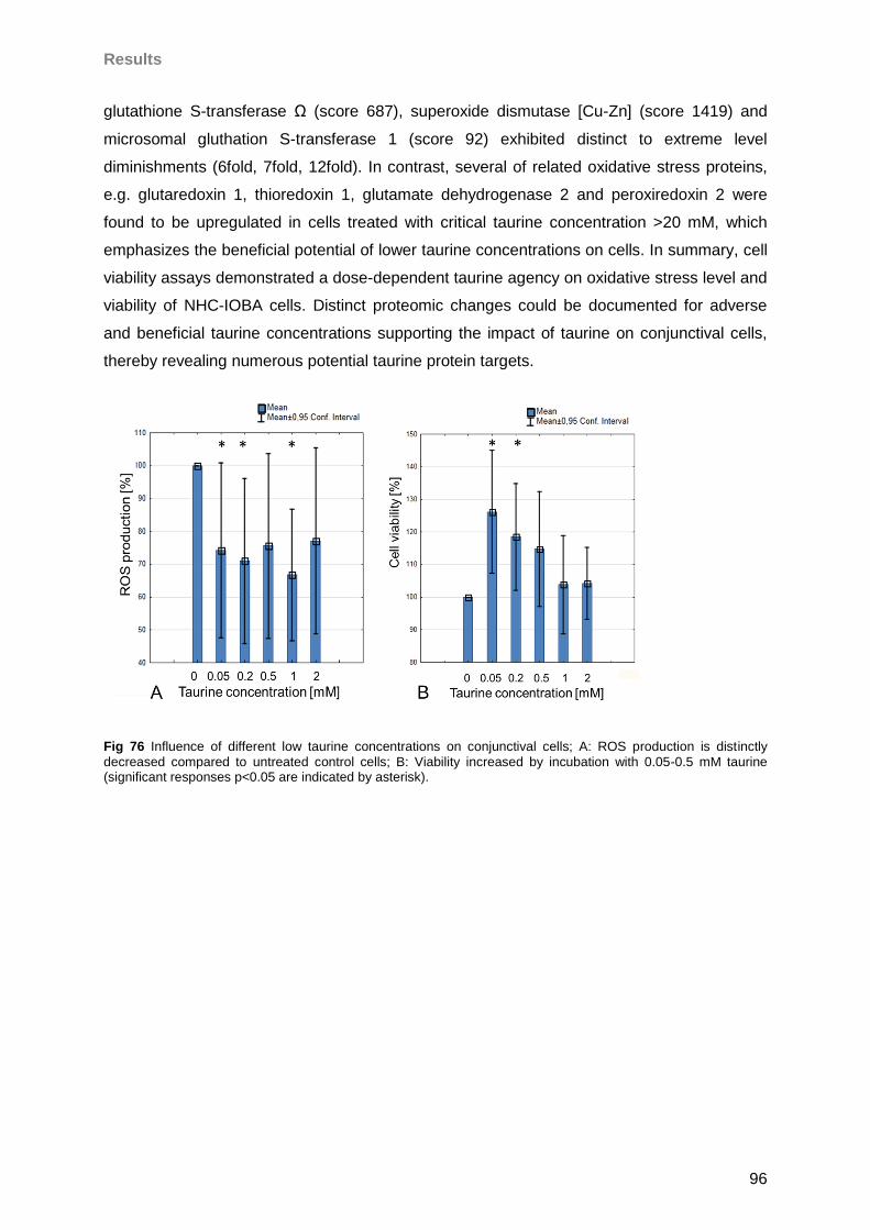
Results
96
glutathione S-transferase Ω (score 687), superoxide dismutase [Cu-Zn] (score 1419) and
microsomal gluthation S-transferase 1 (score 92) exhibited distinct to extreme level
diminishments (6fold, 7fold, 12fold). In contrast, several of related oxidative stress proteins,
e.g. glutaredoxin 1, thioredoxin 1, glutamate dehydrogenase 2 and peroxiredoxin 2 were
found to be upregulated in cells treated with critical taurine concentration >20 mM, which
emphasizes the beneficial potential of lower taurine concentrations on cells. In summary, cell
viability assays demonstrated a dose-dependent taurine agency on oxidative stress level and
viability of NHC-IOBA cells. Distinct proteomic changes could be documented for adverse
and beneficial taurine concentrations supporting the impact of taurine on conjunctival cells,
thereby revealing numerous potential taurine protein targets.
Fig 76 Influence of different low taurine concentrations on conjunctival cells; A: ROS production is distinctly
decreased compared to untreated control cells; B: Viability increased by incubation with 0.05-0.5 mM taurine (significant responses p<0.05 are indicated by asterisk).
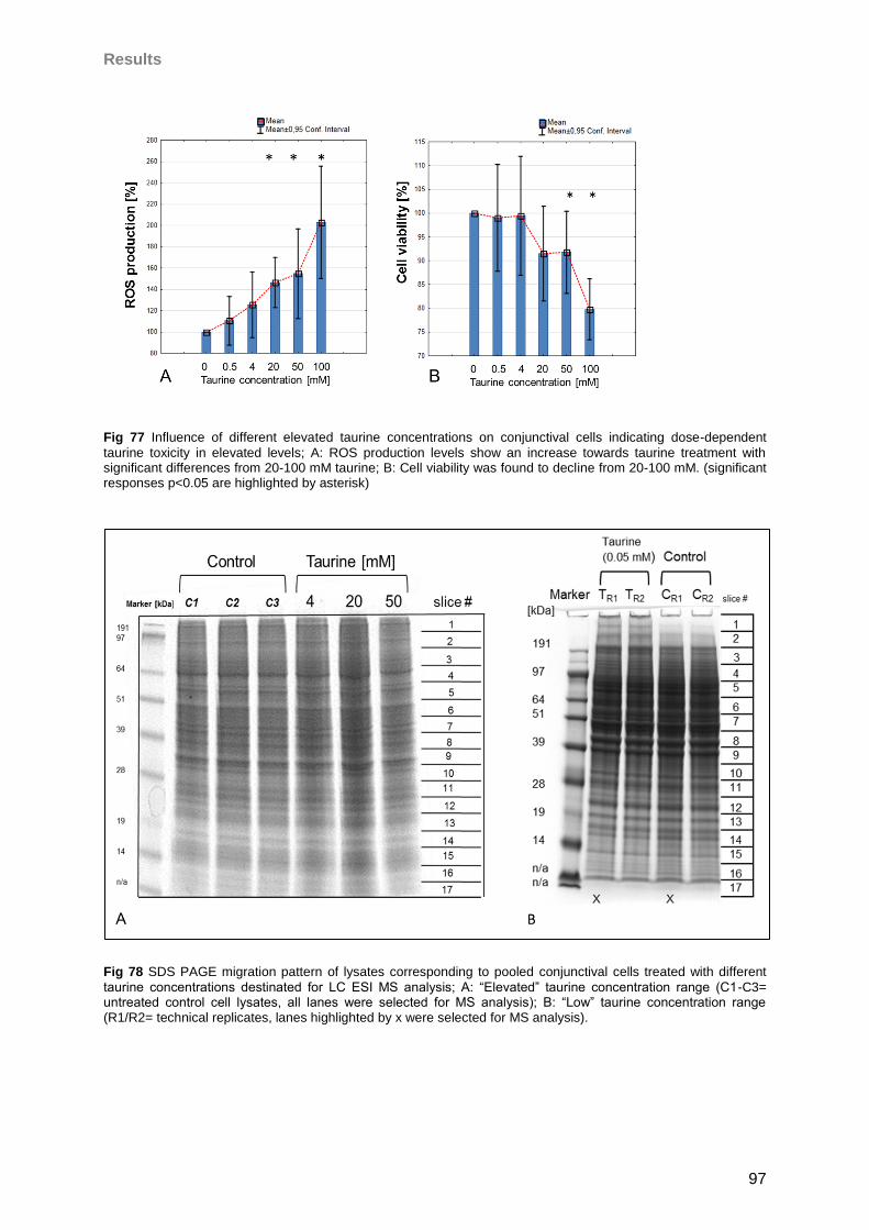
Results
97
Fig 77 Influence of different elevated taurine concentrations on conjunctival cells indicating dose-dependent
taurine toxicity in elevated levels; A: ROS production levels show an increase towards taurine treatment with significant differences from 20-100 mM taurine; B: Cell viability was found to decline from 20-100 mM. (significant responses p<0.05 are highlighted by asterisk)
Fig 78 SDS PAGE migration pattern of lysates corresponding to pooled conjunctival cells treated with different
taurine concentrations destinated for LC ESI MS analysis; A: “Elevated” taurine concentration range (C1-C3= untreated control cell lysates, all lanes were selected for MS analysis); B: “Low” taurine concentration range (R1/R2= technical replicates, lanes highlighted by x were selected for MS analysis).
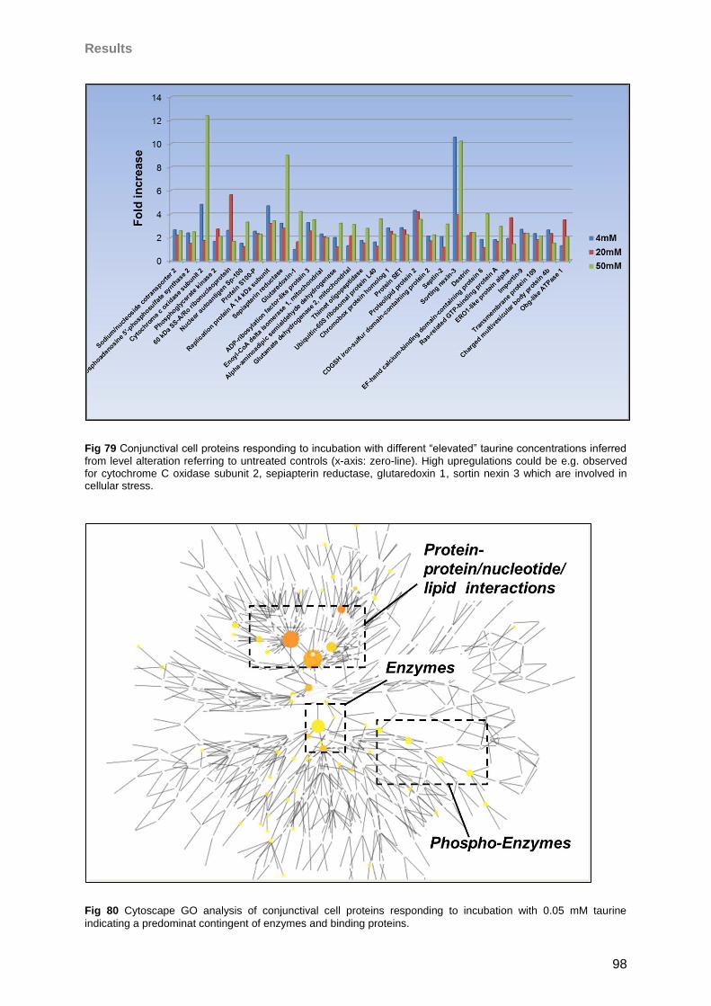
Results
98
Fig 79 Conjunctival cell proteins responding to incubation with different “elevated” taurine concentrations inferred
from level alteration referring to untreated controls (x-axis: zero-line). High upregulations could be e.g. observed for cytochrome C oxidase subunit 2, sepiapterin reductase, glutaredoxin 1, sortin nexin 3 which are involved in cellular stress.
Fig 80 Cytoscape GO analysis of conjunctival cell proteins responding to incubation with 0.05 mM taurine
indicating a predominat contingent of enzymes and binding proteins.
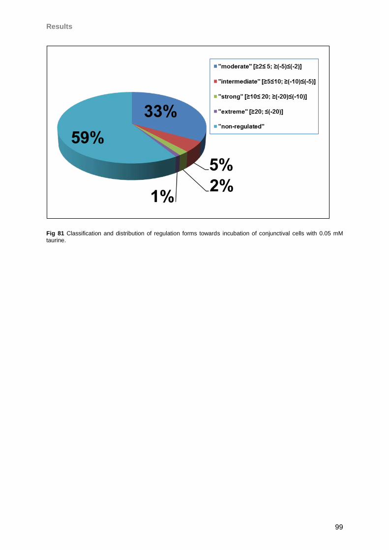
Results
99
Fig 81 Classification and distribution of regulation forms towards incubation of conjunctival cells with 0.05 mM
taurine.
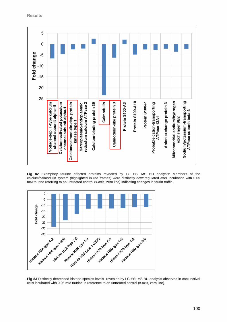
Results
100
Fig 82 Exemplary taurine affected proteins revealed by LC ESI MS BU analysis: Members of the
calcium/calmodulin system (highlighted in red frames) were distinctly downregulated after incubation with 0.05 mM taurine referring to an untreated control (x-axis, zero line) indicating changes in taurin traffic.
Fig 83 Distinctly decreased histone species levels revealed by LC ESI MS BU analysis observed in conjunctival
cells incubated with 0.05 mM taurine in reference to an untreated control (x-axis, zero line).
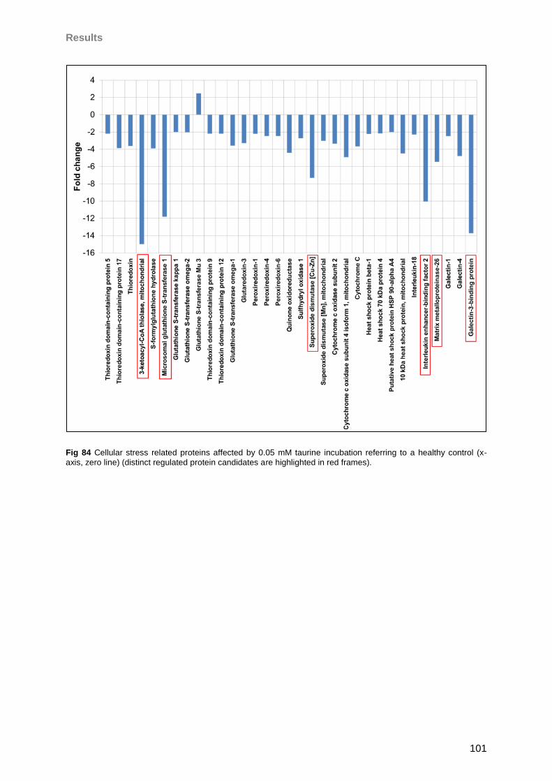
Results
101
Fig 84 Cellular stress related proteins affected by 0.05 mM taurine incubation referring to a healthy control (x-
axis, zero line) (distinct regulated protein candidates are highlighted in red frames).
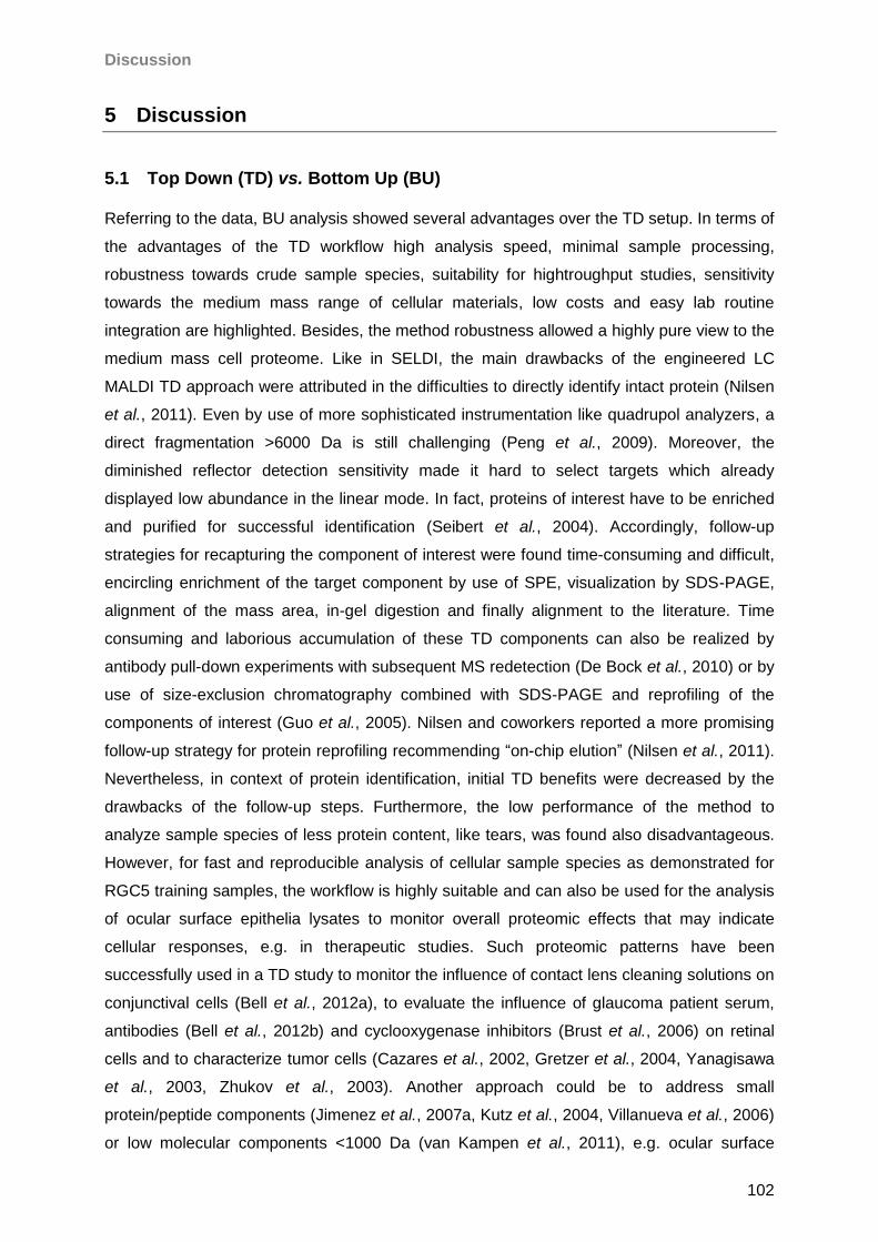
Discussion
102
5 Discussion
5.1 Top Down (TD) vs. Bottom Up (BU)
Referring to the data, BU analysis showed several advantages over the TD setup. In terms of
the advantages of the TD workflow high analysis speed, minimal sample processing,
robustness towards crude sample species, suitability for hightroughput studies, sensitivity
towards the medium mass range of cellular materials, low costs and easy lab routine
integration are highlighted. Besides, the method robustness allowed a highly pure view to the
medium mass cell proteome. Like in SELDI, the main drawbacks of the engineered LC
MALDI TD approach were attributed in the difficulties to directly identify intact protein (Nilsen
et al., 2011). Even by use of more sophisticated instrumentation like quadrupol analyzers, a
direct fragmentation >6000 Da is still challenging (Peng et al., 2009). Moreover, the
diminished reflector detection sensitivity made it hard to select targets which already
displayed low abundance in the linear mode. In fact, proteins of interest have to be enriched
and purified for successful identification (Seibert et al., 2004). Accordingly, follow-up
strategies for recapturing the component of interest were found time-consuming and difficult,
encircling enrichment of the target component by use of SPE, visualization by SDS-PAGE,
alignment of the mass area, in-gel digestion and finally alignment to the literature. Time
consuming and laborious accumulation of these TD components can also be realized by
antibody pull-down experiments with subsequent MS redetection (De Bock et al., 2010) or by
use of size-exclusion chromatography combined with SDS-PAGE and reprofiling of the
components of interest (Guo et al., 2005). Nilsen and coworkers reported a more promising
follow-up strategy for protein reprofiling recommending “on-chip elution” (Nilsen et al., 2011).
Nevertheless, in context of protein identification, initial TD benefits were decreased by the
drawbacks of the follow-up steps. Furthermore, the low performance of the method to
analyze sample species of less protein content, like tears, was found also disadvantageous.
However, for fast and reproducible analysis of cellular sample species as demonstrated for
RGC5 training samples, the workflow is highly suitable and can also be used for the analysis
of ocular surface epithelia lysates to monitor overall proteomic effects that may indicate
cellular responses, e.g. in therapeutic studies. Such proteomic patterns have been
successfully used in a TD study to monitor the influence of contact lens cleaning solutions on
conjunctival cells (Bell et al., 2012a), to evaluate the influence of glaucoma patient serum,
antibodies (Bell et al., 2012b) and cyclooxygenase inhibitors (Brust et al., 2006) on retinal
cells and to characterize tumor cells (Cazares et al., 2002, Gretzer et al., 2004, Yanagisawa
et al., 2003, Zhukov et al., 2003). Another approach could be to address small
protein/peptide components (Jimenez et al., 2007a, Kutz et al., 2004, Villanueva et al., 2006)
or low molecular components <1000 Da (van Kampen et al., 2011), e.g. ocular surface
Discussion
103
degradative fragments (Abalain et al., 2000, Wheater et al., 1999) or profiling of known
masses. To overcome the lack of identification, the method can also be suited for the
analysis of tryptic digests initially facilitating statistics followed by fragmentation of tryptic
peptides of interest (Dekker et al., 2005, Neubert et al., 2008). Nevertheless, in this work for
in depth analysis of the ocular surface, encircling conjunctival cells and tear fluid, the BU
strategy has turned out the more effective approach characterizing proteins >4000 Da. Also,
SDS PAGE as a first fractionation dimension allows visual inspection and densitometric
evaluation if needed (Neuhoff et al., 1990). Regarding in-gel digests it is possible to directly
monitor protein markers in a wide mass range obtaining knowledge of their identity. In
contrast, in TD approaches a high risk remains that significant markers stay unidentified
resulting in a costly loss of biological information and making targeted follow-up studies on
candidate proteins impossible. In fact, by use of the BU approach “in depth” analysis of
human tear fluid and cell samples could be realized providing >1000 proteins per sample
type, whereby proteins could be annotated to biological functions and subcellular
localizations and could be further reconciled with the literature for disease and clinical
acquisition. Nevertheless, also several limitations have to be mentioned. First of all, the
approach is more costly, time and lab intensive, since proteins have to be fractionated by
SDS-PAGE, in-gel digested and in case of LC ESI purified by SPE prior to analysis.
Considering sample preprocessing steps and the more profound analysis protocol the
method is less easy to implement to the daily lab routine as the TD approach. Further
drawbacks are the use of pool samples to reduce costs and analysis time, especially in labs
with a high project contigent. Accordingly, there exists a challenge in statistical evaluation
and the need for time consuming and costly validation experiments, which can be realized by
antibody-based microarray analysis of MS derived marker candidates. In fact, the use of pool
samples is widely practiced in proteomic analysis (Castano et al., 2006, Neubauer et al.,
2006, Tonge et al., 2001) addressing costs and limitation of resources, and was found as a
valid but considerable strategy in terms of reduction of biological variance and comparison
between pool and mean protein expression (Diz et al., 2009). Thus, despite sample pooling
has been proposed to “mute unrelated alterations by dilution” (Weinkauf et al., 2006) it
seems to exist a sample-dependent ratio between technical and biological variation that has
to be considered for the setup of experimental designs (Karp and Lilley, 2009). De facto, an
“in depth” analysis of a large number of individual samples is not realistic in case of the
developed BU strategy considering costs, effort, analysis time and realization of numerous
parallel running projects. However, sensitivity and the possibility to identify large numbers of
proteins allowing orthogonal follow up research on identified candidate proteins and the
possible establishment of large protein maps with knowledge on protein localization and
function achieved by the BU approach surmount the possibilities of the developed TD
Discussion
104
approach. However, there is a high potential in combining both developed strategies in the
analysis of cellular ocular surface sample species, BU studying proteins >4 kDa and TD
monitoring statistical changes in small protein fragments. As an outlook, the fast TD
screening for known signal peptides in digests which have already been proven for particular
proteins by use of the intensive BU proteomic strategy or orthogonal methods may be a
promising future direction to combine benefits of both strategies. For this reason, the protein
maps established in this work could be screened for signature peptides that uniquely
correspond to a particular protein of interest and their fast reprofiling in large sample cohorts
by use of the TD approach, probably allows less costly, lab-intensive and deeper statistical
understanding of ocular surface disease processes. However, such signature peptides have
to be proven in TD analysis in a first step, which is the challenge in such a combinatory
strategy with respect to different ionization mechanism in ESI and MALDI systems.
5.2 Bottom-up (BU) analysis: LC-MALDI vs LC-ESI workflow
The obtained results support the high reproducibility for both approaches. However, there are
distinct differences between MALDI and ESI workflows regarding the developed BU
approaches. The RT reproducibility was excellent in the ESI workflow with low precursor
mass shifting additionally supported by CV values <1% in confidence with the literature
reporting values between 0.1 to 4.5% observed for comparable systems (Duan et al., 2009,
Dunn et al., 2008). In comparison, also the MALDI approach showed a high RT
reproducibility. An important factor which impacts on LC separation efficiency in LC MALDI
experiments is the fraction collection performance and sample space limitation regarding
target plates. In contrast, separation efficiency in continuous LC ESI workflows is
predominantly limited by the column system and flow rate. However for both approaches
there is a need to compromise between efficiency, lab effort, costs and analysis time
regarding analysis of multiple sample sets settling pro and contras between both strategies.
Focusing on the effort/output ratio for analysis of complex sample types, the LC ESI workflow
presented superior over LC MALDI. That is why the sensitivity was found to be clearly higher
in the ESI method regarding severe complex sample species like tear fluid to more complex
sample species like cell lysates. Without a doubt, this is not explicable by the ionization
technique but due to the coupled hybrid MS system providing a higher dynamic range
(Makarov et al., 2006b) for higher performance than TOF systems (Zhang et al., 2000). De
facto, the main reason for the high accuracy is based on the high resolution achieved by the
orbitrap system clearly exceeding TOF systems thereby exceeding a critical value which
allows the discrimination between coeluting components (van der Heeft et al., 2009). In
comparison with the LC ESI based highly accurate hybrid system, the MALDI platform
cannot go strong with its beneficial attributes like high salt and contaminant tolerance, small
Discussion
105
sample consumption and sample storage for reanalysis (van Kampen et al., 2011, Zhang et
al., 2004a). As a matter of fact, regarding practical aspects of the preionization sample
process fraction, deposition complications due to abrasion of the robot tip in the MALDI
workflow balanced with spray incursions due to thermal damages of the spray needle in the
ESI approach. Nevertheless, whereas the MALDI approach saved time in sample pre-
processing due to higher contaminant tolerance it lost speed in fraction deposition and matrix
spotting compared to the ESI approach. A further interesting difference was observed by tear
film analysis, whereby it was shown that the MALDI approach yielded in extremely high
scores for a few prominent tear proteins, whereas the ESI approach identified a higher
number of proteins but did not accomplish the absolute score values resulting from the
MALDI analysis for selected predominant tear proteins, e.g. proline-rich protein 4 or
lysozyme. Most likely this observation reflects adverse of the TOF analyzer resolution
performance for complex protein mixtures in comparison to the orbitrap analyzer. Another
possibility is the concurrence for ionization and ion suppression effects (Annesley, 2003,
Jessome and Volmer, 2006, Muller et al., 2002, Wang et al., 2004), which may lead to a
positive discrimination of abundant peptide ionization in the MALDI approach. Preference of
high intensive peptides is a well-known phenomenon in MALDI experiments and
counteracting strategies like replicate runs considering precursor ion exclusion lists (Chen et
al., 2005) have been developed. Furthermore, fragmentation strategies in both instruments
are different, whereas the MALDI approach was prepared for PSD, the ESI method was
tuned for CID fragmentation leading to different fragment pattern (Hoteling and Owens, 2004,
Li et al., 1999). Although, the possibility of reanalysis and optimization of the measuring
protocol of identical sample sets is an important advantage of MALDI over ESI technology
(Ishihama, 2005), it is hampered by the need for analysis time. Actually, the fragmentation
speed of the TOF/TOF systems was found to clearly underlie the speed of the LTQ Orbitrap
instrument. A problem observed in both developed BU workflows was the high portion of
fragment spectra, which failed to successfully contribute in probability-based protein
identification in data bases (Perkins et al., 1999). Accordingly, the generation of large
amounts of redundant data, whereby a high percentage of high quality MS/MS spectra fail to
contribute to protein identification, is a known phenomenon (Cox et al., 2008, Karty et al.,
2002, Nesvizhskii, 2010, Neuhauser et al., 2012, Xu et al., 2005). Consequently, strategies
are on the way to exploit biological information out of unassigned spectra, whereby e.g. the
use of “de novo sequencing” algorithm was proposed by numerous authors (Ning et al.,
2010, Seidler et al., 2010). From a practical point of view the MALDI method is more lab
intensive, since fractions have to be deposited and matrix has to be applied by pipetting
robotic units, which can be sources of error. On the other hand it is more robust towards
contaminants than ESI, which allowed a direct HPLC analysis without SPE purification of gel
Discussion
106
digests prior to LC MALDI. However, the more obvious advantage is the decoupling of
sample preparation and measuring in the MALDI approach, allowing storage and reanalysis
of samples (Kuzyk et al., 2009), whereas in case of system errors samples are lost in the ESI
approach. In these studies, during test runs, this happened in case of robot error, column
blocking or needle damage. However, in the developed LC ESI workflow each sample
provided enough material for one reanalysis. Thereby the storing was a more critical factor
since freezing of remaining sample runs in sample buffer or relyophilization of samples was
found to impact sample quality. Nevertheless, the complex MALDI software platform was
found to be an important source of error in the course of the studies which was not the case
in LC ESI. Further differences could be attributed to the complementarity of ionization
principles (Bodnar et al., 2003, Stapels and Barofsky, 2004, Stapels et al., 2004, Yang et al.,
2007). In up to date literature, it was reported that more hydrophilic peptides had been
obtained in MALDI (Yang et al., 2007), whereas ESI favors more hydrophobic species (Cech
and Enke, 2000). Moreover, whereas ESI was supported to favor ionization of peptides
ending with lysine over those ending with arginine, the opposite was observed for MALDI
ionization (Stapels and Barofsky, 2004). The complementary ionization character had been
earlier exploited to analyze HDL apolipoproteins (Bondarenko et al., 2002). In confidence,
this work showed the differential character of both ionization techniques combined with two
different analyzer strategies, which balance advantages and limitations. In conclusion, both
approaches have to be used as an amendatory proteomic platform. Whereas the ESI
approach exhibited high performance in getting an encompassing view of the ocular surface
proteome, the MALDI method achieved high accomplishment in the characterization of
particular tear proteins allowing targeted study designs. Both approaches were successfully
used in longitudinal study designs, which are of growing interest in proteomics science
according to the highly dynamic and complex character of the proteome (Cohen et al., 2008,
Corthals et al., 2000, Flood-Nichols et al., 2013, Hawkridge and Muddiman, 2009, Hawkridge
et al., 2010, Pratt et al., 2002, Zabel et al., 2012).
5.3 Characterization of the ocular surface proteome
By use of the developed workflows an in depth view to tear film and conjuntiva proteome
could be obtained. Despite the intensive study of the corneal proteome by Dyrlund and
coworkers, who described 3250 proteins associated to different cornea regions encircling
epithelium, endothelium and stromal cells proteins by use of a sophisticated TripleTOF 5600
system (Dyrlund et al., 2012), only few explorative investigations had been realized
examining conjunctival cells. Actually, Zhou and co-workers identified 600 proteins in NHC-
IOBA cells by MS (Zhou et al., 2011). Nevertheless, by use of the developed LC ESI
workflow >1000 conjunctival proteins could be catalogued. Furthermore, a rough proteomic
Discussion
107
based classification of the IOBA-NHC cells could be achieved in the present work.
Conjunctival cells are classified in five groups, whereas only two groups, goblet cells and
stratified squamous cells had been successfully biochemical defined so far (Dartt, 2002).
Despite the difficulties to characterize conjunctival cell lines, Tong and coworkers found a
high degree of congruency between NHC-IOBA cells and primary cultures proposing NHC-
IOBA cells as a valid substitute model for the conjunctival epithelium, which had been
realized by use of gene expression monitoring (Tong et al., 2009). In confidence with the
literature numerous epithelial key proteins encircling cytokeratines, desmogleins, epiplakin
(Fukuoka et al., 2012) and others could be identified by use of the developed LC ESI BU
workflow, which justified once more the use of NHC-IOBA as an ocular surface epithelia
model, e.g. allowing investigation on cellular integrity focusing on cell-cell contact sites.
Moreover, several secretory proteins, e.g. interleukines, mucins and other classes of
exclusively or secondary secretory proteins have been identified, which additionally
emphasizes the active functionality of NHC-IOBA cells shifting them in direction of goblet
cells since these cells have been defined for their high mucin secretion activity to the tear film
(Dartt, 2002, Dartt et al., 2000, de la Fuente et al., 2010, Watanabe et al., 1995).
Commensurate to this Inatomi, Gipson and collegues recorded expression of mucin species,
especially mucin 5 in the human conjunctival epithelia, (Gipson et al., 2003, Inatomi et al.,
1996), also detected in this work. Mucin 5 was defined as a goblet cell characteristic marker
(Gipson and Inatomi, 1997). In fact, altered expression of mucins by goblet cells have been
associated to dry eye disease (Danjo, 1998) and mucin and interleukine secretion have been
documented for IOBA-NHC cells to be altered under stress condition (Li et al., 2009, Tau,
2012). Moreover, mucin 5AC, expressed in human conjunctiva (McKenzie et al., 2000) and
identified in this work, was found as a reduced protein in Sjögren´s dry eye (Gipson et al.,
2004) documented together with mucin 16 to be related to atopic keratokonjunctivitis (Dogru
et al., 2008) and can therefore be studied in future projects by use of the developed LC ESI
monitoring NHC-IOBA cells in different experimental setups. However it could not be
identified with high frequency and purification attemps might be needed. Actually, most of the
mucin studies focused on RNA or used immune histological approaches for mucin
determination, whereby mucins maybe hampered by low ionization frequencies in MS
workflows due to oligosaccharide side chains (Thomsson et al., 1999). Besides, secretion of
peptides and proteins, e.g. antimicrobial peptides (McDermott, 2009), glycokonjugate
secretions encircling non-mucin glycoproteins (Dartt et al., 1996) or mucin associated
peptides like TFF-peptides (trefoilfactors) (Langer et al., 1999) have been reported for the
human conjunctiva. In accordance, more secretory proteins could be detected, providing a
detailed impression on ocular surface epithelia activity in this work. One of these secretory
proteins is macrophage migration inhibitory factor, a cytokine, which could be detected with
Discussion
108
high score by the LC ESI method and was reported to be upregulated in conjunctival tissues
in case of ocular cicraticial pemphigoid (OCP) (Razzaque et al., 2004). Another identified
secretory protein was serpin B5, an antimicrobial serine proteinase inhibitor, identified in
ocular surface epithelia (Silverman et al., 2001). Actually, serpin B5 could be detected with
high score in tear fluid by the developed LC ESI method, which proves the origin of tear
serpin B5 from conjunctival cells secreting the protein to the tear film. Moreover, the
communicative activity through cytokines could be supported by the identification of a
chemokine receptor, C-C chemokine receptor type 9. In accordance Gulati and coworkers
proved the expression of chemokine receptor CCR5 on the surface of human conjunctival
cells associated to dry eye (Gulati et al., 2006). Interestingly, a GABAnergic and also a
dopaminergic neurotransmitter receptor could be revealed, which supports the finding of
nerve innervation and nerve secretion control of goblet cells of the human conjunctiva
(Diebold et al., 2001). Beside secretory proteins, challenging recovery of membrane proteins
has been realized. Thereby, vinculin, a focal adhesion marker (Sosne et al., 2002), proposed
to be involved in Sjögren´s pathology (Hjelmervik et al., 2009) could be identified by the
developed LC ESI method. Numerous proteins, e.g. the nucleus protein, Ras GTPase-
activating-like protein IQGAP1, which most likely plays a key role in corneal development
associated to Ras (Burgess et al., 2010) have not been identified for conjunctival cells
before. In addition, the identification of oxidative stress proteins in conjunctival cells could
clarify the origin of these proteins in tear film, e.g. superoxide dismutase [Mn], [Cu-Zn];
thioredoxin, thioredoxin reductase 1, thioredoxin domain containing protein 6, 17;
glutaredoxin 3; glutathione synthetase, glutathione S-transferase P/ Ω-1; lactoglutathion
lyase; microsomal glutathione S-transferase 1 and peroxiredoxin 2, 4, 5 and 6. Especially,
peroxiredoxin 2 has been associated to ocular surface disorders with overexpression in
pterygium conjunctiva tissue (Bautista-de Lucio et al., 2013). Moreover, peroxiredoxin 1, 5
and glutathione S-transferase have been found to be overexpressed in tears of
conjunctivochalasis (Acera et al., 2011), which supports the strong pathological relationship
between conjunctival cells and tear film. In this work, a deep insight view of the tear
proteome could be achieved by MALDI and ESI MS with high literature congruency, while
numerous proteins could be catalogued for tear fluid for the first time. For example,
centaurin- 1 as a autism related brain protein (Wassink et al., 2005), furthermore detected in
cephalopod and vertebrate optic lobe neurons of camera eyes (Yoshida and Ogura, 2011).
Also, cytosolic 5'-nucleotidase III-like protein, most likely derived from the lens (Cappiello et
al., 1992) and transmembrane channel-like protein 8, probably originated from serum (Zhang
et al., 2012) and related to Epidermodysplasia verruciformis (Kurima et al., 2003), were
documented for the first time in tears. Also forkhead-associated domain-containing protein 1,
displaying a neurodegenerative relevant sequence motif (Guzik and Goldstein, 2004), could
Discussion
109
be detected in all experiments and was not MS documented for tear fluid before.
Interestingly, certain retina proteins, e.g. retinal dehydrogenase 1 or selenium-binding protein
1 could be revealed from tear fluid. In confidence, retinol dehydrogenase has been identified
in tear fluid of patients with diabetic retinopathy (Csosz et al., 2012) and traffic between
retina and tear film in the course of degenerative processes might be thinkable since
intraocular lesions can effect the ocular surface (Lee and Hirst, 1995). Also, identified
proteins like myozenin 2, a hypertrophic cardiomyopathy biomarker (Osio et al., 2007)
associated to skeletal muscles (Takada et al., 2001) could be suggested to reach the tear
film due to internal processes. In contrast, proteins like cadherin, desmoplakin, plektin,
desmoglein are clearly derived from cell-cell interactions sites (Buxton and Magee, 1992,
Gallicano et al., 1998, Koch et al., 1990, Schmelz and Franke, 1993, Seifert et al., 1992,
Wiche et al., 1983). Also, calreticulin identified in tear fluid is associated to cell-cell contact
and had been demonstrated to play a key role in integrin control (Coppolino et al., 1997).
Without a doubt, these proteins are useful to focus on in studies investigating on epithelia
barrier functionality. Most of the tear proteins were found to be intracellular proteins with
predominance in cytoplasmic organelle-bound proteins inferred from both workflows. This
finding and the predominance in nucleus proteins was also reported by de Souza and
collegues using a comparable LC ESI Orbitrap workflow (de Souza et al., 2006). Since a
comparable protein distribution could be demonstrated for conjunctival cells, tear fluid
provides a reflection of proteomic hierarchy in epithelia. Typical nucleus proteins, e.g.
histones have been described for healthy human tear fluid by use of LC ESI (Zhou, 2006)
and could confidently been detected in tears in this work. Numerous lipid-associated
proteins, e.g. lipocalins, phospholipid transfer protein, apolipoprotein AI or transcobalamin-I
could be detected in tears by use of the developed methods. Transcobalamins are lipid and
vitamin B12 carriers (Carmel, 1972, Rosenfeld et al., 2003) described for human tears
(Csosz et al., 2012). Also, lipocalin a prominent tear protein was intensively studied for its
lipid binding features (Delaire et al., 1992, Millar et al., 2009, Redl, 2000). Interestingly,
beside its affinity to lipids, it was documented to bind thioredoxin (Redl et al., 1999), which
was also detected in tear fluid in this study. Besides, thioredoxin reductase 3, peroxiredoxin
(thioredoxin peroxidase 2) could be identified in tears. Most likely these proteins build a
functional complex in tears counterstriking oxidative stress (Lechner et al., 2001).
Confidently, thioredoxin and associated proteins were detected in conjunctival cells
concluding these proteins to be directly or indirectly released by the cells to the tear film. This
is supported by a study of Sotozono and coworkers, which could prove thioredoxin in human
tear fluid as well as in conjunctival and corneal epithelia proposing a protective role under
oxidative stress condition in case of inflammation (Sotozono et al., 2005). Probably, under
oxidative stress conditions, the rate of lytic ocular surface epithelial cells increases with a
Discussion
110
proportional release rate of thioredoxin and related proteins to the tear film. Since
peroxiredoxin has been reported to display elevated levels in diseased conjunctival tissue in
pterygium and was proposed to protect cells against oxidative stress (Bautista-de Lucio et
al., 2013), it could be released to the tear film after lysis of overexpressing cells, thereby,
restoring the oxidative status of the extracellular milieu. It is interesting, that also a
complement associated protein; complement decay-accelerating factor, could be detected in
the cells. Complement C3 was also detected in tears by use of the developed MALDI and
ESI methods. In confidence, Ballow et al. had found evidence for complement factor
production in conjunctival tissues (Ballow et al., 1985), which underlines the key role of the
conjunctiva for inflammatory processes manifesting in the tear film. In summary, by use of
the developed BU workflows a sensitive image of the ocular surface proteome could be
obtained showing high confidence with recent studies on the tear proteome. Thereby,
numerous new proteins could be mapped for the tear proteome and moreover the origin of
several proteins could be allocated to the conjunctiva. The portion of overlapping proteins
regarding tear and conjunctival cells most likely suggests a more detailed biological role of
the ocular surface epithelium traversing barrier function.
5.4 Effects of Taflotan® sine on the ocular surface proteome
At first instance, interestingly, most responding candidate proteins showed longitudinal
diminishment. Functional analysis classified the majority of these candidates as cellular
proteins deducting evidence for beneficial effects of the therapeutic switch to preservative-
free Taflotan® sine appliance. Ocular surface regenerative processes are most likely reflected
by the observed longitudinal cellular protein decline in in the POAG patients´ tear film, which
can be hypothesized to indicate a decreased leakage rate of epithelium cells. It is proposed,
that most of cellular proteins in the tear film are originated from conjunctiva vessel leakage
(Fukuda et al., 1996, Janssen and van Bijsterveld, 1983, Tiffany, 2008, Tiffany et al., 1989)
and corneal cell leakage events due to cellular breakdown (Choy et al., 2004, de Souza et
al., 2006), which can also be associated to atrophy of lacrimal glands (Stern et al., 2004).
Accordingly, increased epithelial breakdown in the cornea was reported to be correlated to
dry eye (Holly and Lemp, 1977); (Ogawa et al., 2001, Vroman et al., 2005);(Brignole et al.,
2000). Also, Chang and collegues clearly demonstrated that BAC included in latanoprost
causes corneal cell epithelial membrane leakage (Chang et al., 2000). As a matter of fact,
BAC was able to extract intracellular lactat dehydrogenase from rabbit cornea due to its
detergent qualities (Grant and Acosta, 1996), which supports the observation of a high
number of enzymes and other intracellular proteins with high pre-therapeutic shift abundance
in POAG tears in this work. Also, there is clear evidence for cell lytic processes encircling
necrotic and apoptotic reactions of ocular surface epithelia in dose-dependant contact with
Discussion
111
BAC (Burstein, 1980, De Saint Jean et al., 2000, Tonjum, 1975, Tripathi and Tripathi, 1989).
Moreover, BAC was found to alter myosin modification impacting corneal actin cytoskeleton
integribility (Guo et al., 2007). Increased disruption of the perjunctional actomyosin ring
corresponding to increased cell damage of endothelial corneal cells was reported after
treatment of rabbit eyes with BAC eye drops (Chen et al., 2011a). Accordingly, increased
levels of myosin fragments most likely reflect tight junction and corneal endothelial barrier
defects. Xu and coworkers proved SDS comparable detergent qualities of BAC able to
disrupt corneal tight junctions (Nakamura et al., 2010) and increased cornea permeability
(Majumdar et al., 2008). Actually, after the medication switch a linear decrease in myosin 10
was detected, most likely indicating corneal regeneration. Cadherin 5, another corneal
integrity marker was detected to be linearly decreased after the switch. Cadherins are
corneal junction proteins impaired by inflammation (Kimura et al., 2009). This is why a
decline in soluble cadherins is indicative for ocular surface restoring. Furthermore, in this
study, the association to cellular apoptotic processes of several of the candidates fits well
with the observation of an increase in cellular defect indicators. In accordance, Barki & Tahir
reported high toxiticity of BAC while observing apoptotic processes in corneal epithelia of
guinea pigs treated with BAC eye drops (Barki and Tahir, 2007). De facto, numerous
members of the annexin protein family are documented to be strongly correlated to cellular
damage, apoptosis and inflammation (Aubry et al., 1999, Gerke et al., 2005, Gerke and
Moss, 2002, Perretti and D'Acquisto, 2009, Reutelingsperger and van Heerde, 1997).
Annexin A11 detected in tears in this study and significantly exhibiting a linear level decrease
also could reflect ocular surface regeneration since overexpression of tear annexin had been
associated to dry eye (Soria et al., 2013). Interestingly, annexin 1 was found to be
upregulated in tears resulting from parasymphatetic defected lacrimal glands (Nguyen et al.,
2004). Besides, level increase of annexin 5 indicated apoptosis (Buehrlen et al., 2007,
Koopman et al., 1994, Plasier et al., 1999, van Engeland et al., 1998, Vermes et al., 1995) in
cultured conjunctival cells exposed to BAC containing latanoprost, travoprost formulations
and BAC alone in comparison to preservative-free travoprostZ 0.004% (Baudouin et al.,
2007). Since there is a strong relation between annexin family members and their role for
physiological stress condition (Gerke and Moss, 2002), time-related decline of annexin 11 is
most likely linked to a drop in apoptotic events and reflects ocular surface recovery. Beside
its direct relation to annexin 1 as a dry eye marker (Soria et al., 2013), annexin 11 is
associated to calcyclin (S100A6) as a further dry eye marker, because of its calcyclin binding
motif (Sudo and Hidaka, 1998, Tokumitsu et al., 1993). Serotransferrin detected in tears with
high abundance (Li et al., 2005, Srinivasan et al., 2012, Zhou et al., 2009c) was documented
to increase during inflammation (Hallquist and Klasing, 1994) and was found to be
significantly linear reduced in this study. Interestingly, a conjunctival transferrin receptor had
Discussion
112
been identified (Baudouin et al., 1992) and could also be detected in NHC-IOBA cells in this
work. Possibly, increased serotransferrin as an acute phase protein (Hallquist and Klasing,
1994) could be sensed by conjunctival cells, leading to proteomic changes. Also, an
increased iron supply of ocular surface epithelia under stress condition mediated by elevated
tear serotransferrin levels can be assumed (Ismail and Brock, 1993, Kawakami et al., 1990,
Oria et al., 1988). Alternatively, elevated serotransferrin levels in tear fluid indicate a
counterstrategy to bind iron released from lytic cells (Jurado, 1997) due to preservative-
induced damage. Nevertheless, other dry eye associated markers, which showed a clear
level shift, are of speculative expressiveness to evaluate the ocular surface condition. For
example, galectin 7, which had been documented for numerous ocular tissues including the
cornea (Schlotzer-Schrehardt et al., 2012), was found to be clearly receded in the course of
time in response to Taflotan® sine usage. In fact, gene expression of galectin 7 has been
documented to be elevated in injured corneas and has been supposed to be involved in
corneal wound healing (Cao et al., 2002a). This implication fits well with the observed
diminishing of the galectin 7 level, most likely reflecting a regenerating ocular surface.
Contrary, galectin 7 was found to be decreased in tears of dry eye patients (Soria et al.,
2013). However, since galectins, especially galectin 7, are related to apoptosis (Kuwabara et
al., 2001, Kuwabara et al., 2002) and wound healing (Cao et al., 2002b), also documented
for the cornea (Patron et al., 2004), a time-related decrease of the galectin 7 level most likely
corresponds to regenerative processes. Nevertheless it is thinkable that a deficiency of
galectin 7 corresponds to the development of dry eye pathology under certain conditions and
that preservatives induce a special dry eye phenotype due to multifactorial character of the
disease. However, dry eye involvement of galectins (Argueso and Panjwani, 2011, Yabuta et
al., 2014), their abundance in epithelial wounds (Pepe et al., 2014) and postulated role in
apoptotic/necrotic cell clearance (Beer et al., 2008, Mostowy, 2013, Sarter, 2010) makes the
observed galectin 7 highly indicative for ocular surface recovery. The diminishment of
leakage of cellular and tight junction proteins in the study course supports this assumption.
Most likely, in first instance, ocular surface epithelia are affected by BAC followed by lacrimal
and meiobian alterations as a counter reaction to ocular surface damage which is in
confidence of the proposed cornea and lacrimal feedback model of Mathers (Mathers, 2000).
De facto, MS results were supported by MA findings since longitudinal diminished MS marker
candidates encircling e.g. pyruvatekinase isozymes M1/M2, kinectin, annexin A11,
serotransferrin or cadherin 5 displayed a significant convergence to the healthy stage in the
course of Taflotan® sine appliance. Actually, the release of intracellular proteins has been
documented to induce inflammation on target cells (Chase et al., 2007, Harris and
Andersson, 2004, Scaffidi et al., 2002) and ocular surface inflammation, which has been
indicated by elevated cytokine levels (Boehm et al., 2011, de Paiva et al., 2013, De Paiva et
Discussion
113
al., 2007, Luo et al., 2004, Pflugfelder et al., 2013). Interestingly, an increase in interleukine
mRNA in ocular surface epithelia has been shown in response to topical use of
prostaglandine analogues (Lopilly Park et al., 2012), which fits well with MA results of this
work. Accordingly, distinct approximation of the majority of analyzed cytokines to the healthy
stage in the course of the study indicated a clear reduction of ocular surface inflammation in
response to the therapeutic switch. In confidence, Baudouin et al. reported increased
expression of inflammatory conjunctival cell membrane markers, e.g. cytokine receptors in
glaucoma patients in the course of BAC related topical medication (Baudouin et al., 1994).
As already discussed, numerous successfully validated tear proteins are not typical dry eye
markers, underlining the suggestion of a superordinate dry eye phenotype according to
“medicative induced dry eye” (Fraunfelder et al., 2012). Most likely, the observed proteomic
changes indicate reshuffling conditions of the tear film reflected by a diminishment of
epithelia cell leaking events and a correlating drop of inflammation represented by declined
cytokine levels. Interestingly, Wong and collegues could detect clear proteomic alterations
comparing tear fluid of medicated glaucoma patients with healthy and dry eye patients,
whereby the 14-3-3 / protein, absent in dry eye tears, was found to be increased in
medicated groups compared to healthy subjects (Wong et al., 2011). This finding supports a
medicative sicca phenotype. Confidently, 14-3-3 isoforms were also identified in POAG tears
in this work, but displaying no response towards Taflotan® sine. However, the hypothesis is
additionally supported by proteomic illumination of clinical dry eye phenotypes (Boehm et al.,
2013). In contrast, prominent enzymes showing an altered status towards preservative-free
tafluprost utilization like pyruvate kinase isozymes M1/M2 or mitochondrial pantothenate
kinase 2 have not been reported for dry eye. In this work, they certainly refer to cell leakage
and have interestingly been documented for tear and lacrimal gland fluid (Vanhaeri.Nj and
Glasius, 1974a, b). In general, lysosomal enzymes are derived from the lacrimal gland and
levels of cytoplasmic proteins, especially enzymes should be low in tear film “under
conditions where the epithelia cells of the conjunctiva remain intact”, whereby “the
conjunctiva may act as a second source for lysosomal enzymes after mild trauma” (Van
Haeringen, 1981). Recently, Nakamori and Koyama used tear film enzyme activity of lactat
dehydrogenase to estimate ocular epithelial damage since the enzyme was proposed to
reach the tear film through epithelial cell leakage (Koyama et al., 1996, Nakamori et al.,
1993b), which was reported in accordance to early studies proposing cellular leakage of
intracellular enzyme as direct consequence of cytotoxicity (Nakamura et al., 1985). In
confidence, enzymes like pyruvate kinase are known to be highly expressed in cornea
keratocytes (Jester et al., 2005) and have been reported to be released by ocular surface
epithelia (Sariri and Ghafoori, 2008) to the tear film (Baeyens and Gurny, 1997). Actually, it
had been shown that leakage rates correlate with expression levels of intracellular proteins

Discussion
114
(Barnes and Pielak, 2011), which is in confidence with the high abundant detection of
pyruvate kinase isozymes M1/M2 in tear fluid in this work and its abundant detection in NHC-
IOBA cells supporting its epithelial origin. In confidence, Menke and Jokusch used pyruvate
kinase release to monitor effects of hyperosmotic shock in myotubes (Menke and Jockusch,
1995), supposing pyruvate kinase as a predominant releasing protein through membrane
damage. Nevertheless, it remains unknown if level alterations in these enzymes have a
superordinate functions for the ocular surface in case of stress condition. Interestingly
pyruvate kinase had been reported to increase post-cautery and declines after 6-7 days
during rat cornea angiogenesis (Thompson et al., 2003), proposing a highlighted metabolic
function, which fits well with the proposed key role of pyruvate kinase M2 in altered tumor
metabolism (Mazurek, 2012). Furthermore, the supposed role of a specific pyruvate
isozymes M1/M2 derived peptide as an autoantigen in human polymyositis (Kawachi et al.,
2001) may hint to inflammatory functions. Despite the thinkable functions of observed
leakage enzymes, a disruption of the cellular barrier as well as cellular lysis is highly likely
due to Xalatan® application >6 month considering the destructive BAC potential in extended
therapy (Cha et al., 2004), including tear film stability decline (Wilson et al., 1975), tear
production (Labbe et al., 2006) and multifactorial ocular surface complication (Xiong et al.,
2008). Since BAC is also known to interact with protein membrane receptors, which influence
signal transduction (Patarca and Fletcher, 1995), ocular surface processes resulting from
long time application are likely to display high complexity. In summary, the study results
show that switching to preservative-free formulation leads to ocular surface recovery, also
supported by clinical parameters, TBUT and BST. The therapeutic switch provides a patient
perspective for life quality enhancement.
5.5 Taurine effects on the tear and cell proteome
Regarding taurine eye drop application several tear proteins could be revealed responding
with a distinct level decline. Interestingly, these effects were predominantly detected in the
contact lens wearer group over the 5 week study period. However, in contrast, using a linear
trend model no obvious taurine response reactions in the sicca group could be observed. An
explanation could be pathological differences in contact lens ocular surface processes and
related effectiveness of taurine as well as the selected application duration. For example,
Kokke and collegues studied effects of Ω-6 fatty acid utilization in a 6 month study and could
document distinct improvments in the ocular surface health status after 3 month (Kokke et
al., 2008). However, since the substance has been applied orally, time dependent effects
differ most likely from topical appliance in terms of bioavailability. Additionally, differences in
the course of the disease or pathological dispositions may be reflected by the fact, that by
use of linear regression analysis, decreased proteins could exclusively been revealed in the
Discussion
115
contact lens group. In contrast, the non-linear Kruskal-Wallis analysis detected candidate
proteins displaying distinct level decreases in both taurine treatment groups (S_Taurine,
CL_Taurine). This finding suggests that a strict linear regression model is not applicable to
detect all responses in a 5 week study period considering tear protein variation acording to
time (Ng et al., 2000a, Ng et al., 2000b, Ng et al., 2001) and hormone system influence
(Sullivan et al., 1984, Sullivan and Hann, 1989). For example, glycoproteins show a high
degree of diurnal fluctuation (Sack et al., 1997). Regarding the functional context, candidate
proteins detected by use of both models, linear and non-linear, were predominantly
associated to inflammation. Actually, they could be verified to be upregulated in contact lens
and/or dry eye syndrome tears compared to a healthy non contact lens wearer reference.
This fits well with the finding of elevated levels of inflammatory proteins in dry eye (Grus et
al., 2005b, Lam et al., 2008) and contact lens tears (Grus et al., 2005a). Secretoglobin 1D1,
a small anti-inflammatory protein (Mukherjee et al., 2008, Mukherjee et al., 2007), also
known as lipophilin A, which was previously found to be a decreased tear biomarker for
contact lens wear (Kramann et al., 2011) and dry eye (Nichols and Green-Church, 2009,
Versura et al., 2010) was contrary found to be elevated in tears of contact lens and sicca
tears in the present study referring to a healthy non-contact lens wearer reference. The
protein was reported to build a complex with mammaglobin B (Stoeckelhuber et al., 2006).
Interestingly, both proteins; secretoglobin 1D1 and mammaglobin B, were found to be
elevated in tears of aqueous-deficient dry eye patients and patients with a combinatory deficit
in the lipid and aqueous tear phase (Boehm et al., 2013). In this work, secretoglobin 1D1 was
demonstrated to be shifted in direction of the healthy stage in response to taurine utilization.
α-2-macroglobin, another taurine responding candidate, was reported to be expressed in the
cornea (Laurie, 2008, Twining et al., 1994), but probably could also be originated from micro
vascular bleeding (Lembach et al., 2001) since it is a dominant blood protein (McMillan,
1976). It is interesting, that this high molecular weight proteinase can bind a wide range of
targeted molecules, e.g.S100A9 (Eue et al., 2002) or ß-2-microglobulin (Gouin-Charnet et
al., 2000). Moreover, it is suggested to be involved in dry eye referring to a misbalance
between proteinases and inhibitors in the disease pathology (Laurie, 2008), which was also
proposed to play a role in contact lens wearing (Cejkova et al., 1992). Also, a decline in
complement C3 in response to taurine application was detected. Complement C3 activates
the complement cascade and was reported to be the major player in closed eye tear
inflammatory processes, that can be compared to inflammation associated to contact lens
wearing (Sack et al., 1992). Since hemolytic activity of complement C3 was proofed in tears
(Mondino and Zaidman, 1983, Veerhuis and Kijlstra, 1982, Willcox et al., 1997), a potential
taurine depending decline could be associated with the observed level decrease of
haptoglobin related protein and hemopexin, which are both anti-oxidant protective plasma
Discussion
116
proteins occurring in the course of inflammation as acute phase proteins (Ceciliani et al.,
2002, Kuhajda et al., 1989). Haptoglobin and haptoglobin related protein can bind
hemoglobin during red blood cell lysis (Chen et al., 1998, Dobryszycka, 1997, Karring et al.,
2006, Nielsen et al., 2006), whereby haptoglobin has been reported to be increased in tears
of climatic droplet keratopathy patients (Zhou et al., 2009d). Hemopexin, which binds free
heme during inflammatory hemolysis was first detected by Pong et al. 2010 in tear fluid and
moreover, it was demonstrated to be elevated in tears of patients suffering from vernal
keratokunjunctivitis increasing with disease stages (Pong et al., 2010, Pong et al., 2011). The
protein was also correlated to allergic processes of the ocular surface (Enriquez-de-
Salamanca et al., 2012). Hemopexin and α-2-macroglobin are biologically close related and
have been reported to display elevated levels in cerebrospinal fluid and serum during
inflammation (Saso et al., 1999). Hemopexin was also found to be upregulated in
inflammatory nerve injury in the eye (Camborieux et al., 2000). Ig , also detected to respond
to taurine application, has been proven to be elevated in tears of keratoconus patients
(Chandler et al., 1974, Pannebaker et al., 2010) and has been detected in contact lens
deposits (Green-Church and Nichols, 2008), probably reflecting its activity in contact lens
tear inflammation. Regarding the classes of candidate proteins, it can be suggested, that the
taurine impact is predominantely focused on proteins occuring in the course of inflammation
and hemolysis, whereby several of these proteins are known to play key roles in dry eye.
Actually the response of inflammatory proteins like haptoglobin, α-2-macroglobin, hemopexin
together with complement C3 as a prominent type 1 acute phase protein (Baumann and
Schendel, 1991, Immenschuh et al., 1995), as well as the response of immunoglobulines,
clearly indicates the impact of taurine on the immune response of the ocular surface. It was
shown that most of these acute phase proteins are coordinately regulated (Baumann and
Mullereberhard, 1987), which fits well with the observation that the proteins responded
simultaneously. The differential regulation of tear immunoglobuline components like Ig ĸ
chain C region, Ig α-2 chain C region Ig J chain, Ig α-1 chain C region have been furthermore
associated to inflammatory processes in terms of POAG (Pieragostino et al., 2012). Ig ĸ C
level alteration was also documented for keratoconus associated tear fluid (Lema et al.,
2010). However, several limitations of the study design have to be solved in future projects.
Since tear proteins show a high degree of naturally variability, in future studies the monitoring
time has to be extended to detect proteins that correspond later in the time course of taurine
application. Additionally, taurine has to be tested in combination with other cleaning solution
ingredients expected to have beneficial effects on contact lens-induced dry eye. In fact, only
little is known about underlying mechanism how taurine may influence inflammatory
response reflected by the observed proteomic changes. Taurine concentration of the
supplemented eye drops was approximately 20fold higher than the natural taurine
Discussion
117
concentration in tears [195.5 µM] (Nakatsukasa et al., 2011) and therefore it can be
suggested, that this taurine overrun lead to a decline in the taurine uptake rate of ocular
surface epithelia. Actually, Na+ dependent uptake as well as a high affinity transporter system
have been documented for taurine (Karring et al., 2006, Oh et al., 2006, Shioda et al., 2002,
Smith et al., 1992). In confidence, by use of the developed LC ESI method numerous
transmembrane amino acid transporters could be detected in conjunctival epithelia cells, e.g.
large neutral amino acids transporter or sodium coupled neutral amino acid transporter 2,
which could be involved in taurine uptake. Interestingly, probable cation-transporting ATPase
13A5 could be identified in the cells, which fits well with the finding of Garrett and collegues,
who proposed a carrier-mediated organic cation transport process mediating the apsorption
of organic amines, carnitine and drugs supported by the detection of carnitine/organic cation
transporters OCTN1, 2 (Garrett et al., 2008). Nevertheless, the question remains, how
taurine may induce the observed effects regarding decreased inflammation in contact lens
wearers and which cellular targets are affected. In accordance taurine was tested on NHC-
IOBA conjunctival cells and a dose-dependent taurine effect could be determined. Whereas
elevated concentrations >20 mM lead to stress reactions indicated by reduced viability,
increased ROS production and upregulation of stress associated proteins, e.g. elevation of
thioredoxin 1, peroxiredoxin 2 and glutaredoxin 1 levels, lower concentrations showed
beneficial effects on cells, which is somehow contrary to the relative high effective level of
taurine eye drops in the tear study. However, diffusion effects of the tear film are highly likely
in comparison to cell culture experiments. Interestingly, taurine was highly effective to reduce
ROS production in peroxide stressed cells, however, it was more effective to increase the
viability in unstressed than in stressed cells without discrimination between peroxide or
staurosporine stress condition. Cellular levels of numerous stress marker proteins
(gluthathion S-transferases, peroxiredoxins, thioredoxin, superoxide dismutase and
cytochrome C) were clearly found to be decreased. Most of these proteins play a role in
counterstriking oxidative stress. Peroxiredoxins had been intensively studied to be increased
under oxidative stress condition in various tissues (Ishii and Yanagawa, 2007, Shibata et al.,
2003, Sideri et al., 2010) and peroxiredoxin 4 had been demonstrated to succefully remove
peroxide (Tavender and Bulleid, 2010). In addition, thioredoxin and glutaredoxin are two
enzyme key players in the protection system against oxidative stress in case of inflammation
and disease (Arodin et al., 2013, Zhou et al., 2010) and thioredoxin overexpression has been
reported to extend life span in mice (Mitsui et al., 2002). In contrast, Chen et al. reported
glutathione and thioredoxin to be downregulated with hyperosmolarity in NHC-IOBA cells, but
also reported upregulation of certain enzymes like pyrophosphorylase (Chen et al., 2012).
Nevertheless, thioredoxin expression has been reported to be increased by oxidative and
hypoxic stresses documented for cancers (Karlenius et al., 2012, Karlenius, 2010) and

Discussion
118
beneficial effects of overexpression in retinal pigment epithelial cells in response to oxidative
stress could been documented (Sugano, 2013). Most likely, antioxidative effects of taurine
also influence inflammatory and hemolytic processes caused by e.g. contact lens wear.
Interestingly, also increased histone expression has been shown to be associated to life
span extension in yeast (Feser et al., 2010). It is thinkable, that a defined surplus in taurine
availability relieves this anti-stress system, which is reflected by downregulation of these
protein species and by ROS decline and increased cellular viability. Since thioredoxin was
found to regenerate oxidative stress in endothelial cells (Fernando et al., 1992), it can be
suggested, that taurine acts like a substitute since it was documented to protect proteins
from uncontrolled modification, e.g. nitration (Askwith et al., 2012), glycation
(Devamanoharan et al., 1997), carbonylation (Son et al., 2007) and denaturation (Arakawa
and Timasheff, 1985) under stress condition. Interestingly, Kayuya and co-researchers
detected a decline in natural cataract lens taurine levels correlating with glutathione decline
(Kasuya et al., 1992), which supports taurine-anti-oxidative enzyme interactions. Regarding
the detected histone decline, it had been shown, that downregulation of histone H4 is
associated to differentiation processes in single cells (Gerbaulet et al., 1992) and histones
have been found to be involved in inflammation (Gilthorpe, 2013, Nicodeme et al., 2010, Xu
et al., 2011). Allam et al. intensively studied histones released from dying renal cells and
found histones to increase TLR mediated inflammation and kill endothelial and epithelial cells
(Allam et al., 2012), which is interesting in case of conjunctival cells and ocular surface
inflammation. Moreover, the effectivity of taurine regarding unstressed to peroxide stressed
cells may reflect the potential of taurine to improve cell viability in natural to mild stress
environment and to fail rescuing cells under harsh stress conditions. This is fitting well with
the finding of Nonaka and collegues, who reported taurine to prevent superoxide dismutase
expression and secretion in case of homocysteine-induced stress (Nonaka et al., 2001). It is
thinkable, that under normal to mild stress condition taurine may exonerate the anti-oxidative
enzyme system, but fails to prevent cells under strong stress. Furthermore, stress quality
could play a role, since taurine showed good peroxide-counter action but low performance in
case of staurosporine induced stress. Taken together, this might contribute to explain the
differences in taurine tear proteomic response pattern observed between primary dry eye
subjects and contact lens wearers, which might reflect different qualities of stress regarding
molecular mechanism. It can be suggested from these findings, that taurine performs
differentially depending on dry eye phenotype, stress degree and quality. However, taurine
was supported in this work to influence numerous metabolic proteins in conjunctival cells,
which emphasizes its modulative potential to improve cell survival under mild condition. De
facto, the dramatically downregulation of calmodulin and the associated calmodulin/calcium
apparatus in response to 0.05 mM taurine in unstressed cells is highly indicative for a cellular
Discussion
119
taurine response and illustrates the direct impact of taurine on epithelia cells of the ocular
surface. Because calmodulin is a key mediator of taurine transport, a modified cellular
taurine traffic (Falktoft and Lambert, 2004, Law, 1995) and a direct osmolytic effect on
conjunctival cells can be assumed. Actually, for corneal cells a 4.5fold intracellular taurine
concentration increase within 48 h had been demonstrated to elevate cell viability (Shioda et
al., 2002). Confidently, level decrease of galectin-1 and galectin 3-binding protein is
interesting since elevated expression of galectin-1 was found to be associated to apoptosis
in endothelial cells (Qiu et al., 2008) and galectin 7 decline could also be documented as a
potential ocular surface recovery response in the present Taflotan® sine study. Nevertheless,
the discrepancy of beneficial effects obtained by topical application of high taurine
concentrations in tear fluid and the dose-dependent effects observed in cell culture most
likely reflect experimental design differences between in vitro and in vivo studies. The ocular
surface as a highly complex functional unit can react to changes in the biochemical
environment with a whole organ response, whereas cellular responses have limitations.
However, in both study systems, tear flim and conjunctival cells, as representing components
of the ocular surface, taurine performed regeneratively. That is why taurine is highly
interesting for future studies regarding therapeutic strategies of ocular surface pathologies.
Thereby, more detailed studies have to be launched to illuminate the interaction between
taurine and different sources of stress. Also, more investigations have to be tasked
addressing the direct influence of taurine on tear and ocular surface related proteins, e.g. in
terms of post-translational modifications (PTMs) and protein turnover.
5.6 Deviated strategies facing dry eye and ocular surface complications
A taurine counteraction on inflammation could be indicated by a decline in inflammatory tear
proteins predominantly observed in contact lens tears. However, it can be assumed, that
other dry eye forms, also, can be positively affected by taurine appliance, which might be
revealed in prolonged studies. Up to date, taurine, beside other osmolytes, could be
proposed as an effective agency against hyperosmolar stress and preventing of inflammation
and damage of the ocular surface, especially in dry eye therapy (Baudouin et al., 2013),
which is in confidence with the beneficial effects of taurine on conjunctival cells in this work.
Since taurine was demonstrated to benefit peroxide stressed and untreated NHC-IOBA cells,
in this work a loss of mucin-producing goblet cells as well as associated inflammatory
cascades in dry eye disease are likely to be prevented by the amino acid. Moreover, since
neuronal control of goblet cell mucin secretion had been proposed (Diebold et al., 2001) and
taurine was demonstrated for its activity on ionotropic and metabotropic receptors of retina
nerves (Rowan et al., 2013), taurine may directly influence mucin secretion and emending
tear film properties, which has to be examined intensively in future studies. However, care

Discussion
120
has to be taken regarding concentration settings of taurine since a dose dependent cellular
agency could be substantiated in the present work. In case of preservative induced ocular
surface disorders as shown for BAC containing Xalatan® utilization in terms of glaucoma
therapy, taurine could be proposed for its ocular surface damage counteracting potential. De
facto, osmolytes like taurine have been intensively studied for their protein stabilization
features (Anjum et al., 2000, Arakawa and Timasheff, 1985, Haque et al., 2005, Street et al.,
2006) and e.g. trimethylamine n-oxide, a naturally occurring osmolyte had been
demonstrated to stabilize proteins from urea influence (Zou et al., 2002b). Interestingly, some
deep-sea species use taurine derivates as trimethylamine n-oxide substitutes, which
emphasizes their analog role regarding osmolytic activity (Yin et al., 1999, Yin et al., 2000).
The potential of taurine and other osmolytes for protein stabilization and rescuing proteins
from misfolding lead to the definition of osmolytes to represent “chemical chaperones” (Rajan
et al., 2011, Yancey, 2001). Accordingly, stabilizing and refolding of proteins in a detergent
environment could be valuated as a promising key feature of osmolytes, especially a feature
of taurine. In ocular surface complications reffering to glaucoma therapy, the use of
preservative-free Taflotan® sine can be recommended since a clear ocular surface
regeneration could be inferred from the proteomic tear film results. It is thinkable, that a
combination of the preservative-free glaucoma therapy with the use of taurine as an
formulation “additive” (Hamada et al., 2009) could represent a promising therapeutic strategy
for glaucoma medication induced dry eye disease. Moreover, proteins responding to taurine
could be studied more intensively for their value as therapeutic targets. For example,
thioredoxin, glutathione or peroxiredoxin are trigger points of oxidative stress, which can be
addressed for the appliance to counteract inflammatory processes. Actually, research on the
therapeutic value of extracellular thioredoxin as an anti-inflammation tool has already been
launched (Mahmood et al., 2013, Matsuo and Yodoi, 2013). Also, tear enzymes, especially
the pyruvate kinase system has to be studied more intensively, since promising effects have
been postulated for pyruvate kinase in terms of cancer therapy (Mazurek, 2012, Tamada et
al., 2012). Combinatory effects of potential therapeutic proteins, taurine and osmolytes may
show a new direction in dry eye therapy.
Conclusion
121
6 Conclusion
BU clearly exceeded TD performance in this work and a resulting LC MS BU platform
encircling MALDI/ESI techniques could be developed, which enabled a deep insight view to
the ocular surface containing tear film and conjunctival cells. Moreover, by use of the
developed methods the effects of taurine and Taflotan® sine on the ocular surface could be
studied intensively, illuminating new molecular proteomic players indicative for dry eye and
“medication induced dry eye” providing a deeper view to molecular mechanism. Finally, the
proteomic results could give a perspective to develop promising therapeutic strategies
regarding dry eye management and encourage future studies.
Summary
122
7 Summary
In the present work a top down (TD) MALDI approach and two bottom up (BU) MALDI/ESI
mass spectrometric (MS) based methods have been developed integrating HPLC to study
the ocular surface encircling tear film and conjunctival cells. A detailed overview of
development steps, benefits and limitations was given in the work. Whereas the TD
approach was found to be applicable to examine crude cellular specimen, BU approaches
resulted in high sensitivity and accuracy regarding the analysis of processed conjunctival cell
and also tear film proteins. By use of the LC MALDI BU method more than 200 capillary tear
proteins could be detected, LC ESI resulted in more than 1000 capillary tear proteins, which
was also achieved for Schirmer strip tears and conjunctival cells. Regarding quality and
quantity ESI and MALDI methods showed distinct differences discussed to be useful
depending on the proteomic target. Moreover, by use of the developed LC MALDI/ESI BU
platform, which clearly performed advantageous over the TD approach, the influence of
taurine and Taflotan® sine application on the ocular surface could be studied. Taurine could
be demonstrated to lead to anti-inflammatory effects on the ocular surface reflected by
dynamic changes in tear film proteins, predominantly documented in contact lens induced
dry eye. Beneficial effects could also be supported in conjunctival cell studies, however
showing a dose-dependent taurine effect. Regarding the topical application of preservative-
free Taflotan® sine, by use of LC ESI BU analysis, an ocular surface recovery could be
confirmed in primary open angle glaucoma (POAG) patients with dry eye reflected by
dynamic tear film changes after a therapeutic switch from preservative containing Xalatan®.
This could also be validated by microarray (MA) experiments. In taurin studies as well as in
the Taflotan® sine study, characteristic ocular surface protein candidates could be revealed,
which can be used to objectively assess the ocular surface health status. A use of taurine for
dry eye therapy and a combinatory strategy of Taflotan® sine and taurine have been
proposed for future therapeutic strategies in terms of medication induced dry eye in
glaucoma.
Abbreviations
123
8 Abbreviations
1D/2D=1 Dimensional/2 Dimensional
ACN= Acetonitrile
Ambi= Ammoniumbicarbonate
BAC= Benzalkonium chloride
BST= Baal Schirmer tear
BU= Bottom Up
CBB= Coomassie Brilliant Blue
CHCA= α-cyano-4-hydroxy.cinnamic acid
CID= Collision induced decay
DDM= Dodecyl-ß-D-maltoside
DMEM= Dulbecco’s modified eagle’s medium
DPBS= Dulbecco’s phosphate buffered saline
EDTA= Ethylendiamintetraacetate
ESI= Electro spray ionization
FA= Formic acid
FCS= Fetal calf serum
GO= Gene ontology
HPLC= High pressure liquid chromatography
ID= Inner diameter
IOP= Intra ocular pressure
LP= Laser power
MA= Micro array
MALDI= Matrix assisted laser desorption ionization
MeOH= Methanol
MES=2-(N-morpholino)ethanesulfonic acid
MOPS= 3-(N-morpholino)propanesulfonic acid
MS= Mass spectrometry
MWC= Molecular weight cut off
OSDI= Ocular surface disease index
PKM= Pyruvat kinase isozymes M1/M2
POAG= Primary open angle glaucoma
PSD= Post source decay
RP= Reversed phase
SDS PAGE= Sodium dodecyl sulfate poly acrylamide gel electrophorese
Abbreviations
124
SDS= Sodium dodecyl sulfate
SELDI= Siurface enhanced laser desorption ionization
SPE= Solid phase extraction
TBUT= Tear break up time
TD= Top Down
TFA= trifluor acetic acid
TIC= Total ion current
TLR= Toll like receptor
TOF= Time of flight
Instruments/Technical equipment
125
9 Instruments/Technical equipment
428TM array laser scanner (Affymetrix Inc.)
A 316 0.5µm online precolumn filter (Upchurch Scientific, Washington, USA)
Acrodisc 13 mm x 0.45 µm GHP filter (PALL Life Sciences, Ann Arbor, USA)
BioBasic® C18 analytical column (150 x 0.5mm ID) (Thermo Fisher Scientific,
Rockford, USA)
BioBasic® C18 precolumn (30 x 0.5mm ID) (Thermo Fisher Scientific, Rockford, USA)
BioBasic® Phenyl analytical column (100 x 0.5mm ID) (Thermo Fisher Scientific,
Rockford, USA)
Cellstar® V-bottom 96 well plates (Greiner Bio-One)
Concentrator 5301 instrument (Eppendorf, Hamburg, Germany).
DCP-9042 CDN benchtop scanner (Brother International GMBH)
Defined capillary tube (20µl) (Eksigent, Dublin, Ireland)
HeraeusTM Biofuge Primo R centrifuge (Thermo Fisher Scientific, Rockford, USA)
Jupiter® 4 µ Proteo analytical column (150 x 0.5mm ID) (Phenomenex, Torrence, USA)
KS250 Basic Intellimixer (Kika Labortechnik, Cologne, Germany)
Labsonic® M (Sartorius)
Low Flow Metal Needle for API 2 Probes (Thermo Fisher Scientific, Rockford, USA)
LTQ Orbitrap XL mass spectrometer (Thermo Fisher Scientific, Rockford, USA)
MTP 396 Polished/Matt Steel MALDI Target (Bruker Daltonics, Bremen, Germany)
Multiscan Ascent scanner (Thermo Fisher Scientific, Rockford, USA)
Nikon Eclipse TS 100 (Nikon)
Oncyte Avid nitrocellulose slides (Grace Bio-Labs Inc., Oregon, USA)
Onyx® column couplers (Phenomenex, Torrence, USA)
P-470/ M-472 graduated micro-split valves (Upchurch Scientific, Washington, USA)
PAL HTS robot (CTC Analytics, Zwingen, Switzerland)
PEEKTM tubings/Fingertight fittings/ferrules (Upchurch Scientific, Washington, USA)
Prosphere P-HR (4µm, 150mm x 1mm ID) (Alltech Associates, Inc., Deerfield, Illinois,
USA)
Rheos Allegro HPLC Pump (Thermo Fisher Scientific, Rockford, USA)
Ringcaps® Duran® disposable micro pipettes with ring mark (Hirschmann Laborgeräte
GmbH & Co.Kg, Eberstadt, Germany).
sciFLEXARRAYER microarray spotter (Scienion, Dortmund, Germany)
Security GuardTM cartridge system/RP1 (4x2mmID) filter inlet (Phenomenex, Torrence,
USA),
Stainless steel NanoTightTMunion (Upchurch Scientific, Washington, USA)
Instruments/Technical equipment
126
Sun Collect MALDI spotter (Sunchrom, Friedrichsdorf, Switzerland)
Tear Test ophthalmic strips (Optitech® Eyecare, Tarun Enterprises, Allahabad, India)
Teflon-coated needle tip (Tecan, Männedorf, Switzerland)
Ultraflex II mass spectrometer (Bruker Daltonics, Bremen, Germany)
Unimax 1010 shaker
Unimax 1010 shaker (Heidolph Instruments, Schwabach, Germany)
XCell SureLockTM Mini-Cell Electrophoresis System (Invitrogen, Carlsbad, USA)
ZIPTIP® C18 pipette tips (EMD Millipore Corporation, Billerica, USA)
Reaction tubes (0.5/1.5/2 ml) (Eppendorf AG, Hamburg, Germany)
Pipette tips (Eppendorf AG, Hamburg, Germany)
Software
127
10 Software
XCalibur® (Thermo Fisher Scientific, Rockford, USA)
Photoshop C53 (extended version) (Adobe Systems Inc.)
Ingenuity® IPA® tool system (Ingenuity® Systems)
P2M (Proteomics Pipeline Mainz)
Cytoscape (version 2.8.3)
MarathonControl (version 1.5.7) (Lasertechnik Berlin, Berlin, Germany)
Warp-LC (version 1.1) (Bruker Daltonics, Bremen, Germany)
Survey Viewer (version 1.1) (Bruker Daltonics, Bremen, Germany)
Proteome Discoverer® (version 1.1) (Thermo Fisher Scientific, Rockford, USA)
Flex Control (version 2.4) (Bruker Daltonics, Bremen, Germany)
Flex Analysis (version 2.4) (Bruker Daltonics, Bremen, Germany)
ImaGene® (version 5.5) (BioDiscovery, Hawthorne, USA)
Statistica (versions 8/10) (StatSoft, Tulsa, USA)
TramineR
R-Project (version 2.8)
Chemicals
128
11 Chemicals
ACN LC-MS (Applichem GmbH, Ilinois Tool Works Inc., Chicago, Ilinoois, USA)
AMO Complete Moisture Plus® (Abbott Medical Optics Illinois, USA)
CHCA (Fluka Analytical, Sigma-Aldrich, Co. LLC.)
Crystall violet assay (Sigma-Aldrich Co. LLC)
DCFH-DA ROS assays (Sigma-Aldrich Co, St. Louis, USA),
DDM (Fluka Analytical, Sigma-Aldrich Co, St. Louis, USA)
DMEM (Ham’s F12 1:1) (Biochrome, Merck KGaA, Darmstadt, Germany)
DPBS (Sigma-Aldrich Co, St. Louis, USA)
EDTA (Sigma-Aldrich Co, St. Louis, USA)
FCS (Biochrome, Merck KGaA, Darmstadt, Germany)
FA LC-MS (Applichem GmbH, Ilinois Tool Works Inc., Chicago, Ilinoois, USA)
L-alanin/L-glutamine (Biochrome, Merck KGaA, Darmstadt, Germany)
MeOH LC-MS (Applichem GmbH, Ilinois Tool Works Inc., Chicago, Ilinoois, USA)
Novex Colloidal Blue staining kit (Invitrogen, Carlsbad, USA)
NuPage 12% Bis-Tris, 10-well prepared minigels (Invitrogen, Carlsbad, USA)
NuPAGE LDS sample buffer [4x] (Invitrogen, Carlsbad, USA)
Penicillin (Sigma-Aldrich Co, St. Louis, USA)
Sequencing grade modified trypsin (Promega Co, Madison, USA)
Streptomycin (Sigma-Aldrich Co, St. Louis, USA)
Insulin (Sigma-Aldrich Co, St. Louis, USA)
EGF (Invitrogen, Carlsbad, USA)
Hydrocortison (Sigma-Aldrich Co, St. Louis, USA)
Peptide Calibration Standard (Bruker Daltonics, Bremen, Germany)
Peroxide (Carl Roth GMBH & Co.KG, Karlsruhe, Germany)
Pierce® LTQ ESI positive ion calibration mix (Thermo Fisher Scientific, Rockford, USA)
Protease inhibitor for use in tissue culture (Sigma-Aldrich Co, St. Louis, USA)
Protein Calibration Standard I (Bruker Daltonics, Bremen, Germany)
Reducing agent [10x] (Invitrogen, Carlsbad, USA)
SeabluePlus 2 prestained protein standard (Invitrogen, Carlsbad, USA)
Sequencing grade modified trypsin (Promega)
Staurosporine (Calbiochem, Merck KGaA, Darmstadt, Germany)
Streptomycin (Sigma-Aldrich Co, St. Louis, USA)
Taurine (Carl Roth GmbH+Co.KG, Karlsruhe, Germany)
TFA (Merck KGaA, Darmstadt, Germany)
Chemicals
129
Water LC-MS (Applichem GmbH, Ilinois Tool Works Inc., Chicago, Ilinoois, USA)
References
130
12 References
Abalain, J. H., H. Dossou, J. Colin, and H. H. Floch. 2000. Levels of collagen degradation products (telopeptides) in the tear film of patients with keratoconus. Cornea 19:474-476. Acera, A., T. Suarez, I. Rodriguez-Agirretxe, E. Vecino, and J. A. Duran. 2011. Changes in tear protein profile in patients with conjunctivochalasis. Cornea 30:42-49. Aebersold, R., and M. Mann. 2003. Mass spectrometry-based proteomics. Nature 422:198-207. Aguilar, M. I. 2004. HPLC of peptides and proteins: basic theory and methodology. Methods Mol Biol 251:3-8. Aho, V. V., T. J. Nevalainen, V. Paavilainen, and K. M. Saari. 2002. Group II phospholipase A2 content of tears in patients with senile cataract and primary open-angle glaucoma. Eur J Ophthalmol 12:40-43. Akay, C., H. Yaman, M. Oztosun, E. Cakir, A. O. Yildirim, Y. E. Eyi, M. Agilli, E. O. Akgul, I. Aydin, U. Kaldirim, S. K. Tuncer, A. Eken, E. Oztas, Y. Poyrazoglu, M. Yasar, and Y. Ozkan. 2013. The protective effects of taurine on experimental acute pancreatitis in a rat model. Hum Exp Toxicol 32:522-529. Allam, R., C. R. Scherbaum, M. N. Darisipudi, S. R. Mulay, H. Hagele, J. Lichtnekert, J. H. Hagemann, K. V. Rupanagudi, M. Ryu, C. Schwarzenberger, B. Hohenstein, C. Hugo, B. Uhl, C. A. Reichel, F. Krombach, M. Monestier, H. Liapis, K. Moreth, L. Schaefer, and H. J. Anders. 2012. Histones from Dying Renal Cells Aggravate Kidney Injury via TLR2 and TLR4. Journal of the American Society of Nephrology 23:1375-1388. Aluru, S. V., S. Agarwal, B. Srinivasan, G. K. Iyer, S. M. Rajappa, U. Tatu, P. Padmanabhan, N. Subramanian, and A. Narayanasamy. 2012. Lacrimal Proline Rich 4 (LPRR4) Protein in the Tear Fluid Is a Potential Biomarker of Dry Eye Syndrome. PLoS One 7. Alvarez, J. G., and B. T. Storey. 1983. Taurine, Hypotaurine, Epinephrine and Albumin Inhibit Lipid-Peroxidation in Rabbit Spermatozoa and Protect against Loss of Motility. Biol Reprod 29:548-555. Alves, M., R. N. Angerami, and E. M. Rocha. 2013. Dry eye disease caused by viral infection: review. Arq Bras Oftalmol 76:129-132. Ananthi, S., T. Chitra, R. Bini, N. V. Prajna, P. Lalitha, and K. Dharmalingam. 2008. Comparative analysis of the tear protein profile in mycotic keratitis patients. Mol Vis 14:500-507. Anathy, V., S. W. Aesif, A. S. Guala, M. Havermans, N. L. Reynaert, Y. S. Ho, R. C. Budd, and Y. M. Janssen-Heininger. 2009. Redox amplification of apoptosis by caspase-dependent cleavage of glutaredoxin 1 and S-glutathionylation of Fas. J Cell Biol 184:241-252. Anjum, F., V. Rishi, and F. Ahmad. 2000. Compatibility of osmolytes with Gibbs energy of stabilization of proteins. Biochim Biophys Acta 1476:75-84. Annesley, T. M. 2003. Ion suppression in mass spectrometry. Clinical Chemistry 49:1041-1044. Antoine, M., P. Demirev, H. Le, A. Behrens, and J. Castro. 2010. Characterizing the Protein Content in Tears for Early Detection of Disease. J Hopkins Apl Tech D 28:228-229. Apweiler, R., C. Aslanidis, T. Deufel, and e. al. 2009. Approaching clinical proteomics: current state and future fields of application in cellular proteomics. Cytom Part A 75A:816-832. Aquavella, J. V. 2013. Dry eyes: are new ideas drying up? Br J Ophthalmol 97:801-802. Aragona, P., L. Rania, A. M. Roszkowska, R. Spinella, E. Postorino, D. Puzzolo, and A. Micali. 2013. Effects of amino acids enriched tears substitutes on the cornea of patients with dysfunctional tear syndrome. Acta Ophthalmol 91:e437-e444. Arakawa, T., and S. N. Timasheff. 1985. The Stabilization of Proteins by Osmolytes. Biophys J 47:411-414. Argueso, P., and N. Panjwani. 2011. Focus on molecules: galectin-3. Exp Eye Res 92:2-3.
References
131
Argueso, P., S. Spurr-Michaud, C. L. Russo, A. Tisdale, and I. K. Gipson. 2003. MUC16 mucin is expressed by the human ocular surface epithelia and carries the H185 carbohydrate epitope. Invest Ophth Vis Sci 44:2487-2495. Arici, M. K., D. S. Arici, A. Topalkara, and C. Guler. 2000. Adverse effects of topical antiglaucoma drugs on the ocular surface. Clin Experiment Ophthalmol 28:113-117. Arnitz, R., B. Sarg, H. W. Ott, A. Neher, H. Lindner, and M. Nagl. 2006. Protein sites of attack of N-chlorotaurine in Escherichia coli. Proteomics 6:865-869. Arodin, L., H. Lamparter, H. Karlsson, I. Nennesmo, M. Bjornstedt, J. Schroder, and A. P. Fernandes. 2013. Alteration of Thioredoxin and Glutaredoxin in the Progression of Alzheimer's Disease. J Alzheimers Dis. Askwith, T., W. Zeng, M. C. Eggo, and M. J. Stevens. 2012. Taurine reduces nitrosative stress and nitric oxide synthase expression in high glucose-exposed human Schwann cells. Experimental Neurology 233:154-162. Aubry, J. P., H. Blaecke, S. Lecoanet-Henchoz, P. Jeannin, N. Herbault, G. Caron, V. Moine, and J. Y. Bonnefoy. 1999. Annexin V used for measuring apoptosis in the early events of cellular cytotoxicity. Cytometry 37:197-204. Avni, I., H. J. Garzozi, I. S. Barequet, F. Segev, D. Varssano, G. Sartani, N. Chetrit, E. Bakshi, D. Zadok, O. Tomkins, G. Litvin, K. A. Jacobson, S. Fishman, Z. Harpaz, M. Farbstein, S. B. Yehuda, M. H. Silverman, W. D. Kerns, D. R. Bristol, I. Cohn, and P. Fishman. 2010. Treatment of dry eye syndrome with orally administered CF101: data from a phase 2 clinical trial. Ophthalmology 117:1287-1293. Awapara, J., A. J. Landua, and R. Fuerst. 1950. Distribution of Free Amino Acids and Related Substances in Organs of the Rat. Biochimica Et Biophysica Acta 5:457-462. Azzarolo, A. M., K. Brew, S. Kota, O. Ponomareva, J. Schwartz, and C. Zylberberg. 2004. Presence of tear lipocalin and other major proteins in lacrimal fluid of rabbits. Comp Biochem Phys B 138:111-117. Baessmann, C., D. Suckau, S. Hahner, W. Jabs, M. Lubeck, M. Macht, and C. Becker. 2005. Coupling LC with on-line ESI and off-line MALDI-MS/MS for maximum information readout from protein samples. Lc Gc Eur:22-24. Baeyens, V., and R. Gurny. 1997. Chemical and physical parameters of tears relevant for the design of ocular drug delivery formulations. Pharm Acta Helv 72:191-202. Baggerly, K. A., J. S. Morris, and K. R. Coombes. 2004. Reproducibility of SELDI-TOF protein patterns in serum: comparing datasets from different experiments. Bioinformatics 20:777-U710. Bahr, U., A. Deppe, M. Karas, F. Hillenkamp, and U. Giessmann. 1992. Mass-Spectrometry of Synthetic-Polymers by Uv Matrix-Assisted Laser Desorption Ionization. Anal Chem 64:2866-2869. Balkan, J., O. Kanbagli, G. Aykac-Toker, and M. Uysal. 2002. Taurine treatment reduces hepatic lipids and oxidative stress in chronically ethanol-treated rats. Biol Pharm Bull 25:1231-1233. Ballow, M., P. C. Donshik, and L. Mendelson. 1985. Complement proteins and C3 anaphylatoxin in the tears of patients with conjunctivitis. J Allergy Clin Immunol 76:473-476. Barcaroli, M., P. DelBeato, P. Tanzilli, G. DeMattia, and E. M. Vingolo. 1997. Diabetes mellitus and dry eye syndrome: Tear film glucose level in diabetic patients. Invest Ophth Vis Sci 38:734-734. Barka, T., P. A. Asbell, H. Vandernoen, and A. Prasad. 1991. Cystatins in Human Tear Fluid. Current Eye Research 10:25-34. Barki, W. H., and M. Tahir. 2007. Effects of topical benzalkonium chloride on corneal epithelium. Biomedica 23:65-70. Barkman, R., M. Germanis, G. Karpe, and S. Malmborg. 1969. Preservatives in eye drops. Acta Ophthalmol (Copenh) 47:461-475. Barnes, C. O., and G. J. Pielak. 2011. In-cell protein NMR and protein leakage. Proteins-Structure Function and Bioinformatics 79:347-351. Barrera, R., A. Jimenez, R. Lopez, M. C. Mane, J. F. Rodriguez, and J. M. Molleda. 1992. Evaluation of Total Protein-Content in Tears of Dogs by Polyacrylamide-Gel Disk-Electrophoresis. Am J Vet Res 53:454-456.
References
132
Barroso, B., D. Lubda, and R. Bischoff. 2003. Applications of monolithic silica capillary columns in proteomics. J Proteome Res 2:633-642. Baskin, S. I., E. M. Cohn, and Kocsis. 1977. The effect of age on taurine levels in eye tissues. Exp Eye Res 24:315-319. Baudouin, C. 2001. The pathology of dry eye. Survey of Ophthalmology 45:S211-S220. Baudouin, C., P. Aragona, E. M. Messmer, A. Tomlinson, M. Calonge, K. G. Boboridis, Y. A. Akova, G. Geerling, M. Labetoulle, and M. Rolando. 2013. Role of hyperosmolarity in the pathogenesis and management of dry eye disease: proceedings of the OCEAN group meeting. Ocul Surf 11:246-258. Baudouin, C., F. Brignole, D. Fredj-Reygrobellet, F. Negre, J. Bayle, and P. Gastaud. 1992. Transferrin receptor expression by retinal pigment epithelial cells in proliferative vitreoretinopathy. Invest Ophthalmol Vis Sci 33:2822-2829. Baudouin, C., and C. de Lunardo. 1998. Short term comparative study of topical 2% carteolol with and without benzalkonium chloride in healthy volunteers. Brit J Ophthalmol 82:39-42. Baudouin, C., C. Garcher, N. Haouat, A. Bron, and P. Gastaud. 1994. Expression of inflammatory membrane markers by conjunctival cells in chronically treated patients with glaucoma. Ophthalmology 101:454-460. Baudouin, C., L. Riancho, J. M. Warnet, and F. Brignole. 2007. In vitro studies of antiglaucomatous prostaglandin analogues: travoprost with and without benzalkonium chloride and preserved latanoprost. Invest Ophthalmol Vis Sci 48:4123-4128. Baumann, H., and U. Mullereberhard. 1987. Synthesis of Hemopexin and Cysteine Protease Inhibitor Is Coordinately Regulated by Hsf-Ii and Interferon-Beta-2 in Rat Hepatoma-Cells. Biochem Bioph Res Co 146:1218-1226. Baumann, H., and P. Schendel. 1991. Interleukin-11 regulates the hepatic expression of the same plasma protein genes as interleukin-6. J Biol Chem 266:20424-20427. Baumann, S., U. Ceglarek, G. M. Fiedler, J. Lembcke, A. Leichtle, and J. Thiery. 2005. Standardized approach to proteome profiling of human serum based on magnetic bead separation and matrix-assisted laser desorption/ionization time-of-flight mass spectrometry. Clin Chem 51:973-980. Bautista-de Lucio, V. M., N. L. Lopez-Espinosa, A. Robles-Contreras, H. J. Perez-Cano, H. Mejia-Lopez, G. Mendoza, M. C. Jimenez-Martinez, and Y. Garfias. 2013. Overexpression of peroxiredoxin 2 in pterygium. A proteomic approach. Exp Eye Res 110:70-75. Becquet, F., M. Goldschild, M. S. Moldovan, M. Ettaiche, P. Gastaud, and C. Baudouin. 1998. Histopathological effects of topical ophthalmic preservatives on rat corneoconjunctival surface. Curr Eye Res 17:419-425. Beer, A., S. Andre, H. Kaltner, M. Lensch, S. Franz, K. Sarter, C. Schulze, U. S. Gaipl, P. Kern, M. Herrmann, and H. J. Gabius. 2008. Human galectins as sensors for apoptosis/necrosis-associated surface changes of granulocytes and lymphocytes. Cytom Part A 73A:139-147. Bell, K., E. Buksinska, N. Pfeiffer, and F. H. Grus. 2012a. Comparison of the effects of different lens-cleaning solutions on the protein profiles of human conjunctival cells. Graefes Arch Clin Exp Ophthalmol 250:1627-1636. Bell, K., S. Funke, N. Pfeiffer, and F. H. Grus. 2012b. Serum and antibodies of glaucoma patients lead to changes in the proteome, especially cell regulatory proteins, in retinal cells. PLoS One 7:e46910. Benito, M. J., V. Calder, R. M. Corrales, C. Garcia-Vazquez, S. Narayanan, J. M. Herreras, M. E. Stern, M. Calonge, and A. Enriquez-de-Salamanca. 2013. Effect of TGF-beta on ocular surface epithelial cells. Exp Eye Res 107:88-100. Bennett, K. L., M. Funk, M. Tschernutter, F. P. Breitwieser, M. Planyavsky, C. Ubaida Mohien, A. Muller, Z. Trajanoski, J. Colinge, G. Superti-Furga, and U. Schmidt-Erfurth. 2011. Proteomic analysis of human cataract aqueous humour: Comparison of one-dimensional gel LCMS with two-dimensional LCMS of unlabelled and iTRAQ(R)-labelled specimens. J Proteomics 74:151-166.
References
133
Berger, S. L., and V. L. King. 1998. The fluctuation of tear production in the dog. J Am Anim Hosp Assoc 34:79-83. Berger, T. A. 1993. The Effect of Adsorbed Mobile-Phase Components on the Retention Mechanism, Efficiency, and Peak Distortion in Supercritical-Fluid Chromatography. Chromatographia 37:645-652. Berkelman, T., S. Petersen, C. J. Sun, and S. Cater. 2010. Mini-PROTEAN (R) TGX (TM) precast gel for SDS-PAGE with improved stability: comparison with standard Laemmli gels. Biotechniques 48. Berven, F. S., A. C. Kroksveen, M. Berle, T. Rajalahti, K. Flikka, R. Arneberg, K. M. Myhr, C. Vedeler, O. M. Kvalheim, and R. J. Ulvik. 2007. Pre-analytical influence on the low molecular weight cerebrospinal fluid proteome. Proteom Clin Appl 1:699-711. Bhattacharya, S. K., J. S. Crabb, V. L. Bonilha, X. Gu, H. Takahara, and J. W. Crabb. 2006. Proteomics implicates peptidyl arginine deiminase 2 and optic nerve citrullination in glaucoma pathogenesis. Invest Ophthalmol Vis Sci 47:2508-2514. Bhown, A. S., J. Wayland, and J. C. Bennett. 1986. Simple Device for High-Performance Liquid-Chromatographic Separation on Microbore Columns. J Chromatogr 370:508-512. BiomarkersDefinitionWorkingGroup. 2001. Biomarkers and surrogate endpoints: preferred definitions and conceptual framework. Clin Pharmacol Ther 69:89-95. Bird, G. H., A. R. Lajmi, and J. A. Shin. 2002. Manipulation of temperature to improve solubility of hydrophobic proteins and cocrystallization with matrix for analysis by MALDI-TOF mass spectrometry. Anal Chem 74:219-225. Birdsall, T. C. 1998. Therapeutic applications of taurine. Altern Med Rev 3:128-136. Bjorhall, K., T. Miliotis, and P. Davidsson. 2005. Comparison of different depletion strategies for improved resolution in proteomic analysis of human serum samples. Proteomics 5:307-317. Blagoev, B., S. E. Ong, I. Kratchmarova, and M. Mann. 2004. Temporal analysis of phosphotyrosine-dependent signaling networks by quantitative proteomics. Nature Biotechnology 22:1139-1145. Blais, F., and M. Rioux. 1986. Real-Time Numerical Peak Detector. Signal Process 11:145-155. Blehm, C., S. Vishnu, A. Khattak, S. Mitra, and R. W. Yee. 2005. Computer vision syndrome: a review. Surv Ophthalmol 50:253-262. Bodnar, W. M., R. K. Blackburn, J. M. Krise, and M. A. Moseley. 2003. Exploiting the complementary nature of LC/MALDI/MS/MS and LC/ESI/MS/MS for increased proteome coverage. J Am Soc Mass Spectr 14:971-979. Boehm, N., S. Funke, M. Wiegand, N. Wehrwein, N. Pfeiffer, and F. H. Grus. 2013. Alterations in the tear proteome of dry eye patients--a matter of the clinical phenotype. Invest Ophthalmol Vis Sci 54:2385-2392. Boehm, N., A. I. Riechardt, M. Wiegand, N. Pfeiffer, and F. H. Grus. 2011. Proinflammatory Cytokine Profiling of Tears from Dry Eye Patients by Means of Antibody Microarrays. Invest Ophth Vis Sci 52:7725-7730. Boernsen, K. O., Floeckher, J. M., Bruin, G., J., M. 2000. Use of a microplate scintillation counter as a radioactivity detector for miniaturized separation techniques in drug metabolism. Anal. Chem. 72:3956-3959. Bondarenko, R., Z. N. Farwig, C. J. McNeal, and R. D. Macfarlane. 2002. MALDI- and ESI-MS of the HDL apolipoproteins; new isoforms of apoA-I, II. Int J Mass Spectrom 219:671-680. Bons, J. A. P., D. de Boer, M. P. van Dieijen-Visser, and W. K. W. H. Wodzig. 2006. Standardization of calibration and quality control using surface enhanced laser desorption ionization-time of flight-mass spectrometry. Clinica Chimica Acta 366:249-256. Bowermaster, J., and H. Mcnair. 1983. Microbore High-Performance Liquid-Chromatographic Columns - Speed, Efficiency, Sensitivity and Temperature Programming. J Chromatogr 279:431-438. Brasnu, E., F. Brignole-Bauclouin, L. Riancho, J. M. Guenoun, J. M. Warnet, and C. Baudouin. 2008. In vitro effects of preservative-free tafluprost and preserved latanoprost,
References
134
travoprost, and bimatoprost in a conjunctival epithelial cell line. Current Eye Research 33:303-312. Brauer, L., M. Johl, J. Borgermann, U. Pleyer, M. Tsokos, and F. P. Paulsen. 2007a. Detection and localization of the hydrophobic surfactant proteins B and C in human tear fluid and the human lacrimal system. Curr Eye Res 32:931-938. Brauer, L., C. Kindler, K. Jager, S. Sel, B. Nolle, U. Pleyer, M. Ochs, and F. P. Paulsen. 2007b. Detection of surfactant proteins A and D in human tear fluid and the human lacrimal system. Invest Ophthalmol Vis Sci 48:3945-3953. Brignole, F., P. J. Pisella, M. Goldschild, M. De saint Jean, A. Goguel, and C. Baudouin. 2000. Flow cytometric analysis of inflammatory markers in conjunctival epithelial cells of patients with dry eyes. Invest Ophth Vis Sci 41:1356-1363. Brivio, M., R. H. Fokkens, W. Verboom, D. N. Reinhoudt, N. R. Tas, M. Goedbloed, and A. van den Berg. 2002. Integrated microfluidic system enabling (bio)chemical reactions with on-line MALDI-TOF mass spectrometry. Anal Chem 74:3972-3976. Broekhuyse, R. M. 1974. Tear lactoferrin: a bacteriostatic and complexing protein. Invest Ophthalmol 13:550-554. Bron, A. J., J. M. Tiffany, S. M. Gouveia, N. Yokoi, and L. W. Voon. 2004. Functional aspects of the tear film lipid layer. Exp Eye Res 78:347-360. Brosnan, J. T., and M. E. Brosnan. 2006. The sulfur-containing amino acids: an overview. J Nutr 136:1636S-1640S. Brown, W. J., K. Chambers, and A. Doody. 2003. Phospholipase A2 (PLA2) enzymes in membrane trafficking: mediators of membrane shape and function. Traffic 4:214-221. Bruand, J., T. Alexandrov, S. Sistla, M. Wisztorski, C. Meriaux, M. Becker, M. Salzet, I. Fournier, E. Macagno, and V. Bafna. 2011. AMASS: algorithm for MSI analysis by semi-supervised segmentation. J Proteome Res 10:4734-4743. Brust, A. K., H. K. Ulbrich, G. M. Seigel, N. Pfeiffer, and F. H. Grus. 2008. Effects of Cyclooxygenase Inhibitors on Apoptotic Neuroretinal Cells. Biomark Insights 3:387-402. Brust, K., H. Ulbrich, G. Seigel, N. Pfeiffer, and F. H. Grus. 2006. Neuroprotective Effects Of Cyclooxygenase Inhibitors On Apoptotic Retinal Ganglion Cells Invest Ophthalmol Vis Sci 47: E-Abstract 404. Bruzdziak, P., A. Panuszko, and J. Stangret. 2013. Influence of Osmolytes on Protein and Water Structure: A Step To Understanding the Mechanism of Protein Stabilization. J Phys Chem B. Buehrlen, M., U. A. Harreus, F. Gamarra, R. Hagen, and N. H. Kleinsasser. 2007. Cumulative genotoxic and apoptotic effects of xenobiotics in a mini organ culture model of human nasal mucosa as detected by the alkaline single cell microgel electrophoresis assay and the annexin V-affinity assay. Toxicol Lett 169:152-161. Bureau, M. H., and R. W. Olsen. 1991. Taurine Acts on a Subclass of Gaba-a Receptors in Mammalian Brain Invitro. Eur J Pharm-Molec Ph 207:9-16. Burgess, D., Y. Zhang, E. Siefker, R. Vaca, M. R. Kuracha, L. Reneker, P. A. Overbeek, and V. Govindarajan. 2010. Activated Ras alters lens and corneal development through induction of distinct downstream targets. Bmc Dev Biol 10. Buron, N., O. Micheau, S. Cathelin, P. O. Lafontaine, C. Creuzot-Garcher, and E. Solary. 2006. Differential mechanisms of conjunctival cell death induction by ultraviolet irradiation and benzalkonium chloride. Invest Ophthalmol Vis Sci 47:4221-4230. Burstein, N. L. 1980. Preservative cytotoxic threshold for benzalkonium chloride and chlorhexidine digluconate in cat and rabbit corneas. Invest Ophthalmol Vis Sci 19:308-313. Burton, L., G. Ivosev, G. Tate, G. Impey, J. Wingate, and R. Bonner. 2008. Instrumental and experimental effects in LC-MS-based metabolomics. J Chromatogr B 871:227-235. Buxton, R. S., and A. I. Magee. 1992. Structure and interactions of desmosomal and other cadherins. Semin Cell Biol 3:157-167. Caffery, B., E. Joyce, A. Boone, A. Slomovic, T. Simpson, L. Jones, and M. Senchyna. 2008. Tear lipocalin and lysozyme in Sjogren and non-Sjogren dry eye. Optom Vis Sci 85:661-667.
References
135
Caillier, L., E. T. de Givenchy, R. Levy, Y. Vandenberghe, S. Geribaldi, and F. Guittard. 2009. Synthesis and antimicrobial properties of polymerizable quaternary ammoniums. Eur J Med Chem 44:3201-3208. Callesen, A. K., O. Mogensen, A. K. Jensen, T. A. Kruse, T. Martinussen, O. N. Jensen, and J. S. Madsen. 2012. Reproducibility of mass spectrometry based protein profiles for diagnosis of ovarian cancer across clinical studies: A systematic review. Journal of Proteomics 75:2758-2772. Callesen, A. K., W. Vach, P. E. Jorgensen, S. Cold, O. Mogensen, T. A. Kruse, O. N. Jensen, and J. S. Madsen. 2008. Reproducibility of mass spectrometry based protein profiles for diagnosis of breast cancer across clinical studies: A systematic review. J Proteome Res 7:1395-1402. Calonge, M. 2001. The treatment of dry eye. Survey of Ophthalmology 45:S227-S239. Camborieux, L., V. Julia, B. Pipy, and J. P. Swerts. 2000. Respective roles of inflammation and axonal breakdown in the regulation of peripheral nerve hemopexin: an analysis in rats and in C57BL/Wlds mice. J Neuroimmunol 107:29-41. Canas, B., C. Pineiro, E. Calvo, D. Lopez-Ferrer, and J. M. Gallardo. 2007. Trends in sample preparation for classical and second generation proteomics. J Chromatogr A 1153:235-258. Cao, Z., H. K. Wu, A. Bruce, K. Wollenberg, and N. Panjwani. 2002a. Detection of differentially expressed genes in healing mouse corneas, using cDNA microarrays. Invest Ophthalmol Vis Sci 43:2897-2904. Cao, Z. Y., N. Said, S. Amin, H. K. Wu, A. Bruce, M. Garate, D. K. Hsu, I. Kuwabara, F. T. Liu, and N. Panjwani. 2002b. Galectins-3 and-7, but not galectin-1, play a role in re-epithelialization of wounds. Journal of Biological Chemistry 277:42299-42305. Cappiello, M., D. Barsacchi, A. Delcorso, M. G. Tozzi, M. Camici, U. Mura, and P. L. Ipata. 1992. Purine Salvage as a Metabolite and Energy Saving Mechanism in the Ocular Lens. Current Eye Research 11:435-444. Capriotti, A. L., C. Cavaliere, P. Foglia, R. Samperi, and A. Lagana. 2011. Intact protein separation by chromatographic and/or electrophoretic techniques for top-down proteomics. J Chromatogr A 1218:8760-8776. Carmel, R. 1972. The vitamin B 12 -binding proteins of saliva and tears and their relationship to other vitamin B 12 binders. Biochim Biophys Acta 263:747-752. Castano, E. M., A. E. Roher, C. L. Esh, T. A. Kokjohn, and T. Beach. 2006. Comparative proteomics of cerebrospinal fluid in neuropathologically-confirmed Alzheimer's disease and non-demented elderly subjects. Neurol Res 28:155-163. Castells, C. B., and R. C. Castells. 1998. Peak distortion in reversed-phase liquid chromatography as a consequence of viscosity differences between sample solvent and mobile phase. Journal of Chromatography A 805:55-61. Cazares, L. H., B. L. Adam, M. D. Ward, S. Nasim, P. F. Schellhammer, O. J. Semmes, and G. L. Wright. 2002. Normal, benign, preneoplastic, and malignant prostate cells have distinct protein expression profiles resolved by surface enhanced laser desorption/ionization mass spectrometry. Clin Cancer Res 8:2541-2552. Cech, N. B., and C. G. Enke. 2000. Relating electrospray ionization response to nonpolar character of small peptides. Anal Chem 72:2717-2723. Ceciliani, F., A. Giordano, and V. Spagnolo. 2002. The systemic reaction during inflammation: the acute-phase proteins. Protein Pept Lett 9:211-223. Cejkova, J., Z. Lojda, J. Vacik, G. A. Digenis, and S. Dropcova. 1992. Histochemical-Changes in the Rabbit Cornea and Plasmin Activity in the Tear Fluid during Contact-Lens Wear - Favorable Influence of Protease Inhibitors (Aprotinin, Pc5, Elastatinal). Histochemistry 97:69-76. Cha, S. H., J. S. Lee, B. S. Oum, and C. D. Kim. 2004. Corneal epithelial cellular dysfunction from benzalkonium chloride (BAC) in vitro. Clin Exp Ophthalmol 32:180-184. Chalmers, R. L., and C. G. Begley. 2006. Dryness symptoms among an unselected clinical population with and without contact lens wear. Cont Lens Anterior Eye 29:25-30.
References
136
Chandler, J. W., R. Leder, H. E. Kaufman, and J. R. Caldwell. 1974. Quantitative determinations of complement components and immunoglobulins in tears and aqueous humor. Invest Ophthalmol 13:151-153. Chang, J. W., S. H. Lee, Y. Lu, and Y. J. Yoo. 2006. Transforming growth factor-beta1 induces the non-classical secretion of peroxiredoxin-I in A549 cells. Biochem Biophys Res Commun 345:118-123. Chang, L., J. Zhao, J. Xu, W. Jiang, C. S. Tang, and Y. F. Qi. 2004. Effects of taurine and homocysteine on calcium homeostasis and hydrogen peroxide and superoxide anions in rat myocardial mitochondria. Clin Exp Pharmacol Physiol 31:237-243. Chang, S. W., R. F. Chi, C. C. Wu, and M. J. Su. 2000. Benzalkonium chloride and gentamicin cause a leak in corneal epithelial cell membrane. Exp Eye Res 71:3-10. Chase, M. A., D. S. Wheeler, K. M. Lierl, V. S. Hughes, H. R. Wong, and K. Page. 2007. Hsp72 induces inflammation and regulates cytokine production in airway epithelium through a TLR4- and NF-kappa B-Dependent mechanism. J Immunol 179:6318-6324. Chen, E. I., J. Hewel, B. Felding-Habermann, and J. R. Yates. 2006. Large scale protein profiling by combination of protein fractionation and multidimensional protein identification technology (MudPIT). Mol Cell Proteomics 5:53-56. Chen, G., C. Nan, J. Tian, P. Jean-Charles, Y. Li, H. Weissbach, and X. P. Huang. 2012. Protective effects of taurine against oxidative stress in the heart of MsrA knockout mice. J Cell Biochem 113:3559-3566. Chen, H. B., S. Yamabayashi, B. Ou, Y. Tanaka, S. Ohno, and S. Tsukahara. 1997. Structure and composition of rat precorneal tear film. A study by an in vivo cryofixation. Invest Ophthalmol Vis Sci 38:381-387. Chen, H. S., T. Rejtar, V. Andreev, E. Moskovets, and B. L. Karger. 2005. Enhanced characterization of complex proteomic samples using LC-MALDI MS/MS: Exclusion of redundant peptides from MS/MS analysis in replicate runs. Anal Chem 77:7816-7825. Chen, W., Z. Li, J. Hu, Z. Zhang, L. Chen, Y. Chen, and Z. Liu. 2011a. Corneal alterations induced by topical application of benzalkonium chloride in rabbit. PLoS One 6. Chen, W. H., H. S. Lu, K. Dutt, A. Smith, D. M. Hunt, and R. C. Hunt. 1998. Expression of the protective proteins hemopexin and haptoglobin by cells of the neural retina. Experimental Eye Research 67:83-93. Chen, Z. Y., F. A. Shamsi, K. J. Li, Q. A. Huang, A. A. Al-Rajhi, I. A. Chaudhry, and K. L. Wu. 2011b. Comparison of camel tear proteins between summer and winter. Molecular Vision 17:323-331. Chertov, O., A. Biragyn, L. W. Kwak, J. T. Simpson, T. Boronina, V. M. Hoang, D. A. Prieto, T. P. Conrads, T. D. Veenstra, and R. J. Fisher. 2004. Organic solvent extraction of proteins and peptides from serum as an effective sample preparation for detection and identification of biomarkers by mass spectrometry. Proteomics 4:1195-1203. Chertov, O., J. T. Simpson, A. Biragyn, T. P. Conrads, T. D. Veenstra, and R. J. Fisher. 2005. Enrichment of low-molecular-weight proteins from biofluids for biomarker discovery. Expert Rev Proteomic 2:139-145. Chervet, J. P., M. Ursem, and J. B. Salzmann. 1996. Instrumental requirements for nanoscale liquid chromatography. Anal Chem 68:1507-1512. Chesney, R. W. 1985. Taurine: its biological role and clinical implications. Adv Pediatr 32:1-42. Chiba, Y., K. Ando, and T. Fujita. 2002. The protective effects of taurine against renal damage by salt loading in Dahl salt-sensitive rats. J Hypertens 20:2269-2274. Cho, H. K., M. H. Park, and J. I. Moon. 2011. Effects of antiglaucoma drugs on the ocular surface in rabbits: a fixed-combination drug versus two concomitant drugs. Jpn J Ophthalmol 55:670-675. Choe, L. H., K. Aggarwal, Z. Franck, and K. H. Lee. 2005. A comparison of the consistency of proteome quantitation using two-dimensional electrophoresis and shotgun isobaric tagging in Escherichia coli cells. Electrophoresis 26:2437-2449. Chowdhury, U. R., B. J. Madden, M. C. Charlesworth, and M. P. Fautsch. 2010. Proteome Analysis of Human Aqueous Humor. Invest Ophth Vis Sci 51:4921-4931.
References
137
Choy, C. K. M., P. Cho, I. F. F. Benzie, and V. Ng. 2004. Effect of one overnight wear of orthokeratology lenses on tear composition. Optometry Vision Sci 81:414-420. Chromy, B. A., A. D. Gonzales, J. Perkins, M. W. Choi, M. H. Corzett, B. C. Chang, C. H. Corzett, and S. L. McCutchen-Maloney. 2004. Proteomic analysis of human serum by two-dimensional differential gel electrophoresis after depletion of high-abundant proteins. J Proteome Res 3:1120-1127. Claeys, E., L. Uytterhaegen, B. Buts, and D. Demeyer. 1995. Quantification of Beef Myofibrillar Proteins by Sds-Page. Meat Sci 39:177-193. Cohen, A. A., N. Geva-Zatorsky, E. Eden, M. Frenkel-Morgenstern, I. Issaeva, A. Sigal, R. Milo, C. Cohen-Saidon, Y. Liron, Z. Kam, L. Cohen, T. Danon, N. Perzov, and U. Alon. 2008. Dynamic Proteomics of Individual Cancer Cells in Response to a Drug. Science 322:1511-1516. Cojocaru, V. M., C. Ciurtin, E. Uyy, and F. A. Antohe. 2011. Nano-LC mass spectrometry proteomic tear secretion analysis in patients with secondary Sjögrens syndrome. Digest J Nanomaterials and Biostructures 6:507-514. Conder, J. R. 1982. Peak Distortion in Chromatography .2. Kinetically Controlled Factors. J High Res Chromatog 5:397-403. Coppolino, M. G., M. J. Woodside, N. Demaurex, S. Grinstein, R. StArnaud, and S. Dedhar. 1997. Calreticulin is essential for integrin-mediated calcium signalling and cell adhesion. Nature 386:843-847. Corrales, R. M., S. Narayanan, I. Fernandez, A. Mayo, D. J. Galarreta, G. Fuentes-Paez, F. J. Chaves, J. M. Herreras, and M. Calonge. 2011. Ocular mucin gene expression levels as biomarkers for the diagnosis of dry eye syndrome. Invest Ophthalmol Vis Sci 52:8363-8369. Corthals, G. L., V. C. Wasinger, D. F. Hochstrasser, and J. C. Sanchez. 2000. The dynamic range of protein expression: A challenge for proteomic research. Electrophoresis 21:1104-1115. Costagliola, C., A. Del Prete, C. Incorvaia, R. Fusco, F. Parmeggiani, and A. Di Giovanni. 2001. Ocular surface changes induced by topical application of latanoprost and timolol: a short-term study in glaucomatous patients with and without allergic conjunctivitis. Graef Arch Clin Exp 239:809-814. Cox, B., and A. Emili. 2006. Tissue subcellular fractionation and protein extraction for use in mass-spectrometry-based proteomics. Nat Protoc 1:1872-1878. Cox, J., N. C. Hubner, and M. Mann. 2008. How Much Peptide Sequence Information Is Contained in Ion Trap Tandem Mass Spectra? J Am Soc Mass Spectr 19:1813-1820. Cozzi, R., R. Ricordy, F. Bartolini, L. Ramadori, P. Perticone, and R. De Salvia. 1995. Taurine and ellagic acid: two differently-acting natural antioxidants. Environ Mol Mutagen 26:248-254. Crabb , J. W. personal communication 2013. Quantitative Proteomics. 271 Tips and techniques for using proteomics and metabolomics in your clinical and translational research-SIG. Seattle, USA. ARVO Life-changing Research World Conference 2013 Crabb, J. W., X. Yuan, G. Dvoriantchikova, D. Ivanov, J. S. Crabb, and V. I. Shestopalov. 2010. Preliminary quantitative proteomic characterization of glaucomatous rat retinal ganglion cells. Exp Eye Res 91:107-110. Craig, J. P., and A. Tomlinson. 1997. Importance of the lipid layer in human tear film stability and evaporation. Optom Vis Sci 74:8-13. Csosz, E., P. Boross, A. Csutak, A. Berta, F. Toth, S. Poliska, Z. Torok, and J. Tozser. 2012. Quantitative analysis of proteins in the tear fluid of patients with diabetic retinopathy. J Proteomics 75:2196-2204. Cuatrecasas, P. 1970a. Agarose derivatives for purification of protein by affinity chromatography. Nature 228:1327-1328. Cuatrecasas, P. 1970b. Protein purification by affinity chromatography. Derivatizations of agarose and polyacrylamide beads. J Biol Chem 245:3059-3065. Cvenkel, B., and A. Ihan. 2002. Ocular surface changes induced by topical antiglaucoma monotherapy. Ophthalmologica 216:175-179.
References
138
Daniel, J. M., S. D. Friess, S. Rajagopalan, S. Wendt, and R. Zenobi. 2002. Quantitative determination of noncovalent binding interactions using soft ionization mass spectrometry. Int J of Mass Spec 216:1-27. Danjo, Y. W., H.; Tisdale, A. S.; George, M.; Tsumura, T.; Abelson, M. B.; Gipson, I. K. 1998. Alteration of mucin in human conjunctival epithelia in dry eye. Invest Ophthalmol Vis Sci 39:2602-2609. Dartt, D. A. 2002. Regulation of mucin and fluid secretion by conjunctival epithelial cells. Prog Retin Eye Res 21:555-576. Dartt, D. A., T. L. Kessler, E. H. Chung, and J. D. Zieske. 1996. Vasoactive intestinal peptide-stimulated glycoconjugate secretion from conjunctival goblet cells. Exp Eye Res 63:27-34. Dartt, D. A., J. D. Rios, H. Kanno, I. M. Rawe, J. D. Zieske, N. Ralda, R. R. Hodges, and D. Zoukhri. 2000. Regulation of conjunctival goblet cell secretion by Ca(2+)and protein kinase C. Exp Eye Res 71:619-628. Das, J., J. Ghosh, P. Manna, and P. C. Sil. 2011. Taurine suppresses doxorubicin-triggered oxidative stress and cardiac apoptosis in rat via up-regulation of PI3-K/Akt and inhibition of p53, p38-JNK. Biochem Pharmacol 81:891-909. Davidson, H. J., and V. J. Kuonen. 2004. The tear film and ocular mucins. Vet Ophthalmol 7:71-77. Dawson, R., Jr., M. Biasetti, S. Messina, and J. Dominy. 2002. The cytoprotective role of taurine in exercise-induced muscle injury. Amino Acids 22:309-324. De Bock, M., D. de Seny, M. A. Meuwis, J. P. Chapelle, E. Louis, M. Malaise, M. P. Merville, and M. Fillet. 2010. Challenges for Biomarker Discovery in Body Fluids Using SELDI-TOF-MS. Journal of Biomedicine and Biotechnology. de Freitas Campos, C., N. Cole, D. Van Dyk, B. J. Walsh, P. Diakos, D. Almeida, A. Torrecilhas, J. L. Laus, and M. D. Willcox. 2008. Proteomic analysis of dog tears for potential cancer markers. Res Vet Sci 85:349-352. de la Fuente, M., M. Ravina, P. Paolicelli, A. Sanchez, B. Seijo, and M. J. Alonso. 2010. Chitosan-based nanostructures: A delivery platform for ocular therapeutics. Adv Drug Deliver Rev 62:100-117. de Noo, M. E., A. Deelder, M. van der Werff, A. Ozalp, B. Mertens, and R. Tollenaar. 2006. MALDI-TOF serum protein profiling for the detection of breast cancer. Onkologie 29:501-506. de Paiva, C. S., R. M. Corrales, and S. C. Pflugfelder. 2013. Biomarkers in dry eye disease. J Mol Biomark Diagn 4. De Paiva, C. S., R. M. Corrales, A. L. Villarreal, W. J. Farley, D. Q. Li, M. E. Stern, and S. C. Pfiugfelder. 2006. Corticosteroid and doxycycline suppress MMP-9 and inflammatory cytokine expression, MAPK activation in the corneal epithelium in experimental dry eye. Experimental Eye Research 83:526-535. De Paiva, C. S., A. L. Villarreal, R. M. Corrales, H. T. Rahman, V. Y. Chang, W. J. Farley, M. E. Stern, J. Y. Niederkorn, D. Q. Li, and S. C. Pflugfelder. 2007. Dry eye-induced conjunctival epithelial squamous metaplasia is modulated by interferon-gamma. Invest Ophthalmol Vis Sci 48:2553-2560. de Roos, B., I. Duivenvoorden, G. Rucklidge, M. Reid, K. Ross, R. J. Lamers, P. J. Voshol, L. M. Havekes, and B. Teusink. 2005. Response of apolipoprotein E*3-Leiden transgenic mice to dietary fatty acids: combining liver proteomics with physiological data. FASEB J 19:813-815. De Saint Jean, M., C. Debbasch, F. Brignole, P. Rat, J. M. Warnet, and C. Baudouin. 2000. Toxicity of preserved and unpreserved antiglaucoma topical drugs in an in vitro model of conjunctival cells. Current Eye Research 20:85-94. de Souza, G. A., L. M. Godoy, and M. Mann. 2006. Identification of 491 proteins in the tear fluid proteome reveals a large number of proteases and protease inhibitors. Genome Biol 7:R72. Debbasch, C., F. Brignole, P. J. Pisella, J. M. Warnet, P. Rat, and C. Baudouin. 2001. Quaternary ammoniums and other preservatives' contribution in oxidative stress and apoptosis on Chang conjunctival cells. Invest Ophthalmol Vis Sci 42:642-652.
References
139
Deeg, C. A., B. Amann, S. M. Hauck, and B. Kaspers. 2006. Defining cytochemical markers for different cell types in the equine retina. Anat Histol Embryol 35:412-415. Degim, Z., N. Celebi, H. Sayan, A. Babul, D. Erdogan, and G. Take. 2002. An investigation on skin wound healing in mice with a taurine-chitosan gel formulation. Amino Acids 22:187-198. Dejong, C., T. Stolwijk, E. Kuppens, R. Dekeizer, and J. Vanbest. 1994. Topical Timolol with and without Benzalkonium Chloride - Epithelial Permeability and Autofluorescence of the Cornea in Glaucoma. Graef Arch Clin Exp 232:221-224. Dekker, L. J., W. Boogerd, G. Stockhammer, J. C. Dalebout, I. Siccama, P. Zheng, J. M. Bonfrer, J. J. Verschuuren, G. Jenster, M. M. Verbeek, T. M. Luider, and P. A. Smitt. 2005. MALDI-TOF mass spectrometry analysis of cerebrospinal fluid tryptic peptide profiles to diagnose leptomeningeal metastases in patients with breast cancer. Mol Cell Proteomics 4:1341-1349. Delaire, A., H. Lassagne, and A. M. Gachon. 1992. New members of the lipocalin family in human tear fluid. Exp Eye Res 55:645-647. Delmas, V., D. G. Stokes, and R. P. Perry. 1993. A Mammalian DNA-Binding Protein That Contains a Chromodomain and an Snf2 Swi2-Like Helicase Domain. P Natl Acad Sci USA 90:2414-2418. Deng, X., J. Liang, Z. X. Lin, F. S. Wu, Y. P. Zhang, and Z. W. Zhang. 2010. Natural taurine promotes apoptosis of human hepatic stellate cells in proteomic analysis. World J Gastroenterol 16:1916-1923. Deschamps, N., and C. Baudouin. 2013. Dry Eye and Biomarkers: Present and Future. Curr Ophthalmol Rep 1:65-74. Deschamps, N., X. Ricaud, G. Rabut, A. Labbe, C. Baudouin, and A. Denoyer. 2013. The Impact of Dry Eye Disease on Visual Performance While Driving. Am J Ophthalmol 156:184-189. Devamanoharan, P. S., A. H. Ali, and S. D. Varma. 1997. Prevention of lens protein glycation by taurine. Mol Cell Biochem 177:245-250. Di Wu, Q., J. H. Wang, F. Fennessy, H. P. Redmond, and D. Bouchier-Hayes. 1999. Taurine prevents high-glucose-induced human vascular endothelial cell apoptosis. Am J Physiol-Cell Ph 277:C1229-C1238. Diebold, Y., M. Calonge, A. E. de Salamanca, S. Callejo, R. M. Corrales, V. Saez, K. F. Siemasko, and M. E. Stern. 2003. Characterization of a spontaneously immortalized cell line (IOBA-NHC) from normal human conjunctiva. Invest Ophth Vis Sci 44:4263-4274. Diebold, Y., J. D. Rios, R. R. Hodges, I. Rawe, and D. A. Dartt. 2001. Presence of nerves and their receptors in mouse and human conjunctival goblet cells. Invest Ophth Vis Sci 42:2270-2282. Dincer, S., A. Babul, D. Erdogan, C. Ozogul, and S. L. Dincer. 1996. Effect of taurine on wound healing. Amino Acids 10:59-71. Diz, A. P., M. Truebano, and D. O. F. Skibinski. 2009. The consequences of sample pooling in proteomics: An empirical study. Electrophoresis 30:2967-2975. Dobryszycka, W. 1997. Biological functions of haptoglobin - New pieces to an old puzzle. Eur J Clin Chem Clin 35:647-654. Dogru, M., Y. Matsumoto, N. Okada, A. Igarashi, K. Fukagawa, J. Shimazaki, K. Tsubota, and H. Fujishima. 2008. Alterations of the ocular surface epithelial MUC16 and goblet cell MUC5AC in patients with atopic keratoconjunctivitis. Allergy 63:1324-1334. Dogru, M., M. Nakamura, J. Shimazaki, and K. Tsubota. 2013. Changing trends in the treatment of dry-eye disease. Expert Opin Investig Drugs. Dolara, P., F. Franconi, A. Giotti, R. Basosi, and G. Valensin. 1978. Taurine-calcium interaction measured by means of 13C nuclear magnetic resonance. Biochem Pharmacol 27:803-804. Domon, B., and R. Aebersold. 2006. Review - Mass spectrometry and protein analysis. Science 312:212-217. Donnenfeld, E. D., K. Solomon, H. D. Perry, S. J. Doshi, M. Ehrenhaus, R. Solomon, and S. Biser. 2003. The effect of hinge position on corneal sensation and dry eye after LASIK. Ophthalmology 110:1023-1029; discussion 1029-1030.
References
140
Du, Z., Y. L. Yu, and J. H. Wang. 2007. Extraction of proteins from biological fluids by use of an ionic liquid/aqueous two-phase system. Chem-Eur J 13:2130-2137. Dua, H. S., L. A. Faraj, D. G. Said, T. Gray, and J. Lowe. 2013. Human corneal anatomy redefined: a novel pre-Descemet's layer (Dua's layer). Ophthalmology 120:1778-1785. Duan, X. M., Q. J. Lu, P. Xue, H. J. Zhang, Z. Dong, F. Q. Yang, and N. L. Wang. 2008. Proteomic analysis of aqueous humor from patients with myopia. Molecular Vision 14:370-377. Duan, X. T., R. Young, R. M. Straubinger, B. Page, J. Cao, H. Wang, H. Y. Yu, J. M. Canty, and J. Qu. 2009. A Straightforward and Highly Efficient Precipitation/On-Pellet Digestion Procedure Coupled with a Long Gradient Nano-LC Separation and Orbitrap Mass Spectrometry for Label-Free Expression Profiling of the Swine Heart Mitochondrial Proteome. J Proteome Res 8:2838-2850. Duncan, M. W., R. Aebersold, and R. M. Caprioli. 2010. The pros and cons of peptide-centric proteomics. Nat Biotechnol 28:659-664. Dunn, W. B., D. Broadhurst, M. Brown, P. N. Baker, C. W. G. Redman, L. C. Kenny, and D. B. Kell. 2008. Metabolic profiling of serum using Ultra Performance Liquid Chromatography and the LTQ-Orbitrap mass spectrometry system. J Chromatogr B 871:288-298. Dunson, W. A. 1970. Some Aspects of Electrolyte and Water Balance in 3 Estuarine Reptiles, Diamondback Terrapin, American and Salt-Water Crocodiles. Comp Biochem Physiol 32:161-&. Dyrlund, T. F., E. T. Poulsen, C. Scavenius, C. L. Nikolajsen, I. B. Thogersen, H. Vorum, and J. J. Enghild. 2012. Human cornea proteome: identification and quantitation of the proteins of the three main layers including epithelium, stroma, and endothelium. J Proteome Res 11:4231-4239. Ebeling, D. D., M. S. Westphall, M. Scalf, and L. M. Smith. 2000. Corona discharge in charge reduction electrospray mass spectrometry. Anal Chem 72:5158-5161. Ebert, M. P., D. Niemeyer, S. O. Deininger, T. Wex, C. Knippig, J. Hoffmann, J. Sauer, W. Albrecht, P. Malfertheiner, and C. Rocken. 2006. Identification and confirmation of increased fibrinopeptide a serum protein levels in gastric cancer sera by magnet bead assisted MALDI-TOF mass spectrometry. J Proteome Res 5:2152-2158. El Ayed, M., D. Bonnel, R. Longuespee, C. Castellier, J. Franck, D. Vergara, A. Desmons, A. Tasiemski, A. Kenani, D. Vinatier, R. Day, I. Fournier, and M. Salzet. 2010. MALDI imaging mass spectrometry in ovarian cancer for tracking, identifying, and validating biomarkers. Med Sci Monitor 16:Br233-Br245. Endo, Y., K. Hanada, M. Miyake, K. Ogawara, K. Higaki, and T. Kimura. 2002. Mechanisms of cytoprotective effect of amino acids on local toxicity caused by sodium laurate, a drug absorption enhancer, in intestinal epithelium. Journal of Pharmaceutical Sciences 91:730-743. Enoksson, M., A. P. Fernandes, S. Prast, C. H. Lillig, A. Hohngren, and S. Orrenius. 2005. Overexpression of glutaredoxin 2 attenuates apoptosis by preventing cytochrome c release. Biochem Bioph Res Co 327:774-779. Enriquez-de-Salamanca, A., S. Bonini, and M. Calonge. 2012. Molecular and cellular biomarkers in dry eye disease and ocular allergy. Curr Opin Allergy Clin Immunol 12:523-533. Erb, C., U. Gast, and D. Schremmer. 2008. German register for glaucoma patients with dry eye. I. Basic outcome with respect to dry eye. Graef Arch Clin Exp 246:1593-1601. Erb, C., I. Lanzl, S. F. Seidova, and F. Kimmich. 2011. Preservative-free tafluprost 0.0015% in the treatment of patients with glaucoma and ocular hypertension. Adv Ther 28:575-585. Erra-Balsells, R., and H. Nonami. 2003. UV-matrix-assisted laser desorption/ionization time-of-flight mass spectrometry analysis of synthetic polymers by using nor-harmane as matrix. Arkivoc:517-537. Etzioni, R., N. Urban, S. Ramsey, M. McIntosh, S. Schwartz, B. Reid, J. Radich, G. Anderson, and L. Hartwell. 2003. The case for early detection. Nat Rev Cancer 3:243-252.
References
141
Eue, I., S. Konig, J. Pior, and C. Sorg. 2002. S100A8, S100A9 and the S100A8/A9 heterodimer complex specifically bind to human endothelial cells: identification and characterization of ligands for the myeloid-related proteins S100A9 and S100A8/A9 on human dermal microvascular endothelial cell line-1 cells. Int Immunol 14:287-297. Evans, J. E., K. M. Green, R. M. Sullivan, D. A. Schaumberg, M. R. Dana, and D. A. Sullivan. 2003. Proteomic analysis of human meibomian gland secretions. Invest Ophth Vis Sci 44:U432-U432. Evans, V., C. Vockler, M. Friedlander, B. Walsh, and M. D. Willcox. 2001. Lacryglobin in human tears, a potential marker for cancer. Clin Experiment Ophthalmol 29:161-163. Falktoft, B., and I. H. Lambert. 2004. Ca2+-mediated potentiation of the swelling-induced taurine efflux from HeLa cells: On the role of calmodulin and novel protein kinase C isoforms. J Membrane Biol 201:59-75. Fenn, J. B., M. Mann, C. K. Meng, S. F. Wong, and C. M. Whitehouse. 1989. Electrospray Ionization for Mass-Spectrometry of Large Biomolecules. Science 246:64-71. Fenn, J. B., M. Mann, C. K. Meng, S. F. Wong, and C. M. Whitehouse. 1990. Electrospray Ionization-Principles and Practice. Mass Spectrometry Reviews 9:37-70. Fernando, M. R., H. Nanri, S. Yoshitake, K. Nagata-Kuno, and S. Minakami. 1992. Thioredoxin regenerates proteins inactivated by oxidative stress in endothelial cells. Eur J Biochem 209:917-922. Ferro, M., D. Seigneurin-Berny, N. Rolland, A. Chapel, D. Salvi, J. Garin, and J. Joyard. 2000. Organic solvent extraction as a versatile procedure to identify hydrophobic chloroplast membrane proteins. Electrophoresis 21:3517-3526. Feser, J., D. Truong, C. Das, J. J. Carson, J. Kieft, T. Harkness, and J. K. Tyler. 2010. Elevated Histone Expression Promotes Life Span Extension. Mol Cell 39:724-735. Feuerstein, I., M. Rainer, K. Bernardo, G. Stecher, C. W. Huck, K. Kofler, A. Pelzer, W. Horninger, H. Klocker, G. Bartsch, and G. K. Bonn. 2005. Derivatized cellulose combined with MALDI-TOF MS: A new tool for serum protein profiling. J Proteome Res 4:2320-2326. Fiedler, G. M., S. Baumann, A. Leichtle, A. Oltmann, J. Kase, J. Thiery, and U. Ceglarek. 2007. Standardized peptidome profiling of human urine by magnetic bead separation and matrix-assisted laser desorption/ionization time-of-flight mass spectrometry. Clin Chem 53:421-428. Flanagan, J. L., and M. D. P. Willcox. 2009. Role of lactoferrin in the tear film. Biochimie 91:35-43. FlexAnalysis2.4OperatorManual. 2005. Bruker Daltonik GmbH, Bremen, Germany. 1st Edition. Flood-Nichols, S. K., D. Tinnemore, M. A. Wingerd, A. I. Abu-Alya, P. G. Napolitano, J. D. Stallings, and D. L. Ippolito. 2013. Longitudinal analysis of maternal plasma apolipoproteins in pregnancy: a targeted proteomics approach. Mol Cell Proteomics 12:55-64. Flower, D. R. 1996. The lipocalin protein family: structure and function. Biochem J 318 ( Pt 1):1-14. Fluckinger, M., H. Haas, P. Merschak, B. J. Glasgow, and B. Redl. 2004. Human tear lipocalin exhibits antimicrobial activity by scavenging microbial siderophores. Antimicrob Agents Chemother 48:3367-3372. Fonn, D., N. Pritchard, D. Brazeau, and L. Michaud. 1995. Discontinuation of Contact-Lens Wear - the Numbers, Reasons and Patient Profiles. Invest Ophth Vis Sci 36:S312-S312. Foong, A. W., S. M. Saw, J. L. Loo, S. Shen, S. C. Loon, M. Rosman, T. Aung, D. T. Tan, E. S. Tai, and T. Y. Wong. 2007. Rationale and methodology for a population-based study of eye diseases in Malay people: The Singapore Malay eye study (SiMES). Ophthalmic Epidemiol 14:25-35. Fountoulakis, M., and H. Langen. 1997. Identification of proteins by matrix-assisted laser desorption ionization-mass spectrometry following in-gel digestion in low-salt, nonvolatile buffer and simplified peptide recovery. Anal Biochem 250:153-156.
References
142
Fraga, C. G., B. J. Prazen, and R. E. Synovec. 2001. Objective data alignment and chemometric analysis of comprehensive two-dimensional separations with run-to-run peak shifting on both dimensions. Anal Chem 73:5833-5840. Franconi, F., I. Stendardi, P. Failli, R. Matucci, C. Baccaro, L. Montorsi, R. Bandinelli, and A. Giotti. 1985. The protective effects of taurine on hypoxia (performed in the absence of glucose) and on reoxygenation (in the presence of glucose) in guinea-pig heart. Biochem Pharmacol 34:2611-2615. Fratantoni, S. A., S. R. Piersma, and C. R. Jimenez. 2010. Comparison of the performance of two affinity depletion spin filters for quantitative proteomics of CSF: Evaluation of sensitivity and reproducibility of CSF analysis using GeLC-MS/MS and spectral counting. Proteomics Clin Appl 4:613-617. Fraunfelder, F. T., J. J. Sciubba, and W. D. Mathers. 2012. The role of medications in causing dry eye. J Ophthalmol 2012:285851. Freed, G. L., L. H. Cazares, C. E. Fichandler, T. W. Fuller, C. A. Sawyer, B. C. Stack, S. Schraff, O. J. Semmes, J. T. Wadsworth, and R. R. Drake. 2008. Differential Capture of Serum Proteins for Expression Profiling and Biomarker Discovery in Pre- and Posttreatment Head and Neck Cancer Samples. Laryngoscope 118:61-68. Friedman, D. B., S. Hill, J. W. Keller, N. B. Merchant, S. E. Levy, R. J. Coffey, and R. M. Caprioli. 2004. Proteome analysis of human colon cancer by two-dimensional difference gel electrophoresis and mass spectrometry. Proteomics 4:793-811. Froger, N., L. Cadetti, H. Lorach, J. Martins, A. P. Bemelmans, E. Dubus, J. Degardin, D. Pain, V. Forster, L. Chicaud, I. Ivkovic, M. Simonutti, S. Fouquet, F. Jammoul, T. Leveillard, R. Benosman, J. A. Sahel, and S. Picaud. 2012. Taurine provides neuroprotection against retinal ganglion cell degeneration. PLoS One 7:e42017. Frohlich, T., and G. J. Arnold. 2006. Proteome research based on modern liquid chromatography - tandem mass spectrometry: separation, identification and quantification. J Neural Transm 113:973-994. Frost, M. R., and T. T. Norton. 2007. Differential protein expression in tree shrew sclera during development of lens-induced myopia and recovery. Molecular Vision 13:1580-1588. Frost, M. R., and T. T. Norton. 2012. Alterations in Protein Expression in Tree Shrew Sclera during Development of Lens-Induced Myopia and Recovery. Invest Ophth Vis Sci 53:322-336. Fu, Q., C. P. Garnham, S. T. Elliott, D. E. Bovenkamp, and J. E. Van Eyk. 2005. A robust, streamlined, and reproducible method for proteomic analysis of serum by delipidation, albumin and IgG depletion, and two-dimensional gel electrophoresis. Proteomics 5:2656-2664. Fukuda, M., R. J. Fullard, M. D. Willcox, C. Baleriola-Lucas, F. Bestawros, D. Sweeney, and B. A. Holden. 1996. Fibronectin in the tear film. Invest Ophthalmol Vis Sci 37:459-467. Fukuoka, H., S. Kawasaki, N. Yokoi, and S. Kinoshita. 2012. Three-dimensional Culture Of N-cadherin-expressing Conjunctival Epithelial Cells. ARVO 2012 Annual Meeting, Fort Lauderdale, USA Program Number: 3977. Funding, M., H. Vorum, B. Honore, E. Nexo, and N. Ehlers. 2005. Proteomic analysis of aqueous humour from patients with acute corneal rejection. Acta Ophthalmol Scan 83:31-39. Fung, E. T., and C. Enderwick. 2002. ProteinChip clinical proteomics: computational challenges and solutions. Biotechniques Suppl:34-38, 40-31. Fung, K. Y., C. Morris, S. Sathe, R. Sack, and M. W. Duncan. 2004. Characterization of the in vivo forms of lacrimal-specific proline-rich proteins in human tear fluid. Proteomics 4:3953-3959. Funke, S., D. Azimi, D. Wolters, F. H. Grus, and N. Pfeiffer. 2012. Longitudinal analysis of taurine induced effects on the tear proteome of contact lens wearers and dry eye patients using a RP-RP-Capillary-HPLC-MALDI TOF/TOF MS approach. J Proteomics 75:3177-3190. Funke, S., S. Beck, K. Lorenz, M. Kotterer, D. Wolters, N. Pfeiffer, and F. H. Grus. 2014. Analysis of the Effects of Preservative-free Tafluprost on the Tear Proteome Ophthalmology submitted.
References
143
Furrer, P., J. M. Mayer, and R. Gurny. 2002. Ocular tolerance of preservatives and alternatives. Eur J Pharm Biopharm 53:263-280. Gabadinho, A., G. Ritschard, N. S. Muller, and M. Studer. 2011. Analyzing and Visualizing State Sequences in R with TraMineR. J Stat Softw 40:1-37. Gadaria-Rathod, N., K. I. Lee, and P. A. Asbell. 2013. Emerging drugs for the treatment of dry eye disease. Expert Opin Emerg Drugs 18:121-136. Galiacy, S. D., C. Froment, E. Mouton-Barbosa, A. Erraud, K. Chaoui, L. Desjardins, B. Monsarrat, F. Malecaze, and O. Burlet-Schiltz. 2011. Deeper in the human cornea proteome using nanoLC-Orbitrap MS/MS: An improvement for future studies on cornea homeostasis and pathophysiology. Journal of Proteomics 75:81-92. Galletti, J. G., M. L. Gabelloni, P. E. Morande, F. Sabbione, M. E. Vermeulen, A. S. Trevani, and M. N. Giordano. 2013. Benzalkonium chloride breaks down conjunctival immunological tolerance in a murine model. Mucosal Immunol 6:24-34. Gallicano, G. I., P. Kouklis, C. Bauer, M. Yin, V. Vasioukhin, L. Degenstein, and E. Fuchs. 1998. Desmoplakin is required early in development for assembly of desmosomes and cytoskeletal linkage. Journal of Cell Biology 143:2009-2022. Gamez-Pozo, A., I. Sanchez-Navarro, M. Nistal, E. Calvo, R. Madero, E. Diaz, E. Camafeita, J. de Castro, J. A. Lopez, M. Gonzalez-Baron, E. Espinosa, and J. A. F. Vara. 2009. MALDI Profiling of Human Lung Cancer Subtypes. PLoS One 4. Gao, B. B., X. H. Chen, N. Timothy, L. P. Aiello, and E. P. Feener. 2008. Characterization of the vitreous proteome in diabetes without diabetic retinopathy and diabetes with proliferative diabetic retinopathy. J Proteome Res 7:2516-2525. Garcia-Fernandez, M., H. Kissel, S. Brown, T. Gorenc, A. J. Schile, S. Rafii, S. Larisch, and H. Steller. 2010. Sept4/ARTS is required for stem cell apoptosis and tumor suppression. Gene Dev 24:2282-2293. Garcia, J. F., and D. Barcelo. 1993. An Overview of Lc-Ms Interfacing Systems with Selected Applications. Hrc-J High Res Chrom 16:633-641. Garrett, Q., S. Xu, P. A. Simmons, J. Vehige, J. L. Flanagan, and M. D. Willcox. 2008. Expression and localization of carnitine/organic cation transporter OCTN1 and OCTN2 in ocular epithelium. Invest Ophthalmol Vis Sci 49:4844-4849. Gasset, A. R., Y. Ishii, H. E. Kaufman, and T. Miller. 1974. Cytotoxicity of ophthalmic preservatives. Am J Ophthalmol 78:98-105. Gast, M. C. W., C. H. van Gils, L. F. A. Wessels, N. Harris, J. M. G. Bonfrer, E. J. T. Rutgers, J. H. M. Schellens, and J. H. Beijnen. 2009. Influence of sample storage duration on serum protein profiles assessed by surface-enhanced laser desorption/ionisation time-of-flight mass spectrometry (SELDI-TOF MS). Clinical Chemistry and Laboratory Medicine 47:694-705. Gaucher, D., E. Arnault, Z. Husson, N. Froger, E. Dubus, P. Gondouin, D. Dherbecourt, J. Degardin, M. Simonutti, S. Fouquet, M. A. Benahmed, K. Elbayed, I. J. Namer, P. Massin, J. A. Sahel, and S. Picaud. 2012. Taurine deficiency damages retinal neurones: cone photoreceptors and retinal ganglion cells. Amino Acids 43:1979-1993. Gayton, J. L. 2009. Etiology, prevalence, and treatment of dry eye disease. Clin Ophthalmol 3:405-412. Georgiev, G. A. Y., N.; Koev, K.; Kutsarova, E.; Ivanova, S.; Kyumurkov, A.; Jordanova, A.; Krastev, R.; Lalchev, Z. 2011. Surface chemistry study of the interactions of benzalkonium chloride with films of meibom, corneal cells lipids, and whole tears. Invest Ophthalmol Vis Sci 52:4645-4654. Gerbaulet, S. P., A. J. van Wijnen, N. Aronin, M. S. Tassinari, J. B. Lian, J. L. Stein, and G. S. Stein. 1992. Downregulation of histone H4 gene transcription during postnatal development in transgenic mice and at the onset of differentiation in transgenically derived calvarial osteoblast cultures. J Cell Biochem 49:137-147. Gerke, V., C. E. Creutz, and S. E. Moss. 2005. Annexins: Linking Ca2+ signalling to membrane dynamics. Nat Rev Mol Cell Bio 6:449-461. Gerke, V., and S. E. Moss. 2002. Annexins: From structure to function. Physiol Rev 82:331-371.
References
144
Ghiso, J., N. Cowan, and B. Frangione. 1988. Isolation of a sequence encoding human cystatin C. Conservation of exon-intron structure between members of the cysteine proteinase inhibitors superfamily. Biol Chem Hoppe Seyler 369 Suppl:205-208. Gilar, M., P. Olivova, A. E. Daly, and J. C. Gebler. 2005. Two-dimensional separation of peptides using RP-RP-HPLC system with different pH in first and second separation dimensions. Journal of Separation Science 28:1694-1703. Gilthorpe, J. D. O., F.; Nash, J.; Calvo, M.; Bennett, D. L. H.; Lumsden, A.; Pini, A. 2013. Extracellular histone H1 is neurotoxic and drives a pro-inflammatory response in microglia. F1000Research 2013, 2:148 2:1-13. Gipson, I. K. 1994. Evidence That the Entire Ocular Surface Epithelium Produces Mucins for the Tear Film. Invest Ophth Vis Sci 35:1811-1811. Gipson, I. K. 2007. The ocular surface: The challenge to enable and protect vision: The Friedenwald lecture. Invest Ophth Vis Sci 48:4391-4398. Gipson, I. K., and P. Argueso. 2003. Role of mucins in the function of the corneal and conjunctival epithelia. Int Rev Cytol 231:1-49. Gipson, I. K., Y. Hori, and P. Argueso. 2004. Character of ocular surface mucins and their alteration in dry eye disease. Ocul Surf 2:131-148. Gipson, I. K., and T. Inatomi. 1997. Mucin genes expressed by the ocular surface epithelium. Prog Retin Eye Res 16:81-98. Gipson, I. K., and T. Inatomi. 1998. Cellular origin of mucins of the ocular surface tear film. Lacrimal Gland, Tear Film, and Dry Eye Syndromes 2 438:221-227. Gipson, I. K., S. Spurr-Michaud, P. Argueso, A. Tisdale, T. F. Ng, and C. L. Russo. 2003. Mucin gene expression in immortalized human corneal-limbal and conjunctival epithelial cell lines. Invest Ophthalmol Vis Sci 44:2496-2506. Glanzer, J. G., Y. Enose, T. Wang, I. Kadiu, N. Gong, W. Rozek, J. N. Liu, J. D. Schlautman, P. S. Ciborowski, M. P. Thomas, and H. E. Gendelman. 2007. Genomic and proteomic microglial profiling: pathways for neuroprotective inflammatory responses following nerve fragment clearance and activation. J Neurochem 102:627-645. Glasgow, B. J., A. R. Abduragimov, Z. T. Farahbakhsh, K. F. Faull, and W. L. Hubbell. 1995. Tear lipocalins bind a broad array of lipid ligands. Curr Eye Res 14:363-372. Glasson, M. J., F. Stapleton, L. Keay, and M. D. Willcox. 2006. The effect of short term contact lens wear on the tear film and ocular surface characteristics of tolerant and intolerant wearers. Cont Lens Anterior Eye 29:41-47; quiz 49. Goklen, K. E., and T. A. Hatton. 1987. Liquid-Liquid-Extraction of Low-Molecular-Weight Proteins by Selective Solubilization in Reversed Micelles. Separ Sci Technol 22:831-841. Goodman, E. C., and L. L. Iversen. 1986. Calcitonin Gene-Related Peptide - Novel Neuropeptide. Life Sci 38:2169-2178. Gordon, R. E., A. A. Shaked, and D. F. Solano. 1986. Taurine Protects Hamster Bronchioles from Acute No2-Induced Alterations - a Histologic, Ultrastructural, and Freeze-Fracture Study. Am J Pathol 125:585-600. Gorry, P. A. 1990. General Least-Squares Smoothing and Differentiation by the Convolution (Savitzky-Golay) Method. Anal Chem 62:570-573. Goto, E., Y. Yagi, Y. Matsumoto, and K. Tsubota. 2002. Impaired functional visual acuity of dry eye patients. Am J Ophthalmol 133:181-186. Gouin-Charnet, A., D. Laune, C. Granier, J. C. Mani, B. Pau, G. Mourad, and A. Argiles. 2000. alpha2-Macroglobulin, the main serum antiprotease, binds beta2-microglobulin, the light chain of the class I major histocompatibility complex, which is involved in human disease. Clin Sci (Lond) 98:427-433. Gouveia, S. M., and J. M. Tiffany. 2005. Human tear viscosity: an interactive role for proteins and lipids. Biochim Biophys Acta 1753:155-163. Govek, S. P., G. Oshiro, J. V. Anzola, C. Beauregard, J. Chen, A. R. Coyle, D. A. Gamache, M. R. Hellberg, J. N. Hsien, J. M. Lerch, J. C. Liao, J. W. Malecha, L. M. Staszewski, D. J. Thomas, J. M. Yanni, S. A. Noble, and A. K. Shiau. 2010. Water-soluble PDE4 inhibitors for the treatment of dry eye. Bioorg Med Chem Lett 20:2928-2932.
References
145
Granger, J., J. Siddiqui, S. Copeland, and D. Remick. 2005. Albumin depletion of human plasma also removes low abundance proteins including the cytokines. Proteomics 5:4713-4718. Grant, R. L., and D. Acosta. 1996. Prolonged adverse effects of benzalkonium chloride and sodium dodecyl sulfate in a primary culture system of rabbit corneal epithelial cells. Fundam Appl Toxicol 33:71-82. Green-Church, K. B., and J. J. Nichols. 2008. Mass spectrometry-based proteomic analyses of contact lens deposition. Mol Vis 14:291-297. Green-Church, K. B., K. K. Nichols, N. M. Kleinholz, L. W. Zhang, and J. J. Nichols. 2008. Investigation of the human tear film proteome using multiple proteomic approaches. Molecular Vision 14:456-470. Green, D. R., and J. C. Reed. 1998. Mitochondria and apoptosis. Science 281:1309-1312. Gretzer, M. B., D. W. Chan, C. L. van Rootselaar, J. M. Rosenzweig, S. Dalrymple, L. A. Mangold, A. W. Partin, and R. W. Veltri. 2004. Proteomic analysis of dunning prostate cancer cell lines with variable metastatic potential using SELDI-TOF. Prostate 60:325-331. Grey, A. C., and K. L. Schey. 2009. Age-related changes in the spatial distribution of human lens alpha-crystallin products by MALDI imaging mass spectrometry. Invest Ophthalmol Vis Sci 50:4319-4329. Groseclose, M. R., P. R. Massion, P. Chaurand, and R. M. Caprioli. 2008. High-throughput proteomic analysis of formalin-fixed paraffin-embedded tissue microarrays using MALDI imaging mass spectrometry. Proteomics 8:3715-3724. Grus, F. H., and A. J. Augustin. 1999. Analysis of tear protein patterns by a neural network as a diagnostical tool for the detection of dry eyes. Electrophoresis 20:875-880. Grus, F. H., S. C. Joachim, S. Sandmann, U. Thiel, K. Bruns, K. J. Lackner, and N. Pfeiffer. 2008. Transthyretin and complex protein pattern in aqueous humor of patients with primary open-angle glaucoma. Molecular Vision 14:1437-1445. Grus, F. H., C. Kramann, N. Bozkurt, N. Wiegel, K. Bruns, N. Lackner, and N. Pfeiffer. 2005a. Effects of multipurpose contact lens solutions on the protein composition of the tear film. Cont Lens Anterior Eye 28:103-112. Grus, F. H., V. N. Podust, K. Bruns, K. Lackner, S. Fu, E. A. Dalmasso, A. Wirthlin, and N. Pfeiffer. 2005b. SELDI-TOF-MS ProteinChip array profiling of tears from patients with dry eye. Invest Ophthalmol Vis Sci 46:863-876. Guerrera, I. C., and O. Kleiner. 2005. Application of mass spectrometry in proteomics. Biosci Rep 25:71-93. Gulati, A., M. Sacchetti, S. Bonini, and R. Dana. 2006. Chemokine receptor CCR5 expression in conjunctival epithelium of patients with dry eye syndrome. Arch Ophthalmol 124:710-716. Gumerov, D. R., A. Dobo, and I. A. Kaltashov. 2002. Protein-ion charge-state distributions in electrospray ionization mass spectrometry: distinguishing conformational contributions from masking effects. Eur J Mass Spectrom 8:123-129. Guo, B., P. Lu, X. Chen, W. Zhang, and R. Chen. 2010. Prevalence of dry eye disease in Mongolians at high altitude in China: the Henan eye study. Ophthalmic Epidemiol 17:234-241. Guo, J., E. C. Yang, L. Desouza, G. Diehl, M. J. Rodrigues, A. D. Romaschin, T. J. Colgan, and K. W. Siu. 2005. A strategy for high-resolution protein identification in surface-enhanced laser desorption/ionization mass spectrometry: calgranulin A and chaperonin 10 as protein markers for endometrial carcinoma. Proteomics 5:1953-1966. Guo, Y., M. Satpathy, G. Wilson, and S. P. Srinivas. 2007. Benzalkonium chloride induces dephosphorylation of myosin light chain in cultured corneal epithelial cells. Invest Ophthalmol Vis Sci 48:2001-2008. Gupta, K., and R. L. Mathur. 1983. Distribution of taurine in the crystalline lens of vertebrate species and in cataractogenesis. Exp Eye Res 37:379-384. Gustavsson, S. A., J. Samskog, K. E. Markides, and B. Langstrom. 2001. Studies of signal suppression in liquid chromatography-electrospray ionization mass spectrometry using volatile ion-pairing reagents. Journal of Chromatography A 937:41-47.
References
146
Guzik, B. W., and L. S. Goldstein. 2004. Microtubule-dependent transport in neurons: steps towards an understanding of regulation, function and dysfunction. Curr Opin Cell Biol 16:443-450. Hada, J., T. Kaku, K. Morimoto, Y. Hayashi, and K. Nagai. 1996. Adenosine transport inhibitors enhance high K(+)-evoked taurine release from rat hippocampus. Eur J Pharmacol 305:101-107. Hager, J. W. 2004. Recent trends in mass spectrometer development. Anal Bioanal Chem 378:845-850. Hakansson, L. 2007. Development of Biomarkers: What Are the Scientific Hurdles? Ejc Suppl 5:10-11. Hall, C. A. 1975. Transcobalamin I and II as natural transport proteins of vitamin B12. J Clin Invest 56:1125-1131. Hallquist, N. A., and K. C. Klasing. 1994. Serotransferrin, ovotransferrin and metallothionein levels during an immune response in chickens. Comp Biochem Physiol Biochem Mol Biol 108:375-384. Halquist, M. S., and H. T. Karnes. 2011. Quantification of Alefacept, an immunosuppressive fusion protein in human plasma using a protein analogue internal standard, trypsin cleaved signature peptides and liquid chromatography tandem mass spectrometry. J Chromatogr B 879:789-798. Hamacher, T., J. Airaksinen, V. Saarela, M. J. Liinamaa, U. Richter, and A. Ropo. 2008. Efficacy and safety levels of preserved and preservative-free tafluprost are equivalent in patients with glaucoma or ocular hypertension: results from a pharmacodynamics analysis. Acta Ophthalmol Suppl (Oxf ) 242:14-19. Hamada, H., T. Arakawa, and K. Shiraki. 2009. Effect of Additives on Protein Aggregation. Curr Pharm Biotechno 10:400-407. Hanrieder, J. W., G.; Bergquist, J.; Andersson, M.; Fex-Svenningsen, A. 2011. MALDI mass spectrometry based molecular phenotyping of CNS glial cells for prediction in mammalian brain tissue. Anal Bioanal Chem 401:135-147. Hansen, S. H., M. L. Andersen, C. Cornett, R. Gradinaru, and N. Grunnet. 2010. A role for taurine in mitochondrial function. Journal of Biomedical Science 17. Haque, I., R. Singh, F. Ahmad, and A. A. Moosavi-Movahedi. 2005. Testing polyols' compatibility with Gibbs energy of stabilization of proteins under conditions in which they behave as compatible osmolytes. FEBS Lett 579:3891-3898. Hardenborg, E., A. Botling-Taube, J. Hanrieder, M. Andersson, A. Alm, and J. Bergquist. 2009. Protein content in aqueous humor from patients with pseudoexfoliation (PEX) investigated by capillary LC MALDI-TOF/TOF MS. Proteom Clin Appl 3:299-306. Hardman, M., and A. A. Makarov. 2003. Interfacing the orbitrap mass analyzer to an electrospray ion source. Anal Chem 75:1699-1705. Harned, J., L. N. Fleisher, and M. C. McGahan. 2006. Lens epithelial cells synthesize and secrete ceruloplasmin: effects of ceruloplasmin and transferrin on iron efflux and intracellular iron dynamics. Exp Eye Res 83:721-727. Harris, H. E., and U. Andersson. 2004. The nuclear protein HMGB1 as a proinflammatory mediator. European Journal of Immunology 34:1503-1512. Hassan, M. I., A. Waheed, S. Yadav, T. P. Singh, and F. Ahmad. 2008. Zinc alpha 2-glycoprotein: a multidisciplinary protein. Mol Cancer Res 6:892-906. Hawkridge, A. M., and D. C. Muddiman. 2009. Mass spectrometry-based biomarker discovery: toward a global proteome index of individuality. Annu Rev Anal Chem (Palo Alto Calif) 2:265-277. Hawkridge, A. M., R. B. Wysocky, J. N. Petitte, K. E. Anderson, P. E. Mozdziak, O. J. Fletcher, J. M. Horowitz, and D. C. Muddiman. 2010. Measuring the intra-individual variability of the plasma proteome in the chicken model of spontaneous ovarian adenocarcinoma. Anal Bioanal Chem 398:737-749. Haynes, R. J., P. J. Tighe, and H. S. Dua. 1999. Antimicrobial defensin peptides of the human ocular surface. Br J Ophthalmol 83:737-741.
References
147
He, X. G. 2000. On-line identification of phytochemical constituents in botanical extracts by combined high-performance liquid chromatographic-diode array detection-mass spectrometric techniques. Journal of Chromatography A 880:203-232. Heinamaki, A. A., A. S. Muhonen, and R. S. Piha. 1986. Taurine and other free amino acids in the retina, vitreous, lens, iris-ciliary body, and cornea of the rat eye. Neurochem Res 11:535-542. Herber, S., F. H. Grus, P. Sabuncuo, and A. J. Augustin. 2001. Two-dimensional analysis of tear protein patterns of diabetic patients. Electrophoresis 22:1838-1844. Herbert, B. R., J. L. Harry, N. H. Packer, A. A. Gooley, S. K. Pedersen, and K. L. Williams. 2001. What place for polyacrylamide in proteomics? Trends in Biotechnology 19:S3-S9. Herrero Vanrell, R. 2007. Preservatives in ophthalmic formulations: an overview. Arch Soc Esp Oftalmol 82:531-532. Hikichi, T., A. Yoshida, Y. Fukui, T. Hamano, M. Ri, K. Araki, K. Horimoto, E. Takamura, K. Kitagawa, M. Oyama, Y. Danjo, S. Kondo, H. Fujishima, I. Toda, and K. Tsubota. 1995. Prevalence of Dry Eye in Japanese Eye Centers. Graef Arch Clin Exp 233:555-558. Hirabayashi, A., M. Ishimaru, N. Manri, T. Yokosuka, and H. Hanzawa. 2007. Detection of potential ion suppression for peptide analysis in nanoflow liquid chromatography/mass spectrometry. Rapid Commun Mass Sp 21:2860-2866. Hjelmervik, T. O., R. Jonsson, and A. I. Bolstad. 2009. The minor salivary gland proteome in Sjogren's syndrome. Oral Dis 15:342-353. Hoehenwarter, W., N. M. Kumar, M. Wacker, U. Zimny-Arndt, J. Klose, and P. R. Jungblut. 2005. Eye lens proteomics: from global approach to detailed information about phakinin and gamma E and F crystallin genes. Proteomics 5:245-257. Holly, F. J., and M. A. Lemp. 1977. Tear Physiology and Dry Eyes. Survey of Ophthalmology 22:69-87. Holmgren, A. 2000. Antioxidant function of thioredoxin and glutaredoxin systems. Antioxid Redox Signal 2:811-820. Holmquist, L., and K. Carlson. 1977. Selective Extraction of Human-Serum Very Low-Density Apolipoproteins with Organic-Solvents. Biochimica Et Biophysica Acta 493:400-409. Hommer, A., O. Mohammed Ramez, M. Burchert, and F. Kimmich. 2010. IOP-lowering efficacy and tolerability of preservative-free tafluprost 0.0015% among patients with ocular hypertension or glaucoma. Curr Med Res Opin 26:1905-1913. Horsley, M. B., and M. Y. Kahook. 2009. Effects of prostaglandin analog therapy on the ocular surface of glaucoma patients. Clin Ophthalmol 3:291-295. Hortin, G. L. 2006. The MALDI-TOF mass spectrometric view of the plasma proteome and peptidome. Clin Chem 52:1223-1237. Hos, D., K. R. Koch, F. Bock, R. S. Grajewski, T. S. Dietlein, C. Cursiefen, and L. M. Heindl. 2013. Short- and long-term corneal vascular effects of tafluprost eye drops. Graefes Arch Clin Exp Ophthalmol 251:1919-1927. Hoteling, A. J., K. Kawaoka, M. C. Goodberlet, W. M. Yu, and K. G. Owens. 2003. Optimization of matrix-assisted laser desorption/ionization time-of-flight collision-induced dissociation using poly(ethylene glycol). Rapid Commun Mass Sp 17:1671-1676. Hoteling, A. J., and K. G. Owens. 2004. Improved PSD and CID on a MALDI TOFMS. J Am Soc Mass Spectr 15:523-535. Hsu, Y. W., S. M. Yeh, Y. Y. Chen, Y. C. Chen, S. L. Lin, and J. K. Tseng. 2012. Protective effects of taurine against alloxan-induced diabetic cataracts and refraction changes in New Zealand White rabbits. Exp Eye Res 103:71-77. Hu, Q. Z., R. J. Noll, H. Y. Li, A. Makarov, M. Hardman, and R. G. Cooks. 2005. The Orbitrap: a new mass spectrometer. Journal of Mass Spectrometry 40:430-443. Hu, S., T. Yu, Y. Xie, Y. Yang, Y. Li, X. Zhou, S. Tsung, R. R. Loo, J. R. Loo, and D. T. Wong. 2007. Discovery of oral fluid biomarkers for human oral cancer by mass spectrometry. Cancer Genomics Proteomics 4:55-64. Huang, C. C., C. B. Chang, J. Y. Liu, S. Basavappa, and P. H. Lim. 2001. Effects of calcium, calmodulin, protein kinase C and protein tyrosine kinases on volume-activated taurine efflux in human erythroleukemia cells. Journal of Cellular Physiology 189:316-322.
References
148
Huang, H. L., T. Stasyk, S. Morandell, H. Dieplinger, G. Falkensammer, A. Griesmacher, M. Mogg, M. Schreiber, I. Feuerstein, C. W. Huck, G. Stecher, G. K. Bonn, and L. A. Huber. 2006. Biomarker discovery in breast cancer serum using 2-D differential gel electrophoresis/MALDI-TOF/TOF and data validation by routine clinical assays. Electrophoresis 27:1641-1650. Huang, J. C., C. C. Sun, C. K. Chang, D. H. Ma, and Y. F. Lin. 2012. Effect of hinge position on corneal sensation and dry eye parameters after femtosecond laser-assisted LASIK. J Refract Surg 28:625-631. Huber, L. A., K. Pfaller, and I. Vietor. 2003. Organelle proteomics - Implications for subcellular fractionation in proteomics. Circulation Research 92:962-968. Hutchens, T. W., and T. T. Yip. 1993. New Desorption Strategies for the Mass-Spectrometric Analysis of Macromolecules. Rapid Commun Mass Sp 7:576-580. Huxtable, R. J. 1990. The Interaction between Taurine, Calcium and Phospholipids - Further Investigations of a Trinitarian Hypothesis. Prog Clin Biol Res 351:185-196. Huxtable, R. J. 1992. Physiological Actions of Taurine. Physiol Rev 72:101-163. Ilina, E. N., A. D. Borovskaya, M. V. Serebryakova, V. V. Chelysheva, K. T. Momynaliev, T. Maier, M. Kostrzewa, and V. M. Govorun. 2010. Application of matrix-assisted laser desorption/ionization time-of-flight mass spectrometry for the study of Helicobacter pylori. Rapid Commun Mass Spectrom 24:328-334. Immenschuh, S., D. X. Song, H. Satoh, and U. Mullereberhard. 1995. The Type-Ii Hemopexin Interleukin-6 Response Element Predominates the Transcriptional Regulation of the Hemopexin Acute-Phase Responsiveness. Biochem Bioph Res Co 207:202-208. Inatomi, T., S. Spurr-Michaud, A. S. Tisdale, Q. Zhan, S. T. Feldman, and I. K. Gipson. 1996. Expression of secretory mucin genes by human conjunctival epithelia. Invest Ophthalmol Vis Sci 37:1684-1692. Ireland, D. C., M. L. Colgrave, and D. J. Craik. 2006. A novel suite of cyclotides from Viola odorata: sequence variation and the implications for structure, function and stability. Biochem J 400:1-12. Ishihama, Y. 2005. Proteomic LC-MS systems using nanoscale liquid chromatography with tandem mass spectrometry. Journal of Chromatography A 1067:73-83. Ishii, T., and T. Yanagawa. 2007. Stress-induced peroxiredoxins. Subcell Biochem 44:375-384. Ismail, M., and J. H. Brock. 1993. Binding of lactoferrin and transferrin to the human promonocytic cell line U937. Effect on iron uptake and release. J Biol Chem 268:21618-21625. Issaq, H. J. 2001. The role of separation science in proteomics research. Electrophoresis 22:3629-3638. Issaq, H. J., K. C. Chan, G. M. Janini, T. P. Conrads, and T. D. Veenstra. 2005. Multidimensional separation of peptides for effective proteomic analysis. J Chromatogr B Analyt Technol Biomed Life Sci 817:35-47. Issaq, H. J., T. P. Conrads, D. A. Prieto, R. Tirumalai, and T. D. Veenstra. 2003. SELDI-TOF MS for diagnostic proteomics. Anal Chem 75:148a-155a. Issaq, H. J., T. D. Veenstra, T. P. Conrads, and D. Felschow. 2002. The SELDI-TOF MS approach to proteomics: protein profiling and biomarker identification. Biochem Biophys Res Commun 292:587-592. Iwata, S. 1973. Chemical composition of the aqueous phase. Int Ophthalmol Clin 13:29-46. Jacobsen, J. G., and L. H. Smith. 1968. Biochemistry and physiology of taurine and taurine derivatives. Physiol Rev 48:424-511. Jandrokovic, S., S. P. Suic, R. Kordic, T. Kuzman, and I. Petricek. 2013. Tear film status in glaucoma patients. Coll Antropol 37 Suppl 1:137-140. Janssen, P. T., and O. P. van Bijsterveld. 1983. Origin and biosynthesis of human tear fluid proteins. Invest Ophthalmol Vis Sci 24:623-630. Januleviciene, I., I. Derkac, L. Grybauskiene, R. Paulauskaite, R. Gromnickaite, and L. Kuzmiene. 2012. Effects of preservative-free tafluprost on tear film osmolarity, tolerability, and intraocular pressure in previously treated patients with open-angle glaucoma. Clin Ophthalmol 6:103-109.
References
149
Jauhiainen, M., N. L. Setala, C. Ehnholm, J. Metso, T. M. T. Tervo, O. Erikkson, and J. M. Holopainen. 2005. Phospholipid transfer protein is present in human tear fluid. Biochemistry 44:8111-8116. Jensen, O. L., and B. S. Gluud. 1985. Bacterial-Growth in the Conjunctival Sac and the Local Defense of the Outer Eye. Acta Ophthalmol 63:80-82. Jessome, L. L., and D. A. Volmer. 2006. Ion suppression: A major concern in mass spectrometry. Lc Gc N Am 24:498-+. Jester, J. V., A. Budge, S. Fisher, and J. Huang. 2005. Corneal keratocytes: phenotypic and species differences in abundant protein expression and in vitro light-scattering. Invest Ophthalmol Vis Sci 46:2369-2378. Jiang, X. S., H. Zhou, L. Zhang, Q. H. Sheng, S. J. Li, L. Li, P. Hao, Y. X. Li, Q. C. Xia, J. R. Wu, and R. Zeng. 2004. A high-throughput approach for subcellular proteome: identification of rat liver proteins using subcellular fractionation coupled with two-dimensional liquid chromatography tandem mass spectrometry and bioinformatic analysis. Mol Cell Proteomics 3:441-455. Jie, Y., L. Xu, Y. Y. Wu, and J. B. Jonas. 2009. Prevalence of dry eye among adult chinese in the Beijing eye study. Eye 23:688-693. Jimenez, C. R., Z. El Filali, J. C. Knol, K. Hoekman, F. A. Kruyt, G. Giaccone, A. B. Smit, and K. W. Li. 2007a. Automated serum peptide profiling using novel magnetic C18 beads off-line coupled to MALDI-TOF-MS. Proteomics Clin Appl 1:598-604. Jimenez, C. R., M. Koel-Simmelink, T. V. Pham, L. van der Voort, and C. E. Teunissen. 2007b. Endogeneous peptide profiling of cerebrospinal fluid by MALDI-TOF mass spectrometry: Optimization of magnetic bead-based peptide capture and analysis of preanalytical variables. Proteomics Clin Appl 1:1385-1392. Joachim, S. C., K. Bruns, K. J. Lackner, N. Pfeiffer, and F. H. Grus. 2007a. Analysis of IgG antibody patterns against retinal antigens and antibodies to alpha-crystallin, GFAP, and alpha-enolase in sera of patients with "wet" age-related macular degeneration. Graefes Arch Clin Exp Ophthalmol 245:619-626. Joachim, S. C., K. Bruns, K. J. Lackner, N. Pfeiffer, and F. H. Grus. 2007b. Antibodies to alpha B-crystallin, vimentin, and heat shock protein 70 in aqueous humor of patients with normal tension glaucoma and IgG antibody patterns against retinal antigen in aqueous humor. Curr Eye Res 32:501-509. Joachim, S. C., F. H. Grus, D. Kraft, K. White-Farrar, G. Barnes, M. Barbeck, S. Ghanaati, S. Cao, B. Li, and M. B. Wax. 2009. Complex antibody profile changes in an experimental autoimmune glaucoma animal model. Invest Ophthalmol Vis Sci 50:4734-4742. Joachim, S. C., J. Reichelt, S. Berneiser, N. Pfeiffer, and F. H. Grus. 2008. Sera of glaucoma patients show autoantibodies against myelin basic protein and complex autoantibody profiles against human optic nerve antigens. Graef Arch Clin Exp 246:573-580. Joachim, S. C., M. B. Wax, P. Seidel, N. Pfeiffer, and F. H. Grus. 2010. Enhanced characterization of serum autoantibody reactivity following HSP 60 immunization in a rat model of experimental autoimmune glaucoma. Curr Eye Res 35:900-908. Johnson, M. E., and P. J. Murphy. 2004. Changes in the tear film and ocular surface from dry eye syndrome. Prog Retin Eye Res 23:449-474. Jong, C. J., J. Azuma, and S. Schaffer. 2012. Mechanism underlying the antioxidant activity of taurine: prevention of mitochondrial oxidant production. Amino Acids 42:2223-2232. Jonhagen, S., P. Ackermann, T. Saartok, and P. A. Renstrom. 2006. Calcitonin gene related peptide and neuropeptide Y in skeletal muscle after eccentric exercise: a microdialysis study. Brit J Sport Med 40:264-267. Jr, G. W., L. H. Cazares, S. M. Leung, S. Nasim, B. L. Adam, T. T. Yip, P. F. Schellhammer, L. Gong, and A. Vlahou. 1999. Proteinchip(R) surface enhanced laser desorption/ionization (SELDI) mass spectrometry: a novel protein biochip technology for detection of prostate cancer biomarkers in complex protein mixtures. Prostate Cancer Prostatic Dis 2:264-276. Jumblatt, M. M., R. W. McKenzie, and J. E. Jumblatt. 1999. MUC5AC mucin is a component of the human precorneal tear film. Invest Ophth Vis Sci 40:43-49.
References
150
Jurado, R. L. 1997. Iron, infections, and anemia of inflammation. Clin Infect Dis 25:888-895. Kadenbach, B., S. Arnold, I. Lee, and M. Huttemann. 2004. The possible role of cytochrome c oxidase in stress-induced apoptosis and degenerative diseases. Bba-Bioenergetics 1655:400-408. Kaiserman, I., N. Kaiserman, S. Nakar, and S. Vinker. 2005. Dry eye in diabetic patients. Am J Ophthalmol 139:498-503. Kamoi, K., and M. Mochizuki. 2012. HTLV infection and the eye. Curr Opin Ophthalmol 23:557-561. Kampik, A., Grehn, F. et al. . 2002. Augenärztliche Therapie. Georg Thieme Verlag KG, Stuttgart, Germany. Kanamoto, T., T. Ue, T. Yokoyama, N. Souchelnytskyi, and Y. Kiuchi. 2009. Proteomic study of DBA/2J mice retina: Down-regulation of Integrin beta 7 correlated with retinal ganglion cell death. Proteomics 9:4962-4969. Kapp, E., F. Schutz, and R. Simpson. 2005. An evaluation, comparison, and accurate benchmarking of several publicly available MS/MS search algorithms; Sensitivity and specificity analysis. Mol Cell Proteomics 4:S24-S24. Karas, M., U. Bahr, and T. Dulcks. 2000a. Nano-electrospray ionization mass spectrometry: addressing analytical problems beyond routine. Fresen J Anal Chem 366:669-676. Karas, M., M. Gluckmann, and J. Schafer. 2000b. Ionization in matrix-assisted laser desorption/ionization: singly charged molecular ions are the lucky survivors. Journal of Mass Spectrometry 35:1-12. Karas, M., and F. Hillenkamp. 1988. Laser Desorption Ionization of Proteins with Molecular Masses Exceeding 10000 Daltons. Anal Chem 60:2299-2301. Karlenius, T. C., F. Shah, G. Di Trapani, F. M. Clarke, and K. F. Tonissen. 2012. Cycling hypoxia up-regulates thioredoxin levels in human MDA-MB-231 breast cancer cells. Biochem Bioph Res Co 419:350-355. Karlenius, T. C. T., K. F. . 2010. Thioredoxin and Cancer: A Role for Thioredoxin in all States of Tumor Oxygenation. Cancers 2010, 2, 209-232; 2:209-232;. Karnati, R., D. E. Laurie, and G. W. Laurie. 2013. Lacritin and the tear proteome as natural replacement therapy for dry eye. Exp Eye Res. Karp, N. A., and K. S. Lilley. 2009. Investigating sample pooling strategies for DIGE experiments to address biological variability. Proteomics 9:388-397. Karring, H., I. Thogersen, G. Klintworth, T. Moller-Pedersen, and J. Enghild. 2006. The human cornea proteome: Bioinformatic analyses indicate import of plasma proteins into the cornea. Molecular Vision 12:451-460. Karring, H., I. B. Thogersen, G. K. Klintworth, T. Moller-Pedersen, and J. J. Enghild. 2005. A dataset of human cornea proteins identified by peptide mass fingerprinting and tandem mass spectrometry. Mol Cell Proteomics 4:1406-1408. Karty, J. A., M. M. E. Ireland, Y. V. Brun, and J. P. Reilly. 2002. Artifacts and unassigned masses encountered in peptide mass mapping. J Chromatogr B 782:363-383. Kasuya, M., M. Itoi, S. Kobayashi, H. Sunaga, and K. T. Suzuki. 1992. Changes of glutathione and taurine concentrations in lenses of rat eyes induced by galactose-cataract formation or ageing. Exp Eye Res 54:49-53. Katz, A., P. Waridel, A. Shevchenko, and U. Pick. 2007. Salt-induced changes in the plasma membrane proteome of the halotolerant alga Dunaliella salina as revealed by blue native gel electrophoresis and nano-LC-MS/MS analysis. Mol Cell Proteomics 6:1459-1472. Kawachi, I., K. Tanaka, M. Tanaka, and S. Tsuji. 2001. Dendritic cells presenting pyruvate kinase M1/M2 isozyme peptide can induce experimental allergic myositis in BALB/c mice. Journal of Neuroimmunology 117:108-115. Kawakami, H., S. Dosako, and B. Lonnerdal. 1990. Iron uptake from transferrin and lactoferrin by rat intestinal brush-border membrane vesicles. Am J Physiol 258:G535-541. Kedik, S. A., S. M. Levachev, A. V. Panov, I. V. Sakaeva, A. E. Kharlov, O. A. Grigor'eva, E. S. Zhavoronok, Y. V. Cherta, M. A. Zaitsev, and H. K. An. 2011. Formation of Ultrathin Protective Films for Ophthalmological Use from Aqueous Solutions of Polymers and Taurine. Pharm Chem J+ 45:245-247.
References
151
Kelleher, N. L., H. Y. Lin, G. A. Valaskovic, D. J. Aaserud, E. K. Fridriksson, and F. W. McLafferty. 1999. Top down versus bottom up protein characterization by tandem high-resolution mass spectrometry. J Am Chem Soc 121:806-812. Kendler, B. S. 1989. Taurine: an overview of its role in preventive medicine. Prev Med 18:79-100. Kerai, M. D., C. J. Waterfield, S. H. Kenyon, D. S. Asker, and J. A. Timbrell. 1998. Taurine: protective properties against ethanol-induced hepatic steatosis and lipid peroxidation during chronic ethanol consumption in rats. Amino Acids 15:53-76. Ketterlinus, R., S. Y. Hsieh, S. H. Teng, H. Lee, and W. Pusch. 2005. Fishing for biomarkers: analyzing mass spectrometry data with the new ClinProTools software. Biotechniques Suppl:37-40. Kettman, J. R., J. R. Frey, and I. Lefkovits. 2001. Proteome, transcriptome and genome: top down or bottom up analysis? Biomol Eng 18:207-212. Keys, S. A., and W. F. Zimmerman. 1999. Antioxidant activity of retinol, glutathione, and taurine in bovine photoreceptor cell membranes. Exp Eye Res 68:693-702. Kilic, F., R. Bhardwaj, J. Caulfeild, and J. R. Trevithick. 1999. Modelling cortical cataractogenesis 22: Is in vitro reduction of damage in model diabetic rat cataract by taurine due to its antioxidant activity? Experimental Eye Research 69:291-300. Kim, C. 2006. Accumulation of taurine in tumor and inflammatory lesions. Taurine 6 583:213-217. Kim, C., S. H. Lee, G. J. Seong, Y. H. Kim, and M. Y. Lee. 2006. Nuclear translocation and overexpression of GAPDH by the hyper-pressure in retinal ganglion cell. Biochem Bioph Res Co 341:1237-1243. Kim, E. C., J. S. Choi, and C. K. Joo. 2009a. A comparison of vitamin a and cyclosporine a 0.05% eye drops for treatment of dry eye syndrome. Am J Ophthalmol 147:206-213 e203. Kim, J. J., Y. H. Kim, and M. Y. Lee. 2009b. Proteomic characterization of differentially expressed proteins associated with no stress in retinal ganglion cells. Bmb Rep 42:456-461. Kim, K. W., S. B. Han, E. R. Han, S. J. Woo, J. J. Lee, J. C. Yoon, and J. Y. Hyon. 2011. Association between depression and dry eye disease in an elderly population. Invest Ophthalmol Vis Sci 52:7954-7958. Kim, S. J., C. Ramesh, H. Gupta, and W. Lee. 2007a. Taurine-diabetes interaction: from involvement to protection. J Biol Reg Homeos Ag 21:63-77. Kim, T., S. J. Kim, K. Kim, U. B. Kang, C. Lee, K. S. Park, H. G. Yu, and Y. Kim. 2007b. Profiling of vitreous proteomes from proliferative diabetic retinopathy and nondiabetic patients. Proteomics 7:4203-4215. Kim, Y., V. Ignatchenko, C. Q. Yao, I. Kalatskaya, J. O. Nyalwidhe, R. S. Lance, A. O. Gramolini, D. A. Troyer, L. D. Stein, P. C. Boutros, J. A. Medin, O. J. Semmes, R. R. Drake, and T. Kislinger. 2012. Identification of differentially expressed proteins in direct expressed prostatic secretions of men with organ-confined versus extracapsular prostate cancer. Mol Cell Proteomics. Kimura, K., S. Teranishi, and T. Nishida. 2009. Interleukin-1ß-induced disruption of barrier function in cultured human corneal epithelial cells. Invest Ophthalmol Vis Sci 50:597-603. Kinoshita, M. 2003. The septins. Genome Biol 4:236. Kirkland, J. J. 2000. Ultrafast reversed-phase high-performance liquid chromatographic separations: an overview. J Chromatogr Sci 38:535-544. Kislinger, T., A. O. Gramolini, D. H. MacLennan, and A. Emili. 2005. Multidimensional protein identification technology (MudPIT): Technical overview of a profiling method optimized for the comprehensive proteomic investigation of normal and diseased heart tissue. J Am Soc Mass Spectr 16:1207-1220. Kjellstrom, S., and O. N. Jensen. 2003. In situ liquid - Liquid extraction as a sample preparation method for matrix-assisted laser desorption/ionization MS analysis of polypeptide mixtures. Anal Chem 75:2362-2369. Kobayashi, T. K., K. Tsubota, E. Takamura, M. Sawa, Y. Ohashi, and M. Usui. 1997. Effect of retinol palmitate as a treatment for dry eye: A cytological evaluation. Ophthalmologica 211:358-361.
References
152
Koch, P. J., M. J. Walsh, M. Schmelz, M. D. Goldschmidt, R. Zimbelmann, and W. W. Franke. 1990. Identification of Desmoglein, a Constitutive Desmosomal Glycoprotein, as a Member of the Cadherin Family of Cell-Adhesion Molecules. Eur J Cell Biol 53:1-12. Koh, S., C. Ikeda, Y. Takai, H. Watanabe, N. Maeda, and K. Nishida. 2013. Long-term results of treatment with diquafosol ophthalmic solution for aqueous-deficient dry eye. Jpn J Ophthalmol. Kojima, K., S. Asmellash, C. A. Klug, W. E. Grizzle, J. A. Mobley, and J. D. Christein. 2008. Applying proteomic-based biomarker tools for the accurate diagnosis of pancreatic cancer. J Gastrointest Surg 12:1683-1690. Kokke, K. H., J. A. Morris, and J. G. Lawrenson. 2008. Oral omega-6 essential fatty acid treatment in contact lens associated dry eye. Cont Lens Anterior Eye 31:141-146; quiz 170. Koomen, J. M., D. H. Li, L. C. Xiao, T. C. Liu, K. R. Coombes, J. Abbruzzese, and R. Kobayashi. 2005. Direct tandem mass spectrometry reveals limitations in protein profiling experiments for plasma biomarker discovery. J Proteome Res 4:972-981. Koopman, G., C. P. Reutelingsperger, G. A. Kuijten, R. M. Keehnen, S. T. Pals, and M. H. van Oers. 1994. Annexin V for flow cytometric detection of phosphatidylserine expression on B cells undergoing apoptosis. Blood 84:1415-1420. Kosanam, H., S. Makawita, B. Judd, A. Newman, and E. P. Diamandis. 2011. Mining the malignant ascites proteome for pancreatic cancer biomarkers. Proteomics 11:4551-4558. Koubenakis, A., V. Frankevich, J. Zhang, and R. Zenobi. 2004. Time-resolved surface temperature measurement of MALDI matrices under pulsed UV laser irradiation. J Phys Chem A 108:2405-2410. Koyama, I., K. Nakamori, T. Nagahama, M. Ogasawara, and M. Nemoto. 1996. The reactivity of taurine with hypochlorous acid and its application for eye drops. Adv Exp Med Biol 403:9-18. Kozerski, G. E., R. H. Gallavan, and M. J. Ziemelis. 2003. Investigation of trialkoxysilane hydrolysis kinetics using liquid chromatography with inductively coupled plasma atomic emission spectrometric detection and non-linear regression modeling. Analytica Chimica Acta 489:103-114. Kraj, A., and J. Silberring. 2008. Proteomics: Introduction to methods and applications. Wiley: John Wiley & Sons, Inc., New Jersey, USA. Kramann, C., N. Boehm, K. Lorenz, N. Wehrwein, B. M. Stoffelns, N. Pfeiffer, and F. H. Grus. 2011. Effect of contact lenses on the protein composition in tear film: a ProteinChip study. Graefes Arch Clin Exp Ophthalmol 249:233-243. Kramann, C. A., N. Pfeiffer, and F. Grus. 2005. SELDI-TOF-MS ProteinChip array profiling of tears from dry-eye patients: Diagnostic applications of new protein biomarkers. Invest Ophth Vis Sci 46. Krenzer, K. L., and T. F. Freddo. 1997. Cytokeratin expression in normal human bulbar conjunctiva obtained by impression cytology. Invest Ophth Vis Sci 38:142-152. Kuhajda, F. P., S. Piantadosi, and G. R. Pasternack. 1989. Haptoglobin-Related Protein (Hpr) Epitopes in Breast-Cancer as a Predictor of Recurrence of the Disease. New Engl J Med 321:636-641. Kuppens, E. V. M. J., C. A. Dejong, T. R. Stolwijk, R. J. W. Dekeizer, and J. A. Vanbest. 1995. Effect of Timolol with and without Preservative on the Basal Tear Turnover in Glaucoma. Brit J Ophthalmol 79:339-342. Kurima, K., Y. Yang, K. Sorber, and A. J. Griffith. 2003. Characterization of the transmembrane channel-like (TMC) gene family: functional clues from hearing loss and epidermodysplasia verruciformis. Genomics 82:300-308. Kushnirov, V. V. 2000. Rapid and reliable protein extraction from yeast. Yeast 16:857-860. Kutz, K. K., J. J. Schmidt, and L. Li. 2004. In situ tissue analysis of neuropeptides by MALDI FTMS in-cell accumulation. Anal Chem 76:5630-5640. Kuwabara, I., Y. Kuwabara, R. Y. Yang, D. Hsu, and F. T. Liu. 2001. Galectin-7 (PIG1 : p53-induced gene 1) exhibits pro-apoptotic function through mitochondrial cytochrome C release and JNK activation. J Invest Dermatol 117:404-404.
References
153
Kuwabara, I., Y. Kuwabara, R. Y. Yang, M. Schuler, D. R. Green, B. L. Zuraw, D. K. Hsu, and F. T. Liu. 2002. Galectin-7 (PIG1) exhibits pro-apoptotic function through JNK activation and mitochondrial cytochrome c release. Journal of Biological Chemistry 277:3487-3497. Kuwayama, Y., and R. A. Stone. 1987. Distinct Substance-P and Calcitonin Gene-Related Peptide Immunoreactive Nerves in the Guinea-Pig Eye. Invest Ophth Vis Sci 28:1947-1954. Kuzyk, M. A., L. B. Ohlund, M. H. Elliott, D. Smith, H. Qian, A. Delaney, C. L. Hunter, and C. H. Borchers. 2009. A comparison of MS/MS-based, stable-isotope-labeled, quantitation performance on ESI-quadrupole TOF and MALDI-TOF/TOF mass spectrometers. Proteomics 9:3328-3340. Labbe, A., A. Pauly, H. Liang, F. Brignole-Baudouin, C. Martin, J. M. Warnet, and C. Baudouin. 2006. Comparison of toxicological profiles of benzalkonium chloride and polyquaternium-1: an experimental study. J Ocul Pharmacol Ther 22:267-278. Laemmli, U. K. 1970. Cleavage of structural proteins during the assembly of the head of bacteriophage T4. Nature 227:680-685. Lake, N., and C. Verdonesmith. 1989. Immunocytochemical Localization of Taurine in the Mammalian Retina. Current Eye Research 8:163-173. Lam, T. C., R. K. Chun, K. K. Li, and C. H. To. 2008. Application of proteomic technology in eye research: a mini review. Clin Exp Optom 91:23-33. Lang, G., K., Gareis, O., Lang, G., E., Recker, D., Spraul, C., W., Wagner, P. 2008. Augenheilkunde. Georg Thieme Verlag KG, Stuttgart, Germany 4th Edition. Lange, R. 1963. The Osmotic Function of Amino Acids and Taurine in the Mussel, Mytilus Edulis. Comp Biochem Physiol 10:173-179. Langer, G., W. Jagla, W. Behrens-Baumann, S. Walter, and W. Hoffmann. 1999. Secretory peptides TFF1 and TFF3 synthesized in human conjunctival goblet cells. Invest Ophthalmol Vis Sci 40:2220-2224. Lapiere, C. L., and L. Thunus. 1967. Quaternary ammoniums. Farmaco Prat 22:359-402. Laurie, G. A. W. K. O., L. A.; Conway, B. P.; McKown, R. L.; Kitagawa, K.; Nichols, J. J. 2008. Dry eye and designer ophthalmic. Optom Vis Sci 85:1–18. Lavanya, R., V. S. Jeganathan, Y. Zheng, P. Raju, N. Cheung, E. S. Tai, J. J. Wang, E. Lamoureux, P. Mitchell, T. L. Young, H. Cajucom-Uy, P. J. Foster, T. Aung, S. M. Saw, and T. Y. Wong. 2009. Methodology of the Singapore Indian Chinese Cohort (SICC) eye study: quantifying ethnic variations in the epidemiology of eye diseases in Asians. Ophthalmic Epidemiol 16:325-336. Law, R. O. 1995. Taurine efflux and cell volume regulation in cerebral cortical slices during chronic hypernatraemia. Neurosci Lett 185:56-59. Leal, M. F., J. Chung, D. Q. Calcagno, P. P. Assumpcao, S. Demachki, I. D. da Silva, R. Chammas, R. R. Burbano, and M. de Arruda Cardoso Smith. 2012. Differential proteomic analysis of noncardia gastric cancer from individuals of northern Brazil. PLoS One 7:e42255. LeBlanc, A. 1995. Increased production of a 4kDa amyloid ß peptide in serum deprived human primary neuron cultures: possible involvment of apoptosis. J Neurosci 15:7837-7846. Lebrecht, A., D. Boehm, M. Schmidt, H. Koelbl, and F. H. Grus. 2009. Surface-enhanced Laser Desorption/Ionisation Time-of-flight Mass Spectrometry to Detect Breast Cancer Markers in Tears and Serum. Cancer Genomics Proteomics 6:75-83. Lechner, M., P. Wojnar, and B. Redl. 2001. Human tear lipocalin acts as an oxidative-stress-induced scavenger of potentially harmful lipid peroxidation products in a cell culture system. Biochem J 356:129-135. Lee, A. J., J. Lee, S. M. Saw, G. Gazzard, D. Koh, D. Widjaja, and D. T. Tan. 2002. Prevalence and risk factors associated with dry eye symptoms: a population based study in Indonesia. Br J Ophthalmol 86:1347-1351. Lee, G. A., and L. W. Hirst. 1995. Ocular surface squamous neoplasia. Survey of Ophthalmology 39:429-450. Lee, K. A., C. Farnsworth, W. Yu, and L. E. Bonilla. 2011. 24-Hour Lock Mass Protection. J Proteome Res 10:880-885. Lema, I., D. Brea, R. Rodriguez-Gonzalez, E. Diez-Feijoo, and T. Sobrino. 2010. Proteomic analysis of the tear film in patients with keratoconus. Mol Vis 16:2055-2061.
References
154
Lemaire, R., S. A. Menguellet, J. Stauber, V. Marchaudon, J. P. Lucot, P. Collinet, M. O. Farine, D. Vinatier, R. Day, P. Ducoroy, M. Salzet, and I. Fournier. 2007. Specific MALDI imaging and profiling for biomarker hunting and validation: fragment of the 11S proteasome activator complex, Reg alpha fragment, is a new potential ovary cancer biomarker. J Proteome Res 6:4127-4134. Lembach, M., C. Linenberg, S. Sathe, A. Beaton, O. Ucakhan, P. Asbell, and R. Sack. 2001. Effect of external ocular surgery and mode of post-operative care on plasminogen, plasmin, angiostatins and alpha(2)-macroglobulin in tears. Curr Eye Res 22:286-294. Lemp, M. A. 1973. Surfacing the precorneal tear film. Ann Ophthalmol 5:819-826. Lemp, M. A. 1995. Report of the National Eye Institute/Industry workshop on Clinical Trials in Dry Eyes. CLAO J 21:221-232. Lemp, M. A. 2008. Advances in understanding and managing dry eye disease. Am J Ophthalmol 146:350-356. Lemp, M. A., and Foulks, G. N. 2008. The Definition & Classification of Dry Eye Disease-Guidelines from the 2007 International Dry Eye Workshop. http://www.tearfilm.org. Lerdweeraphon, W., J. M. Wyss, T. Boonmars, and S. Roysommuti. 2013. Perinatal taurine exposure affects adult oxidative stress. Am J Physiol Regul Integr Comp Physiol 305:R95-97. Lescuyer, P., D. Hochstrasser, and T. Rabilloud. 2007. How shall we use the proteomics toolbox for biomarker discovery? J Proteome Res 6:3371-3376. Lestari, Y. D., R. Sitompul, L. Edwar, and Z. Djoerban. 2013. Ocular diseases among HIV/AIDS patients in Jakarta, Indonesia. Southeast Asian J Trop Med Public Health 44:62-71. Li, J., H. Y. Zhu, and R. W. Beuerman. 2009. Stimulation of specific cytokines in human conjunctival epithelial cells by defensins HNP1, HBD2, and HBD3. Invest Ophthalmol Vis Sci 50:644-653. Li, L. J., R. W. Garden, E. V. Romanova, and J. V. Sweedler. 1999. In situ sequencing of peptides from biological tissues and single cells using MALDI-PSD/CID analysis. Anal Chem 71:5451-5458. Li, M., L. Gong, W. J. Chapin, and M. Zhu. 2012. Assessment of vision-related quality of life in dry eye patients. Invest Ophthalmol Vis Sci 53:5722-5727. Li, N., N. Wang, J. Zheng, X. M. Liu, O. W. Lever, P. M. Erickson, and L. Li. 2005. Characterization of human tear proteome using multiple proteomic analysis techniques. J Proteome Res 4:2052-2061. Li, Z., J. M. Woo, S. W. Chung, M. Y. Kwon, J. S. Choi, H. J. Oh, and K. C. Yoon. 2013. Therapeutic effect of topical adiponectin in a mouse model of desiccating stress-induced dry eye. Invest Ophthalmol Vis Sci 54:155-162. Liang, H., C. Baudouin, A. Pauly, and F. Brignole-Baudouin. 2008. Conjunctival and corneal reactions in rabbits following short- and repeated exposure to preservative-free tafluprost, commercially available latanoprost and 0.02% benzalkonium chloride. Brit J Ophthalmol 92:1275-1282. Libotte, T., H. Zaim, S. Abraham, and e. al. 2005. Lamin A/C-dependent localization of nesprin-2, a giant scaffolder at the nuclear envelope. Mol Biol Cell 16:3411-3424. Lichtenthaler, R., R. B. Rodrigues, J. G. S. Maia, M. Papagiannopoulos, H. Fabricius, and F. Marx. 2005. Total oxidant scavenging capacities of Euterpe oleracea Mart. (Acai) fruits. Int J Food Sci Nutr 56:53-64. Lima, L., P. Matus, and B. Drujan. 1993. Taurine-Induced Regeneration of Goldfish Retina in Culture May Involve a Calcium-Mediated Mechanism. J Neurochem 60:2153-2158. Link, A. J. 2002. Multidimensional peptide separations in proteomics. Trends Biotechnol 20:S8-13. Lippa, C. F. H., J. E.; Smith, T. W.; Pulaski-Salo, D.; Drachman, D. A.; . 1993. Vascular amyloid deposition in Alzheimer's disease-Neither necessary nor sufficient for the local formation of plaques or tangles. Arch Neurol 50:1088-1092.
References
155
Liu, S. H., J. Li, D. T. H. Tan, and R. W. Beuerman. 2007. Expression and function of muscarinic receptor subtypes on human cornea and conjunctiva. Invest Ophth Vis Sci 48:2987-2996. Liu, Y., and W. Mao. 2013. Tafluprost once daily for treatment of elevated intraocular pressure in patients with open-angle glaucoma. Clin Ophthalmol 7:7-14. Lleu, P. L., S. Croswell, and R. J. Huxtable. 1992. Phospholipids, Phospholipid Methylation and Taurine Content in Synaptosomes of Developing Rat-Brain. Taurine 315:221-228. Löffler, G. 2003. Basiswissen Biochemie mit Pathobiochemie. Springer-Verlag Berlin/Heidelberg, Germany 5th Edition. Lombardini, J. B. 1985. Effects of taurine on calcium ion uptake and protein phosphorylation in rat retinal membrane preparations. J Neurochem 45:268-275. Lombardini, J. B., R. S. L. Young, and C. L. Props. 1996. Taurine depletion increases phosphorylation of a specific protein in the rat retina. Amino Acids 10:153-165. Longtine, M. S., D. J. DeMarini, M. L. Valencik, O. S. AlAwar, H. Fares, C. DeVirgilio, and J. R. Pringle. 1996. The septins: Roles in cytokinesis and other processes. Curr Opin Cell Biol 8:106-119. Lopez-Colome, A. M., and H. Pasantes-Morales. 1981. Taurine binding to membranes from rat brain regions. J Neurosci Res 6:475-485. Lopilly Park, H. Y., J. H. Kim, K. M. Lee, and C. K. Park. 2012. Effect of prostaglandin analogues on tear proteomics and expression of cytokines and matrix metalloproteinases in the conjunctiva and cornea. Exp Eye Res 94:13-21. Louzada, P. R., A. C. P. Lima, D. L. Mendonca-Silva, F. Noel, F. G. De Mello, and S. T. Ferreira. 2004. Taurine prevents the neurotoxicity of beta-amyloid and glutamate receptor agonists: activation of GABA receptors and possible implications for Alzheimer's disease and other neurological disorders. Faseb Journal 18:511-518. Lu, A., A. Zougman, M. Pudelko, M. Bebenek, P. Ziolkowski, M. Mann, and J. R. Wisniewski. 2009. Mapping of lysine monomethylation of linker histones in human breast and its cancer. J Proteome Res 8:4207-4215. Lu, X., and H. Zhu. 2005. Tube-gel digestion: a novel proteomic approach for high throughput analysis of membrane proteins. Mol Cell Proteomics 4:1948-1958. Luo, L., D. Q. Li, A. Doshi, W. Farley, R. M. Corrales, and S. C. Pflugfelder. 2004. Experimental dry eye stimulates production of inflammatory cytokines and MMP-9 and activates MAPK signaling pathways on the ocular surface. Invest Ophthalmol Vis Sci 45:4293-4301. Mackie, I. A. 1985. Contact lenses in dry eyes. Trans Ophthalmol Soc U K 104 ( Pt 4):477-483. Macri, A., and S. Pflugfelder. 2000. Correlation of the Schirmer 1 and fluorescein clearance tests with the severity of corneal epithelial and eyelid disease. Arch Ophthalmol 118:1632-1638. Magagnotti, C., I. Fermo, R. M. Carletti, M. Ferrari, and A. Bachi. 2010. Comparison of different depletion strategies for improving resolution of the human urine proteome. Clin Chem Lab Med 48:531-535. Mahalakshmi, K., G. Pushpakiran, and C. V. Anuradha. 2003. Taurine prevents acrylonitrile-induced oxidative stress in rat brain. Pol J Pharmacol 55:1037-1043. Mahmood, D. F. D., A. Abderrazak, K. El Hadri, T. Simmet, and M. Rouis. 2013. The Thioredoxin System as a Therapeutic Target in Human Health and Disease. Antioxid Redox Sign 19:1266-1303. Majumdar, S., K. Hippalgaonkar, and M. A. Repka. 2008. Effect of chitosan, benzalkonium chloride and ethylenediaminetetraacetic acid on permeation of acyclovir across isolated rabbit cornea. Int J Pharm 348:175-178. Makarov, A. 2000. Electrostatic axially harmonic orbital trapping: A high-performance technique of mass analysis. Anal Chem 72:1156-1162. Makarov, A., E. Denisov, A. Kholomeev, W. Balschun, O. Lange, K. Strupat, and S. Horning. 2006a. Performance evaluation of a hybrid linear ion trap/orbitrap mass spectrometer. Anal Chem 78:2113-2120.

References
156
Makarov, A., E. Denisov, O. Lange, and S. Horning. 2006b. Dynamic range of mass accuracy in LTQ Orbitrap hybrid mass spectrometer. J Am Soc Mass Spectr 17:977-982. Malone, J. I., S. A. Benford, and J. Malone, Jr. 1993. Taurine prevents galactose-induced cataracts. J Diabetes Complications 7:44-48. Malone, J. I., S. Lowitt, and W. R. Cook. 1990. Nonosmotic Diabetic Cataracts. Pediatr Res 27:293-296. Malvitte, L., T. Montange, A. Vejux, C. Baudouin, A. M. Bron, C. Creuzot-Garcher, and G. Lizard. 2007. Measurement of inflammatory cytokines by multicytokine assay in tears of patients with glaucoma topically treated with chronic drugs. Br J Ophthalmol 91:29-32. Manaviat, M. R., M. Rashidi, M. Afkhami-Ardekani, and M. R. Shoja. 2008. Prevalence of dry eye syndrome and diabetic retinopathy in type 2 diabetic patients. Bmc Ophthalmol 8. Mann, M., R. C. Hendrickson, and A. Pandey. 2001a. Analysis of proteins and proteomes by mass spectrometry. Annual Review of Biochemistry 70:437-473. Mann, M., R. C. Hendrickson, and A. Pandey. 2001b. Analysis of proteins and proteomes by mass spectrometry. Annu Rev Biochem 70:437-473. Mann, M., and N. L. Kelleher. 2008. Precision proteomics: The case for high resolution and high mass accuracy. P Natl Acad Sci USA 105:18132-18138. Marcinkiewicz, J. 2009. Taurine bromamine: a new therapeutic option in inflammatory skin diseases. Pol Arch Med Wewn 119:673-675. Marcinkiewicz, J. 2010. Taurine bromamine (TauBr) - its role in immunity and new perspectives for clinical use. Journal of Biomedical Science 17. Marcinkiewicz, J., A. Grabowska, J. Bereta, and T. Stelmaszynska. 1995. Taurine chloramine, a product of activated neutrophils, inhibits in vitro the generation of nitric oxide and other macrophage inflammatory mediators. J Leukocyte Biol 58:667-674. Marcinkiewicz, J., and E. Kontny. 2012. Taurine and inflammatory diseases. Amino Acids. Marshall, A. G., and C. L. Hendrickson. 2008. High-resolution mass spectrometers. Annu Rev Anal Chem (Palo Alto Calif) 1:579-599. Martin, E., J. M. Molleda, P. J. Ginel, M. Novales, R. Lucena, and R. Lopez. 1997. Total protein and immunoglobulin concentrations in equine tears. J Vet Med A 44:461-465. Maruska, A., A. Rocco, O. Kornysova, and S. Fanali. 2007. Synthesis and evaluation of polymeric continuous bed (monolithic) reversed-phase gradient stationary phases for capillary liquid chromatography and capillary electrochromatography. J Biochem Biophys Methods 70:47-55. Mathers, W. D. 2000. Why the eye becomes dry: a cornea and lacrimal gland feedback model. CLAO J 26:159-165. Matsuo, Y., and J. Yodoi. 2013. Extracellular thioredoxin: a therapeutic tool to combat inflammation. Cytokine Growth Factor Rev 24:345-353. Maturo, J., and E. C. Kulakowski. 1988. Taurine Binding to the Purified Insulin-Receptor. Biochem Pharmacol 37:3755-3760. Mazurek, S. 2012. Pyruvate kinase M2: A key enzyme of the tumor metabolome and its medical relevance. Biomed Res-India 23:133-141. McCabe, E., and S. Narayanan. 2009. Advancements in anti-inflammatory therapy for dry eye syndrome. Optometry 80:555-566. McCarty, C. A., A. K. Bansal, P. M. Livingston, Y. L. Stanislavsky, and H. R. Taylor. 1998. The epidemiology of dry eye in Melbourne, Australia. Ophthalmology 105:1114-1119. McCulley, J. P., and W. Shine. 1997. A compositional based model for the tear film lipid layer. Trans Am Ophthalmol Soc 95:79-88; discussion 88-93. McDermott, A. M. 2009. The Role of Antimicrobial Peptides at the Ocular Surface. Ophthal Res 41:60-75. McDonald, W. H., R. Ohi, D. T. Miyamoto, T. J. Mitchison, and J. R. Yates. 2002. Comparison of three directly coupled HPLC MS/MS strategies for identification of proteins from complex mixtures: single-dimension LC-MS/MS, 2-phase MudPIT, and 3-phase MudPIT. Int J Mass Spectrom 219:245-251. McGowan, R. P., F.; Kim, J. et al. 2005. Aß42 is essential for parenchymal and vascular amyloid deposition in mice. Neuron 47:191-199.
References
157
McKenzie, R. W., J. E. Jumblatt, and M. M. Jumblatt. 2000. Quantification of MUC2 and MUC5AC transcripts in human conjunctiva. Invest Ophth Vis Sci 41:703-708. McMillan, D. E. 1976. Plasma protein changes, blood viscosity, and diabetic microangiopathy. Diabetes 25:858-864. McQueen, P., and O. Krokhin. 2012. Optimal selection of 2D reversed-phase-reversed-phase HPLC separation techniques in bottom-up proteomics. Expert Rev Proteomics 9:125-128. Medzihradszky, K. F., J. M. Campbell, M. A. Baldwin, A. M. Falick, P. Juhasz, M. L. Vestal, and A. L. Burlingame. 2000. The characteristics of peptide collision-induced dissociation using a high-performance MALDI-TOF/TOF tandem mass spectrometer. Anal Chem 72:552-558. Menke, A., and H. Jockusch. 1995. Extent of Shock-Induced Membrane Leakage in Human and Mouse Myotubes Depends on Dystrophin. J Cell Sci 108:727-733. Messmer, E. M. 2012. Preservatives in ophthalmology. Ophthalmologe 109:1064-1070. Mezhoud, K., A. L. Bauchet, S. Chateau-Joubert, D. Praseuth, A. Marie, J. C. Francois, J. J. Fontaine, J. P. Jaeg, J. P. Cravedi, S. Puiseux-Dao, and M. Edery. 2008. Proteomic and phosphoproteomic analysis of cellular responses in medaka fish (Oryzias latipes) following oral gavage with microcystin-LR. Toxicon 51:1431-1439. Midwinter, R. G., A. V. Peskin, M. C. Vissers, and C. C. Winterbourn. 2004. Extracellular oxidation by taurine chloramine activates ERK via the epidermal growth factor receptor. J Biol Chem 279:32205-32211. Mikesh, L. M., B. Ueberheide, A. Chi, J. J. Coon, J. E. P. Syka, J. Shabanowitz, and D. F. Hunt. 2006. The utility of ETD mass spectrometry in proteomic analysis. Bba-Proteins Proteom 1764:1811-1822. Militante, J., and J. B. Lombardini. 2004. Age-related retinal degeneration in animal models of aging: Possible involvement of taurine deficiency and oxidative stress. Neurochem Res 29:151-160. Miljanovic, B., R. Dana, D. A. Sullivan, and D. A. Schaumberg. 2007. Impact of dry eye syndrome on vision-related quality of life. Am J Ophthalmol 143:409-415. Miljanovic, B., K. A. Trivedi, M. R. Dana, J. P. Gilbard, J. E. Buring, and D. A. Schaumberg. 2005. Relation between dietary n-3 and n-6 fatty acids and clinically diagnosed dry eye syndrome in women. American Journal of Clinical Nutrition 82:887-893. Miljanovic, B. M., R. Dana, D. A. Sullivan, and D. A. Schaumberg. 2004. Impact of dry eye syndrome on vision-related quality of life among women. Invest Ophth Vis Sci 45:U204-U204. Millar, T. J., P. Mudgil, I. A. Butovich, and C. K. Palaniappan. 2009. Adsorption of human tear lipocalin to human meibomian lipid films. Invest Ophthalmol Vis Sci 50:140-151. Millett, F., V. Darleyusmar, and R. A. Capaldi. 1982. Cytochrome-C Is Cross-Linked to Subunit-Ii of Cytochrome-C Oxidase by a Water-Soluble Carbodiimide. Biochemistry 21:3857-3862. Mitsui, A., J. Hamuro, H. Nakamura, N. Kondo, Y. Hirabayashi, S. Ishizaki-Koizumi, T. Hirakawa, T. Inoue, and J. Yodoi. 2002. Overexpression of human thioredoxin in transgenic mice controls oxidative stress and life span. Antioxid Redox Signal 4:693-696. Mitton, K. P., H. A. Linklater, T. Dzialoszynski, S. E. Sanford, K. Starkey, and J. R. Trevithick. 1999. Modelling cortical cataractogenesis 21: In diabetic rat lenses taurine supplementation partially reduces damage resulting from osmotic compensation leading to osmolyte loss and antioxidant depletion. Experimental Eye Research 69:279-289. Mitulovic, G., and K. Mechtler. 2006. HPLC techniques for proteomics analysis--a short overview of latest developments. Brief Funct Genomic Proteomic 5:249-260. Mo, W., and B. L. Karger. 2002. Analytical aspects of mass spectrometry and proteomics. Curr Opin Chem Biol 6:666-675. Mollenhauer, J., S. Herbertz, B. Helmke, G. Kollender, I. Krebs, J. Madsen, U. Holmskov, K. Sorger, L. Schmitt, S. Wiemann, H. F. Otto, H. J. Grone, and A. Poustka. 2001. Deleted in Malignant Brain Tumors 1 is a versatile mucin-like molecule likely to play a differential role in digestive tract cancer. Cancer Research 61:8880-8886.
References
158
Mollerup, J., and I. H. Lambert. 1996. Phosphorylation is involved in the regulation of the taurine influx via the beta-system in Ehrlich ascites tumor cells. J Membrane Biol 150:73-82. Molloy, M. P., S. Bolis, B. R. Herbert, K. Ou, M. I. Tyler, D. D. van Dyk, M. D. Willcox, A. A. Gooley, K. L. Williams, C. A. Morris, and B. J. Walsh. 1997. Establishment of the human reflex tear two-dimensional polyacrylamide gel electrophoresis reference map: new proteins of potential diagnostic value. Electrophoresis 18:2811-2815. Molloy, M. P., E. E. Brzezinski, J. Hang, M. T. McDowell, and R. A. VanBogelen. 2003. Overcoming technical variation and biological variation in quantitative proteomics. Proteomics 3:1912-1919. Molloy, M. P., B. R. Herbert, K. L. Williams, and A. A. Gooley. 1999. Extraction of Escherichia coli proteins with organic solvents prior to two-dimensional electrophoresis. Electrophoresis 20:701-704. Mondino, B. J., and G. W. Zaidman. 1983. Hemolytic complement in tears. Ophthalmic Res 15:208-211. Moritz, R. L. 2007. Configuration, column construction, and column packing for a capillary liquid chromatography system. CSH Protoc 2007:pdb prot4578. Moss, S. E., R. Klein, and B. E. Klein. 2000. Prevalence of and risk factors for dry eye syndrome. Arch Ophthalmol 118:1264-1268. Mostowy, S. 2013. Autophagy and bacterial clearance: a not so clear picture. Cell Microbiol 15:395-402. Mrukwa-Kominek, E., A. Rogowska-Godela, and S. Gierek-Ciaciura. 2007. [Effect of anti-inflammatory therapy on the treatment of dry eye syndrome]. Klin Oczna 109:79-84. Muhling, J., M. Fuchs, C. Fleck, A. Sablotzki, M. Krull, M. G. Dehne, J. Gonter, S. Weiss, J. Engel, and G. Hempelmann. 2002. Effects of arginine, L-alanyl-L-glutamine or taurine on neutrophil (PMN) free amino acid profiles and immune functions in vitro. Amino Acids 22:39-53. Mukherjee, A. B., Z. Zhang, and B. S. Chilton. 2008. Uteroglobin: A steroid-inducible immunomodulatory protein that founded the secretoglobin superfamily (vol 28, pg 707, 2007). Endocr Rev 29:131-131. Mukherjee, A. B., Z. J. Zhang, and B. S. Chilton. 2007. Uteroglobin: a steroid-inducible immunomodulatory protein that founded the Secretoglobin superfamily. Endocr Rev 28:707-725. Mukhopadhyay, R. 2005. The automated union of LC and MALDI MS. Anal Chem 77:150a-152a. Muller, C., P. Schafer, M. Stortzel, S. Vogt, and W. Weinmann. 2002. Ion suppression effects in liquid chromatography-electrospray-ionisation transport-region collision induced dissociation mass spectrometry with different serum extraction methods for systematic toxicological analysis with mass spectra libraries. J Chromatogr B Analyt Technol Biomed Life Sci 773:47-52. Mulvenna, I., F. Stapleton, P. G. Hains, A. Cengiz, M. Tan, B. Walsh, and B. Holden. 2000. Low molecular weight analysis of tears using matrix assisted laser desorption ionization-time of flight mass spectrometry. Clin Exp Ophthalmol 28:205-207. Murata, H., Y. Ihara, H. Nakamura, J. Yodoi, K. Sumikawa, and T. Kondo. 2003. Glutaredoxin exerts an antiapoptotic effect by regulating the redox state of Akt. J Biol Chem 278:50226-50233. Murray, K. K. 1997. Coupling matrix-assisted laser desorption/ionization to liquid separations. Mass Spectrometry Reviews 16:283-299. Mustafa, D. A. N., P. C. Burgers, L. J. Dekker, H. Charif, M. K. Titulaer, P. A. E. Sillevis Smitt, T. M. Luider, and J. M. Kros. 2007. Identification of glioma neovascularization-related proteins by using MALDI-FTMS and Nano-LC fractionation to microdissected tumor vessels. Mol Cell Proteomics 6:1147-1157. Musyimi, H. K., J. Guy, D. A. Narcisse, S. A. Soper, and K. K. Murray. 2005. Direct coupling of polymer-based microchip electrophoresis to online MALDI-MS using a rotating ball inlet. Electrophoresis 26:4703-4710. Musyimi, H. K., D. A. Narcisse, X. Zhang, W. Stryjewski, S. A. Soper, and K. K. Murray. 2004. Online CE-MALDI-TOF MS using a rotating ball interface. Anal Chem 76:5968-5973.
References
159
Nagl, M., B. Teuchner, E. Pottinger, H. Ulmer, and W. Gottardi. 2000. Tolerance of N-chlorotaurine, a new antimicrobial agent, in infectious conjunctivitis - A phase II pilot study. Ophthalmologica 214:111-114. Najafi, L., M. Malek, A. E. Valojerdi, R. Aghili, M. E. Khamseh, A. E. Fallah, M. R. Tokhmehchi, and M. J. Behrouz. 2013. Dry eye and its correlation to diabetes microvascular complications in people with type 2 diabetes mellitus. J Diabetes Complications. Nakamori, K., I. Koyama, T. Nakamura, M. Nemoto, T. Yoshida, M. Umeda, and K. Inoue. 1993a. Quantitative-Evaluation of the Effectiveness of Taurine in Protecting the Ocular Surface against Oxidant. Chem Pharm Bull 41:335-338. Nakamori, K., I. Koyama, T. Nakamura, M. Nemoto, T. Yoshida, M. Umeda, and K. Inoue. 1993b. Quantitative evaluation of the effectiveness of taurine in protecting the ocular surface against oxidant. Chem Pharm Bull (Tokyo) 41:335-338. Nakamura, M., T. Imanaka, and A. Sakamoto. 2012. Diquafosol ophthalmic solution for dry eye treatment. Adv Ther 29:579-589. Nakamura, T., T. Fujii, and A. Ichihara. 1985. Enzyme Leakage Due to Change of Membrane-Permeability of Primary Cultured Rat Hepatocytes Treated with Various Hepatotoxins and Its Prevention by Glycyrrhizin. Cell Biol Toxicol 1:285-295. Nakamura, T., K. Nishida, A. Dota, M. Matsuki, K. Yamanishi, and S. Kinoshita. 2001. Elevated expression of transglutaminase 1 and keratinization-related proteins in conjunctiva in severe ocular surface disease. Invest Ophth Vis Sci 42:549-556. Nakamura, T., M. Teshima, T. Kitahara, H. Sasaki, M. Uematsu, T. Kitaoka, M. Nakashima, K. Nishida, J. Nakamura, and S. Higuchi. 2010. Sensitive and real-time method for evaluating corneal barrier considering tear flow. Biol Pharm Bull 33:107-110. Nakatsukasa, M., C. Sotozono, K. Shimbo, N. Ono, H. Miyano, A. Okano, J. Hamuro, and S. Kinoshita. 2011. Amino Acid Profiles in Human Tear Fluids Analyzed by High-Performance Liquid Chromatography and Electrospray Ionization Tandem Mass Spectrometry. Am J Ophthalmol 151:799-808. Nandhini, A. T. A., V. Thirunavukkarasu, and C. V. Anuradha. 2005. Taurine modifies insulin signaling enzymes in the fructose-fed insulin resistant-rats. Diabetes Metab 31:337-344. Nandhini, T. A., and C. V. Anuradha. 2003. Inhibition of lipid peroxidation, protein glycation and elevation of membrane ion pump activity by taurine in RBC exposed to high glucose. Clinica Chimica Acta 336:129-135. Naoe, K., M. Nishino, T. Ohsa, M. Kawagoe, and M. Imai. 1999. Protein extraction using sugar ester reverse micelles. J Chem Technol Biot 74:221-226. Naoe, K., O. Ura, M. Hattori, M. Kawagoe, and M. Imai. 1998. Protein extraction using non-ionic reverse micelles of Span 60. Biochem Eng J 2:113-119. Nelson, J. D., H. Helms, R. Fiscella, Y. Southwell, and J. D. Hirsch. 2000. A new look at dry eye disease and its treatment. Adv Ther 17:84-93. Nesvizhskii, A. I. 2010. A survey of computational methods and error rate estimation procedures for peptide and protein identification in shotgun proteomics. Journal of Proteomics 73:2092-2123. Neubauer, H., S. E. Clare, R. Kurek, T. Fehm, D. Wallwiener, K. Sotlar, A. Nordheim, W. Wozny, G. P. Schwall, S. Poznanovic, C. Sastri, C. Hunzinger, W. Stegmann, A. Schrattenholz, and M. A. Cahill. 2006. Breast cancer proteomics by laser capture microdissection, sample pooling, 54-cm IPG IEF, and differential iodine radioisotope detection. Electrophoresis 27:1840-1852. Neubert, H., T. P. Bonnert, K. Rumpel, B. T. Hunt, E. S. Henle, and I. T. James. 2008. Label-free detection of differential protein expression by LC/MALDI mass spectrometry. J Proteome Res 7:2270-2279. Neuhauser, N., A. Michalski, J. Cox, and M. Mann. 2012. Expert System for Computer-assisted Annotation of MS/MS Spectra. Mol Cell Proteomics 11:1500-1509. Neuhoff, N. V., T. Kaiser, S. Wittke, R. Krebs, A. Pitt, A. Burchard, A. Sundmacher, B. Schlegelberger, W. Kolch, and H. Mischak. 2004. Mass spectrometry for the detection of differentially expressed proteins: a comparison of surface-enhanced laser

References
160
desorption/ionization and capillary electrophoresis/mass spectrometry. Rapid Commun Mass Sp 18:149-156. Neuhoff, V., R. Stamm, I. Pardowitz, N. Arold, W. Ehrhardt, and D. Taube. 1990. Essential Problems in Quantification of Proteins Following Colloidal Staining with Coomassie Brilliant Blue Dyes in Polyacrylamide Gels, and Their Solution. Electrophoresis 11:101-117. Ng, V., P. Cho, S. Mak, and A. Lee. 2000a. Variability of tear protein levels in normal young adults: between-day variation. Graef Arch Clin Exp 238:892-899. Ng, V., P. Cho, and C. H. To. 2000b. Tear proteins of normal young Hong Kong Chinese. Graef Arch Clin Exp 238:738-745. Ng, V., P. Cho, F. Wong, and Y. Chan. 2001. Variability of tear protein levels in normal young adults: diurnal (daytime) variation. Graef Arch Clin Exp 239:257-263. Nguyen, D. H., H. Toshida, J. Schurr, and R. W. Beuerman. 2004. Microarray analysis of the rat lacrimal gland following the loss of parasympathetic control of secretion. Physiol Genomics 18:108-118. Nichols, J. J., and K. B. Green-Church. 2009. Mass Spectrometry-Based Proteomic Analyses in Contact Lens-Related Dry Eye. Cornea 28:1109-1117. Nichols, J. J., and L. T. Sinnott. 2006. Tear film, contact lens, and patient-related factors associated with contact lens-related dry eye. Invest Ophth Vis Sci 47:1319-1328. Nichols, K. K., B. Yerxa, and D. J. Kellerman. 2004. Diquafosol tetrasodium: a novel dry eye therapy. Expert Opin Inv Drug 13:47-54. Nicodeme, E., K. L. Jeffrey, U. Schaefer, S. Beinke, S. Dewell, C. W. Chung, R. Chandwani, I. Marazzi, P. Wilson, H. Coste, J. White, J. Kirilovsky, C. M. Rice, J. M. Lora, R. K. Prinjha, K. Lee, and A. Tarakhovsky. 2010. Suppression of inflammation by a synthetic histone mimic. Nature 468:1119-1123. Nielsen, M. J., S. V. Petersen, C. Jacobsen, C. Oxvig, D. Rees, H. J. Moller, and S. K. Moestrup. 2006. Haptoglobin-related protein is a high-affinity hemoglobin-binding plasma protein. Blood 108:2846-2849. Nielsen, N. V., J. U. Prause, and J. S. Eriksen. 1981. Lysozyme, Alpha-1-Antitrypsin and Serum-Albumin in Tear Fluid of Timolol Treated Glaucoma Patients with and without Symptoms of Dry Eyes. Acta Ophthalmol 59:503-509. Nilsen, M. M., E. A. M. Janssen, J. P. A. Baak, O. K. Andersen, and A. Hjelle. 2011. From SELDI-TOF MS to protein identification by on-chip elution. J Proteomics 74:2995-2998. Ning, K., D. Fermin, and A. I. Nesvizhskii. 2010. Computational analysis of unassigned high-quality MS/MS spectra in proteomic data sets. Proteomics 10:2712-2718. Nishikawa, A. H., and P. Bailon. 1975. Affinity purification methods. Nonspecific adsorption of proteins due to ionic groups in cyanogen bromide treated agarose. Arch Biochem Biophys 168:576-584. Niu, S., W. Zhang, and B. T. Chait. 1998. Direct comparison of infrared and ultraviolet wavelength matrix-assisted laser desorption/ionization mass spectrometry of proteins. J Am Soc Mass Spectrom 9:1-7. Noecker, R. 2001. Effects of common ophthalmic preservatives on ocular health. Adv Ther 18:205-215. Noga, M., F. Sucharski, P. Suder, and J. Silberring. 2007. A practical guide to nano-LC troubleshooting. Journal of Separation Science 30:2179-2189. Nonaka, H., T. Tsujino, Y. Watari, N. Emoto, and M. Yokoyama. 2001. Taurine prevents the decrease in expression and secretion of extracellular superoxide dismutase induced by homocysteine - Amelioration of homocysteine-induced endoplasmic reticulum stress by taurine. Circulation 104:1165-1170. Nordmann, J. P., N. Auzanneau, S. Ricard, and G. Berdeaux. 2003. Vision related quality of life and topical glaucoma treatment side effects. Health Qual Life Outcomes 1:75. Nowack, C., and A. Wohrmann-Repenning. 2010. The nasolacrimal duct of anuran amphibians: suggestions on its functional role in vomeronasal perception. J Anat 216:510-517. Obrosova, I. G., and M. J. Stevens. 1999. Effect of dietary taurine supplementation on GSH and NAD(P)-redox status, lipid peroxidation, and energy metabolism in diabetic precataractous lens. Invest Ophthalmol Vis Sci 40:680-688.
References
161
Ogawa, Y., K. Yamazaki, M. Kuwana, Y. Mashima, Y. Nakamura, S. Ishida, I. Toda, Y. Oguchi, K. Tsubota, S. Okamoto, and Y. Kawakami. 2001. A significant role of stromal fibroblasts in rapidly progressive dry eye in patients with chronic GVHD. Invest Ophth Vis Sci 42:111-119. Oh, C., Y. J. Choi, H. G. Kim, and D. H. Lee. 2006. Osmosensitive gene expression of taurine transporter and cyclin C in embryonic fibroblast cells. Adv Exp Med Biol 583:49-57. Ohlmeier, S., A. J. Kastaniotis, J. K. Hiltunen, and U. Bergmann. 2004. The yeast mitochondrial proteome, a study of fermentative and respiratory growth. J Biol Chem 279:3956-3979. Old, W. M., K. Meyer-Arendt, L. Aveline-Wolf, K. G. Pierce, A. Mendoza, J. R. Sevinsky, K. A. Resing, and N. G. Ahn. 2005. Comparison of label-free methods for quantifying human proteins by shotgun proteomics. Mol Cell Proteomics 4:1487-1502. Olsen, J. V., L. M. F. de Godoy, G. Q. Li, B. Macek, P. Mortensen, R. Pesch, A. Makarov, O. Lange, S. Horning, and M. Mann. 2005. Parts per million mass accuracy on an orbitrap mass spectrometer via lock mass injection into a C-trap. Mol Cell Proteomics 4:2010-2021. Olsen, J. V., B. Macek, O. Lange, A. Makarov, S. Horning, and M. Mann. 2007. Higher-energy C-trap dissociation for peptide modification analysis. Nat Methods 4:709-712. Olson, R. J., and G. L. White, Jr. 1990. Preservatives in ophthalmic topical medications: a significant cause of disease. Cornea 9:363-364. Oppenheimer, S. R., D. Mi, M. E. Sanders, and R. M. Caprioli. 2010. Molecular analysis of tumor margins by MALDI mass spectrometry in renal carcinoma. J Proteome Res 9:2182-2190. Oria, R., X. Alvarez-Hernandez, J. Liceaga, and J. H. Brock. 1988. Uptake and handling of iron from transferrin, lactoferrin and immune complexes by a macrophage cell line. Biochem J 252:221-225. Orr, H. T., A. I. Cohen, and O. H. Lowry. 1976. The distribution of taurine in the vertebrate retina. J Neurochem 26:609-611. Osio, A., L. Tan, S. N. Chen, R. Lombardi, S. F. Nagueh, S. Shete, R. Roberts, J. T. Willerson, and A. J. Marian. 2007. Myozenin 2 is a novel gene for human hypertrophic cardiomyopathy. Circulation Research 100:766-768. Ouchi, M., K. West, J. W. Crabb, S. Kinoshita, and M. Kamei. 2005. Proteomic analysis of vitreous from diabetic macular edema. Experimental Eye Research 81:176-182. Padmakumar, V. C., T. Libotte, W. Lu, H. Zaim, S. Abraham, A. A. Noegel, J. Gotzmann, R. Foisner, and I. karakesisoglou. 2005. The inner nuclear membrane protein Sun1 mediates the anchorage of nesprin-2 to the nuclear envelope. J Cell Science 118:3419-3430. Pan, C. L., G. S. Giraldo, H. Prentice, and J. Y. Wu. 2010. Taurine protection of PC12 cells against endoplasmic reticulum stress induced by oxidative stress. Journal of Biomedical Science 17. Pan, C. L., H. Prentice, A. L. Price, and J. Y. Wu. 2012. Beneficial effect of taurine on hypoxia- and glutamate-induced endoplasmic reticulum stress pathways in primary neuronal culture. Amino Acids 43:845-855. Pannebaker, C., H. L. Chandler, and J. J. Nichols. 2010. Tear proteomics in keratoconus. Molecular Vision 16:1949-1957. Papayannopoulos, I. A. 1995. The Interpretation of Collision-Induced Dissociation Tandem Mass-Spectra of Peptides. Mass Spectrometry Reviews 14:49-73. Pappin, D. J., P. Hojrup, and A. J. Bleasby. 1993. Rapid identification of proteins by peptide-mass fingerprinting. Curr Biol 3:327-332. Park, E., M. R. Quinn, and G. Schuller-Levis. 2000. Taurine chloramine attenuates the hydrolytic activity of matrix metalloproteinase-9 in LPS-activated murine peritoneal macrophages. Adv Exp Med Biol 483:389-398. Parma, A. E., A. S. Fernandez, C. G. Santisteban, R. A. Bowden, and S. I. Cerone. 1987. Tears and Aqueous-Humor from Horses Inoculated with Leptospira Contain Antibodies Which Bind to Cornea. Vet Immunol Immunop 14:181-185. Pasantes-Morales, H. 1982. Taurine-calcium interactions in frog rod outer segments: taurine effects on an ATP-dependent calcium translocation process. Vision Res 22:1487-1493.
References
162
Pasantes-Morales, H., and C. Cruz. 1984. Protective effect of taurine and zinc on peroxidation-induced damage in photoreceptor outer segments. J Neurosci Res 11:303-311. Pasantes-Morales, H., and C. Cruz. 1985. Taurine and hypotaurine inhibit light-induced lipid peroxidation and protect rod outer segment structure. Brain Res 330:154-157. Pasantesmorales, H., C. E. Wright, and G. E. Gaull. 1984. Protective Effect of Taurine, Zinc and Tocopherol on Retinol-Induced Damage in Human-Lymphoblastoid Cells. J Nutr 114:2256-2261. Patarca, R., and M. A. Fletcher. 1995. Effects of benzalkonium salts on eukaryotic and microbial G-protein-mediated processes and surface membranes. Crit Rev Oncog 6:327-356. Patron, M. E., Z. Cao, L. Urim, F. T. Liu, M. H. Goldstein, and N. Panjwani. 2004. Immunohistochemical localization of counterreceptors for galectins-3 and-7 in normal and healing corneas. Invest Ophth Vis Sci 45:U556-U556. Patton, E. E., A. R. Willems, and M. Tyers. 1998. Combinatorial control in ubiquitin-dependent proteolysis: don't Skp the F-box hypothesis. Trends Genet 14:236-243. Paweletz, C. P., B. Trock, M. Pennanen, T. Tsangaris, C. Magnant, L. A. Liotta, and E. F. Petricoin. 2001. Proteomic patterns of nipple aspirate fluids obtained by SELDI-TOF: Potential for new biomarkers to aid in the diagnosis of breast cancer. Dis Markers 17:301-307. Peng, C. C., C. Cerretani, R. J. Braun, and C. J. Radke. 2013. Evaporation-driven instability of the precorneal tear film. Adv Colloid Interface Sci. Peng, J., A. J. Stanley, D. Cairns, P. J. Selby, and R. E. Banks. 2009. Using the protein chip interface with quadrupole time-of-flight mass spectrometry to directly identify peaks in SELDI profiles - initial evaluation using low molecular weight serum peaks. Proteomics 9:492-498. Pepe, D., C. G. Elliott, T. Forbes, and D. W. Hamilton. 2014. Detection of galectin-3 and localization of advanced glycation end products (AGE) in human chronic skin wounds. Histol Histopathol 29:251-258. Peral, A., C. O. Dominguez-Godinez, G. Carracedo, and J. Pintor. 2008. Therapeutic targets in dry eye syndrome. Drug News Perspect 21:166-176. Perkins, D. N., D. J. C. Pappin, D. M. Creasy, and J. S. Cottrell. 1999. Probability-based protein identification by searching sequence databases using mass spectrometry data. Electrophoresis 20:3551-3567. Perretti, M., and F. D'Acquisto. 2009. Annexin A1 and glucocorticoids as effectors of the resolution of inflammation. Nat Rev Immunol 9:62-70. Perry, H. D., R. Solomon, E. D. Donnenfeld, A. R. Perry, J. R. Wittpenn, H. E. Greenman, and H. E. Savage. 2008. Evaluation of topical cyclosporine for the treatment of dry eye disease. Arch Ophthalmol 126:1046-1050. Pescosolido, N., B. Imperatrice, A. Koverech, and M. Messano. 2009. L-carnitine and short chain ester in tears from patients with dry eye. Optom Vis Sci 86:E132-138. Peskin, A. V., and C. C. Winterbourn. 2006. Taurine chloramine is more selective than hypochlorous acid at targeting critical cysteines and inactivating creatine kinase and glyceraldehyde-3-phosphate dehydrogenase. Free Radical Bio Med 40:45-53. Petricoin, E., J. Wulfkuhle, V. Espina, and L. A. Liotta. 2004. Clinical proteomics: revolutionizing disease detection and patient tailoring therapy. J Proteome Res 3:209-217. Petrosian, A. M., and J. E. Haroutounian. 2000. Taurine as a universal carrier of lipid soluble vitamins: a hypothesis. Amino Acids 19:409-421. Petznick, A., M. D. M. Evans, M. C. Madigan, Q. Garrett, and D. F. Sweeney. 2012. A preliminary study of changes in tear film proteins in the feline eye following nictitating membrane removal. Vet Ophthalmol 15:164-171. Petznick, A., M. D. M. Evans, M. C. Madigan, M. Markoulli, Q. Garrett, and D. F. Sweeney. 2011. A comparison of basal and eye-flush tears for the analysis of cat tear proteins. Acta Ophthalmol 89:E75-E81. Pfister, R. R., and N. Burstein. 1976. The effects of ophthalmic drugs, vehicles, and preservatives on corneal epithelium: a scanning electron microscope study. Invest Ophthalmol 15:246-259.
References
163
Pflugfelder, S. C. 2003. Anti-inflammatory therapy of dry eye. Ocul Surf 1:31-36. Pflugfelder, S. C. 2004. Antiinflammatory therapy for dry eye. Am J Ophthalmol 137:337-342. Pflugfelder, S. C., R. M. Corrales, and C. S. de Paiva. 2013. T helper cytokines in dry eye disease. Exp Eye Res. Pflugfelder, S. C., D. Jones, Z. H. Ji, A. Afonso, and D. Monroy. 1999. Altered cytokine balance in the tear fluid and conjunctiva of patients with Sjogren's syndrome keratoconjunctivitis sicca. Current Eye Research 19:201-211. Pieragostino, D., L. Agnifili, V. Fasanella, S. D'Aguanno, R. Mastropasqua, C. Di Ilio, P. Sacchetta, A. Urbani, and P. Del Boccio. 2013. Shotgun proteomics reveals specific modulated protein patterns in tears of patients with primary open angle glaucoma naive to therapy. Molecular Biosystems 9:1108-1116. Pieragostino, D., S. Bucci, L. Agnifili, V. Fasanella, S. D'Aguanno, A. Mastropasqua, M. Ciancaglini, L. Mastropasqua, C. Di Ilio, P. Sacchetta, A. Urbani, and P. Del Boccio. 2012. Differential protein expression in tears of patients with primary open angle and pseudoexfoliative glaucoma. Mol Biosyst 8:1017-1028. Pierson, J., J. L. Norris, H. R. Aerni, P. Svenningsson, R. M. Caprioli, and P. E. Andren. 2004. Molecular profiling of experimental Parkinson's disease: direct analysis of peptides and proteins on brain tissue sections by MALDI mass spectrometry. J Proteome Res 3:289-295. Pisella, P. J., F. Brignole, C. Debbasch, P. A. Lozato, C. Creuzot-Garcher, J. Bara, P. Saiag, J. M. Warnet, and C. Baudouin. 2000. Flow cytometric analysis of conjunctival epithelium in ocular rosacea and keratoconjunctivitis sicca. Ophthalmology 107:1841-1849. Piwowar, A. M., N. P. Lockyer, and J. C. Vickerman. 2009. Salt Effects on Ion Formation in Desorption Mass Spectrometry: An Investigation into the Role of Alkali Chlorides on Peak Suppression in Time-of-Flight-Secondary Ion Mass Spectrometry. Anal Chem 81:1040-1048. Plasier, B., D. R. Lloyd, G. C. Paul, C. R. Thomas, and M. Al-Rubeai. 1999. Automatic image analysis for quantification of apoptosis in animal cell culture by annexin-V affinity assay. J Immunol Methods 229:81-95. Pong, J. C., C. Y. Chu, K. O. Chu, T. C. Poon, S. M. Ngai, C. P. Pang, and C. C. Wang. 2010. Identification of hemopexin in tear film. Anal Biochem 404:82-85. Pong, J. C., C. Y. Chu, W. Y. Li, L. Y. Tang, L. Li, W. T. Lui, T. C. Poon, S. K. Rao, D. S. Lam, C. C. Wang, and C. P. Pang. 2011. Association of hemopexin in tear film and conjunctival macrophages with vernal keratoconjunctivitis. Arch Ophthalmol 129:453-461. Pow, D. V., R. Sullivan, P. Reye, and S. Hermanussen. 2002. Localization of taurine transporters, taurine, and (3)H taurine accumulation in the rat retina, pituitary, and brain. Glia 37:153-168. Pratt, J. M., J. Petty, I. Riba-Garcia, D. H. L. Robertson, S. J. Gaskell, S. G. Oliver, and R. J. Beynon. 2002. Dynamics of protein turnover, a missing dimension in proteomics. Mol Cell Proteomics 1:579-591. Preisler, J., F. Foret, and B. L. Karger. 1998. On-line MALDI-TOF MS using a continuous vacuum deposition interface. Anal Chem 70:5278-5287. Profrock, D. 2010. Hyphenation of capillary-LC with ICP-MS and parallel on-line micro fraction collection for MALDI-TOF-TOF analysis-complementary tools for protein phosphorylation analysis. J Anal Atom Spectrom 25:334-344. Qiu, J., H. Q. Gao, B. Y. Li, and L. Shen. 2008. Proteomics investigation of protein expression changes in ouabain induced apoptosis in human umbilical vein endothelial cells. J Cell Biochem 104:1054-1064. Qu, X. D., and R. I. Lehrer. 1998. Secretory phospholipase A(2) is the principal bactericide for staphylococci and other gram-positive bacteria in human tears. Infection and Immunity 66:2791-2797. Rajan, R. S., K. Tsumoto, M. Tokunaga, H. Tokunaga, Y. Kita, and T. Arakawa. 2011. Chemical and pharmacological chaperones: application for recombinant protein production and protein folding diseases. Curr Med Chem 18:1-15. Ramamoorthy, S., M. A. Delmonte, F. H. Leibach, and V. Ganapathy. 1994. Molecular Identity and Calmodulin-Mediated Regulation of the Taurine Transporter in a Human Retinal-Pigment Epithelial-Cell Line. Current Eye Research 13:523-529.
References
164
Ramselaar, J. A., J. P. Boot, N. J. van Haeringen, J. A. van Best, and J. A. Oosterhuis. 1988. Corneal epithelial permeability after instillation of ophthalmic solutions containing local anaesthetics and preservatives. Curr Eye Res 7:947-950. Rashid, S., Y. Jin, T. Ecoiffier, S. Barabino, D. A. Schaumberg, and M. R. Dana. 2008. Topical omega-3 and omega-6 fatty acids for treatment of dry eye. Arch Ophthalmol 126:219-225. Rassin, D. K., J. A. Sturman, K. C. Hayes, and G. E. Gaull. 1978. Taurine deficiency in the kitten subcellular distribution of taurine and [35S]taurine in brain. Neurochem Res 3:401-410. Rattan, S. I. S. 1996. Synthesis, modifications, and turnover of proteins during aging. Exp Gerontol 31:33-47. Razzaque, M. S., C. S. Foster, and A. R. Ahmed. 2004. Role of macrophage migration inhibitory factor in conjunctival pathology in ocular cicatricial pemphigoid. Invest Ophthalmol Vis Sci 45:1174-1181. Reddy, D. V. 1967. Distribution of free amino acids and related compounds in ocular fluids, lens, and plasma of various mammalian species. Invest Ophthalmol 6:478-483. Reddy, V. N. 1970. Studies on Intraocular Transport of Taurine .2. Accumulation in Rabbit Lens. Invest Ophth Visual 9:206-&. Redl, B. 2000. Human tear lipocalin. Biochim Biophys Acta 1482:241-248. Redl, B., P. Merschak, B. Abt, and P. Wojnar. 1999. Phage display reveals a novel interaction of human tear lipocalin and thioredoxin which is relevant for ligand binding. FEBS Lett 460:182-186. Redmond, H. P., P. P. Stapleton, P. Neary, and D. Bouchier-Hayes. 1998. Immunonutrition: the role of taurine. Nutrition 14:599-604. Rehm, H. 2006. Der Experimentator: Proteinbiochemie/Proteomics. Elsevier GmbH, Spektrum Akademischer Verlag, München, Germany 5th Edition. Rehorek, S. J., E. J. Legenzoff, K. Carmody, T. D. Smith, and J. C. Sedlmayr. 2005. Alligator tears: A reevaluation of the lacrimal apparatus of the crocodilians. J Morphol 266:298-308. Reid, G. E., and S. A. McLuckey. 2002. 'Top down' protein characterization via tandem mass spectrometry. J Mass Spectrom 37:663-675. Rejtar, T., P. Hu, P. Juhasz, J. M. Campbell, M. L. Vestal, J. Preisler, and B. L. Karger. 2002. Off-line coupling of high-resolution capillary electrophoresis to MALDI-TOF and TOF/TOF MS. J Proteome Res 1:171-179. Remane, D., M. R. Meyer, D. K. Wissenbach, and H. H. Maurer. 2010a. Ion suppression and enhancement effects of co-eluting analytes in multi-analyte approaches: systematic investigation using ultra-high-performance liquid chromatography/mass spectrometry with atmospheric-pressure chemical ionization or electrospray ionization. Rapid Commun Mass Spectrom 24:3103-3108. Remane, D., M. R. Meyer, D. K. Wissenbach, and H. H. Maurer. 2010b. Ion suppression and enhancement effects of co-eluting analytes in multi-analyte approaches: systematic investigation using ultra-high-performance liquid chromatography/mass spectrometry with atmosphericpressure chemical ionization or electrospray ionization. Rapid Commun Mass Sp 24:3103-3108. Reutelingsperger, C. P., and W. L. van Heerde. 1997. Annexin V, the regulator of phosphatidylserine-catalyzed inflammation and coagulation during apoptosis. Cell Mol Life Sci 53:527-532. Reymond, I., K. Almarghini, and M. Tappaz. 1996. Immunocytochemical localization of cysteine sulfinate decarboxylase in astrocytes in the cerebellum and hippocampus: A quantitative double immunofluorescence study with glial fibrillary acidic protein and S-100 protein. Neuroscience 75:619-633. Rifai, N., M. A. Gillette, and S. A. Carr. 2006. Protein biomarker discovery and validation: the long and uncertain path to clinical utility. Nature Biotechnology 24:971-983. Ripps, H., and W. Shen. 2012. Review: Taurine: A "very essential" amino acid. Molecular Vision 18:2673-2686. Rocha, E. M., F. Mantelli, L. F. Nominato, and S. Bonini. 2013. Hormones and dry eye syndrome: an update on what we do and don't know. Curr Opin Ophthalmol 24:348-355.
References
165
Rodriguez-Martinez, E., C. Rugerio-Vargas, A. I. Rodriguez, G. Borgonio-Perez, and S. Rivas-Arancibia. 2004. Antioxidant effects of taurine, vitamin C, and vitamin E on oxidative damage in hippocampus caused by the administration of 3-nitropropionic acid in rats. Int J Neurosci 114:1133-1145. Roedl, J. B., S. Bleich, U. Reulbach, R. Rejdak, J. Kornhuber, F. E. Kruse, U. Schlotzer-Schrehardt, and A. G. Junemann. 2007. Homocysteine in tear fluid of patients with pseudoexfoliation glaucoma. J Glaucoma 16:234-239. Roedl, J. B., S. Bleich, U. Schlotzer-Schrehardt, N. von Ahsen, J. Kornhuber, G. O. H. Naumann, F. E. Kruse, and A. G. M. Junemann. 2008. Increased homocysteine levels in tear fluid of patients with primary open-angle glaucoma. Ophthal Res 40:249-256. Rogatsky, E., G. Cruikshank, and D. T. Stein. 2009. Reduction in delay time of high-dwell volume pumps in LC-MS applications using short-term low-ratio split flow. J Sep Sci 32:321-327. Rogers, R., M. Dharsee, S. Ackloo, and J. G. Flanagan. 2012. Proteomics analyses of activated human optic nerve head lamina cribrosa cells following biomechanical strain. Invest Ophthalmol Vis Sci 53:3806-3816. Rohner, T. C. S., D.; Stockli, M. 2005. MALDI mass spectrometric imaging of biological tissue sections. Mech Ageing Dev 126:177-185. Rolando, M., and M. Zierhut. 2001. The ocular surface and tear film and their dysfunction in dry eye disease. Survey of Ophthalmology 45:S203-S210. Rosenfeld, J., J. Capdevielle, J. C. Guillemot, and P. Ferrara. 1992. In-gel digestion of proteins for internal sequence analysis after one- or two-dimensional gel electrophoresis. Anal Biochem 203:173-179. Rosenfeld, J. M., R. Vargas, W. Xie, and R. M. Evans. 2003. Genetic profiling defines the xenobiotic gene network controlled by the nuclear receptor pregnane X receptor. Mol Endocrinol 17:1268-1282. Rowan, M. J., S. Bulley, L. A. Purpura, H. Ripps, and W. Shen. 2013. Taurine regulation of voltage-gated channels in retinal neurons. Adv Exp Med Biol 775:85-99. Rozing, G. 2003. Trends in HPLC column formats - Microbore, nanobore and smaller. Lc Gc Eur 16:14-19. Rozing, G., E. Nagele, P. Horth, M. Vollmer, R. Moritz, B. Glatz, and A. Gratzfeld-Husgen. 2004. Instrumentation for advanced microseparations in pharmaceutical analysis and proteomics. J Biochem Biophys Methods 60:233-263. Saad, S. Y., and A. C. Al-Rikabi. 2002. Protection effects of Taurine supplementation against cisplatin-induced nephrotoxicity in rats. Chemotherapy 48:42-48. Sack, R. A., B. I. Bogart, A. Beaton, S. Sathe, and G. Lew. 1997. Diurnal variations in tear glycoproteins: evidence for an epithelial origin for the major non-reducible > or = 450 kDa sialoglycoprotein(s). Curr Eye Res 16:577-588. Sack, R. A., I. Nunes, A. Beaton, and C. Morris. 2001. Host-defense mechanism of the ocular surfaces. Bioscience Rep 21:463-480. Sack, R. A., K. O. Tan, and A. Tan. 1992. Diurnal tear cycle: evidence for a nocturnal inflammatory constitutive tear fluid. Invest Ophthalmol Vis Sci 33:626-640. Sanders, M. E., E. C. Dias, B. J. Xu, J. A. Mobley, D. Billheimer, H. Roder, J. Grigorieva, M. Dowsett, C. L. Arteaga, and R. M. Caprioli. 2008. Differentiating proteomic biomarkers in breast cancer by laser capture microdissection and MALDI MS. J Proteome Res 7:1500-1507. Saratsis, A. M., S. Yadavilli, S. Magge, B. R. Rood, J. Perez, D. A. Hill, E. Hwang, L. Kilburn, R. J. Packer, and J. Nazarian. 2012. Insights into pediatric diffuse intrinsic pontine glioma through proteomic analysis of cerebrospinal fluid. Neuro Oncol 14:547-560. Sariri, R., and H. Ghafoori. 2008. Tear proteins in health, disease, and contact lens wear. Biochemistry-Moscow+ 73:381-392. Sarter, K. 2010. The Role of Galectins in Clearance and Immunogenicity of Apoptotic and Secondary Necrotic Cells. Dissertation: Friedrich-Alexander-Universität Erlangen-Nürnberg. Saso, L., M. G. Leone, M. Y. Mo, E. Grippa, C. Y. Cheng, and B. Silvestrini. 1999. Differential changes in alpha2-macroglobulin and hemopexin in brain and liver in response to acute inflammation. Biochemistry (Mosc) 64:839-844.
References
166
Savitzky, A., and M. J. E. Golay. 1964. Smoothing + Differentiation of Data by Simplified Least Squares Procedures. Anal Chem 36:1627-&. Scaffidi, P., T. Misteli, and M. E. Bianchi. 2002. Release of chromatin protein HMGB1 by necrotic cells triggers inflammation. Nature 418:191-195. Scalf, M., M. S. Westphall, and L. M. Smith. 2000. Charge reduction electrospray mass spectrometry. Anal Chem 72:52-60. Schaffer, S., K. Takahashi, and J. Azuma. 2000. Role of osmoregulation in the actions of taurine. Amino Acids 19:527-546. Schagger, H., and G. von Jagow. 1987. Tricine-sodium dodecyl sulfate-polyacrylamide gel electrophoresis for the separation of proteins in the range from 1 to 100 kDa. Anal Biochem 166:368-379. Schaumberg, D. A., D. A. Sullivan, J. E. Buring, and M. R. Dana. 2003. Prevalence of dry eye syndrome among US women. Am J Ophthalmol 136:318-326. Scheltema, R. A., A. Kamleh, D. Wildridge, C. Ebikeme, D. G. Watson, M. R. Barrett, R. C. Jansen, and R. Breitling. 2008. Increasing the mass accuracy of high-resolution LC-MS data using background ions - a case study on the LTQ-Orbitrap. Proteomics 8:4647-4656. Schiffman, R. M. 2001. Reliability and validity of a proposed dry eye evaluation scheme - Reply. Arch Ophthalmol-Chic 119:456-456. Schiller, J., R. Suss, B. Fuchs, M. Muller, O. Zschornig, and K. Arnold. 2007. MALDI-TOF MS in lipidomics. Front Biosci 12:2568-2579. Schlosser, A., and W. D. Lehmann. 2000. Special feature: Commentary - Five-membered ring formation in unimolecular reactions of peptides: a key structural element controlling low-energy collision-induced dissociation of peptides. Journal of Mass Spectrometry 35:1382-1390. Schlote, T., G. Kadner, and N. Freudenthaler. 2004. Marked reduction and distinct patterns of eye blinking in patients with moderately dry eyes during video display terminal use. Graef Arch Clin Exp 242:306-312. Schlotzer-Schrehardt, U., S. Andre, C. Janko, H. Kaltner, J. Kopitz, H. J. Gabius, and M. Herrmann. 2012. Adhesion/growth-regulatory galectins in the human eye: localization profiles and tissue reactivities as a standard to detect disease-associated alterations. Graef Arch Clin Exp 250:1169-1180. Schmelz, M., and W. W. Franke. 1993. Complexus Adhaerentes, a New Group of Desmoplakin-Containing Junctions in Endothelial-Cells - the Syndesmos Connecting Retothelial Cells of Lymph-Nodes. Eur J Cell Biol 61:274-289. Schmidtnielsen, K., and R. Fange. 1958. Salt Glands in Marine Reptiles. Nature 182:783-785. Schoenwald, R. D., S. Vidvauns, D. E. Wurster, and C. F. Barfknecht. 1997. Tear film stability of protein extracts from dry eye patients administered a sigma agonist. J Ocul Pharmacol Ther 13:151-161. Schuller-Levis, G. B., and E. Park. 2003. Taurine: new implications for an old amino acid. Fems Microbiol Lett 226:195-202. Schuller-Levis, G. B., and E. Park. 2004. Taurine and its chloramine: Modulators of immunity. Neurochem Res 29:117-126. Scigelova, M., and A. Makarov. 2006. Orbitrap mass analyzer--overview and applications in proteomics. Proteomics 6 Suppl 2:16-21. Sebring, L. A., and R. J. Huxtable. 1986. Low Affinity Binding of Taurine to Phospholiposomes and Cardiac Sarcolemma. Biochimica Et Biophysica Acta 884:559-566. Seibert, V., A. Wiesner, T. Buschmann, and J. Meuer. 2004. Surface-enhanced laser desorption ionization time-of-flight mass spectrometry (SELDI TOF-MS) and ProteinChip technology in proteomics research. Pathol Res Pract 200:83-94. Seibold, L. K., D. A. Ammar, and M. Y. Kahook. 2013. Acute effects of glaucoma medications and benzalkonium chloride on pre-adipocyte proliferation and adipocyte cytotoxicity in vitro. Curr Eye Res 38:70-74. Seidler, J., N. Zinn, M. E. Boehm, and W. D. Lehmann. 2010. De novo sequencing of peptides by MS/MS. Proteomics 10:634-649.
References
167
Seifart, U., and I. Strempel. 1994. [The dry eye and diabetes mellitus]. Ophthalmologe 91:235-239. Seifert, G. J., D. Lawson, and G. Wiche. 1992. Immunolocalization of the Intermediate Filament-Associated Protein Plectin at Focal Contacts and Actin Stress Fibers. Eur J Cell Biol 59:138-147. Sekiyama, E., Y. Matsuyama, D. Higo, T. Nirasawa, M. Ikegawa, S. Kinoshita, and K. Tashiro. 2008. Applying magnetic bead separation/MALDI-TOF mass spectrometry to human tear fluid proteome analysis. J Proteom Bioinform 1:368-373. Selber, K., F. Tjerneld, A. Collen, T. Hyytia, T. Nakari-Setala, M. Bailey, R. Fagerstrom, J. Kan, J. van der Laan, M. Penttila, and M. R. Kula. 2004. Large-scale separation and production of engineered proteins, designed for facilitated recovery in detergent-based aqueous two-phase extraction systems. Process Biochem 39:889-896. Selinger, D. S., R. C. Selinger, and W. P. Reed. 1979. Resistance to Infection of the External Eye - Role of Tears. Survey of Ophthalmology 24:33-38. Sener, G., A. O. Sehirli, Y. Ipci, S. Cetinel, E. Cikler, N. Gedik, and I. Alican. 2005. Taurine treatment protects against chronic nicotine-induced oxidative changes. Fund Clin Pharmacol 19:155-164. Shamsi, F. A., Z. Chen, J. Liang, K. Li, A. A. Al-Rajhi, I. A. Chaudhry, M. Li, and K. Wu. 2011. Analysis and comparison of proteomic profiles of tear fluid from human, cow, sheep, and camel eyes. Invest Ophthalmol Vis Sci 52:9156-9165. Shearer, D., W. Ens, K. Standing, and G. Valdimarsson. 2008. Posttranslational modifications in lens fiber connexins identified by off-line-HPLC MALDI-quadrupole time-of-flight mass spectrometry. Invest Ophthalmol Vis Sci 49:1553-1562. Shen, H. L., G. Cheng, H. Z. Fan, J. Zhang, X. M. Zhang, H. J. Lu, C. L. Liu, F. X. Sun, H. Jin, X. J. Xu, G. B. Xu, S. Wang, C. Y. Fang, H. M. Bao, Y. Wang, J. Wang, H. Zhong, Z. I. Yu, Y. K. Liu, Z. Y. Tang, and P. Y. Yang. 2006. Expressed proteome analysis of human hepatocellular carcinoma in nude mice (LCI-D20) with high metastasis potential. Proteomics 6:528-537. Shevchenko, A., H. Tomas, J. Havlis, J. V. Olsen, and M. Mann. 2006. In-gel digestion for mass spectrometric characterization of proteins and proteomes. Nat Protoc 1:2856-2860. Shibata, E., H. Nanri, K. Ejima, M. Araki, J. Fukuda, K. Yoshimura, N. Toki, M. Ikeda, and M. Kashimura. 2003. Enhancement of mitochondrial oxidative stress and up-regulation of antioxidant protein peroxiredoxin III/SP-22 in the mitochondria of human pre-eclamptic placentae. Placenta 24:698-705. Shine, W. E., and J. P. McCulley. 2003. Polar lipids in human meibomian gland secretions. Curr Eye Res 26:89-94. Shioda, R., P. S. Reinach, T. Hisatsune, and Y. Miyamoto. 2002. Osmosensitive taurine transporter expression and activity in human corneal epithelial cells. Invest Ophthalmol Vis Sci 43:2916-2922. Sideri, T. C., K. Stojanovski, M. F. Tuite, and C. M. Grant. 2010. Ribosome-associated peroxiredoxins suppress oxidative stress-induced de novo formation of the [PSI+] prion in yeast. Proc Natl Acad Sci U S A 107:6394-6399. Silverman, G. A., P. I. Bird, R. W. Carrell, F. C. Church, P. B. Coughlin, P. G. Gettins, J. A. Irving, D. A. Lomas, C. J. Luke, R. W. Moyer, P. A. Pemberton, E. Remold-O'Donnell, G. S. Salvesen, J. Travis, and J. C. Whisstock. 2001. The serpins are an expanding superfamily of structurally similar but functionally diverse proteins. Evolution, mechanism of inhibition, novel functions, and a revised nomenclature. J Biol Chem 276:33293-33296. Silverman, J. D., and L. Kruger. 1989. Calcitonin-Gene-Related-Peptide-Immunoreactive Innervation of the Rat Head with Emphasis on Specialized Sensory Structures. J Comp Neurol 280:303-330. Simon, S. A., L. J. Liu, and R. P. Erickson. 2003. Neuropeptides modulate rat chorda tympani responses. Am J Physiol-Reg I 284:R1494-R1505. Sindt, C. W., and G. N. Foulks. 2013. Efficacy of an artificial tear emulsion in patients with dry eye associated with meibomian gland dysfunction. Clin Ophthalmol 7:1713-1722.
References
168
Sjodahl, J., M. Kempka, K. Hermansson, A. Thorsen, and J. Roeraade. 2005. Chip with twin anchors for reduced ion suppression and improved mass accuracy in MALDI-TOF mass spectrometry. Anal Chem 77:827-832. Skalicky, S. E., C. Petsoglou, A. Gurbaxani, C. L. Fraser, and P. McCluskey. 2013. New agents for treating dry eye syndrome. Curr Allergy Asthma Rep 13:322-328. Skofitsch, G., and D. M. Jacobowitz. 1985. Quantitative distribution of calcitonin gene-related peptide in the rat central nervous system. Peptides 6:1069-1073. Smith, K. E., L. A. Borden, C. H. Wang, P. R. Hartig, T. A. Branchek, and R. L. Weinshank. 1992. Cloning and expression of a high affinity taurine transporter from rat brain. Mol Pharmacol 42:563-569. Solomon, A., D. Dursun, Z. G. Liu, Y. H. Xie, A. Macri, and S. C. Pflugfelder. 2001. Pro- and anti-inflammatory forms of interleukin-1 in the tear fluid and conjunctiva of patients with dry-eye disease. Invest Ophth Vis Sci 42:2283-2292. Son, H. Y., H. Kim, and H. K. Y. 2007. Taurine prevents oxidative damage of high glucose-induced cataractogenesis in isolated rat lenses. J Nutr Sci Vitaminol (Tokyo) 53:324-330. Soria, J., J. A. Duran, J. Etxebarria, J. Merayo, N. Gonzalez, R. Reigada, I. Garcia, A. Acera, and T. Suarez. 2013. Tear proteome and protein network analyses reveal a novel pentamarker panel for tear film characterization in dry eye and meibomian gland dysfunction. J Proteomics 78:94-112. Soskic, V., K. Groebe, and A. Schrattenholz. 2008. Nonenzymatic posttranslational protein modifications in ageing. Exp Gerontol 43:247-257. Sosne, G., S. Hafeez, A. L. Greenberry, 2nd, and M. Kurpakus-Wheater. 2002. Thymosin beta4 promotes human conjunctival epithelial cell migration. Curr Eye Res 24:268-273. Sosne, G., P. Qiu, G. W. Ousler, S. P. Dunn, and D. Crockford. 2012. Thymosin beta 4: a potential novel dry eye therapy. Thymosins in Health and Disease Ii 1270:45-50. Sotozono, C., S. Ermis, K. Yamasaki, H. Nakamura, J. Yodoi, and S. Kinoshita. 2005. Thioredoxin in human tears. Invest Ophth Vis Sci 46. Spazierer, D., P. Fuchs, S. Reipert, I. Fischer, M. Schmuth, H. Lassmann, and G. Wiche. 2006. Epiplakin is dispensable for skin barrier function and for integrity of keratin network cytoarchitecture in simple and stratified epithelia. Mol Cell Biol 26:559-568. Spurr-Michaud, S., P. Argueso, and I. Gipson. 2007. Assay of mucins in human tear fluid. Experimental Eye Research 84:939-950. Srinivasan, S., M. Thangavelu, L. Zhang, K. B. Green, and K. K. Nichols. 2012. iTRAQ Quantitative Proteomics in the Analysis of Tears in Dry Eye Patients. Invest Ophthalmol Vis Sci 53:5052-5059. Stapels, M. D., and D. F. Barofsky. 2004. Complementary use of MALDI and ESI for the HPLC-MS/MS analysis of DNA-binding proteins. Anal Chem 76:5423-5430. Stapels, M. D., J. C. Cho, S. J. Giovannoni, and D. F. Barofsky. 2004. Proteomic analysis of novel marine bacteria using MALDI and ESI mass spectrometry. J Biomol Tech 15:191-198. Stasyk, T., and L. A. Huber. 2004. Zooming in: fractionation strategies in proteomics. Proteomics 4:3704-3716. Stern, M. E., R. W. Beuerman, R. I. Fox, J. P. Gao, A. K. Mircheff, and S. C. Pflugfelder. 1998. The pathology of dry eye: The interaction between the ocular surface and lacrimal glands. Cornea 17:584-589. Stern, M. E., J. Gao, K. F. Siemasko, R. W. Beuerman, and S. C. Pflugfelder. 2004. The role of the lacrimal functional unit in the pathophysiology of dry eye. Exp Eye Res 78:409-416. Steven, P., and C. Cursiefen. 2013. Glaucoma and dry eye : Current concepts and future perspectives. Ophthalmologe. Stevenson, T. I., and J. A. Loo. 1998. A simple off-line sample spotter for coupling HPLC with MALDI MS. Lc Gc-Mag Sep Sci 16:54-+. Stewart, W. C., J. A. Stewart, and L. A. Nelson. 2011. Ocular surface disease in patients with ocular hypertension and glaucoma. Curr Eye Res 36:391-398.
References
169
Stoeckelhuber, M., E. M. Messmer, C. Schmidt, F. Xiao, C. Schubert, and J. Klug. 2006. Immunohistochemical analysis of secretoglobin SCGB 2A1 expression in human ocular glands and tissues. Histochem Cell Biol 126:103-109. Stoka, V., M. Nycander, B. Lenarcic, C. Labriola, J. J. Cazzulo, I. Bjork, and V. Turk. 1995. Inhibition of cruzipain, the major cysteine proteinase of the protozoan parasite, Trypanosoma cruzi, by proteinase inhibitors of the cystatin superfamily. FEBS Lett 370:101-104. Streckfus, C. F., L. R. Bigler, and M. Zwick. 2006. The use of surface-enhanced laser desorption/ionization time-of-flight mass spectrometry to detect putative breast cancer markers in saliva: a feasibility study. J Oral Pathol Med 35:292-300. Street, T. O., D. W. Bolen, and G. D. Rose. 2006. A molecular mechanism for osmolyte-induced protein stability (vol 103, pg 13997, 2006). P Natl Acad Sci USA 103:17064-17064. Strimbu, K., and J. A. Tavel. 2010. What are biomarkers? Curr Opin Hiv Aids 5:463-466. Sturman, J. A., D. K. Rassin, K. C. Hayes, and G. E. Gaull. 1978. Taurine deficiency in the kitten: exchange and turnover of [35S] taurine in brain, retina, and other tissues. J Nutr 108:1462-1476. Suckau, D., A. Resemann, M. Schuerenberg, P. Hufnagel, J. Franzen, and A. Holle. 2003. A novel MALDI LIFT-TOF/TOF mass spectrometer for proteomics. Anal Bioanal Chem 376:952-965. Sudo, T., and H. Hidaka. 1998. Regulation of calcyclin (S100A6) binding by alternative splicing in the N-terminal regulatory domain of annexin XI isoforms. Journal of Biological Chemistry 273:6351-6357. Sugano, E. M., N.; Takahashi, M.; Tabata, K.; Tamai, M.; Tomita; H. 2013. Essential Role of Thioredoxin 2 in Mitigating Oxidative Stress in Retinal Epithelial Cells. Journal of Ophthalmology 2013:1-7. Sullivan, D. A. 2004. Tearful relationships? Sex, hormones, the lacrimal gland, and aqueous-deficient dry eye. Ocul Surf 2:92-123. Sullivan, D. A., K. J. Bloch, and M. R. Allansmith. 1984. Hormonal influence on the secretory immune system of the eye: androgen control of secretory component production by the rat exorbital gland. Immunology 52:239-246. Sullivan, D. A., and L. E. Hann. 1989. Hormonal influence on the secretory immune system of the eye: endocrine impact on the lacrimal gland accumulation and secretion of IgA and IgG. J Steroid Biochem 34:253-262. Sullivan, D. A., B. D. Sullivan, J. E. Evans, F. Schirra, H. Yamagami, M. Liu, S. M. Richards, T. Suzuki, D. A. Schaumberg, R. M. Sullivan, and M. R. Dana. 2002. Androgen deficiency, meibomian gland dysfunction, and evaporative dry eye. Neuroendocrine Immune Basis of the Rheumatic Diseases Ii, Proceedings 966:211-222. Sun, M., and C. Xu. 2008. Neuroprotective mechanism of taurine due to up-regulating calpastatin and down-regulating calpain and caspase-3 during focal cerebral ischemia. Cellular and Molecular Neurobiology 28:593-611. Sun, M., Y. Zhao, Y. Gu, and Y. Zhang. 2013. Protective effects of taurine against closed head injury in rats. J Neurotrauma. Sweeney, D. F., T. J. Millar, and S. R. Raju. 2013. Tear film stability: A review. Exp Eye Res. Syka, J. E. P., J. J. Coon, M. J. Schroeder, J. Shabanowitz, and D. F. Hunt. 2004. Peptide and protein sequence analysis by electron transfer dissociation mass spectrometry. P Natl Acad Sci USA 101:9528-9533. Szwergold, B. S. 2005. Intrinsic toxicity of glucose, due to non-enzymatic glycation, is controlled in-vivo by deglycation systems including: FN3K-mediated deglycation of fructosamines and transglycation of aldosamines. Med Hypotheses 65:337-348. Szwergold, B. S. 2006. alpha-Thiolamines such as cysteine and cysteamine act as effective transglycating agents due to formation of irreversible thiazolidine derivatives. Med Hypotheses 66:698-707. Tabb, D. L., L. Vega-Montoto, P. A. Rudnick, A. M. Variyath, A. J. Ham, D. M. Bunk, L. E. Kilpatrick, D. D. Billheimer, R. K. Blackman, H. L. Cardasis, S. A. Carr, K. R. Clauser, J. D. Jaffe, K. A. Kowalski, T. A. Neubert, F. E. Regnier, B. Schilling, T. J. Tegeler, M.
References
170
Wang, P. Wang, J. R. Whiteaker, L. J. Zimmerman, S. J. Fisher, B. W. Gibson, C. R. Kinsinger, M. Mesri, H. Rodriguez, S. E. Stein, P. Tempst, A. G. Paulovich, D. C. Liebler, and C. Spiegelman. 2010. Repeatability and reproducibility in proteomic identifications by liquid chromatography-tandem mass spectrometry. J Proteome Res 9:761-776. Takada, F., D. L. Vander Woude, H. Q. Tong, T. G. Thompson, S. C. Watkins, L. M. Kunkel, and A. H. Beggs. 2001. Myozenin: an alpha-actinin- and gamma-filamin-binding protein of skeletal muscle Z lines. Proc Natl Acad Sci U S A 98:1595-1600. Takagi, Y., T. Nakajima, A. Shimazaki, M. Kageyama, T. Matsugi, Y. Matsumura, B. T. Gabelt, P. L. Kaufman, and H. Hara. 2004. Pharmacological characteristics of AFP-168 (tafluprost), a new prostanoid FP receptor agonist, as an ocular hypotensive drug. Exp Eye Res 78:767-776. Takatani, T., K. Takahashi, Y. Uozumi, E. Shikata, Y. Yamamoto, T. Ito, T. Matsuda, S. W. Schaffer, Y. Fujio, and J. Azuma. 2004. Taurine inhibits apoptosis by preventing formation of the Apaf-1/caspase-9 apoptosome. Am J Physiol-Cell Ph 287:C949-C953. Tamada, M., M. Suematsu, and H. Saya. 2012. Pyruvate Kinase M2: Multiple Faces for Conferring Benefits on Cancer Cells. Clin Cancer Res 18:5554-5561. Tanaka, K., H. Waki, y. Ido, S. Akita, Y. Yoshida, and T. Yoshida. 1988. Protein and polymer analyses up to m/z 100000 by laser ionization time-of-flight mass spectrometry. Rapid Commun Mass Spectrom 2:151-153. Tang, N., P. Tornatore, and S. R. Weinberger. 2004. Current developments in SELDI affinity technology. Mass Spectrometry Reviews 23:34-44. Tarpley, R. J., and S. H. Ridgway. 1991. Orbital Gland Structure and Secretions in the Atlantic Bottle-Nosed-Dolphin (Tursiops-Truncatus). J Morphol 207:173-184. Tau, J., Novaes, P.; Matsuda, M.; Saldiva, P. H. N.; Berra, A. 2012. Diesel Exhaust Particles selectively induce both proinflamatories cytokines and mucins production in Cornea and Conjunctiva Human Cell Lines. Fort Lauderdale, USA: ARVO Translational Research Annual Meeting 2012 Program # 2319 Poster # A336. Tavender, T. J., and N. J. Bulleid. 2010. Peroxiredoxin IV protects cells from oxidative stress by removing H2O2 produced during disulphide formation. J Cell Sci 123:2672-2679. Tesseraud, S., S. Metayer-Coustard, A. Collin, and I. Seiliez. 2009. Role of sulfur amino acids in controlling nutrient metabolism and cell functions: implications for nutrition. Brit J Nutr 101:1132-1139. Teuchner, B., M. Nagl, A. Schidlbauer, H. Ishiko, E. Dragosits, H. Ulmer, K. Aoki, S. Ohno, N. Mizuki, V. Gottardi, and C. Larcher. 2005. Tolerability and efficacy of N-chlorotaurine in epidemic keratoconjunctivitis - a double-blind, randomized, phase-2 clinical trial. J Ocul Pharmacol Th 21:157-165. Tezel, G., X. Yang, and J. Cai. 2005. Proteomic identification of oxidatively modified retinal proteins in a chronic pressure-induced rat model of glaucoma. Invest Ophth Vis Sci 46. Thomas, J., G. P. Jacob, L. Abraham, and B. Noushad. 2012. The effect of smoking on the ocular surface and the precorneal tear film. Australas Med J 5:221-226. Thompson, L. J., F. Wang, A. D. Proia, K. G. Peters, B. Jarrold, and K. D. Greis. 2003. Proteome analysis of the rat cornea during angiogenesis. Proteomics 3:2258-2266. Thomsson, K. A., N. G. Karlsson, and G. C. Hansson. 1999. Liquid chromatography-electrospray mass spectrometry as a tool for the analysis of sulfated oligosaccharides from mucin glycoproteins. Journal of Chromatography A 854:131-139. Thurston, J. H., R. E. Hauhart, and J. A. Dirgo. 1980. Taurine - a Role in Osmotic Regulation of Mammalian Brain and Possible Clinical-Significance. Life Sci 26:1561-1568. Tiffany, J. M. 2008. The normal tear film. Dev Ophthalmol 41:1-20. Tiffany, J. M., N. Winter, and G. Bliss. 1989. Tear film stability and tear surface tension. Curr Eye Res 8:507-515. Timbrell, J. A., V. Seabra, and C. J. Waterfield. 1995. The in-Vivo and in-Vitro Protective Properties of Taurine. Gen Pharmacol-Vasc S 26:453-462. Toda, I. 2007. LASIK and dry eye. Compr Ophthalmol Update 8:79-85; discussion 87-79. Toda, I., N. Asano-Kato, Y. Komai-Hori, and K. Tsubota. 2001. Dry eye after laser in situ keratomileusis. Am J Ophthalmol 132:1-7.
References
171
Toda, I., H. Fujishima, and K. Tsubota. 1993. Ocular Fatigue Is the Major Symptom of Dry Eye. Acta Ophthalmol 71:347-352. Tokumitsu, H., A. Mizutani, and H. Hidaka. 1993. Calcyclin-Binding Site Located on the Nh2-Terminal Domain of Rabbit Cap-50 (Annexin-Xi) - Functional Expression of Cap-50 in Escherichia-Coli. Archives of Biochemistry and Biophysics 303:302-306. Tomlinson, A., S. Khanal, K. Ramaesh, C. Diaper, and A. McFadyen. 2006. Tear film osmolarity: Determination of a referent for dry eye diagnosis. Invest Ophth Vis Sci 47:4309-4315. Tomlinson, A., L. C. McCann, and E. I. Pearce. 2010. Comparison of Human Tear Film Osmolarity Measured by Electrical Impedance and Freezing Point Depression Techniques. Cornea 29:1036-1041. Tomosugi, N., K. Kitagawa, N. Takahashi, S. Sugai, and I. Ishikawa. 2005a. Diagnostic potential of tear proteomic patterns in Sjdgren's syndrome. J Proteome Res 4:820-825. Tomosugi, N., K. Kitagawa, N. Takahashi, S. Sugai, and I. Ishikawa. 2005b. Diagnostic potential of tear proteomic patterns in Sjogren's syndrome. J Proteome Res 4:820-825. Tong, L., Y. Diebold, M. Calonge, J. P. Gao, M. E. Stern, and R. W. Beuerman. 2009. Comparison of Gene Expression Profiles of Conjunctival Cell Lines With Primary Cultured Conjunctival Epithelial Cells and Human Conjunctival Tissue. Gene Expression 14:265-278. Tong, L., L. Zhou, R. W. Beuerman, S. Z. Zhao, and X. R. Li. 2011. Association of tear proteins with Meibomian gland disease and dry eye symptoms. Brit J Ophthalmol 95:848-852. Tonge, R., J. Shaw, B. Middleton, R. Rowlinson, S. Rayner, J. Young, F. Pognan, E. Hawkins, I. Currie, and M. Davison. 2001. Validation and development of fluorescence two-dimensional differential gel electrophoresis proteomics technology. Proteomics 1:377-396. Tonjum, A. M. 1975. Effects of benzalkonium chloride upon the corneal epithelium studied with scanning electron microscopy. Acta Ophthalmol (Copenh) 53:358-366. Trachtman, H., S. Futterweit, and R. S. Bienkowski. 1993. Taurine prevents glucose-induced lipid peroxidation and increased collagen production in cultured rat mesangial cells. Biochem Biophys Res Commun 191:759-765. Tran, N., A. D. Graham, and M. C. Lin. 2013. Ethnic differences in dry eye symptoms: Effects of corneal staining and length of contact lens wear. Cont Lens Anterior Eye. Tripathi, B. J., and R. C. Tripathi. 1989. Cytotoxic effects of benzalkonium chloride and chlorobutanol on human corneal epithelial cells in vitro. Lens Eye Toxic Res 6:395-403. Tripathi, B. J., R. C. Tripathi, and S. P. Kolli. 1992. Cytotoxicity of ophthalmic preservatives on human corneal epithelium. Lens Eye Toxic Res 9:361-375. Tsai, P. S., J. E. Evans, K. M. Green, R. M. Sullivan, D. A. Schaumberg, S. M. Richards, M. R. Dana, and D. A. Sullivan. 2006. Proteomic analysis of human meibomian gland secretions. Br J Ophthalmol 90:372-377. Tschoerner, F. B., N.; Kroll, P.; Blindert, M.; Pfeiffer, N.; Grus, F., H. 2008. Proteomics Pipeline Mainz (P2M) and profiling of mass spectrometric data. Amsterdam, Netherlands: HUPO Annual World Congress. Tsubota, K., and K. Nakamori. 1993. Dry Eyes and Video Display Terminals. New Engl J Med 328:584-584. Tsuji, F. K., K. 2012. Biomarker identification of tear fluid. Metabolomics: Open Access 2:1-7. Tu, E. Y., M. E. Shoff, W. Gao, and C. E. Joslin. 2013. Effect of low concentrations of benzalkonium chloride on acanthamoebal survival and its potential impact on empirical therapy of infectious keratitis. JAMA Ophthalmol 131:595-600. Tutt, R., A. Bradley, C. Begley, and L. N. Thibos. 2000. Optical and visual impact of tear break-up in human eyes. Invest Ophthalmol Vis Sci 41:4117-4123. Twining, S. S., T. Fukuchi, B. Y. J. T. Yue, P. M. Wilson, X. Y. Zhou, and G. Loushin. 1994. Alpha-2-Macroglobulin Is Present in and Synthesized by the Cornea. Invest Ophth Vis Sci 35:3226-3233. Uchida, S., T. Nakanishi, H. M. Kwon, A. S. Preston, and J. S. Handler. 1991. Taurine Behaves as an Osmolyte in Madin-Darby Canine Kidney-Cells - Protection by Polarized, Regulated Transport of Taurine. J Clin Invest 88:656-662.
References
172
Uchino, M., N. Yokoi, Y. Uchino, M. Dogru, M. Kawashima, A. Komuro, Y. Sonomura, H. Kato, S. Kinoshita, D. A. Schaumberg, and K. Tsubota. 2013. Prevalence of dry eye disease and its risk factors in visual display terminal users: the osaka study. Am J Ophthalmol 156:759-766 e751. Ueda, Y., M. K. Duncan, and L. L. David. 2002. Lens proteomics: the accumulation of crystallin modifications in the mouse lens with age. Invest Ophthalmol Vis Sci 43:205-215. UltraflexTOF/TOFOperatorManual. 2001. Bruker Daltonik GmbH, Bremen, Germany 1st Edition. UserGuideforRPZipTipPipetteTipsforSamplePreparation. 2007. EMD Millipore Corporation, Billerica, USA. PR02358, Rev. A, 02/07. Uusitalo, H., E. P. Chen, N. Pfeiffer, F. Brignole-Baudouin, K. Kaarniranta, M. Leino, P. Puska, E. Palmgren, T. Hamacher, G. Hofmann, G. Petzold, U. Richter, T. Riedel, M. Winter, and A. Ropo. 2010. Switching from a preserved to a preservative-free prostaglandin preparation in topical glaucoma medication. Acta Ophthalmol 88:329-336. Van Bergen, N. J., J. P. Wood, G. Chidlow, I. A. Trounce, R. J. Casson, W. K. Ju, R. N. Weinreb, and J. G. Crowston. 2009. Recharacterization of the RGC-5 retinal ganglion cell line. Invest Ophthalmol Vis Sci 50:4267-4272. van der Heeft, E., Y. J. C. Bolck, B. Beumer, A. W. J. M. Nijrolder, A. A. M. Stolker, and M. W. F. Nielen. 2009. Full-Scan Accurate Mass Selectivity of Ultra-Performance Liquid Chromatography Combined with Time-of-Flight and Orbitrap Mass Spectrometry in Hormone and Veterinary Drug Residue Analysis. J Am Soc Mass Spectr 20:451-463. van Engeland, M., L. J. Nieland, F. C. Ramaekers, B. Schutte, and C. P. Reutelingsperger. 1998. Annexin V-affinity assay: a review on an apoptosis detection system based on phosphatidylserine exposure. Cytometry 31:1-9. Van Haeringen, N. J. 1981. Clinical biochemistry of tears. Surv Ophthalmol 26:84-96. van Haeringen, N. J., and E. Glasius. 1976. The origin of some enzymes in tear fluid, determined by comparative investigation with two collection methods. Exp Eye Res 22:267-272. van Hagen, J. 2008. Proteomics sample preparation. WILEY-VCH Verlag GmbH & Co. KGaA, Weinheim, Germany. van Kampen, J. J. A., P. C. Burgers, R. de Groot, R. A. Gruters, and T. M. Luider. 2011. Biomedical Application of Maldi Mass Spectrometry for Small-Molecule Analysis. Mass Spectrometry Reviews 30:101-120. van Landingham, S. W., S. K. West, E. K. Akpek, B. Munoz, and P. Y. Ramulu. 2013. Impact of dry eye on reading in a population based sample of the elderly: the Salisbury Eye Evaluation. Br J Ophthalmol. Vanhaeri.Nj, and E. Glasius. 1974a. Enzymatic Studies in Lacrimal Secretion. Experimental Eye Research 19:135-139. Vanhaeri.Nj, and E. Glasius. 1974b. Enzymes of Energy-Producing Metabolism in Human Tear Fluid. Experimental Eye Research 18:407-409. vanHaeringen, N. J. 1997. Aging and the lacrimal system. Brit J Ophthalmol 81:824-826. Veenstra, T. D., T. P. Conrads, B. L. Hood, A. M. Avellino, R. G. Ellenbogen, and R. S. Morrison. 2005. Biomarkers: mining the biofluid proteome. Mol Cell Proteomics 4:409-418. Veerhuis, R., and A. Kijlstra. 1982. Inhibition of hemolytic complement activity by lactoferrin in tears. Exp Eye Res 34:257-265. Venkatesan, N., P. Venkatesan, J. Karthikeyan, and V. Arumugam. 1997. Protection by taurine against adriamycin-induced proteinuria and hyperlipidemia in rats. Proc Soc Exp Biol Med 215:158-164. Venturi, E., K. Mio, M. Nishi, T. Ogura, T. Moriya, S. J. Pitt, K. Okuda, S. Kakizawa, R. Sitsapesan, C. Sato, and H. Takeshima. 2011. Mitsugumin 23 Forms a Massive Bowl-Shaped Assembly and Cation-Conducting Channel. Biochemistry 50:2623-2632. Verdrengh, M., and A. Tarkowski. 2005. Inhibition of septic arthritis by local administration of taurine chloramine, a product of activated neutrophils. J Rheumatol 32:1513-1517. Vermes, I., C. Haanen, H. Steffens-Nakken, and C. Reutelingsperger. 1995. A novel assay for apoptosis. Flow cytometric detection of phosphatidylserine expression on early apoptotic cells using fluorescein labelled Annexin V. J Immunol Methods 184:39-51.
References
173
Versura, P., P. Nanni, A. Bavelloni, W. L. Blalock, M. Piazzi, A. Roda, and E. C. Campos. 2010. Tear proteomics in evaporative dry eye disease. Eye (Lond) 24:1396-1402. Verzola, D., M. B. Bertolotto, B. Villaggio, L. Ottonello, F. Dallegri, G. Frumento, V. Berruti, M. T. Gandolfo, G. Garibotto, and G. Deferrari. 2002. Taurine prevents apoptosis induced by high ambient glucose in human tubule renal cells. J Invest Med 50:443-451. Vijmasi, T., F. L. Y. T. Chen, Y. T. Chen, M. Gallup, and N. McNamara. 2013. Topical administration of interleukin-1 receptor antagonist as a therapy for aqueous-deficient dry eye in autoimmune disease. Molecular Vision 19:1957-1965. Villanueva, J., K. Lawlor, R. Toledo-Crow, and P. Tempst. 2006. Automated serum peptide profiling. Nat Protoc 1:880-891. Villanueva, J., J. Philip, D. Entenberg, C. A. Chaparro, M. K. Tanwar, E. C. Holland, and P. Tempst. 2004. Serum peptide profiling by magnetic particle-assisted, automated sample processing and MALDI-TOF mass spectrometry. Anal Chem 76:1560-1570. Viso, E., M. T. Rodriguez-Ares, and F. Gude. 2009. Prevalence of and associated factors for dry eye in a spanish adult population (the Salnes eye study). Ophthalmic Epidemiol 16:15-21. Voaden, M. J., N. Lake, J. Marshall, and B. Morjaria. 1977. Studies on the distribution of taurine and other neuroactive amino acids in the retina. Exp Eye Res 25:249-257. Voehringer, D. W. 1999. BCL-2 and glutathione: alterations in cellular redox state that regulate apoptosis sensitivity. Free Radic Biol Med 27:945-950. Von Stroh, R. 1993. Computer vision syndrome. Occup Health Saf 62:62-66. von Thun Und Hohenstein-Blaul, N., S. Funke, and F. H. Grus. 2013. Tears as a source of biomarkers for ocular and systemic diseases. Exp Eye Res. Vroman, D. T., H. P. Sandoval, L. E. F. de Castro, T. J. Kasper, M. P. Holzer, and K. D. Solomon. 2005. Effect of hinge location on corneal sensation and dry eye after laser in situ keratomileusis for myopia. J Cataract Refr Surg 31:1881-1887. Vu, D. H., R. A. Koster, A. M. A. Wessels, B. Greijdanus, J. W. C. Alffenaar, and D. R. A. Uges. 2013. Troubleshooting carry-over of LC-MS/MS method for rifampicin, clarithromycin and metabolites in human plasma. J Chromatogr B 917-918:1-4. Wall, D. B., D. M. Lubman, and S. J. Flynn. 1999. Rapid profiling of induced proteins in bacteria using MALDI-TOF mass spectrometric detection of nonporous RP HPLC-separated whole cell lysates. Anal Chem 71:3894-3900. Wan, H., M. G. Stone, C. Simpson, L. E. Reynolds, J. F. Marshall, I. R. Hart, K. M. Hodivala-Dilke, and R. A. J. Eady. 2003. Desmosomal proteins, including desmoglein 3, serve as novel negative markers for epidermal stem cell-containing population of keratinocytes. J Cell Sci 116:4239-4248. Wang, J., Y. W. Wang, and J. K. Gu. 2004. Ion suppression in proteomics analysis with ESI and MALDI/Mass Spectrometry. Mol Cell Proteomics 3:S142-S142. Wang, J. L., Y. J. Zhang, H. Jiang, Y. Cai, and X. H. Qian. 2006. Phosphopeptide detection using automated online IMAC-capillary LC-ESI-MS/MS. Proteomics 6:404-411. Wang, L., L. Zhang, Y. Yu, Y. Wang, and N. Niu. 2008. The protective effects of taurine against early renal injury in STZ-induced diabetic rats, correlated with inhibition of renal LOX-1-mediated ICAM-1 expression. Ren Fail 30:763-771. Wang, Y. Y., P. Cheng, and D. W. Chan. 2003. A simple affinity spin tube filter method for removing high-abundant common proteins or enriching low-abundant biomarkers for serum proteomic analysis. Proteomics 3:243-248. WARP-LC1.1UserManual. 2006. Bruker Daltonik GmbH, Bremen, Germany. Wasinger, V. C., S. J. Cordwell, A. Cerpa-Poljak, J. X. Yan, A. A. Gooley, M. R. Wilkins, M. W. Duncan, R. Harris, K. L. Williams, and I. Humphery-Smith. 1995. Progress with gene-product mapping of the mollicutes: Mycoplasma genitalium. Electrophoresis 16:1090-1094. Wassink, T. H., J. Piven, V. J. Vieland, L. Jenkins, R. Frantz, C. W. Bartlett, R. Goedken, D. Childress, M. A. Spence, M. Smith, and V. C. Sheffield. 2005. Evaluation of the chromosome 2q37.3 gene CENTG2 as an autism susceptibility gene. Am J Med Genet B 136B:36-44.
References
174
Watanabe, A., B. Araki, K. Noso, H. Kakizaki, and S. Kinoshita. 2006. Histopathology of blepharoptosis induced by prolonged hard contact lens wear. Am J Ophthalmol 141:1092-1096. Watanabe, H., M. Fabricant, A. S. Tisdale, S. J. Spurr-Michaud, K. Lindberg, and I. K. Gipson. 1995. Human corneal and conjunctival epithelia produce a mucin-like glycoprotein for the apical surface. Invest Ophthalmol Vis Sci 36:337-344. Waterfield, C. J., M. Mesquita, P. Parnham, and J. A. Timbrell. 1994. Cytoprotective effects of taurine in isolated rat hepatocytes. Toxicol In Vitro 8:573-575. Weinkauf, M., W. Hiddemann, and M. Dreyling. 2006. Sample pooling in 2-D gel electrophoresis: A new approach to reduce nonspecific expression background. Electrophoresis 27:4555-4558. Weiss, S. J., R. Klein, A. Slivka, and M. Wei. 1982. Chlorination of taurine by human neutrophils. Evidence for hypochlorous acid generation. J Clin Invest 70:598-607. Weston, A., and E. S. Assem. 1994. Possible link between anaphylactoid reactions to anaesthetics and chemicals in cosmetics and biocides. Agents Actions 41 Spec No:C138-139. Wettstein, M., and D. Haussinger. 1997. Cytoprotection by the osmolytes betaine and taurine in ischemia-reoxygenation injury in the perfused rat liver. Hepatology 26:1560-1566. Wheater, M. K., K. A. Kernacki, and L. D. Hazlett. 1999. Corneal cell proteins and ocular surface pathology. Biotech Histochem 74:146-159. Whelan, S. A., M. Lu, J. He, W. Yan, R. E. Saxton, K. F. Faull, J. P. Whitelegge, and H. R. Chang. 2009. Mass spectrometry (LC-MS/MS) site-mapping of N-glycosylated membrane proteins for breast cancer biomarkers. J Proteome Res 8:4151-4160. Whitcher, J. P. 1987. Clinical diagnosis of the dry eye. Int Ophthalmol Clin 27:7-24. Whitehouse, C. M., R. N. Dreyer, M. Yamashita, and J. B. Fenn. 1985. Electrospray Interface for Liquid Chromatographs and Mass Spectrometers. Anal Chem 57:675-679. Whitson, J. T., H. D. Cavanagh, N. Lakshman, and W. M. Petroll. 2006. Assessment of corneal epithelial integrity after acute exposure to ocular hypotensive agents preserved with and without benzalkonium chloride. Adv Ther 23:663-671. Wiche, G., R. Krepler, U. Artlieb, R. Pytela, and H. Denk. 1983. Occurrence and Immunolocalization of Plectin in Tissues. Journal of Cell Biology 97:887-901. Wilkins, M. R., C. Pasquali, R. D. Appel, K. Ou, O. Golaz, J. C. Sanchez, J. X. Yan, A. A. Gooley, G. A. Hughes, I. Humphery-Smith, K. L. Williams, and D. F. Hochstrasser. 1996a. From proteins to proteomes: large scale protein identification by two-dimensional electrophoresis and amino acid analysis. Biotechnology (NY) 14:61-65. Wilkins, M. R., J. C. Sanchez, A. A. Gooley, R. D. Appel, I. Humphery-Smith, D. F. Hochstrasser, and K. L. Williams. 1996b. Progress with proteome projects: why all proteins expressed by a genome should be identified and how to do it. Biotechnology and Genetic Engineering Reviews 13:19-50. Wilkins, M. R. e. a. 2006. Guidelines for the next 10 years of proteomics. Proteomics 6:4-8. Willcox, M. P. P., C. A. Morris, A. Thakur, R. A. Sack, J. Wickson, and W. Boey. 1997. Complement and complement regulatory proteins in human tears. Invest Ophthalmol Vis Sci 38:1-8. Wilmarth, P. A., M. A. Riviere, and L. L. David. 2009. Techniques for accurate protein identification in shotgun proteomic studies of human, mouse, bovine, and chicken lenses. J Ocul Biol Dis Infor 2:223-234. Wilson, W. S., A. J. Duncan, and J. L. Jay. 1975. Effect of benzalkonium chloride on the stability of the precorneal tear film in rabbit and man. Br J Ophthalmol 59:667-669. Winston, J. T., D. M. Koepp, C. H. Zhu, S. J. Elledge, and J. W. Harper. 1999. A family of mammalian F-box proteins. Current Biology 9:1180-1182. Wojcik, O. P., K. L. Koenig, A. Zeleniuch-Jacquotte, M. Costa, and Y. Chen. 2010. The potential protective effects of taurine on coronary heart disease. Atherosclerosis 208:19-25. Wolff, E. 1946. The muco-cutaneous junction of the lid margin and the distribution of the tear fluid. Trans. Ophthalmol. Soc. UK 66:291–308. Wolff, E. 1954. The Anatomy of the Eye and Orbit, fourth ed. H.K. Lewis and Co, London.
References
175
Wolters, D. B., N.; Pfeiffer, N.; Grus, F. H. 2011. A new software pipeline for the analysis and quantification of mass specctrometry based proteomics data. Obergurgl, Austria: ARVO Optic Nerve Degeneration, Protection and Autoimmunity conference 2011. Wolters, D. F., S.; Grus, F. H.; Pfeiffer, N. . 2012. Post transcriptional modifications in mass spectrometry based proteomics data. Obergurgl, Austria: ARVO Optic Nerve Degeneration and Ageing Conference 2012. Wong, T. T., L. Zhou, J. Li, L. Tong, S. Z. Zhao, X. R. Li, S. J. Yu, S. K. Koh, and R. W. Beuerman. 2011. Proteomic profiling of inflammatory signaling molecules in the tears of patients on chronic glaucoma medication. Invest Ophthalmol Vis Sci 52:7385-7391. Wood, J. P., G. Chidlow, T. Tran, J. G. Crowston, and R. J. Casson. 2010. A comparison of differentiation protocols for RGC-5 cells. Invest Ophthalmol Vis Sci 51:3774-3783. Woodward, G., P. Kroon, A. Cassidy, and C. Kay. 2009. Anthocyanin Stability and Recovery: Implications for the Analysis of Clinical and Experimental Samples. J Agr Food Chem 57:5271-5278. Wright, C. E., H. H. Tallan, Y. Y. Lin, and G. E. Gaull. 1986. Taurine - Biological Update. Annual Review of Biochemistry 55:427-453. Wright, J. C., and G. E. Meger. 1962. A review of the Schirmer test for tear production. Arch Ophthalmol 67:564-565. Wu, G. Y. 2009. Amino acids: metabolism, functions, and nutrition. Amino Acids 37:1-17. Wu, J. Y., H. Wu, Y. Jin, J. N. Wei, D. Sha, H. Prentice, H. H. Lee, C. H. Lin, Y. H. Lee, and L. L. Yang. 2009a. Mechanism of Neuroprotective Function of Taurine. Taurine 7 643:169-179. Wu, Q. D., J. H. Wang, F. Fennessy, H. P. Redmond, and D. Bouchier-Hayes. 1999. Taurine prevents high-glucose-induced human vascular endothelial cell apoptosis. Am J Physiol 277:C1229-1238. Wu, Z. Z., J. G. Wang, and X. L. Zhang. 2009b. Diagnostic model of saliva protein finger print analysis of patients with gastric cancer. World J Gastroenterol 15:865-870. Xiang, F., Y. H. Lin, J. Wen, D. W. Matson, and R. D. Smith. 1999. An integrated microfabricated device for dual microdialysis and on-line ESI ion trap mass spectrometry for analysis of complex biological samples. Anal Chem 71:1485-1490. Xiong, C., D. Chen, J. Liu, B. Liu, N. Li, Y. Zhou, X. Liang, P. Ma, C. Ye, J. Ge, and Z. Wang. 2008. A rabbit dry eye model induced by topical medication of a preservative benzalkonium chloride. Invest Ophthalmol Vis Sci 49:1850-1856. Xu, J., X. M. Zhang, M. Monestier, N. L. Esmon, and C. T. Esmon. 2011. Extracellular Histones Are Mediators of Death through TLR2 and TLR4 in Mouse Fatal Liver Injury. J Immunol 187:2626-2631. Xu, M., L. Y. Geer, S. H. Bryant, J. S. Roth, J. A. Kowalak, D. M. Maynard, and S. P. Markey. 2005. Assessing data quality of peptide mass spectra obtained by quadrupole ion trap mass spectrometry. J Proteome Res 4:300-305. Yabuta, C., F. Yano, A. Fujii, T. R. Shearer, and M. Azuma. 2014. Galectin-3 enhances epithelial cell adhesion and wound healing in rat cornea. Ophthalmic Res 51:96-103. Yalvac, I. S., G. Gedikoglu, Y. Karagoz, U. Akgun, A. Nurozler, F. Koc, R. Kasim, and S. Duman. 1995. Effects of antiglaucoma drugs on ocular surface. Acta Ophthalmol Scand 73:246-248. Yamauchitakihara, K., J. Azuma, and S. Kishimoto. 1986. Taurine Protection against Experimental Arterial Calcinosis in Mice. Biochem Bioph Res Co 140:679-683. Yanagisawa, K., Y. Shyr, B. J. Xu, P. P. Massion, P. H. Larsen, B. C. White, J. R. Roberts, M. Edgerton, A. Gonzalez, S. Nadaf, J. H. Moore, R. M. Caprioli, and D. P. Carbone. 2003. Proteomic patterns of tumour subsets in non-small-cell lung cancer. Lancet 362:433-439. Yancey, P. H. 2001. Water stress, osmolytes and proteins. Am Zool 41:699-709. Yang, C., Z. He, and W. Yu. 2009. Comparison of public peak detection algorithms for MALDI mass spectrometry data analysis. BMC Bioinformatics 10:4. Yang, C. Q., W. Sun, and Y. S. Gu. 2006. A clinical study of the efficacy of topical corticosteroids on dry eye. J Zhejiang Univ Sci B 7:675-678.
References
176
Yang, M., S. Fazio, D. Munch, and P. Drumm. 2005. Impact of methanol and acetonitrile on separations based on pi-pi interactions with a reversed-phase phenyl column. Journal of Chromatography A 1097:124-129. Yang, X. J., and G. Tezel. 2005. Proteomic analysis of retinal ganglion cells: Toward retinal ganglion cell protein mapping. Invest Ophth Vis Sci 46. Yang, Y., S. Zhang, K. Howe, D. B. Wilson, F. Moser, D. Irwin, and T. W. Thannhauser. 2007. A comparison of nLC-ESI-MS/MS and nLC-MALDI-MS/MS for GeLC-based protein identification and iTRAQ-based shotgun quantitative proteomics. J Biomol Tech 18:226-237. Yates, J. R. 2000. Mass spectrometry - from genomics to proteomics. Trends Genet 16:5-8. Yates, J. R., C. I. Ruse, and A. Nakorchevsky. 2009. Proteomics by mass spectrometry: approaches, advances, and applications. Annu Rev Biomed Eng 11:49-79. Ye, H. B., H. B. Shi, and S. K. Yin. 2013. Mechanisms underlying taurine protection against glutamate-induced neurotoxicity. Can J Neurol Sci 40:628-634. Yee, R. W., E. G. Norcom, and X. P. C. Zhao. 2006. Comparison of the relative toxicity of travoprost 0.004% without benzalkonium chloride and latanoprost 0.005% in an immortalized human cornea epithelial cell culture system. Adv Ther 23:511-518. Yi, E. C., H. Lee, R. Aebersold, and D. R. Goodlett. 2003. A microcapillary trap cartridge-microcapillary high-performance liquid chromatography electrospray ionization emitter device capable of peptide tandem mass spectrometry at the attomole level on an ion trap mass spectrometer with automated routine operation. Rapid Commun Mass Sp 17:2093-2098. Yildirim, Z. L., N. Kilic, C. Ozer, A. Babul, G. Take, and D. Erdogan. 2007. Effects of taurine in cellular responses to oxidative stress in young and middle-aged rat liver. Biogerontology: Mechanisms and Interventions 1100:553-561. Yin, M., H. R. Palmer, J. J. Bedford, A. Fyfe-Johnson, F. Santoso, J. Suko, and P. H. Yancey. 1999. Unusual osmolytes in deep-sea vestimentiferans, gastropods, and echinoderms. Am Zool 39:65a-65a. Yin, M., H. R. Palmer, A. L. Fyfe-Johnson, J. J. Bedford, R. A. J. Smith, and P. H. Yancey. 2000. Hypotaurine, N-methyltaurine, taurine, and glycine betaine as dominant osmolytes of vestimentiferan tubeworms from hydrothermal vents and cold seeps. Physiol Biochem Zool 73:629-637. Yocum, A. K., K. Yu, T. Oe, and I. A. Blair. 2005. Effect of immunoaffinity depletion of human serum during proteomic investigations. J Proteome Res 4:1722-1731. Yoshida, M. A., and A. Ogura. 2011. Genetic mechanisms involved in the evolution of the cephalopod camera eye revealed by transcriptomic and developmental studies. Bmc Evol Biol 11. Young, G., R. L. Chalmers, L. Napier, C. Hunt, and J. Kern. 2011. Characterizing contact lens-related dryness symptoms in a cross-section of UK soft lens wearers. Cont Lens Anterior Eye 34:64-70. Young, N. M., and W. W. Dawson. 1992. The Ocular Secretions of the Bottle-Nosed-Dolphin Tursiops-Truncatus. Mar Mammal Sci 8:57-68. Yu, J., F. Liu, S. J. Cui, Y. Liu, Z. Y. Song, H. Cao, F. E. Chen, W. J. Wang, T. Sun, and F. Wang. 2008. Vitreous proteomic analysis of proliferative vitreoretinopathy. Proteomics 8:3667-3678. Zabel, M., M. Schrag, C. Mueller, W. D. Zhou, A. Crofton, F. Petersen, A. Dickson, and W. M. Kirsch. 2012. Assessing Candidate Serum Biomarkers for Alzheimer's Disease: A Longitudinal Study. Journal of Alzheimers Disease 30:311-321. Zeng, K., H. Xu, M. Mi, K. Chen, J. Zhu, L. Yi, T. Zhang, Q. Zhang, and X. Yu. 2010. Effects of taurine on glial cells apoptosis and taurine transporter expression in retina under diabetic conditions. Neurochem Res 35:1566-1574. Zeng, Z., M. Hincapie, S. J. Pitteri, S. Hanash, J. Schalkwijk, J. M. Hogan, H. Wang, and W. S. Hancock. 2011. A proteomics platform combining depletion, multi-lectin affinity chromatography (M-LAC), and isoelectric focusing to study the breast cancer proteome. Anal Chem 83:4845-4854. Zhang, J., D. R. Goodlett, J. F. Quinn, E. Peskind, J. A. Kaye, Y. Zhou, C. Pan, E. Yi, J. Eng, Q. Wang, R. H. Aebersold, and T. J. Montine. 2005. Quantitative proteomics of
References
177
cerebrospinal fluid from patients with Alzheimer disease. J Alzheimers Dis 7:125-133; discussion 173-180. Zhang, K., Y. Liu, Y. Shang, H. Zheng, J. Guo, H. Tian, Y. Jin, J. He, and X. Liu. 2012. Analysis of pig serum proteins based on shotgun liquid chromatography-tandem mass spectrometry. African J Biotech 11:12118-12127. Zhang, N., N. Li, and L. Li. 2004a. Liquid chromatography MALDI MS/MS for membrane proteome analysis. J Proteome Res 3:719-727. Zhang, N., A. R. Shaw, N. Li, R. Chen, A. Mak, X. Hu, N. Young, D. Wishart, and L. Li. 2008. Liquid chromatography electrospray ionization and matrix-assisted laser desorption ionization tandem mass spectrometry for the analysis of lipid raft proteome of monocytes. Anal Chim Acta 627:82-90. Zhang, N. Y., S. T. Fountain, H. G. Bi, and D. T. Rossi. 2000. Quantification and rapid metabolite identification in drug discovery using API time-of-flight LC/MS. Anal Chem 72:800-806. Zhang, X., S. M. Leung, C. R. Morris, and M. K. Shigenaga. 2004b. Evaluation of a novel, integrated approach using functionalized magnetic beads, bench-top MALDI-TOF-MS with prestructured sample supports, and pattern recognition software for profiling potential biomarkers in human plasma. J Biomol Tech 15:167-175. Zhang, X. R., M. H. Xiang, Q. Q. Wu, Q. S. Li, Y. Xu, and A. G. Sun. 2009. The tear proteomics analysis of conjunctivochalasis. Zhonghua Yan Ke Za Zhi 45:135-140. Zhao, H., J. E. Jumblatt, T. O. Wood, and M. M. Jumblatt. 2001. Quantification of MUC5AC protein in human tears. Cornea 20:873-877. Zhao, Z., N. A. Carnt, Y. Aliwarga, X. Wei, T. Naduvilath, Q. Garrett, J. Korth, and M. D. Willcox. 2009. Care regimen and lens material influence on silicone hydrogel contact lens deposition. Optom Vis Sci 86:251-259. Zhou, L., R. W. Beuerman, L. P. K. Ang, C. M. Chan, S. F. Y. Li, F. T. Chew, and D. T. H. Tan. 2009a. Elevation of Human alpha-Defensins and S100 Calcium-Binding Proteins A8 and A9 in Tear Fluid of Patients with Pterygium. Invest Ophth Vis Sci 50:2077-2086. Zhou, L., R. W. Beuerman, C. M. Chan, S. Z. Zhao, X. R. Li, H. Yang, L. Tong, S. Liu, M. E. Stern, and D. Tan. 2009b. Identification of tear fluid biomarkers in dry eye syndrome using iTRAQ quantitative proteomics. J Proteome Res 8:4889-4905. Zhou, L., R. W. Beuerman, C. M. Chan, S. Z. Zhao, X. R. Li, H. Yang, L. Tong, S. P. Liu, M. E. Stern, and D. Tan. 2009c. Identification of Tear Fluid Biomarkers in Dry Eye Syndrome Using iTRAQ Quantitative Proteomics. J Proteome Res 8:4889-4905. Zhou, L., R. W. Beuerman, A. P. Chew, S. K. Koh, T. A. Cafaro, E. A. Urrets-Zavalia, J. A. Urrets-Zavalia, S. F. Y. Li, and H. M. Serra. 2009d. Quantitative Analysis of N-Linked Glycoproteins in Tear Fluid of Climatic Droplet Keratopathy by Glycopeptide Capture and iTRAQ. J Proteome Res 8:1992-2003. Zhou, L., R. W. Beuerman, Y. Foo, S. Liu, L. P. K. Ang, and D. T. H. Tan. 2006. Characterisation of human tear proteins using high-resolution mass spectrometry. Ann Acad Med Singap 35:400-407. Zhou, L., R. W. Beuerman, L. Huang, A. Barathi, Y. H. Foo, S. F. Li, F. T. Chew, and D. Tan. 2007. Proteomic analysis of rabbit tear fluid: Defensin levels after an experimental corneal wound are correlated to wound closure. Proteomics 7:3194-3206. Zhou, L., J. Li, S. K. Koh, M. Setiawan, H. L. Kiu, and R. W. Beuerman. 2011. Quantitative proteomic analysis of cellular response to hyperosmolarity in human conjunctival epithelial cells. ARVO 2011 Annual Meeting, Fort Lauderdale, USA Program Number: 3809. Zhou, L., S. Z. Zhao, S. K. Koh, L. Chen, C. Vaz, V. Tanavde, X. R. Li, and R. W. Beuerman. 2012. In-depth analysis of the human tear proteome. J Proteomics 75:3877-3885. Zhou, L. B., R. W.; Foo, Y.; Liu, S.; Ang, L. P. K.; Tan, D. T. H. 2006. Characterisation of human tear proteins using high-resolution mass spectrometry. Ann Acad Med Singapore 35:400-407. Zhou, R., A. Tardivel, B. Thorens, I. Choi, and J. Tschopp. 2010. Thioredoxin-interacting protein links oxidative stress to inflammasome activation. Nat Immunol 11:136-140.
References
178
Zhukov, T. A., R. A. Johanson, A. B. Cantor, R. A. Clark, and M. S. Tockman. 2003. Discovery of distinct protein profiles specific for lung tumors and pre-malignant lung lesions by SELDI mass spectrometry. Lung Cancer 40:267-279. Zou, H., X. Huang, M. Ye, and Q. Luo. 2002a. Monolithic stationary phases for liquid chromatography and capillary electrochromatography. J Chromatogr A 954:5-32. Zou, Q., B. J. Bennion, V. Daggett, and K. P. Murphy. 2002b. The molecular mechanism of stabilization of proteins by TMAO and its ability to counteract the effects of urea. J Am Chem Soc 124:1192-1202.
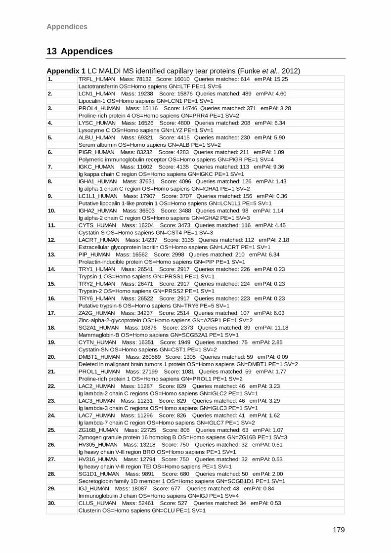
Appendices
179
13 Appendices
Appendix 1 LC MALDI MS identified capillary tear proteins (Funke et al., 2012)
1. TRFL_HUMAN Mass: 78132 Score: 16010 Queries matched: 614 emPAI: 15.25
Lactotransferrin OS=Homo sapiens GN=LTF PE=1 SV=6
2. LCN1_HUMAN Mass: 19238 Score: 15876 Queries matched: 489 emPAI: 4.60
Lipocalin-1 OS=Homo sapiens GN=LCN1 PE=1 SV=1
3. PROL4_HUMAN Mass: 15116 Score: 14746 Queries matched: 371 emPAI: 3.28
Proline-rich protein 4 OS=Homo sapiens GN=PRR4 PE=1 SV=2
4. LYSC_HUMAN Mass: 16526 Score: 4800 Queries matched: 208 emPAI: 6.34
Lysozyme C OS=Homo sapiens GN=LYZ PE=1 SV=1
5. ALBU_HUMAN Mass: 69321 Score: 4415 Queries matched: 230 emPAI: 5.90
Serum albumin OS=Homo sapiens GN=ALB PE=1 SV=2
6. PIGR_HUMAN Mass: 83232 Score: 4283 Queries matched: 211 emPAI: 1.09
Polymeric immunoglobulin receptor OS=Homo sapiens GN=PIGR PE=1 SV=4
7. IGKC_HUMAN Mass: 11602 Score: 4135 Queries matched: 113 emPAI: 9.36
Ig kappa chain C region OS=Homo sapiens GN=IGKC PE=1 SV=1
8. IGHA1_HUMAN Mass: 37631 Score: 4096 Queries matched: 126 emPAI: 1.43
Ig alpha-1 chain C region OS=Homo sapiens GN=IGHA1 PE=1 SV=2
9. LC1L1_HUMAN Mass: 17907 Score: 3707 Queries matched: 156 emPAI: 0.36
Putative lipocalin 1-like protein 1 OS=Homo sapiens GN=LCN1L1 PE=5 SV=1
10. IGHA2_HUMAN Mass: 36503 Score: 3488 Queries matched: 98 emPAI: 1.14
Ig alpha-2 chain C region OS=Homo sapiens GN=IGHA2 PE=1 SV=3
11. CYTS_HUMAN Mass: 16204 Score: 3473 Queries matched: 116 emPAI: 4.45
Cystatin-S OS=Homo sapiens GN=CST4 PE=1 SV=3
12. LACRT_HUMAN Mass: 14237 Score: 3135 Queries matched: 112 emPAI: 2.18
Extracellular glycoprotein lacritin OS=Homo sapiens GN=LACRT PE=1 SV=1
13. PIP_HUMAN Mass: 16562 Score: 2998 Queries matched: 210 emPAI: 6.34
Prolactin-inducible protein OS=Homo sapiens GN=PIP PE=1 SV=1
14. TRY1_HUMAN Mass: 26541 Score: 2917 Queries matched: 226 emPAI: 0.23
Trypsin-1 OS=Homo sapiens GN=PRSS1 PE=1 SV=1
15. TRY2_HUMAN Mass: 26471 Score: 2917 Queries matched: 224 emPAI: 0.23
Trypsin-2 OS=Homo sapiens GN=PRSS2 PE=1 SV=1
16. TRY6_HUMAN Mass: 26522 Score: 2917 Queries matched: 223 emPAI: 0.23
Putative trypsin-6 OS=Homo sapiens GN=TRY6 PE=5 SV=1
17. ZA2G_HUMAN Mass: 34237 Score: 2514 Queries matched: 107 emPAI: 6.03
Zinc-alpha-2-glycoprotein OS=Homo sapiens GN=AZGP1 PE=1 SV=2
18. SG2A1_HUMAN Mass: 10876 Score: 2373 Queries matched: 89 emPAI: 11.18
Mammaglobin-B OS=Homo sapiens GN=SCGB2A1 PE=1 SV=1
19. CYTN_HUMAN Mass: 16351 Score: 1949 Queries matched: 75 emPAI: 2.85
Cystatin-SN OS=Homo sapiens GN=CST1 PE=1 SV=2
20. DMBT1_HUMAN Mass: 260569 Score: 1305 Queries matched: 59 emPAI: 0.09
Deleted in malignant brain tumors 1 protein OS=Homo sapiens GN=DMBT1 PE=1 SV=2
21. PROL1_HUMAN Mass: 27199 Score: 1081 Queries matched: 59 emPAI: 1.77
Proline-rich protein 1 OS=Homo sapiens GN=PROL1 PE=1 SV=2
22. LAC2_HUMAN Mass: 11287 Score: 829 Queries matched: 46 emPAI: 3.23
Ig lambda-2 chain C regions OS=Homo sapiens GN=IGLC2 PE=1 SV=1
23. LAC3_HUMAN Mass: 11231 Score: 829 Queries matched: 46 emPAI: 3.29
Ig lambda-3 chain C regions OS=Homo sapiens GN=IGLC3 PE=1 SV=1
24. LAC7_HUMAN Mass: 11296 Score: 826 Queries matched: 41 emPAI: 1.62
Ig lambda-7 chain C region OS=Homo sapiens GN=IGLC7 PE=1 SV=2
25. ZG16B_HUMAN Mass: 22725 Score: 806 Queries matched: 63 emPAI: 1.07
Zymogen granule protein 16 homolog B OS=Homo sapiens GN=ZG16B PE=1 SV=3
26. HV305_HUMAN Mass: 13218 Score: 750 Queries matched: 32 emPAI: 0.51
Ig heavy chain V-III region BRO OS=Homo sapiens PE=1 SV=1
27. HV316_HUMAN Mass: 12794 Score: 750 Queries matched: 32 emPAI: 0.53
Ig heavy chain V-III region TEI OS=Homo sapiens PE=1 SV=1
28. SG1D1_HUMAN Mass: 9891 Score: 680 Queries matched: 50 emPAI: 2.00
Secretoglobin family 1D member 1 OS=Homo sapiens GN=SCGB1D1 PE=1 SV=1
29. IGJ_HUMAN Mass: 18087 Score: 677 Queries matched: 43 emPAI: 0.84
Immunoglobulin J chain OS=Homo sapiens GN=IGJ PE=1 SV=4
30. CLUS_HUMAN Mass: 52461 Score: 527 Queries matched: 34 emPAI: 0.53
Clusterin OS=Homo sapiens GN=CLU PE=1 SV=1
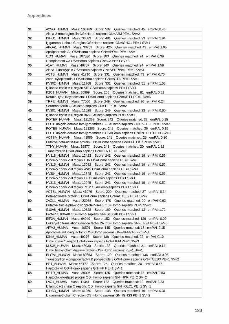
Appendices
180
31. A2MG_HUMAN Mass: 163189 Score: 507 Queries matched: 45 emPAI: 0.46
Alpha-2-macroglobulin OS=Homo sapiens GN=A2M PE=1 SV=2
32. IGHG1_HUMAN Mass: 36083 Score: 481 Queries matched: 23 emPAI: 1.94
Ig gamma-1 chain C region OS=Homo sapiens GN=IGHG1 PE=1 SV=1
33. APOA1_HUMAN Mass: 30759 Score: 425 Queries matched: 43 emPAI: 1.95
Apolipoprotein A-I OS=Homo sapiens GN=APOA1 PE=1 SV=1
34. CO3_HUMAN Mass: 187030 Score: 383 Queries matched: 74 emPAI: 0.39
Complement C3 OS=Homo sapiens GN=C3 PE=1 SV=2
35. A1AT_HUMAN Mass: 46707 Score: 340 Queries matched: 24 emPAI: 1.59
Alpha-1-antitrypsin OS=Homo sapiens GN=SERPINA1 PE=1 SV=3
36. ACTB_HUMAN Mass: 41710 Score: 331 Queries matched: 43 emPAI: 0.70
Actin, cytoplasmic 1 OS=Homo sapiens GN=ACTB PE=1 SV=1
37. KV302_HUMAN Mass: 11768 Score: 331 Queries matched: 51 emPAI: 1.53
Ig kappa chain V-III region SIE OS=Homo sapiens PE=1 SV=1
38. K2C1_HUMAN Mass: 65999 Score: 255 Queries matched: 81 emPAI: 0.81
Keratin, type II cytoskeletal 1 OS=Homo sapiens GN=KRT1 PE=1 SV=6
39. TRFE_HUMAN Mass: 77000 Score: 249 Queries matched: 36 emPAI: 0.24
Serotransferrin OS=Homo sapiens GN=TF PE=1 SV=2
40. KV301_HUMAN Mass: 11628 Score: 249 Queries matched: 33 emPAI: 0.60
Ig kappa chain V-III region B6 OS=Homo sapiens PE=1 SV=1
41. POTEF_HUMAN Mass: 121367 Score: 242 Queries matched: 37 emPAI: 0.15
POTE ankyrin domain family member F OS=Homo sapiens GN=POTEF PE=1 SV=2
42. POTEE_HUMAN Mass: 121286 Score: 242 Queries matched: 36 emPAI: 0.15
POTE ankyrin domain family member E OS=Homo sapiens GN=POTEE PE=1 SV=3
43. ACTBM_HUMAN Mass: 41989 Score: 241 Queries matched: 25 emPAI: 0.30
Putative beta-actin-like protein 3 OS=Homo sapiens GN=POTEKP PE=5 SV=1
44. TTHY_HUMAN Mass: 15877 Score: 241 Queries matched: 20 emPAI: 1.82
Transthyretin OS=Homo sapiens GN=TTR PE=1 SV=1
45. HV318_HUMAN Mass: 12423 Score: 241 Queries matched: 19 emPAI: 0.55
Ig heavy chain V-III region TUR OS=Homo sapiens PE=1 SV=1
46. HV315_HUMAN Mass: 13082 Score: 241 Queries matched: 19 emPAI: 0.52
Ig heavy chain V-III region WAS OS=Homo sapiens PE=1 SV=1
47. HV304_HUMAN Mass: 12348 Score: 241 Queries matched: 19 emPAI: 0.56
Ig heavy chain V-III region TIL OS=Homo sapiens PE=1 SV=1
48. HV313_HUMAN Mass: 12945 Score: 241 Queries matched: 19 emPAI: 0.52
Ig heavy chain V-III region POM OS=Homo sapiens PE=1 SV=1
49. ACTBL_HUMAN Mass: 41976 Score: 200 Queries matched: 27 emPAI: 0.14
Beta-actin-like protein 2 OS=Homo sapiens GN=ACTBL2 PE=1 SV=2
50. ZAGL1_HUMAN Mass: 22965 Score: 178 Queries matched: 20 emPAI: 0.62
Putative zinc-alpha-2-glycoprotein-like 1 OS=Homo sapiens PE=5 SV=2
51. S10A8_HUMAN Mass: 10828 Score: 169 Queries matched: 13 emPAI: 1.72
Protein S100-A8 OS=Homo sapiens GN=S100A8 PE=1 SV=1
52. EIF2A_HUMAN Mass: 64949 Score: 152 Queries matched: 126 emPAI: 0.09
Eukaryotic translation initiation factor 2A OS=Homo sapiens GN=EIF2A PE=1 SV=3
53. AIFM2_HUMAN Mass: 40501 Score: 145 Queries matched: 15 emPAI: 0.15
Apoptosis-inducing factor 2 OS=Homo sapiens GN=AIFM2 PE=2 SV=1
54. IGHM_HUMAN Mass: 49276 Score: 138 Queries matched: 22 emPAI: 0.12
Ig mu chain C region OS=Homo sapiens GN=IGHM PE=1 SV=3
55. MUCB_HUMAN Mass: 43030 Score: 138 Queries matched: 21 emPAI: 0.14
Ig mu heavy chain disease protein OS=Homo sapiens PE=1 SV=1
56. ELOA1_HUMAN Mass: 89853 Score: 129 Queries matched: 136 emPAI: 0.06
Transcription elongation factor B polypeptide 3 OS=Homo sapiens GN=TCEB3 PE=1 SV=2
57. HPT_HUMAN Mass: 45177 Score: 125 Queries matched: 20 emPAI: 0.45
Haptoglobin OS=Homo sapiens GN=HP PE=1 SV=1
58. HPTR_HUMAN Mass: 39005 Score: 125 Queries matched: 13 emPAI: 0.53
Haptoglobin-related protein OS=Homo sapiens GN=HPR PE=2 SV=2
59. LAC1_HUMAN Mass: 11341 Score: 122 Queries matched: 19 emPAI: 3.23
Ig lambda-1 chain C regions OS=Homo sapiens GN=IGLC1 PE=1 SV=1
60. IGHG3_HUMAN Mass: 41260 Score: 108 Queries matched: 16 emPAI: 0.31
Ig gamma-3 chain C region OS=Homo sapiens GN=IGHG3 PE=1 SV=2
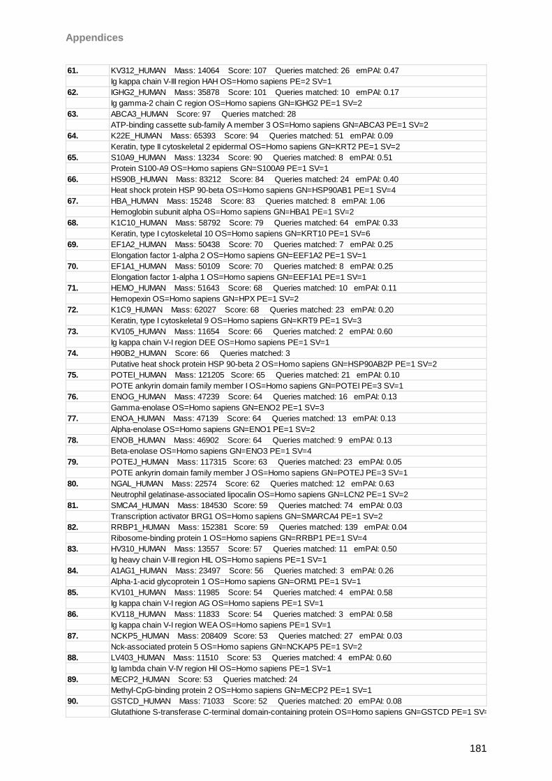
Appendices
181
61. KV312_HUMAN Mass: 14064 Score: 107 Queries matched: 26 emPAI: 0.47
Ig kappa chain V-III region HAH OS=Homo sapiens PE=2 SV=1
62. IGHG2_HUMAN Mass: 35878 Score: 101 Queries matched: 10 emPAI: 0.17
Ig gamma-2 chain C region OS=Homo sapiens GN=IGHG2 PE=1 SV=2
63. ABCA3_HUMAN Score: 97 Queries matched: 28
ATP-binding cassette sub-family A member 3 OS=Homo sapiens GN=ABCA3 PE=1 SV=2
64. K22E_HUMAN Mass: 65393 Score: 94 Queries matched: 51 emPAI: 0.09
Keratin, type II cytoskeletal 2 epidermal OS=Homo sapiens GN=KRT2 PE=1 SV=2
65. S10A9_HUMAN Mass: 13234 Score: 90 Queries matched: 8 emPAI: 0.51
Protein S100-A9 OS=Homo sapiens GN=S100A9 PE=1 SV=1
66. HS90B_HUMAN Mass: 83212 Score: 84 Queries matched: 24 emPAI: 0.40
Heat shock protein HSP 90-beta OS=Homo sapiens GN=HSP90AB1 PE=1 SV=4
67. HBA_HUMAN Mass: 15248 Score: 83 Queries matched: 8 emPAI: 1.06
Hemoglobin subunit alpha OS=Homo sapiens GN=HBA1 PE=1 SV=2
68. K1C10_HUMAN Mass: 58792 Score: 79 Queries matched: 64 emPAI: 0.33
Keratin, type I cytoskeletal 10 OS=Homo sapiens GN=KRT10 PE=1 SV=6
69. EF1A2_HUMAN Mass: 50438 Score: 70 Queries matched: 7 emPAI: 0.25
Elongation factor 1-alpha 2 OS=Homo sapiens GN=EEF1A2 PE=1 SV=1
70. EF1A1_HUMAN Mass: 50109 Score: 70 Queries matched: 8 emPAI: 0.25
Elongation factor 1-alpha 1 OS=Homo sapiens GN=EEF1A1 PE=1 SV=1
71. HEMO_HUMAN Mass: 51643 Score: 68 Queries matched: 10 emPAI: 0.11
Hemopexin OS=Homo sapiens GN=HPX PE=1 SV=2
72. K1C9_HUMAN Mass: 62027 Score: 68 Queries matched: 23 emPAI: 0.20
Keratin, type I cytoskeletal 9 OS=Homo sapiens GN=KRT9 PE=1 SV=3
73. KV105_HUMAN Mass: 11654 Score: 66 Queries matched: 2 emPAI: 0.60
Ig kappa chain V-I region DEE OS=Homo sapiens PE=1 SV=1
74. H90B2_HUMAN Score: 66 Queries matched: 3
Putative heat shock protein HSP 90-beta 2 OS=Homo sapiens GN=HSP90AB2P PE=1 SV=2
75. POTEI_HUMAN Mass: 121205 Score: 65 Queries matched: 21 emPAI: 0.10
POTE ankyrin domain family member I OS=Homo sapiens GN=POTEI PE=3 SV=1
76. ENOG_HUMAN Mass: 47239 Score: 64 Queries matched: 16 emPAI: 0.13
Gamma-enolase OS=Homo sapiens GN=ENO2 PE=1 SV=3
77. ENOA_HUMAN Mass: 47139 Score: 64 Queries matched: 13 emPAI: 0.13
Alpha-enolase OS=Homo sapiens GN=ENO1 PE=1 SV=2
78. ENOB_HUMAN Mass: 46902 Score: 64 Queries matched: 9 emPAI: 0.13
Beta-enolase OS=Homo sapiens GN=ENO3 PE=1 SV=4
79. POTEJ_HUMAN Mass: 117315 Score: 63 Queries matched: 23 emPAI: 0.05
POTE ankyrin domain family member J OS=Homo sapiens GN=POTEJ PE=3 SV=1
80. NGAL_HUMAN Mass: 22574 Score: 62 Queries matched: 12 emPAI: 0.63
Neutrophil gelatinase-associated lipocalin OS=Homo sapiens GN=LCN2 PE=1 SV=2
81. SMCA4_HUMAN Mass: 184530 Score: 59 Queries matched: 74 emPAI: 0.03
Transcription activator BRG1 OS=Homo sapiens GN=SMARCA4 PE=1 SV=2
82. RRBP1_HUMAN Mass: 152381 Score: 59 Queries matched: 139 emPAI: 0.04
Ribosome-binding protein 1 OS=Homo sapiens GN=RRBP1 PE=1 SV=4
83. HV310_HUMAN Mass: 13557 Score: 57 Queries matched: 11 emPAI: 0.50
Ig heavy chain V-III region HIL OS=Homo sapiens PE=1 SV=1
84. A1AG1_HUMAN Mass: 23497 Score: 56 Queries matched: 3 emPAI: 0.26
Alpha-1-acid glycoprotein 1 OS=Homo sapiens GN=ORM1 PE=1 SV=1
85. KV101_HUMAN Mass: 11985 Score: 54 Queries matched: 4 emPAI: 0.58
Ig kappa chain V-I region AG OS=Homo sapiens PE=1 SV=1
86. KV118_HUMAN Mass: 11833 Score: 54 Queries matched: 3 emPAI: 0.58
Ig kappa chain V-I region WEA OS=Homo sapiens PE=1 SV=1
87. NCKP5_HUMAN Mass: 208409 Score: 53 Queries matched: 27 emPAI: 0.03
Nck-associated protein 5 OS=Homo sapiens GN=NCKAP5 PE=1 SV=2
88. LV403_HUMAN Mass: 11510 Score: 53 Queries matched: 4 emPAI: 0.60
Ig lambda chain V-IV region Hil OS=Homo sapiens PE=1 SV=1
89. MECP2_HUMAN Score: 53 Queries matched: 24
Methyl-CpG-binding protein 2 OS=Homo sapiens GN=MECP2 PE=1 SV=1
90. GSTCD_HUMAN Mass: 71033 Score: 52 Queries matched: 20 emPAI: 0.08
Glutathione S-transferase C-terminal domain-containing protein OS=Homo sapiens GN=GSTCD PE=1 SV=2
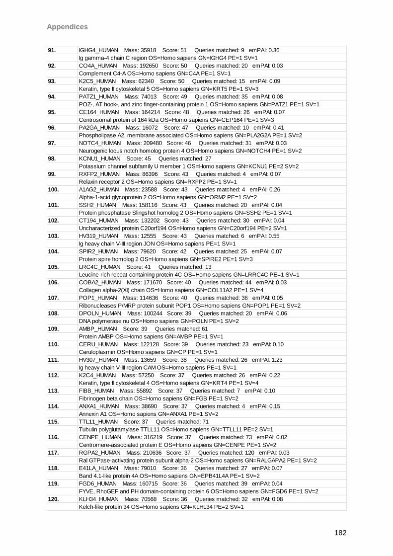
Appendices
182
91. IGHG4_HUMAN Mass: 35918 Score: 51 Queries matched: 9 emPAI: 0.36
Ig gamma-4 chain C region OS=Homo sapiens GN=IGHG4 PE=1 SV=1
92. CO4A_HUMAN Mass: 192650 Score: 50 Queries matched: 20 emPAI: 0.03
Complement C4-A OS=Homo sapiens GN=C4A PE=1 SV=1
93. K2C5_HUMAN Mass: 62340 Score: 50 Queries matched: 15 emPAI: 0.09
Keratin, type II cytoskeletal 5 OS=Homo sapiens GN=KRT5 PE=1 SV=3
94. PATZ1_HUMAN Mass: 74013 Score: 49 Queries matched: 35 emPAI: 0.08
POZ-, AT hook-, and zinc finger-containing protein 1 OS=Homo sapiens GN=PATZ1 PE=1 SV=1
95. CE164_HUMAN Mass: 164214 Score: 48 Queries matched: 26 emPAI: 0.07
Centrosomal protein of 164 kDa OS=Homo sapiens GN=CEP164 PE=1 SV=3
96. PA2GA_HUMAN Mass: 16072 Score: 47 Queries matched: 10 emPAI: 0.41
Phospholipase A2, membrane associated OS=Homo sapiens GN=PLA2G2A PE=1 SV=2
97. NOTC4_HUMAN Mass: 209480 Score: 46 Queries matched: 31 emPAI: 0.03
Neurogenic locus notch homolog protein 4 OS=Homo sapiens GN=NOTCH4 PE=1 SV=2
98. KCNU1_HUMAN Score: 45 Queries matched: 27
Potassium channel subfamily U member 1 OS=Homo sapiens GN=KCNU1 PE=2 SV=2
99. RXFP2_HUMAN Mass: 86396 Score: 43 Queries matched: 4 emPAI: 0.07
Relaxin receptor 2 OS=Homo sapiens GN=RXFP2 PE=1 SV=1
100. A1AG2_HUMAN Mass: 23588 Score: 43 Queries matched: 4 emPAI: 0.26
Alpha-1-acid glycoprotein 2 OS=Homo sapiens GN=ORM2 PE=1 SV=2
101. SSH2_HUMAN Mass: 158116 Score: 43 Queries matched: 20 emPAI: 0.04
Protein phosphatase Slingshot homolog 2 OS=Homo sapiens GN=SSH2 PE=1 SV=1
102. CT194_HUMAN Mass: 132202 Score: 43 Queries matched: 30 emPAI: 0.04
Uncharacterized protein C20orf194 OS=Homo sapiens GN=C20orf194 PE=2 SV=1
103. HV319_HUMAN Mass: 12555 Score: 43 Queries matched: 6 emPAI: 0.55
Ig heavy chain V-III region JON OS=Homo sapiens PE=1 SV=1
104. SPIR2_HUMAN Mass: 79620 Score: 42 Queries matched: 25 emPAI: 0.07
Protein spire homolog 2 OS=Homo sapiens GN=SPIRE2 PE=1 SV=3
105. LRC4C_HUMAN Score: 41 Queries matched: 13
Leucine-rich repeat-containing protein 4C OS=Homo sapiens GN=LRRC4C PE=1 SV=1
106. COBA2_HUMAN Mass: 171670 Score: 40 Queries matched: 44 emPAI: 0.03
Collagen alpha-2(XI) chain OS=Homo sapiens GN=COL11A2 PE=1 SV=4
107. POP1_HUMAN Mass: 114636 Score: 40 Queries matched: 36 emPAI: 0.05
Ribonucleases P/MRP protein subunit POP1 OS=Homo sapiens GN=POP1 PE=1 SV=2
108. DPOLN_HUMAN Mass: 100244 Score: 39 Queries matched: 20 emPAI: 0.06
DNA polymerase nu OS=Homo sapiens GN=POLN PE=1 SV=2
109. AMBP_HUMAN Score: 39 Queries matched: 61
Protein AMBP OS=Homo sapiens GN=AMBP PE=1 SV=1
110. CERU_HUMAN Mass: 122128 Score: 39 Queries matched: 23 emPAI: 0.10
Ceruloplasmin OS=Homo sapiens GN=CP PE=1 SV=1
111. HV307_HUMAN Mass: 13659 Score: 38 Queries matched: 26 emPAI: 1.23
Ig heavy chain V-III region CAM OS=Homo sapiens PE=1 SV=1
112. K2C4_HUMAN Mass: 57250 Score: 37 Queries matched: 26 emPAI: 0.22
Keratin, type II cytoskeletal 4 OS=Homo sapiens GN=KRT4 PE=1 SV=4
113. FIBB_HUMAN Mass: 55892 Score: 37 Queries matched: 7 emPAI: 0.10
Fibrinogen beta chain OS=Homo sapiens GN=FGB PE=1 SV=2
114. ANXA1_HUMAN Mass: 38690 Score: 37 Queries matched: 4 emPAI: 0.15
Annexin A1 OS=Homo sapiens GN=ANXA1 PE=1 SV=2
115. TTL11_HUMAN Score: 37 Queries matched: 71
Tubulin polyglutamylase TTLL11 OS=Homo sapiens GN=TTLL11 PE=2 SV=1
116. CENPE_HUMAN Mass: 316219 Score: 37 Queries matched: 73 emPAI: 0.02
Centromere-associated protein E OS=Homo sapiens GN=CENPE PE=1 SV=2
117. RGPA2_HUMAN Mass: 210636 Score: 37 Queries matched: 120 emPAI: 0.03
Ral GTPase-activating protein subunit alpha-2 OS=Homo sapiens GN=RALGAPA2 PE=1 SV=2
118. E41LA_HUMAN Mass: 79010 Score: 36 Queries matched: 27 emPAI: 0.07
Band 4.1-like protein 4A OS=Homo sapiens GN=EPB41L4A PE=1 SV=2
119. FGD6_HUMAN Mass: 160715 Score: 36 Queries matched: 39 emPAI: 0.04
FYVE, RhoGEF and PH domain-containing protein 6 OS=Homo sapiens GN=FGD6 PE=1 SV=2
120. KLH34_HUMAN Mass: 70568 Score: 36 Queries matched: 32 emPAI: 0.08
Kelch-like protein 34 OS=Homo sapiens GN=KLHL34 PE=2 SV=1
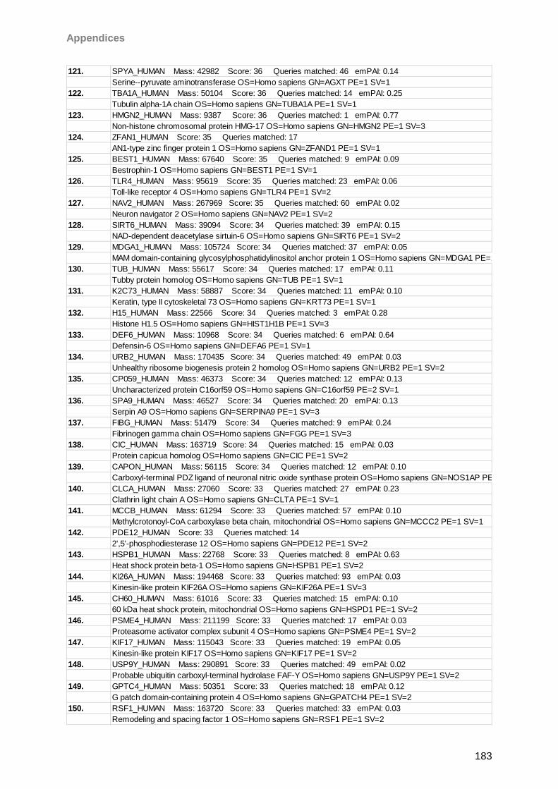
Appendices
183
121. SPYA_HUMAN Mass: 42982 Score: 36 Queries matched: 46 emPAI: 0.14
Serine--pyruvate aminotransferase OS=Homo sapiens GN=AGXT PE=1 SV=1
122. TBA1A_HUMAN Mass: 50104 Score: 36 Queries matched: 14 emPAI: 0.25
Tubulin alpha-1A chain OS=Homo sapiens GN=TUBA1A PE=1 SV=1
123. HMGN2_HUMAN Mass: 9387 Score: 36 Queries matched: 1 emPAI: 0.77
Non-histone chromosomal protein HMG-17 OS=Homo sapiens GN=HMGN2 PE=1 SV=3
124. ZFAN1_HUMAN Score: 35 Queries matched: 17
AN1-type zinc finger protein 1 OS=Homo sapiens GN=ZFAND1 PE=1 SV=1
125. BEST1_HUMAN Mass: 67640 Score: 35 Queries matched: 9 emPAI: 0.09
Bestrophin-1 OS=Homo sapiens GN=BEST1 PE=1 SV=1
126. TLR4_HUMAN Mass: 95619 Score: 35 Queries matched: 23 emPAI: 0.06
Toll-like receptor 4 OS=Homo sapiens GN=TLR4 PE=1 SV=2
127. NAV2_HUMAN Mass: 267969 Score: 35 Queries matched: 60 emPAI: 0.02
Neuron navigator 2 OS=Homo sapiens GN=NAV2 PE=1 SV=2
128. SIRT6_HUMAN Mass: 39094 Score: 34 Queries matched: 39 emPAI: 0.15
NAD-dependent deacetylase sirtuin-6 OS=Homo sapiens GN=SIRT6 PE=1 SV=2
129. MDGA1_HUMAN Mass: 105724 Score: 34 Queries matched: 37 emPAI: 0.05
MAM domain-containing glycosylphosphatidylinositol anchor protein 1 OS=Homo sapiens GN=MDGA1 PE=1 SV=1
130. TUB_HUMAN Mass: 55617 Score: 34 Queries matched: 17 emPAI: 0.11
Tubby protein homolog OS=Homo sapiens GN=TUB PE=1 SV=1
131. K2C73_HUMAN Mass: 58887 Score: 34 Queries matched: 11 emPAI: 0.10
Keratin, type II cytoskeletal 73 OS=Homo sapiens GN=KRT73 PE=1 SV=1
132. H15_HUMAN Mass: 22566 Score: 34 Queries matched: 3 emPAI: 0.28
Histone H1.5 OS=Homo sapiens GN=HIST1H1B PE=1 SV=3
133. DEF6_HUMAN Mass: 10968 Score: 34 Queries matched: 6 emPAI: 0.64
Defensin-6 OS=Homo sapiens GN=DEFA6 PE=1 SV=1
134. URB2_HUMAN Mass: 170435 Score: 34 Queries matched: 49 emPAI: 0.03
Unhealthy ribosome biogenesis protein 2 homolog OS=Homo sapiens GN=URB2 PE=1 SV=2
135. CP059_HUMAN Mass: 46373 Score: 34 Queries matched: 12 emPAI: 0.13
Uncharacterized protein C16orf59 OS=Homo sapiens GN=C16orf59 PE=2 SV=1
136. SPA9_HUMAN Mass: 46527 Score: 34 Queries matched: 20 emPAI: 0.13
Serpin A9 OS=Homo sapiens GN=SERPINA9 PE=1 SV=3
137. FIBG_HUMAN Mass: 51479 Score: 34 Queries matched: 9 emPAI: 0.24
Fibrinogen gamma chain OS=Homo sapiens GN=FGG PE=1 SV=3
138. CIC_HUMAN Mass: 163719 Score: 34 Queries matched: 15 emPAI: 0.03
Protein capicua homolog OS=Homo sapiens GN=CIC PE=1 SV=2
139. CAPON_HUMAN Mass: 56115 Score: 34 Queries matched: 12 emPAI: 0.10
Carboxyl-terminal PDZ ligand of neuronal nitric oxide synthase protein OS=Homo sapiens GN=NOS1AP PE=1 SV=3
140. CLCA_HUMAN Mass: 27060 Score: 33 Queries matched: 27 emPAI: 0.23
Clathrin light chain A OS=Homo sapiens GN=CLTA PE=1 SV=1
141. MCCB_HUMAN Mass: 61294 Score: 33 Queries matched: 57 emPAI: 0.10
Methylcrotonoyl-CoA carboxylase beta chain, mitochondrial OS=Homo sapiens GN=MCCC2 PE=1 SV=1
142. PDE12_HUMAN Score: 33 Queries matched: 14
2',5'-phosphodiesterase 12 OS=Homo sapiens GN=PDE12 PE=1 SV=2
143. HSPB1_HUMAN Mass: 22768 Score: 33 Queries matched: 8 emPAI: 0.63
Heat shock protein beta-1 OS=Homo sapiens GN=HSPB1 PE=1 SV=2
144. KI26A_HUMAN Mass: 194468 Score: 33 Queries matched: 93 emPAI: 0.03
Kinesin-like protein KIF26A OS=Homo sapiens GN=KIF26A PE=1 SV=3
145. CH60_HUMAN Mass: 61016 Score: 33 Queries matched: 15 emPAI: 0.10
60 kDa heat shock protein, mitochondrial OS=Homo sapiens GN=HSPD1 PE=1 SV=2
146. PSME4_HUMAN Mass: 211199 Score: 33 Queries matched: 17 emPAI: 0.03
Proteasome activator complex subunit 4 OS=Homo sapiens GN=PSME4 PE=1 SV=2
147. KIF17_HUMAN Mass: 115043 Score: 33 Queries matched: 19 emPAI: 0.05
Kinesin-like protein KIF17 OS=Homo sapiens GN=KIF17 PE=1 SV=2
148. USP9Y_HUMAN Mass: 290891 Score: 33 Queries matched: 49 emPAI: 0.02
Probable ubiquitin carboxyl-terminal hydrolase FAF-Y OS=Homo sapiens GN=USP9Y PE=1 SV=2
149. GPTC4_HUMAN Mass: 50351 Score: 33 Queries matched: 18 emPAI: 0.12
G patch domain-containing protein 4 OS=Homo sapiens GN=GPATCH4 PE=1 SV=2
150. RSF1_HUMAN Mass: 163720 Score: 33 Queries matched: 33 emPAI: 0.03
Remodeling and spacing factor 1 OS=Homo sapiens GN=RSF1 PE=1 SV=2
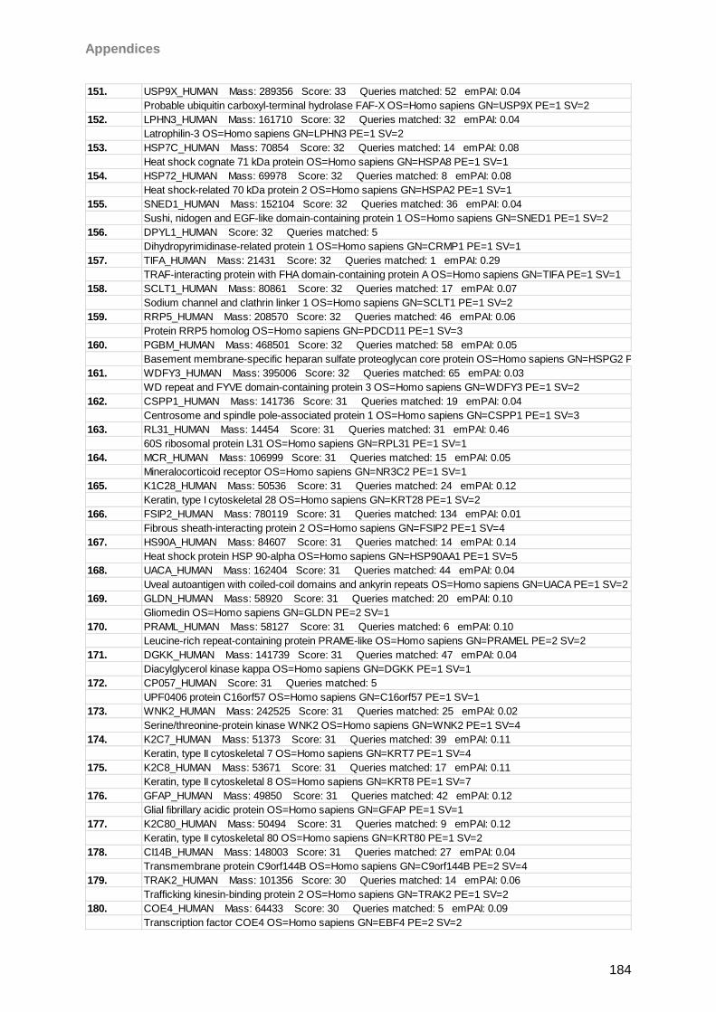
Appendices
184
151. USP9X_HUMAN Mass: 289356 Score: 33 Queries matched: 52 emPAI: 0.04
Probable ubiquitin carboxyl-terminal hydrolase FAF-X OS=Homo sapiens GN=USP9X PE=1 SV=2
152. LPHN3_HUMAN Mass: 161710 Score: 32 Queries matched: 32 emPAI: 0.04
Latrophilin-3 OS=Homo sapiens GN=LPHN3 PE=1 SV=2
153. HSP7C_HUMAN Mass: 70854 Score: 32 Queries matched: 14 emPAI: 0.08
Heat shock cognate 71 kDa protein OS=Homo sapiens GN=HSPA8 PE=1 SV=1
154. HSP72_HUMAN Mass: 69978 Score: 32 Queries matched: 8 emPAI: 0.08
Heat shock-related 70 kDa protein 2 OS=Homo sapiens GN=HSPA2 PE=1 SV=1
155. SNED1_HUMAN Mass: 152104 Score: 32 Queries matched: 36 emPAI: 0.04
Sushi, nidogen and EGF-like domain-containing protein 1 OS=Homo sapiens GN=SNED1 PE=1 SV=2
156. DPYL1_HUMAN Score: 32 Queries matched: 5
Dihydropyrimidinase-related protein 1 OS=Homo sapiens GN=CRMP1 PE=1 SV=1
157. TIFA_HUMAN Mass: 21431 Score: 32 Queries matched: 1 emPAI: 0.29
TRAF-interacting protein with FHA domain-containing protein A OS=Homo sapiens GN=TIFA PE=1 SV=1
158. SCLT1_HUMAN Mass: 80861 Score: 32 Queries matched: 17 emPAI: 0.07
Sodium channel and clathrin linker 1 OS=Homo sapiens GN=SCLT1 PE=1 SV=2
159. RRP5_HUMAN Mass: 208570 Score: 32 Queries matched: 46 emPAI: 0.06
Protein RRP5 homolog OS=Homo sapiens GN=PDCD11 PE=1 SV=3
160. PGBM_HUMAN Mass: 468501 Score: 32 Queries matched: 58 emPAI: 0.05
Basement membrane-specific heparan sulfate proteoglycan core protein OS=Homo sapiens GN=HSPG2 PE=1 SV=3
161. WDFY3_HUMAN Mass: 395006 Score: 32 Queries matched: 65 emPAI: 0.03
WD repeat and FYVE domain-containing protein 3 OS=Homo sapiens GN=WDFY3 PE=1 SV=2
162. CSPP1_HUMAN Mass: 141736 Score: 31 Queries matched: 19 emPAI: 0.04
Centrosome and spindle pole-associated protein 1 OS=Homo sapiens GN=CSPP1 PE=1 SV=3
163. RL31_HUMAN Mass: 14454 Score: 31 Queries matched: 31 emPAI: 0.46
60S ribosomal protein L31 OS=Homo sapiens GN=RPL31 PE=1 SV=1
164. MCR_HUMAN Mass: 106999 Score: 31 Queries matched: 15 emPAI: 0.05
Mineralocorticoid receptor OS=Homo sapiens GN=NR3C2 PE=1 SV=1
165. K1C28_HUMAN Mass: 50536 Score: 31 Queries matched: 24 emPAI: 0.12
Keratin, type I cytoskeletal 28 OS=Homo sapiens GN=KRT28 PE=1 SV=2
166. FSIP2_HUMAN Mass: 780119 Score: 31 Queries matched: 134 emPAI: 0.01
Fibrous sheath-interacting protein 2 OS=Homo sapiens GN=FSIP2 PE=1 SV=4
167. HS90A_HUMAN Mass: 84607 Score: 31 Queries matched: 14 emPAI: 0.14
Heat shock protein HSP 90-alpha OS=Homo sapiens GN=HSP90AA1 PE=1 SV=5
168. UACA_HUMAN Mass: 162404 Score: 31 Queries matched: 44 emPAI: 0.04
Uveal autoantigen with coiled-coil domains and ankyrin repeats OS=Homo sapiens GN=UACA PE=1 SV=2
169. GLDN_HUMAN Mass: 58920 Score: 31 Queries matched: 20 emPAI: 0.10
Gliomedin OS=Homo sapiens GN=GLDN PE=2 SV=1
170. PRAML_HUMAN Mass: 58127 Score: 31 Queries matched: 6 emPAI: 0.10
Leucine-rich repeat-containing protein PRAME-like OS=Homo sapiens GN=PRAMEL PE=2 SV=2
171. DGKK_HUMAN Mass: 141739 Score: 31 Queries matched: 47 emPAI: 0.04
Diacylglycerol kinase kappa OS=Homo sapiens GN=DGKK PE=1 SV=1
172. CP057_HUMAN Score: 31 Queries matched: 5
UPF0406 protein C16orf57 OS=Homo sapiens GN=C16orf57 PE=1 SV=1
173. WNK2_HUMAN Mass: 242525 Score: 31 Queries matched: 25 emPAI: 0.02
Serine/threonine-protein kinase WNK2 OS=Homo sapiens GN=WNK2 PE=1 SV=4
174. K2C7_HUMAN Mass: 51373 Score: 31 Queries matched: 39 emPAI: 0.11
Keratin, type II cytoskeletal 7 OS=Homo sapiens GN=KRT7 PE=1 SV=4
175. K2C8_HUMAN Mass: 53671 Score: 31 Queries matched: 17 emPAI: 0.11
Keratin, type II cytoskeletal 8 OS=Homo sapiens GN=KRT8 PE=1 SV=7
176. GFAP_HUMAN Mass: 49850 Score: 31 Queries matched: 42 emPAI: 0.12
Glial fibrillary acidic protein OS=Homo sapiens GN=GFAP PE=1 SV=1
177. K2C80_HUMAN Mass: 50494 Score: 31 Queries matched: 9 emPAI: 0.12
Keratin, type II cytoskeletal 80 OS=Homo sapiens GN=KRT80 PE=1 SV=2
178. CI14B_HUMAN Mass: 148003 Score: 31 Queries matched: 27 emPAI: 0.04
Transmembrane protein C9orf144B OS=Homo sapiens GN=C9orf144B PE=2 SV=4
179. TRAK2_HUMAN Mass: 101356 Score: 30 Queries matched: 14 emPAI: 0.06
Trafficking kinesin-binding protein 2 OS=Homo sapiens GN=TRAK2 PE=1 SV=2
180. COE4_HUMAN Mass: 64433 Score: 30 Queries matched: 5 emPAI: 0.09
Transcription factor COE4 OS=Homo sapiens GN=EBF4 PE=2 SV=2
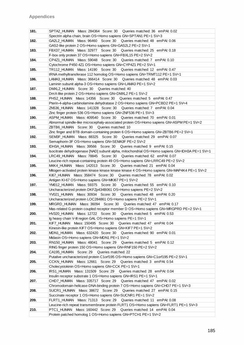
Appendices
185
181. SPTA2_HUMAN Mass: 284364 Score: 30 Queries matched: 36 emPAI: 0.02
Spectrin alpha chain, brain OS=Homo sapiens GN=SPTAN1 PE=1 SV=3
182. GA2L2_HUMAN Mass: 96460 Score: 30 Queries matched: 48 emPAI: 0.06
GAS2-like protein 2 OS=Homo sapiens GN=GAS2L2 PE=2 SV=1
183. FBX37_HUMAN Mass: 32977 Score: 30 Queries matched: 25 emPAI: 0.18
F-box only protein 37 OS=Homo sapiens GN=FBXL15 PE=2 SV=2
184. CP4Z1_HUMAN Mass: 59048 Score: 30 Queries matched: 7 emPAI: 0.10
Cytochrome P450 4Z1 OS=Homo sapiens GN=CYP4Z1 PE=2 SV=1
185. TR112_HUMAN Mass: 14190 Score: 30 Queries matched: 12 emPAI: 0.47
tRNA methyltransferase 112 homolog OS=Homo sapiens GN=TRMT112 PE=1 SV=1
186. LAMA3_HUMAN Mass: 366414 Score: 30 Queries matched: 48 emPAI: 0.03
Laminin subunit alpha-3 OS=Homo sapiens GN=LAMA3 PE=1 SV=2
187. DMXL2_HUMAN Score: 30 Queries matched: 40
DmX-like protein 2 OS=Homo sapiens GN=DMXL2 PE=1 SV=2
188. PHS2_HUMAN Mass: 14356 Score: 30 Queries matched: 5 emPAI: 0.47
Pterin-4-alpha-carbinolamine dehydratase 2 OS=Homo sapiens GN=PCBD2 PE=1 SV=4
189. ZN536_HUMAN Mass: 141328 Score: 30 Queries matched: 7 emPAI: 0.04
Zinc finger protein 536 OS=Homo sapiens GN=ZNF536 PE=1 SV=3
190. ASPM_HUMAN Mass: 409540 Score: 30 Queries matched: 70 emPAI: 0.01
Abnormal spindle-like microcephaly-associated protein OS=Homo sapiens GN=ASPM PE=1 SV=2
191. ZBTB6_HUMAN Score: 30 Queries matched: 10
Zinc finger and BTB domain-containing protein 6 OS=Homo sapiens GN=ZBTB6 PE=2 SV=1
192. SEM3F_HUMAN Mass: 88325 Score: 30 Queries matched: 29 emPAI: 0.07
Semaphorin-3F OS=Homo sapiens GN=SEMA3F PE=2 SV=2
193. IDH3A_HUMAN Mass: 39566 Score: 30 Queries matched: 9 emPAI: 0.15
Isocitrate dehydrogenase [NAD] subunit alpha, mitochondrial OS=Homo sapiens GN=IDH3A PE=1 SV=1
194. LRC49_HUMAN Mass: 78845 Score: 30 Queries matched: 62 emPAI: 0.07
Leucine-rich repeat-containing protein 49 OS=Homo sapiens GN=LRRC49 PE=2 SV=2
195. M4K4_HUMAN Mass: 142013 Score: 30 Queries matched: 21 emPAI: 0.04
Mitogen-activated protein kinase kinase kinase kinase 4 OS=Homo sapiens GN=MAP4K4 PE=1 SV=2
196. KI67_HUMAN Mass: 358474 Score: 30 Queries matched: 78 emPAI: 0.02
Antigen KI-67 OS=Homo sapiens GN=MKI67 PE=1 SV=2
197. YM012_HUMAN Mass: 59375 Score: 30 Queries matched: 55 emPAI: 0.10
Uncharacterized protein DKFZp434B061 OS=Homo sapiens PE=2 SV=2
198. YV021_HUMAN Mass: 30934 Score: 30 Queries matched: 48 emPAI: 0.20
Uncharacterized protein LOC284861 OS=Homo sapiens PE=2 SV=1
199. MRGRD_HUMAN Mass: 36094 Score: 30 Queries matched: 47 emPAI: 0.17
Mas-related G-protein coupled receptor member D OS=Homo sapiens GN=MRGPRD PE=2 SV=1
200. HV320_HUMAN Mass: 12722 Score: 30 Queries matched: 5 emPAI: 0.53
Ig heavy chain V-III region GAL OS=Homo sapiens PE=1 SV=1
201. KIF7_HUMAN Mass: 150495 Score: 30 Queries matched: 47 emPAI: 0.04
Kinesin-like protein KIF7 OS=Homo sapiens GN=KIF7 PE=1 SV=2
202. MDN1_HUMAN Mass: 632420 Score: 30 Queries matched: 90 emPAI: 0.01
Midasin OS=Homo sapiens GN=MDN1 PE=1 SV=2
203. RN150_HUMAN Mass: 48041 Score: 29 Queries matched: 5 emPAI: 0.12
RING finger protein 150 OS=Homo sapiens GN=RNF150 PE=2 SV=2
204. CA195_HUMAN Score: 29 Queries matched: 22
Putative uncharacterized protein C1orf195 OS=Homo sapiens GN=C1orf195 PE=2 SV=1
205. CCKN_HUMAN Mass: 12661 Score: 29 Queries matched: 3 emPAI: 0.54
Cholecystokinin OS=Homo sapiens GN=CCK PE=1 SV=1
206. IRS1_HUMAN Mass: 131509 Score: 29 Queries matched: 28 emPAI: 0.04
Insulin receptor substrate 1 OS=Homo sapiens GN=IRS1 PE=1 SV=1
207. CHD7_HUMAN Mass: 335717 Score: 29 Queries matched: 47 emPAI: 0.02
Chromodomain-helicase-DNA-binding protein 7 OS=Homo sapiens GN=CHD7 PE=1 SV=3
208. SUCR1_HUMAN Mass: 38672 Score: 29 Queries matched: 27 emPAI: 0.15
Succinate receptor 1 OS=Homo sapiens GN=SUCNR1 PE=1 SV=2
209. FLRT1_HUMAN Mass: 71313 Score: 29 Queries matched: 11 emPAI: 0.08
Leucine-rich repeat transmembrane protein FLRT1 OS=Homo sapiens GN=FLRT1 PE=1 SV=3
210. PTC1_HUMAN Mass: 160442 Score: 29 Queries matched: 14 emPAI: 0.04
Protein patched homolog 1 OS=Homo sapiens GN=PTCH1 PE=1 SV=2
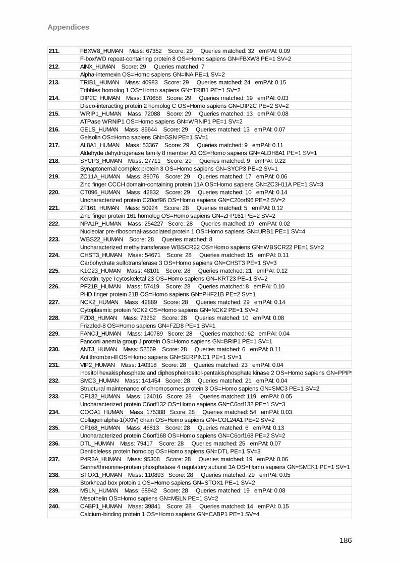
Appendices
186
211. FBXW8_HUMAN Mass: 67352 Score: 29 Queries matched: 32 emPAI: 0.09
F-box/WD repeat-containing protein 8 OS=Homo sapiens GN=FBXW8 PE=1 SV=2
212. AINX_HUMAN Score: 29 Queries matched: 7
Alpha-internexin OS=Homo sapiens GN=INA PE=1 SV=2
213. TRIB1_HUMAN Mass: 40983 Score: 29 Queries matched: 24 emPAI: 0.15
Tribbles homolog 1 OS=Homo sapiens GN=TRIB1 PE=1 SV=2
214. DIP2C_HUMAN Mass: 170658 Score: 29 Queries matched: 19 emPAI: 0.03
Disco-interacting protein 2 homolog C OS=Homo sapiens GN=DIP2C PE=2 SV=2
215. WRIP1_HUMAN Mass: 72088 Score: 29 Queries matched: 13 emPAI: 0.08
ATPase WRNIP1 OS=Homo sapiens GN=WRNIP1 PE=1 SV=2
216. GELS_HUMAN Mass: 85644 Score: 29 Queries matched: 13 emPAI: 0.07
Gelsolin OS=Homo sapiens GN=GSN PE=1 SV=1
217. AL8A1_HUMAN Mass: 53367 Score: 29 Queries matched: 9 emPAI: 0.11
Aldehyde dehydrogenase family 8 member A1 OS=Homo sapiens GN=ALDH8A1 PE=1 SV=1
218. SYCP3_HUMAN Mass: 27711 Score: 29 Queries matched: 9 emPAI: 0.22
Synaptonemal complex protein 3 OS=Homo sapiens GN=SYCP3 PE=2 SV=1
219. ZC11A_HUMAN Mass: 89076 Score: 29 Queries matched: 17 emPAI: 0.06
Zinc finger CCCH domain-containing protein 11A OS=Homo sapiens GN=ZC3H11A PE=1 SV=3
220. CT096_HUMAN Mass: 42832 Score: 29 Queries matched: 10 emPAI: 0.14
Uncharacterized protein C20orf96 OS=Homo sapiens GN=C20orf96 PE=2 SV=2
221. ZF161_HUMAN Mass: 50924 Score: 28 Queries matched: 5 emPAI: 0.12
Zinc finger protein 161 homolog OS=Homo sapiens GN=ZFP161 PE=2 SV=2
222. NPA1P_HUMAN Mass: 254227 Score: 28 Queries matched: 19 emPAI: 0.02
Nucleolar pre-ribosomal-associated protein 1 OS=Homo sapiens GN=URB1 PE=1 SV=4
223. WBS22_HUMAN Score: 28 Queries matched: 8
Uncharacterized methyltransferase WBSCR22 OS=Homo sapiens GN=WBSCR22 PE=1 SV=2
224. CHST3_HUMAN Mass: 54671 Score: 28 Queries matched: 15 emPAI: 0.11
Carbohydrate sulfotransferase 3 OS=Homo sapiens GN=CHST3 PE=1 SV=3
225. K1C23_HUMAN Mass: 48101 Score: 28 Queries matched: 21 emPAI: 0.12
Keratin, type I cytoskeletal 23 OS=Homo sapiens GN=KRT23 PE=1 SV=2
226. PF21B_HUMAN Mass: 57419 Score: 28 Queries matched: 8 emPAI: 0.10
PHD finger protein 21B OS=Homo sapiens GN=PHF21B PE=2 SV=1
227. NCK2_HUMAN Mass: 42889 Score: 28 Queries matched: 29 emPAI: 0.14
Cytoplasmic protein NCK2 OS=Homo sapiens GN=NCK2 PE=1 SV=2
228. FZD8_HUMAN Mass: 73252 Score: 28 Queries matched: 10 emPAI: 0.08
Frizzled-8 OS=Homo sapiens GN=FZD8 PE=1 SV=1
229. FANCJ_HUMAN Mass: 140789 Score: 28 Queries matched: 62 emPAI: 0.04
Fanconi anemia group J protein OS=Homo sapiens GN=BRIP1 PE=1 SV=1
230. ANT3_HUMAN Mass: 52569 Score: 28 Queries matched: 6 emPAI: 0.11
Antithrombin-III OS=Homo sapiens GN=SERPINC1 PE=1 SV=1
231. VIP2_HUMAN Mass: 140318 Score: 28 Queries matched: 23 emPAI: 0.04
Inositol hexakisphosphate and diphosphoinositol-pentakisphosphate kinase 2 OS=Homo sapiens GN=PPIP5K2 PE=1 SV=3
232. SMC3_HUMAN Mass: 141454 Score: 28 Queries matched: 21 emPAI: 0.04
Structural maintenance of chromosomes protein 3 OS=Homo sapiens GN=SMC3 PE=1 SV=2
233. CF132_HUMAN Mass: 124016 Score: 28 Queries matched: 119 emPAI: 0.05
Uncharacterized protein C6orf132 OS=Homo sapiens GN=C6orf132 PE=1 SV=3
234. COOA1_HUMAN Mass: 175388 Score: 28 Queries matched: 54 emPAI: 0.03
Collagen alpha-1(XXIV) chain OS=Homo sapiens GN=COL24A1 PE=2 SV=2
235. CF168_HUMAN Mass: 46813 Score: 28 Queries matched: 6 emPAI: 0.13
Uncharacterized protein C6orf168 OS=Homo sapiens GN=C6orf168 PE=2 SV=2
236. DTL_HUMAN Mass: 79417 Score: 28 Queries matched: 25 emPAI: 0.07
Denticleless protein homolog OS=Homo sapiens GN=DTL PE=1 SV=3
237. P4R3A_HUMAN Mass: 95308 Score: 28 Queries matched: 19 emPAI: 0.06
Serine/threonine-protein phosphatase 4 regulatory subunit 3A OS=Homo sapiens GN=SMEK1 PE=1 SV=1
238. STOX1_HUMAN Mass: 110893 Score: 28 Queries matched: 29 emPAI: 0.05
Storkhead-box protein 1 OS=Homo sapiens GN=STOX1 PE=1 SV=2
239. MSLN_HUMAN Mass: 68942 Score: 28 Queries matched: 19 emPAI: 0.08
Mesothelin OS=Homo sapiens GN=MSLN PE=1 SV=2
240. CABP1_HUMAN Mass: 39841 Score: 28 Queries matched: 14 emPAI: 0.15
Calcium-binding protein 1 OS=Homo sapiens GN=CABP1 PE=1 SV=4
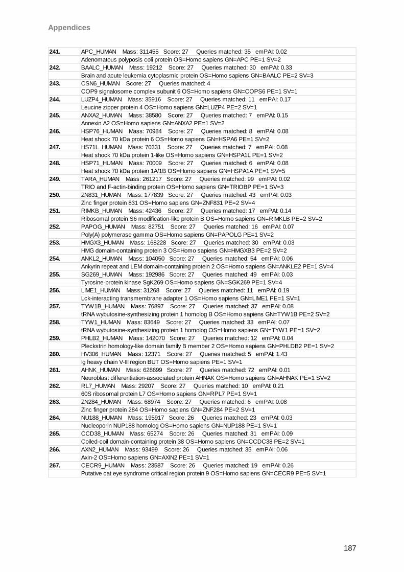
Appendices
187
241. APC_HUMAN Mass: 311455 Score: 27 Queries matched: 35 emPAI: 0.02
Adenomatous polyposis coli protein OS=Homo sapiens GN=APC PE=1 SV=2
242. BAALC_HUMAN Mass: 19212 Score: 27 Queries matched: 30 emPAI: 0.33
Brain and acute leukemia cytoplasmic protein OS=Homo sapiens GN=BAALC PE=2 SV=3
243. CSN6_HUMAN Score: 27 Queries matched: 4
COP9 signalosome complex subunit 6 OS=Homo sapiens GN=COPS6 PE=1 SV=1
244. LUZP4_HUMAN Mass: 35916 Score: 27 Queries matched: 11 emPAI: 0.17
Leucine zipper protein 4 OS=Homo sapiens GN=LUZP4 PE=2 SV=1
245. ANXA2_HUMAN Mass: 38580 Score: 27 Queries matched: 7 emPAI: 0.15
Annexin A2 OS=Homo sapiens GN=ANXA2 PE=1 SV=2
246. HSP76_HUMAN Mass: 70984 Score: 27 Queries matched: 8 emPAI: 0.08
Heat shock 70 kDa protein 6 OS=Homo sapiens GN=HSPA6 PE=1 SV=2
247. HS71L_HUMAN Mass: 70331 Score: 27 Queries matched: 7 emPAI: 0.08
Heat shock 70 kDa protein 1-like OS=Homo sapiens GN=HSPA1L PE=1 SV=2
248. HSP71_HUMAN Mass: 70009 Score: 27 Queries matched: 6 emPAI: 0.08
Heat shock 70 kDa protein 1A/1B OS=Homo sapiens GN=HSPA1A PE=1 SV=5
249. TARA_HUMAN Mass: 261217 Score: 27 Queries matched: 99 emPAI: 0.02
TRIO and F-actin-binding protein OS=Homo sapiens GN=TRIOBP PE=1 SV=3
250. ZN831_HUMAN Mass: 177839 Score: 27 Queries matched: 43 emPAI: 0.03
Zinc finger protein 831 OS=Homo sapiens GN=ZNF831 PE=2 SV=4
251. RIMKB_HUMAN Mass: 42436 Score: 27 Queries matched: 17 emPAI: 0.14
Ribosomal protein S6 modification-like protein B OS=Homo sapiens GN=RIMKLB PE=2 SV=2
252. PAPOG_HUMAN Mass: 82751 Score: 27 Queries matched: 16 emPAI: 0.07
Poly(A) polymerase gamma OS=Homo sapiens GN=PAPOLG PE=1 SV=2
253. HMGX3_HUMAN Mass: 168228 Score: 27 Queries matched: 30 emPAI: 0.03
HMG domain-containing protein 3 OS=Homo sapiens GN=HMGXB3 PE=2 SV=2
254. ANKL2_HUMAN Mass: 104050 Score: 27 Queries matched: 54 emPAI: 0.06
Ankyrin repeat and LEM domain-containing protein 2 OS=Homo sapiens GN=ANKLE2 PE=1 SV=4
255. SG269_HUMAN Mass: 192986 Score: 27 Queries matched: 49 emPAI: 0.03
Tyrosine-protein kinase SgK269 OS=Homo sapiens GN=SGK269 PE=1 SV=4
256. LIME1_HUMAN Mass: 31268 Score: 27 Queries matched: 11 emPAI: 0.19
Lck-interacting transmembrane adapter 1 OS=Homo sapiens GN=LIME1 PE=1 SV=1
257. TYW1B_HUMAN Mass: 76897 Score: 27 Queries matched: 37 emPAI: 0.08
tRNA wybutosine-synthesizing protein 1 homolog B OS=Homo sapiens GN=TYW1B PE=2 SV=2
258. TYW1_HUMAN Mass: 83649 Score: 27 Queries matched: 33 emPAI: 0.07
tRNA wybutosine-synthesizing protein 1 homolog OS=Homo sapiens GN=TYW1 PE=1 SV=2
259. PHLB2_HUMAN Mass: 142070 Score: 27 Queries matched: 12 emPAI: 0.04
Pleckstrin homology-like domain family B member 2 OS=Homo sapiens GN=PHLDB2 PE=1 SV=2
260. HV306_HUMAN Mass: 12371 Score: 27 Queries matched: 5 emPAI: 1.43
Ig heavy chain V-III region BUT OS=Homo sapiens PE=1 SV=1
261. AHNK_HUMAN Mass: 628699 Score: 27 Queries matched: 72 emPAI: 0.01
Neuroblast differentiation-associated protein AHNAK OS=Homo sapiens GN=AHNAK PE=1 SV=2
262. RL7_HUMAN Mass: 29207 Score: 27 Queries matched: 10 emPAI: 0.21
60S ribosomal protein L7 OS=Homo sapiens GN=RPL7 PE=1 SV=1
263. ZN284_HUMAN Mass: 68974 Score: 27 Queries matched: 6 emPAI: 0.08
Zinc finger protein 284 OS=Homo sapiens GN=ZNF284 PE=2 SV=1
264. NU188_HUMAN Mass: 195917 Score: 26 Queries matched: 23 emPAI: 0.03
Nucleoporin NUP188 homolog OS=Homo sapiens GN=NUP188 PE=1 SV=1
265. CCD38_HUMAN Mass: 65274 Score: 26 Queries matched: 31 emPAI: 0.09
Coiled-coil domain-containing protein 38 OS=Homo sapiens GN=CCDC38 PE=2 SV=1
266. AXN2_HUMAN Mass: 93499 Score: 26 Queries matched: 35 emPAI: 0.06
Axin-2 OS=Homo sapiens GN=AXIN2 PE=1 SV=1
267. CECR9_HUMAN Mass: 23587 Score: 26 Queries matched: 19 emPAI: 0.26
Putative cat eye syndrome critical region protein 9 OS=Homo sapiens GN=CECR9 PE=5 SV=1
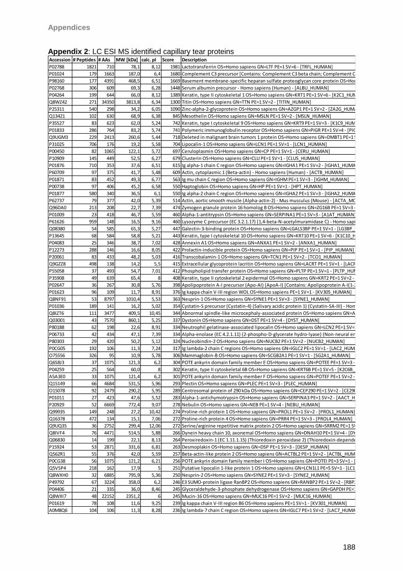
Appendices
188
Appendix 2: LC ESI MS identified capillary tear proteins
Accession # Peptides # AAs MW [kDa] calc. pI Score Description
P02788 1821 710 78,1 8,12 1981 Lactotransferrin OS=Homo sapiens GN=LTF PE=1 SV=6 - [TRFL_HUMAN]
P01024 179 1663 187,0 6,4 1680 Complement C3 precursor [Contains: Complement C3 beta chain; Complement C3 alpha chain; C3a anaphylatoxin; Complement C3b alpha' chain; Complement C3c alpha' chain fragment 1; Complement C3dg fragment; Complement C3g fragment; Complement C3d fragment; Complement C3f fragment; Complement C3c alpha' chain fragment 2] - Homo sapiens (Human) - [CO3_HUMAN]
P98160 177 4391 468,5 6,51 1669 Basement membrane-specific heparan sulfate proteoglycan core protein OS=Homo sapiens GN=HSPG2 PE=1 SV=4 - [PGBM_HUMAN]
P02768 306 609 69,3 6,28 1448 Serum albumin precursor - Homo sapiens (Human) - [ALBU_HUMAN]
P04264 199 644 66,0 8,12 1389 Keratin, type II cytoskeletal 1 OS=Homo sapiens GN=KRT1 PE=1 SV=6 - [K2C1_HUMAN]
Q8WZ42 271 34350 3813,8 6,34 1300 Titin OS=Homo sapiens GN=TTN PE=1 SV=2 - [TITIN_HUMAN]
P25311 540 298 34,2 6,05 1090 Zinc-alpha-2-glycoprotein OS=Homo sapiens GN=AZGP1 PE=1 SV=2 - [ZA2G_HUMAN]
Q13421 102 630 68,9 6,38 845 Mesothelin OS=Homo sapiens GN=MSLN PE=1 SV=2 - [MSLN_HUMAN]
P35527 83 623 62,0 5,24 742 Keratin, type I cytoskeletal 9 OS=Homo sapiens GN=KRT9 PE=1 SV=3 - [K1C9_HUMAN]
P01833 286 764 83,2 5,74 741 Polymeric immunoglobulin receptor OS=Homo sapiens GN=PIGR PE=1 SV=4 - [PIGR_HUMAN]
Q9UGM3 229 2413 260,6 5,44 718 Deleted in malignant brain tumors 1 protein OS=Homo sapiens GN=DMBT1 PE=1 SV=2 - [DMBT1_HUMAN]
P31025 706 176 19,2 5,58 704 Lipocalin-1 OS=Homo sapiens GN=LCN1 PE=1 SV=1 - [LCN1_HUMAN]
P00450 82 1065 122,1 5,72 697 Ceruloplasmin OS=Homo sapiens GN=CP PE=1 SV=1 - [CERU_HUMAN]
P10909 145 449 52,5 6,27 679 Clusterin OS=Homo sapiens GN=CLU PE=1 SV=1 - [CLUS_HUMAN]
P01876 710 353 37,6 6,51 615 Ig alpha-1 chain C region OS=Homo sapiens GN=IGHA1 PE=1 SV=2 - [IGHA1_HUMAN]
P60709 97 375 41,7 5,48 609 Actin, cytoplasmic 1 (Beta-actin) - Homo sapiens (Human) - [ACTB_HUMAN]
P01871 83 452 49,3 6,77 563 Ig mu chain C region OS=Homo sapiens GN=IGHM PE=1 SV=3 - [IGHM_HUMAN]
P00738 97 406 45,2 6,58 550 Haptoglobin OS=Homo sapiens GN=HP PE=1 SV=1 - [HPT_HUMAN]
P01877 580 340 36,5 6,1 550 Ig alpha-2 chain C region OS=Homo sapiens GN=IGHA2 PE=1 SV=3 - [IGHA2_HUMAN]
P62737 79 377 42,0 5,39 514 Actin, aortic smooth muscle (Alpha-actin-2) - Mus musculus (Mouse) - [ACTA_MOUSE]
Q96DA0 213 208 22,7 7,39 474 Zymogen granule protein 16 homolog B OS=Homo sapiens GN=ZG16B PE=1 SV=3 - [ZG16B_HUMAN]
P01009 23 418 46,7 5,59 460 Alpha-1-antitrypsin OS=Homo sapiens GN=SERPINA1 PE=1 SV=3 - [A1AT_HUMAN]
P61626 959 148 16,5 9,16 460 Lysozyme C precursor (EC 3.2.1.17) (1,4-beta-N-acetylmuramidase C) - Homo sapiens (Human) - [LYSC_HUMAN]
Q08380 54 585 65,3 5,27 447 Galectin-3-binding protein OS=Homo sapiens GN=LGALS3BP PE=1 SV=1 - [LG3BP_HUMAN]
P13645 68 584 58,8 5,21 443 Keratin, type I cytoskeletal 10 OS=Homo sapiens GN=KRT10 PE=1 SV=6 - [K1C10_HUMAN]
P04083 25 346 38,7 7,02 428 Annexin A1 OS=Homo sapiens GN=ANXA1 PE=1 SV=2 - [ANXA1_HUMAN]
P12273 288 146 16,6 8,05 422 Prolactin-inducible protein OS=Homo sapiens GN=PIP PE=1 SV=1 - [PIP_HUMAN]
P20061 83 433 48,2 5,03 416 Transcobalamin-1 OS=Homo sapiens GN=TCN1 PE=1 SV=2 - [TCO1_HUMAN]
Q9GZZ8 498 138 14,2 5,5 415 Extracellular glycoprotein lacritin OS=Homo sapiens GN=LACRT PE=1 SV=1 - [LACRT_HUMAN]
P55058 37 493 54,7 7,01 412 Phospholipid transfer protein OS=Homo sapiens GN=PLTP PE=1 SV=1 - [PLTP_HUMAN]
P35908 49 639 65,4 8 408 Keratin, type II cytoskeletal 2 epidermal OS=Homo sapiens GN=KRT2 PE=1 SV=2 - [K22E_HUMAN]
P02647 36 267 30,8 5,76 398 Apolipoprotein A-I precursor (Apo-AI) (ApoA-I) [Contains: Apolipoprotein A-I(1-242)] - Homo sapiens (Human) - [APOA1_HUMAN]
P01623 96 109 11,7 8,91 376 Ig kappa chain V-III region WOL OS=Homo sapiens PE=1 SV=1 - [KV305_HUMAN]
Q8NF91 53 8797 1010,4 5,53 363 Nesprin-1 OS=Homo sapiens GN=SYNE1 PE=1 SV=3 - [SYNE1_HUMAN]
P01036 189 141 16,2 5,02 354 Cystatin-S precursor (Cystatin-4) (Salivary acidic protein 1) (Cystatin-SA-III) - Homo sapiens (Human) - [CYTS_HUMAN]
Q8IZT6 111 3477 409,5 10,45 344 Abnormal spindle-like microcephaly-associated protein OS=Homo sapiens GN=ASPM PE=1 SV=2 - [ASPM_HUMAN]
Q03001 43 7570 860,1 5,25 337 Dystonin OS=Homo sapiens GN=DST PE=1 SV=4 - [DYST_HUMAN]
P80188 62 198 22,6 8,91 334 Neutrophil gelatinase-associated lipocalin OS=Homo sapiens GN=LCN2 PE=1 SV=2 - [NGAL_HUMAN]
P06733 42 434 47,1 7,39 334 Alpha-enolase (EC 4.2.1.11) (2-phospho-D-glycerate hydro-lyase) (Non-neural enolase) (NNE) (Enolase 1) (Phosphopyruvate hydratase) (C-myc promoter-binding protein) (MBP-1) (MPB-1) (Plasminogen-binding protein) - Homo sapiens (Human) - [ENOA_HUMAN]
P80303 29 420 50,2 5,12 324 Nucleobindin-2 OS=Homo sapiens GN=NUCB2 PE=1 SV=2 - [NUCB2_HUMAN]
P0CG05 192 106 11,3 7,24 317 Ig lambda-2 chain C regions OS=Homo sapiens GN=IGLC2 PE=1 SV=1 - [LAC2_HUMAN]
O75556 326 95 10,9 5,78 306 Mammaglobin-B OS=Homo sapiens GN=SCGB2A1 PE=1 SV=1 - [SG2A1_HUMAN]
Q6S8J3 37 1075 121,3 6,2 304 POTE ankyrin domain family member E OS=Homo sapiens GN=POTEE PE=1 SV=3 - [POTEE_HUMAN]
P04259 25 564 60,0 8 302 Keratin, type II cytoskeletal 6B OS=Homo sapiens GN=KRT6B PE=1 SV=5 - [K2C6B_HUMAN]
A5A3E0 33 1075 121,4 6,2 301 POTE ankyrin domain family member F OS=Homo sapiens GN=POTEF PE=1 SV=2 - [POTEF_HUMAN]
Q15149 66 4684 531,5 5,96 293 Plectin OS=Homo sapiens GN=PLEC PE=1 SV=3 - [PLEC_HUMAN]
O15078 92 2479 290,2 5,95 289 Centrosomal protein of 290 kDa OS=Homo sapiens GN=CEP290 PE=1 SV=2 - [CE290_HUMAN]
P01011 27 423 47,6 5,52 283 Alpha-1-antichymotrypsin OS=Homo sapiens GN=SERPINA3 PE=1 SV=2 - [AACT_HUMAN]
P20929 52 6669 772,4 9,07 278 Nebulin OS=Homo sapiens GN=NEB PE=1 SV=4 - [NEBU_HUMAN]
Q99935 149 248 27,2 10,42 274 Proline-rich protein 1 OS=Homo sapiens GN=PROL1 PE=1 SV=2 - [PROL1_HUMAN]
Q16378 472 134 15,1 7,06 272 Proline-rich protein 4 OS=Homo sapiens GN=PRR4 PE=1 SV=3 - [PROL4_HUMAN]
Q9UQ35 36 2752 299,4 12,06 272 Serine/arginine repetitive matrix protein 2 OS=Homo sapiens GN=SRRM2 PE=1 SV=2 - [SRRM2_HUMAN]
Q8IVF4 76 4471 514,5 5,88 266 Dynein heavy chain 10, axonemal OS=Homo sapiens GN=DNAH10 PE=1 SV=4 - [DYH10_HUMAN]
Q06830 14 199 22,1 8,13 264 Peroxiredoxin-1 (EC 1.11.1.15) (Thioredoxin peroxidase 2) (Thioredoxin-dependent peroxide reductase 2) (Proliferation-associated gene protein) (PAG) (Natural killer cell-enhancing factor A) (NKEF-A) - Homo sapiens (Human) - [PRDX1_HUMAN]
P15924 53 2871 331,6 6,81 263 Desmoplakin OS=Homo sapiens GN=DSP PE=1 SV=3 - [DESP_HUMAN]
Q562R1 55 376 42,0 5,59 257 Beta-actin-like protein 2 OS=Homo sapiens GN=ACTBL2 PE=1 SV=2 - [ACTBL_HUMAN]
P0CG38 56 1075 121,2 6,21 256 POTE ankyrin domain family member I OS=Homo sapiens GN=POTEI PE=3 SV=1 - [POTEI_HUMAN]
Q5VSP4 218 162 17,9 5 251 Putative lipocalin 1-like protein 1 OS=Homo sapiens GN=LCN1L1 PE=5 SV=1 - [LC1L1_HUMAN]
Q8WXH0 32 6885 795,9 5,36 250 Nesprin-2 OS=Homo sapiens GN=SYNE2 PE=1 SV=3 - [SYNE2_HUMAN]
P49792 67 3224 358,0 6,2 246 E3 SUMO-protein ligase RanBP2 OS=Homo sapiens GN=RANBP2 PE=1 SV=2 - [RBP2_HUMAN]
P04406 21 335 36,0 8,46 245 Glyceraldehyde-3-phosphate dehydrogenase OS=Homo sapiens GN=GAPDH PE=1 SV=3 - [G3P_HUMAN]
Q8WXI7 48 22152 2351,2 6 245 Mucin-16 OS=Homo sapiens GN=MUC16 PE=1 SV=2 - [MUC16_HUMAN]
P01619 78 108 11,6 9,25 239 Ig kappa chain V-III region B6 OS=Homo sapiens PE=1 SV=1 - [KV301_HUMAN]
A0M8Q6 104 106 11,3 8,28 236 Ig lambda-7 chain C region OS=Homo sapiens GN=IGLC7 PE=1 SV=2 - [LAC7_HUMAN]
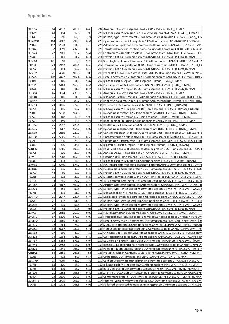
Appendices
189
Q12955 64 4377 480,1 6,49 234 Ankyrin-3 OS=Homo sapiens GN=ANK3 PE=1 SV=3 - [ANK3_HUMAN]
P01625 40 114 12,6 7,93 225 Ig kappa chain V-IV region Len OS=Homo sapiens PE=1 SV=2 - [KV402_HUMAN]
P13647 15 590 62,3 7,74 225 Keratin, type II cytoskeletal 5 OS=Homo sapiens GN=KRT5 PE=1 SV=3 - [K2C5_HUMAN]
Q8NCM8 160 4307 492,3 6,54 223 Cytoplasmic dynein 2 heavy chain 1 OS=Homo sapiens GN=DYNC2H1 PE=1 SV=4 - [DYHC2_HUMAN]
P25054 112 2843 311,5 7,8 221 Adenomatous polyposis coli protein OS=Homo sapiens GN=APC PE=1 SV=2 - [APC_HUMAN]
Q9Y4A5 32 3859 437,3 8,19 220 Transformation/transcription domain-associated protein (350/400 kDa PCAF-associated factor) (PAF350/400) (STAF40) (Tra1 homolog) - Homo sapiens (Human) - [TRRAP_HUMAN]
Q02224 22 2701 316,2 5,64 218 Centromere-associated protein E OS=Homo sapiens GN=CENPE PE=1 SV=2 - [CENPE_HUMAN]
P26447 31 101 11,7 6,11 214 Protein S100-A4 OS=Homo sapiens GN=S100A4 PE=1 SV=1 - [S10A4_HUMAN]
O95968 171 90 9,9 9,25 214 Secretoglobin family 1D member 1 OS=Homo sapiens GN=SCGB1D1 PE=1 SV=1 - [SG1D1_HUMAN]
P46100 28 2492 282,4 6,58 212 Transcriptional regulator ATRX OS=Homo sapiens GN=ATRX PE=1 SV=5 - [ATRX_HUMAN]
P06702 39 114 13,2 6,13 211 Protein S100-A9 OS=Homo sapiens GN=S100A9 PE=1 SV=1 - [S10A9_HUMAN]
O75592 21 4640 509,8 7,03 209 Probable E3 ubiquitin-protein ligase MYCBP2 OS=Homo sapiens GN=MYCBP2 PE=1 SV=3 - [MYCB2_HUMAN]
Q9P225 107 4427 507,4 6,37 209 Dynein heavy chain 2, axonemal OS=Homo sapiens GN=DNAH2 PE=1 SV=3 - [DYH2_HUMAN]
P01834 224 106 11,6 5,87 207 Ig kappa chain C region - Homo sapiens (Human) - [KAC_HUMAN]
Q9Y6V0 44 5183 566,3 6,54 202 Protein piccolo OS=Homo sapiens GN=PCLO PE=1 SV=3 - [PCLO_HUMAN]
P01598 25 108 11,8 8,44 199 Ig kappa chain V-I region EU OS=Homo sapiens PE=1 SV=1 - [KV106_HUMAN]
Q01484 45 3924 430,0 5,12 199 Ankyrin-2 OS=Homo sapiens GN=ANK2 PE=1 SV=3 - [ANK2_HUMAN]
P0CG04 76 106 11,3 7,87 197 Ig lambda-1 chain C regions OS=Homo sapiens GN=IGLC1 PE=1 SV=1 - [LAC1_HUMAN]
P0C6X7 57 7073 789,7 6,62 195 Replicase polyprotein 1ab OS=Human SARS coronavirus GN=rep PE=1 SV=1 - [R1AB_CVHSA]
O95613 20 3336 377,8 5,55 194 Pericentrin OS=Homo sapiens GN=PCNT PE=1 SV=4 - [PCNT_HUMAN]
P01781 55 116 12,7 8,48 191 Ig heavy chain V-III region GAL OS=Homo sapiens PE=1 SV=1 - [HV320_HUMAN]
P21817 49 5038 564,8 5,3 191 Ryanodine receptor 1 OS=Homo sapiens GN=RYR1 PE=1 SV=3 - [RYR1_HUMAN]
P01593 48 108 12,0 5,99 189 Ig kappa chain V-I region AG - Homo sapiens (Human) - [KV101_HUMAN]
P01591 87 159 18,1 5,24 188 Immunoglobulin J chain OS=Homo sapiens GN=IGJ PE=1 SV=4 - [IGJ_HUMAN]
Q5TZA2 25 2017 228,4 5,5 187 Rootletin OS=Homo sapiens GN=CROCC PE=1 SV=1 - [CROCC_HUMAN]
Q92736 47 4967 564,2 6,07 184 Ryanodine receptor 2 OS=Homo sapiens GN=RYR2 PE=1 SV=3 - [RYR2_HUMAN]
Q12789 22 2109 238,7 7,3 184 General transcription factor 3C polypeptide 1 OS=Homo sapiens GN=GTF3C1 PE=1 SV=4 - [TF3C1_HUMAN]
Q2LD37 25 5005 555,1 6,58 183 Uncharacterized protein KIAA1109 OS=Homo sapiens GN=KIAA1109 PE=1 SV=2 - [K1109_HUMAN]
P09211 11 210 23,3 5,64 182 Glutathione S-transferase P OS=Homo sapiens GN=GSTP1 PE=1 SV=2 - [GSTP1_HUMAN]
P01857 16 330 36,1 8,19 182 Ig gamma-1 chain C region - Homo sapiens (Human) - [IGHG1_HUMAN]
A6NKT7 58 1766 198,3 6,39 181 RanBP2-like and GRIP domain-containing protein 3 OS=Homo sapiens GN=RGPD3 PE=2 SV=1 - [RGPD3_HUMAN]
P08758 11 320 35,9 5,05 181 Annexin A5 OS=Homo sapiens GN=ANXA5 PE=1 SV=2 - [ANXA5_HUMAN]
Q5VST9 42 7968 867,9 5,99 181 Obscurin OS=Homo sapiens GN=OBSCN PE=1 SV=3 - [OBSCN_HUMAN]
P06313 35 133 14,6 6,58 181 Ig kappa chain V-IV region JI OS=Homo sapiens PE=4 SV=1 - [KV403_HUMAN]
Q09666 35 5890 628,7 6,15 180 Neuroblast differentiation-associated protein AHNAK OS=Homo sapiens GN=AHNAK PE=1 SV=2 - [AHNK_HUMAN]
Q9BYX7 25 375 42,0 6,33 177 Putative beta-actin-like protein 3 OS=Homo sapiens GN=POTEKP PE=5 SV=1 - [ACTBM_HUMAN]
P06703 43 90 10,2 5,48 177 Protein S100-A6 OS=Homo sapiens GN=S100A6 PE=1 SV=1 - [S10A6_HUMAN]
P00338 11 332 36,7 8,27 177 L-lactate dehydrogenase A chain OS=Homo sapiens GN=LDHA PE=1 SV=2 - [LDHA_HUMAN]
P63104 18 245 27,7 4,79 174 14-3-3 protein zeta/delta OS=Homo sapiens GN=YWHAZ PE=1 SV=1 - [1433Z_HUMAN]
Q8TCU4 23 4167 460,7 6,28 172 Alstrom syndrome protein 1 OS=Homo sapiens GN=ALMS1 PE=1 SV=3 - [ALMS1_HUMAN]
O95678 9 551 59,5 7,74 170 Keratin, type II cytoskeletal 75 OS=Homo sapiens GN=KRT75 PE=1 SV=2 - [K2C75_HUMAN]
P80748 30 111 11,9 5,08 169 Ig lambda chain V-III region LOI OS=Homo sapiens PE=1 SV=1 - [LV302_HUMAN]
Q96HP0 26 2047 229,4 6,74 169 Dedicator of cytokinesis protein 6 OS=Homo sapiens GN=DOCK6 PE=1 SV=3 - [DOCK6_HUMAN]
P02533 21 472 51,5 5,16 168 Keratin, type I cytoskeletal 14 OS=Homo sapiens GN=KRT14 PE=1 SV=4 - [K1C14_HUMAN]
Q5XKE5 27 535 57,8 7,2 168 Keratin, type II cytoskeletal 79 OS=Homo sapiens GN=KRT79 PE=1 SV=2 - [K2C79_HUMAN]
P05109 40 93 10,8 7,03 167 Protein S100-A8 OS=Homo sapiens GN=S100A8 PE=1 SV=1 - [S10A8_HUMAN]
Q8IVL1 29 2488 268,0 9,03 166 Neuron navigator 2 OS=Homo sapiens GN=NAV2 PE=1 SV=3 - [NAV2_HUMAN]
Q4G0P3 67 5120 575,5 6,07 165 Hydrocephalus-inducing protein homolog OS=Homo sapiens GN=HYDIN PE=1 SV=2 - [HYDIN_HUMAN]
Q9UFH2 76 4485 511,5 5,77 165 Dynein heavy chain 17, axonemal OS=Homo sapiens GN=DNAH17 PE=2 SV=2 - [DYH17_HUMAN]
Q14515 12 664 75,2 4,81 164 SPARC-like protein 1 OS=Homo sapiens GN=SPARCL1 PE=1 SV=2 - [SPRL1_HUMAN]
Q5CZC0 34 6907 780,1 6,71 163 Fibrous sheath-interacting protein 2 OS=Homo sapiens GN=FSIP2 PE=1 SV=4 - [FSIP2_HUMAN]
Q15782 17 390 43,5 7,53 162 Chitinase-3-like protein 2 OS=Homo sapiens GN=CHI3L2 PE=1 SV=1 - [CH3L2_HUMAN]
O75122 74 1294 141,0 8,47 161 CLIP-associating protein 2 OS=Homo sapiens GN=CLASP2 PE=1 SV=2 - [CLAP2_HUMAN]
Q5T4S7 28 5183 573,5 6,04 160 E3 ubiquitin-protein ligase UBR4 OS=Homo sapiens GN=UBR4 PE=1 SV=1 - [UBR4_HUMAN]
Q14643 26 2758 313,7 6,04 159 Inositol 1,4,5-trisphosphate receptor type 1 OS=Homo sapiens GN=ITPR1 PE=1 SV=2 - [ITPR1_HUMAN]
Q96T23 21 1441 163,7 5,01 159 Remodeling and spacing factor 1 OS=Homo sapiens GN=RSF1 PE=1 SV=2 - [RSF1_HUMAN]
A6NE01 34 2351 262,6 8,6 159 Protein FAM186A OS=Homo sapiens GN=FAM186A PE=2 SV=3 - [F186A_HUMAN]
P07339 35 412 44,5 6,54 158 Cathepsin D OS=Homo sapiens GN=CTSD PE=1 SV=1 - [CATD_HUMAN]
Q8N3K9 25 4069 448,9 4,78 157 Cardiomyopathy-associated protein 5 OS=Homo sapiens GN=CMYA5 PE=1 SV=3 - [CMYA5_HUMAN]
P01766 68 120 13,2 6,57 157 Ig heavy chain V-III region BRO OS=Homo sapiens PE=1 SV=1 - [HV305_HUMAN]
P61769 64 119 13,7 6,52 156 Beta-2-microglobulin OS=Homo sapiens GN=B2M PE=1 SV=1 - [B2MG_HUMAN]
Q5T200 21 1668 196,5 9,42 155 Zinc finger CCCH domain-containing protein 13 OS=Homo sapiens GN=ZC3H13 PE=1 SV=1 - [ZC3HD_HUMAN]
P49454 13 3210 367,5 5,07 155 Centromere protein F OS=Homo sapiens GN=CENPF PE=1 SV=2 - [CENPF_HUMAN]
Q9UMN6 34 2715 293,3 8,22 154 Histone-lysine N-methyltransferase MLL4 OS=Homo sapiens GN=WBP7 PE=1 SV=1 - [MLL4_HUMAN]
B1AJZ9 324 1412 161,8 6,95 154 Forkhead-associated domain-containing protein 1 OS=Homo sapiens GN=FHAD1 PE=2 SV=2 - [FHAD1_HUMAN]

Appendices
190
P35580 17 1976 228,9 5,54 153 Myosin-10 OS=Homo sapiens GN=MYH10 PE=1 SV=3 - [MYH10_HUMAN]
O14578 59 2027 231,3 6,57 153 Citron Rho-interacting kinase OS=Homo sapiens GN=CIT PE=1 SV=2 - [CTRO_HUMAN]
Q9UIF8 52 2168 240,3 6,54 153 Bromodomain adjacent to zinc finger domain protein 2B OS=Homo sapiens GN=BAZ2B PE=1 SV=3 - [BAZ2B_HUMAN]
O95153 32 1857 199,9 5,11 151 Peripheral-type benzodiazepine receptor-associated protein 1 OS=Homo sapiens GN=BZRAP1 PE=1 SV=2 - [RIMB1_HUMAN]
Q9BXX2 18 1477 167,6 6,16 151 Ankyrin repeat domain-containing protein 30B OS=Homo sapiens GN=ANKRD30B PE=2 SV=2 - [AN30B_HUMAN]
Q07864 36 2286 261,4 6,39 150 DNA polymerase epsilon catalytic subunit A OS=Homo sapiens GN=POLE PE=1 SV=5 - [DPOE1_HUMAN]
Q92614 66 2054 233,0 6,3 149 Myosin-XVIIIa OS=Homo sapiens GN=MYO18A PE=1 SV=3 - [MY18A_HUMAN]
P01594 33 108 11,9 5,33 149 Ig kappa chain V-I region AU OS=Homo sapiens PE=1 SV=1 - [KV102_HUMAN]
Q13029 23 1718 188,8 7,31 149 PR domain zinc finger protein 2 OS=Homo sapiens GN=PRDM2 PE=1 SV=3 - [PRDM2_HUMAN]
Q14152 44 1382 166,5 6,79 149 Eukaryotic translation initiation factor 3 subunit A OS=Homo sapiens GN=EIF3A PE=1 SV=1 - [EIF3A_HUMAN]
P01606 51 108 11,8 9,91 148 Ig kappa chain V-I region OU OS=Homo sapiens PE=1 SV=1 - [KV114_HUMAN]
Q14669 20 1992 220,3 8,48 147 Probable E3 ubiquitin-protein ligase TRIP12 OS=Homo sapiens GN=TRIP12 PE=1 SV=1 - [TRIPC_HUMAN]
Q86VF7 30 1730 197,0 9,2 147 Nebulin-related-anchoring protein OS=Homo sapiens GN=NRAP PE=2 SV=2 - [NRAP_HUMAN]
P01596 24 107 11,7 9,41 147 Ig kappa chain V-I region CAR OS=Homo sapiens PE=1 SV=1 - [KV104_HUMAN]
Q13428 51 1488 152,0 9,04 145 Treacle protein OS=Homo sapiens GN=TCOF1 PE=1 SV=3 - [TCOF_HUMAN]
Q92817 26 2033 231,5 6,96 144 Envoplakin OS=Homo sapiens GN=EVPL PE=1 SV=3 - [EVPL_HUMAN]
Q9UIG0 60 1483 170,8 8,48 142 Tyrosine-protein kinase BAZ1B OS=Homo sapiens GN=BAZ1B PE=1 SV=2 - [BAZ1B_HUMAN]
Q8TE73 23 4624 528,7 6,1 142 Dynein heavy chain 5, axonemal OS=Homo sapiens GN=DNAH5 PE=1 SV=3 - [DYH5_HUMAN]
P22079 11 712 80,2 8,62 140 Lactoperoxidase OS=Homo sapiens GN=LPO PE=1 SV=2 - [PERL_HUMAN]
P01768 60 122 13,7 9,63 140 Ig heavy chain V-III region CAM OS=Homo sapiens PE=1 SV=1 - [HV307_HUMAN]
A7E2Y1 21 1941 221,3 6,01 140 Myosin-7B OS=Homo sapiens GN=MYH7B PE=2 SV=3 - [MYH7B_HUMAN]
P80362 25 108 11,7 5,31 139 Ig kappa chain V-I region WAT OS=Homo sapiens PE=1 SV=1 - [KV125_HUMAN]
P24043 17 3122 343,7 6,4 138 Laminin subunit alpha-2 OS=Homo sapiens GN=LAMA2 PE=1 SV=4 - [LAMA2_HUMAN]
O60673 17 3130 352,6 8,47 138 DNA polymerase zeta catalytic subunit OS=Homo sapiens GN=REV3L PE=1 SV=2 - [DPOLZ_HUMAN]
P07858 10 339 37,8 6,3 137 Cathepsin B OS=Homo sapiens GN=CTSB PE=1 SV=3 - [CATB_HUMAN]
A2RUB6 47 948 109,3 8,25 137 Coiled-coil domain-containing protein 66 OS=Homo sapiens GN=CCDC66 PE=1 SV=4 - [CCD66_HUMAN]
Q96Q89 20 1820 210,5 5,67 137 Kinesin-like protein KIF20B OS=Homo sapiens GN=KIF20B PE=1 SV=3 - [KI20B_HUMAN]
Q8WXG9 20 6307 692,2 4,64 137 G-protein coupled receptor 98 OS=Homo sapiens GN=GPR98 PE=1 SV=1 - [GPR98_HUMAN]
Q68DN6 48 1748 196,5 6,1 137 RANBP2-like and GRIP domain-containing protein 1/2 OS=Homo sapiens GN=RGPD1 PE=1 SV=2 - [RGPD1_HUMAN]
Q7Z5J4 46 1906 203,2 8,79 137 Retinoic acid-induced protein 1 OS=Homo sapiens GN=RAI1 PE=1 SV=2 - [RAI1_HUMAN]
Q5VT06 16 3117 350,7 6,33 136 Centrosome-associated protein 350 OS=Homo sapiens GN=CEP350 PE=1 SV=1 - [CE350_HUMAN]
Q12923 18 2485 276,7 6,42 135 Tyrosine-protein phosphatase non-receptor type 13 OS=Homo sapiens GN=PTPN13 PE=1 SV=2 - [PTN13_HUMAN]
P01607 26 108 11,9 6,51 135 Ig kappa chain V-I region Rei OS=Homo sapiens PE=1 SV=1 - [KV115_HUMAN]
Q8TD26 14 2715 305,2 6,27 135 Chromodomain-helicase-DNA-binding protein 6 OS=Homo sapiens GN=CHD6 PE=1 SV=4 - [CHD6_HUMAN]
Q14980 22 2115 238,1 5,78 135 Nuclear mitotic apparatus protein 1 OS=Homo sapiens GN=NUMA1 PE=1 SV=2 - [NUMA1_HUMAN]
P01765 49 115 12,3 9,13 134 Ig heavy chain V-III region TIL - Homo sapiens (Human) - [HV304_HUMAN]
Q8IZD9 14 2030 233,0 6,98 134 Dedicator of cytokinesis protein 3 OS=Homo sapiens GN=DOCK3 PE=1 SV=1 - [DOCK3_HUMAN]
Q04695 12 432 48,1 5,02 134 Keratin, type I cytoskeletal 17 OS=Homo sapiens GN=KRT17 PE=1 SV=2 - [K1C17_HUMAN]
O60293 33 1989 226,2 8,13 134 Zinc finger C3H1 domain-containing protein OS=Homo sapiens GN=ZFC3H1 PE=1 SV=3 - [ZC3H1_HUMAN]
Q96SN8 74 1893 214,9 5,58 134 CDK5 regulatory subunit-associated protein 2 OS=Homo sapiens GN=CDK5RAP2 PE=1 SV=5 - [CK5P2_HUMAN]
Q96PE2 28 2063 221,5 6,29 134 Rho guanine nucleotide exchange factor 17 OS=Homo sapiens GN=ARHGEF17 PE=1 SV=1 - [ARHGH_HUMAN]
Q7Z6E9 17 1792 201,4 9,64 133 E3 ubiquitin-protein ligase RBBP6 OS=Homo sapiens GN=RBBP6 PE=1 SV=1 - [RBBP6_HUMAN]
Q9Y4F4 167 1720 189,2 8,5 132 Protein FAM179B OS=Homo sapiens GN=FAM179B PE=1 SV=4 - [F179B_HUMAN]
Q9Y4D8 64 3996 439,1 6,19 132 Probable E3 ubiquitin-protein ligase C12orf51 OS=Homo sapiens GN=C12orf51 PE=1 SV=5 - [K0614_HUMAN]
P07711 11 333 37,5 5,45 132 Cathepsin L1 OS=Homo sapiens GN=CTSL1 PE=1 SV=2 - [CATL1_HUMAN]
Q9BV73 31 2442 281,0 5,02 131 Centrosome-associated protein CEP250 OS=Homo sapiens GN=CEP250 PE=1 SV=2 - [CP250_HUMAN]
P01624 32 109 11,9 8,94 131 Ig kappa chain V-III region POM OS=Homo sapiens PE=1 SV=1 - [KV306_HUMAN]
Q8N474 15 314 35,4 8,85 131 Secreted frizzled-related protein 1 OS=Homo sapiens GN=SFRP1 PE=1 SV=1 - [SFRP1_HUMAN]
Q5VU65 13 1888 210,5 7,5 131 Nuclear pore membrane glycoprotein 210-like OS=Homo sapiens GN=NUP210L PE=2 SV=1 - [P210L_HUMAN]
P07602 11 524 58,1 5,17 130 Proactivator polypeptide OS=Homo sapiens GN=PSAP PE=1 SV=2 - [SAP_HUMAN]
Q7Z460 28 1538 169,3 9,03 130 CLIP-associating protein 1 OS=Homo sapiens GN=CLASP1 PE=1 SV=1 - [CLAP1_HUMAN]
P07355 12 339 38,6 7,75 129 Annexin A2 OS=Homo sapiens GN=ANXA2 PE=1 SV=2 - [ANXA2_HUMAN]
Q7Z406 11 1995 227,9 5,92 129 Myosin-14 OS=Homo sapiens GN=MYH14 PE=1 SV=1 - [MYH14_HUMAN]
Q8WZ64 25 1704 193,3 7,39 129 Arf-GAP with Rho-GAP domain, ANK repeat and PH domain-containing protein 2 OS=Homo sapiens GN=ARAP2 PE=1 SV=3 - [ARAP2_HUMAN]
P30414 76 1462 165,6 9,99 129 NK-tumor recognition protein OS=Homo sapiens GN=NKTR PE=1 SV=2 - [NKTR_HUMAN]
Q9H2D6 23 2365 261,2 8,48 128 TRIO and F-actin-binding protein OS=Homo sapiens GN=TRIOBP PE=1 SV=3 - [TARA_HUMAN]
Q7Z7A1 19 2325 268,7 5,55 128 Centriolin OS=Homo sapiens GN=CEP110 PE=1 SV=2 - [CE110_HUMAN]
Q14966 47 1978 220,5 6,38 128 Zinc finger protein 638 OS=Homo sapiens GN=ZNF638 PE=1 SV=2 - [ZN638_HUMAN]
P11717 24 2491 274,2 5,94 127 Cation-independent mannose-6-phosphate receptor OS=Homo sapiens GN=IGF2R PE=1 SV=3 - [MPRI_HUMAN]
Q2KHM9 16 967 109,3 7,68 127 Uncharacterized protein KIAA0753 OS=Homo sapiens GN=KIAA0753 PE=2 SV=3 - [K0753_HUMAN]
Q86TE4 81 346 38,9 8,73 127 Leucine zipper protein 2 OS=Homo sapiens GN=LUZP2 PE=2 SV=2 - [LUZP2_HUMAN]
Q14004 18 1512 164,8 9,69 127 Cyclin-dependent kinase 13 OS=Homo sapiens GN=CDK13 PE=1 SV=2 - [CDK13_HUMAN]
Q63HN8 15 3280 373,7 6,92 126 RING finger protein 213 OS=Homo sapiens GN=RNF213 PE=1 SV=2 - [RN213_HUMAN]
Q9Y2K3 11 1946 224,5 5,85 126 Myosin-15 OS=Homo sapiens GN=MYH15 PE=1 SV=5 - [MYH15_HUMAN]
Q9NR99 50 2828 312,0 8,32 126 Matrix-remodeling-associated protein 5 OS=Homo sapiens GN=MXRA5 PE=2 SV=3 - [MXRA5_HUMAN]
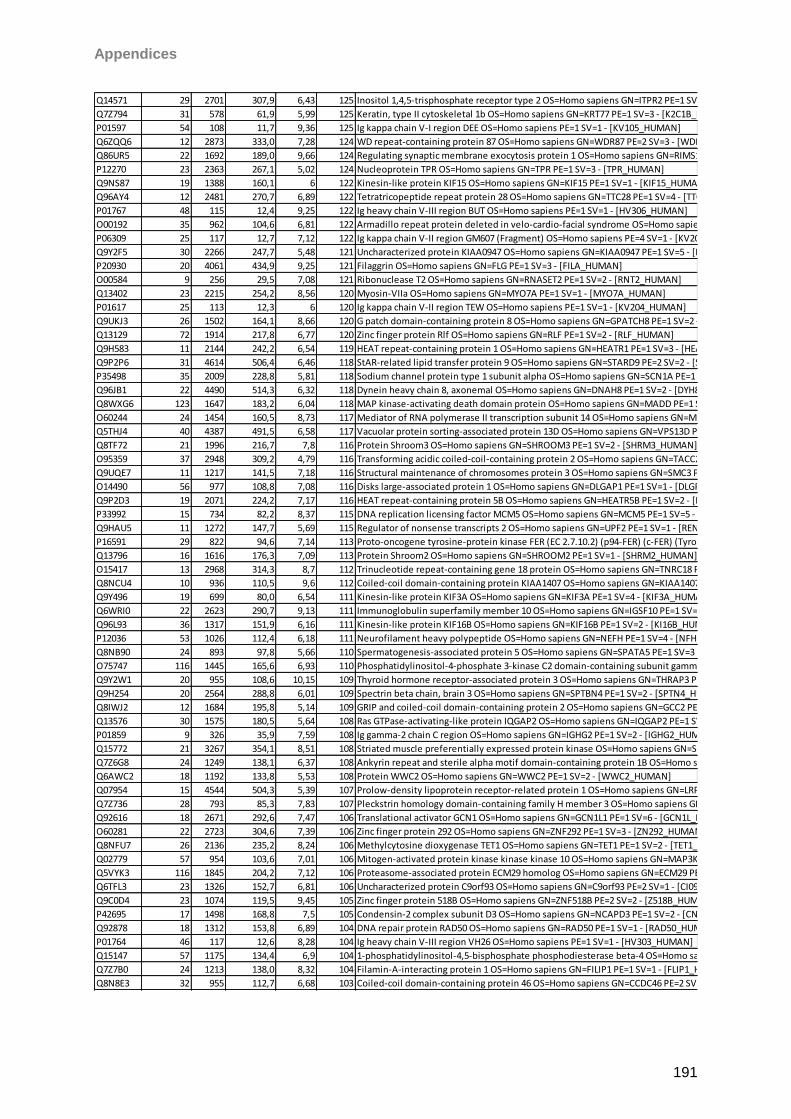
Appendices
191
Q14571 29 2701 307,9 6,43 125 Inositol 1,4,5-trisphosphate receptor type 2 OS=Homo sapiens GN=ITPR2 PE=1 SV=2 - [ITPR2_HUMAN]
Q7Z794 31 578 61,9 5,99 125 Keratin, type II cytoskeletal 1b OS=Homo sapiens GN=KRT77 PE=1 SV=3 - [K2C1B_HUMAN]
P01597 54 108 11,7 9,36 125 Ig kappa chain V-I region DEE OS=Homo sapiens PE=1 SV=1 - [KV105_HUMAN]
Q6ZQQ6 12 2873 333,0 7,28 124 WD repeat-containing protein 87 OS=Homo sapiens GN=WDR87 PE=2 SV=3 - [WDR87_HUMAN]
Q86UR5 22 1692 189,0 9,66 124 Regulating synaptic membrane exocytosis protein 1 OS=Homo sapiens GN=RIMS1 PE=1 SV=1 - [RIMS1_HUMAN]
P12270 23 2363 267,1 5,02 124 Nucleoprotein TPR OS=Homo sapiens GN=TPR PE=1 SV=3 - [TPR_HUMAN]
Q9NS87 19 1388 160,1 6 122 Kinesin-like protein KIF15 OS=Homo sapiens GN=KIF15 PE=1 SV=1 - [KIF15_HUMAN]
Q96AY4 12 2481 270,7 6,89 122 Tetratricopeptide repeat protein 28 OS=Homo sapiens GN=TTC28 PE=1 SV=4 - [TTC28_HUMAN]
P01767 48 115 12,4 9,25 122 Ig heavy chain V-III region BUT OS=Homo sapiens PE=1 SV=1 - [HV306_HUMAN]
O00192 35 962 104,6 6,81 122 Armadillo repeat protein deleted in velo-cardio-facial syndrome OS=Homo sapiens GN=ARVCF PE=1 SV=1 - [ARVC_HUMAN]
P06309 25 117 12,7 7,12 122 Ig kappa chain V-II region GM607 (Fragment) OS=Homo sapiens PE=4 SV=1 - [KV205_HUMAN]
Q9Y2F5 30 2266 247,7 5,48 121 Uncharacterized protein KIAA0947 OS=Homo sapiens GN=KIAA0947 PE=1 SV=5 - [K0947_HUMAN]
P20930 20 4061 434,9 9,25 121 Filaggrin OS=Homo sapiens GN=FLG PE=1 SV=3 - [FILA_HUMAN]
O00584 9 256 29,5 7,08 121 Ribonuclease T2 OS=Homo sapiens GN=RNASET2 PE=1 SV=2 - [RNT2_HUMAN]
Q13402 23 2215 254,2 8,56 120 Myosin-VIIa OS=Homo sapiens GN=MYO7A PE=1 SV=1 - [MYO7A_HUMAN]
P01617 25 113 12,3 6 120 Ig kappa chain V-II region TEW OS=Homo sapiens PE=1 SV=1 - [KV204_HUMAN]
Q9UKJ3 26 1502 164,1 8,66 120 G patch domain-containing protein 8 OS=Homo sapiens GN=GPATCH8 PE=1 SV=2 - [GPTC8_HUMAN]
Q13129 72 1914 217,8 6,77 120 Zinc finger protein Rlf OS=Homo sapiens GN=RLF PE=1 SV=2 - [RLF_HUMAN]
Q9H583 11 2144 242,2 6,54 119 HEAT repeat-containing protein 1 OS=Homo sapiens GN=HEATR1 PE=1 SV=3 - [HEAT1_HUMAN]
Q9P2P6 31 4614 506,4 6,46 118 StAR-related lipid transfer protein 9 OS=Homo sapiens GN=STARD9 PE=2 SV=2 - [STAR9_HUMAN]
P35498 35 2009 228,8 5,81 118 Sodium channel protein type 1 subunit alpha OS=Homo sapiens GN=SCN1A PE=1 SV=2 - [SCN1A_HUMAN]
Q96JB1 22 4490 514,3 6,32 118 Dynein heavy chain 8, axonemal OS=Homo sapiens GN=DNAH8 PE=1 SV=2 - [DYH8_HUMAN]
Q8WXG6 123 1647 183,2 6,04 118 MAP kinase-activating death domain protein OS=Homo sapiens GN=MADD PE=1 SV=2 - [MADD_HUMAN]
O60244 24 1454 160,5 8,73 117 Mediator of RNA polymerase II transcription subunit 14 OS=Homo sapiens GN=MED14 PE=1 SV=2 - [MED14_HUMAN]
Q5THJ4 40 4387 491,5 6,58 117 Vacuolar protein sorting-associated protein 13D OS=Homo sapiens GN=VPS13D PE=1 SV=1 - [VP13D_HUMAN]
Q8TF72 21 1996 216,7 7,8 116 Protein Shroom3 OS=Homo sapiens GN=SHROOM3 PE=1 SV=2 - [SHRM3_HUMAN]
O95359 37 2948 309,2 4,79 116 Transforming acidic coiled-coil-containing protein 2 OS=Homo sapiens GN=TACC2 PE=1 SV=3 - [TACC2_HUMAN]
Q9UQE7 11 1217 141,5 7,18 116 Structural maintenance of chromosomes protein 3 OS=Homo sapiens GN=SMC3 PE=1 SV=2 - [SMC3_HUMAN]
O14490 56 977 108,8 7,08 116 Disks large-associated protein 1 OS=Homo sapiens GN=DLGAP1 PE=1 SV=1 - [DLGP1_HUMAN]
Q9P2D3 19 2071 224,2 7,17 116 HEAT repeat-containing protein 5B OS=Homo sapiens GN=HEATR5B PE=1 SV=2 - [HTR5B_HUMAN]
P33992 15 734 82,2 8,37 115 DNA replication licensing factor MCM5 OS=Homo sapiens GN=MCM5 PE=1 SV=5 - [MCM5_HUMAN]
Q9HAU5 11 1272 147,7 5,69 115 Regulator of nonsense transcripts 2 OS=Homo sapiens GN=UPF2 PE=1 SV=1 - [RENT2_HUMAN]
P16591 29 822 94,6 7,14 113 Proto-oncogene tyrosine-protein kinase FER (EC 2.7.10.2) (p94-FER) (c-FER) (Tyrosine kinase 3) - Homo sapiens (Human) - [FER_HUMAN]
Q13796 16 1616 176,3 7,09 113 Protein Shroom2 OS=Homo sapiens GN=SHROOM2 PE=1 SV=1 - [SHRM2_HUMAN]
O15417 13 2968 314,3 8,7 112 Trinucleotide repeat-containing gene 18 protein OS=Homo sapiens GN=TNRC18 PE=1 SV=3 - [TNC18_HUMAN]
Q8NCU4 10 936 110,5 9,6 112 Coiled-coil domain-containing protein KIAA1407 OS=Homo sapiens GN=KIAA1407 PE=2 SV=1 - [K1407_HUMAN]
Q9Y496 19 699 80,0 6,54 111 Kinesin-like protein KIF3A OS=Homo sapiens GN=KIF3A PE=1 SV=4 - [KIF3A_HUMAN]
Q6WRI0 22 2623 290,7 9,13 111 Immunoglobulin superfamily member 10 OS=Homo sapiens GN=IGSF10 PE=1 SV=1 - [IGS10_HUMAN]
Q96L93 36 1317 151,9 6,16 111 Kinesin-like protein KIF16B OS=Homo sapiens GN=KIF16B PE=1 SV=2 - [KI16B_HUMAN]
P12036 53 1026 112,4 6,18 111 Neurofilament heavy polypeptide OS=Homo sapiens GN=NEFH PE=1 SV=4 - [NFH_HUMAN]
Q8NB90 24 893 97,8 5,66 110 Spermatogenesis-associated protein 5 OS=Homo sapiens GN=SPATA5 PE=1 SV=3 - [SPAT5_HUMAN]
O75747 116 1445 165,6 6,93 110 Phosphatidylinositol-4-phosphate 3-kinase C2 domain-containing subunit gamma OS=Homo sapiens GN=PIK3C2G PE=1 SV=3 - [P3C2G_HUMAN]
Q9Y2W1 20 955 108,6 10,15 109 Thyroid hormone receptor-associated protein 3 OS=Homo sapiens GN=THRAP3 PE=1 SV=2 - [TR150_HUMAN]
Q9H254 20 2564 288,8 6,01 109 Spectrin beta chain, brain 3 OS=Homo sapiens GN=SPTBN4 PE=1 SV=2 - [SPTN4_HUMAN]
Q8IWJ2 12 1684 195,8 5,14 109 GRIP and coiled-coil domain-containing protein 2 OS=Homo sapiens GN=GCC2 PE=1 SV=4 - [GCC2_HUMAN]
Q13576 30 1575 180,5 5,64 108 Ras GTPase-activating-like protein IQGAP2 OS=Homo sapiens GN=IQGAP2 PE=1 SV=4 - [IQGA2_HUMAN]
P01859 9 326 35,9 7,59 108 Ig gamma-2 chain C region OS=Homo sapiens GN=IGHG2 PE=1 SV=2 - [IGHG2_HUMAN]
Q15772 21 3267 354,1 8,51 108 Striated muscle preferentially expressed protein kinase OS=Homo sapiens GN=SPEG PE=1 SV=4 - [SPEG_HUMAN]
Q7Z6G8 24 1249 138,1 6,37 108 Ankyrin repeat and sterile alpha motif domain-containing protein 1B OS=Homo sapiens GN=ANKS1B PE=1 SV=1 - [ANS1B_HUMAN]
Q6AWC2 18 1192 133,8 5,53 108 Protein WWC2 OS=Homo sapiens GN=WWC2 PE=1 SV=2 - [WWC2_HUMAN]
Q07954 15 4544 504,3 5,39 107 Prolow-density lipoprotein receptor-related protein 1 OS=Homo sapiens GN=LRP1 PE=1 SV=2 - [LRP1_HUMAN]
Q7Z736 28 793 85,3 7,83 107 Pleckstrin homology domain-containing family H member 3 OS=Homo sapiens GN=PLEKHH3 PE=1 SV=2 - [PKHH3_HUMAN]
Q92616 18 2671 292,6 7,47 106 Translational activator GCN1 OS=Homo sapiens GN=GCN1L1 PE=1 SV=6 - [GCN1L_HUMAN]
O60281 22 2723 304,6 7,39 106 Zinc finger protein 292 OS=Homo sapiens GN=ZNF292 PE=1 SV=3 - [ZN292_HUMAN]
Q8NFU7 26 2136 235,2 8,24 106 Methylcytosine dioxygenase TET1 OS=Homo sapiens GN=TET1 PE=1 SV=2 - [TET1_HUMAN]
Q02779 57 954 103,6 7,01 106 Mitogen-activated protein kinase kinase kinase 10 OS=Homo sapiens GN=MAP3K10 PE=1 SV=3 - [M3K10_HUMAN]
Q5VYK3 116 1845 204,2 7,12 106 Proteasome-associated protein ECM29 homolog OS=Homo sapiens GN=ECM29 PE=1 SV=2 - [ECM29_HUMAN]
Q6TFL3 23 1326 152,7 6,81 106 Uncharacterized protein C9orf93 OS=Homo sapiens GN=C9orf93 PE=2 SV=1 - [CI093_HUMAN]
Q9C0D4 23 1074 119,5 9,45 105 Zinc finger protein 518B OS=Homo sapiens GN=ZNF518B PE=2 SV=2 - [Z518B_HUMAN]
P42695 17 1498 168,8 7,5 105 Condensin-2 complex subunit D3 OS=Homo sapiens GN=NCAPD3 PE=1 SV=2 - [CNDD3_HUMAN]
Q92878 18 1312 153,8 6,89 104 DNA repair protein RAD50 OS=Homo sapiens GN=RAD50 PE=1 SV=1 - [RAD50_HUMAN]
P01764 46 117 12,6 8,28 104 Ig heavy chain V-III region VH26 OS=Homo sapiens PE=1 SV=1 - [HV303_HUMAN]
Q15147 57 1175 134,4 6,9 104 1-phosphatidylinositol-4,5-bisphosphate phosphodiesterase beta-4 OS=Homo sapiens GN=PLCB4 PE=1 SV=3 - [PLCB4_HUMAN]
Q7Z7B0 24 1213 138,0 8,32 104 Filamin-A-interacting protein 1 OS=Homo sapiens GN=FILIP1 PE=1 SV=1 - [FLIP1_HUMAN]
Q8N8E3 32 955 112,7 6,68 103 Coiled-coil domain-containing protein 46 OS=Homo sapiens GN=CCDC46 PE=2 SV=2 - [CCD46_HUMAN]
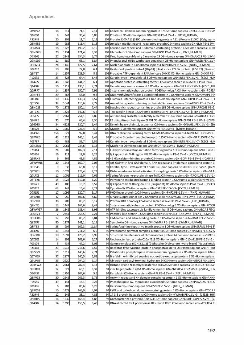
Appendices
192
Q494V2 18 611 71,1 7,11 103 Coiled-coil domain-containing protein 37 OS=Homo sapiens GN=CCDC37 PE=1 SV=1 - [CCD37_HUMAN]
Q16651 8 343 36,4 5,85 103 Prostasin OS=Homo sapiens GN=PRSS8 PE=1 SV=1 - [PRSS8_HUMAN]
P31949 20 105 11,7 7,12 103 Protein S100-A11 (S100 calcium-binding protein A11) (Protein S100C) (Calgizzarin) (MLN 70) - Homo sapiens (Human) - [S10AB_HUMAN]
Q8N9B5 19 988 111,4 6,18 103 Junction-mediating and -regulatory protein OS=Homo sapiens GN=JMY PE=1 SV=2 - [JMY_HUMAN]
Q96JM4 18 1722 199,2 6,19 103 Leucine-rich repeat and IQ domain-containing protein 1 OS=Homo sapiens GN=LRRIQ1 PE=2 SV=3 - [LRIQ1_HUMAN]
Q9NPG3 20 1134 121,4 9,33 102 Ubinuclein-1 OS=Homo sapiens GN=UBN1 PE=1 SV=2 - [UBN1_HUMAN]
O75165 27 2243 254,3 6,74 102 DnaJ homolog subfamily C member 13 OS=Homo sapiens GN=DNAJC13 PE=1 SV=5 - [DJC13_HUMAN]
Q9NSD9 11 589 66,1 6,84 102 Phenylalanyl-tRNA synthetase beta chain OS=Homo sapiens GN=FARSB PE=1 SV=3 - [SYFB_HUMAN]
Q9H6R4 24 1146 127,5 7,64 102 Nucleolar protein 6 OS=Homo sapiens GN=NOL6 PE=1 SV=2 - [NOL6_HUMAN]
P04792 10 205 22,8 6,4 102 Heat-shock protein beta-1 (HspB1) (Heat shock 27 kDa protein) (HSP 27) (Stress-responsive protein 27) (SRP27) (Estrogen-regulated 24 kDa protein) (28 kDa heat shock protein) - Homo sapiens (Human) - [HSPB1_HUMAN]
Q8IY37 14 1157 129,5 8,1 102 Probable ATP-dependent RNA helicase DHX37 OS=Homo sapiens GN=DHX37 PE=1 SV=1 - [DHX37_HUMAN]
P12035 23 628 64,4 6,48 102 Keratin, type II cytoskeletal 3 OS=Homo sapiens GN=KRT3 PE=1 SV=3 - [K2C3_HUMAN]
O14727 46 1248 141,7 6,4 102 Apoptotic protease-activating factor 1 OS=Homo sapiens GN=APAF1 PE=1 SV=2 - [APAF_HUMAN]
Q14687 16 1217 136,1 7,74 101 Genetic suppressor element 1 OS=Homo sapiens GN=GSE1 PE=1 SV=3 - [GSE1_HUMAN]
Q29RF7 14 1337 150,7 7,91 101 Sister chromatid cohesion protein PDS5 homolog A OS=Homo sapiens GN=PDS5A PE=1 SV=1 - [PDS5A_HUMAN]
Q9NPF5 29 467 53,0 9,5 101 DNA methyltransferase 1-associated protein 1 OS=Homo sapiens GN=DMAP1 PE=1 SV=1 - [DMAP1_HUMAN]
Q4L180 10 1135 130,3 6,57 101 Filamin A-interacting protein 1-like OS=Homo sapiens GN=FILIP1L PE=1 SV=2 - [FIL1L_HUMAN]
Q5T2S8 32 1044 115,6 7,77 101 Armadillo repeat-containing protein 4 OS=Homo sapiens GN=ARMC4 PE=2 SV=1 - [ARMC4_HUMAN]
Q8ND23 70 1372 150,1 7,44 101 Leucine-rich repeat-containing protein 16B OS=Homo sapiens GN=LRRC16B PE=2 SV=2 - [LR16B_HUMAN]
Q5TCY1 84 1321 142,6 5,6 100 Tau-tubulin kinase 1 OS=Homo sapiens GN=TTBK1 PE=1 SV=2 - [TTBK1_HUMAN]
O95477 9 2261 254,1 6,86 100 ATP-binding cassette sub-family A member 1 OS=Homo sapiens GN=ABCA1 PE=1 SV=3 - [ABCA1_HUMAN]
Q96JP5 35 570 63,4 7,36 100 E3 ubiquitin-protein ligase ZFP91 OS=Homo sapiens GN=ZFP91 PE=1 SV=1 - [ZFP91_HUMAN]
Q96DT5 34 4523 520,7 6,44 100 Dynein heavy chain 11, axonemal OS=Homo sapiens GN=DNAH11 PE=1 SV=3 - [DYH11_HUMAN]
P35579 17 1960 226,4 5,6 100 Myosin-9 OS=Homo sapiens GN=MYH9 PE=1 SV=4 - [MYH9_HUMAN]
Q14566 156 821 92,8 5,41 100 DNA replication licensing factor MCM6 OS=Homo sapiens GN=MCM6 PE=1 SV=1 - [MCM6_HUMAN]
Q8IWK6 43 1321 146,1 8,48 99 Probable G-protein coupled receptor 125 OS=Homo sapiens GN=GPR125 PE=1 SV=2 - [GP125_HUMAN]
P05787 40 483 53,7 5,59 98 Keratin, type II cytoskeletal 8 OS=Homo sapiens GN=KRT8 PE=1 SV=7 - [K2C8_HUMAN]
Q9NZM1 21 2061 234,6 6,18 98 Myoferlin OS=Homo sapiens GN=MYOF PE=1 SV=1 - [MYOF_HUMAN]
P78344 24 907 102,3 7,14 98 Eukaryotic translation initiation factor 4 gamma 2 OS=Homo sapiens GN=EIF4G2 PE=1 SV=1 - [IF4G2_HUMAN]
P01616 16 112 12,0 9,29 98 Ig kappa chain V-II region MIL OS=Homo sapiens PE=1 SV=1 - [KV203_HUMAN]
Q9BRK5 9 362 41,8 4,86 98 45 kDa calcium-binding protein OS=Homo sapiens GN=SDF4 PE=1 SV=1 - [CAB45_HUMAN]
Q8WWN8 30 1544 169,7 7,08 97 Arf-GAP with Rho-GAP domain, ANK repeat and PH domain-containing protein 3 OS=Homo sapiens GN=ARAP3 PE=1 SV=1 - [ARAP3_HUMAN]
Q01546 9 638 65,8 8,12 97 Keratin, type II cytoskeletal 2 oral OS=Homo sapiens GN=KRT76 PE=1 SV=2 - [K22O_HUMAN]
Q9Y4D1 10 1078 123,4 7,23 97 Disheveled-associated activator of morphogenesis 1 OS=Homo sapiens GN=DAAM1 PE=1 SV=2 - [DAAM1_HUMAN]
Q7L7X3 11 1001 116,0 7,65 97 Serine/threonine-protein kinase TAO1 OS=Homo sapiens GN=TAOK1 PE=1 SV=1 - [TAOK1_HUMAN]
Q8TBY8 15 1022 119,0 6,29 97 Polyamine-modulated factor 1-binding protein 1 OS=Homo sapiens GN=PMFBP1 PE=2 SV=1 - [PMFBP_HUMAN]
P01621 49 100 10,7 6,52 97 Ig kappa chain V-III region NG9 (Fragment) OS=Homo sapiens PE=1 SV=1 - [KV303_HUMAN]
P01037 62 141 16,4 7,21 97 Cystatin-SN OS=Homo sapiens GN=CST1 PE=1 SV=3 - [CYTN_HUMAN]
O75151 17 1096 120,7 9,17 97 PHD finger protein 2 OS=Homo sapiens GN=PHF2 PE=1 SV=4 - [PHF2_HUMAN]
Q6VAB6 42 950 107,6 8,69 96 Kinase suppressor of Ras 2 OS=Homo sapiens GN=KSR2 PE=1 SV=2 - [KSR2_HUMAN]
Q8N9T8 36 709 83,2 5,17 96 Protein KRI1 homolog OS=Homo sapiens GN=KRI1 PE=1 SV=2 - [KRI1_HUMAN]
Q9NTI5 12 1447 164,6 8,47 96 Sister chromatid cohesion protein PDS5 homolog B OS=Homo sapiens GN=PDS5B PE=1 SV=1 - [PDS5B_HUMAN]
Q8WWZ7 26 1642 186,4 6,95 96 ATP-binding cassette sub-family A member 5 OS=Homo sapiens GN=ABCA5 PE=2 SV=2 - [ABCA5_HUMAN]
Q96RV3 7 2341 258,5 7,21 96 Pecanex-like protein 1 OS=Homo sapiens GN=PCNX PE=2 SV=2 - [PCX1_HUMAN]
Q9UHB6 17 759 85,2 6,84 96 LIM domain and actin-binding protein 1 OS=Homo sapiens GN=LIMA1 PE=1 SV=1 - [LIMA1_HUMAN]
Q92797 80 1274 141,1 6,13 95 Symplekin OS=Homo sapiens GN=SYMPK PE=1 SV=2 - [SYMPK_HUMAN]
Q8IYB3 25 904 102,3 11,84 95 Serine/arginine repetitive matrix protein 1 OS=Homo sapiens GN=SRRM1 PE=1 SV=2 - [SRRM1_HUMAN]
Q14997 13 1843 211,2 6,9 95 Proteasome activator complex subunit 4 OS=Homo sapiens GN=PSME4 PE=1 SV=2 - [PSME4_HUMAN]
Q96SB8 10 1091 126,2 6,99 95 Structural maintenance of chromosomes protein 6 OS=Homo sapiens GN=SMC6 PE=1 SV=2 - [SMC6_HUMAN]
Q7Z3E2 14 898 103,6 6,27 95 Uncharacterized protein C10orf118 OS=Homo sapiens GN=C10orf118 PE=2 SV=2 - [CJ118_HUMAN]
P09104 5 434 47,2 5,03 95 Gamma-enolase (EC 4.2.1.11) (2-phospho-D-glycerate hydro-lyase) (Neural enolase) (Neuron-specific enolase) (NSE) (Enolase 2) - Homo sapiens (Human) - [ENOG_HUMAN]
P23468 21 1912 214,6 6,57 95 Receptor-type tyrosine-protein phosphatase delta OS=Homo sapiens GN=PTPRD PE=1 SV=2 - [PTPRD_HUMAN]
Q6ZV29 11 1317 145,6 7,74 95 Patatin-like phospholipase domain-containing protein 7 OS=Homo sapiens GN=PNPLA7 PE=2 SV=3 - [PLPL7_HUMAN]
Q5TH69 37 2177 240,5 5,82 94 Brefeldin A-inhibited guanine nucleotide-exchange protein 3 OS=Homo sapiens GN=ARFGEF3 PE=1 SV=3 - [BIG3_HUMAN]
Q9UPU5 26 2620 294,2 6,14 94 Ubiquitin carboxyl-terminal hydrolase 24 OS=Homo sapiens GN=USP24 PE=1 SV=3 - [UBP24_HUMAN]
Q9BYW2 31 2564 287,4 6,14 94 Histone-lysine N-methyltransferase SETD2 OS=Homo sapiens GN=SETD2 PE=1 SV=3 - [SETD2_HUMAN]
Q9HBT8 12 521 60,1 8,32 94 Zinc finger protein 286A OS=Homo sapiens GN=ZNF286A PE=2 SV=1 - [Z286A_HUMAN]
O60437 23 1756 204,6 5,6 94 Periplakin OS=Homo sapiens GN=PPL PE=1 SV=4 - [PEPL_HUMAN]
Q8IWZ3 30 2542 269,3 5,73 94 Ankyrin repeat and KH domain-containing protein 1 OS=Homo sapiens GN=ANKHD1 PE=1 SV=1 - [ANKH1_HUMAN]
P14555 49 144 16,1 9,23 94 Phospholipase A2, membrane associated OS=Homo sapiens GN=PLA2G2A PE=1 SV=2 - [PA2GA_HUMAN]
P06396 6 782 85,6 6,28 94 Gelsolin OS=Homo sapiens GN=GSN PE=1 SV=1 - [GELS_HUMAN]
Q9BQS8 10 1478 166,9 4,92 94 FYVE and coiled-coil domain-containing protein 1 OS=Homo sapiens GN=FYCO1 PE=1 SV=3 - [FYCO1_HUMAN]
P31946 12 246 28,1 4,83 94 14-3-3 protein beta/alpha OS=Homo sapiens GN=YWHAB PE=1 SV=3 - [1433B_HUMAN]
Q5RHP9 16 1530 168,4 4,88 93 Uncharacterized protein C1orf173 OS=Homo sapiens GN=C1orf173 PE=2 SV=1 - [CA173_HUMAN]
O14802 14 1390 155,5 8,48 93 DNA-directed RNA polymerase III subunit RPC1 OS=Homo sapiens GN=POLR3A PE=1 SV=2 - [RPC1_HUMAN]
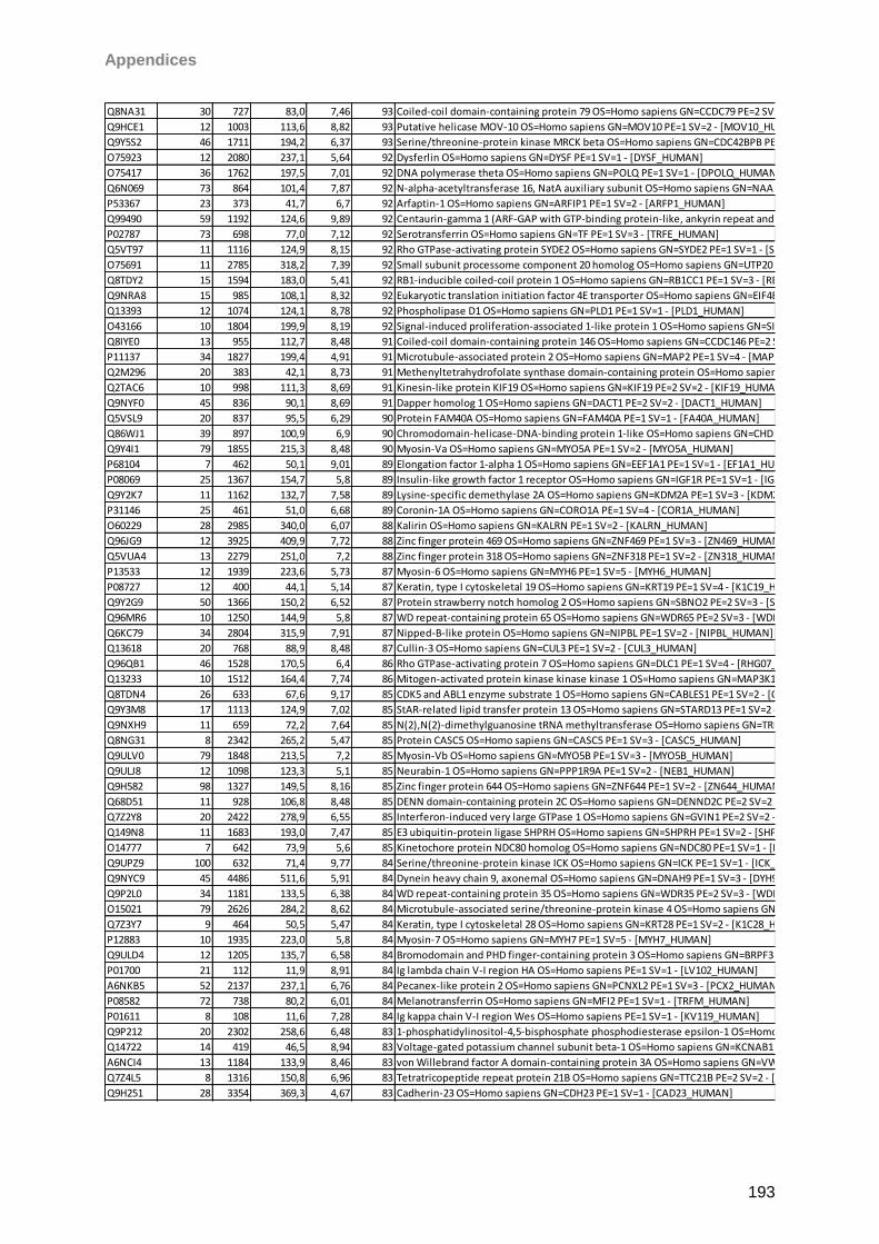
Appendices
193
Q8NA31 30 727 83,0 7,46 93 Coiled-coil domain-containing protein 79 OS=Homo sapiens GN=CCDC79 PE=2 SV=3 - [CCD79_HUMAN]
Q9HCE1 12 1003 113,6 8,82 93 Putative helicase MOV-10 OS=Homo sapiens GN=MOV10 PE=1 SV=2 - [MOV10_HUMAN]
Q9Y5S2 46 1711 194,2 6,37 93 Serine/threonine-protein kinase MRCK beta OS=Homo sapiens GN=CDC42BPB PE=1 SV=2 - [MRCKB_HUMAN]
O75923 12 2080 237,1 5,64 92 Dysferlin OS=Homo sapiens GN=DYSF PE=1 SV=1 - [DYSF_HUMAN]
O75417 36 1762 197,5 7,01 92 DNA polymerase theta OS=Homo sapiens GN=POLQ PE=1 SV=1 - [DPOLQ_HUMAN]
Q6N069 73 864 101,4 7,87 92 N-alpha-acetyltransferase 16, NatA auxiliary subunit OS=Homo sapiens GN=NAA16 PE=1 SV=2 - [NAA16_HUMAN]
P53367 23 373 41,7 6,7 92 Arfaptin-1 OS=Homo sapiens GN=ARFIP1 PE=1 SV=2 - [ARFP1_HUMAN]
Q99490 59 1192 124,6 9,89 92 Centaurin-gamma 1 (ARF-GAP with GTP-binding protein-like, ankyrin repeat and pleckstrin homology domains 2) (AGAP-2) (Phosphatidylinositol-3-kinase enhancer) (PIKE) (GTP-binding and GTPase-activating protein 2) (GGAP2) - Homo sapiens (Human) - [CENG1_HUMAN]
P02787 73 698 77,0 7,12 92 Serotransferrin OS=Homo sapiens GN=TF PE=1 SV=3 - [TRFE_HUMAN]
Q5VT97 11 1116 124,9 8,15 92 Rho GTPase-activating protein SYDE2 OS=Homo sapiens GN=SYDE2 PE=1 SV=1 - [SYDE2_HUMAN]
O75691 11 2785 318,2 7,39 92 Small subunit processome component 20 homolog OS=Homo sapiens GN=UTP20 PE=1 SV=3 - [UTP20_HUMAN]
Q8TDY2 15 1594 183,0 5,41 92 RB1-inducible coiled-coil protein 1 OS=Homo sapiens GN=RB1CC1 PE=1 SV=3 - [RBCC1_HUMAN]
Q9NRA8 15 985 108,1 8,32 92 Eukaryotic translation initiation factor 4E transporter OS=Homo sapiens GN=EIF4ENIF1 PE=1 SV=2 - [4ET_HUMAN]
Q13393 12 1074 124,1 8,78 92 Phospholipase D1 OS=Homo sapiens GN=PLD1 PE=1 SV=1 - [PLD1_HUMAN]
O43166 10 1804 199,9 8,19 92 Signal-induced proliferation-associated 1-like protein 1 OS=Homo sapiens GN=SIPA1L1 PE=1 SV=4 - [SI1L1_HUMAN]
Q8IYE0 13 955 112,7 8,48 91 Coiled-coil domain-containing protein 146 OS=Homo sapiens GN=CCDC146 PE=2 SV=2 - [CC146_HUMAN]
P11137 34 1827 199,4 4,91 91 Microtubule-associated protein 2 OS=Homo sapiens GN=MAP2 PE=1 SV=4 - [MAP2_HUMAN]
Q2M296 20 383 42,1 8,73 91 Methenyltetrahydrofolate synthase domain-containing protein OS=Homo sapiens GN=MTHFSD PE=1 SV=2 - [MTHSD_HUMAN]
Q2TAC6 10 998 111,3 8,69 91 Kinesin-like protein KIF19 OS=Homo sapiens GN=KIF19 PE=2 SV=2 - [KIF19_HUMAN]
Q9NYF0 45 836 90,1 8,69 91 Dapper homolog 1 OS=Homo sapiens GN=DACT1 PE=2 SV=2 - [DACT1_HUMAN]
Q5VSL9 20 837 95,5 6,29 90 Protein FAM40A OS=Homo sapiens GN=FAM40A PE=1 SV=1 - [FA40A_HUMAN]
Q86WJ1 39 897 100,9 6,9 90 Chromodomain-helicase-DNA-binding protein 1-like OS=Homo sapiens GN=CHD1L PE=1 SV=2 - [CHD1L_HUMAN]
Q9Y4I1 79 1855 215,3 8,48 90 Myosin-Va OS=Homo sapiens GN=MYO5A PE=1 SV=2 - [MYO5A_HUMAN]
P68104 7 462 50,1 9,01 89 Elongation factor 1-alpha 1 OS=Homo sapiens GN=EEF1A1 PE=1 SV=1 - [EF1A1_HUMAN]
P08069 25 1367 154,7 5,8 89 Insulin-like growth factor 1 receptor OS=Homo sapiens GN=IGF1R PE=1 SV=1 - [IGF1R_HUMAN]
Q9Y2K7 11 1162 132,7 7,58 89 Lysine-specific demethylase 2A OS=Homo sapiens GN=KDM2A PE=1 SV=3 - [KDM2A_HUMAN]
P31146 25 461 51,0 6,68 89 Coronin-1A OS=Homo sapiens GN=CORO1A PE=1 SV=4 - [COR1A_HUMAN]
O60229 28 2985 340,0 6,07 88 Kalirin OS=Homo sapiens GN=KALRN PE=1 SV=2 - [KALRN_HUMAN]
Q96JG9 12 3925 409,9 7,72 88 Zinc finger protein 469 OS=Homo sapiens GN=ZNF469 PE=1 SV=3 - [ZN469_HUMAN]
Q5VUA4 13 2279 251,0 7,2 88 Zinc finger protein 318 OS=Homo sapiens GN=ZNF318 PE=1 SV=2 - [ZN318_HUMAN]
P13533 12 1939 223,6 5,73 87 Myosin-6 OS=Homo sapiens GN=MYH6 PE=1 SV=5 - [MYH6_HUMAN]
P08727 12 400 44,1 5,14 87 Keratin, type I cytoskeletal 19 OS=Homo sapiens GN=KRT19 PE=1 SV=4 - [K1C19_HUMAN]
Q9Y2G9 50 1366 150,2 6,52 87 Protein strawberry notch homolog 2 OS=Homo sapiens GN=SBNO2 PE=2 SV=3 - [SBNO2_HUMAN]
Q96MR6 10 1250 144,9 5,8 87 WD repeat-containing protein 65 OS=Homo sapiens GN=WDR65 PE=2 SV=3 - [WDR65_HUMAN]
Q6KC79 34 2804 315,9 7,91 87 Nipped-B-like protein OS=Homo sapiens GN=NIPBL PE=1 SV=2 - [NIPBL_HUMAN]
Q13618 20 768 88,9 8,48 87 Cullin-3 OS=Homo sapiens GN=CUL3 PE=1 SV=2 - [CUL3_HUMAN]
Q96QB1 46 1528 170,5 6,4 86 Rho GTPase-activating protein 7 OS=Homo sapiens GN=DLC1 PE=1 SV=4 - [RHG07_HUMAN]
Q13233 10 1512 164,4 7,74 86 Mitogen-activated protein kinase kinase kinase 1 OS=Homo sapiens GN=MAP3K1 PE=1 SV=4 - [M3K1_HUMAN]
Q8TDN4 26 633 67,6 9,17 85 CDK5 and ABL1 enzyme substrate 1 OS=Homo sapiens GN=CABLES1 PE=1 SV=2 - [CABL1_HUMAN]
Q9Y3M8 17 1113 124,9 7,02 85 StAR-related lipid transfer protein 13 OS=Homo sapiens GN=STARD13 PE=1 SV=2 - [STA13_HUMAN]
Q9NXH9 11 659 72,2 7,64 85 N(2),N(2)-dimethylguanosine tRNA methyltransferase OS=Homo sapiens GN=TRMT1 PE=1 SV=1 - [TRM1_HUMAN]
Q8NG31 8 2342 265,2 5,47 85 Protein CASC5 OS=Homo sapiens GN=CASC5 PE=1 SV=3 - [CASC5_HUMAN]
Q9ULV0 79 1848 213,5 7,2 85 Myosin-Vb OS=Homo sapiens GN=MYO5B PE=1 SV=3 - [MYO5B_HUMAN]
Q9ULJ8 12 1098 123,3 5,1 85 Neurabin-1 OS=Homo sapiens GN=PPP1R9A PE=1 SV=2 - [NEB1_HUMAN]
Q9H582 98 1327 149,5 8,16 85 Zinc finger protein 644 OS=Homo sapiens GN=ZNF644 PE=1 SV=2 - [ZN644_HUMAN]
Q68D51 11 928 106,8 8,48 85 DENN domain-containing protein 2C OS=Homo sapiens GN=DENND2C PE=2 SV=2 - [DEN2C_HUMAN]
Q7Z2Y8 20 2422 278,9 6,55 85 Interferon-induced very large GTPase 1 OS=Homo sapiens GN=GVIN1 PE=2 SV=2 - [GVIN1_HUMAN]
Q149N8 11 1683 193,0 7,47 85 E3 ubiquitin-protein ligase SHPRH OS=Homo sapiens GN=SHPRH PE=1 SV=2 - [SHPRH_HUMAN]
O14777 7 642 73,9 5,6 85 Kinetochore protein NDC80 homolog OS=Homo sapiens GN=NDC80 PE=1 SV=1 - [NDC80_HUMAN]
Q9UPZ9 100 632 71,4 9,77 84 Serine/threonine-protein kinase ICK OS=Homo sapiens GN=ICK PE=1 SV=1 - [ICK_HUMAN]
Q9NYC9 45 4486 511,6 5,91 84 Dynein heavy chain 9, axonemal OS=Homo sapiens GN=DNAH9 PE=1 SV=3 - [DYH9_HUMAN]
Q9P2L0 34 1181 133,5 6,38 84 WD repeat-containing protein 35 OS=Homo sapiens GN=WDR35 PE=2 SV=3 - [WDR35_HUMAN]
O15021 79 2626 284,2 8,62 84 Microtubule-associated serine/threonine-protein kinase 4 OS=Homo sapiens GN=MAST4 PE=1 SV=3 - [MAST4_HUMAN]
Q7Z3Y7 9 464 50,5 5,47 84 Keratin, type I cytoskeletal 28 OS=Homo sapiens GN=KRT28 PE=1 SV=2 - [K1C28_HUMAN]
P12883 10 1935 223,0 5,8 84 Myosin-7 OS=Homo sapiens GN=MYH7 PE=1 SV=5 - [MYH7_HUMAN]
Q9ULD4 12 1205 135,7 6,58 84 Bromodomain and PHD finger-containing protein 3 OS=Homo sapiens GN=BRPF3 PE=1 SV=2 - [BRPF3_HUMAN]
P01700 21 112 11,9 8,91 84 Ig lambda chain V-I region HA OS=Homo sapiens PE=1 SV=1 - [LV102_HUMAN]
A6NKB5 52 2137 237,1 6,76 84 Pecanex-like protein 2 OS=Homo sapiens GN=PCNXL2 PE=1 SV=3 - [PCX2_HUMAN]
P08582 72 738 80,2 6,01 84 Melanotransferrin OS=Homo sapiens GN=MFI2 PE=1 SV=1 - [TRFM_HUMAN]
P01611 8 108 11,6 7,28 84 Ig kappa chain V-I region Wes OS=Homo sapiens PE=1 SV=1 - [KV119_HUMAN]
Q9P212 20 2302 258,6 6,48 83 1-phosphatidylinositol-4,5-bisphosphate phosphodiesterase epsilon-1 OS=Homo sapiens GN=PLCE1 PE=1 SV=3 - [PLCE1_HUMAN]
Q14722 14 419 46,5 8,94 83 Voltage-gated potassium channel subunit beta-1 OS=Homo sapiens GN=KCNAB1 PE=2 SV=1 - [KCAB1_HUMAN]
A6NCI4 13 1184 133,9 8,46 83 von Willebrand factor A domain-containing protein 3A OS=Homo sapiens GN=VWA3A PE=2 SV=3 - [VWA3A_HUMAN]
Q7Z4L5 8 1316 150,8 6,96 83 Tetratricopeptide repeat protein 21B OS=Homo sapiens GN=TTC21B PE=2 SV=2 - [TT21B_HUMAN]
Q9H251 28 3354 369,3 4,67 83 Cadherin-23 OS=Homo sapiens GN=CDH23 PE=1 SV=1 - [CAD23_HUMAN]
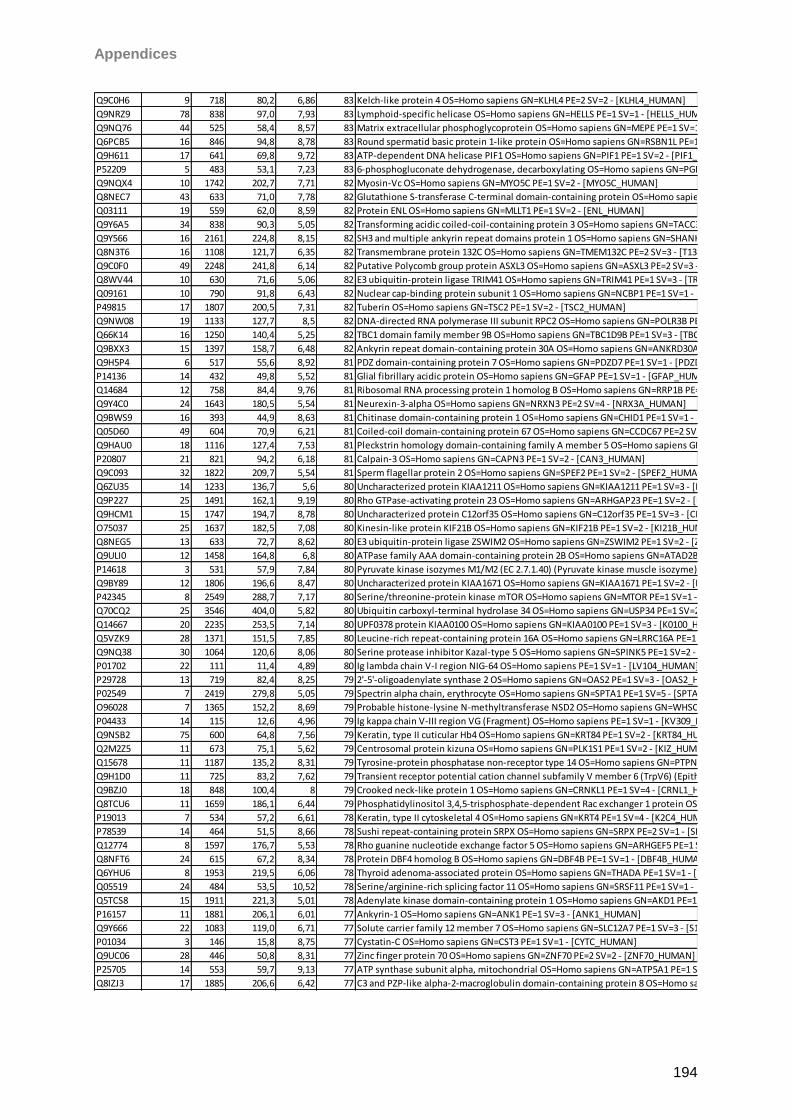
Appendices
194
Q9C0H6 9 718 80,2 6,86 83 Kelch-like protein 4 OS=Homo sapiens GN=KLHL4 PE=2 SV=2 - [KLHL4_HUMAN]
Q9NRZ9 78 838 97,0 7,93 83 Lymphoid-specific helicase OS=Homo sapiens GN=HELLS PE=1 SV=1 - [HELLS_HUMAN]
Q9NQ76 44 525 58,4 8,57 83 Matrix extracellular phosphoglycoprotein OS=Homo sapiens GN=MEPE PE=1 SV=1 - [MEPE_HUMAN]
Q6PCB5 16 846 94,8 8,78 83 Round spermatid basic protein 1-like protein OS=Homo sapiens GN=RSBN1L PE=1 SV=2 - [RSBNL_HUMAN]
Q9H611 17 641 69,8 9,72 83 ATP-dependent DNA helicase PIF1 OS=Homo sapiens GN=PIF1 PE=1 SV=2 - [PIF1_HUMAN]
P52209 5 483 53,1 7,23 83 6-phosphogluconate dehydrogenase, decarboxylating OS=Homo sapiens GN=PGD PE=1 SV=3 - [6PGD_HUMAN]
Q9NQX4 10 1742 202,7 7,71 82 Myosin-Vc OS=Homo sapiens GN=MYO5C PE=1 SV=2 - [MYO5C_HUMAN]
Q8NEC7 43 633 71,0 7,78 82 Glutathione S-transferase C-terminal domain-containing protein OS=Homo sapiens GN=GSTCD PE=1 SV=2 - [GSTCD_HUMAN]
Q03111 19 559 62,0 8,59 82 Protein ENL OS=Homo sapiens GN=MLLT1 PE=1 SV=2 - [ENL_HUMAN]
Q9Y6A5 34 838 90,3 5,05 82 Transforming acidic coiled-coil-containing protein 3 OS=Homo sapiens GN=TACC3 PE=1 SV=1 - [TACC3_HUMAN]
Q9Y566 16 2161 224,8 8,15 82 SH3 and multiple ankyrin repeat domains protein 1 OS=Homo sapiens GN=SHANK1 PE=1 SV=2 - [SHAN1_HUMAN]
Q8N3T6 16 1108 121,7 6,35 82 Transmembrane protein 132C OS=Homo sapiens GN=TMEM132C PE=2 SV=3 - [T132C_HUMAN]
Q9C0F0 49 2248 241,8 6,14 82 Putative Polycomb group protein ASXL3 OS=Homo sapiens GN=ASXL3 PE=2 SV=3 - [ASXL3_HUMAN]
Q8WV44 10 630 71,6 5,06 82 E3 ubiquitin-protein ligase TRIM41 OS=Homo sapiens GN=TRIM41 PE=1 SV=3 - [TRI41_HUMAN]
Q09161 10 790 91,8 6,43 82 Nuclear cap-binding protein subunit 1 OS=Homo sapiens GN=NCBP1 PE=1 SV=1 - [NCBP1_HUMAN]
P49815 17 1807 200,5 7,31 82 Tuberin OS=Homo sapiens GN=TSC2 PE=1 SV=2 - [TSC2_HUMAN]
Q9NW08 19 1133 127,7 8,5 82 DNA-directed RNA polymerase III subunit RPC2 OS=Homo sapiens GN=POLR3B PE=1 SV=2 - [RPC2_HUMAN]
Q66K14 16 1250 140,4 5,25 82 TBC1 domain family member 9B OS=Homo sapiens GN=TBC1D9B PE=1 SV=3 - [TBC9B_HUMAN]
Q9BXX3 15 1397 158,7 6,48 82 Ankyrin repeat domain-containing protein 30A OS=Homo sapiens GN=ANKRD30A PE=1 SV=3 - [AN30A_HUMAN]
Q9H5P4 6 517 55,6 8,92 81 PDZ domain-containing protein 7 OS=Homo sapiens GN=PDZD7 PE=1 SV=1 - [PDZD7_HUMAN]
P14136 14 432 49,8 5,52 81 Glial fibrillary acidic protein OS=Homo sapiens GN=GFAP PE=1 SV=1 - [GFAP_HUMAN]
Q14684 12 758 84,4 9,76 81 Ribosomal RNA processing protein 1 homolog B OS=Homo sapiens GN=RRP1B PE=1 SV=3 - [RRP1B_HUMAN]
Q9Y4C0 24 1643 180,5 5,54 81 Neurexin-3-alpha OS=Homo sapiens GN=NRXN3 PE=2 SV=4 - [NRX3A_HUMAN]
Q9BWS9 16 393 44,9 8,63 81 Chitinase domain-containing protein 1 OS=Homo sapiens GN=CHID1 PE=1 SV=1 - [CHID1_HUMAN]
Q05D60 49 604 70,9 6,21 81 Coiled-coil domain-containing protein 67 OS=Homo sapiens GN=CCDC67 PE=2 SV=2 - [CCD67_HUMAN]
Q9HAU0 18 1116 127,4 7,53 81 Pleckstrin homology domain-containing family A member 5 OS=Homo sapiens GN=PLEKHA5 PE=1 SV=1 - [PKHA5_HUMAN]
P20807 21 821 94,2 6,18 81 Calpain-3 OS=Homo sapiens GN=CAPN3 PE=1 SV=2 - [CAN3_HUMAN]
Q9C093 32 1822 209,7 5,54 81 Sperm flagellar protein 2 OS=Homo sapiens GN=SPEF2 PE=1 SV=2 - [SPEF2_HUMAN]
Q6ZU35 14 1233 136,7 5,6 80 Uncharacterized protein KIAA1211 OS=Homo sapiens GN=KIAA1211 PE=1 SV=3 - [K1211_HUMAN]
Q9P227 25 1491 162,1 9,19 80 Rho GTPase-activating protein 23 OS=Homo sapiens GN=ARHGAP23 PE=1 SV=2 - [RHG23_HUMAN]
Q9HCM1 15 1747 194,7 8,78 80 Uncharacterized protein C12orf35 OS=Homo sapiens GN=C12orf35 PE=1 SV=3 - [CL035_HUMAN]
O75037 25 1637 182,5 7,08 80 Kinesin-like protein KIF21B OS=Homo sapiens GN=KIF21B PE=1 SV=2 - [KI21B_HUMAN]
Q8NEG5 13 633 72,7 8,62 80 E3 ubiquitin-protein ligase ZSWIM2 OS=Homo sapiens GN=ZSWIM2 PE=1 SV=2 - [ZSWM2_HUMAN]
Q9ULI0 12 1458 164,8 6,8 80 ATPase family AAA domain-containing protein 2B OS=Homo sapiens GN=ATAD2B PE=1 SV=3 - [ATD2B_HUMAN]
P14618 3 531 57,9 7,84 80 Pyruvate kinase isozymes M1/M2 (EC 2.7.1.40) (Pyruvate kinase muscle isozyme) (Pyruvate kinase 2/3) (Cytosolic thyroid hormone-binding protein) (CTHBP) (THBP1) - Homo sapiens (Human) - [KPYM_HUMAN]
Q9BY89 12 1806 196,6 8,47 80 Uncharacterized protein KIAA1671 OS=Homo sapiens GN=KIAA1671 PE=1 SV=2 - [K1671_HUMAN]
P42345 8 2549 288,7 7,17 80 Serine/threonine-protein kinase mTOR OS=Homo sapiens GN=MTOR PE=1 SV=1 - [MTOR_HUMAN]
Q70CQ2 25 3546 404,0 5,82 80 Ubiquitin carboxyl-terminal hydrolase 34 OS=Homo sapiens GN=USP34 PE=1 SV=2 - [UBP34_HUMAN]
Q14667 20 2235 253,5 7,14 80 UPF0378 protein KIAA0100 OS=Homo sapiens GN=KIAA0100 PE=1 SV=3 - [K0100_HUMAN]
Q5VZK9 28 1371 151,5 7,85 80 Leucine-rich repeat-containing protein 16A OS=Homo sapiens GN=LRRC16A PE=1 SV=1 - [LR16A_HUMAN]
Q9NQ38 30 1064 120,6 8,06 80 Serine protease inhibitor Kazal-type 5 OS=Homo sapiens GN=SPINK5 PE=1 SV=2 - [ISK5_HUMAN]
P01702 22 111 11,4 4,89 80 Ig lambda chain V-I region NIG-64 OS=Homo sapiens PE=1 SV=1 - [LV104_HUMAN]
P29728 13 719 82,4 8,25 79 2'-5'-oligoadenylate synthase 2 OS=Homo sapiens GN=OAS2 PE=1 SV=3 - [OAS2_HUMAN]
P02549 7 2419 279,8 5,05 79 Spectrin alpha chain, erythrocyte OS=Homo sapiens GN=SPTA1 PE=1 SV=5 - [SPTA1_HUMAN]
O96028 7 1365 152,2 8,69 79 Probable histone-lysine N-methyltransferase NSD2 OS=Homo sapiens GN=WHSC1 PE=1 SV=1 - [NSD2_HUMAN]
P04433 14 115 12,6 4,96 79 Ig kappa chain V-III region VG (Fragment) OS=Homo sapiens PE=1 SV=1 - [KV309_HUMAN]
Q9NSB2 75 600 64,8 7,56 79 Keratin, type II cuticular Hb4 OS=Homo sapiens GN=KRT84 PE=1 SV=2 - [KRT84_HUMAN]
Q2M2Z5 11 673 75,1 5,62 79 Centrosomal protein kizuna OS=Homo sapiens GN=PLK1S1 PE=1 SV=2 - [KIZ_HUMAN]
Q15678 11 1187 135,2 8,31 79 Tyrosine-protein phosphatase non-receptor type 14 OS=Homo sapiens GN=PTPN14 PE=1 SV=2 - [PTN14_HUMAN]
Q9H1D0 11 725 83,2 7,62 79 Transient receptor potential cation channel subfamily V member 6 (TrpV6) (Epithelial calcium channel 2) (ECaC2) (Calcium transport protein 1) (CaT1) (CaT-like) (CaT-L) - Homo sapiens (Human) - [TRPV6_HUMAN]
Q9BZJ0 18 848 100,4 8 79 Crooked neck-like protein 1 OS=Homo sapiens GN=CRNKL1 PE=1 SV=4 - [CRNL1_HUMAN]
Q8TCU6 11 1659 186,1 6,44 79 Phosphatidylinositol 3,4,5-trisphosphate-dependent Rac exchanger 1 protein OS=Homo sapiens GN=PREX1 PE=1 SV=3 - [PREX1_HUMAN]
P19013 7 534 57,2 6,61 78 Keratin, type II cytoskeletal 4 OS=Homo sapiens GN=KRT4 PE=1 SV=4 - [K2C4_HUMAN]
P78539 14 464 51,5 8,66 78 Sushi repeat-containing protein SRPX OS=Homo sapiens GN=SRPX PE=2 SV=1 - [SRPX_HUMAN]
Q12774 8 1597 176,7 5,53 78 Rho guanine nucleotide exchange factor 5 OS=Homo sapiens GN=ARHGEF5 PE=1 SV=3 - [ARHG5_HUMAN]
Q8NFT6 24 615 67,2 8,34 78 Protein DBF4 homolog B OS=Homo sapiens GN=DBF4B PE=1 SV=1 - [DBF4B_HUMAN]
Q6YHU6 8 1953 219,5 6,06 78 Thyroid adenoma-associated protein OS=Homo sapiens GN=THADA PE=1 SV=1 - [THADA_HUMAN]
Q05519 24 484 53,5 10,52 78 Serine/arginine-rich splicing factor 11 OS=Homo sapiens GN=SRSF11 PE=1 SV=1 - [SRS11_HUMAN]
Q5TCS8 15 1911 221,3 5,01 78 Adenylate kinase domain-containing protein 1 OS=Homo sapiens GN=AKD1 PE=1 SV=2 - [AKD1_HUMAN]
P16157 11 1881 206,1 6,01 77 Ankyrin-1 OS=Homo sapiens GN=ANK1 PE=1 SV=3 - [ANK1_HUMAN]
Q9Y666 22 1083 119,0 6,71 77 Solute carrier family 12 member 7 OS=Homo sapiens GN=SLC12A7 PE=1 SV=3 - [S12A7_HUMAN]
P01034 3 146 15,8 8,75 77 Cystatin-C OS=Homo sapiens GN=CST3 PE=1 SV=1 - [CYTC_HUMAN]
Q9UC06 28 446 50,8 8,31 77 Zinc finger protein 70 OS=Homo sapiens GN=ZNF70 PE=2 SV=2 - [ZNF70_HUMAN]
P25705 14 553 59,7 9,13 77 ATP synthase subunit alpha, mitochondrial OS=Homo sapiens GN=ATP5A1 PE=1 SV=1 - [ATPA_HUMAN]
Q8IZJ3 17 1885 206,6 6,42 77 C3 and PZP-like alpha-2-macroglobulin domain-containing protein 8 OS=Homo sapiens GN=CPAMD8 PE=1 SV=2 - [CPMD8_HUMAN]
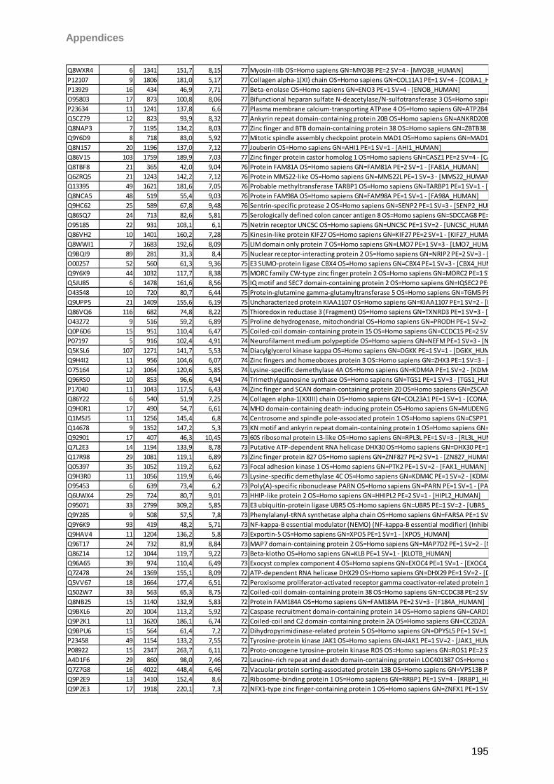
Appendices
195
Q8WXR4 6 1341 151,7 8,15 77 Myosin-IIIb OS=Homo sapiens GN=MYO3B PE=2 SV=4 - [MYO3B_HUMAN]
P12107 9 1806 181,0 5,17 77 Collagen alpha-1(XI) chain OS=Homo sapiens GN=COL11A1 PE=1 SV=4 - [COBA1_HUMAN]
P13929 16 434 46,9 7,71 77 Beta-enolase OS=Homo sapiens GN=ENO3 PE=1 SV=4 - [ENOB_HUMAN]
O95803 17 873 100,8 8,06 77 Bifunctional heparan sulfate N-deacetylase/N-sulfotransferase 3 OS=Homo sapiens GN=NDST3 PE=2 SV=1 - [NDST3_HUMAN]
P23634 11 1241 137,8 6,6 77 Plasma membrane calcium-transporting ATPase 4 OS=Homo sapiens GN=ATP2B4 PE=1 SV=2 - [AT2B4_HUMAN]
Q5CZ79 12 823 93,9 8,32 77 Ankyrin repeat domain-containing protein 20B OS=Homo sapiens GN=ANKRD20B PE=2 SV=2 - [AN20B_HUMAN]
Q8NAP3 7 1195 134,2 8,03 77 Zinc finger and BTB domain-containing protein 38 OS=Homo sapiens GN=ZBTB38 PE=1 SV=2 - [ZBT38_HUMAN]
Q9Y6D9 8 718 83,0 5,92 77 Mitotic spindle assembly checkpoint protein MAD1 OS=Homo sapiens GN=MAD1L1 PE=1 SV=2 - [MD1L1_HUMAN]
Q8N157 20 1196 137,0 7,12 77 Jouberin OS=Homo sapiens GN=AHI1 PE=1 SV=1 - [AHI1_HUMAN]
Q86V15 103 1759 189,9 7,03 77 Zinc finger protein castor homolog 1 OS=Homo sapiens GN=CASZ1 PE=2 SV=4 - [CASZ1_HUMAN]
Q8TBF8 21 365 42,0 9,04 76 Protein FAM81A OS=Homo sapiens GN=FAM81A PE=2 SV=1 - [FA81A_HUMAN]
Q6ZRQ5 21 1243 142,2 7,12 76 Protein MMS22-like OS=Homo sapiens GN=MMS22L PE=1 SV=3 - [MMS22_HUMAN]
Q13395 49 1621 181,6 7,05 76 Probable methyltransferase TARBP1 OS=Homo sapiens GN=TARBP1 PE=1 SV=1 - [TARB1_HUMAN]
Q8NCA5 48 519 55,4 9,03 76 Protein FAM98A OS=Homo sapiens GN=FAM98A PE=1 SV=1 - [FA98A_HUMAN]
Q9HC62 25 589 67,8 9,48 76 Sentrin-specific protease 2 OS=Homo sapiens GN=SENP2 PE=1 SV=3 - [SENP2_HUMAN]
Q86SQ7 24 713 82,6 5,81 75 Serologically defined colon cancer antigen 8 OS=Homo sapiens GN=SDCCAG8 PE=1 SV=1 - [SDCG8_HUMAN]
O95185 22 931 103,1 6,1 75 Netrin receptor UNC5C OS=Homo sapiens GN=UNC5C PE=1 SV=2 - [UNC5C_HUMAN]
Q86VH2 10 1401 160,2 7,28 75 Kinesin-like protein KIF27 OS=Homo sapiens GN=KIF27 PE=2 SV=1 - [KIF27_HUMAN]
Q8WWI1 7 1683 192,6 8,09 75 LIM domain only protein 7 OS=Homo sapiens GN=LMO7 PE=1 SV=3 - [LMO7_HUMAN]
Q9BQI9 89 281 31,3 8,4 75 Nuclear receptor-interacting protein 2 OS=Homo sapiens GN=NRIP2 PE=2 SV=3 - [NRIP2_HUMAN]
O00257 52 560 61,3 9,36 75 E3 SUMO-protein ligase CBX4 OS=Homo sapiens GN=CBX4 PE=1 SV=3 - [CBX4_HUMAN]
Q9Y6X9 44 1032 117,7 8,38 75 MORC family CW-type zinc finger protein 2 OS=Homo sapiens GN=MORC2 PE=1 SV=2 - [MORC2_HUMAN]
Q5JU85 6 1478 161,6 8,56 75 IQ motif and SEC7 domain-containing protein 2 OS=Homo sapiens GN=IQSEC2 PE=1 SV=1 - [IQEC2_HUMAN]
O43548 10 720 80,7 6,44 75 Protein-glutamine gamma-glutamyltransferase 5 OS=Homo sapiens GN=TGM5 PE=1 SV=4 - [TGM5_HUMAN]
Q9UPP5 21 1409 155,6 6,19 75 Uncharacterized protein KIAA1107 OS=Homo sapiens GN=KIAA1107 PE=1 SV=2 - [K1107_HUMAN]
Q86VQ6 116 682 74,8 8,22 75 Thioredoxin reductase 3 (Fragment) OS=Homo sapiens GN=TXNRD3 PE=1 SV=3 - [TRXR3_HUMAN]
O43272 9 516 59,2 6,89 75 Proline dehydrogenase, mitochondrial OS=Homo sapiens GN=PRODH PE=1 SV=2 - [PROD_HUMAN]
Q0P6D6 15 951 110,4 6,47 75 Coiled-coil domain-containing protein 15 OS=Homo sapiens GN=CCDC15 PE=2 SV=2 - [CCD15_HUMAN]
P07197 5 916 102,4 4,91 74 Neurofilament medium polypeptide OS=Homo sapiens GN=NEFM PE=1 SV=3 - [NFM_HUMAN]
Q5KSL6 107 1271 141,7 5,53 74 Diacylglycerol kinase kappa OS=Homo sapiens GN=DGKK PE=1 SV=1 - [DGKK_HUMAN]
Q9H4I2 11 956 104,6 6,07 74 Zinc fingers and homeoboxes protein 3 OS=Homo sapiens GN=ZHX3 PE=1 SV=3 - [ZHX3_HUMAN]
O75164 12 1064 120,6 5,85 74 Lysine-specific demethylase 4A OS=Homo sapiens GN=KDM4A PE=1 SV=2 - [KDM4A_HUMAN]
Q96RS0 10 853 96,6 4,94 74 Trimethylguanosine synthase OS=Homo sapiens GN=TGS1 PE=1 SV=3 - [TGS1_HUMAN]
P17040 11 1043 117,5 6,43 74 Zinc finger and SCAN domain-containing protein 20 OS=Homo sapiens GN=ZSCAN20 PE=2 SV=3 - [ZSC20_HUMAN]
Q86Y22 6 540 51,9 7,25 74 Collagen alpha-1(XXIII) chain OS=Homo sapiens GN=COL23A1 PE=1 SV=1 - [CONA1_HUMAN]
Q9H0R1 17 490 54,7 6,61 74 MHD domain-containing death-inducing protein OS=Homo sapiens GN=MUDENG PE=2 SV=2 - [MUDEN_HUMAN]
Q1MSJ5 11 1256 145,4 6,8 74 Centrosome and spindle pole-associated protein 1 OS=Homo sapiens GN=CSPP1 PE=1 SV=4 - [CSPP1_HUMAN]
Q14678 9 1352 147,2 5,3 73 KN motif and ankyrin repeat domain-containing protein 1 OS=Homo sapiens GN=KANK1 PE=1 SV=3 - [KANK1_HUMAN]
Q92901 17 407 46,3 10,45 73 60S ribosomal protein L3-like OS=Homo sapiens GN=RPL3L PE=1 SV=3 - [RL3L_HUMAN]
Q7L2E3 14 1194 133,9 8,78 73 Putative ATP-dependent RNA helicase DHX30 OS=Homo sapiens GN=DHX30 PE=1 SV=1 - [DHX30_HUMAN]
Q17R98 29 1081 119,1 6,89 73 Zinc finger protein 827 OS=Homo sapiens GN=ZNF827 PE=2 SV=1 - [ZN827_HUMAN]
Q05397 35 1052 119,2 6,62 73 Focal adhesion kinase 1 OS=Homo sapiens GN=PTK2 PE=1 SV=2 - [FAK1_HUMAN]
Q9H3R0 11 1056 119,9 6,46 73 Lysine-specific demethylase 4C OS=Homo sapiens GN=KDM4C PE=1 SV=2 - [KDM4C_HUMAN]
O95453 6 639 73,4 6,2 73 Poly(A)-specific ribonuclease PARN OS=Homo sapiens GN=PARN PE=1 SV=1 - [PARN_HUMAN]
Q6UWX4 29 724 80,7 9,01 73 HHIP-like protein 2 OS=Homo sapiens GN=HHIPL2 PE=2 SV=1 - [HIPL2_HUMAN]
O95071 33 2799 309,2 5,85 73 E3 ubiquitin-protein ligase UBR5 OS=Homo sapiens GN=UBR5 PE=1 SV=2 - [UBR5_HUMAN]
Q9Y285 9 508 57,5 7,8 73 Phenylalanyl-tRNA synthetase alpha chain OS=Homo sapiens GN=FARSA PE=1 SV=3 - [SYFA_HUMAN]
Q9Y6K9 93 419 48,2 5,71 73 NF-kappa-B essential modulator (NEMO) (NF-kappa-B essential modifier) (Inhibitor of nuclear factor kappa-B kinase subunit gamma) (IkB kinase subunit gamma) (I-kappa-B kinase gamma) (IKK-gamma) (IKKG) (IkB kinase-associated protein 1) (IKKAP1) (FIP-3) - Homo sapiens (Human) - [NEMO_HUMAN]
Q9HAV4 11 1204 136,2 5,8 73 Exportin-5 OS=Homo sapiens GN=XPO5 PE=1 SV=1 - [XPO5_HUMAN]
Q96T17 24 732 81,9 8,84 73 MAP7 domain-containing protein 2 OS=Homo sapiens GN=MAP7D2 PE=1 SV=2 - [MA7D2_HUMAN]
Q86Z14 12 1044 119,7 9,22 73 Beta-klotho OS=Homo sapiens GN=KLB PE=1 SV=1 - [KLOTB_HUMAN]
Q96A65 39 974 110,4 6,49 73 Exocyst complex component 4 OS=Homo sapiens GN=EXOC4 PE=1 SV=1 - [EXOC4_HUMAN]
Q7Z478 24 1369 155,1 8,09 72 ATP-dependent RNA helicase DHX29 OS=Homo sapiens GN=DHX29 PE=1 SV=2 - [DHX29_HUMAN]
Q5VV67 18 1664 177,4 6,51 72 Peroxisome proliferator-activated receptor gamma coactivator-related protein 1 OS=Homo sapiens GN=PPRC1 PE=1 SV=1 - [PPRC1_HUMAN]
Q502W7 33 563 65,3 8,75 72 Coiled-coil domain-containing protein 38 OS=Homo sapiens GN=CCDC38 PE=2 SV=1 - [CCD38_HUMAN]
Q8NB25 15 1140 132,9 5,83 72 Protein FAM184A OS=Homo sapiens GN=FAM184A PE=2 SV=3 - [F184A_HUMAN]
Q9BXL6 20 1004 113,2 5,92 72 Caspase recruitment domain-containing protein 14 OS=Homo sapiens GN=CARD14 PE=1 SV=2 - [CAR14_HUMAN]
Q9P2K1 11 1620 186,1 6,74 72 Coiled-coil and C2 domain-containing protein 2A OS=Homo sapiens GN=CC2D2A PE=1 SV=3 - [C2D2A_HUMAN]
Q9BPU6 15 564 61,4 7,2 72 Dihydropyrimidinase-related protein 5 OS=Homo sapiens GN=DPYSL5 PE=1 SV=1 - [DPYL5_HUMAN]
P23458 49 1154 133,2 7,55 72 Tyrosine-protein kinase JAK1 OS=Homo sapiens GN=JAK1 PE=1 SV=2 - [JAK1_HUMAN]
P08922 15 2347 263,7 6,11 72 Proto-oncogene tyrosine-protein kinase ROS OS=Homo sapiens GN=ROS1 PE=2 SV=3 - [ROS_HUMAN]
A4D1F6 29 860 98,0 7,46 72 Leucine-rich repeat and death domain-containing protein LOC401387 OS=Homo sapiens PE=2 SV=2 - [YG015_HUMAN]
Q7Z7G8 16 4022 448,4 6,46 72 Vacuolar protein sorting-associated protein 13B OS=Homo sapiens GN=VPS13B PE=1 SV=2 - [VP13B_HUMAN]
Q9P2E9 13 1410 152,4 8,6 72 Ribosome-binding protein 1 OS=Homo sapiens GN=RRBP1 PE=1 SV=4 - [RRBP1_HUMAN]
Q9P2E3 17 1918 220,1 7,3 72 NFX1-type zinc finger-containing protein 1 OS=Homo sapiens GN=ZNFX1 PE=1 SV=2 - [ZNFX1_HUMAN]
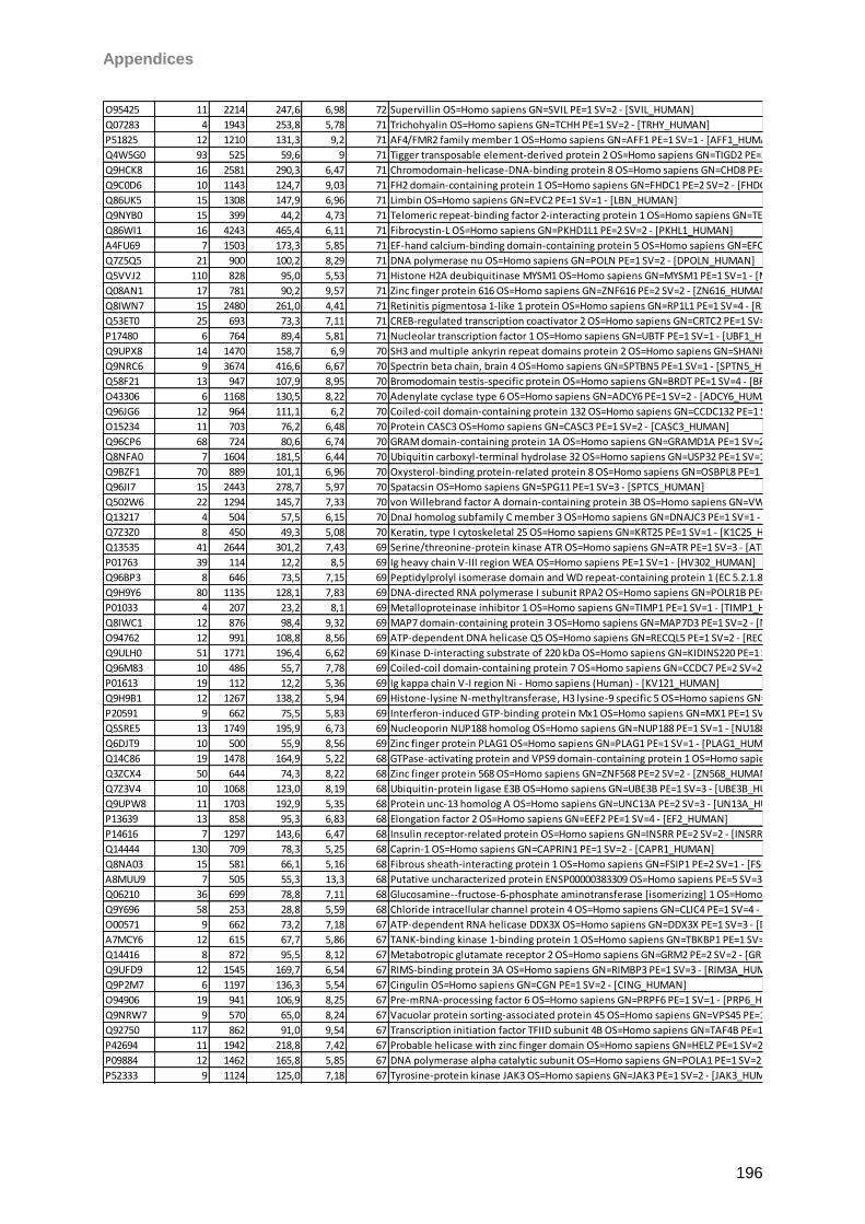
Appendices
196
O95425 11 2214 247,6 6,98 72 Supervillin OS=Homo sapiens GN=SVIL PE=1 SV=2 - [SVIL_HUMAN]
Q07283 4 1943 253,8 5,78 71 Trichohyalin OS=Homo sapiens GN=TCHH PE=1 SV=2 - [TRHY_HUMAN]
P51825 12 1210 131,3 9,2 71 AF4/FMR2 family member 1 OS=Homo sapiens GN=AFF1 PE=1 SV=1 - [AFF1_HUMAN]
Q4W5G0 93 525 59,6 9 71 Tigger transposable element-derived protein 2 OS=Homo sapiens GN=TIGD2 PE=2 SV=1 - [TIGD2_HUMAN]
Q9HCK8 16 2581 290,3 6,47 71 Chromodomain-helicase-DNA-binding protein 8 OS=Homo sapiens GN=CHD8 PE=1 SV=5 - [CHD8_HUMAN]
Q9C0D6 10 1143 124,7 9,03 71 FH2 domain-containing protein 1 OS=Homo sapiens GN=FHDC1 PE=2 SV=2 - [FHDC1_HUMAN]
Q86UK5 15 1308 147,9 6,96 71 Limbin OS=Homo sapiens GN=EVC2 PE=1 SV=1 - [LBN_HUMAN]
Q9NYB0 15 399 44,2 4,73 71 Telomeric repeat-binding factor 2-interacting protein 1 OS=Homo sapiens GN=TERF2IP PE=1 SV=1 - [TE2IP_HUMAN]
Q86WI1 16 4243 465,4 6,11 71 Fibrocystin-L OS=Homo sapiens GN=PKHD1L1 PE=2 SV=2 - [PKHL1_HUMAN]
A4FU69 7 1503 173,3 5,85 71 EF-hand calcium-binding domain-containing protein 5 OS=Homo sapiens GN=EFCAB5 PE=1 SV=2 - [EFCB5_HUMAN]
Q7Z5Q5 21 900 100,2 8,29 71 DNA polymerase nu OS=Homo sapiens GN=POLN PE=1 SV=2 - [DPOLN_HUMAN]
Q5VVJ2 110 828 95,0 5,53 71 Histone H2A deubiquitinase MYSM1 OS=Homo sapiens GN=MYSM1 PE=1 SV=1 - [MYSM1_HUMAN]
Q08AN1 17 781 90,2 9,57 71 Zinc finger protein 616 OS=Homo sapiens GN=ZNF616 PE=2 SV=2 - [ZN616_HUMAN]
Q8IWN7 15 2480 261,0 4,41 71 Retinitis pigmentosa 1-like 1 protein OS=Homo sapiens GN=RP1L1 PE=1 SV=4 - [RP1L1_HUMAN]
Q53ET0 25 693 73,3 7,11 71 CREB-regulated transcription coactivator 2 OS=Homo sapiens GN=CRTC2 PE=1 SV=2 - [CRTC2_HUMAN]
P17480 6 764 89,4 5,81 71 Nucleolar transcription factor 1 OS=Homo sapiens GN=UBTF PE=1 SV=1 - [UBF1_HUMAN]
Q9UPX8 14 1470 158,7 6,9 70 SH3 and multiple ankyrin repeat domains protein 2 OS=Homo sapiens GN=SHANK2 PE=1 SV=3 - [SHAN2_HUMAN]
Q9NRC6 9 3674 416,6 6,67 70 Spectrin beta chain, brain 4 OS=Homo sapiens GN=SPTBN5 PE=1 SV=1 - [SPTN5_HUMAN]
Q58F21 13 947 107,9 8,95 70 Bromodomain testis-specific protein OS=Homo sapiens GN=BRDT PE=1 SV=4 - [BRDT_HUMAN]
O43306 6 1168 130,5 8,22 70 Adenylate cyclase type 6 OS=Homo sapiens GN=ADCY6 PE=1 SV=2 - [ADCY6_HUMAN]
Q96JG6 12 964 111,1 6,2 70 Coiled-coil domain-containing protein 132 OS=Homo sapiens GN=CCDC132 PE=1 SV=3 - [CC132_HUMAN]
O15234 11 703 76,2 6,48 70 Protein CASC3 OS=Homo sapiens GN=CASC3 PE=1 SV=2 - [CASC3_HUMAN]
Q96CP6 68 724 80,6 6,74 70 GRAM domain-containing protein 1A OS=Homo sapiens GN=GRAMD1A PE=1 SV=2 - [GRM1A_HUMAN]
Q8NFA0 7 1604 181,5 6,44 70 Ubiquitin carboxyl-terminal hydrolase 32 OS=Homo sapiens GN=USP32 PE=1 SV=1 - [UBP32_HUMAN]
Q9BZF1 70 889 101,1 6,96 70 Oxysterol-binding protein-related protein 8 OS=Homo sapiens GN=OSBPL8 PE=1 SV=3 - [OSBL8_HUMAN]
Q96JI7 15 2443 278,7 5,97 70 Spatacsin OS=Homo sapiens GN=SPG11 PE=1 SV=3 - [SPTCS_HUMAN]
Q502W6 22 1294 145,7 7,33 70 von Willebrand factor A domain-containing protein 3B OS=Homo sapiens GN=VWA3B PE=2 SV=3 - [VWA3B_HUMAN]
Q13217 4 504 57,5 6,15 70 DnaJ homolog subfamily C member 3 OS=Homo sapiens GN=DNAJC3 PE=1 SV=1 - [DNJC3_HUMAN]
Q7Z3Z0 8 450 49,3 5,08 70 Keratin, type I cytoskeletal 25 OS=Homo sapiens GN=KRT25 PE=1 SV=1 - [K1C25_HUMAN]
Q13535 41 2644 301,2 7,43 69 Serine/threonine-protein kinase ATR OS=Homo sapiens GN=ATR PE=1 SV=3 - [ATR_HUMAN]
P01763 39 114 12,2 8,5 69 Ig heavy chain V-III region WEA OS=Homo sapiens PE=1 SV=1 - [HV302_HUMAN]
Q96BP3 8 646 73,5 7,15 69 Peptidylprolyl isomerase domain and WD repeat-containing protein 1 (EC 5.2.1.8) - Homo sapiens (Human) - [PPWD1_HUMAN]
Q9H9Y6 80 1135 128,1 7,83 69 DNA-directed RNA polymerase I subunit RPA2 OS=Homo sapiens GN=POLR1B PE=1 SV=2 - [RPA2_HUMAN]
P01033 4 207 23,2 8,1 69 Metalloproteinase inhibitor 1 OS=Homo sapiens GN=TIMP1 PE=1 SV=1 - [TIMP1_HUMAN]
Q8IWC1 12 876 98,4 9,32 69 MAP7 domain-containing protein 3 OS=Homo sapiens GN=MAP7D3 PE=1 SV=2 - [MA7D3_HUMAN]
O94762 12 991 108,8 8,56 69 ATP-dependent DNA helicase Q5 OS=Homo sapiens GN=RECQL5 PE=1 SV=2 - [RECQ5_HUMAN]
Q9ULH0 51 1771 196,4 6,62 69 Kinase D-interacting substrate of 220 kDa OS=Homo sapiens GN=KIDINS220 PE=1 SV=3 - [KDIS_HUMAN]
Q96M83 10 486 55,7 7,78 69 Coiled-coil domain-containing protein 7 OS=Homo sapiens GN=CCDC7 PE=2 SV=2 - [CCDC7_HUMAN]
P01613 19 112 12,2 5,36 69 Ig kappa chain V-I region Ni - Homo sapiens (Human) - [KV121_HUMAN]
Q9H9B1 12 1267 138,2 5,94 69 Histone-lysine N-methyltransferase, H3 lysine-9 specific 5 OS=Homo sapiens GN=EHMT1 PE=1 SV=3 - [EHMT1_HUMAN]
P20591 9 662 75,5 5,83 69 Interferon-induced GTP-binding protein Mx1 OS=Homo sapiens GN=MX1 PE=1 SV=4 - [MX1_HUMAN]
Q5SRE5 13 1749 195,9 6,73 69 Nucleoporin NUP188 homolog OS=Homo sapiens GN=NUP188 PE=1 SV=1 - [NU188_HUMAN]
Q6DJT9 10 500 55,9 8,56 69 Zinc finger protein PLAG1 OS=Homo sapiens GN=PLAG1 PE=1 SV=1 - [PLAG1_HUMAN]
Q14C86 19 1478 164,9 5,22 68 GTPase-activating protein and VPS9 domain-containing protein 1 OS=Homo sapiens GN=GAPVD1 PE=1 SV=2 - [GAPD1_HUMAN]
Q3ZCX4 50 644 74,3 8,22 68 Zinc finger protein 568 OS=Homo sapiens GN=ZNF568 PE=2 SV=2 - [ZN568_HUMAN]
Q7Z3V4 10 1068 123,0 8,19 68 Ubiquitin-protein ligase E3B OS=Homo sapiens GN=UBE3B PE=1 SV=3 - [UBE3B_HUMAN]
Q9UPW8 11 1703 192,9 5,35 68 Protein unc-13 homolog A OS=Homo sapiens GN=UNC13A PE=2 SV=3 - [UN13A_HUMAN]
P13639 13 858 95,3 6,83 68 Elongation factor 2 OS=Homo sapiens GN=EEF2 PE=1 SV=4 - [EF2_HUMAN]
P14616 7 1297 143,6 6,47 68 Insulin receptor-related protein OS=Homo sapiens GN=INSRR PE=2 SV=2 - [INSRR_HUMAN]
Q14444 130 709 78,3 5,25 68 Caprin-1 OS=Homo sapiens GN=CAPRIN1 PE=1 SV=2 - [CAPR1_HUMAN]
Q8NA03 15 581 66,1 5,16 68 Fibrous sheath-interacting protein 1 OS=Homo sapiens GN=FSIP1 PE=2 SV=1 - [FSIP1_HUMAN]
A8MUU9 7 505 55,3 13,3 68 Putative uncharacterized protein ENSP00000383309 OS=Homo sapiens PE=5 SV=3 - [YV023_HUMAN]
Q06210 36 699 78,8 7,11 68 Glucosamine--fructose-6-phosphate aminotransferase [isomerizing] 1 OS=Homo sapiens GN=GFPT1 PE=1 SV=3 - [GFPT1_HUMAN]
Q9Y696 58 253 28,8 5,59 68 Chloride intracellular channel protein 4 OS=Homo sapiens GN=CLIC4 PE=1 SV=4 - [CLIC4_HUMAN]
O00571 9 662 73,2 7,18 67 ATP-dependent RNA helicase DDX3X OS=Homo sapiens GN=DDX3X PE=1 SV=3 - [DDX3X_HUMAN]
A7MCY6 12 615 67,7 5,86 67 TANK-binding kinase 1-binding protein 1 OS=Homo sapiens GN=TBKBP1 PE=1 SV=1 - [TBKB1_HUMAN]
Q14416 8 872 95,5 8,12 67 Metabotropic glutamate receptor 2 OS=Homo sapiens GN=GRM2 PE=2 SV=2 - [GRM2_HUMAN]
Q9UFD9 12 1545 169,7 6,54 67 RIMS-binding protein 3A OS=Homo sapiens GN=RIMBP3 PE=1 SV=3 - [RIM3A_HUMAN]
Q9P2M7 6 1197 136,3 5,54 67 Cingulin OS=Homo sapiens GN=CGN PE=1 SV=2 - [CING_HUMAN]
O94906 19 941 106,9 8,25 67 Pre-mRNA-processing factor 6 OS=Homo sapiens GN=PRPF6 PE=1 SV=1 - [PRP6_HUMAN]
Q9NRW7 9 570 65,0 8,24 67 Vacuolar protein sorting-associated protein 45 OS=Homo sapiens GN=VPS45 PE=1 SV=1 - [VPS45_HUMAN]
Q92750 117 862 91,0 9,54 67 Transcription initiation factor TFIID subunit 4B OS=Homo sapiens GN=TAF4B PE=1 SV=2 - [TAF4B_HUMAN]
P42694 11 1942 218,8 7,42 67 Probable helicase with zinc finger domain OS=Homo sapiens GN=HELZ PE=1 SV=2 - [HELZ_HUMAN]
P09884 12 1462 165,8 5,85 67 DNA polymerase alpha catalytic subunit OS=Homo sapiens GN=POLA1 PE=1 SV=2 - [DPOLA_HUMAN]
P52333 9 1124 125,0 7,18 67 Tyrosine-protein kinase JAK3 OS=Homo sapiens GN=JAK3 PE=1 SV=2 - [JAK3_HUMAN]
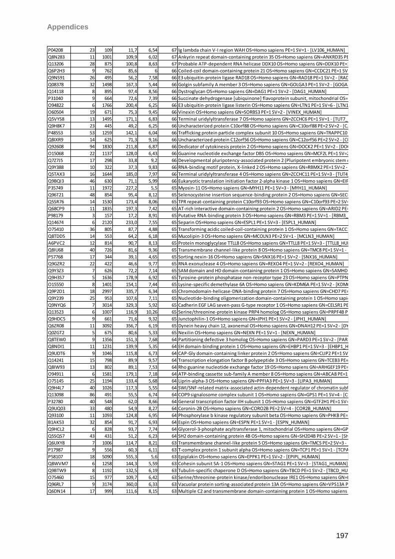
Appendices
197
P04208 23 109 11,7 6,54 67 Ig lambda chain V-I region WAH OS=Homo sapiens PE=1 SV=1 - [LV106_HUMAN]
Q8N283 11 1001 109,9 6,02 67 Ankyrin repeat domain-containing protein 35 OS=Homo sapiens GN=ANKRD35 PE=2 SV=2 - [ANR35_HUMAN]
Q13206 28 875 100,8 8,63 67 Probable ATP-dependent RNA helicase DDX10 OS=Homo sapiens GN=DDX10 PE=1 SV=2 - [DDX10_HUMAN]
Q6P2H3 9 762 85,6 6 66 Coiled-coil domain-containing protein 21 OS=Homo sapiens GN=CCDC21 PE=1 SV=1 - [CCD21_HUMAN]
Q9NS91 26 495 56,2 7,58 66 E3 ubiquitin-protein ligase RAD18 OS=Homo sapiens GN=RAD18 PE=1 SV=2 - [RAD18_HUMAN]
Q08378 32 1498 167,3 5,44 66 Golgin subfamily A member 3 OS=Homo sapiens GN=GOLGA3 PE=1 SV=2 - [GOGA3_HUMAN]
Q14118 8 895 97,4 8,56 66 Dystroglycan OS=Homo sapiens GN=DAG1 PE=1 SV=2 - [DAG1_HUMAN]
P31040 9 664 72,6 7,39 66 Succinate dehydrogenase [ubiquinone] flavoprotein subunit, mitochondrial OS=Homo sapiens GN=SDHA PE=1 SV=2 - [DHSA_HUMAN]
O94822 6 1766 200,4 6,25 66 E3 ubiquitin-protein ligase listerin OS=Homo sapiens GN=LTN1 PE=1 SV=6 - [LTN1_HUMAN]
O60504 19 671 75,3 9,45 66 Vinexin OS=Homo sapiens GN=SORBS3 PE=1 SV=2 - [VINEX_HUMAN]
Q5VYS8 13 1495 171,1 6,83 66 Terminal uridylyltransferase 7 OS=Homo sapiens GN=ZCCHC6 PE=1 SV=1 - [TUT7_HUMAN]
Q9H8K7 23 445 49,2 6,25 66 Uncharacterized protein C10orf88 OS=Homo sapiens GN=C10orf88 PE=2 SV=2 - [CJ088_HUMAN]
P48553 53 1259 142,1 6,04 66 Trafficking protein particle complex subunit 10 OS=Homo sapiens GN=TRAPPC10 PE=1 SV=2 - [TPC10_HUMAN]
Q8IXR9 14 625 71,3 9,16 66 Uncharacterized protein C12orf56 OS=Homo sapiens GN=C12orf56 PE=2 SV=2 - [CL056_HUMAN]
Q92608 94 1830 211,8 6,87 66 Dedicator of cytokinesis protein 2 OS=Homo sapiens GN=DOCK2 PE=1 SV=2 - [DOCK2_HUMAN]
O15068 22 1137 128,0 6,43 66 Guanine nucleotide exchange factor DBS OS=Homo sapiens GN=MCF2L PE=1 SV=2 - [MCF2L_HUMAN]
Q7Z7J5 17 298 33,8 9,2 66 Developmental pluripotency-associated protein 2 (Pluripotent embryonic stem cell-related protein 1) - Homo sapiens (Human) - [DPPA2_HUMAN]
Q9Y388 10 322 37,3 9,83 66 RNA-binding motif protein, X-linked 2 OS=Homo sapiens GN=RBMX2 PE=1 SV=2 - [RBMX2_HUMAN]
Q5TAX3 16 1644 185,0 7,97 66 Terminal uridylyltransferase 4 OS=Homo sapiens GN=ZCCHC11 PE=1 SV=3 - [TUT4_HUMAN]
Q9BQI3 46 630 71,1 5,99 66 Eukaryotic translation initiation factor 2-alpha kinase 1 OS=Homo sapiens GN=EIF2AK1 PE=1 SV=2 - [E2AK1_HUMAN]
P35749 11 1972 227,2 5,5 65 Myosin-11 OS=Homo sapiens GN=MYH11 PE=1 SV=3 - [MYH11_HUMAN]
Q96T21 48 854 95,4 8,12 65 Selenocysteine insertion sequence-binding protein 2 OS=Homo sapiens GN=SECISBP2 PE=1 SV=2 - [SEBP2_HUMAN]
Q5SR76 14 1530 173,4 8,06 65 TPR repeat-containing protein C10orf93 OS=Homo sapiens GN=C10orf93 PE=2 SV=2 - [CJ093_HUMAN]
Q68CP9 11 1835 197,3 7,42 65 AT-rich interactive domain-containing protein 2 OS=Homo sapiens GN=ARID2 PE=1 SV=2 - [ARID2_HUMAN]
P98179 3 157 17,2 8,91 65 Putative RNA-binding protein 3 OS=Homo sapiens GN=RBM3 PE=1 SV=1 - [RBM3_HUMAN]
Q14674 6 2120 233,0 7,55 65 Separin OS=Homo sapiens GN=ESPL1 PE=1 SV=3 - [ESPL1_HUMAN]
O75410 36 805 87,7 4,88 65 Transforming acidic coiled-coil-containing protein 1 OS=Homo sapiens GN=TACC1 PE=1 SV=2 - [TACC1_HUMAN]
Q8TDD5 14 553 64,2 6,18 65 Mucolipin-3 OS=Homo sapiens GN=MCOLN3 PE=2 SV=1 - [MCLN3_HUMAN]
A6PVC2 12 814 90,7 8,13 65 Protein monoglycylase TTLL8 OS=Homo sapiens GN=TTLL8 PE=1 SV=3 - [TTLL8_HUMAN]
Q8IU68 40 726 81,6 9,36 65 Transmembrane channel-like protein 8 OS=Homo sapiens GN=TMC8 PE=1 SV=1 - [TMC8_HUMAN]
P57768 17 344 39,1 4,65 65 Sorting nexin-16 OS=Homo sapiens GN=SNX16 PE=1 SV=2 - [SNX16_HUMAN]
Q9GZR2 22 422 46,6 9,77 65 RNA exonuclease 4 OS=Homo sapiens GN=REXO4 PE=1 SV=2 - [REXO4_HUMAN]
Q9Y3Z3 7 626 72,2 7,14 65 SAM domain and HD domain-containing protein 1 OS=Homo sapiens GN=SAMHD1 PE=1 SV=2 - [SAMH1_HUMAN]
Q9H3S7 5 1636 178,9 6,92 65 Tyrosine-protein phosphatase non-receptor type 23 OS=Homo sapiens GN=PTPN23 PE=1 SV=1 - [PTN23_HUMAN]
O15550 8 1401 154,1 7,44 65 Lysine-specific demethylase 6A OS=Homo sapiens GN=KDM6A PE=1 SV=2 - [KDM6A_HUMAN]
Q9P2D1 18 2997 335,7 6,34 65 Chromodomain-helicase-DNA-binding protein 7 OS=Homo sapiens GN=CHD7 PE=1 SV=3 - [CHD7_HUMAN]
Q9Y239 25 953 107,6 7,11 65 Nucleotide-binding oligomerization domain-containing protein 1 OS=Homo sapiens GN=NOD1 PE=1 SV=1 - [NOD1_HUMAN]
Q9NYQ6 7 3014 329,3 5,92 65 Cadherin EGF LAG seven-pass G-type receptor 1 OS=Homo sapiens GN=CELSR1 PE=1 SV=1 - [CELR1_HUMAN]
Q13523 6 1007 116,9 10,26 65 Serine/threonine-protein kinase PRP4 homolog OS=Homo sapiens GN=PRPF4B PE=1 SV=3 - [PRP4B_HUMAN]
Q9HDC5 9 661 71,6 9,32 65 Junctophilin-1 OS=Homo sapiens GN=JPH1 PE=1 SV=2 - [JPH1_HUMAN]
Q6ZR08 11 3092 356,7 6,19 65 Dynein heavy chain 12, axonemal OS=Homo sapiens GN=DNAH12 PE=1 SV=2 - [DYH12_HUMAN]
Q0ZGT2 5 675 80,6 5,33 65 Nexilin OS=Homo sapiens GN=NEXN PE=1 SV=1 - [NEXN_HUMAN]
Q8TEW0 9 1356 151,3 7,68 64 Partitioning defective 3 homolog OS=Homo sapiens GN=PARD3 PE=1 SV=2 - [PARD3_HUMAN]
Q8NDI1 11 1231 139,9 5,35 64 EH domain-binding protein 1 OS=Homo sapiens GN=EHBP1 PE=1 SV=3 - [EHBP1_HUMAN]
Q9UDT6 9 1046 115,8 6,73 64 CAP-Gly domain-containing linker protein 2 OS=Homo sapiens GN=CLIP2 PE=1 SV=1 - [CLIP2_HUMAN]
Q14241 15 798 89,9 9,57 64 Transcription elongation factor B polypeptide 3 OS=Homo sapiens GN=TCEB3 PE=1 SV=2 - [ELOA1_HUMAN]
Q8IW93 13 802 89,1 7,53 64 Rho guanine nucleotide exchange factor 19 OS=Homo sapiens GN=ARHGEF19 PE=2 SV=1 - [ARHGJ_HUMAN]
O94911 6 1581 179,1 7,18 64 ATP-binding cassette sub-family A member 8 OS=Homo sapiens GN=ABCA8 PE=1 SV=3 - [ABCA8_HUMAN]
O75145 25 1194 133,4 5,68 64 Liprin-alpha-3 OS=Homo sapiens GN=PPFIA3 PE=1 SV=3 - [LIPA3_HUMAN]
Q9H4L7 40 1026 117,3 5,55 64 SWI/SNF-related matrix-associated actin-dependent regulator of chromatin subfamily A containing DEAD/H box 1 OS=Homo sapiens GN=SMARCAD1 PE=1 SV=2 - [SMRCD_HUMAN]
Q13098 86 491 55,5 6,74 64 COP9 signalosome complex subunit 1 OS=Homo sapiens GN=GPS1 PE=1 SV=4 - [CSN1_HUMAN]
P32780 40 548 62,0 8,66 64 General transcription factor IIH subunit 1 OS=Homo sapiens GN=GTF2H1 PE=1 SV=1 - [TF2H1_HUMAN]
Q9UQ03 33 480 54,9 8,27 64 Coronin-2B OS=Homo sapiens GN=CORO2B PE=2 SV=4 - [COR2B_HUMAN]
Q93100 11 1093 124,8 6,95 64 Phosphorylase b kinase regulatory subunit beta OS=Homo sapiens GN=PHKB PE=1 SV=3 - [KPBB_HUMAN]
B1AK53 32 854 91,7 6,93 64 Espin OS=Homo sapiens GN=ESPN PE=1 SV=1 - [ESPN_HUMAN]
Q9HCL2 6 828 93,7 7,74 64 Glycerol-3-phosphate acyltransferase 1, mitochondrial OS=Homo sapiens GN=GPAM PE=1 SV=3 - [GPAT1_HUMAN]
Q5SQS7 43 431 51,2 6,23 64 SH2 domain-containing protein 4B OS=Homo sapiens GN=SH2D4B PE=2 SV=1 - [SH24B_HUMAN]
Q6UXY8 7 1006 114,7 8,21 63 Transmembrane channel-like protein 5 OS=Homo sapiens GN=TMC5 PE=2 SV=3 - [TMC5_HUMAN]
P17987 9 556 60,3 6,11 63 T-complex protein 1 subunit alpha OS=Homo sapiens GN=TCP1 PE=1 SV=1 - [TCPA_HUMAN]
P58107 18 5090 555,3 5,6 63 Epiplakin OS=Homo sapiens GN=EPPK1 PE=1 SV=2 - [EPIPL_HUMAN]
Q8WVM7 6 1258 144,3 5,59 63 Cohesin subunit SA-1 OS=Homo sapiens GN=STAG1 PE=1 SV=3 - [STAG1_HUMAN]
Q9BTW9 8 1192 132,5 6,19 63 Tubulin-specific chaperone D OS=Homo sapiens GN=TBCD PE=1 SV=2 - [TBCD_HUMAN]
O75460 15 977 109,7 6,42 63 Serine/threonine-protein kinase/endoribonuclease IRE1 OS=Homo sapiens GN=ERN1 PE=1 SV=2 - [ERN1_HUMAN]
Q96RL7 9 3174 360,0 6,33 63 Vacuolar protein sorting-associated protein 13A OS=Homo sapiens GN=VPS13A PE=1 SV=2 - [VP13A_HUMAN]
Q6DN14 17 999 111,6 8,15 63 Multiple C2 and transmembrane domain-containing protein 1 OS=Homo sapiens GN=MCTP1 PE=2 SV=2 - [MCTP1_HUMAN]
Appendices
198
Q460N3 38 656 72,5 8,82 63 Poly [ADP-ribose] polymerase 15 OS=Homo sapiens GN=PARP15 PE=1 SV=1 - [PAR15_HUMAN]
O94885 10 1247 136,6 6,09 63 SAM and SH3 domain-containing protein 1 OS=Homo sapiens GN=SASH1 PE=1 SV=3 - [SASH1_HUMAN]
Q7Z6J4 94 655 74,8 6,93 63 FYVE, RhoGEF and PH domain-containing protein 2 OS=Homo sapiens GN=FGD2 PE=2 SV=1 - [FGD2_HUMAN]
Q9UK32 22 745 83,8 6,34 63 Ribosomal protein S6 kinase alpha-6 OS=Homo sapiens GN=RPS6KA6 PE=1 SV=1 - [KS6A6_HUMAN]
Q6PRD1 6 2367 257,2 5,71 63 Probable G-protein coupled receptor 179 OS=Homo sapiens GN=GPR179 PE=2 SV=2 - [GP179_HUMAN]
P58317 31 390 44,7 8,05 63 Zinc finger protein 121 OS=Homo sapiens GN=ZNF121 PE=2 SV=2 - [ZN121_HUMAN]
Q6ZMW3 49 1958 217,8 7,44 63 Echinoderm microtubule-associated protein-like 6 OS=Homo sapiens GN=EML6 PE=2 SV=2 - [EMAL6_HUMAN]
P51523 33 738 85,4 8,78 63 Zinc finger protein 84 (Zinc finger protein HPF2) - Homo sapiens (Human) - [ZNF84_HUMAN]
Q96RU3 7 617 71,3 5,72 63 Formin-binding protein 1 OS=Homo sapiens GN=FNBP1 PE=1 SV=2 - [FNBP1_HUMAN]
Q9H078 10 707 78,7 9,01 63 Caseinolytic peptidase B protein homolog OS=Homo sapiens GN=CLPB PE=1 SV=1 - [CLPB_HUMAN]
Q99743 9 824 91,7 6,81 63 Neuronal PAS domain-containing protein 2 OS=Homo sapiens GN=NPAS2 PE=2 SV=3 - [NPAS2_HUMAN]
P56715 5 2156 240,5 5,8 63 Oxygen-regulated protein 1 OS=Homo sapiens GN=RP1 PE=1 SV=1 - [RP1_HUMAN]
Q8IZY2 7 2146 234,2 7,24 63 ATP-binding cassette sub-family A member 7 OS=Homo sapiens GN=ABCA7 PE=1 SV=3 - [ABCA7_HUMAN]
Q96BY6 7 2183 249,2 6,93 62 Dedicator of cytokinesis protein 10 OS=Homo sapiens GN=DOCK10 PE=1 SV=2 - [DOC10_HUMAN]
Q16363 20 1823 202,4 6,28 62 Laminin subunit alpha-4 OS=Homo sapiens GN=LAMA4 PE=1 SV=4 - [LAMA4_HUMAN]
Q9Y6J0 6 2220 246,2 6,02 62 Calcineurin-binding protein cabin-1 OS=Homo sapiens GN=CABIN1 PE=1 SV=1 - [CABIN_HUMAN]
Q9H1Z4 12 485 53,7 9,14 62 WD repeat-containing protein 13 OS=Homo sapiens GN=WDR13 PE=1 SV=2 - [WDR13_HUMAN]
P33261 30 490 55,9 7,39 62 Cytochrome P450 2C19 OS=Homo sapiens GN=CYP2C19 PE=1 SV=3 - [CP2CJ_HUMAN]
Q9BYB0 15 1741 186,2 9,03 62 SH3 and multiple ankyrin repeat domains protein 3 OS=Homo sapiens GN=SHANK3 PE=1 SV=2 - [SHAN3_HUMAN]
Q8IXH8 15 852 95,2 5,94 62 Cadherin-like protein 26 OS=Homo sapiens GN=CDH26 PE=2 SV=3 - [CAD26_HUMAN]
A4D1E1 84 1349 152,5 8,54 62 Zinc finger protein 804B OS=Homo sapiens GN=ZNF804B PE=1 SV=2 - [Z804B_HUMAN]
O43830 15 326 38,2 9,17 62 Zinc finger protein 73 OS=Homo sapiens GN=ZNF73 PE=3 SV=1 - [ZNF73_HUMAN]
Q9Y2W6 13 606 67,0 5,08 62 Tudor and KH domain-containing protein OS=Homo sapiens GN=TDRKH PE=1 SV=1 - [TDRKH_HUMAN]
O75096 17 1905 211,9 5,27 62 Low-density lipoprotein receptor-related protein 4 OS=Homo sapiens GN=LRP4 PE=1 SV=4 - [LRP4_HUMAN]
Q13813 5 2472 284,4 5,35 62 Spectrin alpha chain, brain OS=Homo sapiens GN=SPTAN1 PE=1 SV=3 - [SPTA2_HUMAN]
Q96A00 10 147 16,7 9,38 62 Protein phosphatase 1 regulatory subunit 14A OS=Homo sapiens GN=PPP1R14A PE=1 SV=1 - [PP14A_HUMAN]
Q6B0I6 7 523 58,6 9,16 62 Lysine-specific demethylase 4D OS=Homo sapiens GN=KDM4D PE=1 SV=3 - [KDM4D_HUMAN]
Q6ZN79 7 300 34,7 9,2 62 Zinc finger protein 705A OS=Homo sapiens GN=ZNF705A PE=2 SV=1 - [Z705A_HUMAN]
P10632 37 490 55,8 8,5 62 Cytochrome P450 2C8 OS=Homo sapiens GN=CYP2C8 PE=1 SV=2 - [CP2C8_HUMAN]
O43852 8 315 37,1 4,64 62 Calumenin OS=Homo sapiens GN=CALU PE=1 SV=2 - [CALU_HUMAN]
Q9HCC0 50 563 61,3 7,68 62 Methylcrotonoyl-CoA carboxylase beta chain, mitochondrial OS=Homo sapiens GN=MCCC2 PE=1 SV=1 - [MCCB_HUMAN]
Q9UKK3 34 1724 192,5 5,66 62 Poly [ADP-ribose] polymerase 4 OS=Homo sapiens GN=PARP4 PE=1 SV=3 - [PARP4_HUMAN]
Q96RR4 5 588 64,7 6,68 62 Calcium/calmodulin-dependent protein kinase kinase 2 OS=Homo sapiens GN=CAMKK2 PE=1 SV=2 - [KKCC2_HUMAN]
O94953 12 1096 121,8 7,09 61 Lysine-specific demethylase 4B OS=Homo sapiens GN=KDM4B PE=1 SV=4 - [KDM4B_HUMAN]
Q8NAN2 23 632 71,0 5,63 61 Protein FAM73A OS=Homo sapiens GN=FAM73A PE=2 SV=1 - [FA73A_HUMAN]
P13942 8 1736 171,7 6,21 61 Collagen alpha-2(XI) chain OS=Homo sapiens GN=COL11A2 PE=1 SV=4 - [COBA2_HUMAN]
Q6F5E8 27 1435 154,6 6,76 61 Leucine-rich repeat-containing protein 16C OS=Homo sapiens GN=RLTPR PE=1 SV=2 - [LR16C_HUMAN]
O75179 7 2603 274,1 6,52 61 Ankyrin repeat domain-containing protein 17 OS=Homo sapiens GN=ANKRD17 PE=1 SV=3 - [ANR17_HUMAN]
Q9NP60 4 686 78,6 6,46 61 X-linked interleukin-1 receptor accessory protein-like 2 OS=Homo sapiens GN=IL1RAPL2 PE=2 SV=1 - [IRPL2_HUMAN]
Q96L91 26 3159 343,3 9,19 61 E1A-binding protein p400 OS=Homo sapiens GN=EP400 PE=1 SV=4 - [EP400_HUMAN]
O60290 9 1169 131,6 7,23 61 Zinc finger protein 862 OS=Homo sapiens GN=ZNF862 PE=2 SV=2 - [ZN862_HUMAN]
Q9UKV0 4 1011 111,2 6,89 61 Histone deacetylase 9 OS=Homo sapiens GN=HDAC9 PE=1 SV=2 - [HDAC9_HUMAN]
Q9Y2R2 8 807 91,6 7,59 61 Tyrosine-protein phosphatase non-receptor type 22 OS=Homo sapiens GN=PTPN22 PE=1 SV=2 - [PTN22_HUMAN]
Q8WVB6 14 975 107,3 7,21 61 Chromosome transmission fidelity protein 18 homolog OS=Homo sapiens GN=CHTF18 PE=1 SV=1 - [CTF18_HUMAN]
P51654 6 580 65,5 6,37 61 Glypican-3 OS=Homo sapiens GN=GPC3 PE=1 SV=1 - [GPC3_HUMAN]
Q8NEE6 8 735 83,9 8,72 61 F-box/LRR-repeat protein 13 OS=Homo sapiens GN=FBXL13 PE=2 SV=3 - [FXL13_HUMAN]
P04745 6 511 57,7 6,93 61 Alpha-amylase 1 OS=Homo sapiens GN=AMY1A PE=1 SV=2 - [AMY1_HUMAN]
P55040 12 296 33,9 8,51 61 GTP-binding protein GEM OS=Homo sapiens GN=GEM PE=1 SV=1 - [GEM_HUMAN]
P06753 19 284 32,8 4,72 61 Tropomyosin alpha-3 chain (Tropomyosin-3) (Tropomyosin gamma) (hTM5) - Homo sapiens (Human) - [TPM3_HUMAN]
P10155 8 538 60,6 8,03 61 60 kDa SS-A/Ro ribonucleoprotein OS=Homo sapiens GN=TROVE2 PE=1 SV=2 - [RO60_HUMAN]
Q15596 33 1464 159,1 6,64 61 Nuclear receptor coactivator 2 OS=Homo sapiens GN=NCOA2 PE=1 SV=2 - [NCOA2_HUMAN]
Q8NAV1 21 312 37,5 9,96 61 Pre-mRNA-splicing factor 38A OS=Homo sapiens GN=PRPF38A PE=1 SV=1 - [PR38A_HUMAN]
Q9NZ08 4 941 107,2 6,46 61 Endoplasmic reticulum aminopeptidase 1 OS=Homo sapiens GN=ERAP1 PE=1 SV=3 - [ERAP1_HUMAN]
Q9Y232 39 598 66,4 9,45 61 Chromodomain Y-like protein OS=Homo sapiens GN=CDYL PE=1 SV=2 - [CDYL1_HUMAN]
Q96SB3 20 815 89,1 4,97 60 Neurabin-2 OS=Homo sapiens GN=PPP1R9B PE=1 SV=2 - [NEB2_HUMAN]
Q8TC71 29 538 61,1 8,63 60 Spermatogenesis-associated protein 18 OS=Homo sapiens GN=SPATA18 PE=1 SV=1 - [SPT18_HUMAN]
Q8N9H9 23 656 69,7 5,43 60 Uncharacterized protein C1orf127 OS=Homo sapiens GN=C1orf127 PE=2 SV=2 - [CA127_HUMAN]
Q8N9M1 11 422 44,7 10,11 60 Uncharacterized protein C19orf47 OS=Homo sapiens GN=C19orf47 PE=1 SV=1 - [CS047_HUMAN]
O75376 13 2440 270,0 7,11 60 Nuclear receptor corepressor 1 OS=Homo sapiens GN=NCOR1 PE=1 SV=2 - [NCOR1_HUMAN]
Q9UL54 7 1235 138,2 7,27 60 Serine/threonine-protein kinase TAO2 (EC 2.7.11.1) (Thousand and one amino acid protein 2) (Prostate-derived STE20-like kinase 1) (PSK-1) (Kinase from chicken homolog C) (hKFC-C) - Homo sapiens (Human) - [TAOK2_HUMAN]
Q7Z591 6 1439 155,0 6,32 60 AT-hook-containing transcription factor OS=Homo sapiens GN=AKNA PE=1 SV=2 - [AKNA_HUMAN]
Q2M2I5 8 525 55,1 4,96 60 Keratin, type I cytoskeletal 24 OS=Homo sapiens GN=KRT24 PE=1 SV=1 - [K1C24_HUMAN]
Q8N961 17 839 93,2 5,9 60 Ankyrin repeat and BTB/POZ domain-containing protein 2 OS=Homo sapiens GN=ABTB2 PE=2 SV=1 - [ABTB2_HUMAN]
O15360 5 1455 162,7 6,6 60 Fanconi anemia group A protein OS=Homo sapiens GN=FANCA PE=1 SV=2 - [FANCA_HUMAN]
Q8N239 101 644 70,6 5,6 60 Kelch-like protein 34 OS=Homo sapiens GN=KLHL34 PE=2 SV=1 - [KLH34_HUMAN]
Appendices
199
Q9Y4E6 32 1490 163,7 6,92 60 WD repeat-containing protein 7 OS=Homo sapiens GN=WDR7 PE=2 SV=2 - [WDR7_HUMAN]
Q5T4T6 12 812 93,5 5,8 60 Synaptonemal complex protein 2-like OS=Homo sapiens GN=SYCP2L PE=1 SV=2 - [SYC2L_HUMAN]
Q13424 8 505 53,9 6,8 60 Alpha-1-syntrophin OS=Homo sapiens GN=SNTA1 PE=1 SV=1 - [SNTA1_HUMAN]
P21580 24 790 89,6 8,22 60 Tumor necrosis factor alpha-induced protein 3 OS=Homo sapiens GN=TNFAIP3 PE=1 SV=1 - [TNAP3_HUMAN]
Q8N1T3 19 1032 119,0 9,09 60 Myosin-Ih OS=Homo sapiens GN=MYO1H PE=2 SV=2 - [MYO1H_HUMAN]
Q92973 41 898 102,3 4,98 60 Transportin-1 OS=Homo sapiens GN=TNPO1 PE=1 SV=2 - [TNPO1_HUMAN]
Q96N67 37 2140 242,4 6,8 60 Dedicator of cytokinesis protein 7 OS=Homo sapiens GN=DOCK7 PE=1 SV=4 - [DOCK7_HUMAN]
Q08462 16 1091 123,5 8,09 60 Adenylate cyclase type 2 OS=Homo sapiens GN=ADCY2 PE=1 SV=5 - [ADCY2_HUMAN]
O15523 7 660 73,1 7,55 60 ATP-dependent RNA helicase DDX3Y OS=Homo sapiens GN=DDX3Y PE=1 SV=2 - [DDX3Y_HUMAN]
Q8NEM0 27 835 92,8 8,25 60 Microcephalin OS=Homo sapiens GN=MCPH1 PE=1 SV=3 - [MCPH1_HUMAN]
Q13075 6 1403 159,5 5,99 60 Baculoviral IAP repeat-containing protein 1 OS=Homo sapiens GN=NAIP PE=1 SV=3 - [BIRC1_HUMAN]
Q6PKG0 72 1096 123,4 8,82 60 La-related protein 1 OS=Homo sapiens GN=LARP1 PE=1 SV=2 - [LARP1_HUMAN]
Q8IZQ1 7 3526 395,0 6,76 60 WD repeat and FYVE domain-containing protein 3 OS=Homo sapiens GN=WDFY3 PE=1 SV=2 - [WDFY3_HUMAN]
Q15413 22 4870 551,6 5,66 60 Ryanodine receptor 3 OS=Homo sapiens GN=RYR3 PE=1 SV=2 - [RYR3_HUMAN]
Q5TID7 10 509 60,1 5,91 59 Uncharacterized protein C1orf114 OS=Homo sapiens GN=C1orf114 PE=2 SV=1 - [CA114_HUMAN]
Q96S53 92 571 63,6 7,06 59 Dual specificity testis-specific protein kinase 2 OS=Homo sapiens GN=TESK2 PE=2 SV=1 - [TESK2_HUMAN]
O94779 25 1100 120,6 6,34 59 Contactin-5 OS=Homo sapiens GN=CNTN5 PE=1 SV=2 - [CNTN5_HUMAN]
A3KN83 5 1393 154,2 7,88 59 Protein strawberry notch homolog 1 OS=Homo sapiens GN=SBNO1 PE=1 SV=1 - [SBNO1_HUMAN]
P49788 5 294 33,3 8,51 59 Retinoic acid receptor responder protein 1 OS=Homo sapiens GN=RARRES1 PE=2 SV=2 - [TIG1_HUMAN]
Q9NQW6 26 1124 124,1 8,07 59 Actin-binding protein anillin OS=Homo sapiens GN=ANLN PE=1 SV=2 - [ANLN_HUMAN]
Q53F19 12 620 70,5 5,73 59 Uncharacterized protein C17orf85 OS=Homo sapiens GN=C17orf85 PE=1 SV=2 - [CQ085_HUMAN]
Q4G0X9 5 1142 130,0 5,29 59 Coiled-coil domain-containing protein 40 OS=Homo sapiens GN=CCDC40 PE=2 SV=2 - [CCD40_HUMAN]
Q9Y6N6 13 1575 171,1 6,58 59 Laminin subunit gamma-3 OS=Homo sapiens GN=LAMC3 PE=2 SV=3 - [LAMC3_HUMAN]
O95197 12 1032 112,5 4,96 59 Reticulon-3 OS=Homo sapiens GN=RTN3 PE=1 SV=2 - [RTN3_HUMAN]
Q9NX58 4 379 43,6 9,54 59 Cell growth-regulating nucleolar protein OS=Homo sapiens GN=LYAR PE=1 SV=2 - [LYAR_HUMAN]
A0AVI2 8 2093 241,8 7,96 59 Fer-1-like protein 5 OS=Homo sapiens GN=FER1L5 PE=2 SV=2 - [FR1L5_HUMAN]
Q9NXD2 12 777 88,2 8,53 59 Myotubularin-related protein 10 OS=Homo sapiens GN=MTMR10 PE=1 SV=3 - [MTMRA_HUMAN]
Q13057 11 564 62,3 6,99 59 Bifunctional coenzyme A synthase OS=Homo sapiens GN=COASY PE=1 SV=4 - [COASY_HUMAN]
Q8NEP3 12 725 80,0 4,67 59 Leucine-rich repeat-containing protein 50 OS=Homo sapiens GN=LRRC50 PE=1 SV=5 - [LRC50_HUMAN]
Q9NQ87 96 328 35,1 10,68 59 Hairy/enhancer-of-split related with YRPW motif-like protein OS=Homo sapiens GN=HEYL PE=2 SV=2 - [HEYL_HUMAN]
Q05BV3 8 1969 219,3 7,74 59 Echinoderm microtubule-associated protein-like 5 OS=Homo sapiens GN=EML5 PE=2 SV=3 - [EMAL5_HUMAN]
O94913 5 1555 172,9 8,48 59 Pre-mRNA cleavage complex 2 protein Pcf11 OS=Homo sapiens GN=PCF11 PE=1 SV=3 - [PCF11_HUMAN]
Q52LW3 5 1261 142,0 6,74 59 Rho GTPase-activating protein 29 OS=Homo sapiens GN=ARHGAP29 PE=1 SV=2 - [RHG29_HUMAN]
Q9Y485 6 3027 337,6 6,34 59 DmX-like protein 1 OS=Homo sapiens GN=DMXL1 PE=1 SV=3 - [DMXL1_HUMAN]
Q9BRD0 4 619 70,5 9,86 59 BUD13 homolog OS=Homo sapiens GN=BUD13 PE=1 SV=1 - [BUD13_HUMAN]
P26599 9 531 57,2 9,17 58 Polypyrimidine tract-binding protein 1 OS=Homo sapiens GN=PTBP1 PE=1 SV=1 - [PTBP1_HUMAN]
Q4FZB7 11 885 99,1 8,78 58 Histone-lysine N-methyltransferase SUV420H1 OS=Homo sapiens GN=SUV420H1 PE=1 SV=3 - [SV421_HUMAN]
O95263 22 885 98,9 6,83 58 High affinity cAMP-specific and IBMX-insensitive 3',5'-cyclic phosphodiesterase 8B OS=Homo sapiens GN=PDE8B PE=1 SV=2 - [PDE8B_HUMAN]
Q6ZRV2 9 1179 127,0 6,98 58 Protein FAM83H OS=Homo sapiens GN=FAM83H PE=1 SV=3 - [FA83H_HUMAN]
O60524 4 1076 122,9 6,35 58 Serologically defined colon cancer antigen 1 OS=Homo sapiens GN=SDCCAG1 PE=1 SV=4 - [SDCG1_HUMAN]
Q9UDY4 20 337 37,8 8,5 58 DnaJ homolog subfamily B member 4 OS=Homo sapiens GN=DNAJB4 PE=1 SV=1 - [DNJB4_HUMAN]
Q9UN79 16 622 69,2 6,71 58 Transcription factor SOX-13 OS=Homo sapiens GN=SOX13 PE=1 SV=3 - [SOX13_HUMAN]
Q5T6S3 11 580 65,5 8,84 58 PHD finger protein 19 OS=Homo sapiens GN=PHF19 PE=1 SV=1 - [PHF19_HUMAN]
Q9HCS4 16 588 62,6 8,94 58 Transcription factor 7-like 1 OS=Homo sapiens GN=TCF7L1 PE=2 SV=1 - [TF7L1_HUMAN]
Q96NH3 21 1257 144,7 6,74 58 Protein broad-minded OS=Homo sapiens GN=BROMI PE=2 SV=4 - [BROMI_HUMAN]
Q6NV74 5 962 102,1 7,96 58 Uncharacterized protein C2orf55 OS=Homo sapiens GN=C2orf55 PE=1 SV=3 - [CB055_HUMAN]
Q2VWP7 31 1150 127,0 7,59 58 Protogenin OS=Homo sapiens GN=PRTG PE=2 SV=1 - [PRTG_HUMAN]
Q9BZ67 11 464 51,2 6,23 58 FERM domain-containing protein 8 OS=Homo sapiens GN=FRMD8 PE=1 SV=1 - [FRMD8_HUMAN]
Q9HCE3 7 1301 141,6 8,65 58 Zinc finger protein 532 OS=Homo sapiens GN=ZNF532 PE=1 SV=2 - [ZN532_HUMAN]
Q8NFP4 50 955 105,7 8,34 58 MAM domain-containing glycosylphosphatidylinositol anchor protein 1 OS=Homo sapiens GN=MDGA1 PE=1 SV=1 - [MDGA1_HUMAN]
Q8NB66 7 2214 250,8 5,92 58 Protein unc-13 homolog C OS=Homo sapiens GN=UNC13C PE=1 SV=3 - [UN13C_HUMAN]
Q9UPW6 30 733 82,5 6,9 58 DNA-binding protein SATB2 OS=Homo sapiens GN=SATB2 PE=1 SV=2 - [SATB2_HUMAN]
Q9BZV3 6 1241 138,5 4,61 58 Interphotoreceptor matrix proteoglycan 2 OS=Homo sapiens GN=IMPG2 PE=1 SV=3 - [IMPG2_HUMAN]
O43424 23 1007 113,3 6,07 58 Glutamate receptor delta-2 subunit OS=Homo sapiens GN=GRID2 PE=2 SV=2 - [GRID2_HUMAN]
Q9NNX1 9 390 44,2 6 58 Tuftelin OS=Homo sapiens GN=TUFT1 PE=2 SV=1 - [TUFT1_HUMAN]
P05177 4 515 58,3 9,06 58 Cytochrome P450 1A2 OS=Homo sapiens GN=CYP1A2 PE=1 SV=3 - [CP1A2_HUMAN]
Q9NQ90 18 1003 113,9 6,55 58 Anoctamin-2 OS=Homo sapiens GN=ANO2 PE=1 SV=2 - [ANO2_HUMAN]
O14964 11 777 86,1 6,16 57 Hepatocyte growth factor-regulated tyrosine kinase substrate OS=Homo sapiens GN=HGS PE=1 SV=1 - [HGS_HUMAN]
Q16352 3 499 55,4 5,4 57 Alpha-internexin OS=Homo sapiens GN=INA PE=1 SV=2 - [AINX_HUMAN]
Q9Y597 15 815 88,9 7,03 57 BTB/POZ domain-containing protein KCTD3 OS=Homo sapiens GN=KCTD3 PE=1 SV=2 - [KCTD3_HUMAN]
Q92613 6 823 93,7 7,18 57 Protein Jade-3 OS=Homo sapiens GN=PHF16 PE=1 SV=1 - [JADE3_HUMAN]
Q5T481 5 1227 134,3 5,69 57 Probable RNA-binding protein 20 OS=Homo sapiens GN=RBM20 PE=1 SV=3 - [RBM20_HUMAN]
Q9BY77 71 421 46,1 9,99 57 Polymerase delta-interacting protein 3 (46 kDa DNA polymerase delta interaction protein) (p46) - Homo sapiens (Human) - [PDIP3_HUMAN]
Q8TDG4 12 1101 124,1 6,58 57 Helicase POLQ-like OS=Homo sapiens GN=HELQ PE=1 SV=2 - [HELQ_HUMAN]
Q9NS56 10 1045 119,1 9,51 57 E3 ubiquitin-protein ligase Topors OS=Homo sapiens GN=TOPORS PE=1 SV=1 - [TOPRS_HUMAN]
Appendices
200
O60522 14 2096 236,4 5,25 57 Tudor domain-containing protein 6 OS=Homo sapiens GN=TDRD6 PE=2 SV=2 - [TDRD6_HUMAN]
P52848 5 882 100,8 7,97 57 Bifunctional heparan sulfate N-deacetylase/N-sulfotransferase 1 OS=Homo sapiens GN=NDST1 PE=1 SV=1 - [NDST1_HUMAN]
Q6NSJ5 9 796 90,2 6,96 57 Leucine-rich repeat-containing protein 8E OS=Homo sapiens GN=LRRC8E PE=2 SV=2 - [LRC8E_HUMAN]
P04430 2 108 11,8 8,44 57 Ig kappa chain V-I region BAN OS=Homo sapiens PE=1 SV=1 - [KV122_HUMAN]
P35711 21 763 84,0 6,6 57 Transcription factor SOX-5 OS=Homo sapiens GN=SOX5 PE=2 SV=3 - [SOX5_HUMAN]
P17213 27 487 53,9 9,38 57 Bactericidal permeability-increasing protein OS=Homo sapiens GN=BPI PE=1 SV=4 - [BPI_HUMAN]
Q9BQA5 28 517 59,6 6,19 57 Histone H4 transcription factor OS=Homo sapiens GN=HINFP PE=1 SV=2 - [HINFP_HUMAN]
P17252 17 672 76,7 7,05 57 Protein kinase C alpha type OS=Homo sapiens GN=PRKCA PE=1 SV=4 - [KPCA_HUMAN]
Q9NYU2 4 1555 177,1 5,63 57 UDP-glucose:glycoprotein glucosyltransferase 1 OS=Homo sapiens GN=UGGT1 PE=1 SV=3 - [UGGG1_HUMAN]
Q6ZNA1 49 936 107,6 9,22 57 Zinc finger protein 836 OS=Homo sapiens GN=ZNF836 PE=2 SV=2 - [ZN836_HUMAN]
P15822 26 2718 296,7 7,84 56 Zinc finger protein 40 OS=Homo sapiens GN=HIVEP1 PE=1 SV=3 - [ZEP1_HUMAN]
A5D8W1 15 941 105,8 7,17 56 Uncharacterized protein C7orf63 OS=Homo sapiens GN=C7orf63 PE=2 SV=3 - [CG063_HUMAN]
Q9NXF1 16 929 105,6 9,36 56 Testis-expressed sequence 10 protein OS=Homo sapiens GN=TEX10 PE=1 SV=2 - [TEX10_HUMAN]
Q8IY92 10 1834 199,9 6,06 56 Structure-specific endonuclease subunit SLX4 OS=Homo sapiens GN=SLX4 PE=1 SV=3 - [SLX4_HUMAN]
Q9NRX1 8 252 27,9 9,73 56 RNA-binding protein PNO1 OS=Homo sapiens GN=PNO1 PE=1 SV=1 - [PNO1_HUMAN]
O75676 15 772 85,6 8,28 56 Ribosomal protein S6 kinase alpha-4 OS=Homo sapiens GN=RPS6KA4 PE=1 SV=1 - [KS6A4_HUMAN]
Q9ULR0 6 285 33,0 5,17 56 Pre-mRNA-splicing factor ISY1 homolog OS=Homo sapiens GN=ISY1 PE=1 SV=3 - [ISY1_HUMAN]
P98196 5 1134 129,7 6,6 56 Probable phospholipid-transporting ATPase IH OS=Homo sapiens GN=ATP11A PE=2 SV=3 - [AT11A_HUMAN]
Q9NQ75 9 786 87,1 7,08 56 Cas scaffolding protein family member 4 OS=Homo sapiens GN=CASS4 PE=1 SV=2 - [CASS4_HUMAN]
Q00341 3 1268 141,4 6,87 56 Vigilin OS=Homo sapiens GN=HDLBP PE=1 SV=2 - [VIGLN_HUMAN]
Q9BWW8 13 343 38,1 8,35 56 Apolipoprotein L6 OS=Homo sapiens GN=APOL6 PE=2 SV=1 - [APOL6_HUMAN]
Q9UPT5 9 735 83,3 6,79 56 Exocyst complex component 7 OS=Homo sapiens GN=EXOC7 PE=1 SV=3 - [EXOC7_HUMAN]
Q5T447 4 861 97,1 5,64 56 E3 ubiquitin-protein ligase HECTD3 OS=Homo sapiens GN=HECTD3 PE=1 SV=1 - [HECD3_HUMAN]
P35251 9 1148 128,2 9,36 56 Replication factor C subunit 1 OS=Homo sapiens GN=RFC1 PE=1 SV=4 - [RFC1_HUMAN]
Q96BN8 32 352 40,2 5,47 56 Protein FAM105B OS=Homo sapiens GN=FAM105B PE=1 SV=3 - [F105B_HUMAN]
Q9UQL6 5 1122 121,9 6,24 56 Histone deacetylase 5 OS=Homo sapiens GN=HDAC5 PE=1 SV=2 - [HDAC5_HUMAN]
O94808 35 682 76,9 7,37 56 Glucosamine--fructose-6-phosphate aminotransferase [isomerizing] 2 OS=Homo sapiens GN=GFPT2 PE=1 SV=3 - [GFPT2_HUMAN]
Q5JTW2 5 689 76,3 8,18 56 Centrosomal protein of 78 kDa OS=Homo sapiens GN=CEP78 PE=1 SV=1 - [CEP78_HUMAN]
Q6ZV70 23 420 46,3 7,09 56 LanC-like protein 3 OS=Homo sapiens GN=LANCL3 PE=2 SV=2 - [LANC3_HUMAN]
Q8TB72 9 1066 114,1 7,08 56 Pumilio homolog 2 OS=Homo sapiens GN=PUM2 PE=1 SV=2 - [PUM2_HUMAN]
Q96C24 46 671 76,0 8,98 56 Synaptotagmin-like protein 4 OS=Homo sapiens GN=SYTL4 PE=1 SV=2 - [SYTL4_HUMAN]
Q6P9B6 7 456 51,0 6,24 55 TLD domain-containing protein KIAA1609 OS=Homo sapiens GN=KIAA1609 PE=1 SV=2 - [K1609_HUMAN]
P05165 66 728 80,0 7,52 55 Propionyl-CoA carboxylase alpha chain, mitochondrial OS=Homo sapiens GN=PCCA PE=1 SV=4 - [PCCA_HUMAN]
Q14D04 21 833 94,7 6,71 55 Ventricular zone-expressed PH domain-containing protein homolog 1 OS=Homo sapiens GN=VEPH1 PE=1 SV=1 - [MELT_HUMAN]
Q5JPE7 12 1267 139,4 5,76 55 Nodal modulator 2 OS=Homo sapiens GN=NOMO2 PE=1 SV=1 - [NOMO2_HUMAN]
P01714 2 108 11,4 6,52 55 Ig lambda chain V-III region SH OS=Homo sapiens PE=1 SV=1 - [LV301_HUMAN]
Q9C0B2 3 762 86,9 5,52 55 Uncharacterized protein KIAA1751 OS=Homo sapiens GN=KIAA1751 PE=2 SV=2 - [K1751_HUMAN]
Q9UKG1 6 709 79,6 5,41 55 DCC-interacting protein 13-alpha OS=Homo sapiens GN=APPL1 PE=1 SV=1 - [DP13A_HUMAN]
Q9HCE0 8 2579 292,3 6,43 55 UPF0493 protein KIAA1632 OS=Homo sapiens GN=KIAA1632 PE=2 SV=2 - [K1632_HUMAN]
Q86XH1 12 822 95,3 9,47 55 IQ and AAA domain-containing protein 1 OS=Homo sapiens GN=IQCA1 PE=2 SV=1 - [IQCA1_HUMAN]
Q8TDI7 6 906 102,5 9,47 55 Transmembrane channel-like protein 2 OS=Homo sapiens GN=TMC2 PE=2 SV=3 - [TMC2_HUMAN]
P29597 10 1187 133,6 7,15 55 Non-receptor tyrosine-protein kinase TYK2 OS=Homo sapiens GN=TYK2 PE=1 SV=3 - [TYK2_HUMAN]
Q99250 11 2005 227,8 5,73 55 Sodium channel protein type 2 subunit alpha OS=Homo sapiens GN=SCN2A PE=1 SV=3 - [SCN2A_HUMAN]
Q7Z403 15 805 90,0 8,66 55 Transmembrane channel-like protein 6 OS=Homo sapiens GN=TMC6 PE=1 SV=2 - [TMC6_HUMAN]
Q96CW5 8 907 103,5 8,12 55 Gamma-tubulin complex component 3 OS=Homo sapiens GN=TUBGCP3 PE=1 SV=2 - [GCP3_HUMAN]
A1L390 16 1219 134,3 6,55 55 Pleckstrin homology domain-containing family G member 3 OS=Homo sapiens GN=PLEKHG3 PE=1 SV=1 - [PKHG3_HUMAN]
P01780 37 115 12,6 9,29 55 Ig heavy chain V-III region JON - Homo sapiens (Human) - [HV319_HUMAN]
O75381 10 377 41,2 4,94 55 Peroxisomal membrane protein PEX14 OS=Homo sapiens GN=PEX14 PE=1 SV=1 - [PEX14_HUMAN]
Q59EK9 22 446 49,7 5,27 55 RUN domain-containing protein 3A OS=Homo sapiens GN=RUNDC3A PE=2 SV=2 - [RUN3A_HUMAN]
Q5T1M5 8 1219 133,5 5,2 55 FK506-binding protein 15 OS=Homo sapiens GN=FKBP15 PE=1 SV=2 - [FKB15_HUMAN]
Q9ULM3 15 1422 150,7 8,98 55 YEATS domain-containing protein 2 OS=Homo sapiens GN=YEATS2 PE=1 SV=2 - [YETS2_HUMAN]
Q96H22 54 339 39,5 9,13 55 Centromere protein N OS=Homo sapiens GN=CENPN PE=1 SV=2 - [CENPN_HUMAN]
Q5U651 3 963 103,4 7,96 55 Ras-interacting protein 1 OS=Homo sapiens GN=RASIP1 PE=1 SV=1 - [RAIN_HUMAN]
P19174 47 1290 148,4 6,05 55 1-phosphatidylinositol-4,5-bisphosphate phosphodiesterase gamma-1 OS=Homo sapiens GN=PLCG1 PE=1 SV=1 - [PLCG1_HUMAN]
Q8N884 101 522 58,8 9,48 55 Uncharacterized protein C6orf150 OS=Homo sapiens GN=C6orf150 PE=1 SV=2 - [CF150_HUMAN]
Q8IXA5 19 215 23,4 7,94 54 Sperm acrosome membrane-associated protein 3 OS=Homo sapiens GN=SPACA3 PE=1 SV=1 - [SACA3_HUMAN]
Q9UP95 14 1085 120,6 6,44 54 Solute carrier family 12 member 4 OS=Homo sapiens GN=SLC12A4 PE=1 SV=2 - [S12A4_HUMAN]
Q8N6Q8 6 603 68,2 7,08 54 Uncharacterized protein C12orf26 OS=Homo sapiens GN=C12orf26 PE=2 SV=2 - [CL026_HUMAN]
Q96FB5 21 475 52,9 7,97 54 UPF0431 protein C1orf66 OS=Homo sapiens GN=C1orf66 PE=2 SV=2 - [CA066_HUMAN]
Q969T7 11 292 33,5 6,68 54 Cytosolic 5'-nucleotidase III-like protein OS=Homo sapiens GN=NT5C3L PE=1 SV=3 - [5NT3L_HUMAN]
Q68D86 11 513 60,4 5,95 54 Coiled-coil domain-containing protein 102B OS=Homo sapiens GN=CCDC102B PE=2 SV=4 - [C102B_HUMAN]
Q63HK5 13 1081 118,5 7,25 54 Teashirt homolog 3 OS=Homo sapiens GN=TSHZ3 PE=1 SV=2 - [TSH3_HUMAN]
P27815 6 886 98,1 5,21 54 cAMP-specific 3',5'-cyclic phosphodiesterase 4A OS=Homo sapiens GN=PDE4A PE=1 SV=3 - [PDE4A_HUMAN]
Q6ZUT9 60 1274 144,9 6,73 54 DENN domain-containing protein 5B OS=Homo sapiens GN=DENND5B PE=1 SV=2 - [DEN5B_HUMAN]
Q8NCM2 14 988 111,8 7,58 54 Potassium voltage-gated channel subfamily H member 5 OS=Homo sapiens GN=KCNH5 PE=1 SV=2 - [KCNH5_HUMAN]
Appendices
201
P48995 19 793 91,2 8,05 54 Short transient receptor potential channel 1 OS=Homo sapiens GN=TRPC1 PE=1 SV=1 - [TRPC1_HUMAN]
Q6EMK4 18 673 71,7 7,39 54 Vasorin OS=Homo sapiens GN=VASN PE=1 SV=1 - [VASN_HUMAN]
Q9UHL9 69 959 106,0 6,87 54 General transcription factor II-I repeat domain-containing protein 1 OS=Homo sapiens GN=GTF2IRD1 PE=1 SV=1 - [GT2D1_HUMAN]
Q3SY89 5 546 59,7 9,85 54 RNA polymerase II transcription factor SIII subunit A3-like-1 OS=Homo sapiens GN=TCEB3CL PE=2 SV=1 - [EA3L1_HUMAN]
Q9H3U1 18 944 103,0 6,07 54 Protein unc-45 homolog A OS=Homo sapiens GN=UNC45A PE=1 SV=1 - [UN45A_HUMAN]
O75152 22 810 89,1 8,37 54 Zinc finger CCCH domain-containing protein 11A OS=Homo sapiens GN=ZC3H11A PE=1 SV=3 - [ZC11A_HUMAN]
Q7Z3T8 13 1539 168,8 4,82 54 Zinc finger FYVE domain-containing protein 16 OS=Homo sapiens GN=ZFYVE16 PE=1 SV=3 - [ZFY16_HUMAN]
P19971 2 482 49,9 5,53 54 Thymidine phosphorylase OS=Homo sapiens GN=TYMP PE=1 SV=2 - [TYPH_HUMAN]
O43150 7 1006 111,6 6,68 54 Arf-GAP with SH3 domain, ANK repeat and PH domain-containing protein 2 OS=Homo sapiens GN=ASAP2 PE=1 SV=3 - [ASAP2_HUMAN]
Q96G42 19 594 63,3 9 54 Kelch domain-containing protein 7B OS=Homo sapiens GN=KLHDC7B PE=2 SV=2 - [KLD7B_HUMAN]
Q86XE3 4 530 60,7 8,21 53 EF-hand domain-containing family member A2 OS=Homo sapiens GN=EFHA2 PE=2 SV=1 - [EFHA2_HUMAN]
Q92771 3 950 105,9 8,24 53 Probable ATP-dependent RNA helicase DDX12 OS=Homo sapiens GN=DDX12 PE=2 SV=3 - [DDX12_HUMAN]
Q8TED9 6 768 86,4 6,8 53 Actin filament-associated protein 1-like 1 OS=Homo sapiens GN=AFAP1L1 PE=1 SV=2 - [AF1L1_HUMAN]
A8MXY4 46 1036 120,0 9,31 53 Zinc finger protein 99 OS=Homo sapiens GN=ZNF99 PE=2 SV=2 - [ZNF99_HUMAN]
Q9BVG8 32 833 92,7 7,69 53 Kinesin-like protein KIFC3 OS=Homo sapiens GN=KIFC3 PE=1 SV=3 - [KIFC3_HUMAN]
Q8NEN9 42 1154 128,5 6,09 53 PDZ domain-containing protein 8 OS=Homo sapiens GN=PDZD8 PE=1 SV=1 - [PDZD8_HUMAN]
Q9H1J1 36 476 54,7 9,03 53 Regulator of nonsense transcripts 3A OS=Homo sapiens GN=UPF3A PE=1 SV=1 - [REN3A_HUMAN]
Q8NC67 8 525 59,4 6,81 53 Neuropilin and tolloid-like protein 2 precursor (Brain-specific transmembrane protein containing 2 CUB and 1 LDL-receptor class A domains protein 2) - Homo sapiens (Human) - [NETO2_HUMAN]
Q5VU57 15 503 58,2 8,41 53 Cytosolic carboxypeptidase 6 OS=Homo sapiens GN=AGBL4 PE=2 SV=3 - [CBPC6_HUMAN]
A6NI56 6 674 76,0 8,38 53 Coiled-coil domain-containing protein 154 OS=Homo sapiens GN=CCDC154 PE=2 SV=4 - [CC154_HUMAN]
Q6P179 4 960 110,4 6,71 53 Endoplasmic reticulum aminopeptidase 2 OS=Homo sapiens GN=ERAP2 PE=1 SV=2 - [ERAP2_HUMAN]
Q9BTV7 8 478 52,2 9,82 53 CDK5 and ABL1 enzyme substrate 2 OS=Homo sapiens GN=CABLES2 PE=1 SV=3 - [CABL2_HUMAN]
Q15005 19 226 25,0 8,47 53 Signal peptidase complex subunit 2 OS=Homo sapiens GN=SPCS2 PE=1 SV=3 - [SPCS2_HUMAN]
P02763 4 201 23,5 5,02 53 Alpha-1-acid glycoprotein 1 precursor (AGP 1) (Orosomucoid-1) (OMD 1) - Homo sapiens (Human) - [A1AG1_HUMAN]
Q6ZNG1 10 722 83,1 9,25 53 Zinc finger protein 600 OS=Homo sapiens GN=ZNF600 PE=1 SV=2 - [ZN600_HUMAN]
Q96A61 16 297 34,6 4,25 53 Tripartite motif-containing protein 52 OS=Homo sapiens GN=TRIM52 PE=2 SV=1 - [TRI52_HUMAN]
Q8WY91 13 577 62,9 9,28 53 THAP domain-containing protein 4 OS=Homo sapiens GN=THAP4 PE=1 SV=2 - [THAP4_HUMAN]
Q587J8 8 217 24,3 9,48 53 ES cell-associated transcript 1 protein OS=Homo sapiens GN=ECAT1 PE=2 SV=1 - [ECAT1_HUMAN]
Q9ULQ0 16 834 95,3 5,91 53 Protein FAM40B OS=Homo sapiens GN=FAM40B PE=2 SV=2 - [FA40B_HUMAN]
Q6NUK1 27 477 53,3 6,33 53 Calcium-binding mitochondrial carrier protein SCaMC-1 OS=Homo sapiens GN=SLC25A24 PE=1 SV=2 - [SCMC1_HUMAN]
P01703 16 103 10,9 9,29 53 Ig lambda chain V-I region NEWM OS=Homo sapiens PE=1 SV=1 - [LV105_HUMAN]
Q01844 9 656 68,4 9,33 53 RNA-binding protein EWS OS=Homo sapiens GN=EWSR1 PE=1 SV=1 - [EWS_HUMAN]
Q9Y679 15 476 53,0 8,09 53 Ancient ubiquitous protein 1 OS=Homo sapiens GN=AUP1 PE=1 SV=1 - [AUP1_HUMAN]
Q8N3Y1 4 598 67,4 5,67 53 F-box/WD repeat-containing protein 8 OS=Homo sapiens GN=FBXW8 PE=1 SV=2 - [FBXW8_HUMAN]
Q9UBT7 102 734 81,8 6,64 53 Alpha-catulin OS=Homo sapiens GN=CTNNAL1 PE=1 SV=2 - [CTNL1_HUMAN]
P08237 16 780 85,1 7,99 53 6-phosphofructokinase, muscle type OS=Homo sapiens GN=PFKM PE=1 SV=2 - [K6PF_HUMAN]
O76074 14 875 99,9 6,09 53 cGMP-specific 3',5'-cyclic phosphodiesterase OS=Homo sapiens GN=PDE5A PE=1 SV=2 - [PDE5A_HUMAN]
Q8NEH6 5 495 60,5 7,12 52 Meiosis-specific nuclear structural protein 1 OS=Homo sapiens GN=MNS1 PE=2 SV=2 - [MNS1_HUMAN]
Q9Y263 12 795 87,1 6,37 52 Phospholipase A-2-activating protein OS=Homo sapiens GN=PLAA PE=1 SV=2 - [PLAP_HUMAN]
Q8N8U2 34 506 56,5 8,79 52 Chromodomain Y-like protein 2 OS=Homo sapiens GN=CDYL2 PE=2 SV=2 - [CDYL2_HUMAN]
Q15057 7 778 88,0 6,8 52 Arf-GAP with coiled-coil, ANK repeat and PH domain-containing protein 2 OS=Homo sapiens GN=ACAP2 PE=1 SV=3 - [ACAP2_HUMAN]
Q9UPS6 7 1923 208,6 4,96 52 Histone-lysine N-methyltransferase SETD1B OS=Homo sapiens GN=SETD1B PE=1 SV=2 - [SET1B_HUMAN]
Q8IXK2 35 581 66,9 6,8 52 Polypeptide N-acetylgalactosaminyltransferase 12 OS=Homo sapiens GN=GALNT12 PE=1 SV=3 - [GLT12_HUMAN]
Q9ULJ1 7 636 73,7 6,48 52 Outer dense fiber protein 2-like OS=Homo sapiens GN=ODF2L PE=2 SV=2 - [ODF2L_HUMAN]
Q07343 6 736 83,3 5,25 52 cAMP-specific 3',5'-cyclic phosphodiesterase 4B OS=Homo sapiens GN=PDE4B PE=1 SV=1 - [PDE4B_HUMAN]
Q13619 10 759 87,6 8,13 52 Cullin-4A OS=Homo sapiens GN=CUL4A PE=1 SV=3 - [CUL4A_HUMAN]
Q8WZ75 4 1007 107,4 6,64 52 Roundabout homolog 4 precursor (Magic roundabout) - Homo sapiens (Human) - [ROBO4_HUMAN]
Q16236 19 605 67,8 4,78 52 Nuclear factor erythroid 2-related factor 2 OS=Homo sapiens GN=NFE2L2 PE=1 SV=3 - [NF2L2_HUMAN]
Q13332 17 1948 216,9 6,46 52 Receptor-type tyrosine-protein phosphatase S OS=Homo sapiens GN=PTPRS PE=1 SV=3 - [PTPRS_HUMAN]
Q8N4A0 35 578 66,6 7,75 52 Polypeptide N-acetylgalactosaminyltransferase 4 OS=Homo sapiens GN=GALNT4 PE=1 SV=1 - [GALT4_HUMAN]
Q96AD5 19 504 55,3 7,08 52 Patatin-like phospholipase domain-containing protein 2 OS=Homo sapiens GN=PNPLA2 PE=1 SV=1 - [PLPL2_HUMAN]
P61247 6 264 29,9 9,73 52 40S ribosomal protein S3a OS=Homo sapiens GN=RPS3A PE=1 SV=2 - [RS3A_HUMAN]
Q8IUF8 8 465 52,8 6,7 52 MYC-induced nuclear antigen OS=Homo sapiens GN=MINA PE=1 SV=1 - [MINA_HUMAN]
O75558 4 287 33,2 6,55 52 Syntaxin-11 OS=Homo sapiens GN=STX11 PE=1 SV=1 - [STX11_HUMAN]
Q9NVE4 134 849 96,3 8,59 52 Coiled-coil domain-containing protein 87 OS=Homo sapiens GN=CCDC87 PE=2 SV=2 - [CCD87_HUMAN]
Q14679 34 1199 133,3 8,85 52 Tubulin polyglutamylase TTLL4 OS=Homo sapiens GN=TTLL4 PE=1 SV=2 - [TTLL4_HUMAN]
P50995 44 505 54,4 7,65 52 Annexin A11 OS=Homo sapiens GN=ANXA11 PE=1 SV=1 - [ANX11_HUMAN]
O75093 45 1534 167,8 6,57 52 Slit homolog 1 protein OS=Homo sapiens GN=SLIT1 PE=2 SV=4 - [SLIT1_HUMAN]
Q96JF6 8 807 93,8 8,7 52 Zinc finger protein 594 OS=Homo sapiens GN=ZNF594 PE=2 SV=3 - [ZN594_HUMAN]
Q15468 10 1287 142,9 6,46 52 SCL-interrupting locus protein OS=Homo sapiens GN=STIL PE=1 SV=2 - [STIL_HUMAN]
Q9ULT8 5 2610 289,2 5,35 52 E3 ubiquitin-protein ligase HECTD1 OS=Homo sapiens GN=HECTD1 PE=1 SV=3 - [HECD1_HUMAN]
O60870 11 393 45,3 8,95 52 DNA/RNA-binding protein KIN17 OS=Homo sapiens GN=KIN PE=1 SV=2 - [KIN17_HUMAN]
Q96MX3 10 618 67,8 9,36 52 Zinc finger protein 48 OS=Homo sapiens GN=ZNF48 PE=2 SV=2 - [ZNF48_HUMAN]
Q9Y267 9 594 66,6 8,38 51 Solute carrier family 22 member 14 OS=Homo sapiens GN=SLC22A14 PE=2 SV=4 - [S22AE_HUMAN]
Q99728 12 777 86,6 8,72 51 BRCA1-associated RING domain protein 1 OS=Homo sapiens GN=BARD1 PE=1 SV=2 - [BARD1_HUMAN]
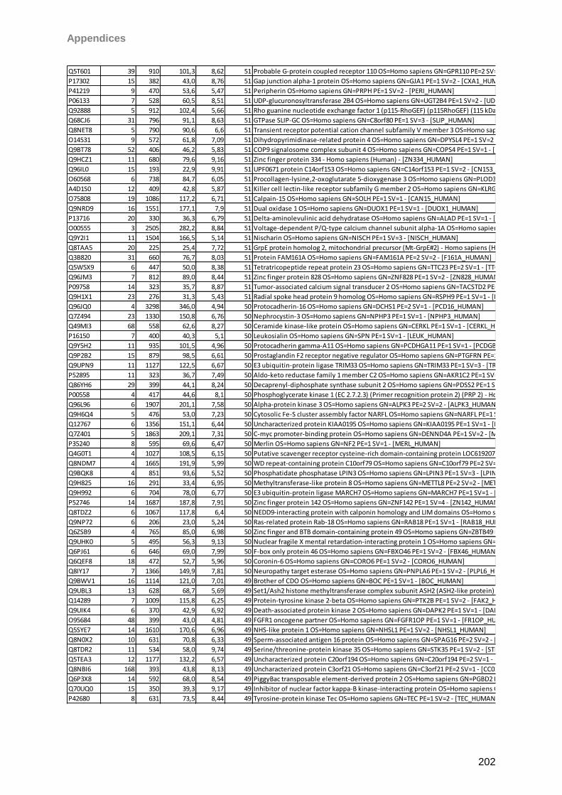
Appendices
202
Q5T601 39 910 101,3 8,62 51 Probable G-protein coupled receptor 110 OS=Homo sapiens GN=GPR110 PE=2 SV=2 - [GP110_HUMAN]
P17302 15 382 43,0 8,76 51 Gap junction alpha-1 protein OS=Homo sapiens GN=GJA1 PE=1 SV=2 - [CXA1_HUMAN]
P41219 9 470 53,6 5,47 51 Peripherin OS=Homo sapiens GN=PRPH PE=1 SV=2 - [PERI_HUMAN]
P06133 7 528 60,5 8,51 51 UDP-glucuronosyltransferase 2B4 OS=Homo sapiens GN=UGT2B4 PE=1 SV=2 - [UD2B4_HUMAN]
Q92888 5 912 102,4 5,66 51 Rho guanine nucleotide exchange factor 1 (p115-RhoGEF) (p115RhoGEF) (115 kDa guanine nucleotide exchange factor) (Sub1.5) - Homo sapiens (Human) - [ARHG1_HUMAN]
Q68CJ6 31 796 91,1 8,63 51 GTPase SLIP-GC OS=Homo sapiens GN=C8orf80 PE=1 SV=3 - [SLIP_HUMAN]
Q8NET8 5 790 90,6 6,6 51 Transient receptor potential cation channel subfamily V member 3 OS=Homo sapiens GN=TRPV3 PE=1 SV=2 - [TRPV3_HUMAN]
O14531 9 572 61,8 7,09 51 Dihydropyrimidinase-related protein 4 OS=Homo sapiens GN=DPYSL4 PE=1 SV=2 - [DPYL4_HUMAN]
Q9BT78 52 406 46,2 5,83 51 COP9 signalosome complex subunit 4 OS=Homo sapiens GN=COPS4 PE=1 SV=1 - [CSN4_HUMAN]
Q9HCZ1 11 680 79,6 9,16 51 Zinc finger protein 334 - Homo sapiens (Human) - [ZN334_HUMAN]
Q96IL0 15 193 22,9 9,91 51 UPF0671 protein C14orf153 OS=Homo sapiens GN=C14orf153 PE=1 SV=2 - [CN153_HUMAN]
O60568 6 738 84,7 6,05 51 Procollagen-lysine,2-oxoglutarate 5-dioxygenase 3 OS=Homo sapiens GN=PLOD3 PE=1 SV=1 - [PLOD3_HUMAN]
A4D1S0 12 409 42,8 5,87 51 Killer cell lectin-like receptor subfamily G member 2 OS=Homo sapiens GN=KLRG2 PE=1 SV=3 - [KLRG2_HUMAN]
O75808 19 1086 117,2 6,71 51 Calpain-15 OS=Homo sapiens GN=SOLH PE=1 SV=1 - [CAN15_HUMAN]
Q9NRD9 16 1551 177,1 7,9 51 Dual oxidase 1 OS=Homo sapiens GN=DUOX1 PE=1 SV=1 - [DUOX1_HUMAN]
P13716 20 330 36,3 6,79 51 Delta-aminolevulinic acid dehydratase OS=Homo sapiens GN=ALAD PE=1 SV=1 - [HEM2_HUMAN]
O00555 3 2505 282,2 8,84 51 Voltage-dependent P/Q-type calcium channel subunit alpha-1A OS=Homo sapiens GN=CACNA1A PE=1 SV=2 - [CAC1A_HUMAN]
Q9Y2I1 11 1504 166,5 5,14 51 Nischarin OS=Homo sapiens GN=NISCH PE=1 SV=3 - [NISCH_HUMAN]
Q8TAA5 20 225 25,4 7,72 51 GrpE protein homolog 2, mitochondrial precursor (Mt-GrpE#2) - Homo sapiens (Human) - [GRPE2_HUMAN]
Q3B820 31 660 76,7 8,03 51 Protein FAM161A OS=Homo sapiens GN=FAM161A PE=2 SV=2 - [F161A_HUMAN]
Q5W5X9 6 447 50,0 8,38 51 Tetratricopeptide repeat protein 23 OS=Homo sapiens GN=TTC23 PE=2 SV=1 - [TTC23_HUMAN]
Q96JM3 7 812 89,0 8,44 51 Zinc finger protein 828 OS=Homo sapiens GN=ZNF828 PE=1 SV=2 - [ZN828_HUMAN]
P09758 14 323 35,7 8,87 51 Tumor-associated calcium signal transducer 2 OS=Homo sapiens GN=TACSTD2 PE=1 SV=3 - [TACD2_HUMAN]
Q9H1X1 23 276 31,3 5,43 51 Radial spoke head protein 9 homolog OS=Homo sapiens GN=RSPH9 PE=1 SV=1 - [RSPH9_HUMAN]
Q96JQ0 4 3298 346,0 4,94 50 Protocadherin-16 OS=Homo sapiens GN=DCHS1 PE=2 SV=1 - [PCD16_HUMAN]
Q7Z494 23 1330 150,8 6,76 50 Nephrocystin-3 OS=Homo sapiens GN=NPHP3 PE=1 SV=1 - [NPHP3_HUMAN]
Q49MI3 68 558 62,6 8,27 50 Ceramide kinase-like protein OS=Homo sapiens GN=CERKL PE=1 SV=1 - [CERKL_HUMAN]
P16150 7 400 40,3 5,1 50 Leukosialin OS=Homo sapiens GN=SPN PE=1 SV=1 - [LEUK_HUMAN]
Q9Y5H2 11 935 101,5 4,96 50 Protocadherin gamma-A11 OS=Homo sapiens GN=PCDHGA11 PE=1 SV=1 - [PCDGB_HUMAN]
Q9P2B2 15 879 98,5 6,61 50 Prostaglandin F2 receptor negative regulator OS=Homo sapiens GN=PTGFRN PE=1 SV=2 - [FPRP_HUMAN]
Q9UPN9 11 1127 122,5 6,67 50 E3 ubiquitin-protein ligase TRIM33 OS=Homo sapiens GN=TRIM33 PE=1 SV=3 - [TRI33_HUMAN]
P52895 11 323 36,7 7,49 50 Aldo-keto reductase family 1 member C2 OS=Homo sapiens GN=AKR1C2 PE=1 SV=3 - [AK1C2_HUMAN]
Q86YH6 29 399 44,1 8,24 50 Decaprenyl-diphosphate synthase subunit 2 OS=Homo sapiens GN=PDSS2 PE=1 SV=2 - [DLP1_HUMAN]
P00558 4 417 44,6 8,1 50 Phosphoglycerate kinase 1 (EC 2.7.2.3) (Primer recognition protein 2) (PRP 2) - Homo sapiens (Human) - [PGK1_HUMAN]
Q96L96 6 1907 201,1 7,58 50 Alpha-protein kinase 3 OS=Homo sapiens GN=ALPK3 PE=2 SV=2 - [ALPK3_HUMAN]
Q9H6Q4 5 476 53,0 7,23 50 Cytosolic Fe-S cluster assembly factor NARFL OS=Homo sapiens GN=NARFL PE=1 SV=1 - [NARFL_HUMAN]
Q12767 6 1356 151,1 6,44 50 Uncharacterized protein KIAA0195 OS=Homo sapiens GN=KIAA0195 PE=1 SV=1 - [K0195_HUMAN]
Q7Z401 5 1863 209,1 7,31 50 C-myc promoter-binding protein OS=Homo sapiens GN=DENND4A PE=1 SV=2 - [MYCPP_HUMAN]
P35240 8 595 69,6 6,47 50 Merlin OS=Homo sapiens GN=NF2 PE=1 SV=1 - [MERL_HUMAN]
Q4G0T1 4 1027 108,5 6,15 50 Putative scavenger receptor cysteine-rich domain-containing protein LOC619207 OS=Homo sapiens PE=5 SV=2 - [SRCRM_HUMAN]
Q8NDM7 4 1665 191,9 5,99 50 WD repeat-containing protein C10orf79 OS=Homo sapiens GN=C10orf79 PE=2 SV=3 - [CJ079_HUMAN]
Q9BQK8 4 851 93,6 5,52 50 Phosphatidate phosphatase LPIN3 OS=Homo sapiens GN=LPIN3 PE=1 SV=3 - [LPIN3_HUMAN]
Q9H825 16 291 33,4 6,95 50 Methyltransferase-like protein 8 OS=Homo sapiens GN=METTL8 PE=2 SV=2 - [METL8_HUMAN]
Q9H992 6 704 78,0 6,77 50 E3 ubiquitin-protein ligase MARCH7 OS=Homo sapiens GN=MARCH7 PE=1 SV=1 - [MARH7_HUMAN]
P52746 14 1687 187,8 7,91 50 Zinc finger protein 142 OS=Homo sapiens GN=ZNF142 PE=1 SV=4 - [ZN142_HUMAN]
Q8TDZ2 6 1067 117,8 6,4 50 NEDD9-interacting protein with calponin homology and LIM domains OS=Homo sapiens GN=MICAL1 PE=1 SV=2 - [MICA1_HUMAN]
Q9NP72 6 206 23,0 5,24 50 Ras-related protein Rab-18 OS=Homo sapiens GN=RAB18 PE=1 SV=1 - [RAB18_HUMAN]
Q6ZSB9 4 765 85,0 6,98 50 Zinc finger and BTB domain-containing protein 49 OS=Homo sapiens GN=ZBTB49 PE=1 SV=3 - [ZBT49_HUMAN]
Q9UHK0 5 495 56,3 9,13 50 Nuclear fragile X mental retardation-interacting protein 1 OS=Homo sapiens GN=NUFIP1 PE=1 SV=2 - [NUFP1_HUMAN]
Q6PJ61 6 646 69,0 7,99 50 F-box only protein 46 OS=Homo sapiens GN=FBXO46 PE=1 SV=2 - [FBX46_HUMAN]
Q6QEF8 18 472 52,7 5,96 50 Coronin-6 OS=Homo sapiens GN=CORO6 PE=1 SV=2 - [CORO6_HUMAN]
Q8IY17 7 1366 149,9 7,81 50 Neuropathy target esterase OS=Homo sapiens GN=PNPLA6 PE=1 SV=2 - [PLPL6_HUMAN]
Q9BWV1 16 1114 121,0 7,01 49 Brother of CDO OS=Homo sapiens GN=BOC PE=1 SV=1 - [BOC_HUMAN]
Q9UBL3 13 628 68,7 5,69 49 Set1/Ash2 histone methyltransferase complex subunit ASH2 (ASH2-like protein) - Homo sapiens (Human) - [ASH2L_HUMAN]
Q14289 7 1009 115,8 6,25 49 Protein-tyrosine kinase 2-beta OS=Homo sapiens GN=PTK2B PE=1 SV=2 - [FAK2_HUMAN]
Q9UIK4 6 370 42,9 6,92 49 Death-associated protein kinase 2 OS=Homo sapiens GN=DAPK2 PE=1 SV=1 - [DAPK2_HUMAN]
O95684 48 399 43,0 4,81 49 FGFR1 oncogene partner OS=Homo sapiens GN=FGFR1OP PE=1 SV=1 - [FR1OP_HUMAN]
Q5SYE7 14 1610 170,6 6,96 49 NHS-like protein 1 OS=Homo sapiens GN=NHSL1 PE=1 SV=2 - [NHSL1_HUMAN]
Q8N0X2 10 631 70,8 6,33 49 Sperm-associated antigen 16 protein OS=Homo sapiens GN=SPAG16 PE=2 SV=2 - [SPG16_HUMAN]
Q8TDR2 11 534 58,0 9,74 49 Serine/threonine-protein kinase 35 OS=Homo sapiens GN=STK35 PE=1 SV=2 - [STK35_HUMAN]
Q5TEA3 12 1177 132,2 6,57 49 Uncharacterized protein C20orf194 OS=Homo sapiens GN=C20orf194 PE=2 SV=1 - [CT194_HUMAN]
Q8NBI6 168 393 43,8 8,13 49 Uncharacterized protein C3orf21 OS=Homo sapiens GN=C3orf21 PE=2 SV=1 - [CC021_HUMAN]
Q6P3X8 14 592 68,0 8,54 49 PiggyBac transposable element-derived protein 2 OS=Homo sapiens GN=PGBD2 PE=2 SV=1 - [PGBD2_HUMAN]
Q70UQ0 15 350 39,3 9,17 49 Inhibitor of nuclear factor kappa-B kinase-interacting protein OS=Homo sapiens GN=IKBIP PE=1 SV=1 - [IKIP_HUMAN]
P42680 8 631 73,5 8,44 49 Tyrosine-protein kinase Tec OS=Homo sapiens GN=TEC PE=1 SV=2 - [TEC_HUMAN]
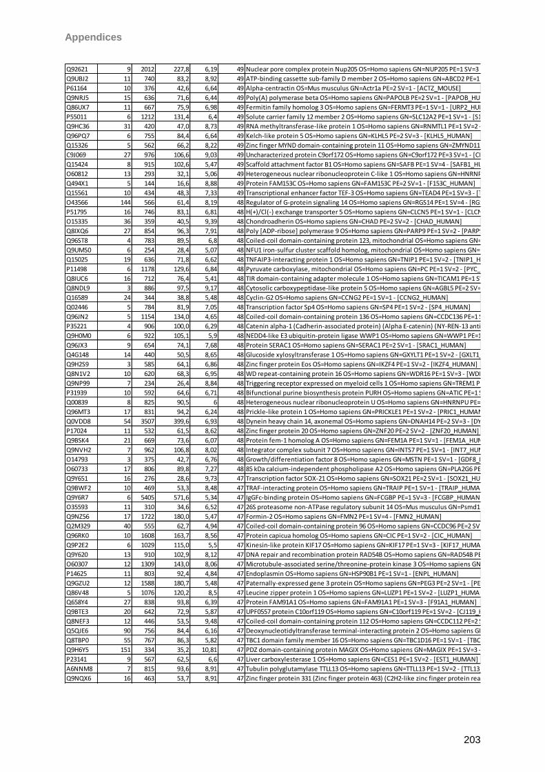
Appendices
203
Q92621 9 2012 227,8 6,19 49 Nuclear pore complex protein Nup205 OS=Homo sapiens GN=NUP205 PE=1 SV=3 - [NU205_HUMAN]
Q9UBJ2 11 740 83,2 8,92 49 ATP-binding cassette sub-family D member 2 OS=Homo sapiens GN=ABCD2 PE=1 SV=1 - [ABCD2_HUMAN]
P61164 10 376 42,6 6,64 49 Alpha-centractin OS=Mus musculus GN=Actr1a PE=2 SV=1 - [ACTZ_MOUSE]
Q9NRJ5 15 636 71,6 6,44 49 Poly(A) polymerase beta OS=Homo sapiens GN=PAPOLB PE=2 SV=1 - [PAPOB_HUMAN]
Q86UX7 11 667 75,9 6,98 49 Fermitin family homolog 3 OS=Homo sapiens GN=FERMT3 PE=1 SV=1 - [URP2_HUMAN]
P55011 6 1212 131,4 6,4 49 Solute carrier family 12 member 2 OS=Homo sapiens GN=SLC12A2 PE=1 SV=1 - [S12A2_HUMAN]
Q9HC36 31 420 47,0 8,73 49 RNA methyltransferase-like protein 1 OS=Homo sapiens GN=RNMTL1 PE=1 SV=2 - [RMTL1_HUMAN]
Q96PQ7 6 755 84,4 6,64 49 Kelch-like protein 5 OS=Homo sapiens GN=KLHL5 PE=2 SV=3 - [KLHL5_HUMAN]
Q15326 5 562 66,2 8,22 49 Zinc finger MYND domain-containing protein 11 OS=Homo sapiens GN=ZMYND11 PE=1 SV=1 - [ZMY11_HUMAN]
C9J069 27 976 106,6 9,03 49 Uncharacterized protein C9orf172 OS=Homo sapiens GN=C9orf172 PE=3 SV=1 - [CI172_HUMAN]
Q15424 8 915 102,6 5,47 49 Scaffold attachment factor B1 OS=Homo sapiens GN=SAFB PE=1 SV=4 - [SAFB1_HUMAN]
O60812 13 293 32,1 5,06 49 Heterogeneous nuclear ribonucleoprotein C-like 1 OS=Homo sapiens GN=HNRNPCL1 PE=1 SV=1 - [HNRCL_HUMAN]
Q494X1 5 144 16,6 8,88 49 Protein FAM153C OS=Homo sapiens GN=FAM153C PE=2 SV=1 - [F153C_HUMAN]
Q15561 10 434 48,3 7,33 49 Transcriptional enhancer factor TEF-3 OS=Homo sapiens GN=TEAD4 PE=1 SV=3 - [TEAD4_HUMAN]
O43566 144 566 61,4 8,19 48 Regulator of G-protein signaling 14 OS=Homo sapiens GN=RGS14 PE=1 SV=4 - [RGS14_HUMAN]
P51795 16 746 83,1 6,81 48 H(+)/Cl(-) exchange transporter 5 OS=Homo sapiens GN=CLCN5 PE=1 SV=1 - [CLCN5_HUMAN]
O15335 36 359 40,5 9,39 48 Chondroadherin OS=Homo sapiens GN=CHAD PE=2 SV=2 - [CHAD_HUMAN]
Q8IXQ6 27 854 96,3 7,91 48 Poly [ADP-ribose] polymerase 9 OS=Homo sapiens GN=PARP9 PE=1 SV=2 - [PARP9_HUMAN]
Q96ST8 4 783 89,5 6,8 48 Coiled-coil domain-containing protein 123, mitochondrial OS=Homo sapiens GN=CCDC123 PE=1 SV=3 - [CC123_HUMAN]
Q9UMS0 6 254 28,4 5,07 48 NFU1 iron-sulfur cluster scaffold homolog, mitochondrial OS=Homo sapiens GN=NFU1 PE=1 SV=2 - [NFU1_HUMAN]
Q15025 19 636 71,8 6,62 48 TNFAIP3-interacting protein 1 OS=Homo sapiens GN=TNIP1 PE=1 SV=2 - [TNIP1_HUMAN]
P11498 6 1178 129,6 6,84 48 Pyruvate carboxylase, mitochondrial OS=Homo sapiens GN=PC PE=1 SV=2 - [PYC_HUMAN]
Q8IUC6 16 712 76,4 5,41 48 TIR domain-containing adapter molecule 1 OS=Homo sapiens GN=TICAM1 PE=1 SV=1 - [TCAM1_HUMAN]
Q8NDL9 3 886 97,5 9,17 48 Cytosolic carboxypeptidase-like protein 5 OS=Homo sapiens GN=AGBL5 PE=2 SV=1 - [CBPC5_HUMAN]
Q16589 24 344 38,8 5,48 48 Cyclin-G2 OS=Homo sapiens GN=CCNG2 PE=1 SV=1 - [CCNG2_HUMAN]
Q02446 5 784 81,9 7,05 48 Transcription factor Sp4 OS=Homo sapiens GN=SP4 PE=1 SV=2 - [SP4_HUMAN]
Q96JN2 5 1154 134,0 4,65 48 Coiled-coil domain-containing protein 136 OS=Homo sapiens GN=CCDC136 PE=1 SV=3 - [CC136_HUMAN]
P35221 4 906 100,0 6,29 48 Catenin alpha-1 (Cadherin-associated protein) (Alpha E-catenin) (NY-REN-13 antigen) - Homo sapiens (Human) - [CTNA1_HUMAN]
Q9H0M0 6 922 105,1 5,9 48 NEDD4-like E3 ubiquitin-protein ligase WWP1 OS=Homo sapiens GN=WWP1 PE=1 SV=1 - [WWP1_HUMAN]
Q96JX3 9 654 74,1 7,68 48 Protein SERAC1 OS=Homo sapiens GN=SERAC1 PE=2 SV=1 - [SRAC1_HUMAN]
Q4G148 14 440 50,5 8,65 48 Glucoside xylosyltransferase 1 OS=Homo sapiens GN=GXYLT1 PE=1 SV=2 - [GXLT1_HUMAN]
Q9H2S9 3 585 64,1 6,86 48 Zinc finger protein Eos OS=Homo sapiens GN=IKZF4 PE=1 SV=2 - [IKZF4_HUMAN]
Q8N1V2 10 620 68,3 6,95 48 WD repeat-containing protein 16 OS=Homo sapiens GN=WDR16 PE=1 SV=3 - [WDR16_HUMAN]
Q9NP99 7 234 26,4 8,84 48 Triggering receptor expressed on myeloid cells 1 OS=Homo sapiens GN=TREM1 PE=1 SV=1 - [TREM1_HUMAN]
P31939 10 592 64,6 6,71 48 Bifunctional purine biosynthesis protein PURH OS=Homo sapiens GN=ATIC PE=1 SV=3 - [PUR9_HUMAN]
Q00839 8 825 90,5 6 48 Heterogeneous nuclear ribonucleoprotein U OS=Homo sapiens GN=HNRNPU PE=1 SV=6 - [HNRPU_HUMAN]
Q96MT3 17 831 94,2 6,24 48 Prickle-like protein 1 OS=Homo sapiens GN=PRICKLE1 PE=1 SV=2 - [PRIC1_HUMAN]
Q0VDD8 54 3507 399,6 6,93 48 Dynein heavy chain 14, axonemal OS=Homo sapiens GN=DNAH14 PE=2 SV=3 - [DYH14_HUMAN]
P17024 11 532 61,5 8,62 48 Zinc finger protein 20 OS=Homo sapiens GN=ZNF20 PE=2 SV=2 - [ZNF20_HUMAN]
Q9BSK4 21 669 73,6 6,07 48 Protein fem-1 homolog A OS=Homo sapiens GN=FEM1A PE=1 SV=1 - [FEM1A_HUMAN]
Q9NVH2 7 962 106,8 8,02 48 Integrator complex subunit 7 OS=Homo sapiens GN=INTS7 PE=1 SV=1 - [INT7_HUMAN]
O14793 3 375 42,7 6,76 48 Growth/differentiation factor 8 OS=Homo sapiens GN=MSTN PE=1 SV=1 - [GDF8_HUMAN]
O60733 17 806 89,8 7,27 48 85 kDa calcium-independent phospholipase A2 OS=Homo sapiens GN=PLA2G6 PE=1 SV=2 - [PA2G6_HUMAN]
Q9Y651 16 276 28,6 9,73 47 Transcription factor SOX-21 OS=Homo sapiens GN=SOX21 PE=2 SV=1 - [SOX21_HUMAN]
Q9BWF2 10 469 53,3 8,48 47 TRAF-interacting protein OS=Homo sapiens GN=TRAIP PE=1 SV=1 - [TRAIP_HUMAN]
Q9Y6R7 6 5405 571,6 5,34 47 IgGFc-binding protein OS=Homo sapiens GN=FCGBP PE=1 SV=3 - [FCGBP_HUMAN]
O35593 11 310 34,6 6,52 47 26S proteasome non-ATPase regulatory subunit 14 OS=Mus musculus GN=Psmd14 PE=1 SV=2 - [PSDE_MOUSE]
Q9NZ56 17 1722 180,0 5,47 47 Formin-2 OS=Homo sapiens GN=FMN2 PE=1 SV=4 - [FMN2_HUMAN]
Q2M329 40 555 62,7 4,94 47 Coiled-coil domain-containing protein 96 OS=Homo sapiens GN=CCDC96 PE=2 SV=2 - [CCD96_HUMAN]
Q96RK0 10 1608 163,7 8,56 47 Protein capicua homolog OS=Homo sapiens GN=CIC PE=1 SV=2 - [CIC_HUMAN]
Q9P2E2 6 1029 115,0 5,5 47 Kinesin-like protein KIF17 OS=Homo sapiens GN=KIF17 PE=1 SV=3 - [KIF17_HUMAN]
Q9Y620 13 910 102,9 8,12 47 DNA repair and recombination protein RAD54B OS=Homo sapiens GN=RAD54B PE=1 SV=1 - [RA54B_HUMAN]
O60307 12 1309 143,0 8,06 47 Microtubule-associated serine/threonine-protein kinase 3 OS=Homo sapiens GN=MAST3 PE=1 SV=2 - [MAST3_HUMAN]
P14625 11 803 92,4 4,84 47 Endoplasmin OS=Homo sapiens GN=HSP90B1 PE=1 SV=1 - [ENPL_HUMAN]
Q9GZU2 12 1588 180,7 5,48 47 Paternally-expressed gene 3 protein OS=Homo sapiens GN=PEG3 PE=2 SV=1 - [PEG3_HUMAN]
Q86V48 5 1076 120,2 8,5 47 Leucine zipper protein 1 OS=Homo sapiens GN=LUZP1 PE=1 SV=2 - [LUZP1_HUMAN]
Q658Y4 27 838 93,8 6,39 47 Protein FAM91A1 OS=Homo sapiens GN=FAM91A1 PE=1 SV=3 - [F91A1_HUMAN]
Q9BTE3 20 642 72,9 5,87 47 UPF0557 protein C10orf119 OS=Homo sapiens GN=C10orf119 PE=1 SV=2 - [CJ119_HUMAN]
Q8NEF3 12 446 53,5 9,48 47 Coiled-coil domain-containing protein 112 OS=Homo sapiens GN=CCDC112 PE=2 SV=2 - [CC112_HUMAN]
Q5QJE6 90 756 84,4 6,16 47 Deoxynucleotidyltransferase terminal-interacting protein 2 OS=Homo sapiens GN=DNTTIP2 PE=1 SV=2 - [TDIF2_HUMAN]
Q8TBP0 55 767 86,3 5,82 47 TBC1 domain family member 16 OS=Homo sapiens GN=TBC1D16 PE=1 SV=1 - [TBC16_HUMAN]
Q9H6Y5 151 334 35,2 10,81 47 PDZ domain-containing protein MAGIX OS=Homo sapiens GN=MAGIX PE=1 SV=3 - [MAGIX_HUMAN]
P23141 9 567 62,5 6,6 47 Liver carboxylesterase 1 OS=Homo sapiens GN=CES1 PE=1 SV=2 - [EST1_HUMAN]
A6NNM8 7 815 93,6 8,91 47 Tubulin polyglutamylase TTLL13 OS=Homo sapiens GN=TTLL13 PE=1 SV=2 - [TTL13_HUMAN]
Q9NQX6 16 463 53,7 8,91 47 Zinc finger protein 331 (Zinc finger protein 463) (C2H2-like zinc finger protein rearranged in thyroid adenomas) - Homo sapiens (Human) - [ZN331_HUMAN]
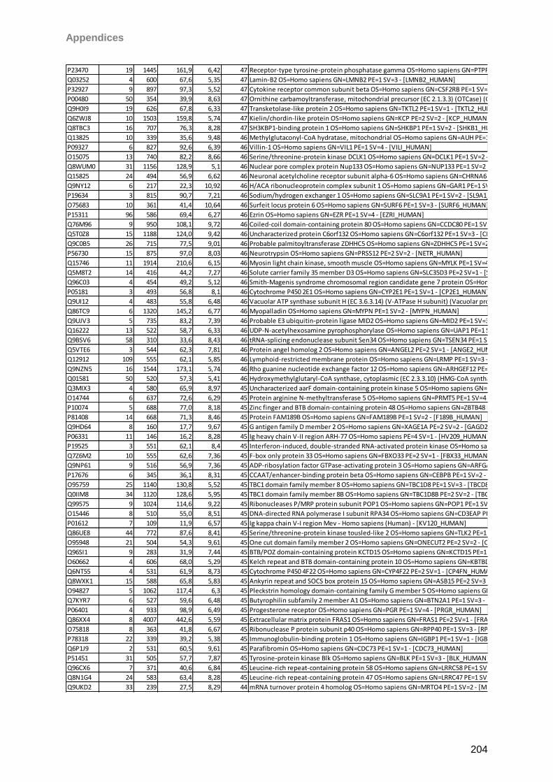
Appendices
204
P23470 19 1445 161,9 6,42 47 Receptor-type tyrosine-protein phosphatase gamma OS=Homo sapiens GN=PTPRG PE=1 SV=4 - [PTPRG_HUMAN]
Q03252 4 600 67,6 5,35 47 Lamin-B2 OS=Homo sapiens GN=LMNB2 PE=1 SV=3 - [LMNB2_HUMAN]
P32927 9 897 97,3 5,52 47 Cytokine receptor common subunit beta OS=Homo sapiens GN=CSF2RB PE=1 SV=2 - [IL3RB_HUMAN]
P00480 50 354 39,9 8,63 47 Ornithine carbamoyltransferase, mitochondrial precursor (EC 2.1.3.3) (OTCase) (Ornithine transcarbamylase) - Homo sapiens (Human) - [OTC_HUMAN]
Q9H0I9 19 626 67,8 6,33 47 Transketolase-like protein 2 OS=Homo sapiens GN=TKTL2 PE=1 SV=1 - [TKTL2_HUMAN]
Q6ZWJ8 10 1503 159,8 5,74 47 Kielin/chordin-like protein OS=Homo sapiens GN=KCP PE=2 SV=2 - [KCP_HUMAN]
Q8TBC3 16 707 76,3 8,28 47 SH3KBP1-binding protein 1 OS=Homo sapiens GN=SHKBP1 PE=1 SV=2 - [SHKB1_HUMAN]
Q13825 10 339 35,6 9,48 46 Methylglutaconyl-CoA hydratase, mitochondrial OS=Homo sapiens GN=AUH PE=1 SV=1 - [AUHM_HUMAN]
P09327 6 827 92,6 6,39 46 Villin-1 OS=Homo sapiens GN=VIL1 PE=1 SV=4 - [VILI_HUMAN]
O15075 13 740 82,2 8,66 46 Serine/threonine-protein kinase DCLK1 OS=Homo sapiens GN=DCLK1 PE=1 SV=2 - [DCLK1_HUMAN]
Q8WUM0 31 1156 128,9 5,1 46 Nuclear pore complex protein Nup133 OS=Homo sapiens GN=NUP133 PE=1 SV=2 - [NU133_HUMAN]
Q15825 24 494 56,9 6,62 46 Neuronal acetylcholine receptor subunit alpha-6 OS=Homo sapiens GN=CHRNA6 PE=1 SV=1 - [ACHA6_HUMAN]
Q9NY12 6 217 22,3 10,92 46 H/ACA ribonucleoprotein complex subunit 1 OS=Homo sapiens GN=GAR1 PE=1 SV=1 - [GAR1_HUMAN]
P19634 3 815 90,7 7,21 46 Sodium/hydrogen exchanger 1 OS=Homo sapiens GN=SLC9A1 PE=1 SV=2 - [SL9A1_HUMAN]
O75683 10 361 41,4 10,64 46 Surfeit locus protein 6 OS=Homo sapiens GN=SURF6 PE=1 SV=3 - [SURF6_HUMAN]
P15311 96 586 69,4 6,27 46 Ezrin OS=Homo sapiens GN=EZR PE=1 SV=4 - [EZRI_HUMAN]
Q76M96 9 950 108,1 9,72 46 Coiled-coil domain-containing protein 80 OS=Homo sapiens GN=CCDC80 PE=1 SV=1 - [CCD80_HUMAN]
Q5T0Z8 15 1188 124,0 9,42 46 Uncharacterized protein C6orf132 OS=Homo sapiens GN=C6orf132 PE=1 SV=3 - [CF132_HUMAN]
Q9C0B5 26 715 77,5 9,01 46 Probable palmitoyltransferase ZDHHC5 OS=Homo sapiens GN=ZDHHC5 PE=1 SV=2 - [ZDHC5_HUMAN]
P56730 15 875 97,0 8,03 46 Neurotrypsin OS=Homo sapiens GN=PRSS12 PE=2 SV=2 - [NETR_HUMAN]
Q15746 11 1914 210,6 6,15 46 Myosin light chain kinase, smooth muscle OS=Homo sapiens GN=MYLK PE=1 SV=4 - [MYLK_HUMAN]
Q5M8T2 14 416 44,2 7,27 46 Solute carrier family 35 member D3 OS=Homo sapiens GN=SLC35D3 PE=2 SV=1 - [S35D3_HUMAN]
Q96C03 4 454 49,2 5,12 46 Smith-Magenis syndrome chromosomal region candidate gene 7 protein OS=Homo sapiens GN=SMCR7 PE=1 SV=1 - [SMCR7_HUMAN]
P05181 3 493 56,8 8,1 46 Cytochrome P450 2E1 OS=Homo sapiens GN=CYP2E1 PE=1 SV=1 - [CP2E1_HUMAN]
Q9UI12 4 483 55,8 6,48 46 Vacuolar ATP synthase subunit H (EC 3.6.3.14) (V-ATPase H subunit) (Vacuolar proton pump subunit H) (V-ATPase 50/57 kDa subunits) (Vacuolar proton pump subunit SFD) (VMA13) (Nef-binding protein 1) (NBP1) - Homo sapiens (Human) - [VATH_HUMAN]
Q86TC9 6 1320 145,2 6,77 46 Myopalladin OS=Homo sapiens GN=MYPN PE=1 SV=2 - [MYPN_HUMAN]
Q9UJV3 5 735 83,2 7,39 46 Probable E3 ubiquitin-protein ligase MID2 OS=Homo sapiens GN=MID2 PE=1 SV=3 - [TRIM1_HUMAN]
Q16222 13 522 58,7 6,33 46 UDP-N-acetylhexosamine pyrophosphorylase OS=Homo sapiens GN=UAP1 PE=1 SV=3 - [UAP1_HUMAN]
Q9BSV6 58 310 33,6 8,43 46 tRNA-splicing endonuclease subunit Sen34 OS=Homo sapiens GN=TSEN34 PE=1 SV=1 - [SEN34_HUMAN]
Q5VTE6 3 544 62,3 7,81 46 Protein angel homolog 2 OS=Homo sapiens GN=ANGEL2 PE=2 SV=1 - [ANGE2_HUMAN]
Q12912 109 555 62,1 5,85 46 Lymphoid-restricted membrane protein OS=Homo sapiens GN=LRMP PE=1 SV=3 - [LRMP_HUMAN]
Q9NZN5 16 1544 173,1 5,74 46 Rho guanine nucleotide exchange factor 12 OS=Homo sapiens GN=ARHGEF12 PE=1 SV=1 - [ARHGC_HUMAN]
Q01581 50 520 57,3 5,41 46 Hydroxymethylglutaryl-CoA synthase, cytoplasmic (EC 2.3.3.10) (HMG-CoA synthase) (3-hydroxy-3-methylglutaryl coenzyme A synthase) - Homo sapiens (Human) - [HMCS1_HUMAN]
Q3MIX3 4 580 65,9 8,97 45 Uncharacterized aarF domain-containing protein kinase 5 OS=Homo sapiens GN=ADCK5 PE=2 SV=2 - [ADCK5_HUMAN]
O14744 6 637 72,6 6,29 45 Protein arginine N-methyltransferase 5 OS=Homo sapiens GN=PRMT5 PE=1 SV=4 - [ANM5_HUMAN]
P10074 5 688 77,0 8,18 45 Zinc finger and BTB domain-containing protein 48 OS=Homo sapiens GN=ZBTB48 PE=1 SV=2 - [ZBT48_HUMAN]
P81408 14 668 71,3 8,46 45 Protein FAM189B OS=Homo sapiens GN=FAM189B PE=1 SV=2 - [F189B_HUMAN]
Q9HD64 8 160 17,7 9,67 45 G antigen family D member 2 OS=Homo sapiens GN=XAGE1A PE=2 SV=2 - [GAGD2_HUMAN]
P06331 11 146 16,2 8,28 45 Ig heavy chain V-II region ARH-77 OS=Homo sapiens PE=4 SV=1 - [HV209_HUMAN]
P19525 3 551 62,1 8,4 45 Interferon-induced, double-stranded RNA-activated protein kinase OS=Homo sapiens GN=EIF2AK2 PE=1 SV=2 - [E2AK2_HUMAN]
Q7Z6M2 10 555 62,6 7,36 45 F-box only protein 33 OS=Homo sapiens GN=FBXO33 PE=2 SV=1 - [FBX33_HUMAN]
Q9NP61 9 516 56,9 7,36 45 ADP-ribosylation factor GTPase-activating protein 3 OS=Homo sapiens GN=ARFGAP3 PE=1 SV=1 - [ARFG3_HUMAN]
P17676 6 345 36,1 8,31 45 CCAAT/enhancer-binding protein beta OS=Homo sapiens GN=CEBPB PE=1 SV=2 - [CEBPB_HUMAN]
O95759 25 1140 130,8 5,52 45 TBC1 domain family member 8 OS=Homo sapiens GN=TBC1D8 PE=1 SV=3 - [TBCD8_HUMAN]
Q0IIM8 34 1120 128,6 5,95 45 TBC1 domain family member 8B OS=Homo sapiens GN=TBC1D8B PE=2 SV=2 - [TBC8B_HUMAN]
Q99575 9 1024 114,6 9,22 45 Ribonucleases P/MRP protein subunit POP1 OS=Homo sapiens GN=POP1 PE=1 SV=2 - [POP1_HUMAN]
O15446 8 510 55,0 8,51 45 DNA-directed RNA polymerase I subunit RPA34 OS=Homo sapiens GN=CD3EAP PE=1 SV=1 - [RPA34_HUMAN]
P01612 7 109 11,9 6,57 45 Ig kappa chain V-I region Mev - Homo sapiens (Human) - [KV120_HUMAN]
Q86UE8 44 772 87,6 8,41 45 Serine/threonine-protein kinase tousled-like 2 OS=Homo sapiens GN=TLK2 PE=1 SV=2 - [TLK2_HUMAN]
O95948 21 504 54,3 9,61 45 One cut domain family member 2 OS=Homo sapiens GN=ONECUT2 PE=2 SV=2 - [ONEC2_HUMAN]
Q96SI1 9 283 31,9 7,44 45 BTB/POZ domain-containing protein KCTD15 OS=Homo sapiens GN=KCTD15 PE=1 SV=1 - [KCD15_HUMAN]
O60662 4 606 68,0 5,29 45 Kelch repeat and BTB domain-containing protein 10 OS=Homo sapiens GN=KBTBD10 PE=1 SV=2 - [KBTBA_HUMAN]
Q6NT55 4 531 61,9 8,73 45 Cytochrome P450 4F22 OS=Homo sapiens GN=CYP4F22 PE=2 SV=1 - [CP4FN_HUMAN]
Q8WXK1 15 588 65,8 5,83 45 Ankyrin repeat and SOCS box protein 15 OS=Homo sapiens GN=ASB15 PE=2 SV=3 - [ASB15_HUMAN]
O94827 5 1062 117,4 6,3 45 Pleckstrin homology domain-containing family G member 5 OS=Homo sapiens GN=PLEKHG5 PE=2 SV=3 - [PKHG5_HUMAN]
Q7KYR7 6 527 59,6 6,48 45 Butyrophilin subfamily 2 member A1 OS=Homo sapiens GN=BTN2A1 PE=1 SV=3 - [BT2A1_HUMAN]
P06401 4 933 98,9 6,49 45 Progesterone receptor OS=Homo sapiens GN=PGR PE=1 SV=4 - [PRGR_HUMAN]
Q86XX4 8 4007 442,6 5,59 45 Extracellular matrix protein FRAS1 OS=Homo sapiens GN=FRAS1 PE=2 SV=1 - [FRAS1_HUMAN]
O75818 8 363 41,8 6,67 45 Ribonuclease P protein subunit p40 OS=Homo sapiens GN=RPP40 PE=1 SV=3 - [RPP40_HUMAN]
P78318 22 339 39,2 5,38 45 Immunoglobulin-binding protein 1 OS=Homo sapiens GN=IGBP1 PE=1 SV=1 - [IGBP1_HUMAN]
Q6P1J9 2 531 60,5 9,61 45 Parafibromin OS=Homo sapiens GN=CDC73 PE=1 SV=1 - [CDC73_HUMAN]
P51451 31 505 57,7 7,87 45 Tyrosine-protein kinase Blk OS=Homo sapiens GN=BLK PE=1 SV=3 - [BLK_HUMAN]
Q96CX6 7 371 40,6 6,84 45 Leucine-rich repeat-containing protein 58 OS=Homo sapiens GN=LRRC58 PE=1 SV=2 - [LRC58_HUMAN]
Q8N1G4 24 583 63,4 8,28 45 Leucine-rich repeat-containing protein 47 OS=Homo sapiens GN=LRRC47 PE=1 SV=1 - [LRC47_HUMAN]
Q9UKD2 33 239 27,5 8,29 44 mRNA turnover protein 4 homolog OS=Homo sapiens GN=MRTO4 PE=1 SV=2 - [MRT4_HUMAN]
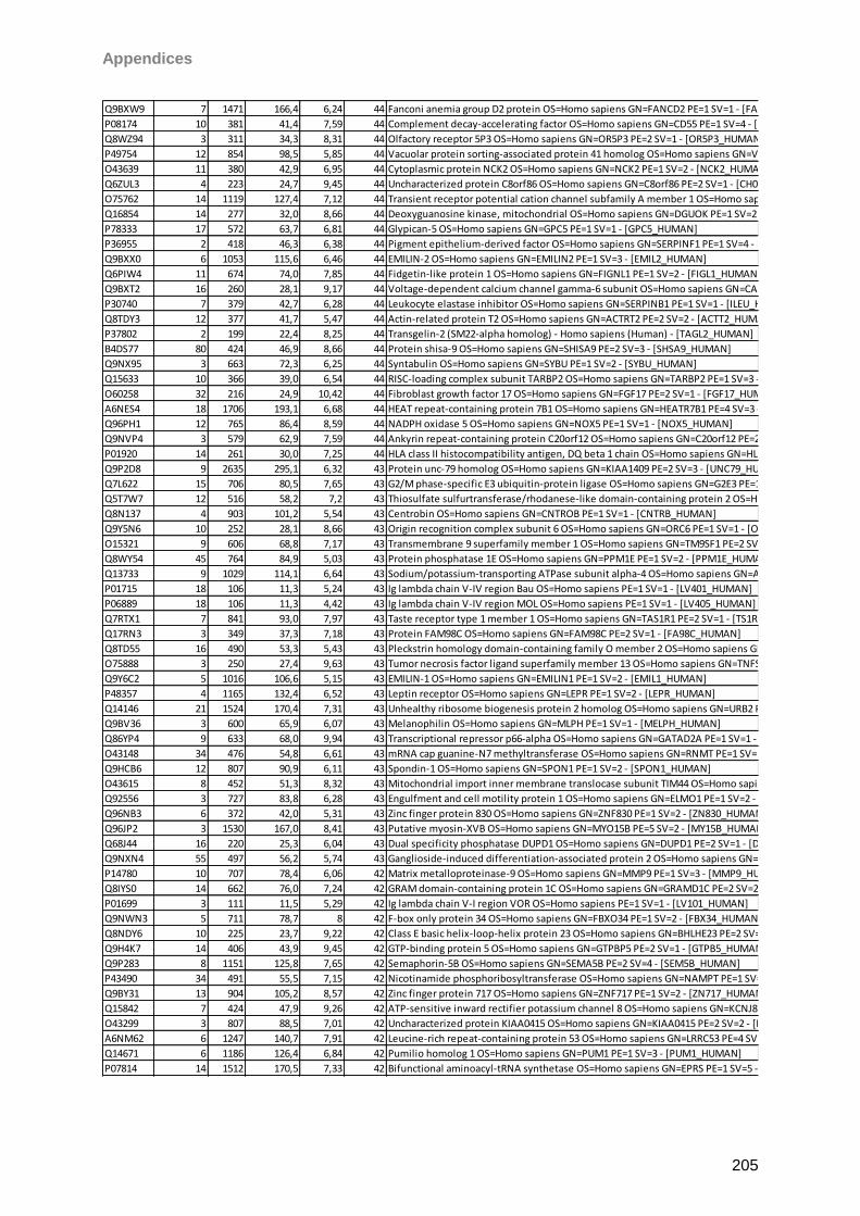
Appendices
205
Q9BXW9 7 1471 166,4 6,24 44 Fanconi anemia group D2 protein OS=Homo sapiens GN=FANCD2 PE=1 SV=1 - [FACD2_HUMAN]
P08174 10 381 41,4 7,59 44 Complement decay-accelerating factor OS=Homo sapiens GN=CD55 PE=1 SV=4 - [DAF_HUMAN]
Q8WZ94 3 311 34,3 8,31 44 Olfactory receptor 5P3 OS=Homo sapiens GN=OR5P3 PE=2 SV=1 - [OR5P3_HUMAN]
P49754 12 854 98,5 5,85 44 Vacuolar protein sorting-associated protein 41 homolog OS=Homo sapiens GN=VPS41 PE=1 SV=3 - [VPS41_HUMAN]
O43639 11 380 42,9 6,95 44 Cytoplasmic protein NCK2 OS=Homo sapiens GN=NCK2 PE=1 SV=2 - [NCK2_HUMAN]
Q6ZUL3 4 223 24,7 9,45 44 Uncharacterized protein C8orf86 OS=Homo sapiens GN=C8orf86 PE=2 SV=1 - [CH086_HUMAN]
O75762 14 1119 127,4 7,12 44 Transient receptor potential cation channel subfamily A member 1 OS=Homo sapiens GN=TRPA1 PE=2 SV=3 - [TRPA1_HUMAN]
Q16854 14 277 32,0 8,66 44 Deoxyguanosine kinase, mitochondrial OS=Homo sapiens GN=DGUOK PE=1 SV=2 - [DGUOK_HUMAN]
P78333 17 572 63,7 6,81 44 Glypican-5 OS=Homo sapiens GN=GPC5 PE=1 SV=1 - [GPC5_HUMAN]
P36955 2 418 46,3 6,38 44 Pigment epithelium-derived factor OS=Homo sapiens GN=SERPINF1 PE=1 SV=4 - [PEDF_HUMAN]
Q9BXX0 6 1053 115,6 6,46 44 EMILIN-2 OS=Homo sapiens GN=EMILIN2 PE=1 SV=3 - [EMIL2_HUMAN]
Q6PIW4 11 674 74,0 7,85 44 Fidgetin-like protein 1 OS=Homo sapiens GN=FIGNL1 PE=1 SV=2 - [FIGL1_HUMAN]
Q9BXT2 16 260 28,1 9,17 44 Voltage-dependent calcium channel gamma-6 subunit OS=Homo sapiens GN=CACNG6 PE=2 SV=1 - [CCG6_HUMAN]
P30740 7 379 42,7 6,28 44 Leukocyte elastase inhibitor OS=Homo sapiens GN=SERPINB1 PE=1 SV=1 - [ILEU_HUMAN]
Q8TDY3 12 377 41,7 5,47 44 Actin-related protein T2 OS=Homo sapiens GN=ACTRT2 PE=2 SV=2 - [ACTT2_HUMAN]
P37802 2 199 22,4 8,25 44 Transgelin-2 (SM22-alpha homolog) - Homo sapiens (Human) - [TAGL2_HUMAN]
B4DS77 80 424 46,9 8,66 44 Protein shisa-9 OS=Homo sapiens GN=SHISA9 PE=2 SV=3 - [SHSA9_HUMAN]
Q9NX95 3 663 72,3 6,25 44 Syntabulin OS=Homo sapiens GN=SYBU PE=1 SV=2 - [SYBU_HUMAN]
Q15633 10 366 39,0 6,54 44 RISC-loading complex subunit TARBP2 OS=Homo sapiens GN=TARBP2 PE=1 SV=3 - [TRBP2_HUMAN]
O60258 32 216 24,9 10,42 44 Fibroblast growth factor 17 OS=Homo sapiens GN=FGF17 PE=2 SV=1 - [FGF17_HUMAN]
A6NES4 18 1706 193,1 6,68 44 HEAT repeat-containing protein 7B1 OS=Homo sapiens GN=HEATR7B1 PE=4 SV=3 - [HTRB1_HUMAN]
Q96PH1 12 765 86,4 8,59 44 NADPH oxidase 5 OS=Homo sapiens GN=NOX5 PE=1 SV=1 - [NOX5_HUMAN]
Q9NVP4 3 579 62,9 7,59 44 Ankyrin repeat-containing protein C20orf12 OS=Homo sapiens GN=C20orf12 PE=2 SV=2 - [CT012_HUMAN]
P01920 14 261 30,0 7,25 44 HLA class II histocompatibility antigen, DQ beta 1 chain OS=Homo sapiens GN=HLA-DQB1 PE=1 SV=2 - [DQB1_HUMAN]
Q9P2D8 9 2635 295,1 6,32 43 Protein unc-79 homolog OS=Homo sapiens GN=KIAA1409 PE=2 SV=3 - [UNC79_HUMAN]
Q7L622 15 706 80,5 7,65 43 G2/M phase-specific E3 ubiquitin-protein ligase OS=Homo sapiens GN=G2E3 PE=1 SV=1 - [G2E3_HUMAN]
Q5T7W7 12 516 58,2 7,2 43 Thiosulfate sulfurtransferase/rhodanese-like domain-containing protein 2 OS=Homo sapiens GN=TSTD2 PE=1 SV=1 - [TSTD2_HUMAN]
Q8N137 4 903 101,2 5,54 43 Centrobin OS=Homo sapiens GN=CNTROB PE=1 SV=1 - [CNTRB_HUMAN]
Q9Y5N6 10 252 28,1 8,66 43 Origin recognition complex subunit 6 OS=Homo sapiens GN=ORC6 PE=1 SV=1 - [ORC6_HUMAN]
O15321 9 606 68,8 7,17 43 Transmembrane 9 superfamily member 1 OS=Homo sapiens GN=TM9SF1 PE=2 SV=2 - [TM9S1_HUMAN]
Q8WY54 45 764 84,9 5,03 43 Protein phosphatase 1E OS=Homo sapiens GN=PPM1E PE=1 SV=2 - [PPM1E_HUMAN]
Q13733 9 1029 114,1 6,64 43 Sodium/potassium-transporting ATPase subunit alpha-4 OS=Homo sapiens GN=ATP1A4 PE=1 SV=3 - [AT1A4_HUMAN]
P01715 18 106 11,3 5,24 43 Ig lambda chain V-IV region Bau OS=Homo sapiens PE=1 SV=1 - [LV401_HUMAN]
P06889 18 106 11,3 4,42 43 Ig lambda chain V-IV region MOL OS=Homo sapiens PE=1 SV=1 - [LV405_HUMAN]
Q7RTX1 7 841 93,0 7,97 43 Taste receptor type 1 member 1 OS=Homo sapiens GN=TAS1R1 PE=2 SV=1 - [TS1R1_HUMAN]
Q17RN3 3 349 37,3 7,18 43 Protein FAM98C OS=Homo sapiens GN=FAM98C PE=2 SV=1 - [FA98C_HUMAN]
Q8TD55 16 490 53,3 5,43 43 Pleckstrin homology domain-containing family O member 2 OS=Homo sapiens GN=PLEKHO2 PE=1 SV=1 - [PKHO2_HUMAN]
O75888 3 250 27,4 9,63 43 Tumor necrosis factor ligand superfamily member 13 OS=Homo sapiens GN=TNFSF13 PE=1 SV=1 - [TNF13_HUMAN]
Q9Y6C2 5 1016 106,6 5,15 43 EMILIN-1 OS=Homo sapiens GN=EMILIN1 PE=1 SV=2 - [EMIL1_HUMAN]
P48357 4 1165 132,4 6,52 43 Leptin receptor OS=Homo sapiens GN=LEPR PE=1 SV=2 - [LEPR_HUMAN]
Q14146 21 1524 170,4 7,31 43 Unhealthy ribosome biogenesis protein 2 homolog OS=Homo sapiens GN=URB2 PE=1 SV=2 - [URB2_HUMAN]
Q9BV36 3 600 65,9 6,07 43 Melanophilin OS=Homo sapiens GN=MLPH PE=1 SV=1 - [MELPH_HUMAN]
Q86YP4 9 633 68,0 9,94 43 Transcriptional repressor p66-alpha OS=Homo sapiens GN=GATAD2A PE=1 SV=1 - [P66A_HUMAN]
O43148 34 476 54,8 6,61 43 mRNA cap guanine-N7 methyltransferase OS=Homo sapiens GN=RNMT PE=1 SV=1 - [MCES_HUMAN]
Q9HCB6 12 807 90,9 6,11 43 Spondin-1 OS=Homo sapiens GN=SPON1 PE=1 SV=2 - [SPON1_HUMAN]
O43615 8 452 51,3 8,32 43 Mitochondrial import inner membrane translocase subunit TIM44 OS=Homo sapiens GN=TIMM44 PE=1 SV=2 - [TIM44_HUMAN]
Q92556 3 727 83,8 6,28 43 Engulfment and cell motility protein 1 OS=Homo sapiens GN=ELMO1 PE=1 SV=2 - [ELMO1_HUMAN]
Q96NB3 6 372 42,0 5,31 43 Zinc finger protein 830 OS=Homo sapiens GN=ZNF830 PE=1 SV=2 - [ZN830_HUMAN]
Q96JP2 3 1530 167,0 8,41 43 Putative myosin-XVB OS=Homo sapiens GN=MYO15B PE=5 SV=2 - [MY15B_HUMAN]
Q68J44 16 220 25,3 6,04 43 Dual specificity phosphatase DUPD1 OS=Homo sapiens GN=DUPD1 PE=2 SV=1 - [DUPD1_HUMAN]
Q9NXN4 55 497 56,2 5,74 43 Ganglioside-induced differentiation-associated protein 2 OS=Homo sapiens GN=GDAP2 PE=2 SV=1 - [GDAP2_HUMAN]
P14780 10 707 78,4 6,06 42 Matrix metalloproteinase-9 OS=Homo sapiens GN=MMP9 PE=1 SV=3 - [MMP9_HUMAN]
Q8IYS0 14 662 76,0 7,24 42 GRAM domain-containing protein 1C OS=Homo sapiens GN=GRAMD1C PE=2 SV=2 - [GRM1C_HUMAN]
P01699 3 111 11,5 5,29 42 Ig lambda chain V-I region VOR OS=Homo sapiens PE=1 SV=1 - [LV101_HUMAN]
Q9NWN3 5 711 78,7 8 42 F-box only protein 34 OS=Homo sapiens GN=FBXO34 PE=1 SV=2 - [FBX34_HUMAN]
Q8NDY6 10 225 23,7 9,22 42 Class E basic helix-loop-helix protein 23 OS=Homo sapiens GN=BHLHE23 PE=2 SV=1 - [BHE23_HUMAN]
Q9H4K7 14 406 43,9 9,45 42 GTP-binding protein 5 OS=Homo sapiens GN=GTPBP5 PE=2 SV=1 - [GTPB5_HUMAN]
Q9P283 8 1151 125,8 7,65 42 Semaphorin-5B OS=Homo sapiens GN=SEMA5B PE=2 SV=4 - [SEM5B_HUMAN]
P43490 34 491 55,5 7,15 42 Nicotinamide phosphoribosyltransferase OS=Homo sapiens GN=NAMPT PE=1 SV=1 - [NAMPT_HUMAN]
Q9BY31 13 904 105,2 8,57 42 Zinc finger protein 717 OS=Homo sapiens GN=ZNF717 PE=1 SV=2 - [ZN717_HUMAN]
Q15842 7 424 47,9 9,26 42 ATP-sensitive inward rectifier potassium channel 8 OS=Homo sapiens GN=KCNJ8 PE=1 SV=1 - [IRK8_HUMAN]
O43299 3 807 88,5 7,01 42 Uncharacterized protein KIAA0415 OS=Homo sapiens GN=KIAA0415 PE=2 SV=2 - [K0415_HUMAN]
A6NM62 6 1247 140,7 7,91 42 Leucine-rich repeat-containing protein 53 OS=Homo sapiens GN=LRRC53 PE=4 SV=2 - [LRC53_HUMAN]
Q14671 6 1186 126,4 6,84 42 Pumilio homolog 1 OS=Homo sapiens GN=PUM1 PE=1 SV=3 - [PUM1_HUMAN]
P07814 14 1512 170,5 7,33 42 Bifunctional aminoacyl-tRNA synthetase OS=Homo sapiens GN=EPRS PE=1 SV=5 - [SYEP_HUMAN]
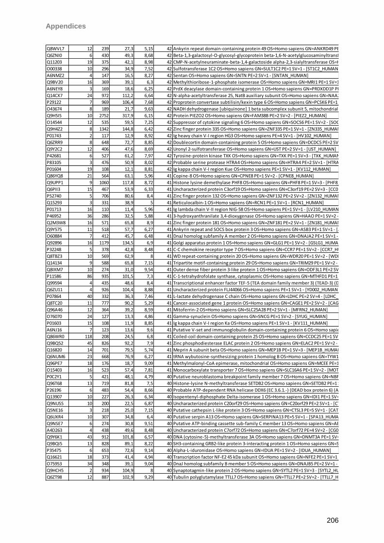
Appendices
206
Q8WVL7 12 239 27,3 5,15 42 Ankyrin repeat domain-containing protein 49 OS=Homo sapiens GN=ANKRD49 PE=2 SV=1 - [ANR49_HUMAN]
Q6ZNI0 6 430 49,3 8,68 42 Beta-1,3-galactosyl-O-glycosyl-glycoprotein beta-1,6-N-acetylglucosaminyltransferase 7 OS=Homo sapiens GN=GCNT7 PE=2 SV=2 - [GCNT7_HUMAN]
Q11203 19 375 42,1 8,98 42 CMP-N-acetylneuraminate-beta-1,4-galactoside alpha-2,3-sialyltransferase OS=Homo sapiens GN=ST3GAL3 PE=2 SV=1 - [SIAT6_HUMAN]
O00338 10 296 34,9 7,52 42 Sulfotransferase 1C2 OS=Homo sapiens GN=SULT1C2 PE=1 SV=1 - [ST1C2_HUMAN]
A6NMZ2 4 147 16,5 8,27 42 Sentan OS=Homo sapiens GN=SNTN PE=2 SV=1 - [SNTAN_HUMAN]
Q9BV20 16 369 39,1 6,3 42 Methylthioribose-1-phosphate isomerase OS=Homo sapiens GN=MRI1 PE=1 SV=1 - [MTNA_HUMAN]
A6NEY8 3 169 18,6 6,25 42 PrdX deacylase domain-containing protein 1 OS=Homo sapiens GN=PRDXDD1P PE=3 SV=3 - [PRXD1_HUMAN]
Q14CX7 24 972 112,2 6,64 42 N-alpha-acetyltransferase 25, NatB auxiliary subunit OS=Homo sapiens GN=NAA25 PE=1 SV=1 - [NAA25_HUMAN]
P29122 7 969 106,4 7,68 42 Proprotein convertase subtilisin/kexin type 6 OS=Homo sapiens GN=PCSK6 PE=1 SV=1 - [PCSK6_HUMAN]
O43674 8 189 21,7 9,63 42 NADH dehydrogenase [ubiquinone] 1 beta subcomplex subunit 5, mitochondrial OS=Homo sapiens GN=NDUFB5 PE=1 SV=1 - [NDUB5_HUMAN]
Q9H5I5 10 2752 317,9 6,15 42 Protein PIEZO2 OS=Homo sapiens GN=FAM38B PE=2 SV=2 - [PIEZ2_HUMAN]
O14544 12 535 59,5 7,25 42 Suppressor of cytokine signaling 6 OS=Homo sapiens GN=SOCS6 PE=1 SV=2 - [SOCS6_HUMAN]
Q9H4Z2 8 1342 144,8 6,42 42 Zinc finger protein 335 OS=Homo sapiens GN=ZNF335 PE=1 SV=1 - [ZN335_HUMAN]
P01743 2 117 12,9 8,92 42 Ig heavy chain V-I region HG3 OS=Homo sapiens PE=4 SV=1 - [HV102_HUMAN]
Q6ZRR9 8 648 72,7 8,85 42 Doublecortin domain-containing protein 5 OS=Homo sapiens GN=DCDC5 PE=2 SV=2 - [DCDC5_HUMAN]
Q9Y2C2 12 406 47,6 8,69 42 Uronyl 2-sulfotransferase OS=Homo sapiens GN=UST PE=2 SV=1 - [UST_HUMAN]
P42681 6 527 61,2 7,97 42 Tyrosine-protein kinase TXK OS=Homo sapiens GN=TXK PE=1 SV=3 - [TXK_HUMAN]
P83105 3 476 50,9 8,02 42 Probable serine protease HTRA4 OS=Homo sapiens GN=HTRA4 PE=2 SV=1 - [HTRA4_HUMAN]
P01604 19 108 12,1 8,81 42 Ig kappa chain V-I region Kue OS=Homo sapiens PE=1 SV=1 - [KV112_HUMAN]
Q86YQ8 21 564 63,1 5,96 41 Copine-8 OS=Homo sapiens GN=CPNE8 PE=1 SV=2 - [CPNE8_HUMAN]
Q9UPP1 9 1060 117,8 8,72 41 Histone lysine demethylase PHF8 OS=Homo sapiens GN=PHF8 PE=1 SV=3 - [PHF8_HUMAN]
Q6PII3 15 467 53,9 6,33 41 Uncharacterized protein C3orf19 OS=Homo sapiens GN=C3orf19 PE=2 SV=3 - [CC019_HUMAN]
P52740 5 706 80,6 8,4 41 Zinc finger protein 132 OS=Homo sapiens GN=ZNF132 PE=2 SV=2 - [ZN132_HUMAN]
Q15293 3 331 38,9 5 41 Reticulocalbin-1 OS=Homo sapiens GN=RCN1 PE=1 SV=1 - [RCN1_HUMAN]
P01713 16 110 11,4 5,96 41 Ig lambda chain V-II region NIG-58 OS=Homo sapiens PE=1 SV=1 - [LV210_HUMAN]
P46952 36 286 32,5 5,88 41 3-hydroxyanthranilate 3,4-dioxygenase OS=Homo sapiens GN=HAAO PE=1 SV=2 - [3HAO_HUMAN]
Q2M3W8 16 571 65,8 8,9 41 Zinc finger protein 181 OS=Homo sapiens GN=ZNF181 PE=2 SV=1 - [ZN181_HUMAN]
Q9Y575 11 518 57,7 6,27 41 Ankyrin repeat and SOCS box protein 3 OS=Homo sapiens GN=ASB3 PE=1 SV=1 - [ASB3_HUMAN]
O60884 7 412 45,7 6,48 41 DnaJ homolog subfamily A member 2 OS=Homo sapiens GN=DNAJA2 PE=1 SV=1 - [DNJA2_HUMAN]
Q92896 16 1179 134,5 6,9 41 Golgi apparatus protein 1 OS=Homo sapiens GN=GLG1 PE=1 SV=2 - [GSLG1_HUMAN]
P32248 5 378 42,8 8,48 41 C-C chemokine receptor type 7 OS=Homo sapiens GN=CCR7 PE=1 SV=2 - [CCR7_HUMAN]
Q8TBZ3 10 569 62,9 8 41 WD repeat-containing protein 20 OS=Homo sapiens GN=WDR20 PE=1 SV=2 - [WDR20_HUMAN]
Q14134 9 588 65,8 7,15 41 Tripartite motif-containing protein 29 OS=Homo sapiens GN=TRIM29 PE=1 SV=2 - [TRI29_HUMAN]
Q8IXM7 10 274 31,0 9,54 41 Outer dense fiber protein 3-like protein 1 OS=Homo sapiens GN=ODF3L1 PE=2 SV=1 - [OD3L1_HUMAN]
P11586 86 935 101,5 7,3 41 C-1-tetrahydrofolate synthase, cytoplasmic OS=Homo sapiens GN=MTHFD1 PE=1 SV=3 - [C1TC_HUMAN]
Q99594 4 435 48,6 8,4 41 Transcriptional enhancer factor TEF-5 (TEA domain family member 3) (TEAD-3) (DTEF-1) - Homo sapiens (Human) - [TEAD3_HUMAN]
Q6ZU11 4 926 104,4 8,88 41 Uncharacterized protein FLJ44066 OS=Homo sapiens PE=1 SV=1 - [YD002_HUMAN]
P07864 40 332 36,3 7,46 41 L-lactate dehydrogenase C chain OS=Homo sapiens GN=LDHC PE=2 SV=4 - [LDHC_HUMAN]
Q8TC20 11 777 90,2 5,29 41 Cancer-associated gene 1 protein OS=Homo sapiens GN=CAGE1 PE=2 SV=2 - [CAGE1_HUMAN]
Q96A46 12 364 39,2 8,59 41 Mitoferrin-2 OS=Homo sapiens GN=SLC25A28 PE=2 SV=1 - [MFRN2_HUMAN]
O76070 24 127 13,3 4,86 41 Gamma-synuclein OS=Homo sapiens GN=SNCG PE=1 SV=2 - [SYUG_HUMAN]
P01603 15 108 11,9 8,85 41 Ig kappa chain V-I region Ka OS=Homo sapiens PE=1 SV=1 - [KV111_HUMAN]
A6NJ16 7 123 13,6 9,6 41 Putative V-set and immunoglobulin domain-containing protein 6 OS=Homo sapiens GN=VSIG6 PE=5 SV=2 - [VSIG6_HUMAN]
Q86WR0 118 208 24,5 6,8 41 Coiled-coil domain-containing protein 25 OS=Homo sapiens GN=CCDC25 PE=1 SV=2 - [CCD25_HUMAN]
Q9BQ52 45 826 92,2 7,9 41 Zinc phosphodiesterase ELAC protein 2 OS=Homo sapiens GN=ELAC2 PE=1 SV=2 - [RNZ2_HUMAN]
Q16820 14 701 79,5 5,74 41 Meprin A subunit beta OS=Homo sapiens GN=MEP1B PE=1 SV=3 - [MEP1B_HUMAN]
Q6NUM6 23 668 76,9 6,27 41 tRNA wybutosine-synthesizing protein 1 homolog B OS=Homo sapiens GN=TYW1B PE=2 SV=2 - [TYW1B_HUMAN]
Q96PE7 18 176 18,7 9,09 41 Methylmalonyl-CoA epimerase, mitochondrial OS=Homo sapiens GN=MCEE PE=1 SV=1 - [MCEE_HUMAN]
O15403 16 523 57,4 7,81 41 Monocarboxylate transporter 7 OS=Homo sapiens GN=SLC16A6 PE=1 SV=2 - [MOT7_HUMAN]
P0C2Y1 5 421 48,1 4,79 40 Putative neuroblastoma breakpoint family member 7 OS=Homo sapiens GN=NBPF7 PE=5 SV=1 - [NBPF7_HUMAN]
Q96T68 13 719 81,8 7,5 40 Histone-lysine N-methyltransferase SETDB2 OS=Homo sapiens GN=SETDB2 PE=1 SV=2 - [SETB2_HUMAN]
P26196 6 483 54,4 8,66 40 Probable ATP-dependent RNA helicase DDX6 (EC 3.6.1.-) (DEAD box protein 6) (ATP-dependent RNA helicase p54) (Oncogene RCK) - Homo sapiens (Human) - [DDX6_HUMAN]
Q13907 10 227 26,3 6,34 40 Isopentenyl-diphosphate Delta-isomerase 1 OS=Homo sapiens GN=IDI1 PE=1 SV=2 - [IDI1_HUMAN]
Q9NUS5 10 200 22,5 6,87 40 Uncharacterized protein C20orf29 OS=Homo sapiens GN=C20orf29 PE=2 SV=1 - [CT029_HUMAN]
Q5NE16 3 218 25,0 7,15 40 Putative cathepsin L-like protein 3 OS=Homo sapiens GN=CTSL3 PE=5 SV=1 - [CATL3_HUMAN]
Q6UXR4 10 307 34,8 6,4 40 Putative serpin A13 OS=Homo sapiens GN=SERPINA13 PE=5 SV=1 - [SPA13_HUMAN]
Q9NSE7 6 274 30,8 9,51 40 Putative ATP-binding cassette sub-family C member 13 OS=Homo sapiens GN=ABCC13 PE=1 SV=2 - [ABCCD_HUMAN]
A4D263 4 438 49,6 8,48 40 Uncharacterized protein C7orf72 OS=Homo sapiens GN=C7orf72 PE=4 SV=2 - [CG072_HUMAN]
Q9Y6K1 43 912 101,8 6,57 40 DNA (cytosine-5)-methyltransferase 3A OS=Homo sapiens GN=DNMT3A PE=1 SV=4 - [DNM3A_HUMAN]
Q9BQI5 13 828 89,1 8,22 40 SH3-containing GRB2-like protein 3-interacting protein 1 OS=Homo sapiens GN=SGIP1 PE=1 SV=2 - [SGIP1_HUMAN]
P35475 6 653 72,6 9,14 40 Alpha-L-iduronidase OS=Homo sapiens GN=IDUA PE=1 SV=2 - [IDUA_HUMAN]
Q16621 18 373 41,4 4,94 40 Transcription factor NF-E2 45 kDa subunit OS=Homo sapiens GN=NFE2 PE=1 SV=1 - [NFE2_HUMAN]
O75953 34 348 39,1 9,04 40 DnaJ homolog subfamily B member 5 OS=Homo sapiens GN=DNAJB5 PE=2 SV=1 - [DNJB5_HUMAN]
Q9HCH5 2 934 104,9 8 40 Synaptotagmin-like protein 2 OS=Homo sapiens GN=SYTL2 PE=1 SV=3 - [SYTL2_HUMAN]
Q6ZT98 12 887 102,9 9,29 40 Tubulin polyglutamylase TTLL7 OS=Homo sapiens GN=TTLL7 PE=2 SV=2 - [TTLL7_HUMAN]
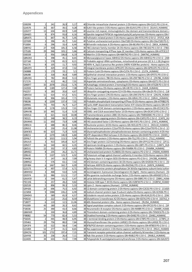
Appendices
207
O00299 1 241 26,9 5,17 40 Chloride intracellular channel protein 1 OS=Homo sapiens GN=CLIC1 PE=1 SV=4 - [CLIC1_HUMAN]
Q14576 35 367 39,5 9,28 40 ELAV-like protein 3 OS=Homo sapiens GN=ELAVL3 PE=1 SV=3 - [ELAV3_HUMAN]
Q3SXY7 13 634 69,9 5,69 40 Leucine-rich repeat, immunoglobulin-like domain and transmembrane domain-containing protein 3 OS=Homo sapiens GN=LRIT3 PE=2 SV=2 - [LRIT3_HUMAN]
Q9H6E5 9 874 93,8 6,16 40 Speckle targeted PIP5K1A-regulated poly(A) polymerase OS=Homo sapiens GN=TUT1 PE=1 SV=2 - [STPAP_HUMAN]
Q8N475 5 847 95,7 5,95 40 Follistatin-related protein 5 OS=Homo sapiens GN=FSTL5 PE=2 SV=2 - [FSTL5_HUMAN]
Q5T699 3 396 45,7 5,62 40 Putative uncharacterized protein C6orf183 OS=Homo sapiens GN=C6orf183 PE=5 SV=2 - [CF183_HUMAN]
P53004 6 296 33,4 6,44 40 Biliverdin reductase A OS=Homo sapiens GN=BLVRA PE=1 SV=2 - [BIEA_HUMAN]
Q9BYX2 27 928 105,3 6,58 40 TBC1 domain family member 2A OS=Homo sapiens GN=TBC1D2 PE=1 SV=3 - [TBD2A_HUMAN]
O75185 29 946 103,1 5,66 40 Calcium-transporting ATPase type 2C member 2 OS=Homo sapiens GN=ATP2C2 PE=1 SV=2 - [AT2C2_HUMAN]
O00339 2 956 106,8 6,19 40 Matrilin-2 OS=Homo sapiens GN=MATN2 PE=1 SV=3 - [MATN2_HUMAN]
Q8N2K0 5 398 45,1 8,65 40 Monoacylglycerol lipase ABHD12 OS=Homo sapiens GN=ABHD12 PE=2 SV=2 - [ABD12_HUMAN]
Q5T160 5 578 65,5 8,21 39 Probable arginyl-tRNA synthetase, mitochondrial precursor (EC 6.1.1.19) (Arginine--tRNA ligase) (ArgRS) (Arginyl-tRNA synthetase-like) - Homo sapiens (Human) - [SYRM_HUMAN]
P61576 8 105 11,7 9,92 39 HERV-K_5q13.3 provirus Rec protein (HERV-K104 Rec protein) - Homo sapiens (Human) - [REC9_HUMAN]
Q8N3F9 7 429 47,0 7,42 39 Integral membrane protein GPR137C OS=Homo sapiens GN=GPR137C PE=2 SV=2 - [G137C_HUMAN]
O60888 5 179 19,1 5,5 39 Protein CutA OS=Homo sapiens GN=CUTA PE=1 SV=2 - [CUTA_HUMAN]
Q96J88 2 318 36,8 9,89 39 Epithelial-stromal interaction protein 1 OS=Homo sapiens GN=EPSTI1 PE=2 SV=2 - [ESIP1_HUMAN]
Q8N393 5 782 89,8 9,13 39 Zinc finger protein 786 OS=Homo sapiens GN=ZNF786 PE=2 SV=2 - [ZN786_HUMAN]
P17174 3 413 46,2 7,01 39 Aspartate aminotransferase, cytoplasmic OS=Homo sapiens GN=GOT1 PE=1 SV=3 - [AATC_HUMAN]
Q96BY7 33 2078 232,6 5,76 39 Autophagy-related protein 2 homolog B OS=Homo sapiens GN=ATG2B PE=1 SV=5 - [ATG2B_HUMAN]
O43593 8 1189 127,4 7,08 39 Protein hairless OS=Homo sapiens GN=HR PE=1 SV=5 - [HAIR_HUMAN]
P62257 9 183 20,6 4,67 39 Ubiquitin-conjugating enzyme E2 H OS=Mus musculus GN=Ube2h PE=2 SV=1 - [UBE2H_MOUSE]
Q8N554 11 614 67,2 8,51 39 Zinc finger protein 276 OS=Homo sapiens GN=ZNF276 PE=1 SV=4 - [ZN276_HUMAN]
P42261 8 906 101,4 7,71 39 Glutamate receptor 1 OS=Homo sapiens GN=GRIA1 PE=1 SV=2 - [GRIA1_HUMAN]
P98198 8 1209 137,4 7,01 39 Probable phospholipid-transporting ATPase ID OS=Homo sapiens GN=ATP8B2 PE=1 SV=2 - [AT8B2_HUMAN]
Q99941 73 703 76,7 6,27 39 Cyclic AMP-dependent transcription factor ATF-6 beta OS=Homo sapiens GN=ATF6B PE=1 SV=2 - [ATF6B_HUMAN]
Q9C0B9 4 1178 125,9 7,01 39 Zinc finger CCHC domain-containing protein 2 OS=Homo sapiens GN=ZCCHC2 PE=1 SV=6 - [ZCHC2_HUMAN]
Q9NV06 10 445 51,4 9,19 39 DDB1- and CUL4-associated factor 13 OS=Homo sapiens GN=DCAF13 PE=1 SV=2 - [DCA13_HUMAN]
A6NKL6 3 621 63,9 10,08 39 Transmembrane protein 200C OS=Homo sapiens GN=TMEM200C PE=2 SV=2 - [T200C_HUMAN]
P40121 12 348 38,5 6,19 39 Macrophage-capping protein OS=Homo sapiens GN=CAPG PE=1 SV=2 - [CAPG_HUMAN]
Q9UNN5 11 650 73,9 4,88 39 FAS-associated factor 1 OS=Homo sapiens GN=FAF1 PE=1 SV=2 - [FAF1_HUMAN]
Q9BSC4 3 688 80,3 8,46 39 Nucleolar protein 10 OS=Homo sapiens GN=NOL10 PE=1 SV=1 - [NOL10_HUMAN]
Q9NSI2 3 230 25,4 11,08 39 Uncharacterized protein C21orf70 OS=Homo sapiens GN=C21orf70 PE=1 SV=2 - [CU070_HUMAN]
Q6W3E5 43 623 71,9 9,03 39 Glycerophosphodiester phosphodiesterase domain-containing protein 4 OS=Homo sapiens GN=GDPD4 PE=2 SV=1 - [GDPD4_HUMAN]
Q08211 2 1270 140,9 6,84 39 ATP-dependent RNA helicase A OS=Homo sapiens GN=DHX9 PE=1 SV=4 - [DHX9_HUMAN]
Q9Y6L7 9 1015 113,5 5,94 39 Tolloid-like protein 2 OS=Homo sapiens GN=TLL2 PE=1 SV=1 - [TLL2_HUMAN]
Q9UJU3 3 913 105,8 8,27 39 Zinc finger protein 112 homolog OS=Homo sapiens GN=ZFP112 PE=2 SV=2 - [ZF112_HUMAN]
Q9NZI7 34 540 60,5 6,35 39 Upstream-binding protein 1 OS=Homo sapiens GN=UBP1 PE=2 SV=1 - [UBIP1_HUMAN]
Q8IZU1 12 332 37,3 4,83 39 Protein FAM9A OS=Homo sapiens GN=FAM9A PE=2 SV=1 - [FAM9A_HUMAN]
Q8N9Z2 8 235 26,2 11,71 39 Uncharacterized protein FLJ36031 OS=Homo sapiens PE=2 SV=2 - [YG034_HUMAN]
Q96L42 7 1107 123,7 6,92 39 Potassium voltage-gated channel subfamily H member 8 OS=Homo sapiens GN=KCNH8 PE=2 SV=2 - [KCNH8_HUMAN]
P04438 7 147 16,3 7,09 39 Ig heavy chain V-II region SESS OS=Homo sapiens PE=2 SV=1 - [HV208_HUMAN]
Q9BRG2 7 576 63,1 7,42 39 SH2 domain-containing protein 3A OS=Homo sapiens GN=SH2D3A PE=1 SV=1 - [SH23A_HUMAN]
Q9Y4B4 6 1467 162,7 6,09 39 Helicase ARIP4 OS=Homo sapiens GN=RAD54L2 PE=1 SV=4 - [ARIP4_HUMAN]
Q15173 4 497 57,4 6,71 38 Serine/threonine-protein phosphatase 2A 56 kDa regulatory subunit beta isoform OS=Homo sapiens GN=PPP2R5B PE=1 SV=1 - [2A5B_HUMAN]
Q8WXD2 3 468 53,0 5,03 38 Secretogranin-3 precursor (Secretogranin III) (SgIII) - Homo sapiens (Human) - [SCG3_HUMAN]
Q92974 15 986 111,5 7,27 38 Rho guanine nucleotide exchange factor 2 OS=Homo sapiens GN=ARHGEF2 PE=1 SV=4 - [ARHG2_HUMAN]
Q9UK59 3 544 61,5 5,47 38 Lariat debranching enzyme OS=Homo sapiens GN=DBR1 PE=1 SV=2 - [DBR1_HUMAN]
P0C1H6 6 257 28,2 9,82 38 Histone H2B type F-M OS=Homo sapiens GN=H2BFM PE=2 SV=1 - [H2BFM_HUMAN]
Q92539 5 896 99,3 5,33 38 Lipin-2 - Homo sapiens (Human) - [LPIN2_HUMAN]
Q9Y426 14 696 75,5 6,92 38 C2 domain-containing protein 2 OS=Homo sapiens GN=C2CD2 PE=1 SV=2 - [CU025_HUMAN]
Q15858 9 1988 226,2 6,99 38 Sodium channel protein type 9 subunit alpha OS=Homo sapiens GN=SCN9A PE=1 SV=2 - [SCN9A_HUMAN]
Q9H0H3 3 589 65,9 6,57 38 Ectoderm-neural cortex protein 2 OS=Homo sapiens GN=KLHL25 PE=2 SV=1 - [ENC2_HUMAN]
P09210 2 222 25,6 8,59 38 Glutathione S-transferase A2 OS=Homo sapiens GN=GSTA2 PE=1 SV=4 - [GSTA2_HUMAN]
P18077 6 110 12,5 11,06 38 60S ribosomal protein L35a - Homo sapiens (Human) - [RL35A_HUMAN]
P61009 43 180 20,3 8,62 38 Signal peptidase complex subunit 3 OS=Homo sapiens GN=SPCS3 PE=1 SV=1 - [SPCS3_HUMAN]
P28336 25 390 43,4 8,66 38 Neuromedin-B receptor OS=Homo sapiens GN=NMBR PE=2 SV=2 - [NMBR_HUMAN]
Q86XW9 1 330 36,8 4,89 38 Thioredoxin domain-containing protein 6 OS=Homo sapiens GN=TXNDC6 PE=1 SV=1 - [TXND6_HUMAN]
P98082 54 770 82,4 5,53 38 Disabled homolog 2 OS=Homo sapiens GN=DAB2 PE=1 SV=3 - [DAB2_HUMAN]
P56545 2 445 48,9 6,95 38 C-terminal-binding protein 2 OS=Homo sapiens GN=CTBP2 PE=1 SV=1 - [CTBP2_HUMAN]
O95461 29 756 88,0 7,84 38 Glycosyltransferase-like protein LARGE1 OS=Homo sapiens GN=LARGE PE=1 SV=1 - [LARGE_HUMAN]
Q9Y2F9 10 522 58,4 7,46 38 BTB/POZ domain-containing protein 3 OS=Homo sapiens GN=BTBD3 PE=2 SV=1 - [BTBD3_HUMAN]
Q15404 52 277 31,5 8,65 38 Ras suppressor protein 1 OS=Homo sapiens GN=RSU1 PE=1 SV=3 - [RSU1_HUMAN]
Q9HCF6 101 1732 197,4 7,15 38 Transient receptor potential cation channel subfamily M member 3 OS=Homo sapiens GN=TRPM3 PE=2 SV=4 - [TRPM3_HUMAN]
Q5HYI8 35 236 26,4 7,11 38 Rab-like protein 3 OS=Homo sapiens GN=RABL3 PE=1 SV=1 - [RABL3_HUMAN]
Q14435 34 633 72,6 7,99 38 Polypeptide N-acetylgalactosaminyltransferase 3 OS=Homo sapiens GN=GALNT3 PE=2 SV=2 - [GALT3_HUMAN]
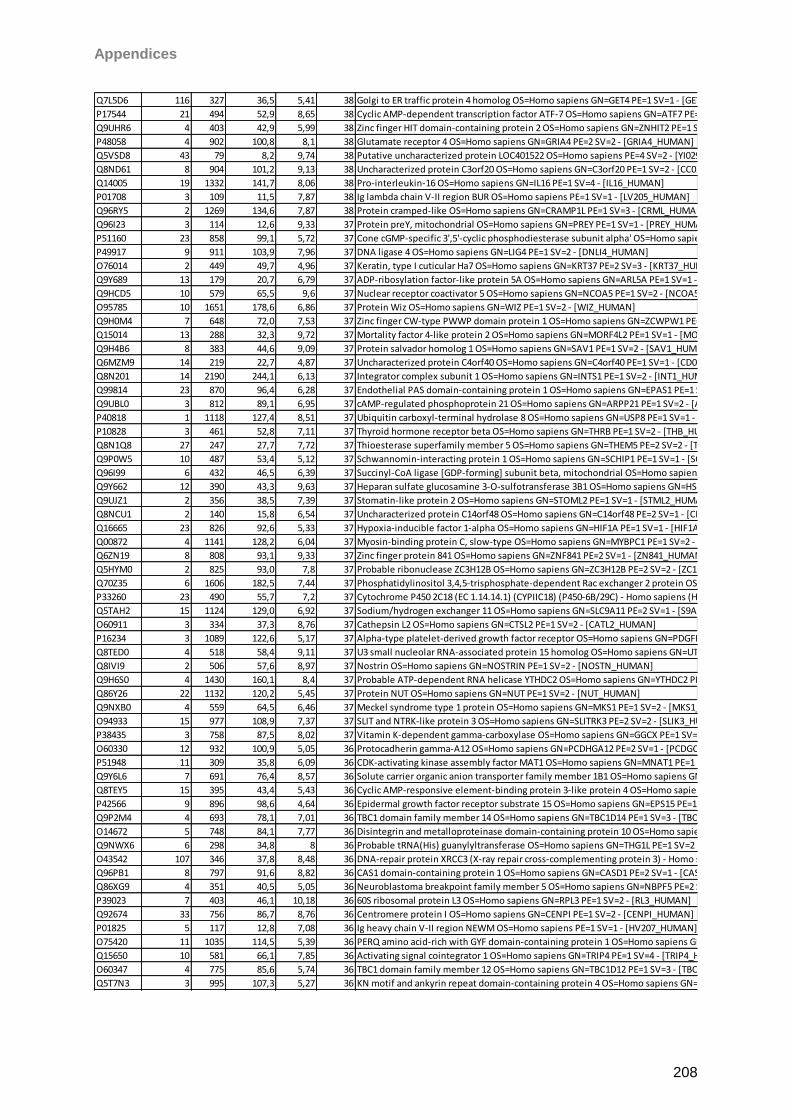
Appendices
208
Q7L5D6 116 327 36,5 5,41 38 Golgi to ER traffic protein 4 homolog OS=Homo sapiens GN=GET4 PE=1 SV=1 - [GET4_HUMAN]
P17544 21 494 52,9 8,65 38 Cyclic AMP-dependent transcription factor ATF-7 OS=Homo sapiens GN=ATF7 PE=1 SV=2 - [ATF7_HUMAN]
Q9UHR6 4 403 42,9 5,99 38 Zinc finger HIT domain-containing protein 2 OS=Homo sapiens GN=ZNHIT2 PE=1 SV=1 - [ZNHI2_HUMAN]
P48058 4 902 100,8 8,1 38 Glutamate receptor 4 OS=Homo sapiens GN=GRIA4 PE=2 SV=2 - [GRIA4_HUMAN]
Q5VSD8 43 79 8,2 9,74 38 Putative uncharacterized protein LOC401522 OS=Homo sapiens PE=4 SV=2 - [YI029_HUMAN]
Q8ND61 8 904 101,2 9,13 38 Uncharacterized protein C3orf20 OS=Homo sapiens GN=C3orf20 PE=1 SV=2 - [CC020_HUMAN]
Q14005 19 1332 141,7 8,06 38 Pro-interleukin-16 OS=Homo sapiens GN=IL16 PE=1 SV=4 - [IL16_HUMAN]
P01708 3 109 11,5 7,87 38 Ig lambda chain V-II region BUR OS=Homo sapiens PE=1 SV=1 - [LV205_HUMAN]
Q96RY5 2 1269 134,6 7,87 38 Protein cramped-like OS=Homo sapiens GN=CRAMP1L PE=1 SV=3 - [CRML_HUMAN]
Q96I23 3 114 12,6 9,33 37 Protein preY, mitochondrial OS=Homo sapiens GN=PREY PE=1 SV=1 - [PREY_HUMAN]
P51160 23 858 99,1 5,72 37 Cone cGMP-specific 3',5'-cyclic phosphodiesterase subunit alpha' OS=Homo sapiens GN=PDE6C PE=1 SV=2 - [PDE6C_HUMAN]
P49917 9 911 103,9 7,96 37 DNA ligase 4 OS=Homo sapiens GN=LIG4 PE=1 SV=2 - [DNLI4_HUMAN]
O76014 2 449 49,7 4,96 37 Keratin, type I cuticular Ha7 OS=Homo sapiens GN=KRT37 PE=2 SV=3 - [KRT37_HUMAN]
Q9Y689 13 179 20,7 6,79 37 ADP-ribosylation factor-like protein 5A OS=Homo sapiens GN=ARL5A PE=1 SV=1 - [ARL5A_HUMAN]
Q9HCD5 10 579 65,5 9,6 37 Nuclear receptor coactivator 5 OS=Homo sapiens GN=NCOA5 PE=1 SV=2 - [NCOA5_HUMAN]
O95785 10 1651 178,6 6,86 37 Protein Wiz OS=Homo sapiens GN=WIZ PE=1 SV=2 - [WIZ_HUMAN]
Q9H0M4 7 648 72,0 7,53 37 Zinc finger CW-type PWWP domain protein 1 OS=Homo sapiens GN=ZCWPW1 PE=1 SV=2 - [ZCPW1_HUMAN]
Q15014 13 288 32,3 9,72 37 Mortality factor 4-like protein 2 OS=Homo sapiens GN=MORF4L2 PE=1 SV=1 - [MO4L2_HUMAN]
Q9H4B6 8 383 44,6 9,09 37 Protein salvador homolog 1 OS=Homo sapiens GN=SAV1 PE=1 SV=2 - [SAV1_HUMAN]
Q6MZM9 14 219 22,7 4,87 37 Uncharacterized protein C4orf40 OS=Homo sapiens GN=C4orf40 PE=1 SV=1 - [CD040_HUMAN]
Q8N201 14 2190 244,1 6,13 37 Integrator complex subunit 1 OS=Homo sapiens GN=INTS1 PE=1 SV=2 - [INT1_HUMAN]
Q99814 23 870 96,4 6,28 37 Endothelial PAS domain-containing protein 1 OS=Homo sapiens GN=EPAS1 PE=1 SV=3 - [EPAS1_HUMAN]
Q9UBL0 3 812 89,1 6,95 37 cAMP-regulated phosphoprotein 21 OS=Homo sapiens GN=ARPP21 PE=1 SV=2 - [ARP21_HUMAN]
P40818 1 1118 127,4 8,51 37 Ubiquitin carboxyl-terminal hydrolase 8 OS=Homo sapiens GN=USP8 PE=1 SV=1 - [UBP8_HUMAN]
P10828 3 461 52,8 7,11 37 Thyroid hormone receptor beta OS=Homo sapiens GN=THRB PE=1 SV=2 - [THB_HUMAN]
Q8N1Q8 27 247 27,7 7,72 37 Thioesterase superfamily member 5 OS=Homo sapiens GN=THEM5 PE=2 SV=2 - [THEM5_HUMAN]
Q9P0W5 10 487 53,4 5,12 37 Schwannomin-interacting protein 1 OS=Homo sapiens GN=SCHIP1 PE=1 SV=1 - [SCHI1_HUMAN]
Q96I99 6 432 46,5 6,39 37 Succinyl-CoA ligase [GDP-forming] subunit beta, mitochondrial OS=Homo sapiens GN=SUCLG2 PE=1 SV=2 - [SUCB2_HUMAN]
Q9Y662 12 390 43,3 9,63 37 Heparan sulfate glucosamine 3-O-sulfotransferase 3B1 OS=Homo sapiens GN=HS3ST3B1 PE=1 SV=1 - [HS3SB_HUMAN]
Q9UJZ1 2 356 38,5 7,39 37 Stomatin-like protein 2 OS=Homo sapiens GN=STOML2 PE=1 SV=1 - [STML2_HUMAN]
Q8NCU1 2 140 15,8 6,54 37 Uncharacterized protein C14orf48 OS=Homo sapiens GN=C14orf48 PE=2 SV=1 - [CN048_HUMAN]
Q16665 23 826 92,6 5,33 37 Hypoxia-inducible factor 1-alpha OS=Homo sapiens GN=HIF1A PE=1 SV=1 - [HIF1A_HUMAN]
Q00872 4 1141 128,2 6,04 37 Myosin-binding protein C, slow-type OS=Homo sapiens GN=MYBPC1 PE=1 SV=2 - [MYPC1_HUMAN]
Q6ZN19 8 808 93,1 9,33 37 Zinc finger protein 841 OS=Homo sapiens GN=ZNF841 PE=2 SV=1 - [ZN841_HUMAN]
Q5HYM0 2 825 93,0 7,8 37 Probable ribonuclease ZC3H12B OS=Homo sapiens GN=ZC3H12B PE=2 SV=2 - [ZC12B_HUMAN]
Q70Z35 6 1606 182,5 7,44 37 Phosphatidylinositol 3,4,5-trisphosphate-dependent Rac exchanger 2 protein OS=Homo sapiens GN=PREX2 PE=2 SV=1 - [PREX2_HUMAN]
P33260 23 490 55,7 7,2 37 Cytochrome P450 2C18 (EC 1.14.14.1) (CYPIIC18) (P450-6B/29C) - Homo sapiens (Human) - [CP2CI_HUMAN]
Q5TAH2 15 1124 129,0 6,92 37 Sodium/hydrogen exchanger 11 OS=Homo sapiens GN=SLC9A11 PE=2 SV=1 - [S9A11_HUMAN]
O60911 3 334 37,3 8,76 37 Cathepsin L2 OS=Homo sapiens GN=CTSL2 PE=1 SV=2 - [CATL2_HUMAN]
P16234 3 1089 122,6 5,17 37 Alpha-type platelet-derived growth factor receptor OS=Homo sapiens GN=PDGFRA PE=1 SV=1 - [PGFRA_HUMAN]
Q8TED0 4 518 58,4 9,11 37 U3 small nucleolar RNA-associated protein 15 homolog OS=Homo sapiens GN=UTP15 PE=1 SV=3 - [UTP15_HUMAN]
Q8IVI9 2 506 57,6 8,97 37 Nostrin OS=Homo sapiens GN=NOSTRIN PE=1 SV=2 - [NOSTN_HUMAN]
Q9H6S0 4 1430 160,1 8,4 37 Probable ATP-dependent RNA helicase YTHDC2 OS=Homo sapiens GN=YTHDC2 PE=1 SV=2 - [YTDC2_HUMAN]
Q86Y26 22 1132 120,2 5,45 37 Protein NUT OS=Homo sapiens GN=NUT PE=1 SV=2 - [NUT_HUMAN]
Q9NXB0 4 559 64,5 6,46 37 Meckel syndrome type 1 protein OS=Homo sapiens GN=MKS1 PE=1 SV=2 - [MKS1_HUMAN]
O94933 15 977 108,9 7,37 37 SLIT and NTRK-like protein 3 OS=Homo sapiens GN=SLITRK3 PE=2 SV=2 - [SLIK3_HUMAN]
P38435 3 758 87,5 8,02 37 Vitamin K-dependent gamma-carboxylase OS=Homo sapiens GN=GGCX PE=1 SV=2 - [VKGC_HUMAN]
O60330 12 932 100,9 5,05 36 Protocadherin gamma-A12 OS=Homo sapiens GN=PCDHGA12 PE=2 SV=1 - [PCDGC_HUMAN]
P51948 11 309 35,8 6,09 36 CDK-activating kinase assembly factor MAT1 OS=Homo sapiens GN=MNAT1 PE=1 SV=1 - [MAT1_HUMAN]
Q9Y6L6 7 691 76,4 8,57 36 Solute carrier organic anion transporter family member 1B1 OS=Homo sapiens GN=SLCO1B1 PE=1 SV=2 - [SO1B1_HUMAN]
Q8TEY5 15 395 43,4 5,43 36 Cyclic AMP-responsive element-binding protein 3-like protein 4 OS=Homo sapiens GN=CREB3L4 PE=1 SV=1 - [CR3L4_HUMAN]
P42566 9 896 98,6 4,64 36 Epidermal growth factor receptor substrate 15 OS=Homo sapiens GN=EPS15 PE=1 SV=2 - [EPS15_HUMAN]
Q9P2M4 4 693 78,1 7,01 36 TBC1 domain family member 14 OS=Homo sapiens GN=TBC1D14 PE=1 SV=3 - [TBC14_HUMAN]
O14672 5 748 84,1 7,77 36 Disintegrin and metalloproteinase domain-containing protein 10 OS=Homo sapiens GN=ADAM10 PE=1 SV=1 - [ADA10_HUMAN]
Q9NWX6 6 298 34,8 8 36 Probable tRNA(His) guanylyltransferase OS=Homo sapiens GN=THG1L PE=1 SV=2 - [THG1_HUMAN]
O43542 107 346 37,8 8,48 36 DNA-repair protein XRCC3 (X-ray repair cross-complementing protein 3) - Homo sapiens (Human) - [XRCC3_HUMAN]
Q96PB1 8 797 91,6 8,82 36 CAS1 domain-containing protein 1 OS=Homo sapiens GN=CASD1 PE=2 SV=1 - [CASD1_HUMAN]
Q86XG9 4 351 40,5 5,05 36 Neuroblastoma breakpoint family member 5 OS=Homo sapiens GN=NBPF5 PE=2 SV=2 - [NBPF5_HUMAN]
P39023 7 403 46,1 10,18 36 60S ribosomal protein L3 OS=Homo sapiens GN=RPL3 PE=1 SV=2 - [RL3_HUMAN]
Q92674 33 756 86,7 8,76 36 Centromere protein I OS=Homo sapiens GN=CENPI PE=1 SV=2 - [CENPI_HUMAN]
P01825 5 117 12,8 7,08 36 Ig heavy chain V-II region NEWM OS=Homo sapiens PE=1 SV=1 - [HV207_HUMAN]
O75420 11 1035 114,5 5,39 36 PERQ amino acid-rich with GYF domain-containing protein 1 OS=Homo sapiens GN=GIGYF1 PE=1 SV=2 - [PERQ1_HUMAN]
Q15650 10 581 66,1 7,85 36 Activating signal cointegrator 1 OS=Homo sapiens GN=TRIP4 PE=1 SV=4 - [TRIP4_HUMAN]
O60347 4 775 85,6 5,74 36 TBC1 domain family member 12 OS=Homo sapiens GN=TBC1D12 PE=1 SV=3 - [TBC12_HUMAN]
Q5T7N3 3 995 107,3 5,27 36 KN motif and ankyrin repeat domain-containing protein 4 OS=Homo sapiens GN=KANK4 PE=1 SV=1 - [KANK4_HUMAN]
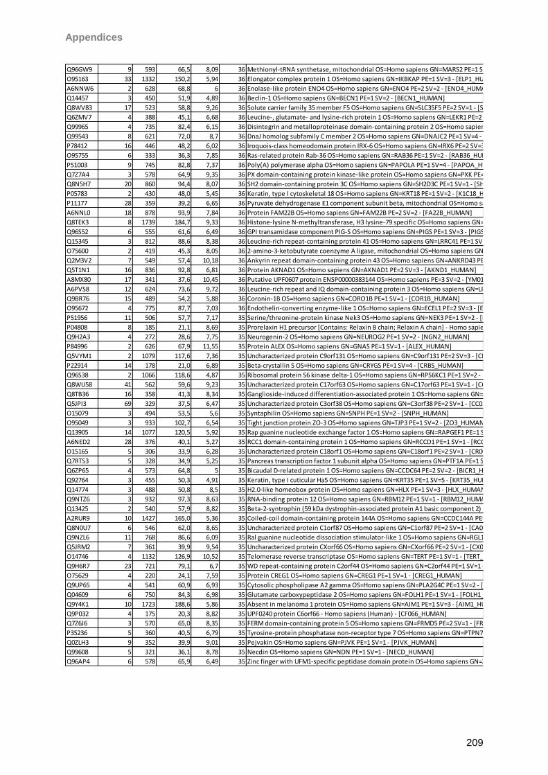
Appendices
209
Q96GW9 9 593 66,5 8,09 36 Methionyl-tRNA synthetase, mitochondrial OS=Homo sapiens GN=MARS2 PE=1 SV=2 - [SYMM_HUMAN]
O95163 33 1332 150,2 5,94 36 Elongator complex protein 1 OS=Homo sapiens GN=IKBKAP PE=1 SV=3 - [ELP1_HUMAN]
A6NNW6 2 628 68,8 6 36 Enolase-like protein ENO4 OS=Homo sapiens GN=ENO4 PE=2 SV=2 - [ENO4_HUMAN]
Q14457 3 450 51,9 4,89 36 Beclin-1 OS=Homo sapiens GN=BECN1 PE=1 SV=2 - [BECN1_HUMAN]
Q8WV83 17 523 58,8 9,26 36 Solute carrier family 35 member F5 OS=Homo sapiens GN=SLC35F5 PE=2 SV=1 - [S35F5_HUMAN]
Q6ZMV7 4 388 45,1 6,68 36 Leucine-, glutamate- and lysine-rich protein 1 OS=Homo sapiens GN=LEKR1 PE=2 SV=2 - [LEKR1_HUMAN]
Q99965 4 735 82,4 6,15 36 Disintegrin and metalloproteinase domain-containing protein 2 OS=Homo sapiens GN=ADAM2 PE=1 SV=2 - [ADAM2_HUMAN]
Q99543 8 621 72,0 8,7 36 DnaJ homolog subfamily C member 2 OS=Homo sapiens GN=DNAJC2 PE=1 SV=4 - [DNJC2_HUMAN]
P78412 16 446 48,2 6,02 36 Iroquois-class homeodomain protein IRX-6 OS=Homo sapiens GN=IRX6 PE=2 SV=3 - [IRX6_HUMAN]
O95755 6 333 36,3 7,85 36 Ras-related protein Rab-36 OS=Homo sapiens GN=RAB36 PE=1 SV=2 - [RAB36_HUMAN]
P51003 9 745 82,8 7,37 36 Poly(A) polymerase alpha OS=Homo sapiens GN=PAPOLA PE=1 SV=4 - [PAPOA_HUMAN]
Q7Z7A4 3 578 64,9 9,35 36 PX domain-containing protein kinase-like protein OS=Homo sapiens GN=PXK PE=1 SV=1 - [PXK_HUMAN]
Q8N5H7 20 860 94,4 8,07 36 SH2 domain-containing protein 3C OS=Homo sapiens GN=SH2D3C PE=1 SV=1 - [SH2D3_HUMAN]
P05783 2 430 48,0 5,45 36 Keratin, type I cytoskeletal 18 OS=Homo sapiens GN=KRT18 PE=1 SV=2 - [K1C18_HUMAN]
P11177 28 359 39,2 6,65 36 Pyruvate dehydrogenase E1 component subunit beta, mitochondrial OS=Homo sapiens GN=PDHB PE=1 SV=3 - [ODPB_HUMAN]
A6NNL0 18 878 93,9 7,84 36 Protein FAM22B OS=Homo sapiens GN=FAM22B PE=2 SV=2 - [FA22B_HUMAN]
Q8TEK3 8 1739 184,7 9,33 36 Histone-lysine N-methyltransferase, H3 lysine-79 specific OS=Homo sapiens GN=DOT1L PE=1 SV=2 - [DOT1L_HUMAN]
Q96S52 6 555 61,6 6,49 36 GPI transamidase component PIG-S OS=Homo sapiens GN=PIGS PE=1 SV=3 - [PIGS_HUMAN]
Q15345 3 812 88,6 8,38 36 Leucine-rich repeat-containing protein 41 OS=Homo sapiens GN=LRRC41 PE=1 SV=3 - [LRC41_HUMAN]
O75600 2 419 45,3 8,05 36 2-amino-3-ketobutyrate coenzyme A ligase, mitochondrial OS=Homo sapiens GN=GCAT PE=2 SV=1 - [KBL_HUMAN]
Q2M3V2 7 549 57,4 10,18 36 Ankyrin repeat domain-containing protein 43 OS=Homo sapiens GN=ANKRD43 PE=1 SV=3 - [ANR43_HUMAN]
Q5T1N1 16 836 92,8 6,81 36 Protein AKNAD1 OS=Homo sapiens GN=AKNAD1 PE=2 SV=3 - [AKND1_HUMAN]
A8MX80 17 341 37,6 10,45 36 Putative UPF0607 protein ENSP00000383144 OS=Homo sapiens PE=3 SV=2 - [YM017_HUMAN]
A6PVS8 12 624 73,6 9,72 36 Leucine-rich repeat and IQ domain-containing protein 3 OS=Homo sapiens GN=LRRIQ3 PE=2 SV=1 - [LRIQ3_HUMAN]
Q9BR76 15 489 54,2 5,88 36 Coronin-1B OS=Homo sapiens GN=CORO1B PE=1 SV=1 - [COR1B_HUMAN]
O95672 4 775 87,7 7,03 36 Endothelin-converting enzyme-like 1 OS=Homo sapiens GN=ECEL1 PE=2 SV=3 - [ECEL1_HUMAN]
P51956 11 506 57,7 7,17 35 Serine/threonine-protein kinase Nek3 OS=Homo sapiens GN=NEK3 PE=1 SV=2 - [NEK3_HUMAN]
P04808 8 185 21,1 8,69 35 Prorelaxin H1 precursor [Contains: Relaxin B chain; Relaxin A chain] - Homo sapiens (Human) - [REL1_HUMAN]
Q9H2A3 4 272 28,6 7,75 35 Neurogenin-2 OS=Homo sapiens GN=NEUROG2 PE=1 SV=2 - [NGN2_HUMAN]
P84996 2 626 67,9 11,55 35 Protein ALEX OS=Homo sapiens GN=GNAS PE=1 SV=1 - [ALEX_HUMAN]
Q5VYM1 2 1079 117,6 7,36 35 Uncharacterized protein C9orf131 OS=Homo sapiens GN=C9orf131 PE=2 SV=3 - [CI131_HUMAN]
P22914 14 178 21,0 6,89 35 Beta-crystallin S OS=Homo sapiens GN=CRYGS PE=1 SV=4 - [CRBS_HUMAN]
Q96S38 2 1066 118,6 4,87 35 Ribosomal protein S6 kinase delta-1 OS=Homo sapiens GN=RPS6KC1 PE=1 SV=2 - [KS6C1_HUMAN]
Q8WU58 41 562 59,6 9,23 35 Uncharacterized protein C17orf63 OS=Homo sapiens GN=C17orf63 PE=1 SV=1 - [CQ063_HUMAN]
Q8TB36 16 358 41,3 8,34 35 Ganglioside-induced differentiation-associated protein 1 OS=Homo sapiens GN=GDAP1 PE=1 SV=3 - [GDAP1_HUMAN]
Q5JPI3 69 329 37,5 6,47 35 Uncharacterized protein C3orf38 OS=Homo sapiens GN=C3orf38 PE=2 SV=1 - [CC038_HUMAN]
O15079 3 494 53,5 5,6 35 Syntaphilin OS=Homo sapiens GN=SNPH PE=1 SV=2 - [SNPH_HUMAN]
O95049 3 933 102,7 6,54 35 Tight junction protein ZO-3 OS=Homo sapiens GN=TJP3 PE=1 SV=2 - [ZO3_HUMAN]
Q13905 14 1077 120,5 5,92 35 Rap guanine nucleotide exchange factor 1 OS=Homo sapiens GN=RAPGEF1 PE=1 SV=3 - [RPGF1_HUMAN]
A6NED2 28 376 40,1 5,27 35 RCC1 domain-containing protein 1 OS=Homo sapiens GN=RCCD1 PE=1 SV=1 - [RCCD1_HUMAN]
O15165 5 306 33,9 6,28 35 Uncharacterized protein C18orf1 OS=Homo sapiens GN=C18orf1 PE=2 SV=1 - [CR001_HUMAN]
Q7RTS3 5 328 34,9 5,25 35 Pancreas transcription factor 1 subunit alpha OS=Homo sapiens GN=PTF1A PE=1 SV=1 - [PTF1A_HUMAN]
Q6ZP65 4 573 64,8 5 35 Bicaudal D-related protein 1 OS=Homo sapiens GN=CCDC64 PE=2 SV=2 - [BICR1_HUMAN]
Q92764 3 455 50,3 4,91 35 Keratin, type I cuticular Ha5 OS=Homo sapiens GN=KRT35 PE=1 SV=5 - [KRT35_HUMAN]
Q14774 3 488 50,8 8,5 35 H2.0-like homeobox protein OS=Homo sapiens GN=HLX PE=1 SV=3 - [HLX_HUMAN]
Q9NTZ6 3 932 97,3 8,63 35 RNA-binding protein 12 OS=Homo sapiens GN=RBM12 PE=1 SV=1 - [RBM12_HUMAN]
Q13425 2 540 57,9 8,82 35 Beta-2-syntrophin (59 kDa dystrophin-associated protein A1 basic component 2) (Syntrophin 3) (SNT3) (Syntrophin-like) (SNTL) - Homo sapiens (Human) - [SNTB2_HUMAN]
A2RUR9 10 1427 165,0 5,36 35 Coiled-coil domain-containing protein 144A OS=Homo sapiens GN=CCDC144A PE=1 SV=1 - [C144A_HUMAN]
Q8N0U7 6 546 62,0 8,65 35 Uncharacterized protein C1orf87 OS=Homo sapiens GN=C1orf87 PE=2 SV=1 - [CA087_HUMAN]
Q9NZL6 11 768 86,6 6,09 35 Ral guanine nucleotide dissociation stimulator-like 1 OS=Homo sapiens GN=RGL1 PE=1 SV=1 - [RGL1_HUMAN]
Q5JRM2 7 361 39,9 9,54 35 Uncharacterized protein CXorf66 OS=Homo sapiens GN=CXorf66 PE=2 SV=1 - [CX066_HUMAN]
O14746 4 1132 126,9 10,52 35 Telomerase reverse transcriptase OS=Homo sapiens GN=TERT PE=1 SV=1 - [TERT_HUMAN]
Q9H6R7 23 721 79,1 6,7 35 WD repeat-containing protein C2orf44 OS=Homo sapiens GN=C2orf44 PE=1 SV=1 - [CB044_HUMAN]
O75629 4 220 24,1 7,59 35 Protein CREG1 OS=Homo sapiens GN=CREG1 PE=1 SV=1 - [CREG1_HUMAN]
Q9UP65 4 541 60,9 6,93 35 Cytosolic phospholipase A2 gamma OS=Homo sapiens GN=PLA2G4C PE=1 SV=2 - [PA24C_HUMAN]
Q04609 6 750 84,3 6,98 35 Glutamate carboxypeptidase 2 OS=Homo sapiens GN=FOLH1 PE=1 SV=1 - [FOLH1_HUMAN]
Q9Y4K1 10 1723 188,6 5,86 35 Absent in melanoma 1 protein OS=Homo sapiens GN=AIM1 PE=1 SV=3 - [AIM1_HUMAN]
Q9P032 4 175 20,3 8,82 35 UPF0240 protein C6orf66 - Homo sapiens (Human) - [CF066_HUMAN]
Q7Z6J6 3 570 65,0 8,35 35 FERM domain-containing protein 5 OS=Homo sapiens GN=FRMD5 PE=2 SV=1 - [FRMD5_HUMAN]
P35236 5 360 40,5 6,79 35 Tyrosine-protein phosphatase non-receptor type 7 OS=Homo sapiens GN=PTPN7 PE=1 SV=3 - [PTN7_HUMAN]
Q0ZLH3 9 352 39,9 9,01 35 Pejvakin OS=Homo sapiens GN=PJVK PE=1 SV=1 - [PJVK_HUMAN]
Q99608 5 321 36,1 8,78 35 Necdin OS=Homo sapiens GN=NDN PE=1 SV=1 - [NECD_HUMAN]
Q96AP4 6 578 65,9 6,49 35 Zinc finger with UFM1-specific peptidase domain protein OS=Homo sapiens GN=ZUFSP PE=1 SV=1 - [ZUFSP_HUMAN]
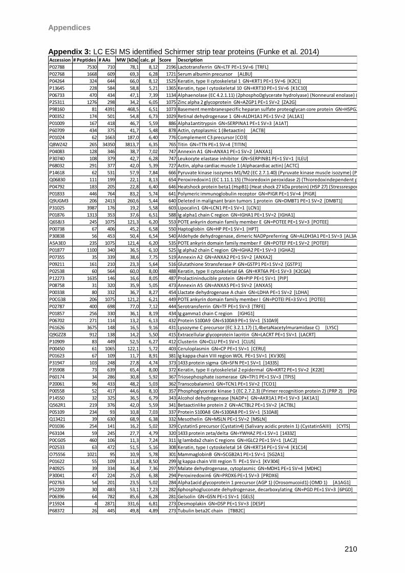
Appendices
210
Appendix 3: LC ESI MS identified Schirmer strip tear proteins (Funke et al. 2014)
Accession # Peptides # AAs MW [kDa] calc. pI Score Description
P02788 7530 710 78,1 8,12 2196 Lactotransferrin GN=LTF PE=1 SV=6 [TRFL]
P02768 1668 609 69,3 6,28 1721 Serum albumin precursor [ALBU]
P04264 324 644 66,0 8,12 1525 Keratin, type II cytoskeletal 1 GN=KRT1 PE=1 SV=6 [K2C1]
P13645 228 584 58,8 5,21 1365 Keratin, type I cytoskeletal 10 GN=KRT10 PE=1 SV=6 [K1C10]
P06733 470 434 47,1 7,39 1134 Alphaenolase (EC 4.2.1.11) (2phosphoDglycerate hydrolyase) (Nonneural enolase) (NNE) (Enolase 1) (Phosphopyruvate hydratase) (Cmyc promoterbinding protein) (MBP1) (MPB1) (Plasminogenbinding protein) [ENOA]
P25311 1276 298 34,2 6,05 1075 Zinc alpha 2 glycoprotein GN=AZGP1 PE=1 SV=2 [ZA2G]
P98160 81 4391 468,5 6,51 1073 Basement membranespecific heparan sulfate proteoglycan core protein GN=HSPG2 PE=1 SV=4 [PGBM]
P00352 174 501 54,8 6,73 1029 Retinal dehydrogenase 1 GN=ALDH1A1 PE=1 SV=2 [AL1A1]
P01009 167 418 46,7 5,59 886 Alpha1antitrypsin GN=SERPINA1 PE=1 SV=3 [A1AT]
P60709 434 375 41,7 5,48 878 Actin, cytoplasmic 1 (Betaactin) [ACTB]
P01024 62 1663 187,0 6,40 776 Complement C3 precursor [CO3]
Q8WZ42 265 34350 3813,7 6,35 765 Titin GN=TTN PE=1 SV=4 [TITIN]
P04083 128 346 38,7 7,02 747 Annexin A1 GN=ANXA1 PE=1 SV=2 [ANXA1]
P30740 108 379 42,7 6,28 747 Leukocyte elastase inhibitor GN=SERPINB1 PE=1 SV=1 [ILEU]
P68032 291 377 42,0 5,39 727 Actin, alpha cardiac muscle 1 (Alphacardiac actin) [ACTC]
P14618 62 531 57,9 7,84 666 Pyruvate kinase isozymes M1/M2 (EC 2.7.1.40) (Pyruvate kinase muscle isozyme) (Pyruvate kinase 2/3) (Cytosolic thyroid hormonebinding protein) (CTHBP) (THBP1) [KPYM]
Q06830 111 199 22,1 8,13 654 Peroxiredoxin1 (EC 1.11.1.15) (Thioredoxin peroxidase 2) (Thioredoxindependent peroxide reductase 2) (Proliferationassociated gene protein) (PAG) (Natural killer cellenhancing factor A) (NKEFA) [PRDX1]
P04792 183 205 22,8 6,40 646 Heatshock protein beta1 (HspB1) (Heat shock 27 kDa protein) (HSP 27) (Stressresponsive protein 27) (SRP27) (Estrogenregulated 24 kDa protein) (28 kDa heat shock protein) [HSPB1]
P01833 446 764 83,2 5,74 641 Polymeric immunoglobulin receptor GN=PIGR PE=1 SV=4 [PIGR]
Q9UGM3 206 2413 260,6 5,44 640 Deleted in malignant brain tumors 1 protein GN=DMBT1 PE=1 SV=2 [DMBT1]
P31025 3987 176 19,2 5,58 603 Lipocalin1 GN=LCN1 PE=1 SV=1 [LCN1]
P01876 1313 353 37,6 6,51 588 Ig alpha1 chain C region GN=IGHA1 PE=1 SV=2 [IGHA1]
Q6S8J3 245 1075 121,3 6,20 553 POTE ankyrin domain family member E GN=POTEE PE=1 SV=3 [POTEE]
P00738 67 406 45,2 6,58 550 Haptoglobin GN=HP PE=1 SV=1 [HPT]
P30838 56 453 50,4 6,54 540 Aldehyde dehydrogenase, dimeric NADPpreferring GN=ALDH3A1 PE=1 SV=3 [AL3A1]
A5A3E0 235 1075 121,4 6,20 535 POTE ankyrin domain family member F GN=POTEF PE=1 SV=2 [POTEF]
P01877 1100 340 36,5 6,10 525 Ig alpha2 chain C region GN=IGHA2 PE=1 SV=3 [IGHA2]
P07355 35 339 38,6 7,75 519 Annexin A2 GN=ANXA2 PE=1 SV=2 [ANXA2]
P09211 161 210 23,3 5,64 516 Glutathione Stransferase P GN=GSTP1 PE=1 SV=2 [GSTP1]
P02538 60 564 60,0 8,00 488 Keratin, type II cytoskeletal 6A GN=KRT6A PE=1 SV=3 [K2C6A]
P12273 1635 146 16,6 8,05 487 Prolactininducible protein GN=PIP PE=1 SV=1 [PIP]
P08758 31 320 35,9 5,05 473 Annexin A5 GN=ANXA5 PE=1 SV=2 [ANXA5]
P00338 80 332 36,7 8,27 454 Llactate dehydrogenase A chain GN=LDHA PE=1 SV=2 [LDHA]
P0CG38 206 1075 121,2 6,21 449 POTE ankyrin domain family member I GN=POTEI PE=3 SV=1 [POTEI]
P02787 400 698 77,0 7,12 444 Serotransferrin GN=TF PE=1 SV=3 [TRFE]
P01857 256 330 36,1 8,19 434 Ig gamma1 chain C region [IGHG1]
P06702 271 114 13,2 6,13 432 Protein S100A9 GN=S100A9 PE=1 SV=1 [S10A9]
P61626 3675 148 16,5 9,16 431 Lysozyme C precursor (EC 3.2.1.17) (1,4betaNacetylmuramidase C) [LYSC]
Q9GZZ8 912 138 14,2 5,50 415 Extracellular glycoprotein lacritin GN=LACRT PE=1 SV=1 [LACRT]
P10909 83 449 52,5 6,27 412 Clusterin GN=CLU PE=1 SV=1 [CLUS]
P00450 61 1065 122,1 5,72 403 Ceruloplasmin GN=CP PE=1 SV=1 [CERU]
P01623 67 109 11,7 8,91 381 Ig kappa chain VIII region WOL PE=1 SV=1 [KV305]
P31947 103 248 27,8 4,74 373 1433 protein sigma GN=SFN PE=1 SV=1 [1433S]
P35908 73 639 65,4 8,00 372 Keratin, type II cytoskeletal 2 epidermal GN=KRT2 PE=1 SV=2 [K22E]
P60174 34 286 30,8 5,92 367 Triosephosphate isomerase GN=TPI1 PE=1 SV=3 [TPIS]
P20061 96 433 48,2 5,03 362 Transcobalamin1 GN=TCN1 PE=1 SV=2 [TCO1]
P00558 52 417 44,6 8,10 357 Phosphoglycerate kinase 1 (EC 2.7.2.3) (Primer recognition protein 2) (PRP 2) [PGK1]
P14550 32 325 36,5 6,79 343 Alcohol dehydrogenase [NADP+] GN=AKR1A1 PE=1 SV=3 [AK1A1]
Q562R1 219 376 42,0 5,59 341 Betaactinlike protein 2 GN=ACTBL2 PE=1 SV=2 [ACTBL]
P05109 234 93 10,8 7,03 337 Protein S100A8 GN=S100A8 PE=1 SV=1 [S10A8]
Q13421 39 630 68,9 6,38 332 Mesothelin GN=MSLN PE=1 SV=2 [MSLN]
P01036 254 141 16,2 5,02 329 CystatinS precursor (Cystatin4) (Salivary acidic protein 1) (CystatinSAIII) [CYTS]
P63104 59 245 27,7 4,79 320 1433 protein zeta/delta GN=YWHAZ PE=1 SV=1 [1433Z]
P0CG05 460 106 11,3 7,24 311 Ig lambda2 chain C regions GN=IGLC2 PE=1 SV=1 [LAC2]
P02533 63 472 51,5 5,16 308 Keratin, type I cytoskeletal 14 GN=KRT14 PE=1 SV=4 [K1C14]
O75556 1021 95 10,9 5,78 301 MammaglobinB GN=SCGB2A1 PE=1 SV=1 [SG2A1]
P01622 55 109 11,8 8,50 299 Ig kappa chain VIII region Ti PE=1 SV=1 [KV304]
P40925 39 334 36,4 7,36 297 Malate dehydrogenase, cytoplasmic GN=MDH1 PE=1 SV=4 [MDHC]
P30041 47 224 25,0 6,38 294 Peroxiredoxin6 GN=PRDX6 PE=1 SV=3 [PRDX6]
P02763 54 201 23,5 5,02 284 Alpha1acid glycoprotein 1 precursor (AGP 1) (Orosomucoid1) (OMD 1) [A1AG1]
P52209 30 483 53,1 7,23 282 6phosphogluconate dehydrogenase, decarboxylating GN=PGD PE=1 SV=3 [6PGD]
P06396 64 782 85,6 6,28 281 Gelsolin GN=GSN PE=1 SV=1 [GELS]
P15924 4 2871 331,6 6,81 273 Desmoplakin GN=DSP PE=1 SV=3 [DESP]
P68372 26 445 49,8 4,89 273 Tubulin beta2C chain [TBB2C]
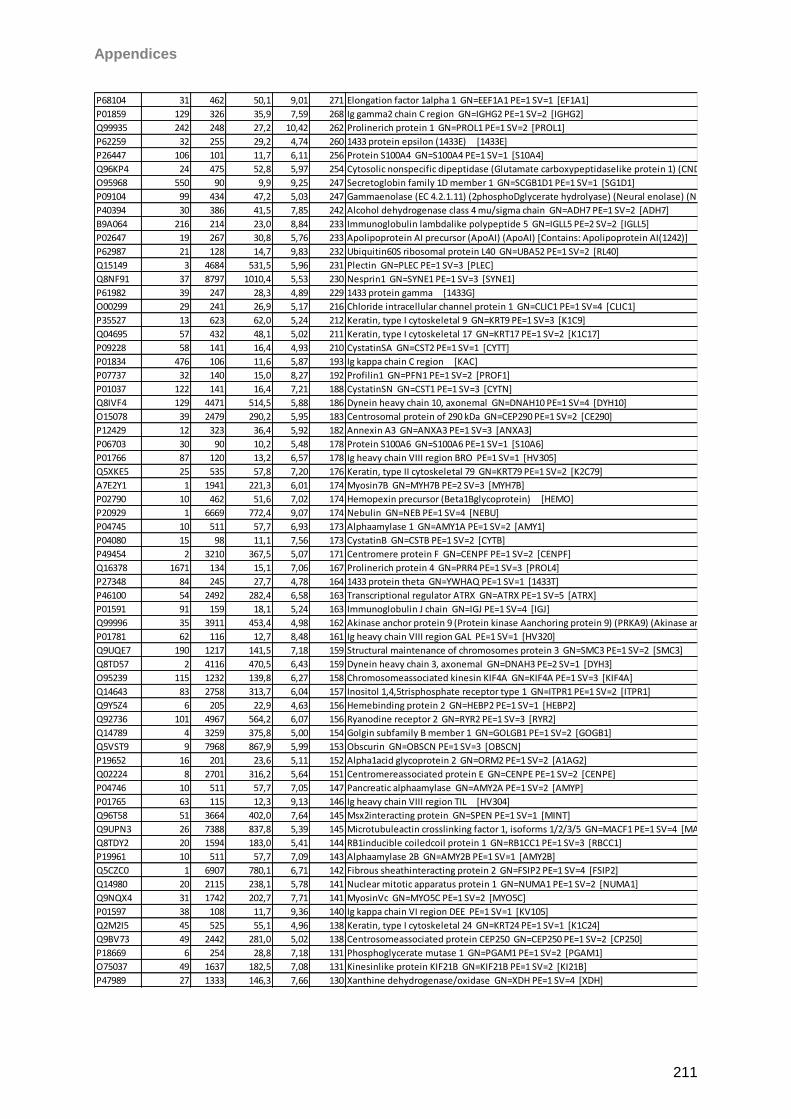
Appendices
211
P68104 31 462 50,1 9,01 271 Elongation factor 1alpha 1 GN=EEF1A1 PE=1 SV=1 [EF1A1]
P01859 129 326 35,9 7,59 268 Ig gamma2 chain C region GN=IGHG2 PE=1 SV=2 [IGHG2]
Q99935 242 248 27,2 10,42 262 Prolinerich protein 1 GN=PROL1 PE=1 SV=2 [PROL1]
P62259 32 255 29,2 4,74 260 1433 protein epsilon (1433E) [1433E]
P26447 106 101 11,7 6,11 256 Protein S100A4 GN=S100A4 PE=1 SV=1 [S10A4]
Q96KP4 24 475 52,8 5,97 254 Cytosolic nonspecific dipeptidase (Glutamate carboxypeptidaselike protein 1) (CNDP dipeptidase 2) [CNDP2]
O95968 550 90 9,9 9,25 247 Secretoglobin family 1D member 1 GN=SCGB1D1 PE=1 SV=1 [SG1D1]
P09104 99 434 47,2 5,03 247 Gammaenolase (EC 4.2.1.11) (2phosphoDglycerate hydrolyase) (Neural enolase) (Neuronspecific enolase) (NSE) (Enolase 2) [ENOG]
P40394 30 386 41,5 7,85 242 Alcohol dehydrogenase class 4 mu/sigma chain GN=ADH7 PE=1 SV=2 [ADH7]
B9A064 216 214 23,0 8,84 233 Immunoglobulin lambdalike polypeptide 5 GN=IGLL5 PE=2 SV=2 [IGLL5]
P02647 19 267 30,8 5,76 233 Apolipoprotein AI precursor (ApoAI) (ApoAI) [Contains: Apolipoprotein AI(1242)] [APOA1]
P62987 21 128 14,7 9,83 232 Ubiquitin60S ribosomal protein L40 GN=UBA52 PE=1 SV=2 [RL40]
Q15149 3 4684 531,5 5,96 231 Plectin GN=PLEC PE=1 SV=3 [PLEC]
Q8NF91 37 8797 1010,4 5,53 230 Nesprin1 GN=SYNE1 PE=1 SV=3 [SYNE1]
P61982 39 247 28,3 4,89 229 1433 protein gamma [1433G]
O00299 29 241 26,9 5,17 216 Chloride intracellular channel protein 1 GN=CLIC1 PE=1 SV=4 [CLIC1]
P35527 13 623 62,0 5,24 212 Keratin, type I cytoskeletal 9 GN=KRT9 PE=1 SV=3 [K1C9]
Q04695 57 432 48,1 5,02 211 Keratin, type I cytoskeletal 17 GN=KRT17 PE=1 SV=2 [K1C17]
P09228 58 141 16,4 4,93 210 CystatinSA GN=CST2 PE=1 SV=1 [CYTT]
P01834 476 106 11,6 5,87 193 Ig kappa chain C region [KAC]
P07737 32 140 15,0 8,27 192 Profilin1 GN=PFN1 PE=1 SV=2 [PROF1]
P01037 122 141 16,4 7,21 188 CystatinSN GN=CST1 PE=1 SV=3 [CYTN]
Q8IVF4 129 4471 514,5 5,88 186 Dynein heavy chain 10, axonemal GN=DNAH10 PE=1 SV=4 [DYH10]
O15078 39 2479 290,2 5,95 183 Centrosomal protein of 290 kDa GN=CEP290 PE=1 SV=2 [CE290]
P12429 12 323 36,4 5,92 182 Annexin A3 GN=ANXA3 PE=1 SV=3 [ANXA3]
P06703 30 90 10,2 5,48 178 Protein S100A6 GN=S100A6 PE=1 SV=1 [S10A6]
P01766 87 120 13,2 6,57 178 Ig heavy chain VIII region BRO PE=1 SV=1 [HV305]
Q5XKE5 25 535 57,8 7,20 176 Keratin, type II cytoskeletal 79 GN=KRT79 PE=1 SV=2 [K2C79]
A7E2Y1 1 1941 221,3 6,01 174 Myosin7B GN=MYH7B PE=2 SV=3 [MYH7B]
P02790 10 462 51,6 7,02 174 Hemopexin precursor (Beta1Bglycoprotein) [HEMO]
P20929 1 6669 772,4 9,07 174 Nebulin GN=NEB PE=1 SV=4 [NEBU]
P04745 10 511 57,7 6,93 173 Alphaamylase 1 GN=AMY1A PE=1 SV=2 [AMY1]
P04080 15 98 11,1 7,56 173 CystatinB GN=CSTB PE=1 SV=2 [CYTB]
P49454 2 3210 367,5 5,07 171 Centromere protein F GN=CENPF PE=1 SV=2 [CENPF]
Q16378 1671 134 15,1 7,06 167 Prolinerich protein 4 GN=PRR4 PE=1 SV=3 [PROL4]
P27348 84 245 27,7 4,78 164 1433 protein theta GN=YWHAQ PE=1 SV=1 [1433T]
P46100 54 2492 282,4 6,58 163 Transcriptional regulator ATRX GN=ATRX PE=1 SV=5 [ATRX]
P01591 91 159 18,1 5,24 163 Immunoglobulin J chain GN=IGJ PE=1 SV=4 [IGJ]
Q99996 35 3911 453,4 4,98 162 Akinase anchor protein 9 (Protein kinase Aanchoring protein 9) (PRKA9) (Akinase anchor protein 450 kDa) (AKAP 450) (Akinase anchor protein 350 kDa) (AKAP 350) (hgAKAP 350) (AKAP 120like protein) (Protein hyperion) (Protein yotiao) (Centrosome and Golgilocalized PKNassociated protein) (CGNAP) [AKAP9]
P01781 62 116 12,7 8,48 161 Ig heavy chain VIII region GAL PE=1 SV=1 [HV320]
Q9UQE7 190 1217 141,5 7,18 159 Structural maintenance of chromosomes protein 3 GN=SMC3 PE=1 SV=2 [SMC3]
Q8TD57 2 4116 470,5 6,43 159 Dynein heavy chain 3, axonemal GN=DNAH3 PE=2 SV=1 [DYH3]
O95239 115 1232 139,8 6,27 158 Chromosomeassociated kinesin KIF4A GN=KIF4A PE=1 SV=3 [KIF4A]
Q14643 83 2758 313,7 6,04 157 Inositol 1,4,5trisphosphate receptor type 1 GN=ITPR1 PE=1 SV=2 [ITPR1]
Q9Y5Z4 6 205 22,9 4,63 156 Hemebinding protein 2 GN=HEBP2 PE=1 SV=1 [HEBP2]
Q92736 101 4967 564,2 6,07 156 Ryanodine receptor 2 GN=RYR2 PE=1 SV=3 [RYR2]
Q14789 4 3259 375,8 5,00 154 Golgin subfamily B member 1 GN=GOLGB1 PE=1 SV=2 [GOGB1]
Q5VST9 9 7968 867,9 5,99 153 Obscurin GN=OBSCN PE=1 SV=3 [OBSCN]
P19652 16 201 23,6 5,11 152 Alpha1acid glycoprotein 2 GN=ORM2 PE=1 SV=2 [A1AG2]
Q02224 8 2701 316,2 5,64 151 Centromereassociated protein E GN=CENPE PE=1 SV=2 [CENPE]
P04746 10 511 57,7 7,05 147 Pancreatic alphaamylase GN=AMY2A PE=1 SV=2 [AMYP]
P01765 63 115 12,3 9,13 146 Ig heavy chain VIII region TIL [HV304]
Q96T58 51 3664 402,0 7,64 145 Msx2interacting protein GN=SPEN PE=1 SV=1 [MINT]
Q9UPN3 26 7388 837,8 5,39 145 Microtubuleactin crosslinking factor 1, isoforms 1/2/3/5 GN=MACF1 PE=1 SV=4 [MACF1]
Q8TDY2 20 1594 183,0 5,41 144 RB1inducible coiledcoil protein 1 GN=RB1CC1 PE=1 SV=3 [RBCC1]
P19961 10 511 57,7 7,09 143 Alphaamylase 2B GN=AMY2B PE=1 SV=1 [AMY2B]
Q5CZC0 1 6907 780,1 6,71 142 Fibrous sheathinteracting protein 2 GN=FSIP2 PE=1 SV=4 [FSIP2]
Q14980 20 2115 238,1 5,78 141 Nuclear mitotic apparatus protein 1 GN=NUMA1 PE=1 SV=2 [NUMA1]
Q9NQX4 31 1742 202,7 7,71 141 MyosinVc GN=MYO5C PE=1 SV=2 [MYO5C]
P01597 38 108 11,7 9,36 140 Ig kappa chain VI region DEE PE=1 SV=1 [KV105]
Q2M2I5 45 525 55,1 4,96 138 Keratin, type I cytoskeletal 24 GN=KRT24 PE=1 SV=1 [K1C24]
Q9BV73 49 2442 281,0 5,02 138 Centrosomeassociated protein CEP250 GN=CEP250 PE=1 SV=2 [CP250]
P18669 6 254 28,8 7,18 131 Phosphoglycerate mutase 1 GN=PGAM1 PE=1 SV=2 [PGAM1]
O75037 49 1637 182,5 7,08 131 Kinesinlike protein KIF21B GN=KIF21B PE=1 SV=2 [KI21B]
P47989 27 1333 146,3 7,66 130 Xanthine dehydrogenase/oxidase GN=XDH PE=1 SV=4 [XDH]
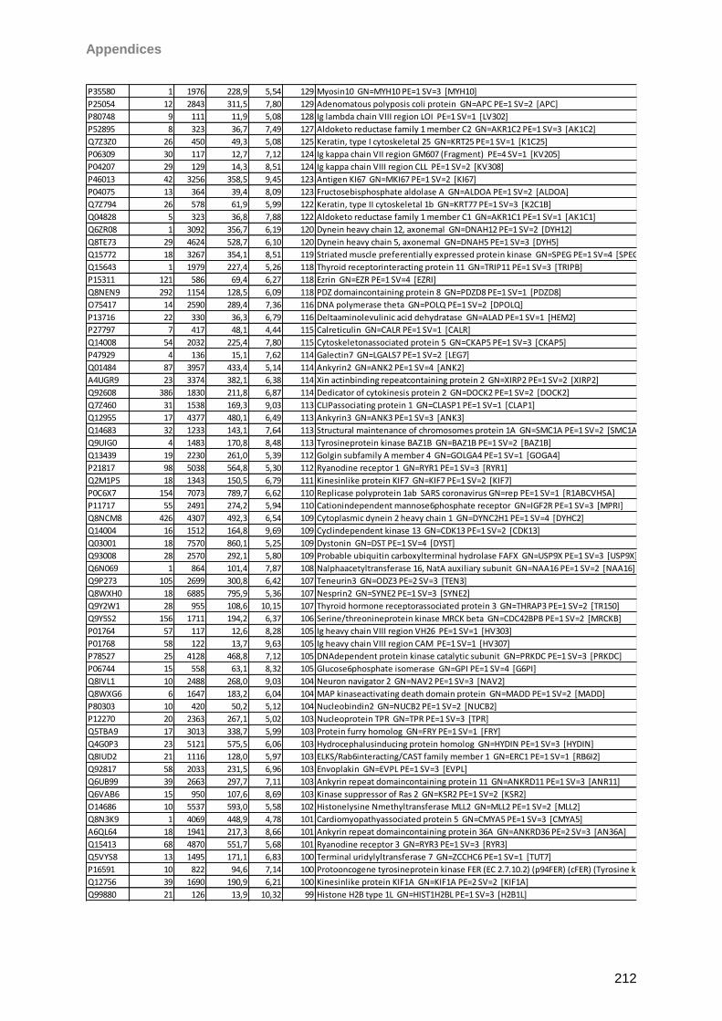
Appendices
212
P35580 1 1976 228,9 5,54 129 Myosin10 GN=MYH10 PE=1 SV=3 [MYH10]
P25054 12 2843 311,5 7,80 129 Adenomatous polyposis coli protein GN=APC PE=1 SV=2 [APC]
P80748 9 111 11,9 5,08 128 Ig lambda chain VIII region LOI PE=1 SV=1 [LV302]
P52895 8 323 36,7 7,49 127 Aldoketo reductase family 1 member C2 GN=AKR1C2 PE=1 SV=3 [AK1C2]
Q7Z3Z0 26 450 49,3 5,08 125 Keratin, type I cytoskeletal 25 GN=KRT25 PE=1 SV=1 [K1C25]
P06309 30 117 12,7 7,12 124 Ig kappa chain VII region GM607 (Fragment) PE=4 SV=1 [KV205]
P04207 29 129 14,3 8,51 124 Ig kappa chain VIII region CLL PE=1 SV=2 [KV308]
P46013 42 3256 358,5 9,45 123 Antigen KI67 GN=MKI67 PE=1 SV=2 [KI67]
P04075 13 364 39,4 8,09 123 Fructosebisphosphate aldolase A GN=ALDOA PE=1 SV=2 [ALDOA]
Q7Z794 26 578 61,9 5,99 122 Keratin, type II cytoskeletal 1b GN=KRT77 PE=1 SV=3 [K2C1B]
Q04828 5 323 36,8 7,88 122 Aldoketo reductase family 1 member C1 GN=AKR1C1 PE=1 SV=1 [AK1C1]
Q6ZR08 1 3092 356,7 6,19 120 Dynein heavy chain 12, axonemal GN=DNAH12 PE=1 SV=2 [DYH12]
Q8TE73 29 4624 528,7 6,10 120 Dynein heavy chain 5, axonemal GN=DNAH5 PE=1 SV=3 [DYH5]
Q15772 18 3267 354,1 8,51 119 Striated muscle preferentially expressed protein kinase GN=SPEG PE=1 SV=4 [SPEG]
Q15643 1 1979 227,4 5,26 118 Thyroid receptorinteracting protein 11 GN=TRIP11 PE=1 SV=3 [TRIPB]
P15311 121 586 69,4 6,27 118 Ezrin GN=EZR PE=1 SV=4 [EZRI]
Q8NEN9 292 1154 128,5 6,09 118 PDZ domaincontaining protein 8 GN=PDZD8 PE=1 SV=1 [PDZD8]
O75417 14 2590 289,4 7,36 116 DNA polymerase theta GN=POLQ PE=1 SV=2 [DPOLQ]
P13716 22 330 36,3 6,79 116 Deltaaminolevulinic acid dehydratase GN=ALAD PE=1 SV=1 [HEM2]
P27797 7 417 48,1 4,44 115 Calreticulin GN=CALR PE=1 SV=1 [CALR]
Q14008 54 2032 225,4 7,80 115 Cytoskeletonassociated protein 5 GN=CKAP5 PE=1 SV=3 [CKAP5]
P47929 4 136 15,1 7,62 114 Galectin7 GN=LGALS7 PE=1 SV=2 [LEG7]
Q01484 87 3957 433,4 5,14 114 Ankyrin2 GN=ANK2 PE=1 SV=4 [ANK2]
A4UGR9 23 3374 382,1 6,38 114 Xin actinbinding repeatcontaining protein 2 GN=XIRP2 PE=1 SV=2 [XIRP2]
Q92608 386 1830 211,8 6,87 114 Dedicator of cytokinesis protein 2 GN=DOCK2 PE=1 SV=2 [DOCK2]
Q7Z460 31 1538 169,3 9,03 113 CLIPassociating protein 1 GN=CLASP1 PE=1 SV=1 [CLAP1]
Q12955 17 4377 480,1 6,49 113 Ankyrin3 GN=ANK3 PE=1 SV=3 [ANK3]
Q14683 32 1233 143,1 7,64 113 Structural maintenance of chromosomes protein 1A GN=SMC1A PE=1 SV=2 [SMC1A]
Q9UIG0 4 1483 170,8 8,48 113 Tyrosineprotein kinase BAZ1B GN=BAZ1B PE=1 SV=2 [BAZ1B]
Q13439 19 2230 261,0 5,39 112 Golgin subfamily A member 4 GN=GOLGA4 PE=1 SV=1 [GOGA4]
P21817 98 5038 564,8 5,30 112 Ryanodine receptor 1 GN=RYR1 PE=1 SV=3 [RYR1]
Q2M1P5 18 1343 150,5 6,79 111 Kinesinlike protein KIF7 GN=KIF7 PE=1 SV=2 [KIF7]
P0C6X7 154 7073 789,7 6,62 110 Replicase polyprotein 1ab SARS coronavirus GN=rep PE=1 SV=1 [R1ABCVHSA]
P11717 55 2491 274,2 5,94 110 Cationindependent mannose6phosphate receptor GN=IGF2R PE=1 SV=3 [MPRI]
Q8NCM8 426 4307 492,3 6,54 109 Cytoplasmic dynein 2 heavy chain 1 GN=DYNC2H1 PE=1 SV=4 [DYHC2]
Q14004 16 1512 164,8 9,69 109 Cyclindependent kinase 13 GN=CDK13 PE=1 SV=2 [CDK13]
Q03001 18 7570 860,1 5,25 109 Dystonin GN=DST PE=1 SV=4 [DYST]
Q93008 28 2570 292,1 5,80 109 Probable ubiquitin carboxylterminal hydrolase FAFX GN=USP9X PE=1 SV=3 [USP9X]
Q6N069 1 864 101,4 7,87 108 Nalphaacetyltransferase 16, NatA auxiliary subunit GN=NAA16 PE=1 SV=2 [NAA16]
Q9P273 105 2699 300,8 6,42 107 Teneurin3 GN=ODZ3 PE=2 SV=3 [TEN3]
Q8WXH0 18 6885 795,9 5,36 107 Nesprin2 GN=SYNE2 PE=1 SV=3 [SYNE2]
Q9Y2W1 28 955 108,6 10,15 107 Thyroid hormone receptorassociated protein 3 GN=THRAP3 PE=1 SV=2 [TR150]
Q9Y5S2 156 1711 194,2 6,37 106 Serine/threonineprotein kinase MRCK beta GN=CDC42BPB PE=1 SV=2 [MRCKB]
P01764 57 117 12,6 8,28 105 Ig heavy chain VIII region VH26 PE=1 SV=1 [HV303]
P01768 58 122 13,7 9,63 105 Ig heavy chain VIII region CAM PE=1 SV=1 [HV307]
P78527 25 4128 468,8 7,12 105 DNAdependent protein kinase catalytic subunit GN=PRKDC PE=1 SV=3 [PRKDC]
P06744 15 558 63,1 8,32 105 Glucose6phosphate isomerase GN=GPI PE=1 SV=4 [G6PI]
Q8IVL1 10 2488 268,0 9,03 104 Neuron navigator 2 GN=NAV2 PE=1 SV=3 [NAV2]
Q8WXG6 6 1647 183,2 6,04 104 MAP kinaseactivating death domain protein GN=MADD PE=1 SV=2 [MADD]
P80303 10 420 50,2 5,12 104 Nucleobindin2 GN=NUCB2 PE=1 SV=2 [NUCB2]
P12270 20 2363 267,1 5,02 103 Nucleoprotein TPR GN=TPR PE=1 SV=3 [TPR]
Q5TBA9 17 3013 338,7 5,99 103 Protein furry homolog GN=FRY PE=1 SV=1 [FRY]
Q4G0P3 23 5121 575,5 6,06 103 Hydrocephalusinducing protein homolog GN=HYDIN PE=1 SV=3 [HYDIN]
Q8IUD2 21 1116 128,0 5,97 103 ELKS/Rab6interacting/CAST family member 1 GN=ERC1 PE=1 SV=1 [RB6I2]
Q92817 58 2033 231,5 6,96 103 Envoplakin GN=EVPL PE=1 SV=3 [EVPL]
Q6UB99 39 2663 297,7 7,11 103 Ankyrin repeat domaincontaining protein 11 GN=ANKRD11 PE=1 SV=3 [ANR11]
Q6VAB6 15 950 107,6 8,69 103 Kinase suppressor of Ras 2 GN=KSR2 PE=1 SV=2 [KSR2]
O14686 10 5537 593,0 5,58 102 Histonelysine Nmethyltransferase MLL2 GN=MLL2 PE=1 SV=2 [MLL2]
Q8N3K9 1 4069 448,9 4,78 101 Cardiomyopathyassociated protein 5 GN=CMYA5 PE=1 SV=3 [CMYA5]
A6QL64 18 1941 217,3 8,66 101 Ankyrin repeat domaincontaining protein 36A GN=ANKRD36 PE=2 SV=3 [AN36A]
Q15413 68 4870 551,7 5,68 101 Ryanodine receptor 3 GN=RYR3 PE=1 SV=3 [RYR3]
Q5VYS8 13 1495 171,1 6,83 100 Terminal uridylyltransferase 7 GN=ZCCHC6 PE=1 SV=1 [TUT7]
P16591 10 822 94,6 7,14 100 Protooncogene tyrosineprotein kinase FER (EC 2.7.10.2) (p94FER) (cFER) (Tyrosine kinase 3) [FER]
Q12756 39 1690 190,9 6,21 100 Kinesinlike protein KIF1A GN=KIF1A PE=2 SV=2 [KIF1A]
Q99880 21 126 13,9 10,32 99 Histone H2B type 1L GN=HIST1H2BL PE=1 SV=3 [H2B1L]
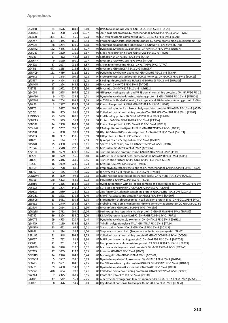
Appendices
213
Q02880 34 1626 183,2 8,00 97 DNA topoisomerase 2beta GN=TOP2B PE=1 SV=3 [TOP2B]
Q9HD33 13 250 29,4 10,37 97 39S ribosomal protein L47, mitochondrial GN=MRPL47 PE=1 SV=2 [RM47]
Q13098 384 491 55,5 6,74 97 COP9 signalosome complex subunit 1 GN=GPS1 PE=1 SV=4 [CSN1]
O75747 394 1445 165,6 6,93 97 Phosphatidylinositol4phosphate 3kinase C2 domaincontaining subunit gamma GN=PIK3C2G PE=1 SV=3 [P3C2G]
Q2VIQ3 68 1234 139,9 6,18 96 Chromosomeassociated kinesin KIF4B GN=KIF4B PE=1 SV=2 [KIF4B]
Q9UFH2 162 4485 511,5 5,77 96 Dynein heavy chain 17, axonemal GN=DNAH17 PE=2 SV=2 [DYH17]
Q96Q89 34 1820 210,5 5,67 96 Kinesinlike protein KIF20B GN=KIF20B PE=1 SV=3 [KI20B]
P07339 37 412 44,5 6,54 95 Cathepsin D GN=CTSD PE=1 SV=1 [CATD]
Q9UKN7 9 3530 395,0 9,17 95 MyosinXV GN=MYO15A PE=1 SV=2 [MYO15]
O14578 57 2027 231,3 6,57 95 Citron Rhointeracting kinase GN=CIT PE=1 SV=2 [CTRO]
Q9Y4I1 447 1855 215,3 8,48 95 MyosinVa GN=MYO5A PE=1 SV=2 [MYO5A]
Q9NYC9 151 4486 511,6 5,91 95 Dynein heavy chain 9, axonemal GN=DNAH9 PE=1 SV=3 [DYH9]
Q5VYK3 3 1845 204,2 7,12 94 Proteasomeassociated protein ECM29 homolog GN=ECM29 PE=1 SV=2 [ECM29]
Q7Z6Z7 14 4374 481,6 5,22 94 E3 ubiquitinprotein ligase HUWE1 GN=HUWE1 PE=1 SV=3 [HUWE1]
Q9UM54 17 1294 149,6 8,53 93 MyosinVI GN=MYO6 PE=1 SV=4 [MYO6]
P35749 13 1972 227,2 5,50 93 Myosin11 GN=MYH11 PE=1 SV=3 [MYH11]
Q14C86 38 1478 164,9 5,22 93 GTPaseactivating protein and VPS9 domaincontaining protein 1 GN=GAPVD1 PE=1 SV=2 [GAPD1]
Q96M86 9 4753 533,3 6,71 93 Dynein heavy chain domaincontaining protein 1 GN=DNHD1 PE=1 SV=2 [DNHD1]
Q8WZ64 26 1704 193,3 7,39 93 ArfGAP with RhoGAP domain, ANK repeat and PH domaincontaining protein 2 GN=ARAP2 PE=1 SV=3 [ARAP2]
Q96L93 3 1317 151,9 6,16 93 Kinesinlike protein KIF16B GN=KIF16B PE=1 SV=2 [KI16B]
Q8IZT6 1 3477 409,5 10,45 92 Abnormal spindlelike microcephalyassociated protein GN=ASPM PE=1 SV=2 [ASPM]
Q5SZL2 26 805 91,8 6,33 92 Coiledcoil domaincontaining protein C6orf204 GN=C6orf204 PE=2 SV=1 [CF204]
A6NNM3 73 1639 180,8 6,77 92 RIMSbinding protein 3B GN=RIMBP3B PE=2 SV=3 [RIM3B]
Q8NCA5 69 519 55,4 9,03 92 Protein FAM98A GN=FAM98A PE=1 SV=1 [FA98A]
Q9NS87 24 1388 160,1 6,00 92 Kinesinlike protein KIF15 GN=KIF15 PE=1 SV=1 [KIF15]
Q63HN8 41 5207 591,0 6,48 92 E3 ubiquitinprotein ligase RNF213 GN=RNF213 PE=1 SV=3 [RN213]
O43290 4 800 90,2 6,13 91 U4/U6.U5 trisnRNPassociated protein 1 GN=SART1 PE=1 SV=1 [SNUT1]
O15083 18 957 110,5 6,99 91 ERC protein 2 GN=ERC2 PE=1 SV=3 [ERC2]
P01625 4 114 12,6 7,93 90 Ig kappa chain VIV region Len PE=1 SV=2 [KV402]
O15020 25 2390 271,2 6,11 90 Spectrin beta chain, brain 2 GN=SPTBN2 PE=1 SV=3 [SPTN2]
B2RTY4 1 2548 292,5 8,88 90 MyosinIXa GN=MYO9A PE=1 SV=2 [MYO9A]
A2VDJ0 18 1609 179,2 6,86 89 Transmembrane protein 131like GN=KIAA0922 PE=1 SV=2 [T131L]
P06576 10 529 56,5 5,40 89 ATP synthase subunit beta, mitochondrial GN=ATP5B PE=1 SV=3 [ATPB]
P31629 15 2446 268,9 6,96 88 Transcription factor HIVEP2 GN=HIVEP2 PE=1 SV=2 [ZEP2]
P13533 16 1939 223,6 5,73 88 Myosin6 GN=MYH6 PE=1 SV=5 [MYH6]
P05165 132 728 80,0 7,52 88 PropionylCoA carboxylase alpha chain, mitochondrial GN=PCCA PE=1 SV=4 [PCCA]
P01767 52 115 12,4 9,25 88 Ig heavy chain VIII region BUT PE=1 SV=1 [HV306]
Q9NQW8 15 809 92,1 7,93 88 Cyclic nucleotidegated cation channel beta3 GN=CNGB3 PE=1 SV=2 [CNGB3]
P98161 178 4303 462,2 6,73 87 Polycystin1 GN=PKD1 PE=1 SV=3 [PKD1]
Q9BZF9 9 1416 162,4 7,03 87 Uveal autoantigen with coiledcoil domains and ankyrin repeats GN=UACA PE=1 SV=2 [UACA]
O75122 28 1294 141,0 8,47 87 CLIPassociating protein 2 GN=CLASP2 PE=1 SV=2 [CLAP2]
O60293 116 1989 226,2 8,13 87 Zinc finger C3H1 domaincontaining protein GN=ZFC3H1 PE=1 SV=3 [ZC3H1]
Q96QB1 53 1528 170,5 6,40 87 Rho GTPaseactivating protein 7 GN=DLC1 PE=1 SV=4 [RHG07]
Q8NFC6 13 3051 330,3 5,08 87 Biorientation of chromosomes in cell division protein 1like GN=BOD1L PE=1 SV=2 [BOD1L]
Q15652 17 2540 284,3 7,87 86 Probable JmjC domaincontaining histone demethylation protein 2C GN=JMJD1C PE=1 SV=2 [JHD2C]
Q92614 28 2054 233,0 6,30 86 MyosinXVIIIa GN=MYO18A PE=1 SV=3 [MY18A]
Q9UQ35 19 2752 299,4 12,06 86 Serine/arginine repetitive matrix protein 2 GN=SRRM2 PE=1 SV=2 [SRRM2]
P49792 59 3224 358,0 6,20 85 E3 SUMOprotein ligase RanBP2 GN=RANBP2 PE=1 SV=2 [RBP2]
Q96DT5 69 4523 520,7 6,44 84 Dynein heavy chain 11, axonemal GN=DNAH11 PE=1 SV=3 [DYH11]
Q14679 27 1199 133,3 8,85 84 Tubulin polyglutamylase TTLL4 GN=TTLL4 PE=1 SV=2 [TTLL4]
Q9UN79 23 622 69,2 6,71 84 Transcription factor SOX13 GN=SOX13 PE=1 SV=3 [SOX13]
P07951 8 284 32,8 4,70 84 Tropomyosin beta chain (Tropomyosin 2) (Betatropomyosin) [TPM2]
A2RUB6 91 948 109,3 8,25 84 Coiledcoil domaincontaining protein 66 GN=CCDC66 PE=1 SV=4 [CCD66]
Q96T17 62 732 81,9 8,84 84 MAP7 domaincontaining protein 2 GN=MAP7D2 PE=1 SV=2 [MA7D2]
P30040 21 261 29,0 7,31 83 Endoplasmic reticulum resident protein 29 GN=ERP29 PE=1 SV=4 [ERP29]
Q9NR99 46 2828 312,0 8,32 83 Matrixremodelingassociated protein 5 GN=MXRA5 PE=2 SV=3 [MXRA5]
Q9Y283 17 1065 117,8 9,35 83 Inversin GN=INVS PE=1 SV=2 [INVS]
Q5VU43 24 2346 264,9 5,44 83 Myomegalin GN=PDE4DIP PE=1 SV=1 [MYOME]
Q0VDD8 5 3507 399,6 6,93 83 Dynein heavy chain 14, axonemal GN=DNAH14 PE=2 SV=3 [DYH14]
Q86VI3 13 1631 184,6 7,65 83 Ras GTPaseactivatinglike protein IQGAP3 GN=IQGAP3 PE=1 SV=2 [IQGA3]
Q96JB1 69 4490 514,3 6,32 83 Dynein heavy chain 8, axonemal GN=DNAH8 PE=1 SV=2 [DYH8]
Q05D60 428 604 70,9 6,21 83 Coiledcoil domaincontaining protein 67 GN=CCDC67 PE=2 SV=2 [CCD67]
Q7Z7A1 7 2325 268,7 5,55 82 Centriolin GN=CEP110 PE=1 SV=2 [CE110]
P47895 17 512 56,1 7,25 82 Aldehyde dehydrogenase family 1 member A3 GN=ALDH1A3 PE=1 SV=2 [AL1A3]
Q9H1J1 8 476 54,7 9,03 82 Regulator of nonsense transcripts 3A GN=UPF3A PE=1 SV=1 [REN3A]
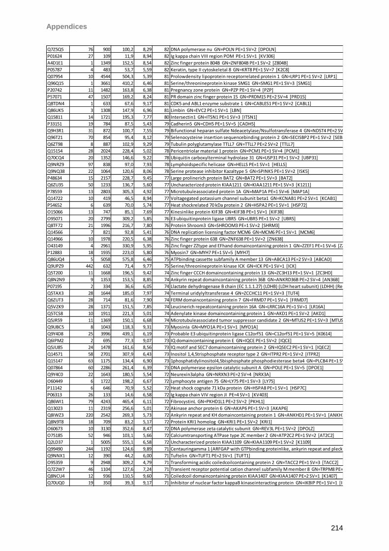
Appendices
214
Q7Z5Q5 76 900 100,2 8,29 82 DNA polymerase nu GN=POLN PE=1 SV=2 [DPOLN]
P01624 27 109 11,9 8,94 82 Ig kappa chain VIII region POM PE=1 SV=1 [KV306]
A4D1E1 1 1349 152,5 8,54 82 Zinc finger protein 804B GN=ZNF804B PE=1 SV=2 [Z804B]
P05787 4 483 53,7 5,59 82 Keratin, type II cytoskeletal 8 GN=KRT8 PE=1 SV=7 [K2C8]
Q07954 10 4544 504,3 5,39 81 Prolowdensity lipoprotein receptorrelated protein 1 GN=LRP1 PE=1 SV=2 [LRP1]
Q96Q15 1 3661 410,2 6,46 81 Serine/threonineprotein kinase SMG1 GN=SMG1 PE=1 SV=3 [SMG1]
P20742 11 1482 163,8 6,38 81 Pregnancy zone protein GN=PZP PE=1 SV=4 [PZP]
P57071 47 1507 169,2 8,24 81 PR domain zinc finger protein 15 GN=PRDM15 PE=2 SV=4 [PRD15]
Q8TDN4 1 633 67,6 9,17 81 CDK5 and ABL1 enzyme substrate 1 GN=CABLES1 PE=1 SV=2 [CABL1]
Q86UK5 3 1308 147,9 6,96 81 Limbin GN=EVC2 PE=1 SV=1 [LBN]
Q15811 14 1721 195,3 7,77 80 Intersectin1 GN=ITSN1 PE=1 SV=3 [ITSN1]
P33151 19 784 87,5 5,43 79 Cadherin5 GN=CDH5 PE=1 SV=5 [CADH5]
Q9H3R1 31 872 100,7 7,55 79 Bifunctional heparan sulfate Ndeacetylase/Nsulfotransferase 4 GN=NDST4 PE=2 SV=1 [NDST4]
Q96T21 70 854 95,4 8,12 79 Selenocysteine insertion sequencebinding protein 2 GN=SECISBP2 PE=1 SV=2 [SEBP2]
Q6ZT98 8 887 102,9 9,29 79 Tubulin polyglutamylase TTLL7 GN=TTLL7 PE=2 SV=2 [TTLL7]
Q15154 28 2024 228,4 5,02 78 Pericentriolar material 1 protein GN=PCM1 PE=1 SV=4 [PCM1]
Q70CQ4 20 1352 146,6 9,22 78 Ubiquitin carboxylterminal hydrolase 31 GN=USP31 PE=1 SV=2 [UBP31]
Q9NRZ9 97 838 97,0 7,93 78 Lymphoidspecific helicase GN=HELLS PE=1 SV=1 [HELLS]
Q9NQ38 22 1064 120,6 8,06 78 Serine protease inhibitor Kazaltype 5 GN=SPINK5 PE=1 SV=2 [ISK5]
P48634 15 2157 228,7 9,45 77 Large prolinerich protein BAT2 GN=BAT2 PE=1 SV=3 [BAT2]
Q6ZU35 50 1233 136,7 5,60 77 Uncharacterized protein KIAA1211 GN=KIAA1211 PE=1 SV=3 [K1211]
P78559 13 2803 305,3 4,92 77 Microtubuleassociated protein 1A GN=MAP1A PE=1 SV=6 [MAP1A]
Q14722 10 419 46,5 8,94 77 Voltagegated potassium channel subunit beta1 GN=KCNAB1 PE=2 SV=1 [KCAB1]
P54652 6 639 70,0 5,74 77 Heat shockrelated 70 kDa protein 2 GN=HSPA2 PE=1 SV=1 [HSP72]
O15066 13 747 85,1 7,69 77 Kinesinlike protein KIF3B GN=KIF3B PE=1 SV=1 [KIF3B]
O95071 20 2799 309,2 5,85 76 E3 ubiquitinprotein ligase UBR5 GN=UBR5 PE=1 SV=2 [UBR5]
Q8TF72 21 1996 216,7 7,80 76 Protein Shroom3 GN=SHROOM3 PE=1 SV=2 [SHRM3]
Q14566 7 821 92,8 5,41 76 DNA replication licensing factor MCM6 GN=MCM6 PE=1 SV=1 [MCM6]
Q14966 10 1978 220,5 6,38 76 Zinc finger protein 638 GN=ZNF638 PE=1 SV=2 [ZN638]
O43149 4 2961 330,9 5,95 76 Zinc finger ZZtype and EFhand domaincontaining protein 1 GN=ZZEF1 PE=1 SV=6 [ZZEF1]
P12883 18 1935 223,0 5,80 76 Myosin7 GN=MYH7 PE=1 SV=5 [MYH7]
Q86UQ4 5 5058 575,8 6,46 75 ATPbinding cassette subfamily A member 13 GN=ABCA13 PE=2 SV=3 [ABCAD]
Q9UPZ9 442 632 71,4 9,77 75 Serine/threonineprotein kinase ICK GN=ICK PE=1 SV=1 [ICK]
Q5T200 11 1668 196,5 9,42 74 Zinc finger CCCH domaincontaining protein 13 GN=ZC3H13 PE=1 SV=1 [ZC3HD]
Q8N2N9 9 1353 153,5 8,85 74 Ankyrin repeat domaincontaining protein 36B GN=ANKRD36B PE=2 SV=4 [AN36B]
P07195 2 334 36,6 6,05 74 Llactate dehydrogenase B chain (EC 1.1.1.27) (LDHB) (LDH heart subunit) (LDHH) (Renal carcinoma antigen NYREN46) [LDHB]
Q5TAX3 28 1644 185,0 7,97 74 Terminal uridylyltransferase 4 GN=ZCCHC11 PE=1 SV=3 [TUT4]
Q6ZUT3 28 714 81,6 7,90 74 FERM domaincontaining protein 7 GN=FRMD7 PE=1 SV=1 [FRMD7]
Q5VZK9 28 1371 151,5 7,85 74 Leucinerich repeatcontaining protein 16A GN=LRRC16A PE=1 SV=1 [LR16A]
Q5TCS8 10 1911 221,3 5,01 74 Adenylate kinase domaincontaining protein 1 GN=AKD1 PE=1 SV=2 [AKD1]
Q5JR59 11 1369 150,1 6,68 74 Microtubuleassociated tumor suppressor candidate 2 GN=MTUS2 PE=1 SV=3 [MTUS2]
Q9UBC5 8 1043 118,3 9,31 73 MyosinIa GN=MYO1A PE=1 SV=1 [MYO1A]
Q9Y4D8 25 3996 439,1 6,19 73 Probable E3 ubiquitinprotein ligase C12orf51 GN=C12orf51 PE=1 SV=5 [K0614]
Q6IPM2 2 695 77,3 9,07 73 IQ domaincontaining protein E GN=IQCE PE=1 SV=2 [IQCE]
Q5JU85 24 1478 161,6 8,56 73 IQ motif and SEC7 domaincontaining protein 2 GN=IQSEC2 PE=1 SV=1 [IQEC2]
Q14571 58 2701 307,9 6,43 73 Inositol 1,4,5trisphosphate receptor type 2 GN=ITPR2 PE=1 SV=2 [ITPR2]
Q15147 63 1175 134,4 6,90 73 1phosphatidylinositol4,5bisphosphate phosphodiesterase beta4 GN=PLCB4 PE=1 SV=3 [PLCB4]
Q07864 60 2286 261,4 6,39 73 DNA polymerase epsilon catalytic subunit A GN=POLE PE=1 SV=5 [DPOE1]
Q9Y4C0 22 1643 180,5 5,54 72 Neurexin3alpha GN=NRXN3 PE=2 SV=4 [NRX3A]
O60449 6 1722 198,2 6,67 72 Lymphocyte antigen 75 GN=LY75 PE=1 SV=3 [LY75]
P11142 6 646 70,9 5,52 72 Heat shock cognate 71 kDa protein GN=HSPA8 PE=1 SV=1 [HSP7C]
P06313 26 133 14,6 6,58 72 Ig kappa chain VIV region JI PE=4 SV=1 [KV403]
Q86WI1 79 4243 465,4 6,11 72 FibrocystinL GN=PKHD1L1 PE=2 SV=2 [PKHL1]
Q13023 11 2319 256,6 5,01 72 Akinase anchor protein 6 GN=AKAP6 PE=1 SV=3 [AKAP6]
Q8IWZ3 220 2542 269,3 5,73 72 Ankyrin repeat and KH domaincontaining protein 1 GN=ANKHD1 PE=1 SV=1 [ANKH1]
Q8N9T8 18 709 83,2 5,17 72 Protein KRI1 homolog GN=KRI1 PE=1 SV=2 [KRI1]
O60673 10 3130 352,6 8,47 72 DNA polymerase zeta catalytic subunit GN=REV3L PE=1 SV=2 [DPOLZ]
O75185 52 946 103,1 5,66 72 Calciumtransporting ATPase type 2C member 2 GN=ATP2C2 PE=1 SV=2 [AT2C2]
Q2LD37 1 5005 555,1 6,58 72 Uncharacterized protein KIAA1109 GN=KIAA1109 PE=1 SV=2 [K1109]
Q99490 244 1192 124,6 9,89 71 Centauringamma 1 (ARFGAP with GTPbinding proteinlike, ankyrin repeat and pleckstrin homology domains 2) (AGAP2) (Phosphatidylinositol3kinase enhancer) (PIKE) (GTPbinding and GTPaseactivating protein 2) (GGAP2) [CENG1]
Q9NNX1 12 390 44,2 6,00 71 Tuftelin GN=TUFT1 PE=2 SV=1 [TUFT1]
O95359 9 2948 309,2 4,79 71 Transforming acidic coiledcoilcontaining protein 2 GN=TACC2 PE=1 SV=3 [TACC2]
Q7Z2W7 46 1104 127,6 7,24 71 Transient receptor potential cation channel subfamily M member 8 GN=TRPM8 PE=2 SV=2 [TRPM8]
Q8NCU4 12 936 110,5 9,60 71 Coiledcoil domaincontaining protein KIAA1407 GN=KIAA1407 PE=2 SV=1 [K1407]
Q70UQ0 19 350 39,3 9,17 71 Inhibitor of nuclear factor kappaB kinaseinteracting protein GN=IKBIP PE=1 SV=1 [IKIP]
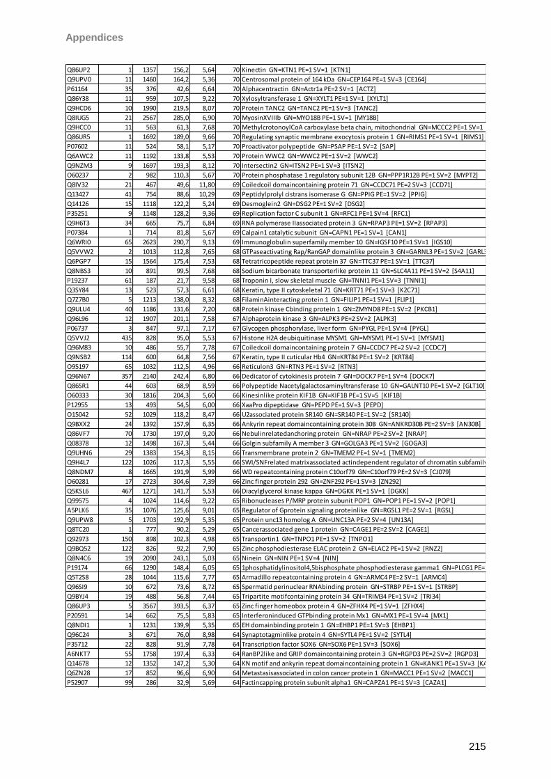
Appendices
215
Q86UP2 1 1357 156,2 5,64 70 Kinectin GN=KTN1 PE=1 SV=1 [KTN1]
Q9UPV0 11 1460 164,2 5,36 70 Centrosomal protein of 164 kDa GN=CEP164 PE=1 SV=3 [CE164]
P61164 35 376 42,6 6,64 70 Alphacentractin GN=Actr1a PE=2 SV=1 [ACTZ]
Q86Y38 11 959 107,5 9,22 70 Xylosyltransferase 1 GN=XYLT1 PE=1 SV=1 [XYLT1]
Q9HCD6 10 1990 219,5 8,07 70 Protein TANC2 GN=TANC2 PE=1 SV=3 [TANC2]
Q8IUG5 21 2567 285,0 6,90 70 MyosinXVIIIb GN=MYO18B PE=1 SV=1 [MY18B]
Q9HCC0 11 563 61,3 7,68 70 MethylcrotonoylCoA carboxylase beta chain, mitochondrial GN=MCCC2 PE=1 SV=1 [MCCB]
Q86UR5 1 1692 189,0 9,66 70 Regulating synaptic membrane exocytosis protein 1 GN=RIMS1 PE=1 SV=1 [RIMS1]
P07602 11 524 58,1 5,17 70 Proactivator polypeptide GN=PSAP PE=1 SV=2 [SAP]
Q6AWC2 11 1192 133,8 5,53 70 Protein WWC2 GN=WWC2 PE=1 SV=2 [WWC2]
Q9NZM3 9 1697 193,3 8,12 70 Intersectin2 GN=ITSN2 PE=1 SV=3 [ITSN2]
O60237 2 982 110,3 5,67 70 Protein phosphatase 1 regulatory subunit 12B GN=PPP1R12B PE=1 SV=2 [MYPT2]
Q8IV32 21 467 49,6 11,80 69 Coiledcoil domaincontaining protein 71 GN=CCDC71 PE=2 SV=3 [CCD71]
Q13427 41 754 88,6 10,29 69 Peptidylprolyl cistrans isomerase G GN=PPIG PE=1 SV=2 [PPIG]
Q14126 15 1118 122,2 5,24 69 Desmoglein2 GN=DSG2 PE=1 SV=2 [DSG2]
P35251 9 1148 128,2 9,36 69 Replication factor C subunit 1 GN=RFC1 PE=1 SV=4 [RFC1]
Q9H6T3 34 665 75,7 6,84 69 RNA polymerase IIassociated protein 3 GN=RPAP3 PE=1 SV=2 [RPAP3]
P07384 1 714 81,8 5,67 69 Calpain1 catalytic subunit GN=CAPN1 PE=1 SV=1 [CAN1]
Q6WRI0 65 2623 290,7 9,13 69 Immunoglobulin superfamily member 10 GN=IGSF10 PE=1 SV=1 [IGS10]
Q5VVW2 2 1013 112,8 7,65 68 GTPaseactivating Rap/RanGAP domainlike protein 3 GN=GARNL3 PE=1 SV=2 [GARL3]
Q6PGP7 15 1564 175,4 7,53 68 Tetratricopeptide repeat protein 37 GN=TTC37 PE=1 SV=1 [TTC37]
Q8NBS3 10 891 99,5 7,68 68 Sodium bicarbonate transporterlike protein 11 GN=SLC4A11 PE=1 SV=2 [S4A11]
P19237 61 187 21,7 9,58 68 Troponin I, slow skeletal muscle GN=TNNI1 PE=1 SV=3 [TNNI1]
Q3SY84 13 523 57,3 6,61 68 Keratin, type II cytoskeletal 71 GN=KRT71 PE=1 SV=3 [K2C71]
Q7Z7B0 5 1213 138,0 8,32 68 FilaminAinteracting protein 1 GN=FILIP1 PE=1 SV=1 [FLIP1]
Q9ULU4 40 1186 131,6 7,20 68 Protein kinase Cbinding protein 1 GN=ZMYND8 PE=1 SV=2 [PKCB1]
Q96L96 12 1907 201,1 7,58 67 Alphaprotein kinase 3 GN=ALPK3 PE=2 SV=2 [ALPK3]
P06737 3 847 97,1 7,17 67 Glycogen phosphorylase, liver form GN=PYGL PE=1 SV=4 [PYGL]
Q5VVJ2 435 828 95,0 5,53 67 Histone H2A deubiquitinase MYSM1 GN=MYSM1 PE=1 SV=1 [MYSM1]
Q96M83 10 486 55,7 7,78 67 Coiledcoil domaincontaining protein 7 GN=CCDC7 PE=2 SV=2 [CCDC7]
Q9NSB2 114 600 64,8 7,56 67 Keratin, type II cuticular Hb4 GN=KRT84 PE=1 SV=2 [KRT84]
O95197 65 1032 112,5 4,96 66 Reticulon3 GN=RTN3 PE=1 SV=2 [RTN3]
Q96N67 357 2140 242,4 6,80 66 Dedicator of cytokinesis protein 7 GN=DOCK7 PE=1 SV=4 [DOCK7]
Q86SR1 44 603 68,9 8,59 66 Polypeptide Nacetylgalactosaminyltransferase 10 GN=GALNT10 PE=1 SV=2 [GLT10]
O60333 30 1816 204,3 5,60 66 Kinesinlike protein KIF1B GN=KIF1B PE=1 SV=5 [KIF1B]
P12955 13 493 54,5 6,00 66 XaaPro dipeptidase GN=PEPD PE=1 SV=3 [PEPD]
O15042 52 1029 118,2 8,47 66 U2associated protein SR140 GN=SR140 PE=1 SV=2 [SR140]
Q9BXX2 24 1392 157,9 6,35 66 Ankyrin repeat domaincontaining protein 30B GN=ANKRD30B PE=2 SV=3 [AN30B]
Q86VF7 70 1730 197,0 9,20 66 Nebulinrelatedanchoring protein GN=NRAP PE=2 SV=2 [NRAP]
Q08378 12 1498 167,3 5,44 66 Golgin subfamily A member 3 GN=GOLGA3 PE=1 SV=2 [GOGA3]
Q9UHN6 29 1383 154,3 8,15 66 Transmembrane protein 2 GN=TMEM2 PE=1 SV=1 [TMEM2]
Q9H4L7 122 1026 117,3 5,55 66 SWI/SNFrelated matrixassociated actindependent regulator of chromatin subfamily A containing DEAD/H box 1 GN=SMARCAD1 PE=1 SV=2 [SMRCD]
Q8NDM7 8 1665 191,9 5,99 66 WD repeatcontaining protein C10orf79 GN=C10orf79 PE=2 SV=3 [CJ079]
O60281 17 2723 304,6 7,39 66 Zinc finger protein 292 GN=ZNF292 PE=1 SV=3 [ZN292]
Q5KSL6 467 1271 141,7 5,53 66 Diacylglycerol kinase kappa GN=DGKK PE=1 SV=1 [DGKK]
Q99575 4 1024 114,6 9,22 65 Ribonucleases P/MRP protein subunit POP1 GN=POP1 PE=1 SV=2 [POP1]
A5PLK6 35 1076 125,6 9,01 65 Regulator of Gprotein signaling proteinlike GN=RGSL1 PE=2 SV=1 [RGSL]
Q9UPW8 5 1703 192,9 5,35 65 Protein unc13 homolog A GN=UNC13A PE=2 SV=4 [UN13A]
Q8TC20 1 777 90,2 5,29 65 Cancerassociated gene 1 protein GN=CAGE1 PE=2 SV=2 [CAGE1]
Q92973 150 898 102,3 4,98 65 Transportin1 GN=TNPO1 PE=1 SV=2 [TNPO1]
Q9BQ52 122 826 92,2 7,90 65 Zinc phosphodiesterase ELAC protein 2 GN=ELAC2 PE=1 SV=2 [RNZ2]
Q8N4C6 19 2090 243,1 5,03 65 Ninein GN=NIN PE=1 SV=4 [NIN]
P19174 66 1290 148,4 6,05 65 1phosphatidylinositol4,5bisphosphate phosphodiesterase gamma1 GN=PLCG1 PE=1 SV=1 [PLCG1]
Q5T2S8 28 1044 115,6 7,77 65 Armadillo repeatcontaining protein 4 GN=ARMC4 PE=2 SV=1 [ARMC4]
Q96SI9 10 672 73,6 8,72 65 Spermatid perinuclear RNAbinding protein GN=STRBP PE=1 SV=1 [STRBP]
Q9BYJ4 19 488 56,8 7,44 65 Tripartite motifcontaining protein 34 GN=TRIM34 PE=1 SV=2 [TRI34]
Q86UP3 5 3567 393,5 6,37 65 Zinc finger homeobox protein 4 GN=ZFHX4 PE=1 SV=1 [ZFHX4]
P20591 14 662 75,5 5,83 65 Interferoninduced GTPbinding protein Mx1 GN=MX1 PE=1 SV=4 [MX1]
Q8NDI1 1 1231 139,9 5,35 65 EH domainbinding protein 1 GN=EHBP1 PE=1 SV=3 [EHBP1]
Q96C24 3 671 76,0 8,98 64 Synaptotagminlike protein 4 GN=SYTL4 PE=1 SV=2 [SYTL4]
P35712 22 828 91,9 7,78 64 Transcription factor SOX6 GN=SOX6 PE=1 SV=3 [SOX6]
A6NKT7 55 1758 197,4 6,33 64 RanBP2like and GRIP domaincontaining protein 3 GN=RGPD3 PE=2 SV=2 [RGPD3]
Q14678 12 1352 147,2 5,30 64 KN motif and ankyrin repeat domaincontaining protein 1 GN=KANK1 PE=1 SV=3 [KANK1]
Q6ZN28 17 852 96,6 6,90 64 Metastasisassociated in colon cancer protein 1 GN=MACC1 PE=1 SV=2 [MACC1]
P52907 99 286 32,9 5,69 64 Factincapping protein subunit alpha1 GN=CAPZA1 PE=1 SV=3 [CAZA1]
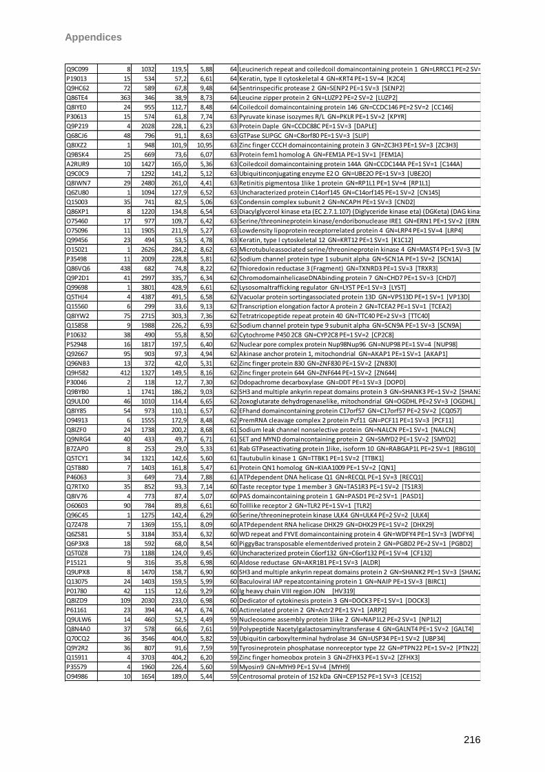
Appendices
216
Q9C099 8 1032 119,5 5,88 64 Leucinerich repeat and coiledcoil domaincontaining protein 1 GN=LRRCC1 PE=2 SV=2 [LRCC1]
P19013 15 534 57,2 6,61 64 Keratin, type II cytoskeletal 4 GN=KRT4 PE=1 SV=4 [K2C4]
Q9HC62 72 589 67,8 9,48 64 Sentrinspecific protease 2 GN=SENP2 PE=1 SV=3 [SENP2]
Q86TE4 363 346 38,9 8,73 64 Leucine zipper protein 2 GN=LUZP2 PE=2 SV=2 [LUZP2]
Q8IYE0 24 955 112,7 8,48 64 Coiledcoil domaincontaining protein 146 GN=CCDC146 PE=2 SV=2 [CC146]
P30613 15 574 61,8 7,74 63 Pyruvate kinase isozymes R/L GN=PKLR PE=1 SV=2 [KPYR]
Q9P219 4 2028 228,1 6,23 63 Protein Daple GN=CCDC88C PE=1 SV=3 [DAPLE]
Q68CJ6 48 796 91,1 8,63 63 GTPase SLIPGC GN=C8orf80 PE=1 SV=3 [SLIP]
Q8IXZ2 1 948 101,9 10,95 63 Zinc finger CCCH domaincontaining protein 3 GN=ZC3H3 PE=1 SV=3 [ZC3H3]
Q9BSK4 25 669 73,6 6,07 63 Protein fem1 homolog A GN=FEM1A PE=1 SV=1 [FEM1A]
A2RUR9 10 1427 165,0 5,36 63 Coiledcoil domaincontaining protein 144A GN=CCDC144A PE=1 SV=1 [C144A]
Q9C0C9 7 1292 141,2 5,12 63 Ubiquitinconjugating enzyme E2 O GN=UBE2O PE=1 SV=3 [UBE2O]
Q8IWN7 29 2480 261,0 4,41 63 Retinitis pigmentosa 1like 1 protein GN=RP1L1 PE=1 SV=4 [RP1L1]
Q6ZU80 1 1094 127,9 6,52 63 Uncharacterized protein C14orf145 GN=C14orf145 PE=1 SV=2 [CN145]
Q15003 35 741 82,5 5,06 63 Condensin complex subunit 2 GN=NCAPH PE=1 SV=3 [CND2]
Q86XP1 8 1220 134,8 6,54 63 Diacylglycerol kinase eta (EC 2.7.1.107) (Diglyceride kinase eta) (DGKeta) (DAG kinase eta) [DGKH]
O75460 17 977 109,7 6,42 63 Serine/threonineprotein kinase/endoribonuclease IRE1 GN=ERN1 PE=1 SV=2 [ERN1]
O75096 11 1905 211,9 5,27 63 Lowdensity lipoprotein receptorrelated protein 4 GN=LRP4 PE=1 SV=4 [LRP4]
Q99456 23 494 53,5 4,78 63 Keratin, type I cytoskeletal 12 GN=KRT12 PE=1 SV=1 [K1C12]
O15021 1 2626 284,2 8,62 63 Microtubuleassociated serine/threonineprotein kinase 4 GN=MAST4 PE=1 SV=3 [MAST4]
P35498 11 2009 228,8 5,81 62 Sodium channel protein type 1 subunit alpha GN=SCN1A PE=1 SV=2 [SCN1A]
Q86VQ6 438 682 74,8 8,22 62 Thioredoxin reductase 3 (Fragment) GN=TXNRD3 PE=1 SV=3 [TRXR3]
Q9P2D1 41 2997 335,7 6,34 62 ChromodomainhelicaseDNAbinding protein 7 GN=CHD7 PE=1 SV=3 [CHD7]
Q99698 1 3801 428,9 6,61 62 Lysosomaltrafficking regulator GN=LYST PE=1 SV=3 [LYST]
Q5THJ4 4 4387 491,5 6,58 62 Vacuolar protein sortingassociated protein 13D GN=VPS13D PE=1 SV=1 [VP13D]
Q15560 6 299 33,6 9,13 62 Transcription elongation factor A protein 2 GN=TCEA2 PE=1 SV=1 [TCEA2]
Q8IYW2 75 2715 303,3 7,36 62 Tetratricopeptide repeat protein 40 GN=TTC40 PE=2 SV=3 [TTC40]
Q15858 9 1988 226,2 6,93 62 Sodium channel protein type 9 subunit alpha GN=SCN9A PE=1 SV=3 [SCN9A]
P10632 38 490 55,8 8,50 62 Cytochrome P450 2C8 GN=CYP2C8 PE=1 SV=2 [CP2C8]
P52948 16 1817 197,5 6,40 62 Nuclear pore complex protein Nup98Nup96 GN=NUP98 PE=1 SV=4 [NUP98]
Q92667 95 903 97,3 4,94 62 Akinase anchor protein 1, mitochondrial GN=AKAP1 PE=1 SV=1 [AKAP1]
Q96NB3 13 372 42,0 5,31 62 Zinc finger protein 830 GN=ZNF830 PE=1 SV=2 [ZN830]
Q9H582 412 1327 149,5 8,16 62 Zinc finger protein 644 GN=ZNF644 PE=1 SV=2 [ZN644]
P30046 2 118 12,7 7,30 62 Ddopachrome decarboxylase GN=DDT PE=1 SV=3 [DOPD]
Q9BYB0 1 1741 186,2 9,03 62 SH3 and multiple ankyrin repeat domains protein 3 GN=SHANK3 PE=1 SV=2 [SHAN3]
Q9ULD0 46 1010 114,4 6,65 62 2oxoglutarate dehydrogenaselike, mitochondrial GN=OGDHL PE=2 SV=3 [OGDHL]
Q8IY85 54 973 110,1 6,57 62 EFhand domaincontaining protein C17orf57 GN=C17orf57 PE=2 SV=2 [CQ057]
O94913 6 1555 172,9 8,48 62 PremRNA cleavage complex 2 protein Pcf11 GN=PCF11 PE=1 SV=3 [PCF11]
Q8IZF0 24 1738 200,2 8,68 61 Sodium leak channel nonselective protein GN=NALCN PE=1 SV=1 [NALCN]
Q9NRG4 40 433 49,7 6,71 61 SET and MYND domaincontaining protein 2 GN=SMYD2 PE=1 SV=2 [SMYD2]
B7ZAP0 8 253 29,0 5,33 61 Rab GTPaseactivating protein 1like, isoform 10 GN=RABGAP1L PE=2 SV=1 [RBG10]
Q5TCY1 34 1321 142,6 5,60 61 Tautubulin kinase 1 GN=TTBK1 PE=1 SV=2 [TTBK1]
Q5TB80 7 1403 161,8 5,47 61 Protein QN1 homolog GN=KIAA1009 PE=1 SV=2 [QN1]
P46063 3 649 73,4 7,88 61 ATPdependent DNA helicase Q1 GN=RECQL PE=1 SV=3 [RECQ1]
Q7RTX0 35 852 93,3 7,14 60 Taste receptor type 1 member 3 GN=TAS1R3 PE=1 SV=2 [TS1R3]
Q8IV76 4 773 87,4 5,07 60 PAS domaincontaining protein 1 GN=PASD1 PE=2 SV=1 [PASD1]
O60603 90 784 89,8 6,61 60 Tolllike receptor 2 GN=TLR2 PE=1 SV=1 [TLR2]
Q96C45 1 1275 142,4 6,29 60 Serine/threonineprotein kinase ULK4 GN=ULK4 PE=2 SV=2 [ULK4]
Q7Z478 7 1369 155,1 8,09 60 ATPdependent RNA helicase DHX29 GN=DHX29 PE=1 SV=2 [DHX29]
Q6ZS81 5 3184 353,4 6,32 60 WD repeat and FYVE domaincontaining protein 4 GN=WDFY4 PE=1 SV=3 [WDFY4]
Q6P3X8 18 592 68,0 8,54 60 PiggyBac transposable elementderived protein 2 GN=PGBD2 PE=2 SV=1 [PGBD2]
Q5T0Z8 73 1188 124,0 9,45 60 Uncharacterized protein C6orf132 GN=C6orf132 PE=1 SV=4 [CF132]
P15121 9 316 35,8 6,98 60 Aldose reductase GN=AKR1B1 PE=1 SV=3 [ALDR]
Q9UPX8 8 1470 158,7 6,90 60 SH3 and multiple ankyrin repeat domains protein 2 GN=SHANK2 PE=1 SV=3 [SHAN2]
Q13075 24 1403 159,5 5,99 60 Baculoviral IAP repeatcontaining protein 1 GN=NAIP PE=1 SV=3 [BIRC1]
P01780 42 115 12,6 9,29 60 Ig heavy chain VIII region JON [HV319]
Q8IZD9 109 2030 233,0 6,98 60 Dedicator of cytokinesis protein 3 GN=DOCK3 PE=1 SV=1 [DOCK3]
P61161 23 394 44,7 6,74 60 Actinrelated protein 2 GN=Actr2 PE=1 SV=1 [ARP2]
Q9ULW6 14 460 52,5 4,49 59 Nucleosome assembly protein 1like 2 GN=NAP1L2 PE=2 SV=1 [NP1L2]
Q8N4A0 37 578 66,6 7,61 59 Polypeptide Nacetylgalactosaminyltransferase 4 GN=GALNT4 PE=1 SV=2 [GALT4]
Q70CQ2 36 3546 404,0 5,82 59 Ubiquitin carboxylterminal hydrolase 34 GN=USP34 PE=1 SV=2 [UBP34]
Q9Y2R2 36 807 91,6 7,59 59 Tyrosineprotein phosphatase nonreceptor type 22 GN=PTPN22 PE=1 SV=2 [PTN22]
Q15911 4 3703 404,2 6,20 59 Zinc finger homeobox protein 3 GN=ZFHX3 PE=1 SV=2 [ZFHX3]
P35579 4 1960 226,4 5,60 59 Myosin9 GN=MYH9 PE=1 SV=4 [MYH9]
O94986 10 1654 189,0 5,44 59 Centrosomal protein of 152 kDa GN=CEP152 PE=1 SV=3 [CE152]
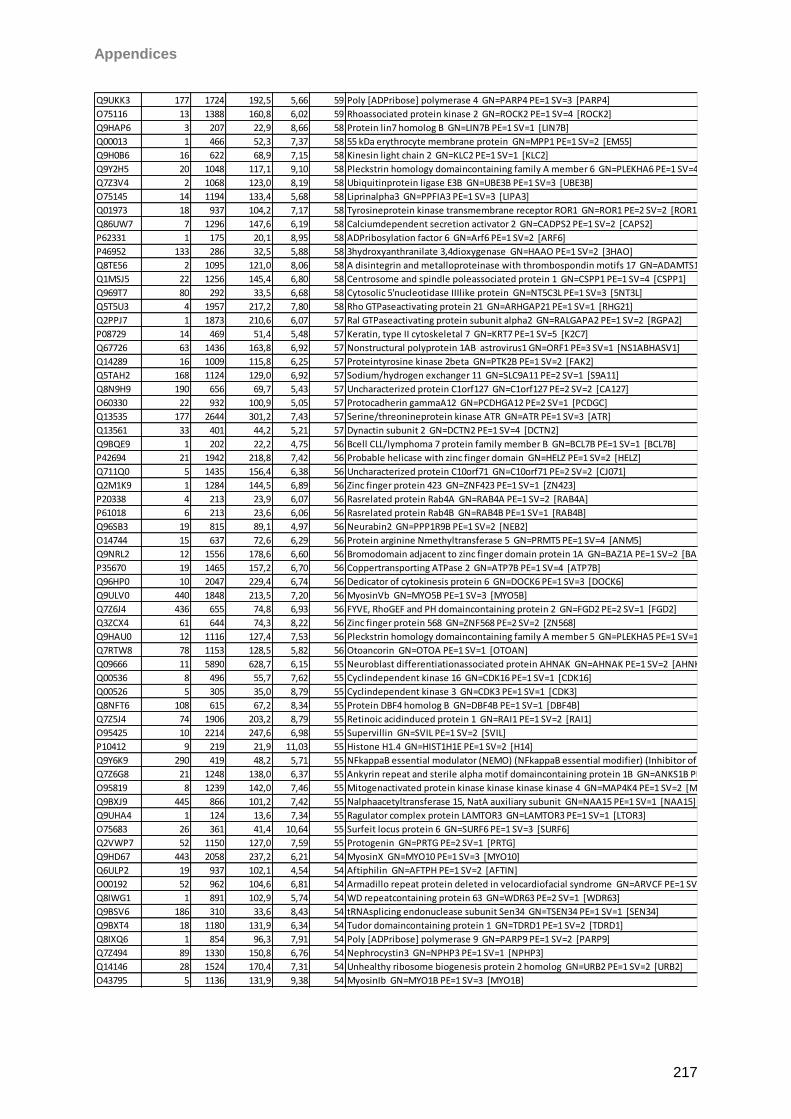
Appendices
217
Q9UKK3 177 1724 192,5 5,66 59 Poly [ADPribose] polymerase 4 GN=PARP4 PE=1 SV=3 [PARP4]
O75116 13 1388 160,8 6,02 59 Rhoassociated protein kinase 2 GN=ROCK2 PE=1 SV=4 [ROCK2]
Q9HAP6 3 207 22,9 8,66 58 Protein lin7 homolog B GN=LIN7B PE=1 SV=1 [LIN7B]
Q00013 1 466 52,3 7,37 58 55 kDa erythrocyte membrane protein GN=MPP1 PE=1 SV=2 [EM55]
Q9H0B6 16 622 68,9 7,15 58 Kinesin light chain 2 GN=KLC2 PE=1 SV=1 [KLC2]
Q9Y2H5 20 1048 117,1 9,10 58 Pleckstrin homology domaincontaining family A member 6 GN=PLEKHA6 PE=1 SV=4 [PKHA6]
Q7Z3V4 2 1068 123,0 8,19 58 Ubiquitinprotein ligase E3B GN=UBE3B PE=1 SV=3 [UBE3B]
O75145 14 1194 133,4 5,68 58 Liprinalpha3 GN=PPFIA3 PE=1 SV=3 [LIPA3]
Q01973 18 937 104,2 7,17 58 Tyrosineprotein kinase transmembrane receptor ROR1 GN=ROR1 PE=2 SV=2 [ROR1]
Q86UW7 7 1296 147,6 6,19 58 Calciumdependent secretion activator 2 GN=CADPS2 PE=1 SV=2 [CAPS2]
P62331 1 175 20,1 8,95 58 ADPribosylation factor 6 GN=Arf6 PE=1 SV=2 [ARF6]
P46952 133 286 32,5 5,88 58 3hydroxyanthranilate 3,4dioxygenase GN=HAAO PE=1 SV=2 [3HAO]
Q8TE56 2 1095 121,0 8,06 58 A disintegrin and metalloproteinase with thrombospondin motifs 17 GN=ADAMTS17 PE=2 SV=2 [ATS17]
Q1MSJ5 22 1256 145,4 6,80 58 Centrosome and spindle poleassociated protein 1 GN=CSPP1 PE=1 SV=4 [CSPP1]
Q969T7 80 292 33,5 6,68 58 Cytosolic 5'nucleotidase IIIlike protein GN=NT5C3L PE=1 SV=3 [5NT3L]
Q5T5U3 4 1957 217,2 7,80 58 Rho GTPaseactivating protein 21 GN=ARHGAP21 PE=1 SV=1 [RHG21]
Q2PPJ7 1 1873 210,6 6,07 57 Ral GTPaseactivating protein subunit alpha2 GN=RALGAPA2 PE=1 SV=2 [RGPA2]
P08729 14 469 51,4 5,48 57 Keratin, type II cytoskeletal 7 GN=KRT7 PE=1 SV=5 [K2C7]
Q67726 63 1436 163,8 6,92 57 Nonstructural polyprotein 1AB astrovirus1 GN=ORF1 PE=3 SV=1 [NS1ABHASV1]
Q14289 16 1009 115,8 6,25 57 Proteintyrosine kinase 2beta GN=PTK2B PE=1 SV=2 [FAK2]
Q5TAH2 168 1124 129,0 6,92 57 Sodium/hydrogen exchanger 11 GN=SLC9A11 PE=2 SV=1 [S9A11]
Q8N9H9 190 656 69,7 5,43 57 Uncharacterized protein C1orf127 GN=C1orf127 PE=2 SV=2 [CA127]
O60330 22 932 100,9 5,05 57 Protocadherin gammaA12 GN=PCDHGA12 PE=2 SV=1 [PCDGC]
Q13535 177 2644 301,2 7,43 57 Serine/threonineprotein kinase ATR GN=ATR PE=1 SV=3 [ATR]
Q13561 33 401 44,2 5,21 57 Dynactin subunit 2 GN=DCTN2 PE=1 SV=4 [DCTN2]
Q9BQE9 1 202 22,2 4,75 56 Bcell CLL/lymphoma 7 protein family member B GN=BCL7B PE=1 SV=1 [BCL7B]
P42694 21 1942 218,8 7,42 56 Probable helicase with zinc finger domain GN=HELZ PE=1 SV=2 [HELZ]
Q711Q0 5 1435 156,4 6,38 56 Uncharacterized protein C10orf71 GN=C10orf71 PE=2 SV=2 [CJ071]
Q2M1K9 1 1284 144,5 6,89 56 Zinc finger protein 423 GN=ZNF423 PE=1 SV=1 [ZN423]
P20338 4 213 23,9 6,07 56 Rasrelated protein Rab4A GN=RAB4A PE=1 SV=2 [RAB4A]
P61018 6 213 23,6 6,06 56 Rasrelated protein Rab4B GN=RAB4B PE=1 SV=1 [RAB4B]
Q96SB3 19 815 89,1 4,97 56 Neurabin2 GN=PPP1R9B PE=1 SV=2 [NEB2]
O14744 15 637 72,6 6,29 56 Protein arginine Nmethyltransferase 5 GN=PRMT5 PE=1 SV=4 [ANM5]
Q9NRL2 12 1556 178,6 6,60 56 Bromodomain adjacent to zinc finger domain protein 1A GN=BAZ1A PE=1 SV=2 [BAZ1A]
P35670 19 1465 157,2 6,70 56 Coppertransporting ATPase 2 GN=ATP7B PE=1 SV=4 [ATP7B]
Q96HP0 10 2047 229,4 6,74 56 Dedicator of cytokinesis protein 6 GN=DOCK6 PE=1 SV=3 [DOCK6]
Q9ULV0 440 1848 213,5 7,20 56 MyosinVb GN=MYO5B PE=1 SV=3 [MYO5B]
Q7Z6J4 436 655 74,8 6,93 56 FYVE, RhoGEF and PH domaincontaining protein 2 GN=FGD2 PE=2 SV=1 [FGD2]
Q3ZCX4 61 644 74,3 8,22 56 Zinc finger protein 568 GN=ZNF568 PE=2 SV=2 [ZN568]
Q9HAU0 12 1116 127,4 7,53 56 Pleckstrin homology domaincontaining family A member 5 GN=PLEKHA5 PE=1 SV=1 [PKHA5]
Q7RTW8 78 1153 128,5 5,82 56 Otoancorin GN=OTOA PE=1 SV=1 [OTOAN]
Q09666 11 5890 628,7 6,15 55 Neuroblast differentiationassociated protein AHNAK GN=AHNAK PE=1 SV=2 [AHNK]
Q00536 8 496 55,7 7,62 55 Cyclindependent kinase 16 GN=CDK16 PE=1 SV=1 [CDK16]
Q00526 5 305 35,0 8,79 55 Cyclindependent kinase 3 GN=CDK3 PE=1 SV=1 [CDK3]
Q8NFT6 108 615 67,2 8,34 55 Protein DBF4 homolog B GN=DBF4B PE=1 SV=1 [DBF4B]
Q7Z5J4 74 1906 203,2 8,79 55 Retinoic acidinduced protein 1 GN=RAI1 PE=1 SV=2 [RAI1]
O95425 10 2214 247,6 6,98 55 Supervillin GN=SVIL PE=1 SV=2 [SVIL]
P10412 9 219 21,9 11,03 55 Histone H1.4 GN=HIST1H1E PE=1 SV=2 [H14]
Q9Y6K9 290 419 48,2 5,71 55 NFkappaB essential modulator (NEMO) (NFkappaB essential modifier) (Inhibitor of nuclear factor kappaB kinase subunit gamma) (IkB kinase subunit gamma) (IkappaB kinase gamma) (IKKgamma) (IKKG) (IkB kinaseassociated protein 1) (IKKAP1) (FIP3) [NEMO]
Q7Z6G8 21 1248 138,0 6,37 55 Ankyrin repeat and sterile alpha motif domaincontaining protein 1B GN=ANKS1B PE=1 SV=2 [ANS1B]
O95819 8 1239 142,0 7,46 55 Mitogenactivated protein kinase kinase kinase kinase 4 GN=MAP4K4 PE=1 SV=2 [M4K4]
Q9BXJ9 445 866 101,2 7,42 55 Nalphaacetyltransferase 15, NatA auxiliary subunit GN=NAA15 PE=1 SV=1 [NAA15]
Q9UHA4 1 124 13,6 7,34 55 Ragulator complex protein LAMTOR3 GN=LAMTOR3 PE=1 SV=1 [LTOR3]
O75683 26 361 41,4 10,64 55 Surfeit locus protein 6 GN=SURF6 PE=1 SV=3 [SURF6]
Q2VWP7 52 1150 127,0 7,59 55 Protogenin GN=PRTG PE=2 SV=1 [PRTG]
Q9HD67 443 2058 237,2 6,21 54 MyosinX GN=MYO10 PE=1 SV=3 [MYO10]
Q6ULP2 19 937 102,1 4,54 54 Aftiphilin GN=AFTPH PE=1 SV=2 [AFTIN]
O00192 52 962 104,6 6,81 54 Armadillo repeat protein deleted in velocardiofacial syndrome GN=ARVCF PE=1 SV=1 [ARVC]
Q8IWG1 1 891 102,9 5,74 54 WD repeatcontaining protein 63 GN=WDR63 PE=2 SV=1 [WDR63]
Q9BSV6 186 310 33,6 8,43 54 tRNAsplicing endonuclease subunit Sen34 GN=TSEN34 PE=1 SV=1 [SEN34]
Q9BXT4 18 1180 131,9 6,34 54 Tudor domaincontaining protein 1 GN=TDRD1 PE=1 SV=2 [TDRD1]
Q8IXQ6 1 854 96,3 7,91 54 Poly [ADPribose] polymerase 9 GN=PARP9 PE=1 SV=2 [PARP9]
Q7Z494 89 1330 150,8 6,76 54 Nephrocystin3 GN=NPHP3 PE=1 SV=1 [NPHP3]
Q14146 28 1524 170,4 7,31 54 Unhealthy ribosome biogenesis protein 2 homolog GN=URB2 PE=1 SV=2 [URB2]
O43795 5 1136 131,9 9,38 54 MyosinIb GN=MYO1B PE=1 SV=3 [MYO1B]
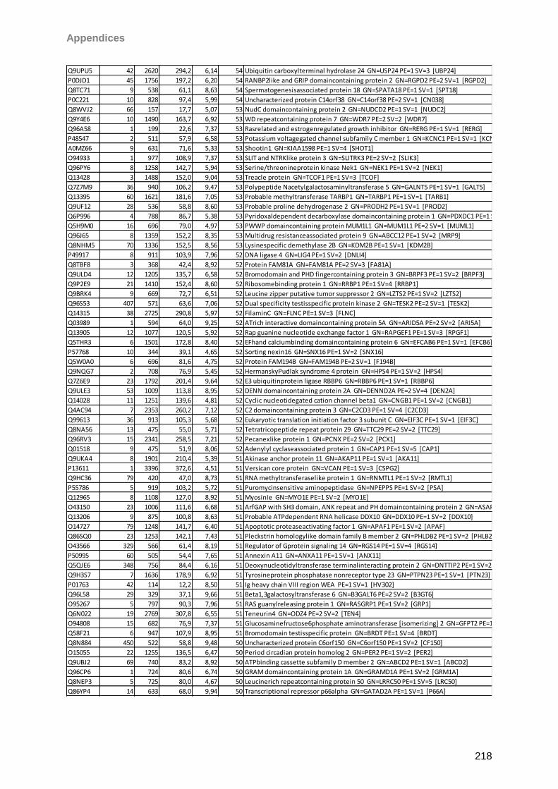
Appendices
218
Q9UPU5 42 2620 294,2 6,14 54 Ubiquitin carboxylterminal hydrolase 24 GN=USP24 PE=1 SV=3 [UBP24]
P0DJD1 45 1756 197,2 6,20 54 RANBP2like and GRIP domaincontaining protein 2 GN=RGPD2 PE=2 SV=1 [RGPD2]
Q8TC71 9 538 61,1 8,63 54 Spermatogenesisassociated protein 18 GN=SPATA18 PE=1 SV=1 [SPT18]
P0C221 10 828 97,4 5,99 54 Uncharacterized protein C14orf38 GN=C14orf38 PE=2 SV=1 [CN038]
Q8WVJ2 66 157 17,7 5,07 53 NudC domaincontaining protein 2 GN=NUDCD2 PE=1 SV=1 [NUDC2]
Q9Y4E6 10 1490 163,7 6,92 53 WD repeatcontaining protein 7 GN=WDR7 PE=2 SV=2 [WDR7]
Q96A58 1 199 22,6 7,37 53 Rasrelated and estrogenregulated growth inhibitor GN=RERG PE=1 SV=1 [RERG]
P48547 2 511 57,9 6,58 53 Potassium voltagegated channel subfamily C member 1 GN=KCNC1 PE=1 SV=1 [KCNC1]
A0MZ66 9 631 71,6 5,33 53 Shootin1 GN=KIAA1598 PE=1 SV=4 [SHOT1]
O94933 1 977 108,9 7,37 53 SLIT and NTRKlike protein 3 GN=SLITRK3 PE=2 SV=2 [SLIK3]
Q96PY6 8 1258 142,7 5,94 53 Serine/threonineprotein kinase Nek1 GN=NEK1 PE=1 SV=2 [NEK1]
Q13428 3 1488 152,0 9,04 53 Treacle protein GN=TCOF1 PE=1 SV=3 [TCOF]
Q7Z7M9 36 940 106,2 9,47 53 Polypeptide Nacetylgalactosaminyltransferase 5 GN=GALNT5 PE=1 SV=1 [GALT5]
Q13395 60 1621 181,6 7,05 53 Probable methyltransferase TARBP1 GN=TARBP1 PE=1 SV=1 [TARB1]
Q9UF12 28 536 58,8 8,60 53 Probable proline dehydrogenase 2 GN=PRODH2 PE=1 SV=1 [PROD2]
Q6P996 4 788 86,7 5,38 53 Pyridoxaldependent decarboxylase domaincontaining protein 1 GN=PDXDC1 PE=1 SV=2 [PDXD1]
Q5H9M0 16 696 79,0 4,97 53 PWWP domaincontaining protein MUM1L1 GN=MUM1L1 PE=2 SV=1 [MUML1]
Q96J65 8 1359 152,2 8,35 53 Multidrug resistanceassociated protein 9 GN=ABCC12 PE=1 SV=2 [MRP9]
Q8NHM5 70 1336 152,5 8,56 53 Lysinespecific demethylase 2B GN=KDM2B PE=1 SV=1 [KDM2B]
P49917 8 911 103,9 7,96 52 DNA ligase 4 GN=LIG4 PE=1 SV=2 [DNLI4]
Q8TBF8 3 368 42,4 8,92 52 Protein FAM81A GN=FAM81A PE=2 SV=3 [FA81A]
Q9ULD4 12 1205 135,7 6,58 52 Bromodomain and PHD fingercontaining protein 3 GN=BRPF3 PE=1 SV=2 [BRPF3]
Q9P2E9 21 1410 152,4 8,60 52 Ribosomebinding protein 1 GN=RRBP1 PE=1 SV=4 [RRBP1]
Q9BRK4 9 669 72,7 6,51 52 Leucine zipper putative tumor suppressor 2 GN=LZTS2 PE=1 SV=2 [LZTS2]
Q96S53 407 571 63,6 7,06 52 Dual specificity testisspecific protein kinase 2 GN=TESK2 PE=2 SV=1 [TESK2]
Q14315 38 2725 290,8 5,97 52 FilaminC GN=FLNC PE=1 SV=3 [FLNC]
Q03989 1 594 64,0 9,25 52 ATrich interactive domaincontaining protein 5A GN=ARID5A PE=2 SV=2 [ARI5A]
Q13905 12 1077 120,5 5,92 52 Rap guanine nucleotide exchange factor 1 GN=RAPGEF1 PE=1 SV=3 [RPGF1]
Q5THR3 6 1501 172,8 8,40 52 EFhand calciumbinding domaincontaining protein 6 GN=EFCAB6 PE=1 SV=1 [EFCB6]
P57768 10 344 39,1 4,65 52 Sorting nexin16 GN=SNX16 PE=1 SV=2 [SNX16]
Q5W0A0 6 696 81,6 4,75 52 Protein FAM194B GN=FAM194B PE=2 SV=1 [F194B]
Q9NQG7 2 708 76,9 5,45 52 HermanskyPudlak syndrome 4 protein GN=HPS4 PE=1 SV=2 [HPS4]
Q7Z6E9 23 1792 201,4 9,64 52 E3 ubiquitinprotein ligase RBBP6 GN=RBBP6 PE=1 SV=1 [RBBP6]
Q9ULE3 53 1009 113,8 8,95 52 DENN domaincontaining protein 2A GN=DENND2A PE=2 SV=4 [DEN2A]
Q14028 11 1251 139,6 4,81 52 Cyclic nucleotidegated cation channel beta1 GN=CNGB1 PE=1 SV=2 [CNGB1]
Q4AC94 7 2353 260,2 7,12 52 C2 domaincontaining protein 3 GN=C2CD3 PE=1 SV=4 [C2CD3]
Q99613 36 913 105,3 5,68 52 Eukaryotic translation initiation factor 3 subunit C GN=EIF3C PE=1 SV=1 [EIF3C]
Q8NA56 13 475 55,0 5,71 52 Tetratricopeptide repeat protein 29 GN=TTC29 PE=2 SV=2 [TTC29]
Q96RV3 15 2341 258,5 7,21 52 Pecanexlike protein 1 GN=PCNX PE=2 SV=2 [PCX1]
Q01518 9 475 51,9 8,06 52 Adenylyl cyclaseassociated protein 1 GN=CAP1 PE=1 SV=5 [CAP1]
Q9UKA4 8 1901 210,4 5,39 51 Akinase anchor protein 11 GN=AKAP11 PE=1 SV=1 [AKA11]
P13611 1 3396 372,6 4,51 51 Versican core protein GN=VCAN PE=1 SV=3 [CSPG2]
Q9HC36 79 420 47,0 8,73 51 RNA methyltransferaselike protein 1 GN=RNMTL1 PE=1 SV=2 [RMTL1]
P55786 5 919 103,2 5,72 51 Puromycinsensitive aminopeptidase GN=NPEPPS PE=1 SV=2 [PSA]
Q12965 8 1108 127,0 8,92 51 MyosinIe GN=MYO1E PE=1 SV=2 [MYO1E]
O43150 23 1006 111,6 6,68 51 ArfGAP with SH3 domain, ANK repeat and PH domaincontaining protein 2 GN=ASAP2 PE=1 SV=3 [ASAP2]
Q13206 9 875 100,8 8,63 51 Probable ATPdependent RNA helicase DDX10 GN=DDX10 PE=1 SV=2 [DDX10]
O14727 79 1248 141,7 6,40 51 Apoptotic proteaseactivating factor 1 GN=APAF1 PE=1 SV=2 [APAF]
Q86SQ0 23 1253 142,1 7,43 51 Pleckstrin homologylike domain family B member 2 GN=PHLDB2 PE=1 SV=2 [PHLB2]
O43566 329 566 61,4 8,19 51 Regulator of Gprotein signaling 14 GN=RGS14 PE=1 SV=4 [RGS14]
P50995 60 505 54,4 7,65 51 Annexin A11 GN=ANXA11 PE=1 SV=1 [ANX11]
Q5QJE6 348 756 84,4 6,16 51 Deoxynucleotidyltransferase terminalinteracting protein 2 GN=DNTTIP2 PE=1 SV=2 [TDIF2]
Q9H3S7 7 1636 178,9 6,92 51 Tyrosineprotein phosphatase nonreceptor type 23 GN=PTPN23 PE=1 SV=1 [PTN23]
P01763 42 114 12,2 8,50 51 Ig heavy chain VIII region WEA PE=1 SV=1 [HV302]
Q96L58 29 329 37,1 9,66 51 Beta1,3galactosyltransferase 6 GN=B3GALT6 PE=2 SV=2 [B3GT6]
O95267 5 797 90,3 7,96 51 RAS guanylreleasing protein 1 GN=RASGRP1 PE=1 SV=2 [GRP1]
Q6N022 19 2769 307,8 6,55 51 Teneurin4 GN=ODZ4 PE=2 SV=2 [TEN4]
O94808 15 682 76,9 7,37 51 Glucosaminefructose6phosphate aminotransferase [isomerizing] 2 GN=GFPT2 PE=1 SV=3 [GFPT2]
Q58F21 6 947 107,9 8,95 51 Bromodomain testisspecific protein GN=BRDT PE=1 SV=4 [BRDT]
Q8N884 450 522 58,8 9,48 50 Uncharacterized protein C6orf150 GN=C6orf150 PE=1 SV=2 [CF150]
O15055 22 1255 136,5 6,47 50 Period circadian protein homolog 2 GN=PER2 PE=1 SV=2 [PER2]
Q9UBJ2 69 740 83,2 8,92 50 ATPbinding cassette subfamily D member 2 GN=ABCD2 PE=1 SV=1 [ABCD2]
Q96CP6 1 724 80,6 6,74 50 GRAM domaincontaining protein 1A GN=GRAMD1A PE=1 SV=2 [GRM1A]
Q8NEP3 5 725 80,0 4,67 50 Leucinerich repeatcontaining protein 50 GN=LRRC50 PE=1 SV=5 [LRC50]
Q86YP4 14 633 68,0 9,94 50 Transcriptional repressor p66alpha GN=GATAD2A PE=1 SV=1 [P66A]
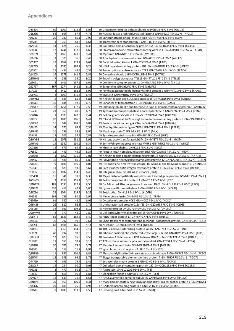
Appendices
219
O43424 69 1007 113,3 6,07 50 Glutamate receptor delta2 subunit GN=GRID2 PE=2 SV=2 [GRID2]
Q16236 56 605 67,8 4,78 50 Nuclear factor erythroid 2related factor 2 GN=NFE2L2 PE=1 SV=3 [NF2L2]
P08237 20 780 85,1 7,99 50 6phosphofructokinase, muscle type GN=PFKM PE=1 SV=2 [K6PF]
P02786 1 760 84,8 6,61 50 Transferrin receptor protein 1 GN=TFRC PE=1 SV=2 [TFR1]
A6NI56 14 674 76,0 8,38 50 Coiledcoil domaincontaining protein 154 GN=CCDC154 PE=2 SV=4 [CC154]
P23634 12 1241 137,8 6,60 50 Plasma membrane calciumtransporting ATPase 4 GN=ATP2B4 PE=1 SV=2 [AT2B4]
O00159 7 1063 121,6 9,41 50 MyosinIc GN=MYO1C PE=1 SV=4 [MYO1C]
Q96JD6 3 320 36,6 7,49 50 1,5anhydroDfructose reductase GN=AKR1E2 PE=1 SV=2 [AKCL2]
Q05397 18 1052 119,2 6,62 50 Focal adhesion kinase 1 GN=PTK2 PE=1 SV=2 [FAK1]
Q7Z745 5 1585 180,7 6,28 50 HEAT repeatcontaining protein 7B2 GN=HEATR7B2 PE=2 SV=3 [HTRB2]
Q15561 21 434 48,3 7,33 50 Transcriptional enhancer factor TEF3 GN=TEAD4 PE=1 SV=3 [TEAD4]
Q14203 16 1278 141,6 5,81 50 Dynactin subunit 1 GN=DCTN1 PE=1 SV=3 [DCTN1]
Q8NHH1 7 538 58,0 9,20 50 Tubulin polyglutamylase TTLL11 GN=TTLL11 PE=2 SV=1 [TTL11]
Q15021 8 1401 157,1 6,61 49 Condensin complex subunit 1 GN=NCAPD2 PE=1 SV=3 [CND1]
Q92797 367 1274 141,1 6,13 49 Symplekin GN=SYMPK PE=1 SV=2 [SYMPK]
B1AJZ9 4 1412 161,8 6,95 49 Forkheadassociated domaincontaining protein 1 GN=FHAD1 PE=2 SV=2 [FHAD1]
Q9BXX0 4 1053 115,6 6,46 49 EMILIN2 GN=EMILIN2 PE=1 SV=3 [EMIL2]
Q8WXK1 8 588 65,8 5,83 49 Ankyrin repeat and SOCS box protein 15 GN=ASB15 PE=2 SV=3 [ASB15]
Q14511 10 834 92,8 6,70 49 Enhancer of filamentation 1 GN=NEDD9 PE=1 SV=1 [CASL]
Q86VF2 8 1251 137,7 7,56 49 Immunoglobulinlike and fibronectin type III domaincontaining protein 1 GN=IGFN1 PE=1 SV=2 [IGFN1]
P35236 21 360 40,5 6,79 49 Tyrosineprotein phosphatase nonreceptor type 7 GN=PTPN7 PE=1 SV=3 [PTN7]
Q02846 1 1103 120,0 7,44 49 Retinal guanylyl cyclase 1 GN=GUCY2D PE=1 SV=2 [GUC2D]
Q8IZJ3 20 1885 206,6 6,42 49 C3 and PZPlike alpha2macroglobulin domaincontaining protein 8 GN=CPAMD8 PE=1 SV=2 [CPMD8]
Q9H3U1 16 944 103,0 6,07 49 Protein unc45 homolog A GN=UNC45A PE=1 SV=1 [UN45A]
Q96JP5 14 570 63,4 7,36 49 E3 ubiquitinprotein ligase ZFP91 GN=ZFP91 PE=1 SV=1 [ZFP91]
Q9UH92 16 298 33,3 8,06 49 Maxlike protein X GN=MLX PE=1 SV=2 [MLX]
P51451 18 505 57,7 7,87 49 Tyrosineprotein kinase Blk GN=BLK PE=1 SV=3 [BLK]
Q8WYB5 13 2073 231,2 5,96 49 Histone acetyltransferase MYST4 GN=MYST4 PE=1 SV=3 [MYST4]
Q9H4A3 15 2382 250,6 6,34 49 Serine/threonineprotein kinase WNK1 GN=WNK1 PE=1 SV=2 [WNK1]
Q07866 14 573 65,3 6,20 49 Kinesin light chain 1 GN=KLC1 PE=1 SV=2 [KLC1]
Q7L592 11 441 49,2 8,34 49 Protein midA homolog, mitochondrial GN=C2orf56 PE=1 SV=1 [MIDA]
Q6UB98 1 2062 235,5 7,01 49 Ankyrin repeat domaincontaining protein 12 GN=ANKRD12 PE=1 SV=3 [ANR12]
Q8IXK2 36 581 66,9 6,80 49 Polypeptide Nacetylgalactosaminyltransferase 12 GN=GALNT12 PE=1 SV=3 [GLT12]
Q96L73 9 2696 296,5 8,03 49 Histonelysine Nmethyltransferase, H3 lysine36 and H4 lysine20 specific GN=NSD1 PE=1 SV=1 [NSD1]
P56945 11 870 93,3 5,67 48 Breast cancer antiestrogen resistance protein 1 GN=BCAR1 PE=1 SV=2 [BCAR1]
P13612 26 1032 114,8 6,48 48 Integrin alpha4 GN=ITGA4 PE=1 SV=3 [ITA4]
Q95460 16 341 39,3 6,30 48 Major histocompatibility complex class Irelated gene protein GN=MR1 PE=1 SV=1 [HMR1]
A6NKG5 3 1359 155,1 5,20 48 Retrotransposonlike protein 1 GN=RTL1 PE=2 SV=2 [RTL1]
Q9NW08 181 1133 127,7 8,50 48 DNAdirected RNA polymerase III subunit RPC2 GN=POLR3B PE=1 SV=2 [RPC2]
Q8N371 339 416 47,2 5,80 48 Lysinespecific demethylase 8 GN=JMJD5 PE=1 SV=1 [KDM8]
Q86Z14 25 1044 119,7 9,22 48 Betaklotho GN=KLB PE=1 SV=1 [KLOTB]
P08582 363 738 80,2 5,94 48 Melanotransferrin GN=MFI2 PE=1 SV=2 [TRFM]
O43639 91 380 42,9 6,95 48 Cytoplasmic protein NCK2 GN=NCK2 PE=1 SV=2 [NCK2]
Q96N23 26 812 92,3 7,71 48 Uncharacterized protein C12orf55 GN=C12orf55 PE=2 SV=2 [CL055]
O95185 34 931 103,1 6,10 48 Netrin receptor UNC5C GN=UNC5C PE=1 SV=2 [UNC5C]
Q9UMW8 8 372 43,0 7,80 48 Ubl carboxylterminal hydrolase 18 GN=USP18 PE=1 SV=1 [UBP18]
Q9BXT8 58 1623 184,5 5,40 48 RING finger protein 17 GN=RNF17 PE=1 SV=3 [RNF17]
Q8TEL6 70 797 90,8 7,61 48 Short transient receptor potential channel 4associated protein GN=TRPC4AP PE=1 SV=2 [TP4AP]
Q9Y2I1 68 1504 166,5 5,14 48 Nischarin GN=NISCH PE=1 SV=3 [NISCH]
Q9UKE5 6 1360 154,8 7,17 48 TRAF2 and NCKinteracting protein kinase GN=TNIK PE=1 SV=1 [TNIK]
P23921 40 792 90,0 7,15 48 Ribonucleosidediphosphate reductase large subunit GN=RRM1 PE=1 SV=1 [RIR1]
Q9BUQ8 12 820 95,5 9,55 48 Probable ATPdependent RNA helicase DDX23 GN=DDX23 PE=1 SV=3 [DDX23]
P25705 11 553 59,7 9,13 47 ATP synthase subunit alpha, mitochondrial GN=ATP5A1 PE=1 SV=1 [ATPA]
Q16820 20 701 79,5 5,74 47 Meprin A subunit beta GN=MEP1B PE=1 SV=3 [MEP1B]
P01700 9 112 11,9 8,91 47 Ig lambda chain VI region HA PE=1 SV=1 [LV102]
Q8NEB9 11 887 101,5 6,81 47 Phosphatidylinositol 3kinase catalytic subunit type 3 GN=PIK3C3 PE=1 SV=1 [PK3C3]
Q6NT04 13 549 63,2 8,75 47 Tigger transposable elementderived protein 7 GN=TIGD7 PE=2 SV=1 [TIGD7]
O94769 7 699 79,7 5,41 47 Extracellular matrix protein 2 GN=ECM2 PE=2 SV=1 [ECM2]
Q4G0S7 14 254 30,0 9,06 47 Coiledcoil domaincontaining protein 152 GN=CCDC152 PE=2 SV=3 [CC152]
P09131 3 477 50,3 7,77 47 P3 protein GN=SLC10A3 PE=2 SV=1 [P3]
P13639 4 858 95,3 6,83 47 Elongation factor 2 GN=EEF2 PE=1 SV=4 [EF2]
O94927 18 633 71,6 8,51 47 HAUS augminlike complex subunit 5 GN=HAUS5 PE=1 SV=2 [HAUS5]
Q8NFP4 39 955 105,7 8,34 47 MAM domaincontaining glycosylphosphatidylinositol anchor protein 1 GN=MDGA1 PE=1 SV=1 [MDGA1]
Q9Y426 29 696 75,5 6,92 47 C2 domaincontaining protein 2 GN=C2CD2 PE=1 SV=2 [CU025]
Q86SJ6 8 1040 113,8 4,56 47 Desmoglein4 GN=DSG4 PE=1 SV=1 [DSG4]
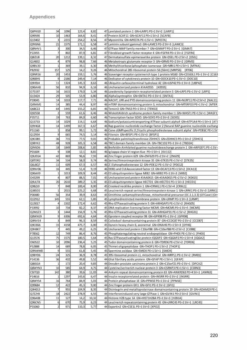
Appendices
220
Q6PKG0 34 1096 123,4 8,82 47 Larelated protein 1 GN=LARP1 PE=1 SV=2 [LARP1]
Q99590 10 1463 164,6 8,41 47 Protein SCAF11 GN=SCAF11 PE=1 SV=2 [SCAFB]
Q13402 3 2215 254,2 8,56 47 MyosinVIIa GN=MYO7A PE=1 SV=1 [MYO7A]
Q9Y6N6 21 1575 171,1 6,58 47 Laminin subunit gamma3 GN=LAMC3 PE=2 SV=3 [LAMC3]
Q8NHV1 3 300 34,5 6,46 47 GTPase IMAP family member 7 GN=GIMAP7 PE=2 SV=1 [GIMA7]
P22455 7 802 87,9 6,81 47 Fibroblast growth factor receptor 4 GN=FGFR4 PE=1 SV=2 [FGFR4]
Q8IZU8 20 1212 139,1 8,32 47 Dermatansulfate epimeraselike protein GN=DSEL PE=2 SV=2 [DSEL]
Q14832 4 879 98,8 7,66 46 Metabotropic glutamate receptor 3 GN=GRM3 PE=2 SV=2 [GRM3]
Q9BV20 1 369 39,1 6,30 46 Methylthioribose1phosphate isomerase GN=MRI1 PE=1 SV=1 [MTNA]
P82932 157 125 14,2 9,26 46 Mitochondrial 28S ribosomal protein S6 (S6mt) (MRPS6) [RT06]
Q9NR16 20 1453 159,1 5,76 46 Scavenger receptor cysteinerich type 1 protein M160 GN=CD163L1 PE=1 SV=2 [C163B]
Q96BY6 9 2186 249,4 7,14 46 Dedicator of cytokinesis protein 10 GN=DOCK10 PE=1 SV=3 [DOC10]
Q9H9J4 11 1324 145,3 8,63 46 Ubiquitin carboxylterminal hydrolase 42 GN=USP42 PE=1 SV=3 [UBP42]
Q96AA8 56 810 94,9 6,16 46 Uncharacterized protein KIAA0555 [K0555]
O75197 16 1615 179,0 5,34 46 Lowdensity lipoprotein receptorrelated protein 5 GN=LRP5 PE=1 SV=2 [LRP5]
Q13424 29 505 53,9 6,80 46 Alpha1syntrophin GN=SNTA1 PE=1 SV=1 [SNTA1]
P59045 14 1033 117,7 7,71 46 NACHT, LRR and PYD domainscontaining protein 11 GN=NLRP11 PE=2 SV=2 [NAL11]
Q49AM1 14 385 44,4 8,97 46 mTERF domaincontaining protein 3, mitochondrial GN=MTERFD3 PE=1 SV=2 [MTER3]
Q68CZ2 64 1445 155,2 6,81 46 Tensin3 GN=TNS3 PE=1 SV=2 [TENS3]
Q92558 2 559 61,6 6,46 46 WiskottAldrich syndrome protein family member 1 GN=WASF1 PE=1 SV=1 [WASF1]
P35711 19 763 84,0 6,60 46 Transcription factor SOX5 GN=SOX5 PE=2 SV=3 [SOX5]
Q13733 14 1029 114,1 6,64 46 Sodium/potassiumtransporting ATPase subunit alpha4 GN=ATP1A4 PE=1 SV=3 [AT1A4]
Q9Y4G8 14 1499 167,3 6,67 46 Rap guanine nucleotide exchange factor 2 (Neural RAP guanine nucleotide exchange protein) (nRap GEP) (PDZ domaincontaining guanine nucleotide exchange factor 1) (PDZGEF1) (RAGEF) [RPGF2]
P51160 1 858 99,1 5,72 46 Cone cGMPspecific 3',5'cyclic phosphodiesterase subunit alpha' GN=PDE6C PE=1 SV=2 [PDE6C]
Q12934 9 665 74,5 5,14 46 Filensin GN=BFSP1 PE=1 SV=3 [BFSP1]
Q9C0B5 36 715 77,5 9,01 46 Probable palmitoyltransferase ZDHHC5 GN=ZDHHC5 PE=1 SV=2 [ZDHC5]
Q9BYX2 48 928 105,3 6,58 46 TBC1 domain family member 2A GN=TBC1D2 PE=1 SV=3 [TBD2A]
Q9Y6D6 18 1849 208,6 5,85 46 Brefeldin Ainhibited guanine nucleotideexchange protein 1 GN=ARFGEF1 PE=1 SV=2 [BIG1]
P01604 9 108 12,1 8,81 46 Ig kappa chain VI region Kue PE=1 SV=1 [KV112]
Q9UEG4 42 869 96,6 7,93 46 Zinc finger protein 629 GN=ZNF629 PE=2 SV=2 [ZN629]
Q8TDR2 34 534 58,0 9,74 46 Serine/threonineprotein kinase 35 GN=STK35 PE=1 SV=2 [STK35]
Q6L8Q7 11 609 67,3 6,57 46 2',5'phosphodiesterase 12 GN=PDE12 PE=1 SV=2 [PDE12]
Q9NV06 16 445 51,4 9,19 46 DDB1 and CUL4associated factor 13 GN=DCAF13 PE=1 SV=2 [DCA13]
Q96AX9 1 1013 109,9 8,44 45 E3 ubiquitinprotein ligase MIB2 GN=MIB2 PE=1 SV=3 [MIB2]
O43299 4 807 88,5 7,01 45 Uncharacterized protein KIAA0415 GN=KIAA0415 PE=2 SV=2 [K0415]
Q9ULT8 20 2610 289,2 5,35 45 E3 ubiquitinprotein ligase HECTD1 GN=HECTD1 PE=1 SV=3 [HECD1]
Q9BZJ0 7 848 100,4 8,00 45 Crooked necklike protein 1 GN=CRNKL1 PE=1 SV=4 [CRNL1]
Q38SD2 1 2015 225,2 6,68 45 Leucinerich repeat serine/threonineprotein kinase 1 GN=LRRK1 PE=1 SV=3 [LRRK1]
P00480 56 354 39,9 8,63 45 Ornithine carbamoyltransferase, mitochondrial precursor (EC 2.1.3.3) (OTCase) (Ornithine transcarbamylase) [OTC]
Q12912 145 555 62,1 5,85 45 Lymphoidrestricted membrane protein GN=LRMP PE=1 SV=3 [LRMP]
Q13017 4 1502 172,4 6,62 45 Rho GTPaseactivating protein 5 GN=ARHGAP5 PE=1 SV=2 [RHG05]
P33992 10 734 82,2 8,37 45 DNA replication licensing factor MCM5 GN=MCM5 PE=1 SV=5 [MCM5]
Q2M1Z3 3 1444 156,9 5,76 45 Rho GTPaseactivating protein 31 GN=ARHGAP31 PE=1 SV=2 [RHG31]
Q8WXG9 9 6306 692,6 4,64 45 Gprotein coupled receptor 98 GN=GPR98 PE=1 SV=2 [GPR98]
Q9NVE4 3 849 96,3 8,59 45 Coiledcoil domaincontaining protein 87 GN=CCDC87 PE=2 SV=2 [CCD87]
Q9C0G6 15 4158 475,7 6,00 45 Dynein heavy chain 6, axonemal GN=DNAH6 PE=1 SV=3 [DYH6]
Q9H8K7 7 445 49,2 6,25 45 Uncharacterized protein C10orf88 GN=C10orf88 PE=2 SV=2 [CJ088]
P78562 12 749 86,4 8,76 45 Phosphateregulating neutral endopeptidase GN=PHEX PE=1 SV=1 [PHEX]
Q13576 75 1575 180,5 5,64 45 Ras GTPaseactivatinglike protein IQGAP2 GN=IQGAP2 PE=1 SV=4 [IQGA2]
O60522 10 2096 236,4 5,25 45 Tudor domaincontaining protein 6 GN=TDRD6 PE=2 SV=2 [TDRD6]
P52888 14 689 78,8 6,05 45 Thimet oligopeptidase GN=THOP1 PE=1 SV=2 [THOP1]
Q9NWM0 9 555 61,8 5,45 45 Spermine oxidase GN=SMOX PE=1 SV=1 [SMOX]
Q9BYD6 24 325 36,9 8,78 45 39S ribosomal protein L1, mitochondrial GN=MRPL1 PE=1 SV=2 [RM01]
P14136 36 432 49,8 5,52 44 Glial fibrillary acidic protein GN=GFAP PE=1 SV=1 [GFAP]
Q86SG4 1 172 20,4 9,83 44 Dresden prostate carcinoma protein 2 GN=C15orf21 PE=2 SV=1 [DPCA2]
Q8WYN3 10 585 64,9 4,75 44 Cysteine/serinerich nuclear protein 3 GN=CSRNP3 PE=1 SV=1 [CSRN3]
C9JTQ0 142 380 39,6 11,03 44 Ankyrin repeat domaincontaining protein 63 GN=ANKRD63 PE=4 SV=1 [ANR63]
P14616 1 1297 143,6 6,47 44 Insulin receptorrelated protein GN=INSRR PE=2 SV=2 [INSRR]
Q8WY54 16 764 84,9 5,03 44 Protein phosphatase 1E GN=PPM1E PE=1 SV=2 [PPM1E]
Q99684 12 422 45,3 9,00 44 Zinc finger protein Gfi1 GN=GFI1 PE=1 SV=2 [GFI1]
Q9H013 7 955 104,9 8,35 44 Disintegrin and metalloproteinase domaincontaining protein 19 GN=ADAM19 PE=1 SV=3 [ADA19]
Q7Z2Y8 18 2422 278,9 6,55 44 Interferoninduced very large GTPase 1 GN=GVIN1 PE=2 SV=2 [GVIN1]
Q96A08 11 127 14,2 10,32 44 Histone H2B type 1A GN=HIST1H2BA PE=1 SV=3 [H2B1A]
Q96CN5 6 670 75,9 6,23 44 Leucinerich repeatcontaining protein 45 GN=LRRC45 PE=2 SV=1 [LRC45]
P55060 2 971 110,3 5,77 44 Exportin2 GN=CSE1L PE=1 SV=3 [XPO2]
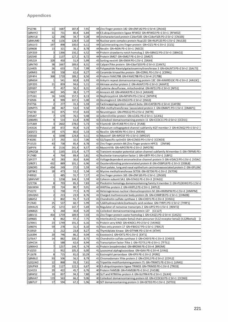
Appendices
221
P52746 11 1687 187,8 7,91 44 Zinc finger protein 142 GN=ZNF142 PE=1 SV=4 [ZN142]
Q8NHY2 31 731 80,4 6,84 44 E3 ubiquitinprotein ligase RFWD2 GN=RFWD2 PE=1 SV=1 [RFWD2]
Q9NVL8 12 296 34,7 9,28 44 Uncharacterized protein C14orf105 GN=C14orf105 PE=2 SV=2 [CN105]
Q8WUM0 43 1156 128,9 5,10 44 Nuclear pore complex protein Nup133 GN=NUP133 PE=1 SV=2 [NU133]
Q9ULV3 197 898 100,0 6,11 44 Cip1interacting zinc finger protein GN=CIZ1 PE=1 SV=2 [CIZ1]
Q99608 13 321 36,1 8,78 44 Necdin GN=NDN PE=1 SV=1 [NECD]
Q9Y2G9 3 1366 150,2 6,52 44 Protein strawberry notch homolog 2 GN=SBNO2 PE=2 SV=3 [SBNO2]
Q92540 2 1137 127,2 8,72 44 Protein SMG7 GN=SMG7 PE=1 SV=2 [SMG7]
O95219 328 450 51,9 5,99 43 Sorting nexin4 GN=SNX4 PE=1 SV=1 [SNX4]
Q8N7X0 38 1667 189,6 8,31 43 Calpain7like protein GN=C6orf103 PE=2 SV=3 [CAN7L]
Q14435 26 633 72,6 7,99 43 Polypeptide Nacetylgalactosaminyltransferase 3 GN=GALNT3 PE=2 SV=2 [GALT3]
Q49MI3 93 558 62,6 8,27 43 Ceramide kinaselike protein GN=CERKL PE=1 SV=1 [CERKL]
Q9Y4F4 368 1720 189,2 8,50 43 Protein FAM179B GN=FAM179B PE=1 SV=4 [F179B]
Q8N6S4 1 541 60,8 6,93 43 Ankyrin repeat domaincontaining protein 13C GN=ANKRD13C PE=2 SV=2 [AN13C]
Q9Y2D5 21 859 94,6 5,11 43 Akinase anchor protein 2 GN=AKAP2 PE=1 SV=3 [AKAP2]
Q9Y697 7 457 50,2 8,31 43 Cysteine desulfurase, mitochondrial GN=NFS1 PE=1 SV=3 [NFS1]
O76027 442 345 38,3 5,77 43 Annexin A9 GN=ANXA9 PE=1 SV=3 [ANXA9]
O75161 16 1426 157,5 8,13 43 Nephrocystin4 GN=NPHP4 PE=1 SV=2 [NPHP4]
P32926 8 999 107,5 5,00 43 Desmoglein3 GN=DSG3 PE=1 SV=2 [DSG3]
P47756 2 277 31,3 5,59 43 Factincapping protein subunit beta GN=CAPZB PE=1 SV=4 [CAPZB]
Q9NPF5 28 467 53,0 9,50 43 DNA methyltransferase 1associated protein 1 GN=DMAP1 PE=1 SV=1 [DMAP1]
P56730 31 875 97,0 8,03 43 Neurotrypsin GN=PRSS12 PE=2 SV=2 [NETR]
O95447 7 670 76,5 9,48 42 Lebercilinlike protein GN=LCA5L PE=2 SV=1 [LCA5L]
Q96M91 9 514 61,8 8,90 42 Coiledcoil domaincontaining protein 11 GN=CCDC11 PE=2 SV=2 [CCD11]
O75369 3 2602 278,0 5,73 42 FilaminB GN=FLNB PE=1 SV=2 [FLNB]
O43526 29 872 95,8 9,23 42 Potassium voltagegated channel subfamily KQT member 2 GN=KCNQ2 PE=1 SV=2 [KCNQ2]
Q0ZGT2 19 675 80,6 5,33 42 Nexilin GN=NEXN PE=1 SV=1 [NEXN]
O00160 6 1098 124,8 9,11 42 MyosinIf GN=MYO1F PE=1 SV=3 [MYO1F]
P30281 17 292 32,5 7,06 42 G1/Sspecific cyclinD3 GN=CCND3 PE=1 SV=2 [CCND3]
P51523 43 738 85,4 8,78 42 Zinc finger protein 84 (Zinc finger protein HPF2) [ZNF84]
Q6PIF6 9 2116 241,4 8,57 42 MyosinVIIb GN=MYO7B PE=2 SV=2 [MYO7B]
Q9NZQ8 5 1165 131,4 6,77 42 Transient receptor potential cation channel subfamily M member 5 GN=TRPM5 PE=1 SV=1 [TRPM5]
P17480 4 764 89,4 5,81 42 Nucleolar transcription factor 1 GN=UBTF PE=1 SV=1 [UBF1]
Q9Y277 42 283 30,6 8,66 42 Voltagedependent anionselective channel protein 3 GN=VDAC3 PE=1 SV=1 [VDAC3]
Q9BZF1 432 889 101,1 6,96 42 Oxysterolbinding proteinrelated protein 8 GN=OSBPL8 PE=1 SV=3 [OSBL8]
Q96DR5 434 249 27,0 5,59 42 Short palate, lung and nasal epithelium carcinomaassociated protein 2 GN=SPLUNC2 PE=1 SV=2 [SPLC2]
Q8TBK2 18 473 53,2 5,34 42 Nlysine methyltransferase SETD6 GN=SETD6 PE=1 SV=2 [SETD6]
P49910 1 485 55,7 7,17 42 Zinc finger protein 165 GN=ZNF165 PE=1 SV=1 [ZN165]
Q8WVM7 1 1258 144,3 5,59 42 Cohesin subunit SA1 GN=STAG1 PE=1 SV=3 [STAG1]
Q7Z736 8 793 85,3 7,83 42 Pleckstrin homology domaincontaining family H member 3 GN=PLEKHH3 PE=1 SV=2 [PKHH3]
Q6UWX4 19 724 80,7 9,01 42 HHIPlike protein 2 GN=HHIPL2 PE=2 SV=1 [HIPL2]
P52272 1 730 77,5 8,70 42 Heterogeneous nuclear ribonucleoprotein M GN=HNRNPM PE=1 SV=3 [HNRPM]
Q9UQN3 2 213 23,9 8,76 41 Charged multivesicular body protein 2b GN=CHMP2B PE=1 SV=1 [CHM2B]
Q86X52 1 802 91,7 9,23 41 Chondroitin sulfate synthase 1 GN=CHSY1 PE=1 SV=3 [CHSS1]
P17643 25 537 60,7 5,99 41 5,6dihydroxyindole2carboxylic acid oxidase GN=TYRP1 PE=1 SV=2 [TYRP1]
Q9HAU5 25 1272 147,7 5,69 41 Regulator of nonsense transcripts 2 GN=UPF2 PE=1 SV=1 [RENT2]
Q96BQ5 5 260 30,8 9,20 41 Coiledcoil domaincontaining protein 127 [CC127]
Q86V15 454 1759 189,9 7,03 41 Zinc finger protein castor homolog 1 GN=CASZ1 PE=2 SV=4 [CASZ1]
Q99665 6 862 97,1 7,75 41 Interleukin12 receptor beta2 chain precursor (IL12 receptor beta2) (IL12Rbeta2) [I12R2]
Q76NI1 23 1749 191,3 6,16 41 Protein very KIND GN=KNDC1 PE=2 SV=2 [VKIND]
Q96EF6 59 278 31,5 8,10 41 Fbox only protein 17 GN=FBXO17 PE=1 SV=1 [FBX17]
P23919 1 212 23,8 8,27 41 Thymidylate kinase GN=DTYMK PE=1 SV=4 [KTHY]
Q16394 20 746 86,2 9,04 41 Exostosin1 GN=EXT1 PE=1 SV=2 [EXT1]
Q70JA7 14 882 100,2 8,75 41 Chondroitin sulfate synthase 3 GN=CHSY3 PE=1 SV=3 [CHSS3]
Q9HCS4 1 588 62,6 8,94 41 Transcription factor 7like 1 GN=TCF7L1 PE=2 SV=1 [TF7L1]
Q96NH3 7 1257 144,7 6,74 41 Protein broadminded GN=BROMI PE=2 SV=4 [BROMI]
P10253 1 952 105,3 6,00 41 Lysosomal alphaglucosidase GN=GAA PE=1 SV=4 [LYAG]
P11678 8 715 81,0 10,29 41 Eosinophil peroxidase GN=EPX PE=1 SV=2 [PERE]
Q8N8U2 93 506 56,5 8,79 41 Chromodomain Ylike protein 2 GN=CDYL2 PE=2 SV=2 [CDYL2]
Q2Q1W2 12 868 93,3 7,61 41 Tripartite motifcontaining protein 71 GN=TRIM71 PE=2 SV=1 [LIN41]
Q9UPN9 19 1127 122,5 6,67 40 E3 ubiquitinprotein ligase TRIM33 GN=TRIM33 PE=1 SV=3 [TRI33]
Q14153 20 422 45,7 6,76 40 Protein FAM53B GN=FAM53B PE=1 SV=2 [FA53B]
Q8IW52 10 837 94,3 7,80 40 SLIT and NTRKlike protein 4 GN=SLITRK4 PE=2 SV=1 [SLIK4]
Q8NA47 119 563 66,2 9,06 40 Coiledcoil domaincontaining protein 63 GN=CCDC63 PE=1 SV=1 [CCD63]
Q86TU7 17 594 67,2 5,96 40 SET domaincontaining protein 3 GN=SETD3 PE=1 SV=1 [SETD3]
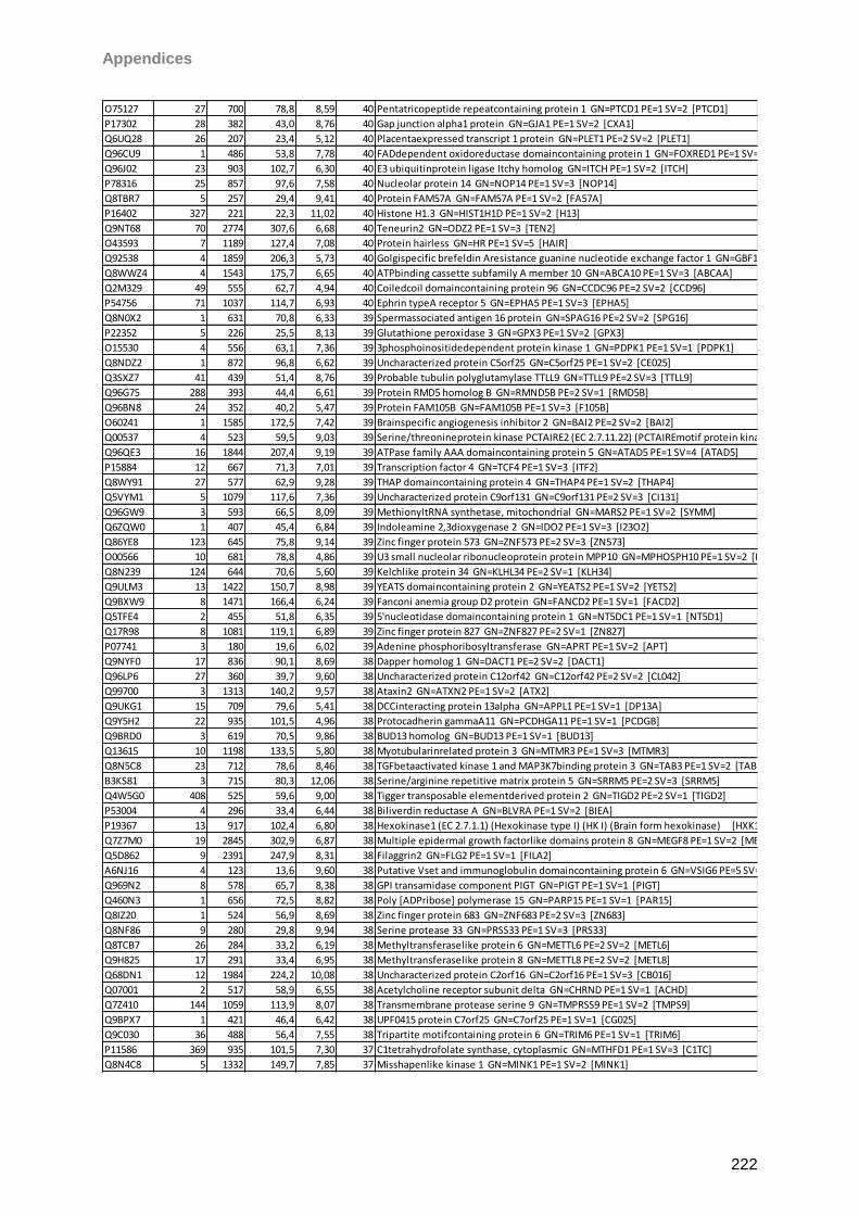
Appendices
222
O75127 27 700 78,8 8,59 40 Pentatricopeptide repeatcontaining protein 1 GN=PTCD1 PE=1 SV=2 [PTCD1]
P17302 28 382 43,0 8,76 40 Gap junction alpha1 protein GN=GJA1 PE=1 SV=2 [CXA1]
Q6UQ28 26 207 23,4 5,12 40 Placentaexpressed transcript 1 protein GN=PLET1 PE=2 SV=2 [PLET1]
Q96CU9 1 486 53,8 7,78 40 FADdependent oxidoreductase domaincontaining protein 1 GN=FOXRED1 PE=1 SV=2 [FXRD1]
Q96J02 23 903 102,7 6,30 40 E3 ubiquitinprotein ligase Itchy homolog GN=ITCH PE=1 SV=2 [ITCH]
P78316 25 857 97,6 7,58 40 Nucleolar protein 14 GN=NOP14 PE=1 SV=3 [NOP14]
Q8TBR7 5 257 29,4 9,41 40 Protein FAM57A GN=FAM57A PE=1 SV=2 [FA57A]
P16402 327 221 22,3 11,02 40 Histone H1.3 GN=HIST1H1D PE=1 SV=2 [H13]
Q9NT68 70 2774 307,6 6,68 40 Teneurin2 GN=ODZ2 PE=1 SV=3 [TEN2]
O43593 7 1189 127,4 7,08 40 Protein hairless GN=HR PE=1 SV=5 [HAIR]
Q92538 4 1859 206,3 5,73 40 Golgispecific brefeldin Aresistance guanine nucleotide exchange factor 1 GN=GBF1 PE=1 SV=2 [GBF1]
Q8WWZ4 4 1543 175,7 6,65 40 ATPbinding cassette subfamily A member 10 GN=ABCA10 PE=1 SV=3 [ABCAA]
Q2M329 49 555 62,7 4,94 40 Coiledcoil domaincontaining protein 96 GN=CCDC96 PE=2 SV=2 [CCD96]
P54756 71 1037 114,7 6,93 40 Ephrin typeA receptor 5 GN=EPHA5 PE=1 SV=3 [EPHA5]
Q8N0X2 1 631 70,8 6,33 39 Spermassociated antigen 16 protein GN=SPAG16 PE=2 SV=2 [SPG16]
P22352 5 226 25,5 8,13 39 Glutathione peroxidase 3 GN=GPX3 PE=1 SV=2 [GPX3]
O15530 4 556 63,1 7,36 39 3phosphoinositidedependent protein kinase 1 GN=PDPK1 PE=1 SV=1 [PDPK1]
Q8NDZ2 1 872 96,8 6,62 39 Uncharacterized protein C5orf25 GN=C5orf25 PE=1 SV=2 [CE025]
Q3SXZ7 41 439 51,4 8,76 39 Probable tubulin polyglutamylase TTLL9 GN=TTLL9 PE=2 SV=3 [TTLL9]
Q96G75 288 393 44,4 6,61 39 Protein RMD5 homolog B GN=RMND5B PE=2 SV=1 [RMD5B]
Q96BN8 24 352 40,2 5,47 39 Protein FAM105B GN=FAM105B PE=1 SV=3 [F105B]
O60241 1 1585 172,5 7,42 39 Brainspecific angiogenesis inhibitor 2 GN=BAI2 PE=2 SV=2 [BAI2]
Q00537 4 523 59,5 9,03 39 Serine/threonineprotein kinase PCTAIRE2 (EC 2.7.11.22) (PCTAIREmotif protein kinase 2) [PCTK2]
Q96QE3 16 1844 207,4 9,19 39 ATPase family AAA domaincontaining protein 5 GN=ATAD5 PE=1 SV=4 [ATAD5]
P15884 12 667 71,3 7,01 39 Transcription factor 4 GN=TCF4 PE=1 SV=3 [ITF2]
Q8WY91 27 577 62,9 9,28 39 THAP domaincontaining protein 4 GN=THAP4 PE=1 SV=2 [THAP4]
Q5VYM1 5 1079 117,6 7,36 39 Uncharacterized protein C9orf131 GN=C9orf131 PE=2 SV=3 [CI131]
Q96GW9 3 593 66,5 8,09 39 MethionyltRNA synthetase, mitochondrial GN=MARS2 PE=1 SV=2 [SYMM]
Q6ZQW0 1 407 45,4 6,84 39 Indoleamine 2,3dioxygenase 2 GN=IDO2 PE=1 SV=3 [I23O2]
Q86YE8 123 645 75,8 9,14 39 Zinc finger protein 573 GN=ZNF573 PE=2 SV=3 [ZN573]
O00566 10 681 78,8 4,86 39 U3 small nucleolar ribonucleoprotein protein MPP10 GN=MPHOSPH10 PE=1 SV=2 [MPP10]
Q8N239 124 644 70,6 5,60 39 Kelchlike protein 34 GN=KLHL34 PE=2 SV=1 [KLH34]
Q9ULM3 13 1422 150,7 8,98 39 YEATS domaincontaining protein 2 GN=YEATS2 PE=1 SV=2 [YETS2]
Q9BXW9 8 1471 166,4 6,24 39 Fanconi anemia group D2 protein GN=FANCD2 PE=1 SV=1 [FACD2]
Q5TFE4 2 455 51,8 6,35 39 5'nucleotidase domaincontaining protein 1 GN=NT5DC1 PE=1 SV=1 [NT5D1]
Q17R98 8 1081 119,1 6,89 39 Zinc finger protein 827 GN=ZNF827 PE=2 SV=1 [ZN827]
P07741 3 180 19,6 6,02 39 Adenine phosphoribosyltransferase GN=APRT PE=1 SV=2 [APT]
Q9NYF0 17 836 90,1 8,69 38 Dapper homolog 1 GN=DACT1 PE=2 SV=2 [DACT1]
Q96LP6 27 360 39,7 9,60 38 Uncharacterized protein C12orf42 GN=C12orf42 PE=2 SV=2 [CL042]
Q99700 3 1313 140,2 9,57 38 Ataxin2 GN=ATXN2 PE=1 SV=2 [ATX2]
Q9UKG1 15 709 79,6 5,41 38 DCCinteracting protein 13alpha GN=APPL1 PE=1 SV=1 [DP13A]
Q9Y5H2 22 935 101,5 4,96 38 Protocadherin gammaA11 GN=PCDHGA11 PE=1 SV=1 [PCDGB]
Q9BRD0 3 619 70,5 9,86 38 BUD13 homolog GN=BUD13 PE=1 SV=1 [BUD13]
Q13615 10 1198 133,5 5,80 38 Myotubularinrelated protein 3 GN=MTMR3 PE=1 SV=3 [MTMR3]
Q8N5C8 23 712 78,6 8,46 38 TGFbetaactivated kinase 1 and MAP3K7binding protein 3 GN=TAB3 PE=1 SV=2 [TAB3]
B3KS81 3 715 80,3 12,06 38 Serine/arginine repetitive matrix protein 5 GN=SRRM5 PE=2 SV=3 [SRRM5]
Q4W5G0 408 525 59,6 9,00 38 Tigger transposable elementderived protein 2 GN=TIGD2 PE=2 SV=1 [TIGD2]
P53004 4 296 33,4 6,44 38 Biliverdin reductase A GN=BLVRA PE=1 SV=2 [BIEA]
P19367 13 917 102,4 6,80 38 Hexokinase1 (EC 2.7.1.1) (Hexokinase type I) (HK I) (Brain form hexokinase) [HXK1]
Q7Z7M0 19 2845 302,9 6,87 38 Multiple epidermal growth factorlike domains protein 8 GN=MEGF8 PE=1 SV=2 [MEGF8]
Q5D862 9 2391 247,9 8,31 38 Filaggrin2 GN=FLG2 PE=1 SV=1 [FILA2]
A6NJ16 4 123 13,6 9,60 38 Putative Vset and immunoglobulin domaincontaining protein 6 GN=VSIG6 PE=5 SV=2 [VSIG6]
Q969N2 8 578 65,7 8,38 38 GPI transamidase component PIGT GN=PIGT PE=1 SV=1 [PIGT]
Q460N3 1 656 72,5 8,82 38 Poly [ADPribose] polymerase 15 GN=PARP15 PE=1 SV=1 [PAR15]
Q8IZ20 1 524 56,9 8,69 38 Zinc finger protein 683 GN=ZNF683 PE=2 SV=3 [ZN683]
Q8NF86 9 280 29,8 9,94 38 Serine protease 33 GN=PRSS33 PE=1 SV=3 [PRS33]
Q8TCB7 26 284 33,2 6,19 38 Methyltransferaselike protein 6 GN=METTL6 PE=2 SV=2 [METL6]
Q9H825 17 291 33,4 6,95 38 Methyltransferaselike protein 8 GN=METTL8 PE=2 SV=2 [METL8]
Q68DN1 12 1984 224,2 10,08 38 Uncharacterized protein C2orf16 GN=C2orf16 PE=1 SV=3 [CB016]
Q07001 2 517 58,9 6,55 38 Acetylcholine receptor subunit delta GN=CHRND PE=1 SV=1 [ACHD]
Q7Z410 144 1059 113,9 8,07 38 Transmembrane protease serine 9 GN=TMPRSS9 PE=1 SV=2 [TMPS9]
Q9BPX7 1 421 46,4 6,42 38 UPF0415 protein C7orf25 GN=C7orf25 PE=1 SV=1 [CG025]
Q9C030 36 488 56,4 7,55 38 Tripartite motifcontaining protein 6 GN=TRIM6 PE=1 SV=1 [TRIM6]
P11586 369 935 101,5 7,30 37 C1tetrahydrofolate synthase, cytoplasmic GN=MTHFD1 PE=1 SV=3 [C1TC]
Q8N4C8 5 1332 149,7 7,85 37 Misshapenlike kinase 1 GN=MINK1 PE=1 SV=2 [MINK1]
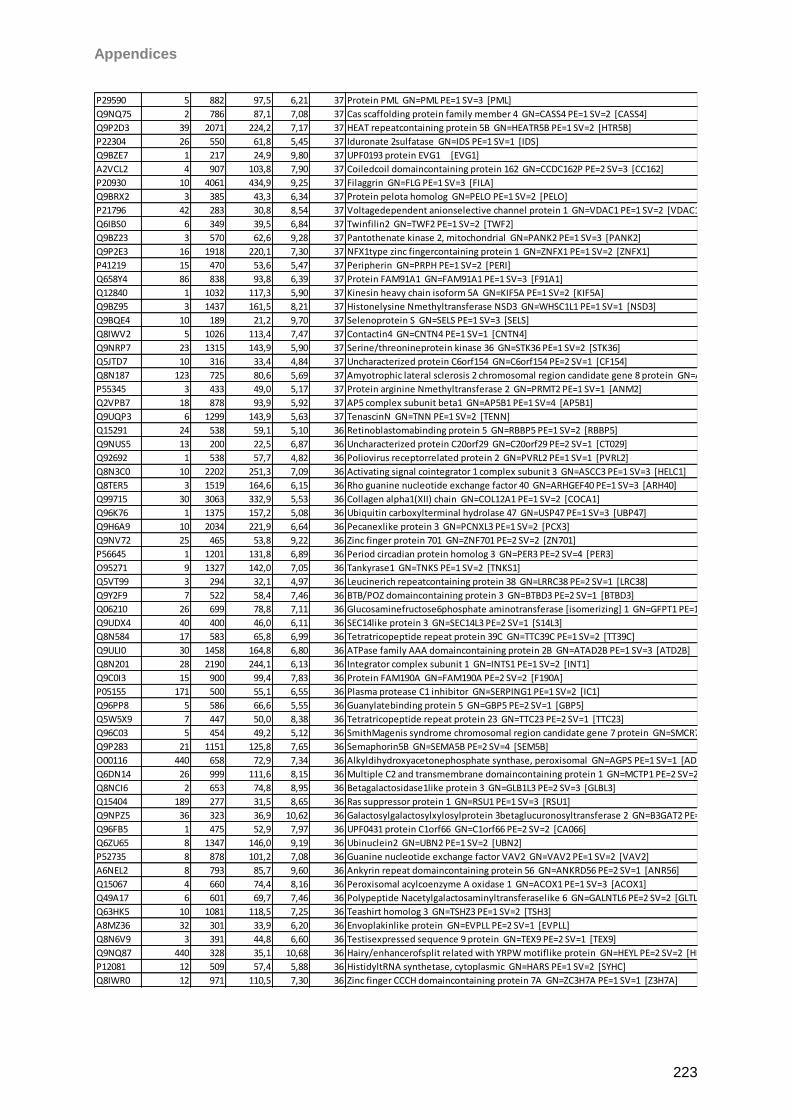
Appendices
223
P29590 5 882 97,5 6,21 37 Protein PML GN=PML PE=1 SV=3 [PML]
Q9NQ75 2 786 87,1 7,08 37 Cas scaffolding protein family member 4 GN=CASS4 PE=1 SV=2 [CASS4]
Q9P2D3 39 2071 224,2 7,17 37 HEAT repeatcontaining protein 5B GN=HEATR5B PE=1 SV=2 [HTR5B]
P22304 26 550 61,8 5,45 37 Iduronate 2sulfatase GN=IDS PE=1 SV=1 [IDS]
Q9BZE7 1 217 24,9 9,80 37 UPF0193 protein EVG1 [EVG1]
A2VCL2 4 907 103,8 7,90 37 Coiledcoil domaincontaining protein 162 GN=CCDC162P PE=2 SV=3 [CC162]
P20930 10 4061 434,9 9,25 37 Filaggrin GN=FLG PE=1 SV=3 [FILA]
Q9BRX2 3 385 43,3 6,34 37 Protein pelota homolog GN=PELO PE=1 SV=2 [PELO]
P21796 42 283 30,8 8,54 37 Voltagedependent anionselective channel protein 1 GN=VDAC1 PE=1 SV=2 [VDAC1]
Q6IBS0 6 349 39,5 6,84 37 Twinfilin2 GN=TWF2 PE=1 SV=2 [TWF2]
Q9BZ23 3 570 62,6 9,28 37 Pantothenate kinase 2, mitochondrial GN=PANK2 PE=1 SV=3 [PANK2]
Q9P2E3 16 1918 220,1 7,30 37 NFX1type zinc fingercontaining protein 1 GN=ZNFX1 PE=1 SV=2 [ZNFX1]
P41219 15 470 53,6 5,47 37 Peripherin GN=PRPH PE=1 SV=2 [PERI]
Q658Y4 86 838 93,8 6,39 37 Protein FAM91A1 GN=FAM91A1 PE=1 SV=3 [F91A1]
Q12840 1 1032 117,3 5,90 37 Kinesin heavy chain isoform 5A GN=KIF5A PE=1 SV=2 [KIF5A]
Q9BZ95 3 1437 161,5 8,21 37 Histonelysine Nmethyltransferase NSD3 GN=WHSC1L1 PE=1 SV=1 [NSD3]
Q9BQE4 10 189 21,2 9,70 37 Selenoprotein S GN=SELS PE=1 SV=3 [SELS]
Q8IWV2 5 1026 113,4 7,47 37 Contactin4 GN=CNTN4 PE=1 SV=1 [CNTN4]
Q9NRP7 23 1315 143,9 5,90 37 Serine/threonineprotein kinase 36 GN=STK36 PE=1 SV=2 [STK36]
Q5JTD7 10 316 33,4 4,84 37 Uncharacterized protein C6orf154 GN=C6orf154 PE=2 SV=1 [CF154]
Q8N187 123 725 80,6 5,69 37 Amyotrophic lateral sclerosis 2 chromosomal region candidate gene 8 protein GN=ALS2CR8 PE=2 SV=2 [AL2S8]
P55345 3 433 49,0 5,17 37 Protein arginine Nmethyltransferase 2 GN=PRMT2 PE=1 SV=1 [ANM2]
Q2VPB7 18 878 93,9 5,92 37 AP5 complex subunit beta1 GN=AP5B1 PE=1 SV=4 [AP5B1]
Q9UQP3 6 1299 143,9 5,63 37 TenascinN GN=TNN PE=1 SV=2 [TENN]
Q15291 24 538 59,1 5,10 36 Retinoblastomabinding protein 5 GN=RBBP5 PE=1 SV=2 [RBBP5]
Q9NUS5 13 200 22,5 6,87 36 Uncharacterized protein C20orf29 GN=C20orf29 PE=2 SV=1 [CT029]
Q92692 1 538 57,7 4,82 36 Poliovirus receptorrelated protein 2 GN=PVRL2 PE=1 SV=1 [PVRL2]
Q8N3C0 10 2202 251,3 7,09 36 Activating signal cointegrator 1 complex subunit 3 GN=ASCC3 PE=1 SV=3 [HELC1]
Q8TER5 3 1519 164,6 6,15 36 Rho guanine nucleotide exchange factor 40 GN=ARHGEF40 PE=1 SV=3 [ARH40]
Q99715 30 3063 332,9 5,53 36 Collagen alpha1(XII) chain GN=COL12A1 PE=1 SV=2 [COCA1]
Q96K76 1 1375 157,2 5,08 36 Ubiquitin carboxylterminal hydrolase 47 GN=USP47 PE=1 SV=3 [UBP47]
Q9H6A9 10 2034 221,9 6,64 36 Pecanexlike protein 3 GN=PCNXL3 PE=1 SV=2 [PCX3]
Q9NV72 25 465 53,8 9,22 36 Zinc finger protein 701 GN=ZNF701 PE=2 SV=2 [ZN701]
P56645 1 1201 131,8 6,89 36 Period circadian protein homolog 3 GN=PER3 PE=2 SV=4 [PER3]
O95271 9 1327 142,0 7,05 36 Tankyrase1 GN=TNKS PE=1 SV=2 [TNKS1]
Q5VT99 3 294 32,1 4,97 36 Leucinerich repeatcontaining protein 38 GN=LRRC38 PE=2 SV=1 [LRC38]
Q9Y2F9 7 522 58,4 7,46 36 BTB/POZ domaincontaining protein 3 GN=BTBD3 PE=2 SV=1 [BTBD3]
Q06210 26 699 78,8 7,11 36 Glucosaminefructose6phosphate aminotransferase [isomerizing] 1 GN=GFPT1 PE=1 SV=3 [GFPT1]
Q9UDX4 40 400 46,0 6,11 36 SEC14like protein 3 GN=SEC14L3 PE=2 SV=1 [S14L3]
Q8N584 17 583 65,8 6,99 36 Tetratricopeptide repeat protein 39C GN=TTC39C PE=1 SV=2 [TT39C]
Q9ULI0 30 1458 164,8 6,80 36 ATPase family AAA domaincontaining protein 2B GN=ATAD2B PE=1 SV=3 [ATD2B]
Q8N201 28 2190 244,1 6,13 36 Integrator complex subunit 1 GN=INTS1 PE=1 SV=2 [INT1]
Q9C0I3 15 900 99,4 7,83 36 Protein FAM190A GN=FAM190A PE=2 SV=2 [F190A]
P05155 171 500 55,1 6,55 36 Plasma protease C1 inhibitor GN=SERPING1 PE=1 SV=2 [IC1]
Q96PP8 5 586 66,6 5,55 36 Guanylatebinding protein 5 GN=GBP5 PE=2 SV=1 [GBP5]
Q5W5X9 7 447 50,0 8,38 36 Tetratricopeptide repeat protein 23 GN=TTC23 PE=2 SV=1 [TTC23]
Q96C03 5 454 49,2 5,12 36 SmithMagenis syndrome chromosomal region candidate gene 7 protein GN=SMCR7 PE=1 SV=1 [SMCR7]
Q9P283 21 1151 125,8 7,65 36 Semaphorin5B GN=SEMA5B PE=2 SV=4 [SEM5B]
O00116 440 658 72,9 7,34 36 Alkyldihydroxyacetonephosphate synthase, peroxisomal GN=AGPS PE=1 SV=1 [ADAS]
Q6DN14 26 999 111,6 8,15 36 Multiple C2 and transmembrane domaincontaining protein 1 GN=MCTP1 PE=2 SV=2 [MCTP1]
Q8NCI6 2 653 74,8 8,95 36 Betagalactosidase1like protein 3 GN=GLB1L3 PE=2 SV=3 [GLBL3]
Q15404 189 277 31,5 8,65 36 Ras suppressor protein 1 GN=RSU1 PE=1 SV=3 [RSU1]
Q9NPZ5 36 323 36,9 10,62 36 Galactosylgalactosylxylosylprotein 3betaglucuronosyltransferase 2 GN=B3GAT2 PE=1 SV=2 [B3GA2]
Q96FB5 1 475 52,9 7,97 36 UPF0431 protein C1orf66 GN=C1orf66 PE=2 SV=2 [CA066]
Q6ZU65 8 1347 146,0 9,19 36 Ubinuclein2 GN=UBN2 PE=1 SV=2 [UBN2]
P52735 8 878 101,2 7,08 36 Guanine nucleotide exchange factor VAV2 GN=VAV2 PE=1 SV=2 [VAV2]
A6NEL2 8 793 85,7 9,60 36 Ankyrin repeat domaincontaining protein 56 GN=ANKRD56 PE=2 SV=1 [ANR56]
Q15067 4 660 74,4 8,16 36 Peroxisomal acylcoenzyme A oxidase 1 GN=ACOX1 PE=1 SV=3 [ACOX1]
Q49A17 6 601 69,7 7,46 36 Polypeptide Nacetylgalactosaminyltransferaselike 6 GN=GALNTL6 PE=2 SV=2 [GLTL6]
Q63HK5 10 1081 118,5 7,25 36 Teashirt homolog 3 GN=TSHZ3 PE=1 SV=2 [TSH3]
A8MZ36 32 301 33,9 6,20 36 Envoplakinlike protein GN=EVPLL PE=2 SV=1 [EVPLL]
Q8N6V9 3 391 44,8 6,60 36 Testisexpressed sequence 9 protein GN=TEX9 PE=2 SV=1 [TEX9]
Q9NQ87 440 328 35,1 10,68 36 Hairy/enhancerofsplit related with YRPW motiflike protein GN=HEYL PE=2 SV=2 [HEYL]
P12081 12 509 57,4 5,88 36 HistidyltRNA synthetase, cytoplasmic GN=HARS PE=1 SV=2 [SYHC]
Q8IWR0 12 971 110,5 7,30 36 Zinc finger CCCH domaincontaining protein 7A GN=ZC3H7A PE=1 SV=1 [Z3H7A]
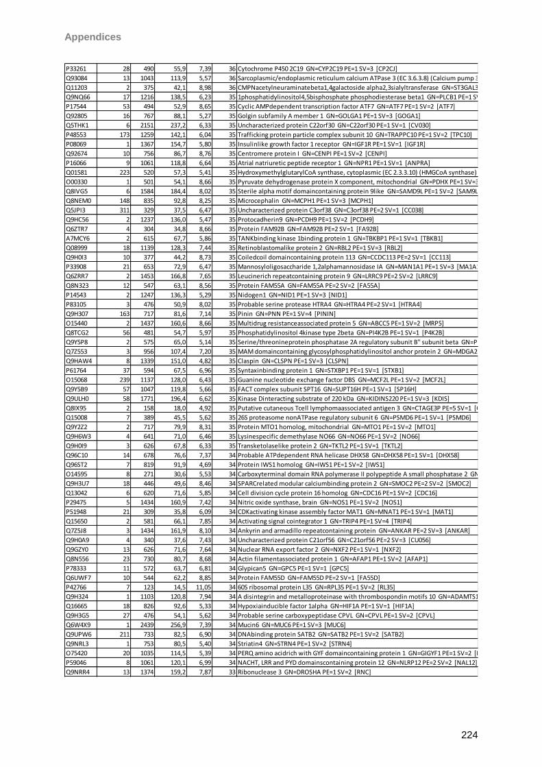
Appendices
224
P33261 28 490 55,9 7,39 36 Cytochrome P450 2C19 GN=CYP2C19 PE=1 SV=3 [CP2CJ]
Q93084 13 1043 113,9 5,57 36 Sarcoplasmic/endoplasmic reticulum calcium ATPase 3 (EC 3.6.3.8) (Calcium pump 3) (SERCA3) (SR Ca(2+)ATPase 3) [AT2A3]
Q11203 2 375 42,1 8,98 36 CMPNacetylneuraminatebeta1,4galactoside alpha2,3sialyltransferase GN=ST3GAL3 PE=2 SV=1 [SIAT6]
Q9NQ66 17 1216 138,5 6,23 35 1phosphatidylinositol4,5bisphosphate phosphodiesterase beta1 GN=PLCB1 PE=1 SV=1 [PLCB1]
P17544 53 494 52,9 8,65 35 Cyclic AMPdependent transcription factor ATF7 GN=ATF7 PE=1 SV=2 [ATF7]
Q92805 16 767 88,1 5,27 35 Golgin subfamily A member 1 GN=GOLGA1 PE=1 SV=3 [GOGA1]
Q5THK1 6 2151 237,2 6,33 35 Uncharacterized protein C22orf30 GN=C22orf30 PE=1 SV=1 [CV030]
P48553 173 1259 142,1 6,04 35 Trafficking protein particle complex subunit 10 GN=TRAPPC10 PE=1 SV=2 [TPC10]
P08069 1 1367 154,7 5,80 35 Insulinlike growth factor 1 receptor GN=IGF1R PE=1 SV=1 [IGF1R]
Q92674 10 756 86,7 8,76 35 Centromere protein I GN=CENPI PE=1 SV=2 [CENPI]
P16066 9 1061 118,8 6,64 35 Atrial natriuretic peptide receptor 1 GN=NPR1 PE=1 SV=1 [ANPRA]
Q01581 223 520 57,3 5,41 35 HydroxymethylglutarylCoA synthase, cytoplasmic (EC 2.3.3.10) (HMGCoA synthase) (3hydroxy3methylglutaryl coenzyme A synthase) [HMCS1]
O00330 1 501 54,1 8,66 35 Pyruvate dehydrogenase protein X component, mitochondrial GN=PDHX PE=1 SV=3 [ODPX]
Q8IVG5 6 1584 184,4 8,02 35 Sterile alpha motif domaincontaining protein 9like GN=SAMD9L PE=1 SV=2 [SAM9L]
Q8NEM0 148 835 92,8 8,25 35 Microcephalin GN=MCPH1 PE=1 SV=3 [MCPH1]
Q5JPI3 311 329 37,5 6,47 35 Uncharacterized protein C3orf38 GN=C3orf38 PE=2 SV=1 [CC038]
Q9HC56 2 1237 136,0 5,47 35 Protocadherin9 GN=PCDH9 PE=1 SV=2 [PCDH9]
Q6ZTR7 4 304 34,8 8,66 35 Protein FAM92B GN=FAM92B PE=2 SV=1 [FA92B]
A7MCY6 2 615 67,7 5,86 35 TANKbinding kinase 1binding protein 1 GN=TBKBP1 PE=1 SV=1 [TBKB1]
Q08999 18 1139 128,3 7,44 35 Retinoblastomalike protein 2 GN=RBL2 PE=1 SV=3 [RBL2]
Q9H0I3 10 377 44,2 8,73 35 Coiledcoil domaincontaining protein 113 GN=CCDC113 PE=2 SV=1 [CC113]
P33908 21 653 72,9 6,47 35 Mannosyloligosaccharide 1,2alphamannosidase IA GN=MAN1A1 PE=1 SV=3 [MA1A1]
Q6ZRR7 2 1453 166,8 7,65 35 Leucinerich repeatcontaining protein 9 GN=LRRC9 PE=2 SV=2 [LRRC9]
Q8N323 12 547 63,1 8,56 35 Protein FAM55A GN=FAM55A PE=2 SV=2 [FA55A]
P14543 2 1247 136,3 5,29 35 Nidogen1 GN=NID1 PE=1 SV=3 [NID1]
P83105 3 476 50,9 8,02 35 Probable serine protease HTRA4 GN=HTRA4 PE=2 SV=1 [HTRA4]
Q9H307 163 717 81,6 7,14 35 Pinin GN=PNN PE=1 SV=4 [PININ]
O15440 2 1437 160,6 8,66 35 Multidrug resistanceassociated protein 5 GN=ABCC5 PE=1 SV=2 [MRP5]
Q8TCG2 56 481 54,7 5,97 35 Phosphatidylinositol 4kinase type 2beta GN=PI4K2B PE=1 SV=1 [P4K2B]
Q9Y5P8 2 575 65,0 5,14 35 Serine/threonineprotein phosphatase 2A regulatory subunit B'' subunit beta GN=PPP2R3B PE=1 SV=2 [P2R3B]
Q7Z553 3 956 107,4 7,20 35 MAM domaincontaining glycosylphosphatidylinositol anchor protein 2 GN=MDGA2 PE=2 SV=2 [MDGA2]
Q9HAW4 8 1339 151,0 4,82 35 Claspin GN=CLSPN PE=1 SV=3 [CLSPN]
P61764 37 594 67,5 6,96 35 Syntaxinbinding protein 1 GN=STXBP1 PE=1 SV=1 [STXB1]
O15068 239 1137 128,0 6,43 35 Guanine nucleotide exchange factor DBS GN=MCF2L PE=1 SV=2 [MCF2L]
Q9Y5B9 57 1047 119,8 5,66 35 FACT complex subunit SPT16 GN=SUPT16H PE=1 SV=1 [SP16H]
Q9ULH0 58 1771 196,4 6,62 35 Kinase Dinteracting substrate of 220 kDa GN=KIDINS220 PE=1 SV=3 [KDIS]
Q8IX95 2 158 18,0 4,92 35 Putative cutaneous Tcell lymphomaassociated antigen 3 GN=CTAGE3P PE=5 SV=1 [CTGE3]
Q15008 7 389 45,5 5,62 35 26S proteasome nonATPase regulatory subunit 6 GN=PSMD6 PE=1 SV=1 [PSMD6]
Q9Y2Z2 2 717 79,9 8,31 35 Protein MTO1 homolog, mitochondrial GN=MTO1 PE=1 SV=2 [MTO1]
Q9H6W3 4 641 71,0 6,46 35 Lysinespecific demethylase NO66 GN=NO66 PE=1 SV=2 [NO66]
Q9H0I9 3 626 67,8 6,33 35 Transketolaselike protein 2 GN=TKTL2 PE=1 SV=1 [TKTL2]
Q96C10 14 678 76,6 7,37 34 Probable ATPdependent RNA helicase DHX58 GN=DHX58 PE=1 SV=1 [DHX58]
Q96ST2 7 819 91,9 4,69 34 Protein IWS1 homolog GN=IWS1 PE=1 SV=2 [IWS1]
O14595 8 271 30,6 5,53 34 Carboxyterminal domain RNA polymerase II polypeptide A small phosphatase 2 GN=CTDSP2 PE=1 SV=2 [CTDS2]
Q9H3U7 18 446 49,6 8,46 34 SPARCrelated modular calciumbinding protein 2 GN=SMOC2 PE=2 SV=2 [SMOC2]
Q13042 6 620 71,6 5,85 34 Cell division cycle protein 16 homolog GN=CDC16 PE=1 SV=2 [CDC16]
P29475 5 1434 160,9 7,42 34 Nitric oxide synthase, brain GN=NOS1 PE=1 SV=2 [NOS1]
P51948 21 309 35,8 6,09 34 CDKactivating kinase assembly factor MAT1 GN=MNAT1 PE=1 SV=1 [MAT1]
Q15650 2 581 66,1 7,85 34 Activating signal cointegrator 1 GN=TRIP4 PE=1 SV=4 [TRIP4]
Q7Z5J8 3 1434 161,9 8,10 34 Ankyrin and armadillo repeatcontaining protein GN=ANKAR PE=2 SV=3 [ANKAR]
Q9H0A9 4 340 37,6 7,43 34 Uncharacterized protein C21orf56 GN=C21orf56 PE=2 SV=3 [CU056]
Q9GZY0 13 626 71,6 7,64 34 Nuclear RNA export factor 2 GN=NXF2 PE=1 SV=1 [NXF2]
Q8N556 23 730 80,7 8,68 34 Actin filamentassociated protein 1 GN=AFAP1 PE=1 SV=2 [AFAP1]
P78333 11 572 63,7 6,81 34 Glypican5 GN=GPC5 PE=1 SV=1 [GPC5]
Q6UWF7 10 544 62,2 8,85 34 Protein FAM55D GN=FAM55D PE=2 SV=1 [FA55D]
P42766 7 123 14,5 11,05 34 60S ribosomal protein L35 GN=RPL35 PE=1 SV=2 [RL35]
Q9H324 1 1103 120,8 7,94 34 A disintegrin and metalloproteinase with thrombospondin motifs 10 GN=ADAMTS10 PE=1 SV=2 [ATS10]
Q16665 18 826 92,6 5,33 34 Hypoxiainducible factor 1alpha GN=HIF1A PE=1 SV=1 [HIF1A]
Q9H3G5 27 476 54,1 5,62 34 Probable serine carboxypeptidase CPVL GN=CPVL PE=1 SV=2 [CPVL]
Q6W4X9 1 2439 256,9 7,39 34 Mucin6 GN=MUC6 PE=1 SV=3 [MUC6]
Q9UPW6 211 733 82,5 6,90 34 DNAbinding protein SATB2 GN=SATB2 PE=1 SV=2 [SATB2]
Q9NRL3 1 753 80,5 5,40 34 Striatin4 GN=STRN4 PE=1 SV=2 [STRN4]
O75420 20 1035 114,5 5,39 34 PERQ amino acidrich with GYF domaincontaining protein 1 GN=GIGYF1 PE=1 SV=2 [PERQ1]
P59046 8 1061 120,1 6,99 34 NACHT, LRR and PYD domainscontaining protein 12 GN=NLRP12 PE=2 SV=2 [NAL12]
Q9NRR4 13 1374 159,2 7,87 33 Ribonuclease 3 GN=DROSHA PE=1 SV=2 [RNC]
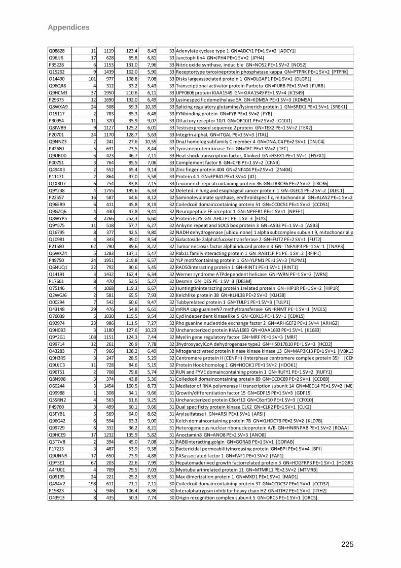
Appendices
225
Q08828 11 1119 123,4 8,43 33 Adenylate cyclase type 1 GN=ADCY1 PE=1 SV=2 [ADCY1]
Q96JJ6 17 628 65,8 6,81 33 Junctophilin4 GN=JPH4 PE=1 SV=2 [JPH4]
P35228 6 1153 131,0 7,96 33 Nitric oxide synthase, inducible GN=NOS2 PE=1 SV=2 [NOS2]
Q15262 9 1439 162,0 5,90 33 Receptortype tyrosineprotein phosphatase kappa GN=PTPRK PE=1 SV=2 [PTPRK]
O14490 101 977 108,8 7,08 33 Disks largeassociated protein 1 GN=DLGAP1 PE=1 SV=1 [DLGP1]
Q96QR8 4 312 33,2 5,43 33 Transcriptional activator protein Purbeta GN=PURB PE=1 SV=3 [PURB]
Q9HCM3 37 1950 210,6 6,11 33 UPF0606 protein KIAA1549 GN=KIAA1549 PE=1 SV=4 [K1549]
P29375 12 1690 192,0 6,49 33 Lysinespecific demethylase 5A GN=KDM5A PE=1 SV=3 [KDM5A]
Q8WXA9 24 508 59,3 10,39 33 Splicing regulatory glutamine/lysinerich protein 1 GN=SREK1 PE=1 SV=1 [SREK1]
O15117 2 783 85,3 6,48 33 FYNbinding protein GN=FYB PE=1 SV=2 [FYB]
P30954 11 320 35,9 9,07 33 Olfactory receptor 10J1 GN=OR10J1 PE=2 SV=2 [O10J1]
Q8IWB9 9 1127 125,2 6,01 33 Testisexpressed sequence 2 protein GN=TEX2 PE=1 SV=2 [TEX2]
P20701 24 1170 128,7 5,63 33 Integrin alphaL GN=ITGAL PE=1 SV=3 [ITAL]
Q9NNZ3 2 241 27,6 10,55 33 DnaJ homolog subfamily C member 4 GN=DNAJC4 PE=2 SV=1 [DNJC4]
P42680 5 631 73,5 8,44 33 Tyrosineprotein kinase Tec GN=TEC PE=1 SV=2 [TEC]
Q9UBD0 6 423 46,7 7,11 33 Heat shock transcription factor, Xlinked GN=HSFX1 PE=1 SV=1 [HSFX1]
P00751 3 764 85,5 7,06 33 Complement factor B GN=CFB PE=1 SV=2 [CFAB]
Q494X3 2 552 65,4 9,14 33 Zinc finger protein 404 GN=ZNF404 PE=2 SV=1 [ZN404]
P11171 2 864 97,0 5,58 33 Protein 4.1 GN=EPB41 PE=1 SV=4 [41]
Q1X8D7 6 754 83,8 7,15 33 Leucinerich repeatcontaining protein 36 GN=LRRC36 PE=2 SV=2 [LRC36]
Q9Y238 4 1755 195,6 6,33 32 Deleted in lung and esophageal cancer protein 1 GN=DLEC1 PE=2 SV=2 [DLEC1]
P22557 16 587 64,6 8,12 32 5aminolevulinate synthase, erythroidspecific, mitochondrial GN=ALAS2 PE=1 SV=2 [HEM0]
Q96ER9 6 411 45,8 8,19 32 Coiledcoil domaincontaining protein 51 GN=CCDC51 PE=1 SV=2 [CCD51]
Q9GZQ6 4 430 47,8 9,41 32 Neuropeptide FF receptor 1 GN=NPFFR1 PE=1 SV=1 [NPFF1]
Q8WYP5 3 2266 252,3 6,60 32 Protein ELYS GN=AHCTF1 PE=1 SV=3 [ELYS]
Q9Y575 11 518 57,7 6,27 32 Ankyrin repeat and SOCS box protein 3 GN=ASB3 PE=1 SV=1 [ASB3]
Q16795 8 377 42,5 9,80 32 NADH dehydrogenase [ubiquinone] 1 alpha subcomplex subunit 9, mitochondrial precursor (EC 1.6.5.3) (EC 1.6.99.3) (NADHubiquinone oxidoreductase 39 kDa subunit) (Complex I39kD) (CI39kD) [NDUA9]
Q10981 4 343 39,0 8,54 32 Galactoside 2alphaLfucosyltransferase 2 GN=FUT2 PE=2 SV=1 [FUT2]
P21580 62 790 89,6 8,22 32 Tumor necrosis factor alphainduced protein 3 GN=TNFAIP3 PE=1 SV=1 [TNAP3]
Q6WKZ4 5 1283 137,1 5,47 32 Rab11 familyinteracting protein 1 GN=RAB11FIP1 PE=1 SV=2 [RFIP1]
P49750 24 1951 219,8 6,57 32 YLP motifcontaining protein 1 GN=YLPM1 PE=1 SV=3 [YLPM1]
Q6NUQ1 22 792 90,6 5,45 32 RAD50interacting protein 1 GN=RINT1 PE=1 SV=1 [RINT1]
Q14191 3 1432 162,4 6,34 32 Werner syndrome ATPdependent helicase GN=WRN PE=1 SV=2 [WRN]
P17661 8 470 53,5 5,27 32 Desmin GN=DES PE=1 SV=3 [DESM]
O75146 4 1068 119,3 6,67 32 Huntingtininteracting protein 1related protein GN=HIP1R PE=1 SV=2 [HIP1R]
Q2WGJ6 2 581 65,5 7,93 32 Kelchlike protein 38 GN=KLHL38 PE=2 SV=3 [KLH38]
O00294 7 542 60,6 9,47 32 Tubbyrelated protein 1 GN=TULP1 PE=1 SV=3 [TULP1]
O43148 29 476 54,8 6,61 32 mRNA cap guanineN7 methyltransferase GN=RNMT PE=1 SV=1 [MCES]
O76039 5 1030 115,5 9,54 32 Cyclindependent kinaselike 5 GN=CDKL5 PE=1 SV=1 [CDKL5]
Q92974 23 986 111,5 7,27 32 Rho guanine nucleotide exchange factor 2 GN=ARHGEF2 PE=1 SV=4 [ARHG2]
Q9H0B3 3 1180 127,6 10,23 32 Uncharacterized protein KIAA1683 GN=KIAA1683 PE=1 SV=1 [K1683]
Q9Y2G1 108 1151 124,3 7,44 32 Myelin gene regulatory factor GN=MRF PE=1 SV=3 [MRF]
Q99714 12 261 26,9 7,78 32 3hydroxyacylCoA dehydrogenase type2 GN=HSD17B10 PE=1 SV=3 [HCD2]
O43283 7 966 108,2 6,49 32 Mitogenactivated protein kinase kinase kinase 13 GN=MAP3K13 PE=1 SV=1 [M3K13]
Q9H3R5 3 247 28,5 5,29 32 Centromere protein H (CENPH) (Interphase centromere complex protein 35) [CENPH]
Q9UJC3 11 728 84,6 5,15 32 Protein Hook homolog 1 GN=HOOK1 PE=1 SV=2 [HOOK1]
Q96T51 2 708 79,8 5,74 32 RUN and FYVE domaincontaining protein 1 GN=RUFY1 PE=1 SV=2 [RUFY1]
Q8N998 3 374 43,8 5,36 31 Coiledcoil domaincontaining protein 89 GN=CCDC89 PE=2 SV=1 [CCD89]
O60244 3 1454 160,5 8,73 31 Mediator of RNA polymerase II transcription subunit 14 GN=MED14 PE=1 SV=2 [MED14]
Q99988 1 308 34,1 9,66 31 Growth/differentiation factor 15 GN=GDF15 PE=1 SV=3 [GDF15]
Q5SRN2 4 563 61,6 9,25 31 Uncharacterized protein C6orf10 GN=C6orf10 PE=1 SV=3 [CF010]
P49760 3 499 60,1 9,66 31 Dual specificity protein kinase CLK2 GN=CLK2 PE=1 SV=1 [CLK2]
Q5FYB1 5 569 64,0 8,62 31 Arylsulfatase I GN=ARSI PE=1 SV=1 [ARSI]
Q96G42 6 594 63,3 9,00 31 Kelch domaincontaining protein 7B GN=KLHDC7B PE=2 SV=2 [KLD7B]
Q99729 6 332 36,2 8,21 31 Heterogeneous nuclear ribonucleoprotein A/B GN=HNRNPAB PE=1 SV=2 [ROAA]
Q9HCE9 17 1232 135,9 5,82 31 Anoctamin8 GN=ANO8 PE=2 SV=3 [ANO8]
Q5T7V8 2 394 45,0 7,08 31 RAB6interacting golgin GN=GORAB PE=1 SV=1 [GORAB]
P17213 3 487 53,9 9,38 31 Bactericidal permeabilityincreasing protein GN=BPI PE=1 SV=4 [BPI]
Q9UNN5 17 650 73,9 4,88 31 FASassociated factor 1 GN=FAF1 PE=1 SV=2 [FAF1]
Q9Y3E1 67 203 22,6 7,99 31 Hepatomaderived growth factorrelated protein 3 GN=HDGFRP3 PE=1 SV=1 [HDGR3]
A4FU01 4 709 79,5 7,03 31 Myotubularinrelated protein 11 GN=MTMR11 PE=2 SV=2 [MTMRB]
Q05195 24 221 25,2 8,53 31 Max dimerization protein 1 GN=MXD1 PE=1 SV=1 [MAD1]
Q494V2 198 611 71,1 7,11 30 Coiledcoil domaincontaining protein 37 GN=CCDC37 PE=1 SV=1 [CCD37]
P19823 5 946 106,4 6,86 30 Interalphatrypsin inhibitor heavy chain H2 GN=ITIH2 PE=1 SV=2 [ITIH2]
O43913 8 435 50,3 7,74 30 Origin recognition complex subunit 5 GN=ORC5 PE=1 SV=1 [ORC5]
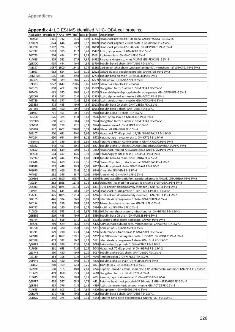
Appendices
226
Appendix 4: LC ESI MS identified NHC-IOBA cell proteins.
Accession #Peptides # AAs MW [kDa] calc. pI Score Description
P07900 1111 732 84,6 5,02 17236 Heat shock protein HSP 90-alpha GN=HSP90AA1 PE=1 SV=5
A2Q0Z1 874 646 70,9 5,52 16909 Heat shock cognate 71 kDa protein GN=HSPA8 PE=2 SV=1
P08238 1102 724 83,2 5,03 16858 Heat shock protein HSP 90-beta GN=HSP90AB1 PE=1 SV=4
P60712 1063 375 41,7 5,48 15447 Actin, cytoplasmic 1 GN=ACTB PE=1 SV=1
P06733 904 434 47,1 7,39 15161 Alpha-enolase GN=ENO1 PE=1 SV=2
P14618 809 531 57,9 7,84 14932 Pyruvate kinase isozymes M1/M2 GN=PKM2 PE=1 SV=4
Q2KJD0 635 444 49,6 4,89 13736 Tubulin beta-5 chain GN=TUBB5 PE=2 SV=1
P31327 1057 1500 164,8 6,74 13385 Carbamoyl-phosphate synthase [ammonia], mitochondrial GN=CPS1 PE=1 SV=2
P11021 662 654 72,3 5,16 12923 78 kDa glucose-regulated protein GN=HSPA5 PE=1 SV=2
Q3MHM5 606 445 49,8 4,89 12793 Tubulin beta-4B chain GN=TUBB4B PE=2 SV=1
P07355 760 339 38,6 7,75 12539 Annexin A2 GN=ANXA2 PE=1 SV=2
P21333 1072 2647 280,6 6,06 12012 Filamin-A GN=FLNA PE=1 SV=4
P68103 898 462 50,1 9,01 11679 Elongation factor 1-alpha 1 GN=EEF1A1 PE=2 SV=1
P04406 695 335 36,0 8,46 11605 Glyceraldehyde-3-phosphate dehydrogenase GN=GAPDH PE=1 SV=3
Q3ZC07 823 377 42,0 5,39 11333 Actin, alpha cardiac muscle 1 GN=ACTC1 PE=2 SV=1
P62739 758 377 42,0 5,39 10443 Actin, aortic smooth muscle GN=ACTA2 PE=1 SV=1
Q13885 478 445 49,9 4,89 10178 Tubulin beta-2A chain GN=TUBB2A PE=1 SV=1
Q2T9S0 450 450 50,4 4,93 10153 Tubulin beta-3 chain GN=TUBB3 PE=2 SV=1
P81947 701 451 50,1 5,06 9968 Tubulin alpha-1B chain PE=1 SV=2
P63258 626 375 41,8 5,48 9606 Actin, cytoplasmic 2 GN=ACTG1 PE=1 SV=1
Q32PH8 650 463 50,4 9,03 9574 Elongation factor 1-alpha 2 GN=EEF1A2 PE=2 SV=1
Q06830 780 199 22,1 8,13 9544 Peroxiredoxin-1 GN=PRDX1 PE=1 SV=1
O75369 847 2602 278,0 5,73 9474 Filamin-B GN=FLNB PE=1 SV=2
P08107 500 641 70,0 5,66 9053 Heat shock 70 kDa protein 1A/1B GN=HSPA1A PE=1 SV=5
P04264 650 644 66,0 8,12 8972 Keratin, type II cytoskeletal 1 GN=KRT1 PE=1 SV=6
A6NMY6 567 339 38,6 6,95 8312 Putative annexin A2-like protein GN=ANXA2P2 PE=5 SV=2
P68362 648 451 50,1 5,06 8271 Tubulin alpha-1A chain OS=Cricetulus griseus GN=TUBA1A PE=2 SV=1
P54652 448 639 70,0 5,74 7852 Heat shock-related 70 kDa protein 2 GN=HSPA2 PE=1 SV=1
P00558 564 417 44,6 8,10 7648 Phosphoglycerate kinase 1 GN=PGK1 PE=1 SV=3
Q3ZBU7 429 444 49,6 4,88 7498 Tubulin beta-4A chain GN=TUBB4A PE=2 SV=1
P38646 386 679 73,6 6,16 7254 Stress-70 protein, mitochondrial GN=HSPA9 PE=1 SV=2
P81948 513 448 49,9 5,06 6852 Tubulin alpha-4A chain GN=TUBA4A PE=1 SV=2
P08670 412 466 53,6 5,12 6846 Vimentin GN=VIM PE=1 SV=4
P04083 282 346 38,7 7,02 6596 Annexin A1 GN=ANXA1 PE=1 SV=2
Q09666 1140 5890 628,7 6,15 6396 Neuroblast differentiation-associated protein AHNAK GN=AHNAK PE=1 SV=2
P22314 383 1058 117,8 5,76 6353 Ubiquitin-like modifier-activating enzyme 1 GN=UBA1 PE=1 SV=3
Q6S8J3 456 1075 121,3 6,20 6331 POTE ankyrin domain family member E GN=POTEE PE=1 SV=3
P34931 306 641 70,3 6,02 6284 Heat shock 70 kDa protein 1-like GN=HSPA1L PE=1 SV=2
A5A3E0 425 1075 121,4 6,20 6266 POTE ankyrin domain family member F GN=POTEF PE=1 SV=2
P07195 446 334 36,6 6,05 6193 L-lactate dehydrogenase B chain GN=LDHB PE=1 SV=2
P60174 293 286 30,8 5,92 5602 Triosephosphate isomerase GN=TPI1 PE=1 SV=3
P07737 264 140 15,0 8,27 5594 Profilin-1 GN=PFN1 PE=1 SV=2
P10809 405 573 61,0 5,87 5582 60 kDa heat shock protein, mitochondrial GN=HSPD1 PE=1 SV=2
Q6B856 219 445 49,9 4,89 5580 Tubulin beta-2B chain GN=TUBB2B PE=1 SV=2
P06744 331 558 63,1 8,32 5578 Glucose-6-phosphate isomerase GN=GPI PE=1 SV=4
P06576 269 529 56,5 5,40 5405 ATP synthase subunit beta, mitochondrial GN=ATP5B PE=1 SV=3
P08758 338 320 35,9 5,05 5291 Annexin A5 GN=ANXA5 PE=1 SV=2
P09211 179 210 23,3 5,64 5286 Glutathione S-transferase P GN=GSTP1 PE=1 SV=2
P46940 411 1657 189,1 6,48 5207 Ras GTPase-activating-like protein IQGAP1 GN=IQGAP1 PE=1 SV=1
P00338 439 332 36,7 8,27 5121 L-lactate dehydrogenase A chain GN=LDHA PE=1 SV=2
Q562R1 468 376 42,0 5,59 5088 Beta-actin-like protein 2 GN=ACTBL2 PE=1 SV=2
P17066 261 643 71,0 6,14 5043 Heat shock 70 kDa protein 6 GN=HSPA6 PE=1 SV=2
Q13748 460 450 49,9 5,10 5037 Tubulin alpha-3C/D chain GN=TUBA3C PE=1 SV=3
P32119 384 198 21,9 5,97 4946 Peroxiredoxin-2 GN=PRDX2 PE=1 SV=5
Q6PEY2 405 450 49,8 5,14 4876 Tubulin alpha-3E chain GN=TUBA3E PE=1 SV=2
P37802 260 199 22,4 8,25 4871 Transgelin-2 GN=TAGLN2 PE=1 SV=3
P62938 500 165 18,0 7,81 4726 Peptidyl-prolyl cis-trans isomerase A OS=Chlorocebus aethiops GN=PPIA PE=2 SV=2
P13639 409 858 95,3 6,83 4618 Elongation factor 2 GN=EEF2 PE=1 SV=4
P13645 319 584 58,8 5,21 4601 Keratin, type I cytoskeletal 10 GN=KRT10 PE=1 SV=6
Q58FF7 428 597 68,3 4,79 4411 Putative heat shock protein HSP 90-beta-3 GN=HSP90AB3P PE=5 SV=1
Q5E9B5 335 376 41,8 5,48 4399 Actin, gamma-enteric smooth muscle GN=ACTG2 PE=2 SV=1
P14625 353 803 92,4 4,84 4209 Endoplasmin GN=HSP90B1 PE=1 SV=1
Q9BUF5 271 446 49,8 4,88 4182 Tubulin beta-6 chain GN=TUBB6 PE=1 SV=1
Q9BYX7 256 375 42,0 6,33 4143 Putative beta-actin-like protein 3 GN=POTEKP PE=5 SV=1
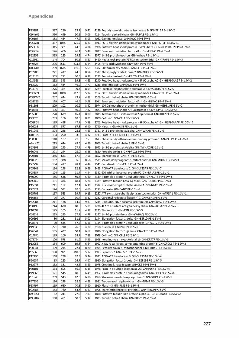
Appendices
227
P23284 397 216 23,7 9,41 4139 Peptidyl-prolyl cis-trans isomerase B GN=PPIB PE=1 SV=2
Q9NY65 310 449 50,1 5,06 4114 Tubulin alpha-8 chain GN=TUBA8 PE=1 SV=1
P09104 163 434 47,2 5,03 4082 Gamma-enolase GN=ENO2 PE=1 SV=3
P0CG38 387 1075 121,2 6,21 3941 POTE ankyrin domain family member I GN=POTEI PE=3 SV=1
Q58FF8 315 381 44,3 4,84 3906 Putative heat shock protein HSP 90-beta 2 GN=HSP90AB2P PE=1 SV=2
Q3SZ54 176 406 46,1 5,48 3837 Eukaryotic initiation factor 4A-I GN=EIF4A1 PE=2 SV=1
P62259 315 255 29,2 4,74 3577 14-3-3 protein epsilon GN=Ywhae PE=1 SV=1
Q12931 144 704 80,1 8,21 3460 Heat shock protein 75 kDa, mitochondrial GN=TRAP1 PE=1 SV=3
P49327 296 2511 273,3 6,44 3405 Fatty acid synthase GN=FASN PE=1 SV=3
Q00610 299 1675 191,5 5,69 3382 Clathrin heavy chain 1 GN=CLTC PE=1 SV=5
P07205 221 417 44,8 8,54 3317 Phosphoglycerate kinase 2 GN=PGK2 PE=1 SV=3
Q13162 305 271 30,5 6,29 3295 Peroxiredoxin-4 GN=PRDX4 PE=1 SV=1
Q14568 252 343 39,3 4,65 3240 Putative heat shock protein HSP 90-alpha A2 GN=HSP90AA2 PE=1 SV=2
P13929 112 434 46,9 7,71 3236 Beta-enolase GN=ENO3 PE=1 SV=4
P04075 276 364 39,4 8,09 3229 Fructose-bisphosphate aldolase A GN=ALDOA PE=1 SV=2
P0CG39 328 1038 117,3 5,97 3152 POTE ankyrin domain family member J GN=POTEJ PE=3 SV=1
Q3ZCM7 237 444 49,7 4,89 3109 Tubulin beta-8 chain GN=TUBB8 PE=1 SV=2
Q3SZ65 129 407 46,4 5,48 3011 Eukaryotic initiation factor 4A-II GN=EIF4A2 PE=2 SV=1
P61603 209 102 10,9 8,92 2974 10 kDa heat shock protein, mitochondrial GN=HSPE1 PE=3 SV=2
P48741 136 367 40,2 7,87 2873 Putative heat shock 70 kDa protein 7 GN=HSPA7 PE=5 SV=2
P35908 228 639 65,4 8,00 2835 Keratin, type II cytoskeletal 2 epidermal GN=KRT2 PE=1 SV=2
P23528 233 166 18,5 8,09 2800 Cofilin-1 GN=CFL1 PE=1 SV=3
Q58FG1 129 418 47,7 5,19 2763 Putative heat shock protein HSP 90-alpha A4 GN=HSP90AA4P PE=5 SV=1
P26038 237 577 67,8 6,40 2740 Moesin GN=MSN PE=1 SV=3
P31946 304 246 28,1 4,83 2721 14-3-3 protein beta/alpha GN=YWHAB PE=1 SV=3
Q01105 194 290 33,5 4,32 2713 Protein SET GN=SET PE=1 SV=3
P30086 110 187 21,0 7,53 2673 Phosphatidylethanolamine-binding protein 1 GN=PEBP1 PE=1 SV=3
A6NNZ2 215 444 49,5 4,86 2663 Tubulin beta-8 chain B PE=1 SV=1
P63103 239 245 27,7 4,79 2645 14-3-3 protein zeta/delta GN=YWHAZ PE=1 SV=1
P30041 271 224 25,0 6,38 2630 Peroxiredoxin-6 GN=PRDX6 PE=1 SV=3
P29401 346 623 67,8 7,66 2602 Transketolase GN=TKT PE=1 SV=3
P40926 102 338 35,5 8,68 2571 Malate dehydrogenase, mitochondrial GN=MDH2 PE=1 SV=3
P27797 184 417 48,1 4,44 2541 Calreticulin GN=CALR PE=1 SV=1
P05141 194 298 32,8 9,69 2463 ADP/ATP translocase 2 GN=SLC25A5 PE=1 SV=7
P05387 104 115 11,7 4,54 2352 60S acidic ribosomal protein P2 GN=RPLP2 PE=1 SV=1
P50990 155 548 59,6 5,60 2339 T-complex protein 1 subunit theta GN=CCT8 PE=1 SV=4
Q99867 181 434 48,4 5,26 2329 Putative tubulin beta-4q chain GN=TUBB4Q PE=5 SV=1
P15531 241 152 17,1 6,19 2312 Nucleoside diphosphate kinase A GN=NME1 PE=1 SV=1
P27824 124 592 67,5 4,60 2272 Calnexin GN=CANX PE=1 SV=2
P25705 167 553 59,7 9,13 2257 ATP synthase subunit alpha, mitochondrial GN=ATP5A1 PE=1 SV=1
P16152 76 277 30,4 8,32 2237 Carbonyl reductase [NADPH] 1 GN=CBR1 PE=1 SV=3
P62984 211 128 14,7 9,83 2235 Ubiquitin-60S ribosomal protein L40 GN=Uba52 PE=1 SV=2
P08195 244 630 68,0 5,01 2226 4F2 cell-surface antigen heavy chain GN=SLC3A2 PE=1 SV=3
P10599 125 105 11,7 4,92 2225 Thioredoxin GN=TXN PE=1 SV=3
Q3SZI4 225 245 27,7 4,78 2147 14-3-3 protein theta GN=YWHAQ PE=2 SV=1
P29692 80 281 31,1 5,01 2144 Elongation factor 1-delta GN=EEF1D PE=1 SV=5
P78371 90 535 57,5 6,46 2144 T-complex protein 1 subunit beta GN=CCT2 PE=1 SV=4
P19338 221 710 76,6 4,70 2108 Nucleolin GN=NCL PE=1 SV=3
P26641 195 437 50,1 6,67 2076 Elongation factor 1-gamma GN=EEF1G PE=1 SV=3
Q148F1 129 166 18,7 7,88 2048 Cofilin-2 GN=CFL2 PE=2 SV=1
Q7Z794 100 578 61,9 5,99 1998 Keratin, type II cytoskeletal 1b GN=KRT77 PE=1 SV=3
P12956 154 609 69,8 6,64 1997 X-ray repair cross-complementing protein 6 GN=XRCC6 PE=1 SV=2
P30044 139 214 22,1 8,70 1995 Peroxiredoxin-5, mitochondrial GN=PRDX5 PE=1 SV=4
P55060 198 971 110,3 5,77 1993 Exportin-2 GN=CSE1L PE=1 SV=3
P12236 158 298 32,8 9,74 1991 ADP/ATP translocase 3 GN=SLC25A6 PE=1 SV=4
P24534 93 225 24,7 4,67 1980 Elongation factor 1-beta GN=EEF1B2 PE=1 SV=3
P12277 152 381 42,6 5,59 1978 Creatine kinase B-type GN=CKB PE=1 SV=1
P30101 164 505 56,7 6,35 1978 Protein disulfide-isomerase A3 GN=PDIA3 PE=1 SV=4
P49368 121 545 60,5 6,49 1962 T-complex protein 1 subunit gamma GN=CCT3 PE=1 SV=4
P31948 259 543 62,6 6,80 1959 Stress-induced-phosphoprotein 1 GN=STIP1 PE=1 SV=1
P67936 196 248 28,5 4,69 1921 Tropomyosin alpha-4 chain GN=TPM4 PE=1 SV=3
P13797 199 630 70,8 5,60 1910 Plastin-3 GN=PLS3 PE=1 SV=4
P02786 153 760 84,8 6,61 1908 Transferrin receptor protein 1 GN=TFRC PE=1 SV=2
Q9H853 82 241 27,5 7,83 1888 Putative tubulin-like protein alpha-4B GN=TUBA4B PE=5 SV=2
Q9H4B7 160 451 50,3 5,17 1883 Tubulin beta-1 chain GN=TUBB1 PE=1 SV=1
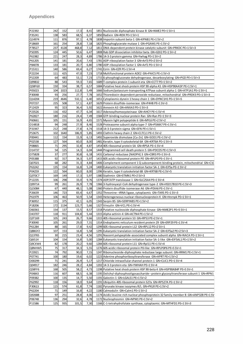
Appendices
228
P22392 242 152 17,3 8,41 1851 Nucleoside diphosphate kinase B GN=NME2 PE=1 SV=1
P35241 138 583 68,5 6,37 1850 Radixin GN=RDX PE=1 SV=1
Q14974 111 876 97,1 4,78 1838 Importin subunit beta-1 GN=KPNB1 PE=1 SV=2
P18669 161 254 28,8 7,18 1825 Phosphoglycerate mutase 1 GN=PGAM1 PE=1 SV=2
P78527 237 4128 468,8 7,12 1811 DNA-dependent protein kinase catalytic subunit GN=PRKDC PE=1 SV=3
P50395 124 445 50,6 6,47 1808 Rab GDP dissociation inhibitor beta GN=GDI2 PE=1 SV=2
P61982 221 247 28,3 4,89 1786 14-3-3 protein gamma GN=Ywhag PE=1 SV=2
P61205 141 181 20,6 7,43 1781 ADP-ribosylation factor 3 GN=Arf3 PE=2 SV=2
P84078 133 181 20,7 6,80 1780 ADP-ribosylation factor 1 GN=Arf1 PE=1 SV=2
P15311 140 586 69,4 6,27 1741 Ezrin GN=EZR PE=1 SV=4
P22234 111 425 47,0 7,23 1718 Multifunctional protein ADE2 GN=PAICS PE=1 SV=3
P52209 64 483 53,1 7,23 1715 6-phosphogluconate dehydrogenase, decarboxylating GN=PGD PE=1 SV=3
Q99832 88 543 59,3 7,65 1689 T-complex protein 1 subunit eta GN=CCT7 PE=1 SV=2
Q58FG0 150 334 38,7 6,57 1684 Putative heat shock protein HSP 90-alpha A5 GN=HSP90AA5P PE=1 SV=1
P05023 104 1023 112,8 5,49 1666 Sodium/potassium-transporting ATPase subunit alpha-1 GN=ATP1A1 PE=1 SV=1
P30048 93 256 27,7 7,78 1652 Thioredoxin-dependent peroxide reductase, mitochondrial GN=PRDX3 PE=1 SV=3
Q14204 219 4646 532,1 6,40 1651 Cytoplasmic dynein 1 heavy chain 1 GN=DYNC1H1 PE=1 SV=5
P07237 225 508 57,1 4,87 1629 Protein disulfide-isomerase GN=P4HB PE=1 SV=3
P12429 95 323 36,4 5,92 1622 Annexin A3 GN=ANXA3 PE=1 SV=3
P23526 116 432 47,7 6,34 1617 Adenosylhomocysteinase GN=AHCY PE=1 SV=4
P62827 180 216 24,4 7,49 1584 GTP-binding nuclear protein Ran GN=Ran PE=1 SV=3
P60661 105 151 16,9 4,65 1571 Myosin light polypeptide 6 GN=MYL6 PE=2 SV=2
O14818 94 248 27,9 8,46 1528 Proteasome subunit alpha type-7 GN=PSMA7 PE=1 SV=1
P31947 212 248 27,8 4,74 1518 14-3-3 protein sigma GN=SFN PE=1 SV=1
P53675 102 1640 186,9 5,85 1493 Clathrin heavy chain 2 GN=CLTCL1 PE=1 SV=2
P00441 112 154 15,9 6,13 1492 Superoxide dismutase [Cu-Zn] GN=SOD1 PE=1 SV=2
P02538 127 564 60,0 8,00 1481 Keratin, type II cytoskeletal 6A GN=KRT6A PE=1 SV=3
P08865 71 295 32,8 4,87 1454 40S ribosomal protein SA GN=RPSA PE=1 SV=4
O14737 54 125 14,3 6,04 1444 Programmed cell death protein 5 GN=PDCD5 PE=1 SV=3
O75828 37 277 30,8 6,18 1431 Carbonyl reductase [NADPH] 3 GN=CBR3 PE=1 SV=3
P05388 92 317 34,3 5,97 1415 60S acidic ribosomal protein P0 GN=RPLP0 PE=1 SV=1
Q07021 68 282 31,3 4,84 1406 Complement component 1 Q subcomponent-binding protein, mitochondrial GN=C1QBP PE=1 SV=1
P63242 135 154 16,8 5,24 1400 Eukaryotic translation initiation factor 5A-1 GN=Eif5a PE=1 SV=2
P04259 122 564 60,0 8,00 1396 Keratin, type II cytoskeletal 6B GN=KRT6B PE=1 SV=5
Q3T0C7 169 149 17,3 5,97 1380 Stathmin GN=STMN1 PE=2 SV=3
P12235 106 298 33,0 9,76 1374 ADP/ATP translocase 1 GN=SLC25A4 PE=1 SV=4
Q99714 99 261 26,9 7,78 1366 3-hydroxyacyl-CoA dehydrogenase type-2 GN=HSD17B10 PE=1 SV=3
Q15084 67 440 48,1 5,08 1360 Protein disulfide-isomerase A6 GN=PDIA6 PE=1 SV=1
P26639 146 723 83,4 6,67 1352 Threonine--tRNA ligase, cytoplasmic GN=TARS PE=1 SV=3
P61979 106 463 50,9 5,54 1343 Heterogeneous nuclear ribonucleoprotein K GN=Hnrnpk PE=1 SV=1
P36952 115 375 42,1 6,05 1343 Serpin B5 GN=SERPINB5 PE=1 SV=2
P18206 172 1134 123,7 5,66 1327 Vinculin GN=VCL PE=1 SV=4
O60361 154 137 15,5 8,57 1318 Putative nucleoside diphosphate kinase GN=NME2P1 PE=5 SV=1
O43707 118 911 104,8 5,44 1315 Alpha-actinin-4 GN=ACTN4 PE=1 SV=2
Q3T169 105 243 26,7 9,66 1315 40S ribosomal protein S3 GN=RPS3 PE=2 SV=1
P30040 105 261 29,0 7,31 1314 Endoplasmic reticulum resident protein 29 GN=ERP29 PE=1 SV=4
P61284 88 165 17,8 9,42 1299 60S ribosomal protein L12 GN=RPL12 PE=2 SV=1
Q8BGY2 107 153 16,8 5,58 1295 Eukaryotic translation initiation factor 5A-2 GN=Eif5a2 PE=2 SV=3
Q13765 30 215 23,4 4,56 1291 Nascent polypeptide-associated complex subunit alpha GN=NACA PE=1 SV=1
Q6IS14 104 154 16,8 5,00 1284 Eukaryotic translation initiation factor 5A-1-like GN=EIF5AL1 PE=1 SV=2
Q9CXW4 82 178 20,2 9,60 1284 60S ribosomal protein L11 GN=Rpl11 PE=1 SV=4
Q8NHW5 75 317 34,3 5,55 1276 60S acidic ribosomal protein P0-like GN=RPLP0P6 PE=5 SV=1
P23921 74 792 90,0 7,15 1247 Ribonucleoside-diphosphate reductase large subunit GN=RRM1 PE=1 SV=1
P07741 100 180 19,6 6,02 1223 Adenine phosphoribosyltransferase GN=APRT PE=1 SV=2
O00299 71 241 26,9 5,17 1217 Chloride intracellular channel protein 1 GN=CLIC1 PE=1 SV=4
Q04917 182 246 28,2 4,84 1202 14-3-3 protein eta GN=YWHAH PE=1 SV=4
Q58FF6 168 505 58,2 4,73 1198 Putative heat shock protein HSP 90-beta 4 GN=HSP90AB4P PE=5 SV=1
P04843 116 607 68,5 6,38 1195 Dolichyl-diphosphooligosaccharide--protein glycosyltransferase subunit 1 GN=RPN1 PE=1 SV=1
P09382 100 135 14,7 5,50 1191 Galectin-1 GN=LGALS1 PE=1 SV=2
P62992 118 156 18,0 9,64 1191 Ubiquitin-40S ribosomal protein S27a GN=RPS27A PE=1 SV=2
P30613 115 574 61,8 7,74 1182 Pyruvate kinase isozymes R/L GN=PKLR PE=1 SV=2
P62204 71 149 16,8 4,22 1180 Calmodulin GN=Calm1 PE=1 SV=2
Q92688 97 251 28,8 4,06 1174 Acidic leucine-rich nuclear phosphoprotein 32 family member B GN=ANP32B PE=1 SV=1
P06748 136 294 32,6 4,78 1172 Nucleophosmin GN=NPM1 PE=1 SV=2
P11586 125 935 101,5 7,30 1166 C-1-tetrahydrofolate synthase, cytoplasmic GN=MTHFD1 PE=1 SV=3
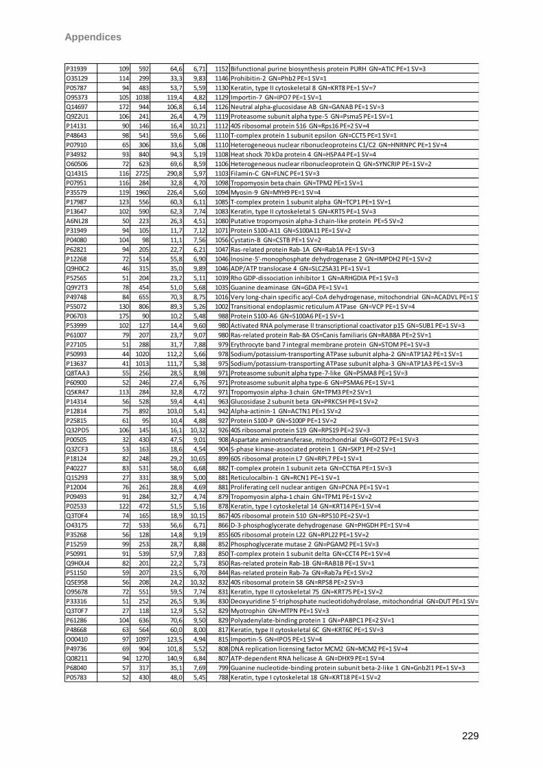
Appendices
229
P31939 109 592 64,6 6,71 1152 Bifunctional purine biosynthesis protein PURH GN=ATIC PE=1 SV=3
O35129 114 299 33,3 9,83 1146 Prohibitin-2 GN=Phb2 PE=1 SV=1
P05787 94 483 53,7 5,59 1130 Keratin, type II cytoskeletal 8 GN=KRT8 PE=1 SV=7
O95373 105 1038 119,4 4,82 1129 Importin-7 GN=IPO7 PE=1 SV=1
Q14697 172 944 106,8 6,14 1126 Neutral alpha-glucosidase AB GN=GANAB PE=1 SV=3
Q9Z2U1 106 241 26,4 4,79 1119 Proteasome subunit alpha type-5 GN=Psma5 PE=1 SV=1
P14131 90 146 16,4 10,21 1112 40S ribosomal protein S16 GN=Rps16 PE=2 SV=4
P48643 98 541 59,6 5,66 1110 T-complex protein 1 subunit epsilon GN=CCT5 PE=1 SV=1
P07910 65 306 33,6 5,08 1110 Heterogeneous nuclear ribonucleoproteins C1/C2 GN=HNRNPC PE=1 SV=4
P34932 93 840 94,3 5,19 1108 Heat shock 70 kDa protein 4 GN=HSPA4 PE=1 SV=4
O60506 72 623 69,6 8,59 1106 Heterogeneous nuclear ribonucleoprotein Q GN=SYNCRIP PE=1 SV=2
Q14315 116 2725 290,8 5,97 1103 Filamin-C GN=FLNC PE=1 SV=3
P07951 116 284 32,8 4,70 1098 Tropomyosin beta chain GN=TPM2 PE=1 SV=1
P35579 119 1960 226,4 5,60 1094 Myosin-9 GN=MYH9 PE=1 SV=4
P17987 123 556 60,3 6,11 1085 T-complex protein 1 subunit alpha GN=TCP1 PE=1 SV=1
P13647 102 590 62,3 7,74 1083 Keratin, type II cytoskeletal 5 GN=KRT5 PE=1 SV=3
A6NL28 50 223 26,3 4,51 1080 Putative tropomyosin alpha-3 chain-like protein PE=5 SV=2
P31949 94 105 11,7 7,12 1071 Protein S100-A11 GN=S100A11 PE=1 SV=2
P04080 104 98 11,1 7,56 1056 Cystatin-B GN=CSTB PE=1 SV=2
P62821 94 205 22,7 6,21 1047 Ras-related protein Rab-1A GN=Rab1A PE=1 SV=3
P12268 72 514 55,8 6,90 1046 Inosine-5'-monophosphate dehydrogenase 2 GN=IMPDH2 PE=1 SV=2
Q9H0C2 46 315 35,0 9,89 1046 ADP/ATP translocase 4 GN=SLC25A31 PE=1 SV=1
P52565 51 204 23,2 5,11 1039 Rho GDP-dissociation inhibitor 1 GN=ARHGDIA PE=1 SV=3
Q9Y2T3 78 454 51,0 5,68 1035 Guanine deaminase GN=GDA PE=1 SV=1
P49748 84 655 70,3 8,75 1016 Very long-chain specific acyl-CoA dehydrogenase, mitochondrial GN=ACADVL PE=1 SV=1
P55072 130 806 89,3 5,26 1002 Transitional endoplasmic reticulum ATPase GN=VCP PE=1 SV=4
P06703 175 90 10,2 5,48 988 Protein S100-A6 GN=S100A6 PE=1 SV=1
P53999 102 127 14,4 9,60 980 Activated RNA polymerase II transcriptional coactivator p15 GN=SUB1 PE=1 SV=3
P61007 79 207 23,7 9,07 980 Ras-related protein Rab-8A OS=Canis familiaris GN=RAB8A PE=2 SV=1
P27105 51 288 31,7 7,88 979 Erythrocyte band 7 integral membrane protein GN=STOM PE=1 SV=3
P50993 44 1020 112,2 5,66 978 Sodium/potassium-transporting ATPase subunit alpha-2 GN=ATP1A2 PE=1 SV=1
P13637 41 1013 111,7 5,38 975 Sodium/potassium-transporting ATPase subunit alpha-3 GN=ATP1A3 PE=1 SV=3
Q8TAA3 55 256 28,5 8,98 971 Proteasome subunit alpha type-7-like GN=PSMA8 PE=1 SV=3
P60900 52 246 27,4 6,76 971 Proteasome subunit alpha type-6 GN=PSMA6 PE=1 SV=1
Q5KR47 113 284 32,8 4,72 971 Tropomyosin alpha-3 chain GN=TPM3 PE=2 SV=1
P14314 56 528 59,4 4,41 963 Glucosidase 2 subunit beta GN=PRKCSH PE=1 SV=2
P12814 75 892 103,0 5,41 942 Alpha-actinin-1 GN=ACTN1 PE=1 SV=2
P25815 61 95 10,4 4,88 927 Protein S100-P GN=S100P PE=1 SV=2
Q32PD5 106 145 16,1 10,32 926 40S ribosomal protein S19 GN=RPS19 PE=2 SV=3
P00505 32 430 47,5 9,01 908 Aspartate aminotransferase, mitochondrial GN=GOT2 PE=1 SV=3
Q3ZCF3 53 163 18,6 4,54 904 S-phase kinase-associated protein 1 GN=SKP1 PE=2 SV=1
P18124 82 248 29,2 10,65 899 60S ribosomal protein L7 GN=RPL7 PE=1 SV=1
P40227 83 531 58,0 6,68 882 T-complex protein 1 subunit zeta GN=CCT6A PE=1 SV=3
Q15293 27 331 38,9 5,00 881 Reticulocalbin-1 GN=RCN1 PE=1 SV=1
P12004 76 261 28,8 4,69 881 Proliferating cell nuclear antigen GN=PCNA PE=1 SV=1
P09493 91 284 32,7 4,74 879 Tropomyosin alpha-1 chain GN=TPM1 PE=1 SV=2
P02533 122 472 51,5 5,16 878 Keratin, type I cytoskeletal 14 GN=KRT14 PE=1 SV=4
Q3T0F4 74 165 18,9 10,15 867 40S ribosomal protein S10 GN=RPS10 PE=2 SV=1
O43175 72 533 56,6 6,71 866 D-3-phosphoglycerate dehydrogenase GN=PHGDH PE=1 SV=4
P35268 56 128 14,8 9,19 855 60S ribosomal protein L22 GN=RPL22 PE=1 SV=2
P15259 99 253 28,7 8,88 852 Phosphoglycerate mutase 2 GN=PGAM2 PE=1 SV=3
P50991 91 539 57,9 7,83 850 T-complex protein 1 subunit delta GN=CCT4 PE=1 SV=4
Q9H0U4 82 201 22,2 5,73 850 Ras-related protein Rab-1B GN=RAB1B PE=1 SV=1
P51150 59 207 23,5 6,70 844 Ras-related protein Rab-7a GN=Rab7a PE=1 SV=2
Q5E958 56 208 24,2 10,32 832 40S ribosomal protein S8 GN=RPS8 PE=2 SV=3
O95678 72 551 59,5 7,74 831 Keratin, type II cytoskeletal 75 GN=KRT75 PE=1 SV=2
P33316 51 252 26,5 9,36 830 Deoxyuridine 5'-triphosphate nucleotidohydrolase, mitochondrial GN=DUT PE=1 SV=4
Q3T0F7 27 118 12,9 5,52 829 Myotrophin GN=MTPN PE=1 SV=3
P61286 104 636 70,6 9,50 829 Polyadenylate-binding protein 1 GN=PABPC1 PE=2 SV=1
P48668 63 564 60,0 8,00 817 Keratin, type II cytoskeletal 6C GN=KRT6C PE=1 SV=3
O00410 97 1097 123,5 4,94 815 Importin-5 GN=IPO5 PE=1 SV=4
P49736 69 904 101,8 5,52 808 DNA replication licensing factor MCM2 GN=MCM2 PE=1 SV=4
Q08211 94 1270 140,9 6,84 807 ATP-dependent RNA helicase A GN=DHX9 PE=1 SV=4
P68040 57 317 35,1 7,69 799 Guanine nucleotide-binding protein subunit beta-2-like 1 GN=Gnb2l1 PE=1 SV=3
P05783 52 430 48,0 5,45 788 Keratin, type I cytoskeletal 18 GN=KRT18 PE=1 SV=2

Appendices
230
P28074 46 263 28,5 6,92 788 Proteasome subunit beta type-5 GN=PSMB5 PE=1 SV=3
Q04837 34 148 17,2 9,60 772 Single-stranded DNA-binding protein, mitochondrial GN=SSBP1 PE=1 SV=1
P61027 56 200 22,5 8,38 767 Ras-related protein Rab-10 GN=Rab10 PE=1 SV=1
P00491 61 289 32,1 6,95 762 Purine nucleoside phosphorylase GN=PNP PE=1 SV=2
Q969H8 68 173 18,8 6,68 758 UPF0556 protein C19orf10 GN=C19orf10 PE=1 SV=1
Q07020 49 188 21,6 11,72 756 60S ribosomal protein L18 GN=RPL18 PE=1 SV=2
Q91V41 54 215 23,9 6,21 744 Ras-related protein Rab-14 GN=Rab14 PE=1 SV=3
O43776 61 548 62,9 6,25 740 Asparagine--tRNA ligase, cytoplasmic GN=NARS PE=1 SV=1
Q56K14 32 114 11,5 4,32 739 60S acidic ribosomal protein P1 GN=RPLP1 PE=3 SV=1
P78417 99 241 27,5 6,60 738 Glutathione S-transferase omega-1 GN=GSTO1 PE=1 SV=2
P38919 42 411 46,8 6,73 738 Eukaryotic initiation factor 4A-III GN=EIF4A3 PE=1 SV=4
P62264 45 151 16,3 10,05 737 40S ribosomal protein S14 GN=Rps14 PE=2 SV=3
Q92930 61 207 23,6 9,07 724 Ras-related protein Rab-8B GN=RAB8B PE=1 SV=2
Q02790 96 459 51,8 5,43 721 Peptidyl-prolyl cis-trans isomerase FKBP4 GN=FKBP4 PE=1 SV=3
P59190 53 212 24,4 5,71 719 Ras-related protein Rab-15 GN=RAB15 PE=1 SV=1
Q6PHN9 57 201 23,0 8,29 719 Ras-related protein Rab-35 GN=Rab35 PE=1 SV=1
Q5XKE5 64 535 57,8 7,20 716 Keratin, type II cytoskeletal 79 GN=KRT79 PE=1 SV=2
P08729 63 469 51,4 5,48 706 Keratin, type II cytoskeletal 7 GN=KRT7 PE=1 SV=5
Q3T165 57 272 29,8 5,76 703 Prohibitin GN=PHB PE=2 SV=1
P62852 70 125 13,7 10,11 703 40S ribosomal protein S25 GN=Rps25 PE=2 SV=1
P62702 87 263 29,6 10,15 703 40S ribosomal protein S4, X isoform GN=Rps4x PE=2 SV=2
P13693 41 172 19,6 4,93 698 Translationally-controlled tumor protein GN=TPT1 PE=1 SV=1
P16442 355 354 40,9 8,98 698 Histo-blood group ABO system transferase GN=ABO PE=1 SV=2
Q9NPC6 350 264 29,9 8,25 698 Myozenin-2 GN=MYOZ2 PE=1 SV=1
Q6DD88 39 541 60,5 5,66 688 Atlastin-3 GN=ATL3 PE=1 SV=1
P30049 23 168 17,5 5,49 681 ATP synthase subunit delta, mitochondrial GN=ATP5D PE=1 SV=2
P53396 89 1101 120,8 7,33 677 ATP-citrate synthase GN=ACLY PE=1 SV=3
P18085 77 180 20,5 7,14 673 ADP-ribosylation factor 4 GN=ARF4 PE=1 SV=3
P60902 89 97 11,2 7,37 673 Protein S100-A10 GN=S100A10 PE=1 SV=2
O43399 45 206 22,2 5,36 670 Tumor protein D54 GN=TPD52L2 PE=1 SV=2
P52292 39 529 57,8 5,40 669 Importin subunit alpha-2 GN=KPNA2 PE=1 SV=1
P56192 41 900 101,1 6,16 665 Methionine--tRNA ligase, cytoplasmic GN=MARS PE=1 SV=2
P62270 93 152 17,7 10,99 663 40S ribosomal protein S18 GN=Rps18 PE=1 SV=3
P30046 68 118 12,7 7,30 660 D-dopachrome decarboxylase GN=DDT PE=1 SV=3
P42704 86 1394 157,8 6,13 654 Leucine-rich PPR motif-containing protein, mitochondrial GN=LRPPRC PE=1 SV=3
Q60972 16 425 47,6 4,89 654 Histone-binding protein RBBP4 GN=Rbbp4 PE=1 SV=5
P00492 45 218 24,6 6,68 652 Hypoxanthine-guanine phosphoribosyltransferase GN=HPRT1 PE=1 SV=2
Q6PDM2 67 248 27,7 10,36 650 Serine/arginine-rich splicing factor 1 GN=Srsf1 PE=1 SV=3
P39687 75 249 28,6 4,09 649 Acidic leucine-rich nuclear phosphoprotein 32 family member A GN=ANP32A PE=1 SV=1
P37108 29 136 14,6 10,04 649 Signal recognition particle 14 kDa protein GN=SRP14 PE=1 SV=2
P07814 91 1512 170,5 7,33 644 Bifunctional glutamate/proline--tRNA ligase GN=EPRS PE=1 SV=5
Q92928 54 201 22,0 5,43 643 Putative Ras-related protein Rab-1C GN=RAB1C PE=5 SV=2
P21796 46 283 30,8 8,54 636 Voltage-dependent anion-selective channel protein 1 GN=VDAC1 PE=1 SV=2
Q99497 116 189 19,9 6,79 633 Protein DJ-1 GN=PARK7 PE=1 SV=2
P08779 96 473 51,2 5,05 633 Keratin, type I cytoskeletal 16 GN=KRT16 PE=1 SV=4
Q96FW1 25 271 31,3 4,94 632 Ubiquitin thioesterase OTUB1 GN=OTUB1 PE=1 SV=2
P84084 77 180 20,5 6,79 630 ADP-ribosylation factor 5 GN=Arf5 PE=2 SV=2
Q02878 58 288 32,7 10,58 620 60S ribosomal protein L6 GN=RPL6 PE=1 SV=3
P22102 48 1010 107,7 6,70 614 Trifunctional purine biosynthetic protein adenosine-3 GN=GART PE=1 SV=1
P61087 33 200 22,4 5,44 613 Ubiquitin-conjugating enzyme E2 K GN=Ube2k PE=1 SV=3
O14980 64 1071 123,3 6,06 612 Exportin-1 GN=XPO1 PE=1 SV=1
Q9UKK9 36 219 24,3 4,94 609 ADP-sugar pyrophosphatase GN=NUDT5 PE=1 SV=1
P17931 56 250 26,1 8,56 608 Galectin-3 GN=LGALS3 PE=1 SV=5
O14684 65 152 17,1 9,50 603 Prostaglandin E synthase GN=PTGES PE=1 SV=2
Q99733 41 375 42,8 4,69 597 Nucleosome assembly protein 1-like 4 GN=NAP1L4 PE=1 SV=1
Q9BSJ8 51 1104 122,8 5,83 595 Extended synaptotagmin-1 GN=ESYT1 PE=1 SV=1
Q9Y266 44 331 38,2 5,38 594 Nuclear migration protein nudC GN=NUDC PE=1 SV=1
P46777 52 297 34,3 9,72 585 60S ribosomal protein L5 GN=RPL5 PE=1 SV=3
P49755 36 219 25,0 7,44 581 Transmembrane emp24 domain-containing protein 10 GN=TMED10 PE=1 SV=2
P54819 48 239 26,5 7,81 574 Adenylate kinase 2, mitochondrial GN=AK2 PE=1 SV=2
P55854 29 103 11,6 5,49 568 Small ubiquitin-related modifier 3 GN=SUMO3 PE=1 SV=2
P57088 21 247 28,0 9,70 566 Transmembrane protein 33 GN=TMEM33 PE=1 SV=2
P62245 53 130 14,8 10,13 562 40S ribosomal protein S15a GN=Rps15a PE=2 SV=2
P42126 23 302 32,8 8,54 561 Enoyl-CoA delta isomerase 1, mitochondrial GN=ECI1 PE=1 SV=1
P61018 27 213 23,6 6,06 557 Ras-related protein Rab-4B GN=RAB4B PE=1 SV=1
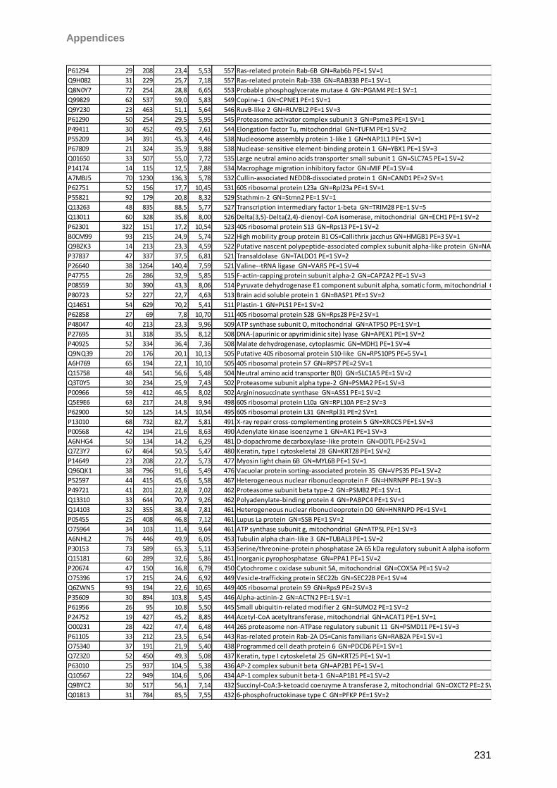
Appendices
231
P61294 29 208 23,4 5,53 557 Ras-related protein Rab-6B GN=Rab6b PE=1 SV=1
Q9H082 31 229 25,7 7,18 557 Ras-related protein Rab-33B GN=RAB33B PE=1 SV=1
Q8N0Y7 72 254 28,8 6,65 553 Probable phosphoglycerate mutase 4 GN=PGAM4 PE=1 SV=1
Q99829 62 537 59,0 5,83 549 Copine-1 GN=CPNE1 PE=1 SV=1
Q9Y230 23 463 51,1 5,64 546 RuvB-like 2 GN=RUVBL2 PE=1 SV=3
P61290 50 254 29,5 5,95 545 Proteasome activator complex subunit 3 GN=Psme3 PE=1 SV=1
P49411 30 452 49,5 7,61 544 Elongation factor Tu, mitochondrial GN=TUFM PE=1 SV=2
P55209 34 391 45,3 4,46 538 Nucleosome assembly protein 1-like 1 GN=NAP1L1 PE=1 SV=1
P67809 21 324 35,9 9,88 538 Nuclease-sensitive element-binding protein 1 GN=YBX1 PE=1 SV=3
Q01650 33 507 55,0 7,72 535 Large neutral amino acids transporter small subunit 1 GN=SLC7A5 PE=1 SV=2
P14174 14 115 12,5 7,88 534 Macrophage migration inhibitory factor GN=MIF PE=1 SV=4
A7MBJ5 70 1230 136,3 5,78 532 Cullin-associated NEDD8-dissociated protein 1 GN=CAND1 PE=2 SV=1
P62751 52 156 17,7 10,45 531 60S ribosomal protein L23a GN=Rpl23a PE=1 SV=1
P55821 92 179 20,8 8,32 529 Stathmin-2 GN=Stmn2 PE=1 SV=1
Q13263 48 835 88,5 5,77 527 Transcription intermediary factor 1-beta GN=TRIM28 PE=1 SV=5
Q13011 60 328 35,8 8,00 526 Delta(3,5)-Delta(2,4)-dienoyl-CoA isomerase, mitochondrial GN=ECH1 PE=1 SV=2
P62301 322 151 17,2 10,54 523 40S ribosomal protein S13 GN=Rps13 PE=1 SV=2
B0CM99 93 215 24,9 5,74 522 High mobility group protein B1 OS=Callithrix jacchus GN=HMGB1 PE=3 SV=1
Q9BZK3 14 213 23,3 4,59 522 Putative nascent polypeptide-associated complex subunit alpha-like protein GN=NACAP1 PE=5 SV=1
P37837 47 337 37,5 6,81 521 Transaldolase GN=TALDO1 PE=1 SV=2
P26640 38 1264 140,4 7,59 521 Valine--tRNA ligase GN=VARS PE=1 SV=4
P47755 26 286 32,9 5,85 515 F-actin-capping protein subunit alpha-2 GN=CAPZA2 PE=1 SV=3
P08559 30 390 43,3 8,06 514 Pyruvate dehydrogenase E1 component subunit alpha, somatic form, mitochondrial GN=PDHA1 PE=1 SV=3
P80723 52 227 22,7 4,63 513 Brain acid soluble protein 1 GN=BASP1 PE=1 SV=2
Q14651 54 629 70,2 5,41 511 Plastin-1 GN=PLS1 PE=1 SV=2
P62858 27 69 7,8 10,70 511 40S ribosomal protein S28 GN=Rps28 PE=2 SV=1
P48047 40 213 23,3 9,96 509 ATP synthase subunit O, mitochondrial GN=ATP5O PE=1 SV=1
P27695 31 318 35,5 8,12 508 DNA-(apurinic or apyrimidinic site) lyase GN=APEX1 PE=1 SV=2
P40925 52 334 36,4 7,36 508 Malate dehydrogenase, cytoplasmic GN=MDH1 PE=1 SV=4
Q9NQ39 20 176 20,1 10,13 505 Putative 40S ribosomal protein S10-like GN=RPS10P5 PE=5 SV=1
A6H769 65 194 22,1 10,10 505 40S ribosomal protein S7 GN=RPS7 PE=2 SV=1
Q15758 48 541 56,6 5,48 504 Neutral amino acid transporter B(0) GN=SLC1A5 PE=1 SV=2
Q3T0Y5 30 234 25,9 7,43 502 Proteasome subunit alpha type-2 GN=PSMA2 PE=1 SV=3
P00966 59 412 46,5 8,02 502 Argininosuccinate synthase GN=ASS1 PE=1 SV=2
Q5E9E6 63 217 24,8 9,94 498 60S ribosomal protein L10a GN=RPL10A PE=2 SV=3
P62900 50 125 14,5 10,54 495 60S ribosomal protein L31 GN=Rpl31 PE=2 SV=1
P13010 68 732 82,7 5,81 491 X-ray repair cross-complementing protein 5 GN=XRCC5 PE=1 SV=3
P00568 42 194 21,6 8,63 490 Adenylate kinase isoenzyme 1 GN=AK1 PE=1 SV=3
A6NHG4 50 134 14,2 6,29 481 D-dopachrome decarboxylase-like protein GN=DDTL PE=2 SV=1
Q7Z3Y7 67 464 50,5 5,47 480 Keratin, type I cytoskeletal 28 GN=KRT28 PE=1 SV=2
P14649 23 208 22,7 5,73 477 Myosin light chain 6B GN=MYL6B PE=1 SV=1
Q96QK1 38 796 91,6 5,49 476 Vacuolar protein sorting-associated protein 35 GN=VPS35 PE=1 SV=2
P52597 44 415 45,6 5,58 467 Heterogeneous nuclear ribonucleoprotein F GN=HNRNPF PE=1 SV=3
P49721 41 201 22,8 7,02 462 Proteasome subunit beta type-2 GN=PSMB2 PE=1 SV=1
Q13310 33 644 70,7 9,26 462 Polyadenylate-binding protein 4 GN=PABPC4 PE=1 SV=1
Q14103 32 355 38,4 7,81 461 Heterogeneous nuclear ribonucleoprotein D0 GN=HNRNPD PE=1 SV=1
P05455 25 408 46,8 7,12 461 Lupus La protein GN=SSB PE=1 SV=2
O75964 34 103 11,4 9,64 461 ATP synthase subunit g, mitochondrial GN=ATP5L PE=1 SV=3
A6NHL2 76 446 49,9 6,05 453 Tubulin alpha chain-like 3 GN=TUBAL3 PE=1 SV=2
P30153 73 589 65,3 5,11 453 Serine/threonine-protein phosphatase 2A 65 kDa regulatory subunit A alpha isoform GN=PPP2R1A PE=1 SV=4
Q15181 60 289 32,6 5,86 451 Inorganic pyrophosphatase GN=PPA1 PE=1 SV=2
P20674 47 150 16,8 6,79 450 Cytochrome c oxidase subunit 5A, mitochondrial GN=COX5A PE=1 SV=2
O75396 17 215 24,6 6,92 449 Vesicle-trafficking protein SEC22b GN=SEC22B PE=1 SV=4
Q6ZWN5 93 194 22,6 10,65 449 40S ribosomal protein S9 GN=Rps9 PE=2 SV=3
P35609 30 894 103,8 5,45 446 Alpha-actinin-2 GN=ACTN2 PE=1 SV=1
P61956 26 95 10,8 5,50 445 Small ubiquitin-related modifier 2 GN=SUMO2 PE=1 SV=2
P24752 19 427 45,2 8,85 444 Acetyl-CoA acetyltransferase, mitochondrial GN=ACAT1 PE=1 SV=1
O00231 28 422 47,4 6,48 444 26S proteasome non-ATPase regulatory subunit 11 GN=PSMD11 PE=1 SV=3
P61105 33 212 23,5 6,54 443 Ras-related protein Rab-2A OS=Canis familiaris GN=RAB2A PE=1 SV=1
O75340 37 191 21,9 5,40 438 Programmed cell death protein 6 GN=PDCD6 PE=1 SV=1
Q7Z3Z0 52 450 49,3 5,08 437 Keratin, type I cytoskeletal 25 GN=KRT25 PE=1 SV=1
P63010 25 937 104,5 5,38 436 AP-2 complex subunit beta GN=AP2B1 PE=1 SV=1
Q10567 22 949 104,6 5,06 434 AP-1 complex subunit beta-1 GN=AP1B1 PE=1 SV=2
Q9BYC2 30 517 56,1 7,14 432 Succinyl-CoA:3-ketoacid coenzyme A transferase 2, mitochondrial GN=OXCT2 PE=2 SV=2
Q01813 31 784 85,5 7,55 432 6-phosphofructokinase type C GN=PFKP PE=1 SV=2
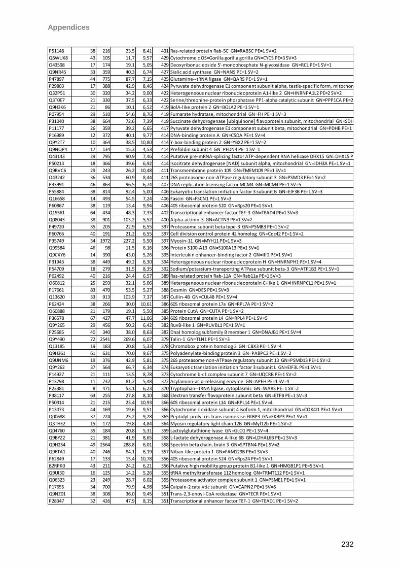
Appendices
232
P51148 38 216 23,5 8,41 431 Ras-related protein Rab-5C GN=RAB5C PE=1 SV=2
Q6WUX8 43 105 11,7 9,57 429 Cytochrome c OS=Gorilla gorilla gorilla GN=CYCS PE=3 SV=3
O43598 17 174 19,1 5,05 429 Deoxyribonucleoside 5'-monophosphate N-glycosidase GN=RCL PE=1 SV=1
Q9NR45 33 359 40,3 6,74 427 Sialic acid synthase GN=NANS PE=1 SV=2
P47897 44 775 87,7 7,15 425 Glutamine--tRNA ligase GN=QARS PE=1 SV=1
P29803 17 388 42,9 8,46 424 Pyruvate dehydrogenase E1 component subunit alpha, testis-specific form, mitochondrial GN=PDHA2 PE=1 SV=1
Q32P51 30 320 34,2 9,00 422 Heterogeneous nuclear ribonucleoprotein A1-like 2 GN=HNRNPA1L2 PE=2 SV=2
Q3T0E7 21 330 37,5 6,33 422 Serine/threonine-protein phosphatase PP1-alpha catalytic subunit GN=PPP1CA PE=2 SV=1
Q9H3K6 21 86 10,1 6,52 419 BolA-like protein 2 GN=BOLA2 PE=1 SV=1
P07954 29 510 54,6 8,76 419 Fumarate hydratase, mitochondrial GN=FH PE=1 SV=3
P31040 38 664 72,6 7,39 419 Succinate dehydrogenase [ubiquinone] flavoprotein subunit, mitochondrial GN=SDHA PE=1 SV=2
P11177 26 359 39,2 6,65 417 Pyruvate dehydrogenase E1 component subunit beta, mitochondrial GN=PDHB PE=1 SV=3
P16989 12 372 40,1 9,77 414 DNA-binding protein A GN=CSDA PE=1 SV=4
Q9Y2T7 10 364 38,5 10,80 414 Y-box-binding protein 2 GN=YBX2 PE=1 SV=2
Q9NQP4 17 134 15,3 4,53 414 Prefoldin subunit 4 GN=PFDN4 PE=1 SV=1
O43143 29 795 90,9 7,46 414 Putative pre-mRNA-splicing factor ATP-dependent RNA helicase DHX15 GN=DHX15 PE=1 SV=2
P50213 18 366 39,6 6,92 414 Isocitrate dehydrogenase [NAD] subunit alpha, mitochondrial GN=IDH3A PE=1 SV=1
Q9BVC6 29 243 26,2 10,48 411 Transmembrane protein 109 GN=TMEM109 PE=1 SV=1
O43242 36 534 60,9 8,44 411 26S proteasome non-ATPase regulatory subunit 3 GN=PSMD3 PE=1 SV=2
P33991 46 863 96,5 6,74 407 DNA replication licensing factor MCM4 GN=MCM4 PE=1 SV=5
P55884 38 814 92,4 5,00 406 Eukaryotic translation initiation factor 3 subunit B GN=EIF3B PE=1 SV=3
Q16658 14 493 54,5 7,24 406 Fascin GN=FSCN1 PE=1 SV=3
P60867 38 119 13,4 9,94 406 40S ribosomal protein S20 GN=Rps20 PE=1 SV=1
Q15561 64 434 48,3 7,33 402 Transcriptional enhancer factor TEF-3 GN=TEAD4 PE=1 SV=3
Q08043 38 901 103,2 5,52 400 Alpha-actinin-3 GN=ACTN3 PE=1 SV=2
P49720 35 205 22,9 6,55 397 Proteasome subunit beta type-3 GN=PSMB3 PE=1 SV=2
P60766 40 191 21,2 6,55 397 Cell division control protein 42 homolog GN=Cdc42 PE=1 SV=2
P35749 34 1972 227,2 5,50 397 Myosin-11 GN=MYH11 PE=1 SV=3
Q99584 46 98 11,5 6,16 396 Protein S100-A13 GN=S100A13 PE=1 SV=1
Q9CXY6 14 390 43,0 5,26 395 Interleukin enhancer-binding factor 2 GN=Ilf2 PE=1 SV=1
P31943 38 449 49,2 6,30 394 Heterogeneous nuclear ribonucleoprotein H GN=HNRNPH1 PE=1 SV=4
P54709 18 279 31,5 8,35 392 Sodium/potassium-transporting ATPase subunit beta-3 GN=ATP1B3 PE=1 SV=1
P62492 40 216 24,4 6,57 389 Ras-related protein Rab-11A GN=Rab11a PE=1 SV=3
O60812 25 293 32,1 5,06 389 Heterogeneous nuclear ribonucleoprotein C-like 1 GN=HNRNPCL1 PE=1 SV=1
P17661 83 470 53,5 5,27 388 Desmin GN=DES PE=1 SV=3
Q13620 33 913 103,9 7,37 387 Cullin-4B GN=CUL4B PE=1 SV=4
P62424 38 266 30,0 10,61 386 60S ribosomal protein L7a GN=RPL7A PE=1 SV=2
O60888 21 179 19,1 5,50 385 Protein CutA GN=CUTA PE=1 SV=2
P36578 67 427 47,7 11,06 384 60S ribosomal protein L4 GN=RPL4 PE=1 SV=5
Q9Y265 29 456 50,2 6,42 382 RuvB-like 1 GN=RUVBL1 PE=1 SV=1
P25685 40 340 38,0 8,63 382 DnaJ homolog subfamily B member 1 GN=DNAJB1 PE=1 SV=4
Q9Y490 72 2541 269,6 6,07 379 Talin-1 GN=TLN1 PE=1 SV=3
Q13185 19 183 20,8 5,33 378 Chromobox protein homolog 3 GN=CBX3 PE=1 SV=4
Q9H361 61 631 70,0 9,67 375 Polyadenylate-binding protein 3 GN=PABPC3 PE=1 SV=2
Q9UNM6 19 376 42,9 5,81 375 26S proteasome non-ATPase regulatory subunit 13 GN=PSMD13 PE=1 SV=2
Q9Y262 37 564 66,7 6,34 374 Eukaryotic translation initiation factor 3 subunit L GN=EIF3L PE=1 SV=1
P14927 21 111 13,5 8,78 373 Cytochrome b-c1 complex subunit 7 GN=UQCRB PE=1 SV=2
P13798 11 732 81,2 5,48 372 Acylamino-acid-releasing enzyme GN=APEH PE=1 SV=4
P23381 8 471 53,1 6,23 370 Tryptophan--tRNA ligase, cytoplasmic GN=WARS PE=1 SV=2
P38117 63 255 27,8 8,10 368 Electron transfer flavoprotein subunit beta GN=ETFB PE=1 SV=3
P50914 21 215 23,4 10,93 366 60S ribosomal protein L14 GN=RPL14 PE=1 SV=4
P13073 44 169 19,6 9,51 366 Cytochrome c oxidase subunit 4 isoform 1, mitochondrial GN=COX4I1 PE=1 SV=1
Q00688 37 224 25,2 9,28 365 Peptidyl-prolyl cis-trans isomerase FKBP3 GN=FKBP3 PE=1 SV=1
Q3THE2 15 172 19,8 4,84 364 Myosin regulatory light chain 12B GN=Myl12b PE=1 SV=2
Q04760 35 184 20,8 5,31 359 Lactoylglutathione lyase GN=GLO1 PE=1 SV=4
Q9BYZ2 21 381 41,9 8,65 358 L-lactate dehydrogenase A-like 6B GN=LDHAL6B PE=1 SV=3
Q9H254 49 2564 288,8 6,01 358 Spectrin beta chain, brain 3 GN=SPTBN4 PE=1 SV=2
Q96TA1 40 746 84,1 6,19 357 Niban-like protein 1 GN=FAM129B PE=1 SV=3
P62849 17 133 15,4 10,78 356 40S ribosomal protein S24 GN=Rps24 PE=1 SV=1
B2RPK0 43 211 24,2 6,21 356 Putative high mobility group protein B1-like 1 GN=HMGB1P1 PE=5 SV=1
Q9UI30 16 125 14,2 5,26 355 tRNA methyltransferase 112 homolog GN=TRMT112 PE=1 SV=1
Q06323 23 249 28,7 6,02 355 Proteasome activator complex subunit 1 GN=PSME1 PE=1 SV=1
P17655 34 700 79,9 4,98 354 Calpain-2 catalytic subunit GN=CAPN2 PE=1 SV=6
Q9NZ01 38 308 36,0 9,45 351 Trans-2,3-enoyl-CoA reductase GN=TECR PE=1 SV=1
P28347 32 426 47,9 8,15 351 Transcriptional enhancer factor TEF-1 GN=TEAD1 PE=1 SV=2
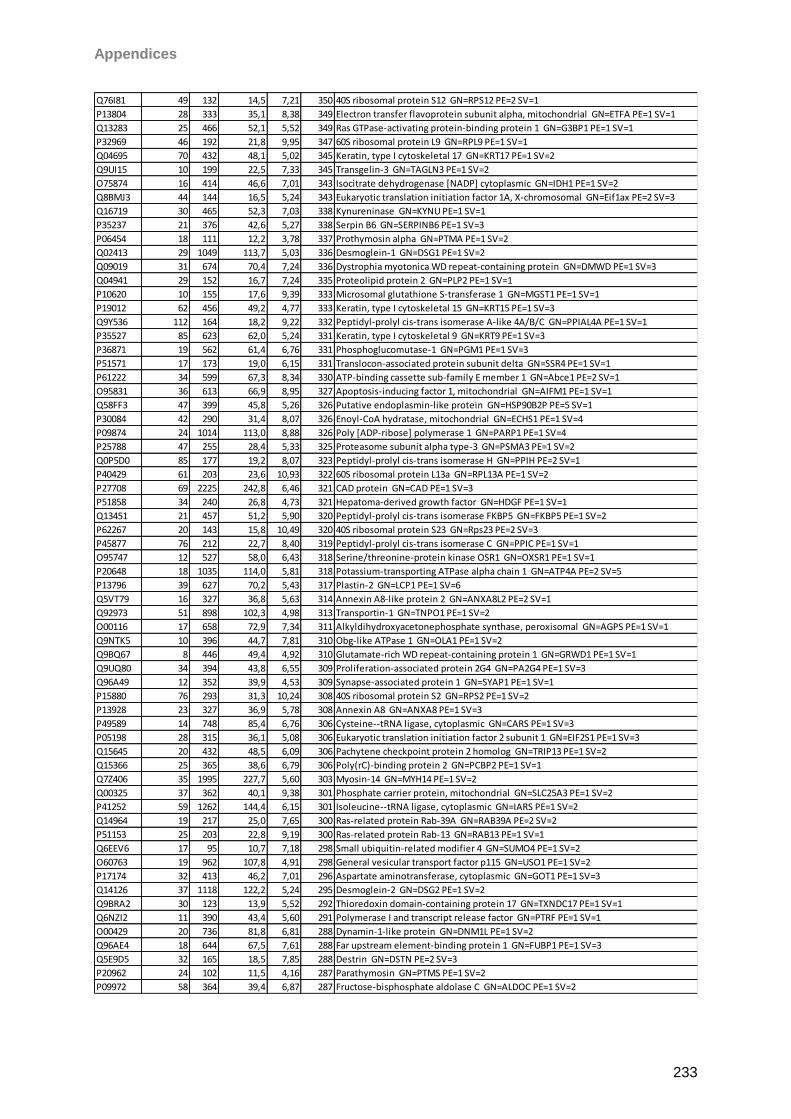
Appendices
233
Q76I81 49 132 14,5 7,21 350 40S ribosomal protein S12 GN=RPS12 PE=2 SV=1
P13804 28 333 35,1 8,38 349 Electron transfer flavoprotein subunit alpha, mitochondrial GN=ETFA PE=1 SV=1
Q13283 25 466 52,1 5,52 349 Ras GTPase-activating protein-binding protein 1 GN=G3BP1 PE=1 SV=1
P32969 46 192 21,8 9,95 347 60S ribosomal protein L9 GN=RPL9 PE=1 SV=1
Q04695 70 432 48,1 5,02 345 Keratin, type I cytoskeletal 17 GN=KRT17 PE=1 SV=2
Q9UI15 10 199 22,5 7,33 345 Transgelin-3 GN=TAGLN3 PE=1 SV=2
O75874 16 414 46,6 7,01 343 Isocitrate dehydrogenase [NADP] cytoplasmic GN=IDH1 PE=1 SV=2
Q8BMJ3 44 144 16,5 5,24 343 Eukaryotic translation initiation factor 1A, X-chromosomal GN=Eif1ax PE=2 SV=3
Q16719 30 465 52,3 7,03 338 Kynureninase GN=KYNU PE=1 SV=1
P35237 21 376 42,6 5,27 338 Serpin B6 GN=SERPINB6 PE=1 SV=3
P06454 18 111 12,2 3,78 337 Prothymosin alpha GN=PTMA PE=1 SV=2
Q02413 29 1049 113,7 5,03 336 Desmoglein-1 GN=DSG1 PE=1 SV=2
Q09019 31 674 70,4 7,24 336 Dystrophia myotonica WD repeat-containing protein GN=DMWD PE=1 SV=3
Q04941 29 152 16,7 7,24 335 Proteolipid protein 2 GN=PLP2 PE=1 SV=1
P10620 10 155 17,6 9,39 333 Microsomal glutathione S-transferase 1 GN=MGST1 PE=1 SV=1
P19012 62 456 49,2 4,77 333 Keratin, type I cytoskeletal 15 GN=KRT15 PE=1 SV=3
Q9Y536 112 164 18,2 9,22 332 Peptidyl-prolyl cis-trans isomerase A-like 4A/B/C GN=PPIAL4A PE=1 SV=1
P35527 85 623 62,0 5,24 331 Keratin, type I cytoskeletal 9 GN=KRT9 PE=1 SV=3
P36871 19 562 61,4 6,76 331 Phosphoglucomutase-1 GN=PGM1 PE=1 SV=3
P51571 17 173 19,0 6,15 331 Translocon-associated protein subunit delta GN=SSR4 PE=1 SV=1
P61222 34 599 67,3 8,34 330 ATP-binding cassette sub-family E member 1 GN=Abce1 PE=2 SV=1
O95831 36 613 66,9 8,95 327 Apoptosis-inducing factor 1, mitochondrial GN=AIFM1 PE=1 SV=1
Q58FF3 47 399 45,8 5,26 326 Putative endoplasmin-like protein GN=HSP90B2P PE=5 SV=1
P30084 42 290 31,4 8,07 326 Enoyl-CoA hydratase, mitochondrial GN=ECHS1 PE=1 SV=4
P09874 24 1014 113,0 8,88 326 Poly [ADP-ribose] polymerase 1 GN=PARP1 PE=1 SV=4
P25788 47 255 28,4 5,33 325 Proteasome subunit alpha type-3 GN=PSMA3 PE=1 SV=2
Q0P5D0 85 177 19,2 8,07 323 Peptidyl-prolyl cis-trans isomerase H GN=PPIH PE=2 SV=1
P40429 61 203 23,6 10,93 322 60S ribosomal protein L13a GN=RPL13A PE=1 SV=2
P27708 69 2225 242,8 6,46 321 CAD protein GN=CAD PE=1 SV=3
P51858 34 240 26,8 4,73 321 Hepatoma-derived growth factor GN=HDGF PE=1 SV=1
Q13451 21 457 51,2 5,90 320 Peptidyl-prolyl cis-trans isomerase FKBP5 GN=FKBP5 PE=1 SV=2
P62267 20 143 15,8 10,49 320 40S ribosomal protein S23 GN=Rps23 PE=2 SV=3
P45877 76 212 22,7 8,40 319 Peptidyl-prolyl cis-trans isomerase C GN=PPIC PE=1 SV=1
O95747 12 527 58,0 6,43 318 Serine/threonine-protein kinase OSR1 GN=OXSR1 PE=1 SV=1
P20648 18 1035 114,0 5,81 318 Potassium-transporting ATPase alpha chain 1 GN=ATP4A PE=2 SV=5
P13796 39 627 70,2 5,43 317 Plastin-2 GN=LCP1 PE=1 SV=6
Q5VT79 16 327 36,8 5,63 314 Annexin A8-like protein 2 GN=ANXA8L2 PE=2 SV=1
Q92973 51 898 102,3 4,98 313 Transportin-1 GN=TNPO1 PE=1 SV=2
O00116 17 658 72,9 7,34 311 Alkyldihydroxyacetonephosphate synthase, peroxisomal GN=AGPS PE=1 SV=1
Q9NTK5 10 396 44,7 7,81 310 Obg-like ATPase 1 GN=OLA1 PE=1 SV=2
Q9BQ67 8 446 49,4 4,92 310 Glutamate-rich WD repeat-containing protein 1 GN=GRWD1 PE=1 SV=1
Q9UQ80 34 394 43,8 6,55 309 Proliferation-associated protein 2G4 GN=PA2G4 PE=1 SV=3
Q96A49 12 352 39,9 4,53 309 Synapse-associated protein 1 GN=SYAP1 PE=1 SV=1
P15880 76 293 31,3 10,24 308 40S ribosomal protein S2 GN=RPS2 PE=1 SV=2
P13928 23 327 36,9 5,78 308 Annexin A8 GN=ANXA8 PE=1 SV=3
P49589 14 748 85,4 6,76 306 Cysteine--tRNA ligase, cytoplasmic GN=CARS PE=1 SV=3
P05198 28 315 36,1 5,08 306 Eukaryotic translation initiation factor 2 subunit 1 GN=EIF2S1 PE=1 SV=3
Q15645 20 432 48,5 6,09 306 Pachytene checkpoint protein 2 homolog GN=TRIP13 PE=1 SV=2
Q15366 25 365 38,6 6,79 306 Poly(rC)-binding protein 2 GN=PCBP2 PE=1 SV=1
Q7Z406 35 1995 227,7 5,60 303 Myosin-14 GN=MYH14 PE=1 SV=2
Q00325 37 362 40,1 9,38 301 Phosphate carrier protein, mitochondrial GN=SLC25A3 PE=1 SV=2
P41252 59 1262 144,4 6,15 301 Isoleucine--tRNA ligase, cytoplasmic GN=IARS PE=1 SV=2
Q14964 19 217 25,0 7,65 300 Ras-related protein Rab-39A GN=RAB39A PE=2 SV=2
P51153 25 203 22,8 9,19 300 Ras-related protein Rab-13 GN=RAB13 PE=1 SV=1
Q6EEV6 17 95 10,7 7,18 298 Small ubiquitin-related modifier 4 GN=SUMO4 PE=1 SV=2
O60763 19 962 107,8 4,91 298 General vesicular transport factor p115 GN=USO1 PE=1 SV=2
P17174 32 413 46,2 7,01 296 Aspartate aminotransferase, cytoplasmic GN=GOT1 PE=1 SV=3
Q14126 37 1118 122,2 5,24 295 Desmoglein-2 GN=DSG2 PE=1 SV=2
Q9BRA2 30 123 13,9 5,52 292 Thioredoxin domain-containing protein 17 GN=TXNDC17 PE=1 SV=1
Q6NZI2 11 390 43,4 5,60 291 Polymerase I and transcript release factor GN=PTRF PE=1 SV=1
O00429 20 736 81,8 6,81 288 Dynamin-1-like protein GN=DNM1L PE=1 SV=2
Q96AE4 18 644 67,5 7,61 288 Far upstream element-binding protein 1 GN=FUBP1 PE=1 SV=3
Q5E9D5 32 165 18,5 7,85 288 Destrin GN=DSTN PE=2 SV=3
P20962 24 102 11,5 4,16 287 Parathymosin GN=PTMS PE=1 SV=2
P09972 58 364 39,4 6,87 287 Fructose-bisphosphate aldolase C GN=ALDOC PE=1 SV=2
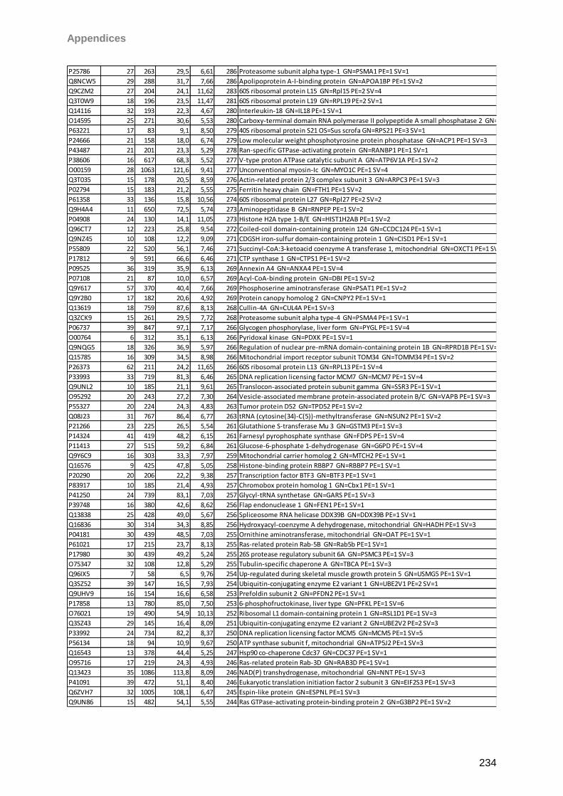
Appendices
234
P25786 27 263 29,5 6,61 286 Proteasome subunit alpha type-1 GN=PSMA1 PE=1 SV=1
Q8NCW5 29 288 31,7 7,66 286 Apolipoprotein A-I-binding protein GN=APOA1BP PE=1 SV=2
Q9CZM2 27 204 24,1 11,62 283 60S ribosomal protein L15 GN=Rpl15 PE=2 SV=4
Q3T0W9 18 196 23,5 11,47 281 60S ribosomal protein L19 GN=RPL19 PE=2 SV=1
Q14116 32 193 22,3 4,67 280 Interleukin-18 GN=IL18 PE=1 SV=1
O14595 25 271 30,6 5,53 280 Carboxy-terminal domain RNA polymerase II polypeptide A small phosphatase 2 GN=CTDSP2 PE=1 SV=2
P63221 17 83 9,1 8,50 279 40S ribosomal protein S21 OS=Sus scrofa GN=RPS21 PE=3 SV=1
P24666 21 158 18,0 6,74 279 Low molecular weight phosphotyrosine protein phosphatase GN=ACP1 PE=1 SV=3
P43487 21 201 23,3 5,29 278 Ran-specific GTPase-activating protein GN=RANBP1 PE=1 SV=1
P38606 16 617 68,3 5,52 277 V-type proton ATPase catalytic subunit A GN=ATP6V1A PE=1 SV=2
O00159 28 1063 121,6 9,41 277 Unconventional myosin-Ic GN=MYO1C PE=1 SV=4
Q3T035 15 178 20,5 8,59 276 Actin-related protein 2/3 complex subunit 3 GN=ARPC3 PE=1 SV=3
P02794 15 183 21,2 5,55 275 Ferritin heavy chain GN=FTH1 PE=1 SV=2
P61358 33 136 15,8 10,56 274 60S ribosomal protein L27 GN=Rpl27 PE=2 SV=2
Q9H4A4 11 650 72,5 5,74 273 Aminopeptidase B GN=RNPEP PE=1 SV=2
P04908 24 130 14,1 11,05 273 Histone H2A type 1-B/E GN=HIST1H2AB PE=1 SV=2
Q96CT7 12 223 25,8 9,54 272 Coiled-coil domain-containing protein 124 GN=CCDC124 PE=1 SV=1
Q9NZ45 10 108 12,2 9,09 271 CDGSH iron-sulfur domain-containing protein 1 GN=CISD1 PE=1 SV=1
P55809 22 520 56,1 7,46 271 Succinyl-CoA:3-ketoacid coenzyme A transferase 1, mitochondrial GN=OXCT1 PE=1 SV=1
P17812 9 591 66,6 6,46 271 CTP synthase 1 GN=CTPS1 PE=1 SV=2
P09525 36 319 35,9 6,13 269 Annexin A4 GN=ANXA4 PE=1 SV=4
P07108 21 87 10,0 6,57 269 Acyl-CoA-binding protein GN=DBI PE=1 SV=2
Q9Y617 57 370 40,4 7,66 269 Phosphoserine aminotransferase GN=PSAT1 PE=1 SV=2
Q9Y2B0 17 182 20,6 4,92 269 Protein canopy homolog 2 GN=CNPY2 PE=1 SV=1
Q13619 18 759 87,6 8,13 268 Cullin-4A GN=CUL4A PE=1 SV=3
Q3ZCK9 15 261 29,5 7,72 268 Proteasome subunit alpha type-4 GN=PSMA4 PE=1 SV=1
P06737 39 847 97,1 7,17 266 Glycogen phosphorylase, liver form GN=PYGL PE=1 SV=4
O00764 6 312 35,1 6,13 266 Pyridoxal kinase GN=PDXK PE=1 SV=1
Q9NQG5 18 326 36,9 5,97 266 Regulation of nuclear pre-mRNA domain-containing protein 1B GN=RPRD1B PE=1 SV=1
Q15785 16 309 34,5 8,98 266 Mitochondrial import receptor subunit TOM34 GN=TOMM34 PE=1 SV=2
P26373 62 211 24,2 11,65 266 60S ribosomal protein L13 GN=RPL13 PE=1 SV=4
P33993 33 719 81,3 6,46 265 DNA replication licensing factor MCM7 GN=MCM7 PE=1 SV=4
Q9UNL2 10 185 21,1 9,61 265 Translocon-associated protein subunit gamma GN=SSR3 PE=1 SV=1
O95292 20 243 27,2 7,30 264 Vesicle-associated membrane protein-associated protein B/C GN=VAPB PE=1 SV=3
P55327 20 224 24,3 4,83 263 Tumor protein D52 GN=TPD52 PE=1 SV=2
Q08J23 31 767 86,4 6,77 263 tRNA (cytosine(34)-C(5))-methyltransferase GN=NSUN2 PE=1 SV=2
P21266 23 225 26,5 5,54 261 Glutathione S-transferase Mu 3 GN=GSTM3 PE=1 SV=3
P14324 41 419 48,2 6,15 261 Farnesyl pyrophosphate synthase GN=FDPS PE=1 SV=4
P11413 27 515 59,2 6,84 261 Glucose-6-phosphate 1-dehydrogenase GN=G6PD PE=1 SV=4
Q9Y6C9 16 303 33,3 7,97 259 Mitochondrial carrier homolog 2 GN=MTCH2 PE=1 SV=1
Q16576 9 425 47,8 5,05 258 Histone-binding protein RBBP7 GN=RBBP7 PE=1 SV=1
P20290 20 206 22,2 9,38 257 Transcription factor BTF3 GN=BTF3 PE=1 SV=1
P83917 10 185 21,4 4,93 257 Chromobox protein homolog 1 GN=Cbx1 PE=1 SV=1
P41250 24 739 83,1 7,03 257 Glycyl-tRNA synthetase GN=GARS PE=1 SV=3
P39748 16 380 42,6 8,62 256 Flap endonuclease 1 GN=FEN1 PE=1 SV=1
Q13838 25 428 49,0 5,67 256 Spliceosome RNA helicase DDX39B GN=DDX39B PE=1 SV=1
Q16836 30 314 34,3 8,85 256 Hydroxyacyl-coenzyme A dehydrogenase, mitochondrial GN=HADH PE=1 SV=3
P04181 30 439 48,5 7,03 255 Ornithine aminotransferase, mitochondrial GN=OAT PE=1 SV=1
P61021 17 215 23,7 8,13 255 Ras-related protein Rab-5B GN=Rab5b PE=1 SV=1
P17980 30 439 49,2 5,24 255 26S protease regulatory subunit 6A GN=PSMC3 PE=1 SV=3
O75347 32 108 12,8 5,29 255 Tubulin-specific chaperone A GN=TBCA PE=1 SV=3
Q96IX5 7 58 6,5 9,76 254 Up-regulated during skeletal muscle growth protein 5 GN=USMG5 PE=1 SV=1
Q3SZ52 39 147 16,5 7,93 254 Ubiquitin-conjugating enzyme E2 variant 1 GN=UBE2V1 PE=2 SV=1
Q9UHV9 16 154 16,6 6,58 253 Prefoldin subunit 2 GN=PFDN2 PE=1 SV=1
P17858 13 780 85,0 7,50 253 6-phosphofructokinase, liver type GN=PFKL PE=1 SV=6
O76021 19 490 54,9 10,13 252 Ribosomal L1 domain-containing protein 1 GN=RSL1D1 PE=1 SV=3
Q3SZ43 29 145 16,4 8,09 251 Ubiquitin-conjugating enzyme E2 variant 2 GN=UBE2V2 PE=2 SV=3
P33992 24 734 82,2 8,37 250 DNA replication licensing factor MCM5 GN=MCM5 PE=1 SV=5
P56134 18 94 10,9 9,67 250 ATP synthase subunit f, mitochondrial GN=ATP5J2 PE=1 SV=3
Q16543 13 378 44,4 5,25 247 Hsp90 co-chaperone Cdc37 GN=CDC37 PE=1 SV=1
O95716 17 219 24,3 4,93 246 Ras-related protein Rab-3D GN=RAB3D PE=1 SV=1
Q13423 35 1086 113,8 8,09 246 NAD(P) transhydrogenase, mitochondrial GN=NNT PE=1 SV=3
P41091 39 472 51,1 8,40 246 Eukaryotic translation initiation factor 2 subunit 3 GN=EIF2S3 PE=1 SV=3
Q6ZVH7 32 1005 108,1 6,47 245 Espin-like protein GN=ESPNL PE=1 SV=3
Q9UN86 15 482 54,1 5,55 244 Ras GTPase-activating protein-binding protein 2 GN=G3BP2 PE=1 SV=2
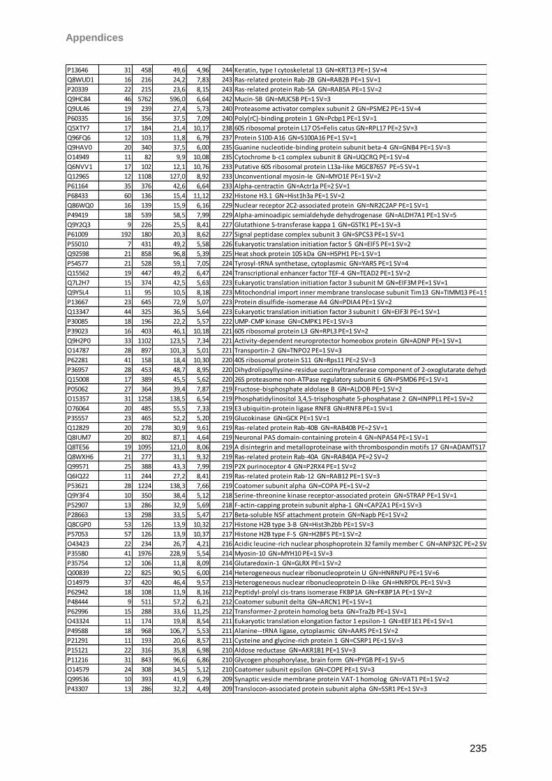
Appendices
235
P13646 31 458 49,6 4,96 244 Keratin, type I cytoskeletal 13 GN=KRT13 PE=1 SV=4
Q8WUD1 16 216 24,2 7,83 243 Ras-related protein Rab-2B GN=RAB2B PE=1 SV=1
P20339 22 215 23,6 8,15 243 Ras-related protein Rab-5A GN=RAB5A PE=1 SV=2
Q9HC84 46 5762 596,0 6,64 242 Mucin-5B GN=MUC5B PE=1 SV=3
Q9UL46 19 239 27,4 5,73 240 Proteasome activator complex subunit 2 GN=PSME2 PE=1 SV=4
P60335 16 356 37,5 7,09 240 Poly(rC)-binding protein 1 GN=Pcbp1 PE=1 SV=1
Q5XTY7 17 184 21,4 10,17 238 60S ribosomal protein L17 OS=Felis catus GN=RPL17 PE=2 SV=3
Q96FQ6 12 103 11,8 6,79 237 Protein S100-A16 GN=S100A16 PE=1 SV=1
Q9HAV0 20 340 37,5 6,00 235 Guanine nucleotide-binding protein subunit beta-4 GN=GNB4 PE=1 SV=3
O14949 11 82 9,9 10,08 235 Cytochrome b-c1 complex subunit 8 GN=UQCRQ PE=1 SV=4
Q6NVV1 17 102 12,1 10,76 233 Putative 60S ribosomal protein L13a-like MGC87657 PE=5 SV=1
Q12965 12 1108 127,0 8,92 233 Unconventional myosin-Ie GN=MYO1E PE=1 SV=2
P61164 35 376 42,6 6,64 233 Alpha-centractin GN=Actr1a PE=2 SV=1
P68433 60 136 15,4 11,12 232 Histone H3.1 GN=Hist1h3a PE=1 SV=2
Q86WQ0 16 139 15,9 6,16 229 Nuclear receptor 2C2-associated protein GN=NR2C2AP PE=1 SV=1
P49419 18 539 58,5 7,99 229 Alpha-aminoadipic semialdehyde dehydrogenase GN=ALDH7A1 PE=1 SV=5
Q9Y2Q3 9 226 25,5 8,41 227 Glutathione S-transferase kappa 1 GN=GSTK1 PE=1 SV=3
P61009 192 180 20,3 8,62 227 Signal peptidase complex subunit 3 GN=SPCS3 PE=1 SV=1
P55010 7 431 49,2 5,58 226 Eukaryotic translation initiation factor 5 GN=EIF5 PE=1 SV=2
Q92598 21 858 96,8 5,39 225 Heat shock protein 105 kDa GN=HSPH1 PE=1 SV=1
P54577 21 528 59,1 7,05 224 Tyrosyl-tRNA synthetase, cytoplasmic GN=YARS PE=1 SV=4
Q15562 19 447 49,2 6,47 224 Transcriptional enhancer factor TEF-4 GN=TEAD2 PE=1 SV=2
Q7L2H7 15 374 42,5 5,63 223 Eukaryotic translation initiation factor 3 subunit M GN=EIF3M PE=1 SV=1
Q9Y5L4 11 95 10,5 8,18 223 Mitochondrial import inner membrane translocase subunit Tim13 GN=TIMM13 PE=1 SV=1
P13667 23 645 72,9 5,07 223 Protein disulfide-isomerase A4 GN=PDIA4 PE=1 SV=2
Q13347 44 325 36,5 5,64 223 Eukaryotic translation initiation factor 3 subunit I GN=EIF3I PE=1 SV=1
P30085 18 196 22,2 5,57 222 UMP-CMP kinase GN=CMPK1 PE=1 SV=3
P39023 16 403 46,1 10,18 221 60S ribosomal protein L3 GN=RPL3 PE=1 SV=2
Q9H2P0 33 1102 123,5 7,34 221 Activity-dependent neuroprotector homeobox protein GN=ADNP PE=1 SV=1
O14787 28 897 101,3 5,01 221 Transportin-2 GN=TNPO2 PE=1 SV=3
P62281 41 158 18,4 10,30 220 40S ribosomal protein S11 GN=Rps11 PE=2 SV=3
P36957 28 453 48,7 8,95 220 Dihydrolipoyllysine-residue succinyltransferase component of 2-oxoglutarate dehydrogenase complex, mitochondrial GN=DLST PE=1 SV=4
Q15008 17 389 45,5 5,62 220 26S proteasome non-ATPase regulatory subunit 6 GN=PSMD6 PE=1 SV=1
P05062 27 364 39,4 7,87 219 Fructose-bisphosphate aldolase B GN=ALDOB PE=1 SV=2
O15357 31 1258 138,5 6,54 219 Phosphatidylinositol 3,4,5-trisphosphate 5-phosphatase 2 GN=INPPL1 PE=1 SV=2
O76064 20 485 55,5 7,33 219 E3 ubiquitin-protein ligase RNF8 GN=RNF8 PE=1 SV=1
P35557 23 465 52,2 5,20 219 Glucokinase GN=GCK PE=1 SV=1
Q12829 20 278 30,9 9,61 219 Ras-related protein Rab-40B GN=RAB40B PE=2 SV=1
Q8IUM7 20 802 87,1 4,64 219 Neuronal PAS domain-containing protein 4 GN=NPAS4 PE=1 SV=1
Q8TE56 19 1095 121,0 8,06 219 A disintegrin and metalloproteinase with thrombospondin motifs 17 GN=ADAMTS17 PE=2 SV=2
Q8WXH6 21 277 31,1 9,32 219 Ras-related protein Rab-40A GN=RAB40A PE=2 SV=2
Q99571 25 388 43,3 7,99 219 P2X purinoceptor 4 GN=P2RX4 PE=1 SV=2
Q6IQ22 11 244 27,2 8,41 219 Ras-related protein Rab-12 GN=RAB12 PE=1 SV=3
P53621 28 1224 138,3 7,66 219 Coatomer subunit alpha GN=COPA PE=1 SV=2
Q9Y3F4 10 350 38,4 5,12 218 Serine-threonine kinase receptor-associated protein GN=STRAP PE=1 SV=1
P52907 13 286 32,9 5,69 218 F-actin-capping protein subunit alpha-1 GN=CAPZA1 PE=1 SV=3
P28663 13 298 33,5 5,47 217 Beta-soluble NSF attachment protein GN=Napb PE=1 SV=2
Q8CGP0 53 126 13,9 10,32 217 Histone H2B type 3-B GN=Hist3h2bb PE=1 SV=3
P57053 57 126 13,9 10,37 217 Histone H2B type F-S GN=H2BFS PE=1 SV=2
O43423 22 234 26,7 4,21 216 Acidic leucine-rich nuclear phosphoprotein 32 family member C GN=ANP32C PE=2 SV=1
P35580 41 1976 228,9 5,54 214 Myosin-10 GN=MYH10 PE=1 SV=3
P35754 12 106 11,8 8,09 214 Glutaredoxin-1 GN=GLRX PE=1 SV=2
Q00839 22 825 90,5 6,00 214 Heterogeneous nuclear ribonucleoprotein U GN=HNRNPU PE=1 SV=6
O14979 37 420 46,4 9,57 213 Heterogeneous nuclear ribonucleoprotein D-like GN=HNRPDL PE=1 SV=3
P62942 18 108 11,9 8,16 212 Peptidyl-prolyl cis-trans isomerase FKBP1A GN=FKBP1A PE=1 SV=2
P48444 9 511 57,2 6,21 212 Coatomer subunit delta GN=ARCN1 PE=1 SV=1
P62996 15 288 33,6 11,25 212 Transformer-2 protein homolog beta GN=Tra2b PE=1 SV=1
O43324 11 174 19,8 8,54 211 Eukaryotic translation elongation factor 1 epsilon-1 GN=EEF1E1 PE=1 SV=1
P49588 18 968 106,7 5,53 211 Alanine--tRNA ligase, cytoplasmic GN=AARS PE=1 SV=2
P21291 11 193 20,6 8,57 211 Cysteine and glycine-rich protein 1 GN=CSRP1 PE=1 SV=3
P15121 22 316 35,8 6,98 210 Aldose reductase GN=AKR1B1 PE=1 SV=3
P11216 31 843 96,6 6,86 210 Glycogen phosphorylase, brain form GN=PYGB PE=1 SV=5
O14579 24 308 34,5 5,12 210 Coatomer subunit epsilon GN=COPE PE=1 SV=3
Q99536 10 393 41,9 6,29 209 Synaptic vesicle membrane protein VAT-1 homolog GN=VAT1 PE=1 SV=2
P43307 13 286 32,2 4,49 209 Translocon-associated protein subunit alpha GN=SSR1 PE=1 SV=3
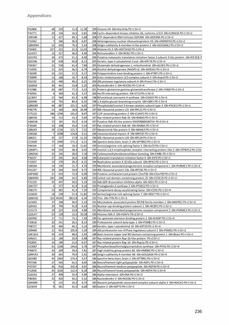
Appendices
236
P62806 48 103 11,4 11,36 209 Histone H4 GN=Hist1h4a PE=1 SV=2
P42771 20 156 16,5 5,81 208 Cyclin-dependent kinase inhibitor 2A, isoforms 1/2/3 GN=CDKN2A PE=1 SV=2
O00148 13 427 49,1 5,68 208 ATP-dependent RNA helicase DDX39A GN=DDX39A PE=1 SV=2
P31942 7 346 36,9 6,87 208 Heterogeneous nuclear ribonucleoprotein H3 GN=HNRNPH3 PE=1 SV=2
Q8N9W4 52 650 79,3 5,54 208 Golgin subfamily A member 6-like protein 2 GN=GOLGA6L2 PE=2 SV=2
P16402 317 221 22,3 11,02 208 Histone H1.3 GN=HIST1H1D PE=1 SV=2
Q14257 23 317 36,9 4,40 206 Reticulocalbin-2 GN=RCN2 PE=1 SV=1
Q2VIR3 34 472 51,2 8,40 206 Putative eukaryotic translation initiation factor 2 subunit 3-like protein GN=EIF2S3L PE=5 SV=2
Q01546 33 638 65,8 8,12 205 Keratin, type II cytoskeletal 2 oral GN=KRT76 PE=1 SV=2
P00367 22 558 61,4 7,80 205 Glutamate dehydrogenase 1, mitochondrial GN=GLUD1 PE=1 SV=2
P14550 21 325 36,5 6,79 204 Alcohol dehydrogenase [NADP(+)] GN=AKR1A1 PE=1 SV=3
P26599 33 531 57,2 9,17 204 Polypyrimidine tract-binding protein 1 GN=PTBP1 PE=1 SV=1
P59999 13 168 19,7 8,43 204 Actin-related protein 2/3 complex subunit 4 GN=Arpc4 PE=1 SV=3
P62192 22 440 49,2 6,21 204 26S protease regulatory subunit 4 GN=Psmc1 PE=1 SV=1
Q02818 30 461 53,8 5,25 203 Nucleobindin-1 GN=NUCB1 PE=1 SV=4
P21980 20 687 77,3 5,22 201 Protein-glutamine gamma-glutamyltransferase 2 GN=TGM2 PE=1 SV=2
P50502 9 369 41,3 5,27 200 Hsc70-interacting protein GN=ST13 PE=1 SV=2
Q13057 12 564 62,3 6,99 198 Bifunctional coenzyme A synthase GN=COASY PE=1 SV=4
Q04446 14 702 80,4 6,32 198 1,4-alpha-glucan-branching enzyme GN=GBE1 PE=1 SV=3
Q8NEB9 44 887 101,5 6,81 197 Phosphatidylinositol 3-kinase catalytic subunit type 3 GN=PIK3C3 PE=1 SV=1
P46778 13 160 18,6 10,49 197 60S ribosomal protein L21 GN=RPL21 PE=1 SV=2
O75122 186 1294 141,0 8,47 197 CLIP-associating protein 2 GN=CLASP2 PE=1 SV=2
Q86YS6 10 212 23,3 5,64 197 Ras-related protein Rab-43 GN=RAB43 PE=1 SV=1
A6NDJ8 12 181 20,2 6,54 197 Putative Rab-43-like protein ENSP00000330714 PE=5 SV=3
P20340 10 208 23,6 5,54 197 Ras-related protein Rab-6A GN=RAB6A PE=1 SV=3
Q96JK9 29 1134 121,7 7,52 197 Mastermind-like protein 3 GN=MAML3 PE=1 SV=3
O00160 7 1098 124,8 9,11 196 Unconventional myosin-If GN=MYO1F PE=1 SV=3
Q862I1 25 157 17,8 11,25 196 60S ribosomal protein L24 GN=RPL24 PE=2 SV=2
O15020 71 2390 271,2 6,11 196 Spectrin beta chain, brain 2 GN=SPTBN2 PE=1 SV=3
P84104 17 164 19,3 11,65 195 Serine/arginine-rich splicing factor 3 GN=Srsf3 PE=1 SV=1
Q3MIP1 43 535 58,4 9,42 195 Inositol 1,4,5-trisphosphate receptor-interacting protein-like 2 GN=ITPRIPL2 PE=2 SV=1
Q96DG6 25 245 28,0 7,18 195 Carboxymethylenebutenolidase homolog GN=CMBL PE=1 SV=1
P56537 27 245 26,6 4,68 194 Eukaryotic translation initiation factor 6 GN=EIF6 PE=1 SV=1
P15927 13 270 29,2 6,15 194 Replication protein A 32 kDa subunit GN=RPA2 PE=1 SV=1
O00264 14 195 21,7 4,70 193 Membrane-associated progesterone receptor component 1 GN=PGRMC1 PE=1 SV=3
P61247 29 264 29,9 9,73 193 40S ribosomal protein S3a GN=RPS3A PE=1 SV=2
A6PW82 21 633 72,0 6,09 193 Putative uncharacterized protein CXorf30 GN=CXorf30 PE=2 SV=2
Q86WR0 292 208 24,5 6,80 193 Coiled-coil domain-containing protein 25 GN=CCDC25 PE=1 SV=2
P31150 35 447 50,6 5,14 192 Rab GDP dissociation inhibitor alpha GN=GDI1 PE=1 SV=2
Q9H7Z7 6 377 41,9 9,16 192 Prostaglandin E synthase 2 GN=PTGES2 PE=1 SV=1
P08174 23 381 41,4 7,59 192 Complement decay-accelerating factor GN=CD55 PE=1 SV=4
Q16629 18 238 27,4 11,82 192 Serine/arginine-rich splicing factor 7 GN=SRSF7 PE=1 SV=1
Q8WZ42 323 34350 3813,8 6,34 192 Titin GN=TTN PE=1 SV=3
Q15691 37 268 30,0 5,14 191 Microtubule-associated protein RP/EB family member 1 GN=MAPRE1 PE=1 SV=3
Q09161 29 790 91,8 6,43 191 Nuclear cap-binding protein subunit 1 GN=NCBP1 PE=1 SV=1
O15173 19 223 23,8 4,88 191 Membrane-associated progesterone receptor component 2 GN=PGRMC2 PE=1 SV=1
Q32LA7 53 128 13,5 10,58 190 Histone H2A.V GN=H2AFV PE=2 SV=3
Q92945 17 711 73,1 7,30 190 Far upstream element-binding protein 2 GN=KHSRP PE=1 SV=4
P20618 20 241 26,5 8,13 189 Proteasome subunit beta type-1 GN=PSMB1 PE=1 SV=2
P08727 45 400 44,1 5,14 189 Keratin, type I cytoskeletal 19 GN=KRT19 PE=1 SV=4
Q99460 22 953 105,8 5,39 189 26S proteasome non-ATPase regulatory subunit 1 GN=PSMD1 PE=1 SV=2
Q9CQC6 59 419 48,0 5,92 188 Basic leucine zipper and W2 domain-containing protein 1 GN=Bzw1 PE=1 SV=1
A6NIZ1 12 184 20,9 5,48 187 Ras-related protein Rap-1b-like protein PE=2 SV=1
P62835 14 184 21,0 6,67 187 Ras-related protein Rap-1A GN=Rap1a PE=2 SV=1
O15067 41 1338 144,6 5,76 187 Phosphoribosylformylglycinamidine synthase GN=PFAS PE=1 SV=4
P40673 43 209 24,0 7,81 187 High mobility group protein B2 GN=HMGB2 PE=1 SV=3
Q9NYA3 39 693 79,9 5,41 186 Golgin subfamily A member 6A GN=GOLGA6A PE=1 SV=3
Q01082 45 2364 274,4 5,57 186 Spectrin beta chain, brain 1 GN=SPTBN1 PE=1 SV=2
P07196 27 543 61,5 4,65 186 Neurofilament light polypeptide GN=NEFL PE=1 SV=3
P07197 36 916 102,4 4,91 186 Neurofilament medium polypeptide GN=NEFM PE=1 SV=3
P12036 45 1026 112,4 6,18 186 Neurofilament heavy polypeptide GN=NEFH PE=1 SV=4
Q16352 27 499 55,4 5,40 186 Alpha-internexin GN=INA PE=1 SV=2
P80303 32 420 50,2 5,12 186 Nucleobindin-2 GN=NUCB2 PE=1 SV=2
Q9H009 5 215 23,2 4,73 185 Nascent polypeptide-associated complex subunit alpha-2 GN=NACA2 PE=1 SV=1
Q15019 9 361 41,5 6,60 185 Septin-2 GN=SEPT2 PE=1 SV=1
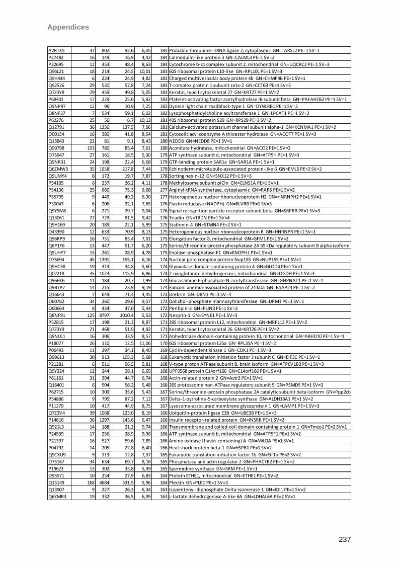
Appendices
237
A2RTX5 37 802 92,6 6,05 185 Probable threonine--tRNA ligase 2, cytoplasmic GN=TARSL2 PE=1 SV=1
P27482 16 149 16,9 4,42 184 Calmodulin-like protein 3 GN=CALML3 PE=1 SV=2
P22695 12 453 48,4 8,63 184 Cytochrome b-c1 complex subunit 2, mitochondrial GN=UQCRC2 PE=1 SV=3
Q96L21 18 214 24,5 10,01 183 60S ribosomal protein L10-like GN=RPL10L PE=1 SV=3
Q9H444 6 224 24,9 4,82 183 Charged multivesicular body protein 4b GN=CHMP4B PE=1 SV=1
Q92526 29 530 57,8 7,24 183 T-complex protein 1 subunit zeta-2 GN=CCT6B PE=1 SV=5
Q7Z3Y8 29 459 49,8 5,05 183 Keratin, type I cytoskeletal 27 GN=KRT27 PE=1 SV=2
P68401 17 229 25,6 5,92 183 Platelet-activating factor acetylhydrolase IB subunit beta GN=PAFAH1B2 PE=1 SV=1
Q9NP97 12 96 10,9 7,25 182 Dynein light chain roadblock-type 1 GN=DYNLRB1 PE=1 SV=3
Q8NF37 7 534 59,1 6,02 182 Lysophosphatidylcholine acyltransferase 1 GN=LPCAT1 PE=1 SV=2
P62276 25 56 6,7 10,13 181 40S ribosomal protein S29 GN=RPS29 PE=3 SV=2
Q12791 36 1236 137,5 7,06 181 Calcium-activated potassium channel subunit alpha-1 GN=KCNMA1 PE=1 SV=2
O00154 16 380 41,8 8,54 181 Cytosolic acyl coenzyme A thioester hydrolase GN=ACOT7 PE=1 SV=3
Q15843 22 81 9,1 8,43 180 NEDD8 GN=NEDD8 PE=1 SV=1
Q99798 193 780 85,4 7,61 180 Aconitate hydratase, mitochondrial GN=ACO2 PE=1 SV=2
O75947 27 161 18,5 5,30 179 ATP synthase subunit d, mitochondrial GN=ATP5H PE=1 SV=3
Q9NR31 24 198 22,4 6,68 179 GTP-binding protein SAR1a GN=SAR1A PE=1 SV=1
Q6ZMW3 35 1958 217,8 7,44 179 Echinoderm microtubule-associated protein-like 6 GN=EML6 PE=2 SV=2
Q9UMY4 8 172 19,7 7,87 178 Sorting nexin-12 GN=SNX12 PE=1 SV=3
P54105 6 237 26,2 4,11 178 Methylosome subunit pICln GN=CLNS1A PE=1 SV=1
P54136 25 660 75,3 6,68 177 Arginyl-tRNA synthetase, cytoplasmic GN=RARS PE=1 SV=2
P55795 9 449 49,2 6,30 177 Heterogeneous nuclear ribonucleoprotein H2 GN=HNRNPH2 PE=1 SV=1
P30043 6 206 22,1 7,65 176 Flavin reductase (NADPH) GN=BLVRB PE=1 SV=3
Q9Y5M8 6 271 29,7 9,04 176 Signal recognition particle receptor subunit beta GN=SRPRB PE=1 SV=3
Q13061 27 729 81,5 9,42 176 Triadin GN=TRDN PE=1 SV=4
Q9H169 20 189 22,1 5,99 175 Stathmin-4 GN=STMN4 PE=2 SV=1
O43390 12 633 70,9 8,13 175 Heterogeneous nuclear ribonucleoprotein R GN=HNRNPR PE=1 SV=1
Q96RP9 16 751 83,4 7,01 175 Elongation factor G, mitochondrial GN=GFM1 PE=1 SV=2
Q6P1F6 13 447 51,7 6,20 175 Serine/threonine-protein phosphatase 2A 55 kDa regulatory subunit B alpha isoform GN=Ppp2r2a PE=1 SV=1
Q9UHY7 15 261 28,9 4,78 175 Enolase-phosphatase E1 GN=ENOPH1 PE=1 SV=1
O75694 45 1391 155,1 6,16 174 Nuclear pore complex protein Nup155 GN=NUP155 PE=1 SV=1
Q9HC38 19 313 34,8 5,60 174 Glyoxalase domain-containing protein 4 GN=GLOD4 PE=1 SV=1
Q02218 35 1023 115,9 6,86 174 2-oxoglutarate dehydrogenase, mitochondrial GN=OGDH PE=1 SV=3
Q96EK6 11 184 20,7 7,99 174 Glucosamine 6-phosphate N-acetyltransferase GN=GNPNAT1 PE=1 SV=1
Q9BTP7 14 215 23,9 9,19 174 Fanconi anemia-associated protein of 24 kDa GN=FAAP24 PE=1 SV=2
Q16643 7 649 71,4 4,45 173 Drebrin GN=DBN1 PE=1 SV=4
O60762 34 260 29,6 9,57 173 Dolichol-phosphate mannosyltransferase GN=DPM1 PE=1 SV=1
O60664 8 434 47,0 5,44 172 Perilipin-3 GN=PLIN3 PE=1 SV=3
Q8NF91 125 8797 1010,4 5,53 172 Nesprin-1 GN=SYNE1 PE=1 SV=3
P52815 17 198 21,3 8,87 171 39S ribosomal protein L12, mitochondrial GN=MRPL12 PE=1 SV=2
Q7Z3Y9 21 468 51,9 4,92 171 Keratin, type I cytoskeletal 26 GN=KRT26 PE=1 SV=2
Q9NUJ1 16 306 33,9 8,57 171 Abhydrolase domain-containing protein 10, mitochondrial GN=ABHD10 PE=1 SV=1
P18077 26 110 12,5 11,06 170 60S ribosomal protein L35a GN=RPL35A PE=1 SV=2
P06493 11 297 34,1 8,40 169 Cyclin-dependent kinase 1 GN=CDK1 PE=1 SV=3
Q99613 30 913 105,3 5,68 168 Eukaryotic translation initiation factor 3 subunit C GN=EIF3C PE=1 SV=1
P21281 6 511 56,5 5,81 168 V-type proton ATPase subunit B, brain isoform GN=ATP6V1B2 PE=1 SV=3
Q9Y224 12 244 28,1 6,65 168 UPF0568 protein C14orf166 GN=C14orf166 PE=1 SV=1
P61161 31 394 44,7 6,74 168 Actin-related protein 2 GN=Actr2 PE=1 SV=1
Q16401 6 504 56,2 5,48 168 26S proteasome non-ATPase regulatory subunit 5 GN=PSMD5 PE=1 SV=3
P62715 10 309 35,6 5,43 167 Serine/threonine-protein phosphatase 2A catalytic subunit beta isoform GN=Ppp2cb PE=1 SV=1
P54886 9 795 87,2 7,12 167 Delta-1-pyrroline-5-carboxylate synthase GN=ALDH18A1 PE=1 SV=2
P11279 10 417 44,9 8,75 167 Lysosome-associated membrane glycoprotein 1 GN=LAMP1 PE=1 SV=3
Q7Z3V4 39 1068 123,0 8,19 166 Ubiquitin-protein ligase E3B GN=UBE3B PE=1 SV=3
P14616 36 1297 143,6 6,47 166 Insulin receptor-related protein GN=INSRR PE=1 SV=2
Q921L3 14 188 21,2 9,74 166 Transmembrane and coiled-coil domain-containing protein 1 GN=Tmco1 PE=2 SV=1
P24539 17 256 28,9 9,36 166 ATP synthase subunit b, mitochondrial GN=ATP5F1 PE=1 SV=2
P21397 16 527 59,6 7,85 166 Amine oxidase [flavin-containing] A GN=MAOA PE=1 SV=1
P04792 14 205 22,8 6,40 166 Heat shock protein beta-1 GN=HSPB1 PE=1 SV=2
Q9CXU9 9 113 12,8 7,37 165 Eukaryotic translation initiation factor 1b GN=Eif1b PE=2 SV=2
O75167 34 634 69,7 8,16 165 Phosphatase and actin regulator 2 GN=PHACTR2 PE=1 SV=2
P19623 13 302 33,8 5,49 165 Spermidine synthase GN=SRM PE=1 SV=1
O95571 10 254 27,9 6,83 164 Protein ETHE1, mitochondrial GN=ETHE1 PE=1 SV=2
Q15149 168 4684 531,5 5,96 164 Plectin GN=PLEC PE=1 SV=3
Q13907 9 227 26,3 6,34 163 Isopentenyl-diphosphate Delta-isomerase 1 GN=IDI1 PE=1 SV=2
Q6ZMR3 19 332 36,5 6,99 163 L-lactate dehydrogenase A-like 6A GN=LDHAL6A PE=2 SV=1
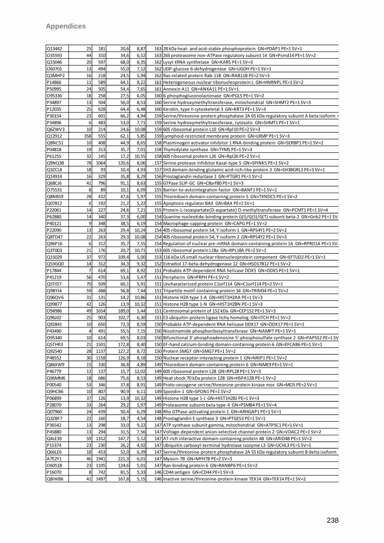
Appendices
238
Q13442 25 181 20,6 8,87 163 28 kDa heat- and acid-stable phosphoprotein GN=PDAP1 PE=1 SV=1
O35593 44 310 34,6 6,52 163 26S proteasome non-ATPase regulatory subunit 14 GN=Psmd14 PE=1 SV=2
Q15046 20 597 68,0 6,35 162 Lysyl-tRNA synthetase GN=KARS PE=1 SV=3
O60701 13 494 55,0 7,12 162 UDP-glucose 6-dehydrogenase GN=UGDH PE=1 SV=1
Q3MHP2 16 218 24,5 5,94 162 Ras-related protein Rab-11B GN=RAB11B PE=2 SV=3
P14866 11 589 64,1 8,22 161 Heterogeneous nuclear ribonucleoprotein L GN=HNRNPL PE=1 SV=2
P50995 24 505 54,4 7,65 161 Annexin A11 GN=ANXA11 PE=1 SV=1
O95336 18 258 27,5 6,05 160 6-phosphogluconolactonase GN=PGLS PE=1 SV=2
P34897 13 504 56,0 8,53 160 Serine hydroxymethyltransferase, mitochondrial GN=SHMT2 PE=1 SV=3
P12035 25 628 64,4 6,48 160 Keratin, type II cytoskeletal 3 GN=KRT3 PE=1 SV=3
P30154 23 601 66,2 4,94 159 Serine/threonine-protein phosphatase 2A 65 kDa regulatory subunit A beta isoform GN=PPP2R1B PE=1 SV=3
P34896 6 483 53,0 7,71 159 Serine hydroxymethyltransferase, cytosolic GN=SHMT1 PE=1 SV=1
Q6ZWV3 10 214 24,6 10,08 159 60S ribosomal protein L10 GN=Rpl10 PE=2 SV=3
Q12912 358 555 62,1 5,85 159 Lymphoid-restricted membrane protein GN=LRMP PE=1 SV=3
Q8NC51 10 408 44,9 8,65 158 Plasminogen activator inhibitor 1 RNA-binding protein GN=SERBP1 PE=1 SV=2
P04818 19 313 35,7 7,01 158 Thymidylate synthase GN=TYMS PE=1 SV=3
P61255 32 145 17,2 10,55 158 60S ribosomal protein L26 GN=Rpl26 PE=2 SV=1
Q9NQ38 78 1064 120,6 8,06 157 Serine protease inhibitor Kazal-type 5 GN=SPINK5 PE=1 SV=2
Q3ZCL8 18 93 10,4 4,93 157 SH3 domain-binding glutamic acid-rich-like protein 3 GN=SH3BGRL3 PE=3 SV=1
Q14914 16 329 35,8 8,29 156 Prostaglandin reductase 1 GN=PTGR1 PE=1 SV=2
Q68CJ6 41 796 91,1 8,63 155 GTPase SLIP-GC GN=C8orf80 PE=1 SV=3
O75531 8 89 10,1 6,09 155 Barrier-to-autointegration factor GN=BANF1 PE=1 SV=1
Q8NBS9 28 432 47,6 5,97 155 Thioredoxin domain-containing protein 5 GN=TXNDC5 PE=1 SV=2
Q07812 6 192 21,2 5,22 155 Apoptosis regulator BAX GN=BAX PE=1 SV=1
P22061 14 227 24,6 7,21 155 Protein-L-isoaspartate(D-aspartate) O-methyltransferase GN=PCMT1 PE=1 SV=4
P62880 14 340 37,3 6,00 154 Guanine nucleotide-binding protein G(I)/G(S)/G(T) subunit beta-2 GN=Gnb2 PE=1 SV=3
P40121 9 348 38,5 6,19 154 Macrophage-capping protein GN=CAPG PE=1 SV=2
P22090 13 263 29,4 10,24 154 40S ribosomal protein S4, Y isoform 1 GN=RPS4Y1 PE=2 SV=2
Q8TD47 22 263 29,3 10,08 154 40S ribosomal protein S4, Y isoform 2 GN=RPS4Y2 PE=1 SV=3
Q96P16 6 312 35,7 7,55 154 Regulation of nuclear pre-mRNA domain-containing protein 1A GN=RPRD1A PE=1 SV=1
Q3T003 21 176 20,7 10,71 153 60S ribosomal protein L18a GN=RPL18A PE=2 SV=1
Q15029 37 972 109,4 5,00 153 116 kDa U5 small nuclear ribonucleoprotein component GN=EFTUD2 PE=1 SV=1
Q53GQ0 14 312 34,3 9,32 152 Estradiol 17-beta-dehydrogenase 12 GN=HSD17B12 PE=1 SV=2
P17844 7 614 69,1 8,92 151 Probable ATP-dependent RNA helicase DDX5 GN=DDX5 PE=1 SV=1
P41219 56 470 53,6 5,47 151 Peripherin GN=PRPH PE=1 SV=2
Q5TID7 70 509 60,1 5,91 151 Uncharacterized protein C1orf114 GN=C1orf114 PE=2 SV=1
Q9BYJ4 59 488 56,8 7,44 151 Tripartite motif-containing protein 34 GN=TRIM34 PE=1 SV=2
Q96QV6 31 131 14,2 10,86 151 Histone H2A type 1-A GN=HIST1H2AA PE=1 SV=3
Q99877 42 126 13,9 10,32 151 Histone H2B type 1-N GN=HIST1H2BN PE=1 SV=3
O94986 49 1654 189,0 5,44 151 Centrosomal protein of 152 kDa GN=CEP152 PE=1 SV=3
Q96J02 25 903 102,7 6,30 151 E3 ubiquitin-protein ligase Itchy homolog GN=ITCH PE=1 SV=2
Q92841 10 650 72,3 8,59 150 Probable ATP-dependent RNA helicase DDX17 GN=DDX17 PE=1 SV=1
P43490 4 491 55,5 7,15 150 Nicotinamide phosphoribosyltransferase GN=NAMPT PE=1 SV=1
O95340 10 614 69,5 8,03 150 Bifunctional 3'-phosphoadenosine 5'-phosphosulfate synthase 2 GN=PAPSS2 PE=1 SV=2
Q5THR3 21 1501 172,8 8,40 150 EF-hand calcium-binding domain-containing protein 6 GN=EFCAB6 PE=1 SV=1
Q92540 28 1137 127,2 8,72 150 Protein SMG7 GN=SMG7 PE=1 SV=2
P48552 30 1158 126,9 8,18 150 Nuclear receptor-interacting protein 1 GN=NRIP1 PE=1 SV=2
Q86XW9 15 330 36,8 4,89 149 Thioredoxin domain-containing protein 6 GN=NME9 PE=1 SV=1
P46779 12 137 15,7 12,02 149 60S ribosomal protein L28 GN=RPL28 PE=1 SV=3
Q96MM6 18 686 75,6 8,53 149 Heat shock 70 kDa protein 12B GN=HSPA12B PE=1 SV=2
P00540 53 346 37,8 8,91 149 Proto-oncogene serine/threonine-protein kinase mos GN=MOS PE=2 SV=1
Q9HCB6 10 807 90,9 6,11 149 Spondin-1 GN=SPON1 PE=1 SV=2
P06899 37 126 13,9 10,32 149 Histone H2B type 1-J GN=HIST1H2BJ PE=1 SV=3
P28070 33 264 29,2 5,97 149 Proteasome subunit beta type-4 GN=PSMB4 PE=1 SV=4
Q07960 24 439 50,4 6,29 148 Rho GTPase-activating protein 1 GN=ARHGAP1 PE=1 SV=1
Q3ZBF7 22 160 18,7 4,54 148 Prostaglandin E synthase 3 GN=PTGES3 PE=1 SV=1
P36542 13 298 33,0 9,22 147 ATP synthase subunit gamma, mitochondrial GN=ATP5C1 PE=1 SV=1
P45880 13 294 31,5 7,56 147 Voltage-dependent anion-selective channel protein 2 GN=VDAC2 PE=1 SV=2
Q4LE39 59 1312 147,7 5,12 147 AT-rich interactive domain-containing protein 4B GN=ARID4B PE=1 SV=2
P15374 23 230 26,2 4,92 147 Ubiquitin carboxyl-terminal hydrolase isozyme L3 GN=UCHL3 PE=1 SV=1
Q66LE6 18 453 52,0 6,39 147 Serine/threonine-protein phosphatase 2A 55 kDa regulatory subunit B delta isoform GN=PPP2R2D PE=1 SV=1
A7E2Y1 46 1941 221,3 6,01 147 Myosin-7B GN=MYH7B PE=2 SV=3
O60518 23 1105 124,6 5,01 147 Ran-binding protein 6 GN=RANBP6 PE=1 SV=2
P16070 8 742 81,5 5,33 146 CD44 antigen GN=CD44 PE=1 SV=3
Q8IWB6 41 1497 167,8 5,15 146 Inactive serine/threonine-protein kinase TEX14 GN=TEX14 PE=1 SV=2
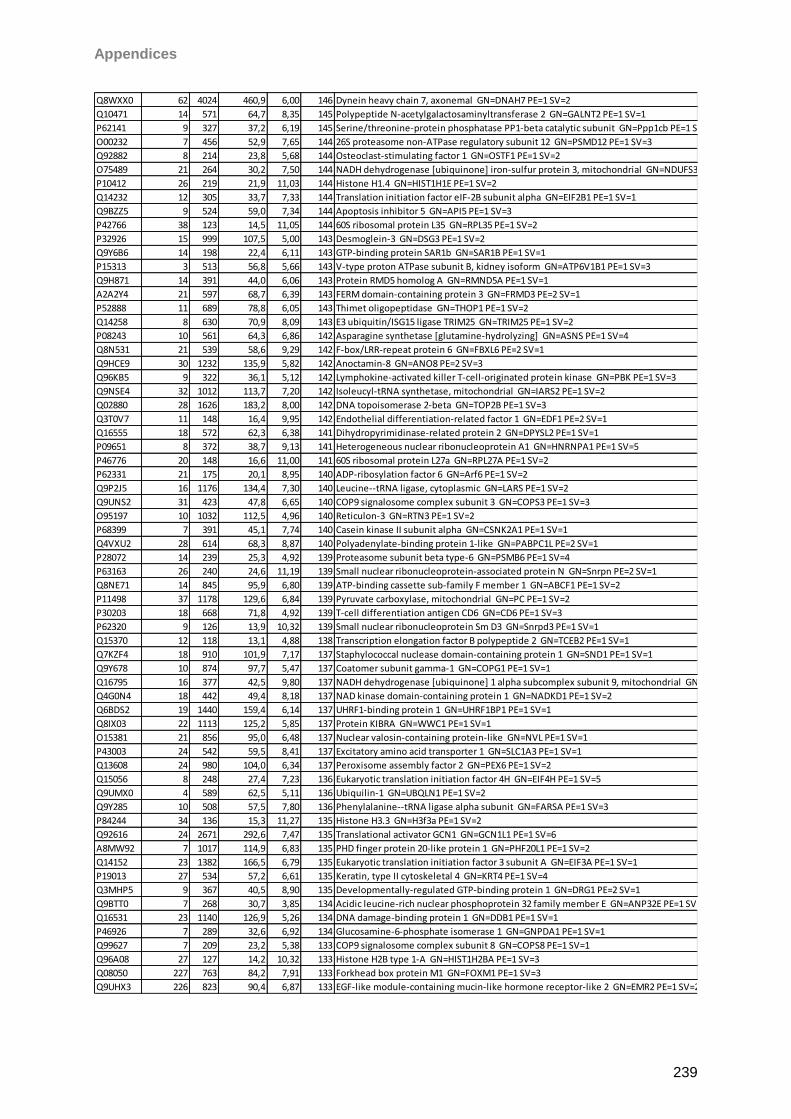
Appendices
239
Q8WXX0 62 4024 460,9 6,00 146 Dynein heavy chain 7, axonemal GN=DNAH7 PE=1 SV=2
Q10471 14 571 64,7 8,35 145 Polypeptide N-acetylgalactosaminyltransferase 2 GN=GALNT2 PE=1 SV=1
P62141 9 327 37,2 6,19 145 Serine/threonine-protein phosphatase PP1-beta catalytic subunit GN=Ppp1cb PE=1 SV=3
O00232 7 456 52,9 7,65 144 26S proteasome non-ATPase regulatory subunit 12 GN=PSMD12 PE=1 SV=3
Q92882 8 214 23,8 5,68 144 Osteoclast-stimulating factor 1 GN=OSTF1 PE=1 SV=2
O75489 21 264 30,2 7,50 144 NADH dehydrogenase [ubiquinone] iron-sulfur protein 3, mitochondrial GN=NDUFS3 PE=1 SV=1
P10412 26 219 21,9 11,03 144 Histone H1.4 GN=HIST1H1E PE=1 SV=2
Q14232 12 305 33,7 7,33 144 Translation initiation factor eIF-2B subunit alpha GN=EIF2B1 PE=1 SV=1
Q9BZZ5 9 524 59,0 7,34 144 Apoptosis inhibitor 5 GN=API5 PE=1 SV=3
P42766 38 123 14,5 11,05 144 60S ribosomal protein L35 GN=RPL35 PE=1 SV=2
P32926 15 999 107,5 5,00 143 Desmoglein-3 GN=DSG3 PE=1 SV=2
Q9Y6B6 14 198 22,4 6,11 143 GTP-binding protein SAR1b GN=SAR1B PE=1 SV=1
P15313 3 513 56,8 5,66 143 V-type proton ATPase subunit B, kidney isoform GN=ATP6V1B1 PE=1 SV=3
Q9H871 14 391 44,0 6,06 143 Protein RMD5 homolog A GN=RMND5A PE=1 SV=1
A2A2Y4 21 597 68,7 6,39 143 FERM domain-containing protein 3 GN=FRMD3 PE=2 SV=1
P52888 11 689 78,8 6,05 143 Thimet oligopeptidase GN=THOP1 PE=1 SV=2
Q14258 8 630 70,9 8,09 143 E3 ubiquitin/ISG15 ligase TRIM25 GN=TRIM25 PE=1 SV=2
P08243 10 561 64,3 6,86 142 Asparagine synthetase [glutamine-hydrolyzing] GN=ASNS PE=1 SV=4
Q8N531 21 539 58,6 9,29 142 F-box/LRR-repeat protein 6 GN=FBXL6 PE=2 SV=1
Q9HCE9 30 1232 135,9 5,82 142 Anoctamin-8 GN=ANO8 PE=2 SV=3
Q96KB5 9 322 36,1 5,12 142 Lymphokine-activated killer T-cell-originated protein kinase GN=PBK PE=1 SV=3
Q9NSE4 32 1012 113,7 7,20 142 Isoleucyl-tRNA synthetase, mitochondrial GN=IARS2 PE=1 SV=2
Q02880 28 1626 183,2 8,00 142 DNA topoisomerase 2-beta GN=TOP2B PE=1 SV=3
Q3T0V7 11 148 16,4 9,95 142 Endothelial differentiation-related factor 1 GN=EDF1 PE=2 SV=1
Q16555 18 572 62,3 6,38 141 Dihydropyrimidinase-related protein 2 GN=DPYSL2 PE=1 SV=1
P09651 8 372 38,7 9,13 141 Heterogeneous nuclear ribonucleoprotein A1 GN=HNRNPA1 PE=1 SV=5
P46776 20 148 16,6 11,00 141 60S ribosomal protein L27a GN=RPL27A PE=1 SV=2
P62331 21 175 20,1 8,95 140 ADP-ribosylation factor 6 GN=Arf6 PE=1 SV=2
Q9P2J5 16 1176 134,4 7,30 140 Leucine--tRNA ligase, cytoplasmic GN=LARS PE=1 SV=2
Q9UNS2 31 423 47,8 6,65 140 COP9 signalosome complex subunit 3 GN=COPS3 PE=1 SV=3
O95197 10 1032 112,5 4,96 140 Reticulon-3 GN=RTN3 PE=1 SV=2
P68399 7 391 45,1 7,74 140 Casein kinase II subunit alpha GN=CSNK2A1 PE=1 SV=1
Q4VXU2 28 614 68,3 8,87 140 Polyadenylate-binding protein 1-like GN=PABPC1L PE=2 SV=1
P28072 14 239 25,3 4,92 139 Proteasome subunit beta type-6 GN=PSMB6 PE=1 SV=4
P63163 26 240 24,6 11,19 139 Small nuclear ribonucleoprotein-associated protein N GN=Snrpn PE=2 SV=1
Q8NE71 14 845 95,9 6,80 139 ATP-binding cassette sub-family F member 1 GN=ABCF1 PE=1 SV=2
P11498 37 1178 129,6 6,84 139 Pyruvate carboxylase, mitochondrial GN=PC PE=1 SV=2
P30203 18 668 71,8 4,92 139 T-cell differentiation antigen CD6 GN=CD6 PE=1 SV=3
P62320 9 126 13,9 10,32 139 Small nuclear ribonucleoprotein Sm D3 GN=Snrpd3 PE=1 SV=1
Q15370 12 118 13,1 4,88 138 Transcription elongation factor B polypeptide 2 GN=TCEB2 PE=1 SV=1
Q7KZF4 18 910 101,9 7,17 137 Staphylococcal nuclease domain-containing protein 1 GN=SND1 PE=1 SV=1
Q9Y678 10 874 97,7 5,47 137 Coatomer subunit gamma-1 GN=COPG1 PE=1 SV=1
Q16795 16 377 42,5 9,80 137 NADH dehydrogenase [ubiquinone] 1 alpha subcomplex subunit 9, mitochondrial GN=NDUFA9 PE=1 SV=2
Q4G0N4 18 442 49,4 8,18 137 NAD kinase domain-containing protein 1 GN=NADKD1 PE=1 SV=2
Q6BDS2 19 1440 159,4 6,14 137 UHRF1-binding protein 1 GN=UHRF1BP1 PE=1 SV=1
Q8IX03 22 1113 125,2 5,85 137 Protein KIBRA GN=WWC1 PE=1 SV=1
O15381 21 856 95,0 6,48 137 Nuclear valosin-containing protein-like GN=NVL PE=1 SV=1
P43003 24 542 59,5 8,41 137 Excitatory amino acid transporter 1 GN=SLC1A3 PE=1 SV=1
Q13608 24 980 104,0 6,34 137 Peroxisome assembly factor 2 GN=PEX6 PE=1 SV=2
Q15056 8 248 27,4 7,23 136 Eukaryotic translation initiation factor 4H GN=EIF4H PE=1 SV=5
Q9UMX0 4 589 62,5 5,11 136 Ubiquilin-1 GN=UBQLN1 PE=1 SV=2
Q9Y285 10 508 57,5 7,80 136 Phenylalanine--tRNA ligase alpha subunit GN=FARSA PE=1 SV=3
P84244 34 136 15,3 11,27 135 Histone H3.3 GN=H3f3a PE=1 SV=2
Q92616 24 2671 292,6 7,47 135 Translational activator GCN1 GN=GCN1L1 PE=1 SV=6
A8MW92 7 1017 114,9 6,83 135 PHD finger protein 20-like protein 1 GN=PHF20L1 PE=1 SV=2
Q14152 23 1382 166,5 6,79 135 Eukaryotic translation initiation factor 3 subunit A GN=EIF3A PE=1 SV=1
P19013 27 534 57,2 6,61 135 Keratin, type II cytoskeletal 4 GN=KRT4 PE=1 SV=4
Q3MHP5 9 367 40,5 8,90 135 Developmentally-regulated GTP-binding protein 1 GN=DRG1 PE=2 SV=1
Q9BTT0 7 268 30,7 3,85 134 Acidic leucine-rich nuclear phosphoprotein 32 family member E GN=ANP32E PE=1 SV=1
Q16531 23 1140 126,9 5,26 134 DNA damage-binding protein 1 GN=DDB1 PE=1 SV=1
P46926 7 289 32,6 6,92 134 Glucosamine-6-phosphate isomerase 1 GN=GNPDA1 PE=1 SV=1
Q99627 7 209 23,2 5,38 133 COP9 signalosome complex subunit 8 GN=COPS8 PE=1 SV=1
Q96A08 27 127 14,2 10,32 133 Histone H2B type 1-A GN=HIST1H2BA PE=1 SV=3
Q08050 227 763 84,2 7,91 133 Forkhead box protein M1 GN=FOXM1 PE=1 SV=3
Q9UHX3 226 823 90,4 6,87 133 EGF-like module-containing mucin-like hormone receptor-like 2 GN=EMR2 PE=1 SV=2
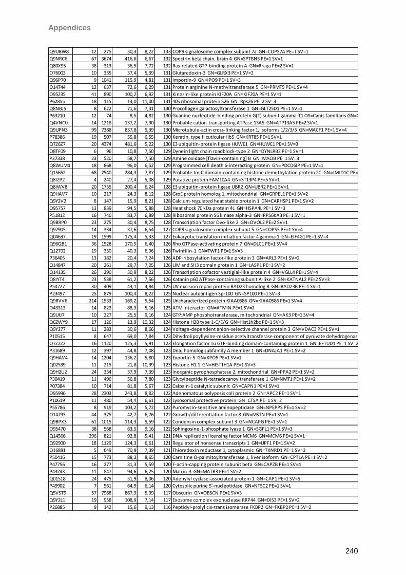
Appendices
240
Q9UBW8 12 275 30,3 8,22 133 COP9 signalosome complex subunit 7a GN=COPS7A PE=1 SV=1
Q9NRC6 67 3674 416,6 6,67 132 Spectrin beta chain, brain 4 GN=SPTBN5 PE=1 SV=1
Q80X95 38 313 36,5 7,72 132 Ras-related GTP-binding protein A GN=Rraga PE=2 SV=1
O76003 10 335 37,4 5,39 131 Glutaredoxin-3 GN=GLRX3 PE=1 SV=2
Q96P70 9 1041 115,9 4,81 131 Importin-9 GN=IPO9 PE=1 SV=3
O14744 12 637 72,6 6,29 131 Protein arginine N-methyltransferase 5 GN=PRMT5 PE=1 SV=4
O95235 41 890 100,2 6,92 131 Kinesin-like protein KIF20A GN=KIF20A PE=1 SV=1
P62855 18 115 13,0 11,00 131 40S ribosomal protein S26 GN=Rps26 PE=2 SV=3
Q8NBJ5 8 622 71,6 7,31 130 Procollagen galactosyltransferase 1 GN=GLT25D1 PE=1 SV=1
P63210 12 74 8,5 4,82 130 Guanine nucleotide-binding protein G(T) subunit gamma-T1 OS=Canis familiaris GN=GNGT1 PE=2 SV=2
Q4VNC0 14 1218 137,2 7,90 130 Probable cation-transporting ATPase 13A5 GN=ATP13A5 PE=2 SV=1
Q9UPN3 99 7388 837,8 5,39 130 Microtubule-actin cross-linking factor 1, isoforms 1/2/3/5 GN=MACF1 PE=1 SV=4
P78386 19 507 55,8 6,55 130 Keratin, type II cuticular Hb5 GN=KRT85 PE=1 SV=1
Q7Z6Z7 20 4374 481,6 5,22 130 E3 ubiquitin-protein ligase HUWE1 GN=HUWE1 PE=1 SV=3
Q8TF09 6 96 10,8 7,50 129 Dynein light chain roadblock-type 2 GN=DYNLRB2 PE=1 SV=1
P27338 23 520 58,7 7,50 129 Amine oxidase [flavin-containing] B GN=MAOB PE=1 SV=3
Q8WUM4 18 868 96,0 6,52 129 Programmed cell death 6-interacting protein GN=PDCD6IP PE=1 SV=1
Q15652 68 2540 284,3 7,87 129 Probable JmjC domain-containing histone demethylation protein 2C GN=JMJD1C PE=1 SV=2
Q8IZP2 4 240 27,4 5,08 129 Putative protein FAM10A4 GN=ST13P4 PE=5 SV=1
Q8IWV8 20 1755 200,4 6,24 128 E3 ubiquitin-protein ligase UBR2 GN=UBR2 PE=1 SV=1
Q9HAV7 10 217 24,3 8,12 128 GrpE protein homolog 1, mitochondrial GN=GRPEL1 PE=1 SV=2
Q9Y2V2 8 147 15,9 8,21 128 Calcium-regulated heat stable protein 1 GN=CARHSP1 PE=1 SV=2
O95757 13 839 94,5 5,88 128 Heat shock 70 kDa protein 4L GN=HSPA4L PE=1 SV=3
P51812 16 740 83,7 6,89 128 Ribosomal protein S6 kinase alpha-3 GN=RPS6KA3 PE=1 SV=1
Q9BRP0 23 275 30,4 8,75 128 Transcription factor Ovo-like 2 GN=OVOL2 PE=2 SV=1
Q92905 14 334 37,6 6,54 127 COP9 signalosome complex subunit 5 GN=COPS5 PE=1 SV=4
Q04637 19 1599 175,4 5,33 127 Eukaryotic translation initiation factor 4 gamma 1 GN=EIF4G1 PE=1 SV=4
Q96QB1 36 1528 170,5 6,40 126 Rho GTPase-activating protein 7 GN=DLC1 PE=1 SV=4
Q12792 19 350 40,3 6,96 126 Twinfilin-1 GN=TWF1 PE=1 SV=3
P36405 13 182 20,4 7,24 126 ADP-ribosylation factor-like protein 3 GN=ARL3 PE=1 SV=2
Q14847 20 261 29,7 7,05 126 LIM and SH3 domain protein 1 GN=LASP1 PE=1 SV=2
Q14135 26 290 30,9 8,22 126 Transcription cofactor vestigial-like protein 4 GN=VGLL4 PE=1 SV=4
Q8IYT4 23 538 61,2 7,56 126 Katanin p60 ATPase-containing subunit A-like 2 GN=KATNAL2 PE=2 SV=3
P54727 30 409 43,1 4,84 125 UV excision repair protein RAD23 homolog B GN=RAD23B PE=1 SV=1
P23497 25 879 100,4 8,22 125 Nuclear autoantigen Sp-100 GN=SP100 PE=1 SV=3
Q9BVV6 214 1533 169,2 5,54 125 Uncharacterized protein KIAA0586 GN=KIAA0586 PE=1 SV=4
O43313 14 823 88,3 5,16 125 ATM interactor GN=ATMIN PE=1 SV=2
Q9UIJ7 10 227 25,5 9,16 124 GTP:AMP phosphotransferase, mitochondrial GN=AK3 PE=1 SV=4
Q6ZWY9 17 126 13,9 10,32 124 Histone H2B type 1-C/E/G GN=Hist1h2bc PE=1 SV=3
Q9Y277 11 283 30,6 8,66 124 Voltage-dependent anion-selective channel protein 3 GN=VDAC3 PE=1 SV=1
P10515 8 647 69,0 7,84 123 Dihydrolipoyllysine-residue acetyltransferase component of pyruvate dehydrogenase complex, mitochondrial GN=DLAT PE=1 SV=3
Q7Z2Z2 16 1120 125,3 5,91 123 Elongation factor Tu GTP-binding domain-containing protein 1 GN=EFTUD1 PE=1 SV=2
P31689 12 397 44,8 7,08 123 DnaJ homolog subfamily A member 1 GN=DNAJA1 PE=1 SV=2
Q9HAV4 14 1204 136,2 5,80 123 Exportin-5 GN=XPO5 PE=1 SV=1
Q02539 11 215 21,8 10,99 123 Histone H1.1 GN=HIST1H1A PE=1 SV=3
Q9H2U2 24 334 37,9 7,39 123 Inorganic pyrophosphatase 2, mitochondrial GN=PPA2 PE=1 SV=2
P30419 11 496 56,8 7,80 123 Glycylpeptide N-tetradecanoyltransferase 1 GN=NMT1 PE=1 SV=2
P07384 10 714 81,8 5,67 122 Calpain-1 catalytic subunit GN=CAPN1 PE=1 SV=1
O95996 28 2303 243,8 8,82 122 Adenomatous polyposis coli protein 2 GN=APC2 PE=1 SV=1
P10619 11 480 54,4 6,61 122 Lysosomal protective protein GN=CTSA PE=1 SV=2
P55786 8 919 103,2 5,72 122 Puromycin-sensitive aminopeptidase GN=NPEPPS PE=1 SV=2
O14793 44 375 42,7 6,76 122 Growth/differentiation factor 8 GN=MSTN PE=1 SV=1
Q9BPX3 61 1015 114,3 5,59 122 Condensin complex subunit 3 GN=NCAPG PE=1 SV=1
O95470 38 568 63,5 9,16 122 Sphingosine-1-phosphate lyase 1 GN=SGPL1 PE=1 SV=3
Q14566 296 821 92,8 5,41 121 DNA replication licensing factor MCM6 GN=MCM6 PE=1 SV=1
Q92900 18 1129 124,3 6,61 121 Regulator of nonsense transcripts 1 GN=UPF1 PE=1 SV=2
Q16881 5 649 70,9 7,39 121 Thioredoxin reductase 1, cytoplasmic GN=TXNRD1 PE=1 SV=3
P50416 15 773 88,3 8,65 120 Carnitine O-palmitoyltransferase 1, liver isoform GN=CPT1A PE=1 SV=2
P47756 16 277 31,3 5,59 120 F-actin-capping protein subunit beta GN=CAPZB PE=1 SV=4
P43243 11 847 94,6 6,25 120 Matrin-3 GN=MATR3 PE=1 SV=2
Q01518 24 475 51,9 8,06 120 Adenylyl cyclase-associated protein 1 GN=CAP1 PE=1 SV=5
P49902 7 561 64,9 6,14 120 Cytosolic purine 5'-nucleotidase GN=NT5C2 PE=1 SV=1
Q5VST9 57 7968 867,9 5,99 117 Obscurin GN=OBSCN PE=1 SV=3
Q9Y2L1 19 958 108,9 7,14 117 Exosome complex exonuclease RRP44 GN=DIS3 PE=1 SV=2
P26885 9 142 15,6 9,13 116 Peptidyl-prolyl cis-trans isomerase FKBP2 GN=FKBP2 PE=1 SV=2
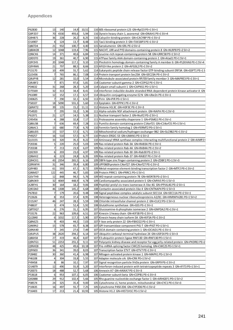
Appendices
241
P62830 13 140 14,9 10,51 116 60S ribosomal protein L23 GN=Rpl23 PE=1 SV=1
Q9P2D7 70 4330 493,6 5,94 116 Dynein heavy chain 1, axonemal GN=DNAH1 PE=1 SV=4
Q9HB71 36 228 26,2 8,25 116 Calcyclin-binding protein GN=CACYBP PE=1 SV=2
O14907 4 124 13,7 8,48 115 Tax1-binding protein 3 GN=TAX1BP3 PE=1 SV=2
Q86TD4 21 932 100,7 4,40 115 Sarcalumenin GN=SRL PE=2 SV=2
Q86W28 12 1048 119,4 7,96 115 NACHT, LRR and PYD domains-containing protein 8 GN=NLRP8 PE=2 SV=2
Q96CX6 14 371 40,6 6,84 115 Leucine-rich repeat-containing protein 58 GN=LRRC58 PE=1 SV=2
Q9D5T0 19 361 40,7 6,90 115 ATPase family AAA domain-containing protein 1 GN=Atad1 PE=1 SV=1
Q9Y2H5 20 1048 117,1 9,10 115 Pleckstrin homology domain-containing family A member 6 GN=PLEKHA6 PE=1 SV=4
Q9Y4W6 21 797 88,5 8,66 115 AFG3-like protein 2 GN=AFG3L2 PE=1 SV=2
P15170 10 499 55,7 5,62 114 Eukaryotic peptide chain release factor GTP-binding subunit ERF3A GN=GSPT1 PE=1 SV=1
Q15436 7 765 86,1 7,08 114 Protein transport protein Sec23A GN=SEC23A PE=1 SV=2
Q9UPY8 12 281 32,0 5,54 114 Microtubule-associated protein RP/EB family member 3 GN=MAPRE3 PE=1 SV=1
Q9UBF2 7 871 97,6 5,81 114 Coatomer subunit gamma-2 GN=COPG2 PE=1 SV=1
P04632 31 268 28,3 5,20 114 Calpain small subunit 1 GN=CAPNS1 PE=1 SV=1
O75569 32 313 34,4 8,41 114 Interferon-inducible double stranded RNA-dependent protein kinase activator A GN=PRKRA PE=1 SV=1
P61089 12 152 17,1 6,57 114 Ubiquitin-conjugating enzyme E2 N GN=Ube2n PE=1 SV=1
O00625 7 290 32,1 6,92 113 Pirin GN=PIR PE=1 SV=1
P58107 18 5090 555,3 5,60 113 Epiplakin GN=EPPK1 PE=1 SV=2
Q6NXT2 39 135 15,2 11,11 112 Histone H3.3C GN=H3F3C PE=1 SV=3
P54920 7 295 33,2 5,36 111 Alpha-soluble NSF attachment protein GN=NAPA PE=1 SV=3
P61971 21 127 14,5 5,38 111 Nuclear transport factor 2 GN=Nutf2 PE=2 SV=1
O95456 4 288 32,8 7,17 111 Proteasome assembly chaperone 1 GN=PSMG1 PE=1 SV=1
Q86U38 7 636 69,4 7,28 111 Pumilio domain-containing protein C14orf21 GN=C14orf21 PE=1 SV=1
Q96AC1 15 680 77,8 6,70 110 Fermitin family homolog 2 GN=FERMT2 PE=1 SV=1
Q86UD5 15 537 57,5 6,71 110 Mitochondrial sodium/hydrogen exchanger 9B2 GN=SLC9B2 PE=1 SV=2
P49257 16 510 57,5 6,77 110 Protein ERGIC-53 GN=LMAN1 PE=1 SV=2
Q13155 33 320 35,3 8,22 110 Aminoacyl tRNA synthase complex-interacting multifunctional protein 2 GN=AIMP2 PE=1 SV=2
P20336 5 220 25,0 5,03 109 Ras-related protein Rab-3A GN=RAB3A PE=1 SV=1
P20338 7 213 23,9 6,07 109 Ras-related protein Rab-4A GN=RAB4A PE=1 SV=2
Q923S9 6 203 23,0 4,97 109 Ras-related protein Rab-30 GN=Rab30 PE=2 SV=1
Q96AX2 6 223 24,8 6,35 109 Ras-related protein Rab-37 GN=RAB37 PE=1 SV=3
Q9HCK1 43 2354 265,5 6,16 109 DBF4-type zinc finger-containing protein 2 GN=ZDBF2 PE=1 SV=3
Q9NWY4 31 346 39,4 6,80 109 UPF0609 protein C4orf27 GN=C4orf27 PE=1 SV=2
Q9Y483 61 593 67,1 8,72 109 Metal-response element-binding transcription factor 2 GN=MTF2 PE=1 SV=2
Q96M27 122 445 46,7 5,83 109 Protein PRRC1 GN=PRRC1 PE=1 SV=1
Q5VTH9 12 848 94,5 5,76 109 WD repeat-containing protein 78 GN=WDR78 PE=2 SV=1
Q8N3K9 32 4069 448,9 4,78 109 Cardiomyopathy-associated protein 5 GN=CMYA5 PE=1 SV=3
A2BFH1 30 164 18,2 9,06 108 Peptidyl-prolyl cis-trans isomerase A-like 4G GN=PPIAL4G PE=2 SV=1
Q9C0A0 46 1308 145,2 6,68 108 Contactin-associated protein-like 4 GN=CNTNAP4 PE=1 SV=3
P67810 7 179 20,6 9,48 108 Signal peptidase complex catalytic subunit SEC11A GN=SEC11A PE=2 SV=1
P22626 8 353 37,4 8,95 108 Heterogeneous nuclear ribonucleoproteins A2/B1 GN=HNRNPA2B1 PE=1 SV=2
O15247 46 247 28,3 5,59 108 Chloride intracellular channel protein 2 GN=CLIC2 PE=1 SV=3
P48637 3 474 52,4 5,92 108 Glutathione synthetase GN=GSS PE=1 SV=1
Q8TDQ7 6 276 31,1 6,95 107 Glucosamine-6-phosphate isomerase 2 GN=GNPDA2 PE=1 SV=1
P33176 22 963 109,6 6,51 107 Kinesin-1 heavy chain GN=KIF5B PE=1 SV=1
Q12840 6 1032 117,3 5,90 107 Kinesin heavy chain isoform 5A GN=KIF5A PE=1 SV=2
Q8NEZ5 9 403 44,5 7,03 107 F-box only protein 22 GN=FBXO22 PE=1 SV=1
Q969N2 15 578 65,7 8,38 107 GPI transamidase component PIG-T GN=PIGT PE=1 SV=1
Q9NX40 7 245 27,6 7,49 107 OCIA domain-containing protein 1 GN=OCIAD1 PE=1 SV=1
Q9UPU5 38 2620 294,2 6,14 107 Ubiquitin carboxyl-terminal hydrolase 24 GN=USP24 PE=1 SV=3
Q86XS8 27 419 46,4 8,87 107 E3 ubiquitin-protein ligase RNF130 GN=RNF130 PE=1 SV=1
Q9NTG1 51 2253 255,3 9,11 107 Polycystic kidney disease and receptor for egg jelly-related protein GN=PKDREJ PE=2 SV=2
Q9NXE8 48 425 49,6 10,18 107 Pre-mRNA-splicing factor CWC25 homolog GN=CWC25 PE=1 SV=1
Q9Y603 36 341 39,0 8,03 107 Transcription factor ETV7 GN=ETV7 PE=1 SV=1
P28482 30 360 41,4 6,98 107 Mitogen-activated protein kinase 1 GN=MAPK1 PE=1 SV=3
P46108 4 304 33,8 5,55 107 Adapter molecule crk GN=CRK PE=1 SV=2
P49458 8 86 10,1 7,97 107 Signal recognition particle 9 kDa protein GN=SRP9 PE=1 SV=2
O14879 12 490 55,9 5,20 107 Interferon-induced protein with tetratricopeptide repeats 3 GN=IFIT3 PE=1 SV=1
P20073 18 488 52,7 5,68 106 Annexin A7 GN=ANXA7 PE=1 SV=3
P53618 6 953 107,1 6,05 106 Coatomer subunit beta GN=COPB1 PE=1 SV=3
Q92888 29 912 102,4 5,66 105 Rho guanine nucleotide exchange factor 1 GN=ARHGEF1 PE=1 SV=2
P08574 24 325 35,4 9,00 105 Cytochrome c1, heme protein, mitochondrial GN=CYC1 PE=1 SV=3
P10635 16 497 55,7 7,25 105 Cytochrome P450 2D6 GN=CYP2D6 PE=1 SV=2
P16403 17 213 21,4 10,93 105 Histone H1.2 GN=HIST1H1C PE=1 SV=2
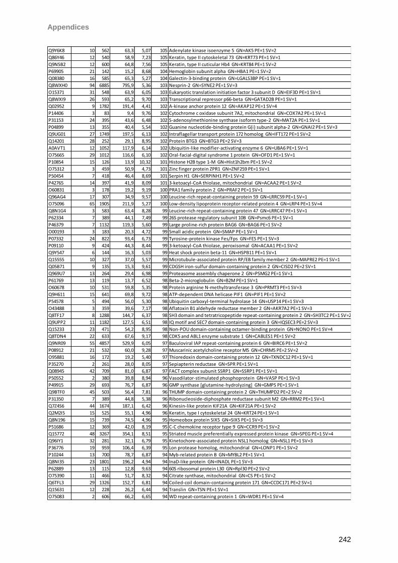
Appendices
242
Q9Y6K8 10 562 63,3 5,07 105 Adenylate kinase isoenzyme 5 GN=AK5 PE=1 SV=2
Q86Y46 12 540 58,9 7,23 105 Keratin, type II cytoskeletal 73 GN=KRT73 PE=1 SV=1
Q9NSB2 12 600 64,8 7,56 105 Keratin, type II cuticular Hb4 GN=KRT84 PE=1 SV=2
P69905 21 142 15,2 8,68 104 Hemoglobin subunit alpha GN=HBA1 PE=1 SV=2
Q08380 16 585 65,3 5,27 104 Galectin-3-binding protein GN=LGALS3BP PE=1 SV=1
Q8WXH0 94 6885 795,9 5,36 103 Nesprin-2 GN=SYNE2 PE=1 SV=3
O15371 31 548 63,9 6,05 103 Eukaryotic translation initiation factor 3 subunit D GN=EIF3D PE=1 SV=1
Q8WXI9 26 593 65,2 9,70 103 Transcriptional repressor p66-beta GN=GATAD2B PE=1 SV=1
Q02952 9 1782 191,4 4,41 102 A-kinase anchor protein 12 GN=AKAP12 PE=1 SV=4
P14406 3 83 9,4 9,76 102 Cytochrome c oxidase subunit 7A2, mitochondrial GN=COX7A2 PE=1 SV=1
P31153 24 395 43,6 6,48 102 S-adenosylmethionine synthase isoform type-2 GN=MAT2A PE=1 SV=1
P04899 13 355 40,4 5,54 102 Guanine nucleotide-binding protein G(i) subunit alpha-2 GN=GNAI2 PE=1 SV=3
Q9UG01 27 1749 197,5 6,13 102 Intraflagellar transport protein 172 homolog GN=IFT172 PE=1 SV=2
Q14201 28 252 29,1 8,95 102 Protein BTG3 GN=BTG3 PE=2 SV=3
A0AVT1 12 1052 117,9 6,14 102 Ubiquitin-like modifier-activating enzyme 6 GN=UBA6 PE=1 SV=1
O75665 29 1012 116,6 6,10 102 Oral-facial-digital syndrome 1 protein GN=OFD1 PE=1 SV=1
P10854 15 126 13,9 10,32 101 Histone H2B type 1-M GN=Hist1h2bm PE=1 SV=2
O75312 3 459 50,9 4,73 101 Zinc finger protein ZPR1 GN=ZNF259 PE=1 SV=1
P50454 7 418 46,4 8,69 101 Serpin H1 GN=SERPINH1 PE=1 SV=2
P42765 14 397 41,9 8,09 101 3-ketoacyl-CoA thiolase, mitochondrial GN=ACAA2 PE=1 SV=2
O60831 3 178 19,2 9,19 100 PRA1 family protein 2 GN=PRAF2 PE=1 SV=1
Q96AG4 17 307 34,9 9,57 100 Leucine-rich repeat-containing protein 59 GN=LRRC59 PE=1 SV=1
O75096 65 1905 211,9 5,27 100 Low-density lipoprotein receptor-related protein 4 GN=LRP4 PE=1 SV=4
Q8N1G4 3 583 63,4 8,28 99 Leucine-rich repeat-containing protein 47 GN=LRRC47 PE=1 SV=1
P62334 7 389 44,1 7,49 99 26S protease regulatory subunit 10B GN=Psmc6 PE=1 SV=1
P46379 7 1132 119,3 5,60 99 Large proline-rich protein BAG6 GN=BAG6 PE=1 SV=2
O00193 3 183 20,3 4,72 99 Small acidic protein GN=SMAP PE=1 SV=1
P07332 24 822 93,4 6,73 99 Tyrosine-protein kinase Fes/Fps GN=FES PE=1 SV=3
P09110 9 424 44,3 8,44 99 3-ketoacyl-CoA thiolase, peroxisomal GN=ACAA1 PE=1 SV=2
Q9Y547 6 144 16,3 5,03 99 Heat shock protein beta-11 GN=HSPB11 PE=1 SV=1
Q15555 10 327 37,0 5,57 99 Microtubule-associated protein RP/EB family member 2 GN=MAPRE2 PE=1 SV=1
Q05B71 9 135 15,3 9,61 99 CDGSH iron-sulfur domain-containing protein 2 GN=CISD2 PE=2 SV=1
Q969U7 13 264 29,4 6,98 99 Proteasome assembly chaperone 2 GN=PSMG2 PE=1 SV=1
P61769 13 119 13,7 6,52 98 Beta-2-microglobulin GN=B2M PE=1 SV=1
O60678 10 531 59,8 5,35 98 Protein arginine N-methyltransferase 3 GN=PRMT3 PE=1 SV=3
Q9H611 15 641 69,8 9,72 98 ATP-dependent DNA helicase PIF1 GN=PIF1 PE=1 SV=2
P54578 5 494 56,0 5,30 98 Ubiquitin carboxyl-terminal hydrolase 14 GN=USP14 PE=1 SV=3
O43488 3 359 39,6 7,17 98 Aflatoxin B1 aldehyde reductase member 2 GN=AKR7A2 PE=1 SV=3
Q8TF17 8 1288 144,7 6,37 98 SH3 domain and tetratricopeptide repeat-containing protein 2 GN=SH3TC2 PE=1 SV=2
Q9UPP2 11 1182 127,5 6,51 98 IQ motif and SEC7 domain-containing protein 3 GN=IQSEC3 PE=2 SV=3
Q15233 23 471 54,2 8,95 98 Non-POU domain-containing octamer-binding protein GN=NONO PE=1 SV=4
Q8TDN4 22 633 67,6 9,17 98 CDK5 and ABL1 enzyme substrate 1 GN=CABLES1 PE=1 SV=2
Q9NR09 55 4857 529,9 6,05 97 Baculoviral IAP repeat-containing protein 6 GN=BIRC6 PE=1 SV=2
P08912 21 532 60,0 9,28 97 Muscarinic acetylcholine receptor M5 GN=CHRM5 PE=2 SV=2
O95881 16 172 19,2 5,40 97 Thioredoxin domain-containing protein 12 GN=TXNDC12 PE=1 SV=1
P35270 2 261 28,0 8,05 97 Sepiapterin reductase GN=SPR PE=1 SV=1
Q08945 42 709 81,0 6,87 97 FACT complex subunit SSRP1 GN=SSRP1 PE=1 SV=1
P50552 2 380 39,8 8,94 96 Vasodilator-stimulated phosphoprotein GN=VASP PE=1 SV=3
P49915 29 693 76,7 6,87 96 GMP synthase [glutamine-hydrolyzing] GN=GMPS PE=1 SV=1
Q9BTF0 45 503 56,4 7,81 96 THUMP domain-containing protein 2 GN=THUMPD2 PE=2 SV=2
P31350 7 389 44,8 5,38 96 Ribonucleoside-diphosphate reductase subunit M2 GN=RRM2 PE=1 SV=1
Q7Z4S6 44 1674 187,1 6,42 96 Kinesin-like protein KIF21A GN=KIF21A PE=1 SV=2
Q2M2I5 15 525 55,1 4,96 96 Keratin, type I cytoskeletal 24 GN=KRT24 PE=1 SV=1
Q8N196 15 739 74,5 4,96 95 Homeobox protein SIX5 GN=SIX5 PE=1 SV=3
P51686 12 369 42,0 8,19 95 C-C chemokine receptor type 9 GN=CCR9 PE=1 SV=2
Q15772 48 3267 354,1 8,51 95 Striated muscle preferentially expressed protein kinase GN=SPEG PE=1 SV=4
Q96IY1 32 281 32,1 6,79 95 Kinetochore-associated protein NSL1 homolog GN=NSL1 PE=1 SV=3
P36776 19 959 106,4 6,39 95 Lon protease homolog, mitochondrial GN=LONP1 PE=1 SV=2
P10244 13 700 78,7 6,87 94 Myb-related protein B GN=MYBL2 PE=1 SV=1
Q8NI35 23 1801 196,2 4,94 94 InaD-like protein GN=INADL PE=1 SV=3
P62889 13 115 12,8 9,63 94 60S ribosomal protein L30 GN=Rpl30 PE=2 SV=2
O75390 11 466 51,7 8,32 94 Citrate synthase, mitochondrial GN=CS PE=1 SV=2
Q6TFL3 29 1326 152,7 6,81 94 Coiled-coil domain-containing protein 171 GN=CCDC171 PE=2 SV=1
Q15631 12 228 26,2 6,44 94 Translin GN=TSN PE=1 SV=1
O75083 2 606 66,2 6,65 94 WD repeat-containing protein 1 GN=WDR1 PE=1 SV=4
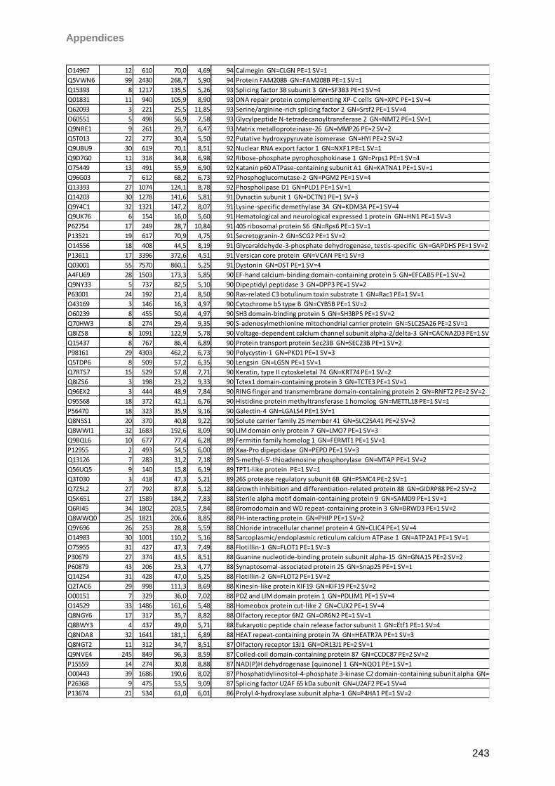
Appendices
243
O14967 12 610 70,0 4,69 94 Calmegin GN=CLGN PE=1 SV=1
Q5VWN6 99 2430 268,7 5,90 94 Protein FAM208B GN=FAM208B PE=1 SV=1
Q15393 8 1217 135,5 5,26 93 Splicing factor 3B subunit 3 GN=SF3B3 PE=1 SV=4
Q01831 11 940 105,9 8,90 93 DNA repair protein complementing XP-C cells GN=XPC PE=1 SV=4
Q62093 3 221 25,5 11,85 93 Serine/arginine-rich splicing factor 2 GN=Srsf2 PE=1 SV=4
O60551 5 498 56,9 7,58 93 Glycylpeptide N-tetradecanoyltransferase 2 GN=NMT2 PE=1 SV=1
Q9NRE1 9 261 29,7 6,47 93 Matrix metalloproteinase-26 GN=MMP26 PE=2 SV=2
Q5T013 22 277 30,4 5,50 92 Putative hydroxypyruvate isomerase GN=HYI PE=2 SV=2
Q9UBU9 30 619 70,1 8,51 92 Nuclear RNA export factor 1 GN=NXF1 PE=1 SV=1
Q9D7G0 11 318 34,8 6,98 92 Ribose-phosphate pyrophosphokinase 1 GN=Prps1 PE=1 SV=4
O75449 13 491 55,9 6,90 92 Katanin p60 ATPase-containing subunit A1 GN=KATNA1 PE=1 SV=1
Q96G03 7 612 68,2 6,73 92 Phosphoglucomutase-2 GN=PGM2 PE=1 SV=4
Q13393 27 1074 124,1 8,78 92 Phospholipase D1 GN=PLD1 PE=1 SV=1
Q14203 30 1278 141,6 5,81 91 Dynactin subunit 1 GN=DCTN1 PE=1 SV=3
Q9Y4C1 32 1321 147,2 8,07 91 Lysine-specific demethylase 3A GN=KDM3A PE=1 SV=4
Q9UK76 6 154 16,0 5,60 91 Hematological and neurological expressed 1 protein GN=HN1 PE=1 SV=3
P62754 17 249 28,7 10,84 91 40S ribosomal protein S6 GN=Rps6 PE=1 SV=1
P13521 19 617 70,9 4,75 91 Secretogranin-2 GN=SCG2 PE=1 SV=2
O14556 18 408 44,5 8,19 91 Glyceraldehyde-3-phosphate dehydrogenase, testis-specific GN=GAPDHS PE=1 SV=2
P13611 17 3396 372,6 4,51 91 Versican core protein GN=VCAN PE=1 SV=3
Q03001 55 7570 860,1 5,25 91 Dystonin GN=DST PE=1 SV=4
A4FU69 28 1503 173,3 5,85 90 EF-hand calcium-binding domain-containing protein 5 GN=EFCAB5 PE=1 SV=2
Q9NY33 5 737 82,5 5,10 90 Dipeptidyl peptidase 3 GN=DPP3 PE=1 SV=2
P63001 24 192 21,4 8,50 90 Ras-related C3 botulinum toxin substrate 1 GN=Rac1 PE=1 SV=1
O43169 3 146 16,3 4,97 90 Cytochrome b5 type B GN=CYB5B PE=1 SV=2
O60239 8 455 50,4 4,97 90 SH3 domain-binding protein 5 GN=SH3BP5 PE=1 SV=2
Q70HW3 8 274 29,4 9,35 90 S-adenosylmethionine mitochondrial carrier protein GN=SLC25A26 PE=2 SV=1
Q8IZS8 8 1091 122,9 5,78 90 Voltage-dependent calcium channel subunit alpha-2/delta-3 GN=CACNA2D3 PE=1 SV=1
Q15437 8 767 86,4 6,89 90 Protein transport protein Sec23B GN=SEC23B PE=1 SV=2
P98161 29 4303 462,2 6,73 90 Polycystin-1 GN=PKD1 PE=1 SV=3
Q5TDP6 8 509 57,2 6,35 90 Lengsin GN=LGSN PE=1 SV=1
Q7RTS7 15 529 57,8 7,71 90 Keratin, type II cytoskeletal 74 GN=KRT74 PE=1 SV=2
Q8IZS6 3 198 23,2 9,33 90 Tctex1 domain-containing protein 3 GN=TCTE3 PE=1 SV=1
Q96EX2 3 444 48,9 7,84 90 RING finger and transmembrane domain-containing protein 2 GN=RNFT2 PE=2 SV=2
O95568 18 372 42,1 6,76 90 Histidine protein methyltransferase 1 homolog GN=METTL18 PE=1 SV=1
P56470 18 323 35,9 9,16 90 Galectin-4 GN=LGALS4 PE=1 SV=1
Q8N5S1 20 370 40,8 9,22 90 Solute carrier family 25 member 41 GN=SLC25A41 PE=2 SV=2
Q8WWI1 32 1683 192,6 8,09 90 LIM domain only protein 7 GN=LMO7 PE=1 SV=3
Q9BQL6 10 677 77,4 6,28 89 Fermitin family homolog 1 GN=FERMT1 PE=1 SV=1
P12955 2 493 54,5 6,00 89 Xaa-Pro dipeptidase GN=PEPD PE=1 SV=3
Q13126 7 283 31,2 7,18 89 S-methyl-5'-thioadenosine phosphorylase GN=MTAP PE=1 SV=2
Q56UQ5 9 140 15,8 6,19 89 TPT1-like protein PE=1 SV=1
Q3T030 3 418 47,3 5,21 89 26S protease regulatory subunit 6B GN=PSMC4 PE=2 SV=1
Q7Z5L2 27 792 87,8 5,12 88 Growth inhibition and differentiation-related protein 88 GN=GIDRP88 PE=2 SV=2
Q5K651 27 1589 184,2 7,83 88 Sterile alpha motif domain-containing protein 9 GN=SAMD9 PE=1 SV=1
Q6RI45 34 1802 203,5 7,84 88 Bromodomain and WD repeat-containing protein 3 GN=BRWD3 PE=1 SV=2
Q8WWQ0 25 1821 206,6 8,85 88 PH-interacting protein GN=PHIP PE=1 SV=2
Q9Y696 26 253 28,8 5,59 88 Chloride intracellular channel protein 4 GN=CLIC4 PE=1 SV=4
O14983 30 1001 110,2 5,16 88 Sarcoplasmic/endoplasmic reticulum calcium ATPase 1 GN=ATP2A1 PE=1 SV=1
O75955 31 427 47,3 7,49 88 Flotillin-1 GN=FLOT1 PE=1 SV=3
P30679 27 374 43,5 8,51 88 Guanine nucleotide-binding protein subunit alpha-15 GN=GNA15 PE=2 SV=2
P60879 43 206 23,3 4,77 88 Synaptosomal-associated protein 25 GN=Snap25 PE=1 SV=1
Q14254 31 428 47,0 5,25 88 Flotillin-2 GN=FLOT2 PE=1 SV=2
Q2TAC6 29 998 111,3 8,69 88 Kinesin-like protein KIF19 GN=KIF19 PE=2 SV=2
O00151 7 329 36,0 7,02 88 PDZ and LIM domain protein 1 GN=PDLIM1 PE=1 SV=4
O14529 33 1486 161,6 5,48 88 Homeobox protein cut-like 2 GN=CUX2 PE=1 SV=4
Q8NGY6 17 317 35,7 8,82 88 Olfactory receptor 6N2 GN=OR6N2 PE=1 SV=1
Q8BWY3 4 437 49,0 5,71 88 Eukaryotic peptide chain release factor subunit 1 GN=Etf1 PE=1 SV=4
Q8NDA8 32 1641 181,1 6,89 88 HEAT repeat-containing protein 7A GN=HEATR7A PE=1 SV=3
Q8NGT2 11 312 34,7 8,51 87 Olfactory receptor 13J1 GN=OR13J1 PE=2 SV=1
Q9NVE4 245 849 96,3 8,59 87 Coiled-coil domain-containing protein 87 GN=CCDC87 PE=2 SV=2
P15559 14 274 30,8 8,88 87 NAD(P)H dehydrogenase [quinone] 1 GN=NQO1 PE=1 SV=1
O00443 39 1686 190,6 8,02 87 Phosphatidylinositol-4-phosphate 3-kinase C2 domain-containing subunit alpha GN=PIK3C2A PE=1 SV=2
P26368 9 475 53,5 9,09 87 Splicing factor U2AF 65 kDa subunit GN=U2AF2 PE=1 SV=4
P13674 21 534 61,0 6,01 86 Prolyl 4-hydroxylase subunit alpha-1 GN=P4HA1 PE=1 SV=2
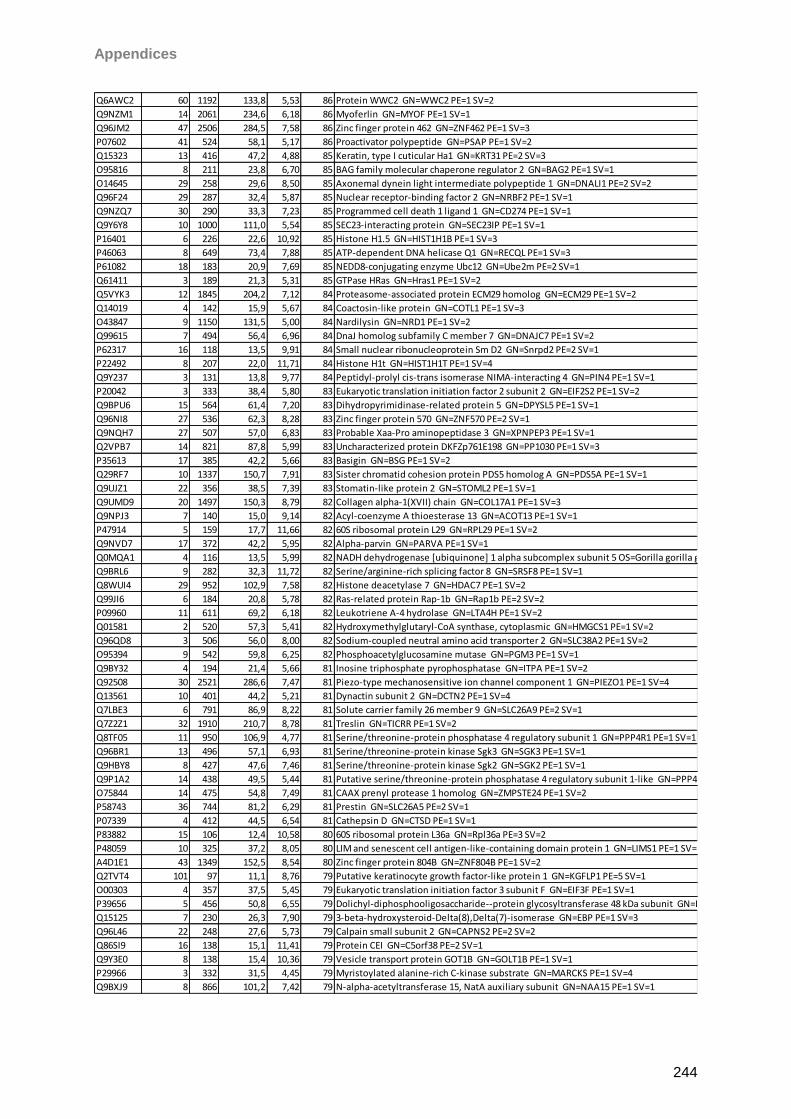
Appendices
244
Q6AWC2 60 1192 133,8 5,53 86 Protein WWC2 GN=WWC2 PE=1 SV=2
Q9NZM1 14 2061 234,6 6,18 86 Myoferlin GN=MYOF PE=1 SV=1
Q96JM2 47 2506 284,5 7,58 86 Zinc finger protein 462 GN=ZNF462 PE=1 SV=3
P07602 41 524 58,1 5,17 86 Proactivator polypeptide GN=PSAP PE=1 SV=2
Q15323 13 416 47,2 4,88 85 Keratin, type I cuticular Ha1 GN=KRT31 PE=2 SV=3
O95816 8 211 23,8 6,70 85 BAG family molecular chaperone regulator 2 GN=BAG2 PE=1 SV=1
O14645 29 258 29,6 8,50 85 Axonemal dynein light intermediate polypeptide 1 GN=DNALI1 PE=2 SV=2
Q96F24 29 287 32,4 5,87 85 Nuclear receptor-binding factor 2 GN=NRBF2 PE=1 SV=1
Q9NZQ7 30 290 33,3 7,23 85 Programmed cell death 1 ligand 1 GN=CD274 PE=1 SV=1
Q9Y6Y8 10 1000 111,0 5,54 85 SEC23-interacting protein GN=SEC23IP PE=1 SV=1
P16401 6 226 22,6 10,92 85 Histone H1.5 GN=HIST1H1B PE=1 SV=3
P46063 8 649 73,4 7,88 85 ATP-dependent DNA helicase Q1 GN=RECQL PE=1 SV=3
P61082 18 183 20,9 7,69 85 NEDD8-conjugating enzyme Ubc12 GN=Ube2m PE=2 SV=1
Q61411 3 189 21,3 5,31 85 GTPase HRas GN=Hras1 PE=1 SV=2
Q5VYK3 12 1845 204,2 7,12 84 Proteasome-associated protein ECM29 homolog GN=ECM29 PE=1 SV=2
Q14019 4 142 15,9 5,67 84 Coactosin-like protein GN=COTL1 PE=1 SV=3
O43847 9 1150 131,5 5,00 84 Nardilysin GN=NRD1 PE=1 SV=2
Q99615 7 494 56,4 6,96 84 DnaJ homolog subfamily C member 7 GN=DNAJC7 PE=1 SV=2
P62317 16 118 13,5 9,91 84 Small nuclear ribonucleoprotein Sm D2 GN=Snrpd2 PE=2 SV=1
P22492 8 207 22,0 11,71 84 Histone H1t GN=HIST1H1T PE=1 SV=4
Q9Y237 3 131 13,8 9,77 84 Peptidyl-prolyl cis-trans isomerase NIMA-interacting 4 GN=PIN4 PE=1 SV=1
P20042 3 333 38,4 5,80 83 Eukaryotic translation initiation factor 2 subunit 2 GN=EIF2S2 PE=1 SV=2
Q9BPU6 15 564 61,4 7,20 83 Dihydropyrimidinase-related protein 5 GN=DPYSL5 PE=1 SV=1
Q96NI8 27 536 62,3 8,28 83 Zinc finger protein 570 GN=ZNF570 PE=2 SV=1
Q9NQH7 27 507 57,0 6,83 83 Probable Xaa-Pro aminopeptidase 3 GN=XPNPEP3 PE=1 SV=1
Q2VPB7 14 821 87,8 5,99 83 Uncharacterized protein DKFZp761E198 GN=PP1030 PE=1 SV=3
P35613 17 385 42,2 5,66 83 Basigin GN=BSG PE=1 SV=2
Q29RF7 10 1337 150,7 7,91 83 Sister chromatid cohesion protein PDS5 homolog A GN=PDS5A PE=1 SV=1
Q9UJZ1 22 356 38,5 7,39 83 Stomatin-like protein 2 GN=STOML2 PE=1 SV=1
Q9UMD9 20 1497 150,3 8,79 82 Collagen alpha-1(XVII) chain GN=COL17A1 PE=1 SV=3
Q9NPJ3 7 140 15,0 9,14 82 Acyl-coenzyme A thioesterase 13 GN=ACOT13 PE=1 SV=1
P47914 5 159 17,7 11,66 82 60S ribosomal protein L29 GN=RPL29 PE=1 SV=2
Q9NVD7 17 372 42,2 5,95 82 Alpha-parvin GN=PARVA PE=1 SV=1
Q0MQA1 4 116 13,5 5,99 82 NADH dehydrogenase [ubiquinone] 1 alpha subcomplex subunit 5 OS=Gorilla gorilla gorilla GN=NDUFA5 PE=3 SV=3
Q9BRL6 9 282 32,3 11,72 82 Serine/arginine-rich splicing factor 8 GN=SRSF8 PE=1 SV=1
Q8WUI4 29 952 102,9 7,58 82 Histone deacetylase 7 GN=HDAC7 PE=1 SV=2
Q99JI6 6 184 20,8 5,78 82 Ras-related protein Rap-1b GN=Rap1b PE=2 SV=2
P09960 11 611 69,2 6,18 82 Leukotriene A-4 hydrolase GN=LTA4H PE=1 SV=2
Q01581 2 520 57,3 5,41 82 Hydroxymethylglutaryl-CoA synthase, cytoplasmic GN=HMGCS1 PE=1 SV=2
Q96QD8 3 506 56,0 8,00 82 Sodium-coupled neutral amino acid transporter 2 GN=SLC38A2 PE=1 SV=2
O95394 9 542 59,8 6,25 82 Phosphoacetylglucosamine mutase GN=PGM3 PE=1 SV=1
Q9BY32 4 194 21,4 5,66 81 Inosine triphosphate pyrophosphatase GN=ITPA PE=1 SV=2
Q92508 30 2521 286,6 7,47 81 Piezo-type mechanosensitive ion channel component 1 GN=PIEZO1 PE=1 SV=4
Q13561 10 401 44,2 5,21 81 Dynactin subunit 2 GN=DCTN2 PE=1 SV=4
Q7LBE3 6 791 86,9 8,22 81 Solute carrier family 26 member 9 GN=SLC26A9 PE=2 SV=1
Q7Z2Z1 32 1910 210,7 8,78 81 Treslin GN=TICRR PE=1 SV=2
Q8TF05 11 950 106,9 4,77 81 Serine/threonine-protein phosphatase 4 regulatory subunit 1 GN=PPP4R1 PE=1 SV=1
Q96BR1 13 496 57,1 6,93 81 Serine/threonine-protein kinase Sgk3 GN=SGK3 PE=1 SV=1
Q9HBY8 8 427 47,6 7,46 81 Serine/threonine-protein kinase Sgk2 GN=SGK2 PE=1 SV=1
Q9P1A2 14 438 49,5 5,44 81 Putative serine/threonine-protein phosphatase 4 regulatory subunit 1-like GN=PPP4R1L PE=5 SV=2
O75844 14 475 54,8 7,49 81 CAAX prenyl protease 1 homolog GN=ZMPSTE24 PE=1 SV=2
P58743 36 744 81,2 6,29 81 Prestin GN=SLC26A5 PE=2 SV=1
P07339 4 412 44,5 6,54 81 Cathepsin D GN=CTSD PE=1 SV=1
P83882 15 106 12,4 10,58 80 60S ribosomal protein L36a GN=Rpl36a PE=3 SV=2
P48059 10 325 37,2 8,05 80 LIM and senescent cell antigen-like-containing domain protein 1 GN=LIMS1 PE=1 SV=4
A4D1E1 43 1349 152,5 8,54 80 Zinc finger protein 804B GN=ZNF804B PE=1 SV=2
Q2TVT4 101 97 11,1 8,76 79 Putative keratinocyte growth factor-like protein 1 GN=KGFLP1 PE=5 SV=1
O00303 4 357 37,5 5,45 79 Eukaryotic translation initiation factor 3 subunit F GN=EIF3F PE=1 SV=1
P39656 5 456 50,8 6,55 79 Dolichyl-diphosphooligosaccharide--protein glycosyltransferase 48 kDa subunit GN=DDOST PE=1 SV=4
Q15125 7 230 26,3 7,90 79 3-beta-hydroxysteroid-Delta(8),Delta(7)-isomerase GN=EBP PE=1 SV=3
Q96L46 22 248 27,6 5,73 79 Calpain small subunit 2 GN=CAPNS2 PE=2 SV=2
Q86SI9 16 138 15,1 11,41 79 Protein CEI GN=C5orf38 PE=2 SV=1
Q9Y3E0 8 138 15,4 10,36 79 Vesicle transport protein GOT1B GN=GOLT1B PE=1 SV=1
P29966 3 332 31,5 4,45 79 Myristoylated alanine-rich C-kinase substrate GN=MARCKS PE=1 SV=4
Q9BXJ9 8 866 101,2 7,42 79 N-alpha-acetyltransferase 15, NatA auxiliary subunit GN=NAA15 PE=1 SV=1
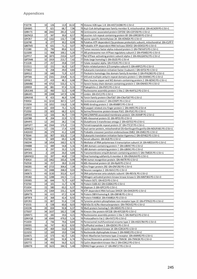
Appendices
245
P33778 10 126 13,9 10,32 79 Histone H2B type 1-B GN=HIST1H2BB PE=1 SV=2
Q9H845 7 621 68,7 7,96 79 Acyl-CoA dehydrogenase family member 9, mitochondrial GN=ACAD9 PE=1 SV=1
Q9BV73 48 2442 281,0 5,02 78 Centrosome-associated protein CEP250 GN=CEP250 PE=1 SV=2
Q6ZNQ3 17 347 39,6 8,57 78 Leucine-rich repeat-containing protein 69 GN=LRRC69 PE=2 SV=2
Q9Y2K7 21 1162 132,7 7,58 78 Lysine-specific demethylase 2A GN=KDM2A PE=1 SV=3
Q16740 6 277 30,2 8,09 78 Putative ATP-dependent Clp protease proteolytic subunit, mitochondrial GN=CLPP PE=1 SV=1
Q86TM3 9 631 71,1 9,07 78 Probable ATP-dependent RNA helicase DDX53 GN=DDX53 PE=1 SV=3
P21580 25 790 89,6 8,22 77 Tumor necrosis factor alpha-induced protein 3 GN=TNFAIP3 PE=1 SV=1
Q13200 11 908 100,1 5,20 77 26S proteasome non-ATPase regulatory subunit 2 GN=PSMD2 PE=1 SV=3
Q2PPJ7 34 1873 210,6 6,07 77 Ral GTPase-activating protein subunit alpha-2 GN=RALGAPA2 PE=1 SV=2
Q8TDM6 32 1919 213,7 7,42 77 Disks large homolog 5 GN=DLG5 PE=1 SV=4
P15328 27 257 29,8 7,97 77 Folate receptor alpha GN=FOLR1 PE=1 SV=3
O15511 12 151 16,3 5,67 77 Actin-related protein 2/3 complex subunit 5 GN=ARPC5 PE=1 SV=3
O75822 5 258 29,0 4,83 77 Eukaryotic translation initiation factor 3 subunit J GN=EIF3J PE=1 SV=2
Q6NSJ2 19 640 71,9 6,57 77 Pleckstrin homology-like domain family B member 3 GN=PHLDB3 PE=2 SV=3
Q9Y566 15 2161 224,8 8,15 77 SH3 and multiple ankyrin repeat domains protein 1 GN=SHANK1 PE=1 SV=2
Q9Y6E2 27 419 48,1 6,68 77 Basic leucine zipper and W2 domain-containing protein 2 GN=BZW2 PE=1 SV=1
Q96M86 44 4753 533,3 6,71 77 Dynein heavy chain domain-containing protein 1 GN=DNHD1 PE=1 SV=2
Q99959 24 881 97,4 9,33 77 Plakophilin-2 GN=PKP2 PE=1 SV=2
Q9ULW6 22 460 52,5 4,49 77 Nucleosome assembly protein 1-like 2 GN=NAP1L2 PE=2 SV=1
Q86UK5 39 1308 147,9 6,96 77 Limbin GN=EVC2 PE=1 SV=1
Q9NXH8 9 423 46,9 9,94 76 Torsin family protein C9orf167 GN=C9orf167 PE=1 SV=2
P49454 61 3210 367,5 5,07 76 Centromere protein F GN=CENPF PE=1 SV=2
O15034 23 1052 116,0 5,30 76 RIMS-binding protein 2 GN=RIMBP2 PE=1 SV=3
P10072 22 659 75,1 9,25 76 Krueppel-related zinc finger protein 1 GN=HKR1 PE=2 SV=4
P53350 23 603 68,2 8,91 76 Serine/threonine-protein kinase PLK1 GN=PLK1 PE=1 SV=1
Q9P055 12 326 36,7 7,93 75 JNK1/MAPK8-associated membrane protein GN=JKAMP PE=2 SV=2
Q5E988 8 204 22,9 9,72 75 40S ribosomal protein S5 GN=RPS5 PE=2 SV=3
Q9H4Y5 12 243 28,2 7,56 75 Glutathione S-transferase omega-2 GN=GSTO2 PE=2 SV=1
Q6PGP7 40 1564 175,4 7,53 75 Tetratricopeptide repeat protein 37 GN=TTC37 PE=1 SV=1
Q0MQC2 12 156 17,4 4,93 75 Acyl carrier protein, mitochondrial OS=Gorilla gorilla gorilla GN=NDUFAB1 PE=2 SV=1
A4GXA9 26 379 41,2 6,89 75 Probable crossover junction endonuclease EME2 GN=EME2 PE=1 SV=2
P78344 33 907 102,3 7,14 75 Eukaryotic translation initiation factor 4 gamma 2 GN=EIF4G2 PE=1 SV=1
P02768 5 609 69,3 6,28 75 Serum albumin GN=ALB PE=1 SV=2
O60244 18 1454 160,5 8,73 75 Mediator of RNA polymerase II transcription subunit 14 GN=MED14 PE=1 SV=2
O94888 11 489 54,8 5,16 75 UBX domain-containing protein 7 GN=UBXN7 PE=1 SV=2
Q04323 3 297 33,3 5,25 75 UBX domain-containing protein 1 GN=UBXN1 PE=1 SV=2
Q9Y3A6 2 229 26,0 4,84 75 Transmembrane emp24 domain-containing protein 5 GN=TMED5 PE=1 SV=1
Q8WW22 15 397 44,8 7,59 74 DnaJ homolog subfamily A member 4 GN=DNAJA4 PE=1 SV=1
P30414 22 1462 165,6 9,99 74 NK-tumor recognition protein GN=NKTR PE=1 SV=2
P62918 16 257 28,0 11,03 74 60S ribosomal protein L8 GN=Rpl8 PE=2 SV=2
O60281 40 2723 304,6 7,39 74 Zinc finger protein 292 GN=ZNF292 PE=1 SV=3
Q8IVL1 31 2488 268,0 9,03 74 Neuron navigator 2 GN=NAV2 PE=1 SV=3
O60673 43 3130 352,6 8,47 74 DNA polymerase zeta catalytic subunit GN=REV3L PE=1 SV=2
O95382 31 1288 142,5 7,11 74 Mitogen-activated protein kinase kinase kinase 6 GN=MAP3K6 PE=1 SV=3
O95905 16 644 72,7 4,87 74 Protein SGT1 GN=ECD PE=1 SV=1
P33764 14 101 11,7 4,78 74 Protein S100-A3 GN=S100A3 PE=1 SV=1
P51654 15 580 65,5 6,37 74 Glypican-3 GN=GPC3 PE=1 SV=1
Q7Z478 25 1369 155,1 8,09 74 ATP-dependent RNA helicase DHX29 GN=DHX29 PE=1 SV=2
Q8NFT6 16 615 67,2 8,34 74 Protein DBF4 homolog B GN=DBF4B PE=1 SV=1
Q8TBF8 23 368 42,4 8,92 74 Protein FAM81A GN=FAM81A PE=2 SV=3
Q9Y2R2 15 807 91,6 7,59 74 Tyrosine-protein phosphatase non-receptor type 22 GN=PTPN22 PE=1 SV=2
P10155 3 538 60,6 8,03 74 60 kDa SS-A/Ro ribonucleoprotein GN=TROVE2 PE=1 SV=2
O43196 20 834 92,8 6,37 74 MutS protein homolog 5 GN=MSH5 PE=1 SV=1
O75037 34 1637 182,5 7,08 74 Kinesin-like protein KIF21B GN=KIF21B PE=1 SV=2
Q96NT1 15 182 19,6 4,21 74 Nucleosome assembly protein 1-like 5 GN=NAP1L5 PE=2 SV=1
Q9NYQ8 18 4349 479,0 5,16 74 Protocadherin Fat 2 GN=FAT2 PE=1 SV=2
P51659 2 736 79,6 8,84 74 Peroxisomal multifunctional enzyme type 2 GN=HSD17B4 PE=1 SV=3
O00391 16 747 82,5 8,92 73 Sulfhydryl oxidase 1 GN=QSOX1 PE=1 SV=3
O94921 24 469 53,0 8,92 73 Cyclin-dependent kinase 14 GN=CDK14 PE=1 SV=3
Q13232 12 169 19,0 7,84 73 Nucleoside diphosphate kinase 3 GN=NME3 PE=1 SV=2
Q16671 13 573 62,7 5,81 73 Anti-Muellerian hormone type-2 receptor GN=AMHR2 PE=1 SV=1
Q59H18 28 835 92,8 6,74 73 Serine/threonine-protein kinase TNNI3K GN=TNNI3K PE=1 SV=3
Q92772 14 493 56,0 8,21 73 Cyclin-dependent kinase-like 2 GN=CDKL2 PE=2 SV=1
Q9BXT8 19 1623 184,5 5,40 73 RING finger protein 17 GN=RNF17 PE=1 SV=3
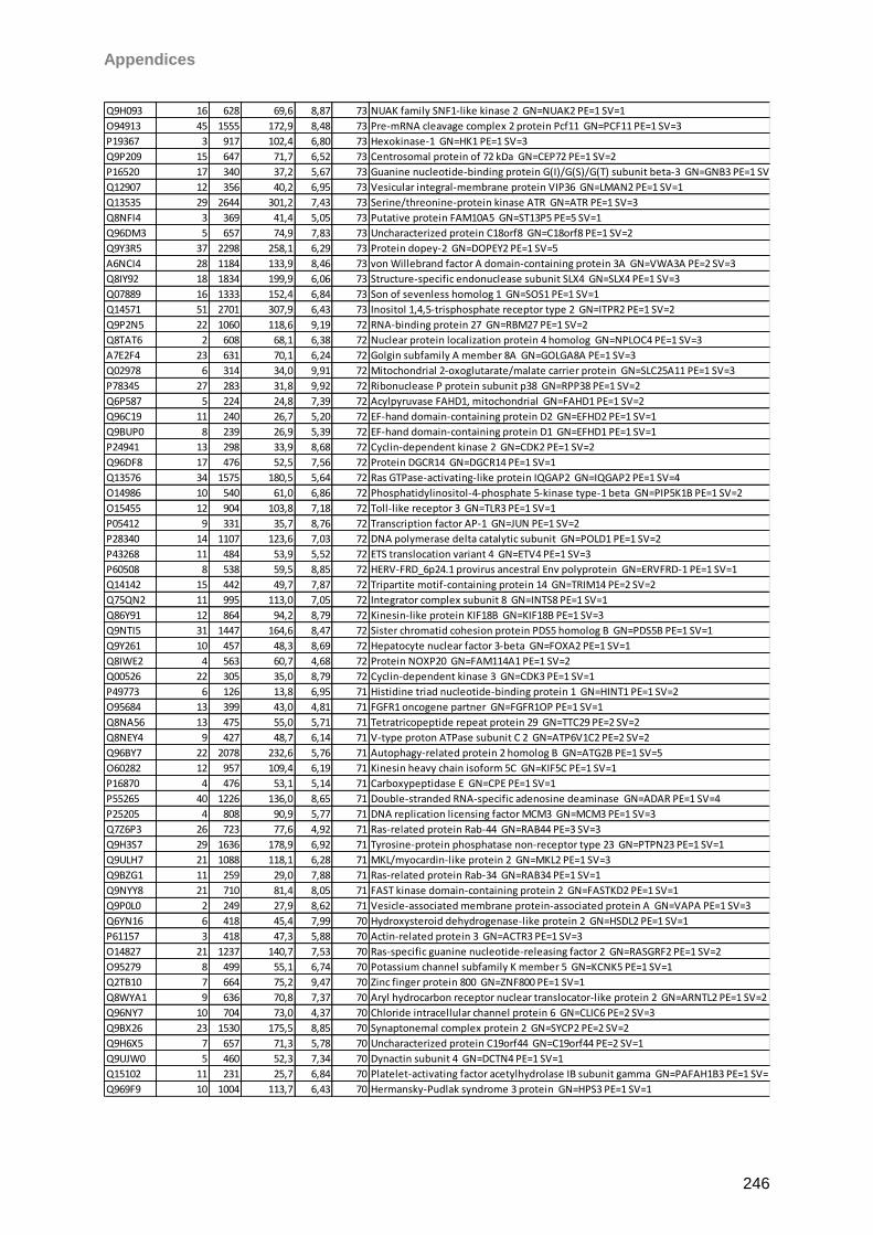
Appendices
246
Q9H093 16 628 69,6 8,87 73 NUAK family SNF1-like kinase 2 GN=NUAK2 PE=1 SV=1
O94913 45 1555 172,9 8,48 73 Pre-mRNA cleavage complex 2 protein Pcf11 GN=PCF11 PE=1 SV=3
P19367 3 917 102,4 6,80 73 Hexokinase-1 GN=HK1 PE=1 SV=3
Q9P209 15 647 71,7 6,52 73 Centrosomal protein of 72 kDa GN=CEP72 PE=1 SV=2
P16520 17 340 37,2 5,67 73 Guanine nucleotide-binding protein G(I)/G(S)/G(T) subunit beta-3 GN=GNB3 PE=1 SV=1
Q12907 12 356 40,2 6,95 73 Vesicular integral-membrane protein VIP36 GN=LMAN2 PE=1 SV=1
Q13535 29 2644 301,2 7,43 73 Serine/threonine-protein kinase ATR GN=ATR PE=1 SV=3
Q8NFI4 3 369 41,4 5,05 73 Putative protein FAM10A5 GN=ST13P5 PE=5 SV=1
Q96DM3 5 657 74,9 7,83 73 Uncharacterized protein C18orf8 GN=C18orf8 PE=1 SV=2
Q9Y3R5 37 2298 258,1 6,29 73 Protein dopey-2 GN=DOPEY2 PE=1 SV=5
A6NCI4 28 1184 133,9 8,46 73 von Willebrand factor A domain-containing protein 3A GN=VWA3A PE=2 SV=3
Q8IY92 18 1834 199,9 6,06 73 Structure-specific endonuclease subunit SLX4 GN=SLX4 PE=1 SV=3
Q07889 16 1333 152,4 6,84 73 Son of sevenless homolog 1 GN=SOS1 PE=1 SV=1
Q14571 51 2701 307,9 6,43 73 Inositol 1,4,5-trisphosphate receptor type 2 GN=ITPR2 PE=1 SV=2
Q9P2N5 22 1060 118,6 9,19 72 RNA-binding protein 27 GN=RBM27 PE=1 SV=2
Q8TAT6 2 608 68,1 6,38 72 Nuclear protein localization protein 4 homolog GN=NPLOC4 PE=1 SV=3
A7E2F4 23 631 70,1 6,24 72 Golgin subfamily A member 8A GN=GOLGA8A PE=1 SV=3
Q02978 6 314 34,0 9,91 72 Mitochondrial 2-oxoglutarate/malate carrier protein GN=SLC25A11 PE=1 SV=3
P78345 27 283 31,8 9,92 72 Ribonuclease P protein subunit p38 GN=RPP38 PE=1 SV=2
Q6P587 5 224 24,8 7,39 72 Acylpyruvase FAHD1, mitochondrial GN=FAHD1 PE=1 SV=2
Q96C19 11 240 26,7 5,20 72 EF-hand domain-containing protein D2 GN=EFHD2 PE=1 SV=1
Q9BUP0 8 239 26,9 5,39 72 EF-hand domain-containing protein D1 GN=EFHD1 PE=1 SV=1
P24941 13 298 33,9 8,68 72 Cyclin-dependent kinase 2 GN=CDK2 PE=1 SV=2
Q96DF8 17 476 52,5 7,56 72 Protein DGCR14 GN=DGCR14 PE=1 SV=1
Q13576 34 1575 180,5 5,64 72 Ras GTPase-activating-like protein IQGAP2 GN=IQGAP2 PE=1 SV=4
O14986 10 540 61,0 6,86 72 Phosphatidylinositol-4-phosphate 5-kinase type-1 beta GN=PIP5K1B PE=1 SV=2
O15455 12 904 103,8 7,18 72 Toll-like receptor 3 GN=TLR3 PE=1 SV=1
P05412 9 331 35,7 8,76 72 Transcription factor AP-1 GN=JUN PE=1 SV=2
P28340 14 1107 123,6 7,03 72 DNA polymerase delta catalytic subunit GN=POLD1 PE=1 SV=2
P43268 11 484 53,9 5,52 72 ETS translocation variant 4 GN=ETV4 PE=1 SV=3
P60508 8 538 59,5 8,85 72 HERV-FRD_6p24.1 provirus ancestral Env polyprotein GN=ERVFRD-1 PE=1 SV=1
Q14142 15 442 49,7 7,87 72 Tripartite motif-containing protein 14 GN=TRIM14 PE=2 SV=2
Q75QN2 11 995 113,0 7,05 72 Integrator complex subunit 8 GN=INTS8 PE=1 SV=1
Q86Y91 12 864 94,2 8,79 72 Kinesin-like protein KIF18B GN=KIF18B PE=1 SV=3
Q9NTI5 31 1447 164,6 8,47 72 Sister chromatid cohesion protein PDS5 homolog B GN=PDS5B PE=1 SV=1
Q9Y261 10 457 48,3 8,69 72 Hepatocyte nuclear factor 3-beta GN=FOXA2 PE=1 SV=1
Q8IWE2 4 563 60,7 4,68 72 Protein NOXP20 GN=FAM114A1 PE=1 SV=2
Q00526 22 305 35,0 8,79 72 Cyclin-dependent kinase 3 GN=CDK3 PE=1 SV=1
P49773 6 126 13,8 6,95 71 Histidine triad nucleotide-binding protein 1 GN=HINT1 PE=1 SV=2
O95684 13 399 43,0 4,81 71 FGFR1 oncogene partner GN=FGFR1OP PE=1 SV=1
Q8NA56 13 475 55,0 5,71 71 Tetratricopeptide repeat protein 29 GN=TTC29 PE=2 SV=2
Q8NEY4 9 427 48,7 6,14 71 V-type proton ATPase subunit C 2 GN=ATP6V1C2 PE=2 SV=2
Q96BY7 22 2078 232,6 5,76 71 Autophagy-related protein 2 homolog B GN=ATG2B PE=1 SV=5
O60282 12 957 109,4 6,19 71 Kinesin heavy chain isoform 5C GN=KIF5C PE=1 SV=1
P16870 4 476 53,1 5,14 71 Carboxypeptidase E GN=CPE PE=1 SV=1
P55265 40 1226 136,0 8,65 71 Double-stranded RNA-specific adenosine deaminase GN=ADAR PE=1 SV=4
P25205 4 808 90,9 5,77 71 DNA replication licensing factor MCM3 GN=MCM3 PE=1 SV=3
Q7Z6P3 26 723 77,6 4,92 71 Ras-related protein Rab-44 GN=RAB44 PE=3 SV=3
Q9H3S7 29 1636 178,9 6,92 71 Tyrosine-protein phosphatase non-receptor type 23 GN=PTPN23 PE=1 SV=1
Q9ULH7 21 1088 118,1 6,28 71 MKL/myocardin-like protein 2 GN=MKL2 PE=1 SV=3
Q9BZG1 11 259 29,0 7,88 71 Ras-related protein Rab-34 GN=RAB34 PE=1 SV=1
Q9NYY8 21 710 81,4 8,05 71 FAST kinase domain-containing protein 2 GN=FASTKD2 PE=1 SV=1
Q9P0L0 2 249 27,9 8,62 71 Vesicle-associated membrane protein-associated protein A GN=VAPA PE=1 SV=3
Q6YN16 6 418 45,4 7,99 70 Hydroxysteroid dehydrogenase-like protein 2 GN=HSDL2 PE=1 SV=1
P61157 3 418 47,3 5,88 70 Actin-related protein 3 GN=ACTR3 PE=1 SV=3
O14827 21 1237 140,7 7,53 70 Ras-specific guanine nucleotide-releasing factor 2 GN=RASGRF2 PE=1 SV=2
O95279 8 499 55,1 6,74 70 Potassium channel subfamily K member 5 GN=KCNK5 PE=1 SV=1
Q2TB10 7 664 75,2 9,47 70 Zinc finger protein 800 GN=ZNF800 PE=1 SV=1
Q8WYA1 9 636 70,8 7,37 70 Aryl hydrocarbon receptor nuclear translocator-like protein 2 GN=ARNTL2 PE=1 SV=2
Q96NY7 10 704 73,0 4,37 70 Chloride intracellular channel protein 6 GN=CLIC6 PE=2 SV=3
Q9BX26 23 1530 175,5 8,85 70 Synaptonemal complex protein 2 GN=SYCP2 PE=2 SV=2
Q9H6X5 7 657 71,3 5,78 70 Uncharacterized protein C19orf44 GN=C19orf44 PE=2 SV=1
Q9UJW0 5 460 52,3 7,34 70 Dynactin subunit 4 GN=DCTN4 PE=1 SV=1
Q15102 11 231 25,7 6,84 70 Platelet-activating factor acetylhydrolase IB subunit gamma GN=PAFAH1B3 PE=1 SV=1
Q969F9 10 1004 113,7 6,43 70 Hermansky-Pudlak syndrome 3 protein GN=HPS3 PE=1 SV=1
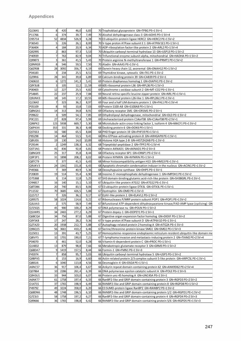
Appendices
247
Q13641 8 420 46,0 6,83 70 Trophoblast glycoprotein GN=TPBG PE=1 SV=1
P11766 6 374 39,7 7,49 70 Alcohol dehydrogenase class-3 GN=ADH5 PE=1 SV=4
O95714 52 4834 526,9 6,28 70 E3 ubiquitin-protein ligase HERC2 GN=HERC2 PE=1 SV=2
P36543 5 226 26,1 8,00 70 V-type proton ATPase subunit E 1 GN=ATP6V1E1 PE=1 SV=1
P36404 4 184 20,9 6,34 70 ADP-ribosylation factor-like protein 2 GN=ARL2 PE=1 SV=4
Q92995 2 863 97,3 5,53 70 Ubiquitin carboxyl-terminal hydrolase 13 GN=USP13 PE=1 SV=2
P40939 7 763 82,9 9,04 70 Trifunctional enzyme subunit alpha, mitochondrial GN=HADHA PE=1 SV=2
Q99873 3 361 41,5 5,43 70 Protein arginine N-methyltransferase 1 GN=PRMT1 PE=1 SV=2
Q9NRG9 8 546 59,5 7,50 70 Aladin GN=AAAS PE=1 SV=1
Q6ZR08 28 3092 356,7 6,19 69 Dynein heavy chain 12, axonemal GN=DNAH12 PE=1 SV=2
P04183 3 234 25,5 8,51 69 Thymidine kinase, cytosolic GN=TK1 PE=1 SV=2
Q29RI6 28 341 39,8 6,89 69 Calcium-binding protein 39 GN=CAB39 PE=2 SV=1
O60610 6 1272 141,3 5,41 69 Protein diaphanous homolog 1 GN=DIAPH1 PE=1 SV=2
Q9Y3U8 3 105 12,2 11,59 69 60S ribosomal protein L36 GN=RPL36 PE=1 SV=3
P00403 12 227 25,5 4,82 69 Cytochrome c oxidase subunit 2 GN=MT-CO2 PE=1 SV=1
P54845 22 237 25,9 7,88 69 Neural retina-specific leucine zipper protein GN=NRL PE=1 SV=1
Q9UNX3 18 145 17,2 10,55 69 60S ribosomal protein L26-like 1 GN=RPL26L1 PE=1 SV=1
Q13642 7 323 36,2 8,97 69 Four and a half LIM domains protein 1 GN=FHL1 PE=1 SV=4
P05109 8 93 10,8 7,03 69 Protein S100-A8 GN=S100A8 PE=1 SV=1
Q8NGA1 14 313 34,8 8,91 69 Olfactory receptor 1M1 GN=OR1M1 PE=2 SV=1
P09622 3 509 54,1 7,85 69 Dihydrolipoyl dehydrogenase, mitochondrial GN=DLD PE=1 SV=2
P0C221 27 828 97,4 5,99 69 Uncharacterized protein C14orf38 GN=C14orf38 PE=2 SV=1
Q96PK2 123 5938 669,7 5,30 68 Microtubule-actin cross-linking factor 1, isoform 4 GN=MACF1 PE=1 SV=2
Q6PKX4 353 331 38,3 8,48 68 Docking protein 6 GN=DOK6 PE=1 SV=1
Q5T6S3 16 580 65,5 8,84 68 PHD finger protein 19 GN=PHF19 PE=1 SV=1
P85298 14 464 53,5 9,41 68 Rho GTPase-activating protein 8 GN=ARHGAP8 PE=1 SV=1
Q8IUE6 19 130 14,0 10,89 68 Histone H2A type 2-B GN=HIST2H2AB PE=1 SV=3
P29144 2 1249 138,3 6,32 68 Tripeptidyl-peptidase 2 GN=TPP2 PE=1 SV=4
Q5T1N1 41 836 92,8 6,81 68 Protein AKNAD1 GN=AKNAD1 PE=2 SV=3
Q8NGX9 25 317 35,8 8,44 68 Olfactory receptor 6P1 GN=OR6P1 PE=2 SV=1
Q9P2P1 31 1898 208,2 8,02 68 Protein NYNRIN GN=NYNRIN PE=1 SV=3
Q8TCT9 7 377 41,5 6,43 68 Minor histocompatibility antigen H13 GN=HM13 PE=1 SV=1
Q9UKV3 24 1341 151,8 6,43 68 Apoptotic chromatin condensation inducer in the nucleus GN=ACIN1 PE=1 SV=2
P49366 7 369 40,9 5,36 68 Deoxyhypusine synthase GN=DHPS PE=1 SV=1
P20839 19 514 55,4 6,90 68 Inosine-5'-monophosphate dehydrogenase 1 GN=IMPDH1 PE=1 SV=2
O75368 3 114 12,8 5,25 67 SH3 domain-binding glutamic acid-rich-like protein GN=SH3BGRL PE=1 SV=1
O94817 16 140 15,1 5,10 67 Ubiquitin-like protein ATG12 GN=ATG12 PE=1 SV=1
Q8TDB6 20 740 83,5 8,06 67 E3 ubiquitin-protein ligase DTX3L GN=DTX3L PE=1 SV=1
P11532 70 3685 426,5 5,88 67 Dystrophin GN=DMD PE=1 SV=3
Q15717 7 326 36,1 9,17 67 ELAV-like protein 1 GN=ELAVL1 PE=1 SV=2
Q99575 58 1024 114,6 9,22 67 Ribonucleases P/MRP protein subunit POP1 GN=POP1 PE=1 SV=2
Q3LXA3 2 575 58,9 7,49 67 Bifunctional ATP-dependent dihydroxyacetone kinase/FAD-AMP lyase (cyclizing) GN=DAK PE=1 SV=2
Q7Z5Q5 45 900 100,2 8,29 67 DNA polymerase nu GN=POLN PE=1 SV=2
Q5JWR5 34 2465 277,2 6,29 67 Protein dopey-1 GN=DOPEY1 PE=1 SV=1
Q68CQ4 34 756 87,0 5,88 67 Digestive organ expansion factor homolog GN=DIEXF PE=1 SV=2
Q9Y5K8 17 247 28,2 9,36 67 V-type proton ATPase subunit D GN=ATP6V1D PE=1 SV=1
Q2TAZ0 20 1938 212,7 5,88 67 Autophagy-related protein 2 homolog A GN=ATG2A PE=1 SV=3
Q96Q15 56 3661 410,2 6,46 67 Serine/threonine-protein kinase SMG1 GN=SMG1 PE=1 SV=3
Q15011 13 391 43,7 5,25 67 Homocysteine-responsive endoplasmic reticulum-resident ubiquitin-like domain member 1 protein GN=HERPUD1 PE=1 SV=1
Q8IVF5 32 1701 190,0 7,21 67 T-lymphoma invasion and metastasis-inducing protein 2 GN=TIAM2 PE=2 SV=4
P04070 3 461 52,0 6,28 66 Vitamin K-dependent protein C GN=PROC PE=1 SV=1
Q14832 13 879 98,8 7,66 66 Metabotropic glutamate receptor 3 GN=GRM3 PE=1 SV=2
Q68DA7 9 1419 157,5 8,44 66 Formin-1 GN=FMN1 PE=1 SV=3
P45974 7 858 95,7 5,03 66 Ubiquitin carboxyl-terminal hydrolase 5 GN=USP5 PE=1 SV=2
Q9BPX5 3 153 16,9 6,60 66 Actin-related protein 2/3 complex subunit 5-like protein GN=ARPC5L PE=1 SV=1
Q86SJ6 6 1040 113,8 4,56 66 Desmoglein-4 GN=DSG4 PE=1 SV=1
A6NC57 36 917 106,4 6,67 66 Ankyrin repeat domain-containing protein 62 GN=ANKRD62 PE=2 SV=4
Q07864 10 2286 261,4 6,39 66 DNA polymerase epsilon catalytic subunit A GN=POLE PE=1 SV=5
Q9H3U1 33 944 103,0 6,07 66 Protein unc-45 homolog A GN=UNC45A PE=1 SV=1
A6NKT7 61 1758 197,4 6,33 66 RanBP2-like and GRIP domain-containing protein 3 GN=RGPD3 PE=2 SV=2
O14715 37 1765 198,9 6,49 66 RANBP2-like and GRIP domain-containing protein 8 GN=RGPD8 PE=1 SV=2
P49792 49 3224 358,0 6,20 66 E3 SUMO-protein ligase RanBP2 GN=RANBP2 PE=1 SV=2
Q68DN6 48 1748 196,5 6,10 66 RANBP2-like and GRIP domain-containing protein 1/2 GN=RGPD1 PE=1 SV=2
Q7Z3J3 58 1758 197,2 6,27 66 RanBP2-like and GRIP domain-containing protein 4 GN=RGPD4 PE=2 SV=3
Q99666 36 1765 198,8 6,42 66 RANBP2-like and GRIP domain-containing protein 5/6 GN=RGPD5 PE=1 SV=3
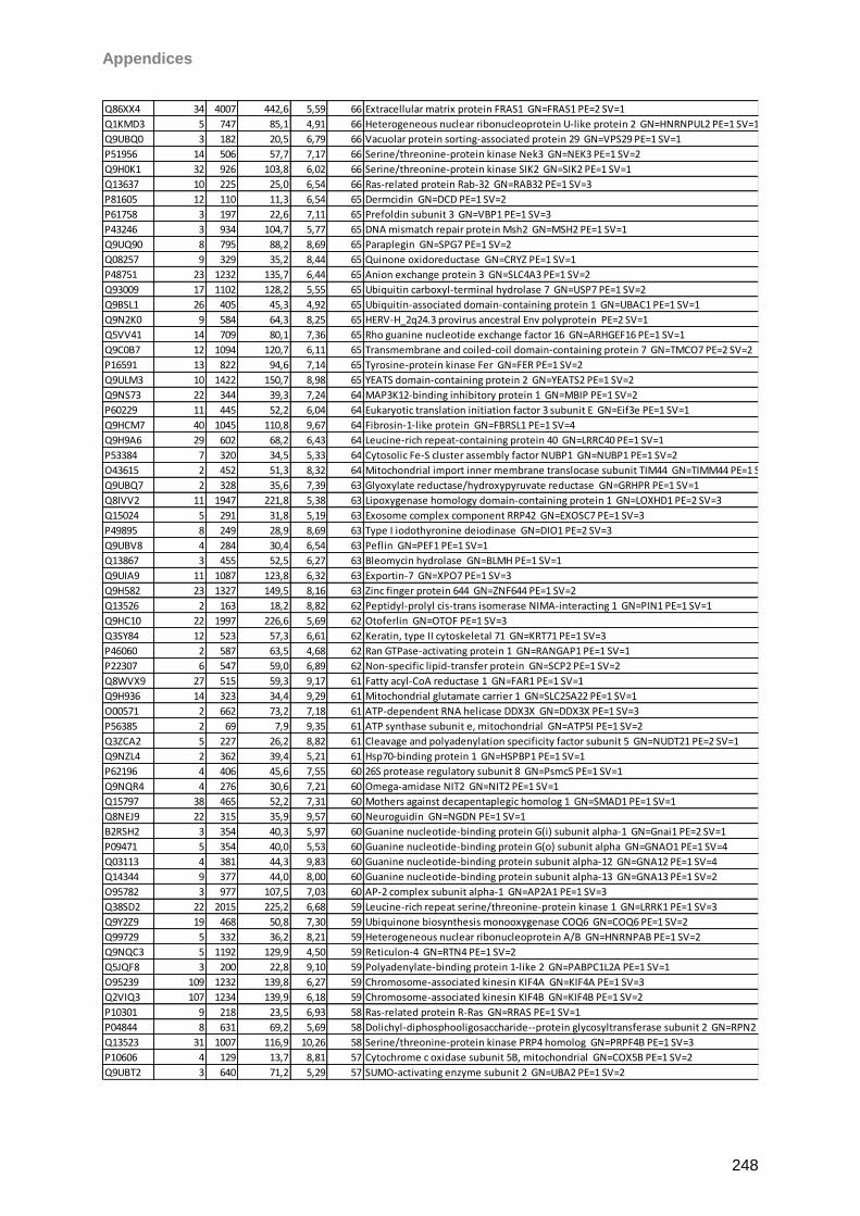
Appendices
248
Q86XX4 34 4007 442,6 5,59 66 Extracellular matrix protein FRAS1 GN=FRAS1 PE=2 SV=1
Q1KMD3 5 747 85,1 4,91 66 Heterogeneous nuclear ribonucleoprotein U-like protein 2 GN=HNRNPUL2 PE=1 SV=1
Q9UBQ0 3 182 20,5 6,79 66 Vacuolar protein sorting-associated protein 29 GN=VPS29 PE=1 SV=1
P51956 14 506 57,7 7,17 66 Serine/threonine-protein kinase Nek3 GN=NEK3 PE=1 SV=2
Q9H0K1 32 926 103,8 6,02 66 Serine/threonine-protein kinase SIK2 GN=SIK2 PE=1 SV=1
Q13637 10 225 25,0 6,54 66 Ras-related protein Rab-32 GN=RAB32 PE=1 SV=3
P81605 12 110 11,3 6,54 65 Dermcidin GN=DCD PE=1 SV=2
P61758 3 197 22,6 7,11 65 Prefoldin subunit 3 GN=VBP1 PE=1 SV=3
P43246 3 934 104,7 5,77 65 DNA mismatch repair protein Msh2 GN=MSH2 PE=1 SV=1
Q9UQ90 8 795 88,2 8,69 65 Paraplegin GN=SPG7 PE=1 SV=2
Q08257 9 329 35,2 8,44 65 Quinone oxidoreductase GN=CRYZ PE=1 SV=1
P48751 23 1232 135,7 6,44 65 Anion exchange protein 3 GN=SLC4A3 PE=1 SV=2
Q93009 17 1102 128,2 5,55 65 Ubiquitin carboxyl-terminal hydrolase 7 GN=USP7 PE=1 SV=2
Q9BSL1 26 405 45,3 4,92 65 Ubiquitin-associated domain-containing protein 1 GN=UBAC1 PE=1 SV=1
Q9N2K0 9 584 64,3 8,25 65 HERV-H_2q24.3 provirus ancestral Env polyprotein PE=2 SV=1
Q5VV41 14 709 80,1 7,36 65 Rho guanine nucleotide exchange factor 16 GN=ARHGEF16 PE=1 SV=1
Q9C0B7 12 1094 120,7 6,11 65 Transmembrane and coiled-coil domain-containing protein 7 GN=TMCO7 PE=2 SV=2
P16591 13 822 94,6 7,14 65 Tyrosine-protein kinase Fer GN=FER PE=1 SV=2
Q9ULM3 10 1422 150,7 8,98 65 YEATS domain-containing protein 2 GN=YEATS2 PE=1 SV=2
Q9NS73 22 344 39,3 7,24 64 MAP3K12-binding inhibitory protein 1 GN=MBIP PE=1 SV=2
P60229 11 445 52,2 6,04 64 Eukaryotic translation initiation factor 3 subunit E GN=Eif3e PE=1 SV=1
Q9HCM7 40 1045 110,8 9,67 64 Fibrosin-1-like protein GN=FBRSL1 PE=1 SV=4
Q9H9A6 29 602 68,2 6,43 64 Leucine-rich repeat-containing protein 40 GN=LRRC40 PE=1 SV=1
P53384 7 320 34,5 5,33 64 Cytosolic Fe-S cluster assembly factor NUBP1 GN=NUBP1 PE=1 SV=2
O43615 2 452 51,3 8,32 64 Mitochondrial import inner membrane translocase subunit TIM44 GN=TIMM44 PE=1 SV=2
Q9UBQ7 2 328 35,6 7,39 63 Glyoxylate reductase/hydroxypyruvate reductase GN=GRHPR PE=1 SV=1
Q8IVV2 11 1947 221,8 5,38 63 Lipoxygenase homology domain-containing protein 1 GN=LOXHD1 PE=2 SV=3
Q15024 5 291 31,8 5,19 63 Exosome complex component RRP42 GN=EXOSC7 PE=1 SV=3
P49895 8 249 28,9 8,69 63 Type I iodothyronine deiodinase GN=DIO1 PE=2 SV=3
Q9UBV8 4 284 30,4 6,54 63 Peflin GN=PEF1 PE=1 SV=1
Q13867 3 455 52,5 6,27 63 Bleomycin hydrolase GN=BLMH PE=1 SV=1
Q9UIA9 11 1087 123,8 6,32 63 Exportin-7 GN=XPO7 PE=1 SV=3
Q9H582 23 1327 149,5 8,16 63 Zinc finger protein 644 GN=ZNF644 PE=1 SV=2
Q13526 2 163 18,2 8,82 62 Peptidyl-prolyl cis-trans isomerase NIMA-interacting 1 GN=PIN1 PE=1 SV=1
Q9HC10 22 1997 226,6 5,69 62 Otoferlin GN=OTOF PE=1 SV=3
Q3SY84 12 523 57,3 6,61 62 Keratin, type II cytoskeletal 71 GN=KRT71 PE=1 SV=3
P46060 2 587 63,5 4,68 62 Ran GTPase-activating protein 1 GN=RANGAP1 PE=1 SV=1
P22307 6 547 59,0 6,89 62 Non-specific lipid-transfer protein GN=SCP2 PE=1 SV=2
Q8WVX9 27 515 59,3 9,17 61 Fatty acyl-CoA reductase 1 GN=FAR1 PE=1 SV=1
Q9H936 14 323 34,4 9,29 61 Mitochondrial glutamate carrier 1 GN=SLC25A22 PE=1 SV=1
O00571 2 662 73,2 7,18 61 ATP-dependent RNA helicase DDX3X GN=DDX3X PE=1 SV=3
P56385 2 69 7,9 9,35 61 ATP synthase subunit e, mitochondrial GN=ATP5I PE=1 SV=2
Q3ZCA2 5 227 26,2 8,82 61 Cleavage and polyadenylation specificity factor subunit 5 GN=NUDT21 PE=2 SV=1
Q9NZL4 2 362 39,4 5,21 61 Hsp70-binding protein 1 GN=HSPBP1 PE=1 SV=1
P62196 4 406 45,6 7,55 60 26S protease regulatory subunit 8 GN=Psmc5 PE=1 SV=1
Q9NQR4 4 276 30,6 7,21 60 Omega-amidase NIT2 GN=NIT2 PE=1 SV=1
Q15797 38 465 52,2 7,31 60 Mothers against decapentaplegic homolog 1 GN=SMAD1 PE=1 SV=1
Q8NEJ9 22 315 35,9 9,57 60 Neuroguidin GN=NGDN PE=1 SV=1
B2RSH2 3 354 40,3 5,97 60 Guanine nucleotide-binding protein G(i) subunit alpha-1 GN=Gnai1 PE=2 SV=1
P09471 5 354 40,0 5,53 60 Guanine nucleotide-binding protein G(o) subunit alpha GN=GNAO1 PE=1 SV=4
Q03113 4 381 44,3 9,83 60 Guanine nucleotide-binding protein subunit alpha-12 GN=GNA12 PE=1 SV=4
Q14344 9 377 44,0 8,00 60 Guanine nucleotide-binding protein subunit alpha-13 GN=GNA13 PE=1 SV=2
O95782 3 977 107,5 7,03 60 AP-2 complex subunit alpha-1 GN=AP2A1 PE=1 SV=3
Q38SD2 22 2015 225,2 6,68 59 Leucine-rich repeat serine/threonine-protein kinase 1 GN=LRRK1 PE=1 SV=3
Q9Y2Z9 19 468 50,8 7,30 59 Ubiquinone biosynthesis monooxygenase COQ6 GN=COQ6 PE=1 SV=2
Q99729 5 332 36,2 8,21 59 Heterogeneous nuclear ribonucleoprotein A/B GN=HNRNPAB PE=1 SV=2
Q9NQC3 5 1192 129,9 4,50 59 Reticulon-4 GN=RTN4 PE=1 SV=2
Q5JQF8 3 200 22,8 9,10 59 Polyadenylate-binding protein 1-like 2 GN=PABPC1L2A PE=1 SV=1
O95239 109 1232 139,8 6,27 59 Chromosome-associated kinesin KIF4A GN=KIF4A PE=1 SV=3
Q2VIQ3 107 1234 139,9 6,18 59 Chromosome-associated kinesin KIF4B GN=KIF4B PE=1 SV=2
P10301 9 218 23,5 6,93 58 Ras-related protein R-Ras GN=RRAS PE=1 SV=1
P04844 8 631 69,2 5,69 58 Dolichyl-diphosphooligosaccharide--protein glycosyltransferase subunit 2 GN=RPN2 PE=1 SV=3
Q13523 31 1007 116,9 10,26 58 Serine/threonine-protein kinase PRP4 homolog GN=PRPF4B PE=1 SV=3
P10606 4 129 13,7 8,81 57 Cytochrome c oxidase subunit 5B, mitochondrial GN=COX5B PE=1 SV=2
Q9UBT2 3 640 71,2 5,29 57 SUMO-activating enzyme subunit 2 GN=UBA2 PE=1 SV=2
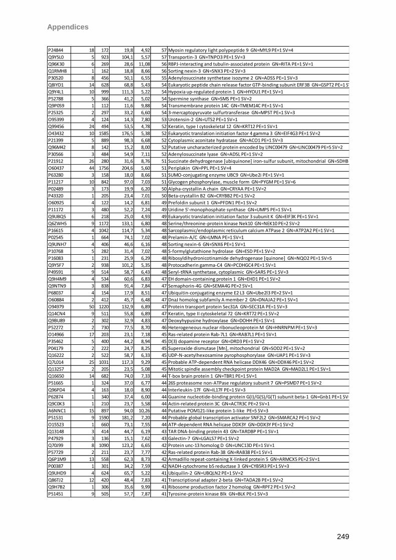
Appendices
249
P24844 18 172 19,8 4,92 57 Myosin regulatory light polypeptide 9 GN=MYL9 PE=1 SV=4
Q9Y5L0 5 923 104,1 5,57 57 Transportin-3 GN=TNPO3 PE=1 SV=3
Q96K30 6 269 28,6 11,08 56 RBPJ-interacting and tubulin-associated protein GN=RITA PE=1 SV=1
Q1RMH8 1 162 18,8 8,66 56 Sorting nexin-3 GN=SNX3 PE=2 SV=3
P30520 8 456 50,1 6,55 55 Adenylosuccinate synthetase isozyme 2 GN=ADSS PE=1 SV=3
Q8IYD1 14 628 68,8 5,43 54 Eukaryotic peptide chain release factor GTP-binding subunit ERF3B GN=GSPT2 PE=1 SV=2
Q9Y4L1 10 999 111,3 5,22 54 Hypoxia up-regulated protein 1 GN=HYOU1 PE=1 SV=1
P52788 5 366 41,2 5,02 54 Spermine synthase GN=SMS PE=1 SV=2
Q9P0S9 1 112 11,6 9,88 54 Transmembrane protein 14C GN=TMEM14C PE=1 SV=1
P25325 2 297 33,2 6,60 54 3-mercaptopyruvate sulfurtransferase GN=MPST PE=1 SV=3
O95399 4 124 14,3 7,80 53 Urotensin-2 GN=UTS2 PE=1 SV=1
Q99456 24 494 53,5 4,78 52 Keratin, type I cytoskeletal 12 GN=KRT12 PE=1 SV=1
O43432 10 1585 176,5 5,38 52 Eukaryotic translation initiation factor 4 gamma 3 GN=EIF4G3 PE=1 SV=2
P21399 5 889 98,3 6,68 52 Cytoplasmic aconitate hydratase GN=ACO1 PE=1 SV=3
Q96M42 8 142 15,2 8,00 52 Putative uncharacterized protein encoded by LINC00479 GN=LINC00479 PE=5 SV=2
P30566 3 484 54,9 7,11 52 Adenylosuccinate lyase GN=ADSL PE=1 SV=2
P21912 26 280 31,6 8,76 51 Succinate dehydrogenase [ubiquinone] iron-sulfur subunit, mitochondrial GN=SDHB PE=1 SV=3
O60437 44 1756 204,6 5,60 51 Periplakin GN=PPL PE=1 SV=4
P63280 3 158 18,0 8,66 51 SUMO-conjugating enzyme UBC9 GN=Ube2i PE=1 SV=1
P11217 10 842 97,0 7,03 51 Glycogen phosphorylase, muscle form GN=PYGM PE=1 SV=6
P02489 3 173 19,9 6,20 50 Alpha-crystallin A chain GN=CRYAA PE=1 SV=2
P43320 1 205 23,4 7,01 50 Beta-crystallin B2 GN=CRYBB2 PE=1 SV=2
O60925 4 122 14,2 6,81 49 Prefoldin subunit 1 GN=PFDN1 PE=1 SV=2
P11172 3 480 52,2 7,24 49 Uridine 5'-monophosphate synthase GN=UMPS PE=1 SV=1
Q9UBQ5 6 218 25,0 4,93 49 Eukaryotic translation initiation factor 3 subunit K GN=EIF3K PE=1 SV=1
Q6ZWH5 9 1172 133,1 6,80 48 Serine/threonine-protein kinase Nek10 GN=NEK10 PE=2 SV=2
P16615 4 1042 114,7 5,34 48 Sarcoplasmic/endoplasmic reticulum calcium ATPase 2 GN=ATP2A2 PE=1 SV=1
P02545 1 664 74,1 7,02 48 Prelamin-A/C GN=LMNA PE=1 SV=1
Q9UNH7 4 406 46,6 6,16 48 Sorting nexin-6 GN=SNX6 PE=1 SV=1
P10768 5 282 31,4 7,02 48 S-formylglutathione hydrolase GN=ESD PE=1 SV=2
P16083 1 231 25,9 6,29 48 Ribosyldihydronicotinamide dehydrogenase [quinone] GN=NQO2 PE=1 SV=5
Q9Y5F7 2 938 101,2 5,35 48 Protocadherin gamma-C4 GN=PCDHGC4 PE=1 SV=1
P49591 9 514 58,7 6,43 48 Seryl-tRNA synthetase, cytoplasmic GN=SARS PE=1 SV=3
Q9H4M9 4 534 60,6 6,83 47 EH domain-containing protein 1 GN=EHD1 PE=1 SV=2
Q9NTN9 3 838 91,4 7,84 47 Semaphorin-4G GN=SEMA4G PE=2 SV=1
P68037 4 154 17,9 8,51 47 Ubiquitin-conjugating enzyme E2 L3 GN=Ube2l3 PE=2 SV=1
O60884 2 412 45,7 6,48 47 DnaJ homolog subfamily A member 2 GN=DNAJA2 PE=1 SV=1
O94979 50 1220 132,9 6,89 47 Protein transport protein Sec31A GN=SEC31A PE=1 SV=3
Q14CN4 9 511 55,8 6,89 47 Keratin, type II cytoskeletal 72 GN=KRT72 PE=1 SV=2
Q9BU89 2 302 32,9 4,83 47 Deoxyhypusine hydroxylase GN=DOHH PE=1 SV=1
P52272 2 730 77,5 8,70 46 Heterogeneous nuclear ribonucleoprotein M GN=HNRNPM PE=1 SV=3
O14966 17 203 23,1 7,18 45 Ras-related protein Rab-7L1 GN=RAB7L1 PE=1 SV=1
P35462 5 400 44,2 8,94 45 D(3) dopamine receptor GN=DRD3 PE=1 SV=2
P04179 2 222 24,7 8,25 45 Superoxide dismutase [Mn], mitochondrial GN=SOD2 PE=1 SV=2
Q16222 2 522 58,7 6,33 45 UDP-N-acetylhexosamine pyrophosphorylase GN=UAP1 PE=1 SV=3
Q7L014 25 1031 117,3 9,29 45 Probable ATP-dependent RNA helicase DDX46 GN=DDX46 PE=1 SV=2
Q13257 2 205 23,5 5,08 45 Mitotic spindle assembly checkpoint protein MAD2A GN=MAD2L1 PE=1 SV=1
Q16650 14 682 74,0 7,33 44 T-box brain protein 1 GN=TBR1 PE=1 SV=1
P51665 1 324 37,0 6,77 44 26S proteasome non-ATPase regulatory subunit 7 GN=PSMD7 PE=1 SV=2
Q96PD4 4 163 18,0 8,90 44 Interleukin-17F GN=IL17F PE=1 SV=3
P62874 1 340 37,4 6,00 44 Guanine nucleotide-binding protein G(I)/G(S)/G(T) subunit beta-1 GN=Gnb1 PE=1 SV=3
Q9C0K3 1 210 23,7 5,58 44 Actin-related protein 3C GN=ACTR3C PE=2 SV=1
A6NNC1 15 897 94,0 10,26 44 Putative POM121-like protein 1-like PE=5 SV=3
P51531 9 1590 181,2 7,20 44 Probable global transcription activator SNF2L2 GN=SMARCA2 PE=1 SV=2
O15523 1 660 73,1 7,55 44 ATP-dependent RNA helicase DDX3Y GN=DDX3Y PE=1 SV=2
Q13148 3 414 44,7 6,19 43 TAR DNA-binding protein 43 GN=TARDBP PE=1 SV=1
P47929 3 136 15,1 7,62 43 Galectin-7 GN=LGALS7 PE=1 SV=2
Q70J99 8 1090 123,2 6,65 42 Protein unc-13 homolog D GN=UNC13D PE=1 SV=1
P57729 2 211 23,7 7,77 42 Ras-related protein Rab-38 GN=RAB38 PE=1 SV=1
Q6P1M9 13 558 62,3 8,73 42 Armadillo repeat-containing X-linked protein 5 GN=ARMCX5 PE=2 SV=1
P00387 1 301 34,2 7,59 42 NADH-cytochrome b5 reductase 3 GN=CYB5R3 PE=1 SV=3
Q9UHD9 4 624 65,7 5,22 41 Ubiquilin-2 GN=UBQLN2 PE=1 SV=2
Q86TJ2 12 420 48,4 7,83 41 Transcriptional adapter 2-beta GN=TADA2B PE=1 SV=2
Q9H7B2 1 306 35,6 9,99 41 Ribosome production factor 2 homolog GN=RPF2 PE=1 SV=2
P51451 9 505 57,7 7,87 41 Tyrosine-protein kinase Blk GN=BLK PE=1 SV=3
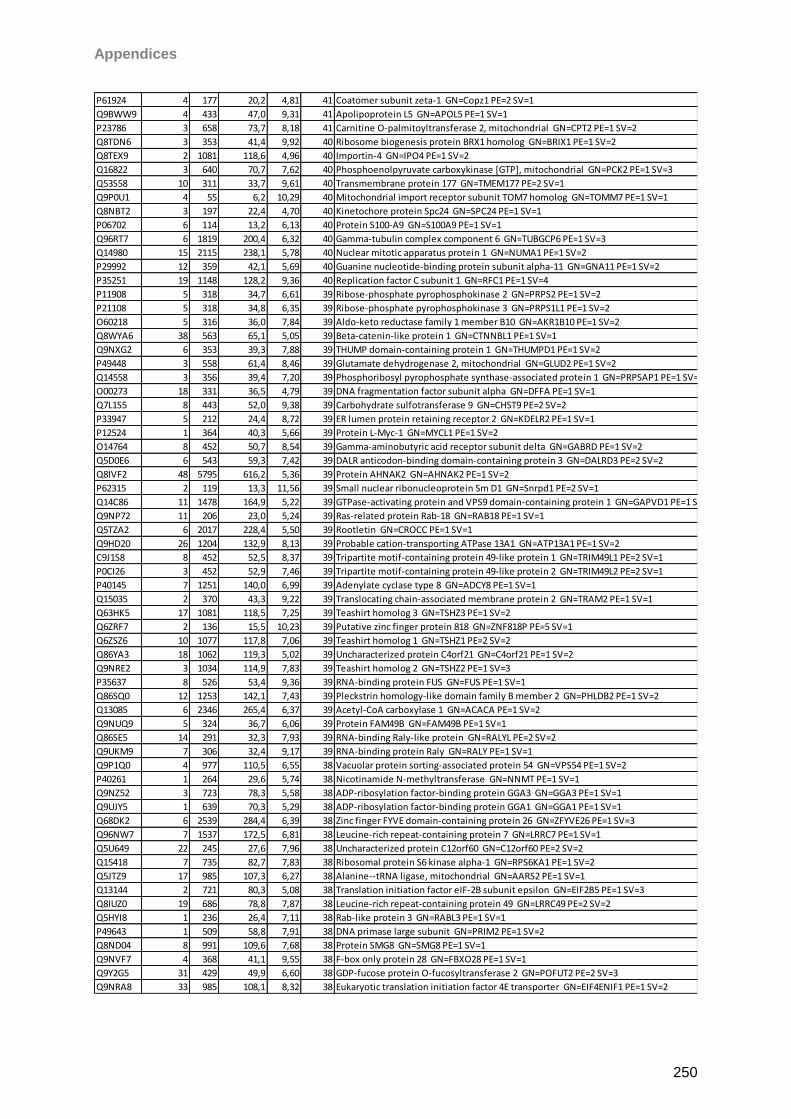
Appendices
250
P61924 4 177 20,2 4,81 41 Coatomer subunit zeta-1 GN=Copz1 PE=2 SV=1
Q9BWW9 4 433 47,0 9,31 41 Apolipoprotein L5 GN=APOL5 PE=1 SV=1
P23786 3 658 73,7 8,18 41 Carnitine O-palmitoyltransferase 2, mitochondrial GN=CPT2 PE=1 SV=2
Q8TDN6 3 353 41,4 9,92 40 Ribosome biogenesis protein BRX1 homolog GN=BRIX1 PE=1 SV=2
Q8TEX9 2 1081 118,6 4,96 40 Importin-4 GN=IPO4 PE=1 SV=2
Q16822 3 640 70,7 7,62 40 Phosphoenolpyruvate carboxykinase [GTP], mitochondrial GN=PCK2 PE=1 SV=3
Q53S58 10 311 33,7 9,61 40 Transmembrane protein 177 GN=TMEM177 PE=2 SV=1
Q9P0U1 4 55 6,2 10,29 40 Mitochondrial import receptor subunit TOM7 homolog GN=TOMM7 PE=1 SV=1
Q8NBT2 3 197 22,4 4,70 40 Kinetochore protein Spc24 GN=SPC24 PE=1 SV=1
P06702 6 114 13,2 6,13 40 Protein S100-A9 GN=S100A9 PE=1 SV=1
Q96RT7 6 1819 200,4 6,32 40 Gamma-tubulin complex component 6 GN=TUBGCP6 PE=1 SV=3
Q14980 15 2115 238,1 5,78 40 Nuclear mitotic apparatus protein 1 GN=NUMA1 PE=1 SV=2
P29992 12 359 42,1 5,69 40 Guanine nucleotide-binding protein subunit alpha-11 GN=GNA11 PE=1 SV=2
P35251 19 1148 128,2 9,36 40 Replication factor C subunit 1 GN=RFC1 PE=1 SV=4
P11908 5 318 34,7 6,61 39 Ribose-phosphate pyrophosphokinase 2 GN=PRPS2 PE=1 SV=2
P21108 5 318 34,8 6,35 39 Ribose-phosphate pyrophosphokinase 3 GN=PRPS1L1 PE=1 SV=2
O60218 5 316 36,0 7,84 39 Aldo-keto reductase family 1 member B10 GN=AKR1B10 PE=1 SV=2
Q8WYA6 38 563 65,1 5,05 39 Beta-catenin-like protein 1 GN=CTNNBL1 PE=1 SV=1
Q9NXG2 6 353 39,3 7,88 39 THUMP domain-containing protein 1 GN=THUMPD1 PE=1 SV=2
P49448 3 558 61,4 8,46 39 Glutamate dehydrogenase 2, mitochondrial GN=GLUD2 PE=1 SV=2
Q14558 3 356 39,4 7,20 39 Phosphoribosyl pyrophosphate synthase-associated protein 1 GN=PRPSAP1 PE=1 SV=2
O00273 18 331 36,5 4,79 39 DNA fragmentation factor subunit alpha GN=DFFA PE=1 SV=1
Q7L1S5 8 443 52,0 9,38 39 Carbohydrate sulfotransferase 9 GN=CHST9 PE=2 SV=2
P33947 5 212 24,4 8,72 39 ER lumen protein retaining receptor 2 GN=KDELR2 PE=1 SV=1
P12524 1 364 40,3 5,66 39 Protein L-Myc-1 GN=MYCL1 PE=1 SV=2
O14764 8 452 50,7 8,54 39 Gamma-aminobutyric acid receptor subunit delta GN=GABRD PE=1 SV=2
Q5D0E6 6 543 59,3 7,42 39 DALR anticodon-binding domain-containing protein 3 GN=DALRD3 PE=2 SV=2
Q8IVF2 48 5795 616,2 5,36 39 Protein AHNAK2 GN=AHNAK2 PE=1 SV=2
P62315 2 119 13,3 11,56 39 Small nuclear ribonucleoprotein Sm D1 GN=Snrpd1 PE=2 SV=1
Q14C86 11 1478 164,9 5,22 39 GTPase-activating protein and VPS9 domain-containing protein 1 GN=GAPVD1 PE=1 SV=2
Q9NP72 11 206 23,0 5,24 39 Ras-related protein Rab-18 GN=RAB18 PE=1 SV=1
Q5TZA2 6 2017 228,4 5,50 39 Rootletin GN=CROCC PE=1 SV=1
Q9HD20 26 1204 132,9 8,13 39 Probable cation-transporting ATPase 13A1 GN=ATP13A1 PE=1 SV=2
C9J1S8 8 452 52,5 8,37 39 Tripartite motif-containing protein 49-like protein 1 GN=TRIM49L1 PE=2 SV=1
P0CI26 3 452 52,9 7,46 39 Tripartite motif-containing protein 49-like protein 2 GN=TRIM49L2 PE=2 SV=1
P40145 7 1251 140,0 6,99 39 Adenylate cyclase type 8 GN=ADCY8 PE=1 SV=1
Q15035 2 370 43,3 9,22 39 Translocating chain-associated membrane protein 2 GN=TRAM2 PE=1 SV=1
Q63HK5 17 1081 118,5 7,25 39 Teashirt homolog 3 GN=TSHZ3 PE=1 SV=2
Q6ZRF7 2 136 15,5 10,23 39 Putative zinc finger protein 818 GN=ZNF818P PE=5 SV=1
Q6ZSZ6 10 1077 117,8 7,06 39 Teashirt homolog 1 GN=TSHZ1 PE=2 SV=2
Q86YA3 18 1062 119,3 5,02 39 Uncharacterized protein C4orf21 GN=C4orf21 PE=1 SV=2
Q9NRE2 3 1034 114,9 7,83 39 Teashirt homolog 2 GN=TSHZ2 PE=1 SV=3
P35637 8 526 53,4 9,36 39 RNA-binding protein FUS GN=FUS PE=1 SV=1
Q86SQ0 12 1253 142,1 7,43 39 Pleckstrin homology-like domain family B member 2 GN=PHLDB2 PE=1 SV=2
Q13085 6 2346 265,4 6,37 39 Acetyl-CoA carboxylase 1 GN=ACACA PE=1 SV=2
Q9NUQ9 5 324 36,7 6,06 39 Protein FAM49B GN=FAM49B PE=1 SV=1
Q86SE5 14 291 32,3 7,93 39 RNA-binding Raly-like protein GN=RALYL PE=2 SV=2
Q9UKM9 7 306 32,4 9,17 39 RNA-binding protein Raly GN=RALY PE=1 SV=1
Q9P1Q0 4 977 110,5 6,55 38 Vacuolar protein sorting-associated protein 54 GN=VPS54 PE=1 SV=2
P40261 1 264 29,6 5,74 38 Nicotinamide N-methyltransferase GN=NNMT PE=1 SV=1
Q9NZ52 3 723 78,3 5,58 38 ADP-ribosylation factor-binding protein GGA3 GN=GGA3 PE=1 SV=1
Q9UJY5 1 639 70,3 5,29 38 ADP-ribosylation factor-binding protein GGA1 GN=GGA1 PE=1 SV=1
Q68DK2 6 2539 284,4 6,39 38 Zinc finger FYVE domain-containing protein 26 GN=ZFYVE26 PE=1 SV=3
Q96NW7 7 1537 172,5 6,81 38 Leucine-rich repeat-containing protein 7 GN=LRRC7 PE=1 SV=1
Q5U649 22 245 27,6 7,96 38 Uncharacterized protein C12orf60 GN=C12orf60 PE=2 SV=2
Q15418 7 735 82,7 7,83 38 Ribosomal protein S6 kinase alpha-1 GN=RPS6KA1 PE=1 SV=2
Q5JTZ9 17 985 107,3 6,27 38 Alanine--tRNA ligase, mitochondrial GN=AARS2 PE=1 SV=1
Q13144 2 721 80,3 5,08 38 Translation initiation factor eIF-2B subunit epsilon GN=EIF2B5 PE=1 SV=3
Q8IUZ0 19 686 78,8 7,87 38 Leucine-rich repeat-containing protein 49 GN=LRRC49 PE=2 SV=2
Q5HYI8 1 236 26,4 7,11 38 Rab-like protein 3 GN=RABL3 PE=1 SV=1
P49643 1 509 58,8 7,91 38 DNA primase large subunit GN=PRIM2 PE=1 SV=2
Q8ND04 8 991 109,6 7,68 38 Protein SMG8 GN=SMG8 PE=1 SV=1
Q9NVF7 4 368 41,1 9,55 38 F-box only protein 28 GN=FBXO28 PE=1 SV=1
Q9Y2G5 31 429 49,9 6,60 38 GDP-fucose protein O-fucosyltransferase 2 GN=POFUT2 PE=2 SV=3
Q9NRA8 33 985 108,1 8,32 38 Eukaryotic translation initiation factor 4E transporter GN=EIF4ENIF1 PE=1 SV=2
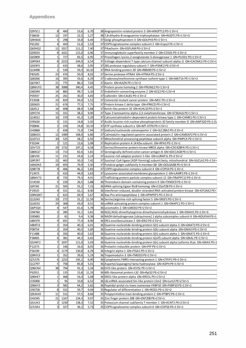
Appendices
251
Q9Y5C1 8 460 53,6 6,70 38 Angiopoietin-related protein 3 GN=ANGPTL3 PE=1 SV=1
P36639 12 197 22,5 5,27 38 7,8-dihydro-8-oxoguanine triphosphatase GN=NUDT1 PE=1 SV=3
Q9H4A6 5 298 33,8 6,44 37 Golgi phosphoprotein 3 GN=GOLPH3 PE=1 SV=1
P61202 4 443 51,6 5,53 37 COP9 signalosome complex subunit 2 GN=Cops2 PE=1 SV=1
Q63HQ2 11 1017 111,2 7,40 37 Pikachurin GN=EGFLAM PE=1 SV=2
Q93033 8 1021 115,0 6,96 37 Immunoglobulin superfamily member 2 GN=CD101 PE=1 SV=2
Q02809 1 727 83,5 6,95 37 Procollagen-lysine,2-oxoglutarate 5-dioxygenase 1 GN=PLOD1 PE=1 SV=2
Q9P0X4 8 2223 244,9 6,54 37 Voltage-dependent T-type calcium channel subunit alpha-1I GN=CACNA1I PE=1 SV=1
Q5E9F9 2 433 48,6 5,95 37 26S protease regulatory subunit 7 GN=PSMC2 PE=2 SV=3
Q14498 2 530 59,3 10,10 37 RNA-binding protein 39 GN=RBM39 PE=1 SV=2
P83105 23 476 50,9 8,02 37 Serine protease HTRA4 GN=HTRA4 PE=2 SV=1
Q00266 16 395 43,6 6,29 37 S-adenosylmethionine synthase isoform type-1 GN=MAT1A PE=1 SV=2
Q674X7 21 775 86,3 7,03 37 Kazrin GN=KAZN PE=1 SV=2
Q8WUY3 38 3088 340,4 4,45 37 Protein prune homolog 2 GN=PRUNE2 PE=1 SV=3
O60344 14 883 99,7 5,10 37 Endothelin-converting enzyme 2 GN=ECE2 PE=1 SV=4
P05937 25 261 30,0 4,83 37 Calbindin GN=CALB1 PE=1 SV=2
P23508 23 829 93,0 5,52 37 Colorectal mutant cancer protein GN=MCC PE=1 SV=2
Q05655 21 676 77,5 7,75 37 Protein kinase C delta type GN=PRKCD PE=1 SV=2
Q6JEL2 16 608 68,9 5,68 37 Kelch-like protein 10 GN=KLHL10 PE=2 SV=1
Q9Y274 15 331 38,2 9,00 37 Type 2 lactosamine alpha-2,3-sialyltransferase GN=ST3GAL6 PE=1 SV=1
Q14012 10 370 41,3 5,29 37 Calcium/calmodulin-dependent protein kinase type 1 GN=CAMK1 PE=1 SV=1
O95626 5 131 14,8 5,02 37 Acidic leucine-rich nuclear phosphoprotein 32 family member D GN=ANP32D PE=1 SV=2
P00846 2 226 24,8 10,10 37 ATP synthase subunit a GN=MT-ATP6 PE=1 SV=1
O43868 2 658 71,9 7,94 37 Sodium/nucleoside cotransporter 2 GN=SLC28A2 PE=2 SV=2
Q08AD1 12 1489 168,0 6,80 37 Calmodulin-regulated spectrin-associated protein 2 GN=CAMSAP2 PE=1 SV=3
Q10713 20 525 58,2 6,92 37 Mitochondrial-processing peptidase subunit alpha GN=PMPCA PE=1 SV=2
P35244 2 121 13,6 5,08 37 Replication protein A 14 kDa subunit GN=RPA3 PE=1 SV=1
Q5VT25 29 1732 197,2 6,58 37 Serine/threonine-protein kinase MRCK alpha GN=CDC42BPA PE=1 SV=1
Q86SQ7 13 713 82,6 5,81 37 Serologically defined colon cancer antigen 8 GN=SDCCAG8 PE=1 SV=1
Q8IV03 11 231 24,6 5,14 37 Leucine rich adaptor protein 1-like GN=LURAP1L PE=2 SV=2
Q9P2R7 22 463 50,3 7,42 37 Succinyl-CoA ligase [ADP-forming] subunit beta, mitochondrial GN=SUCLA2 PE=1 SV=3
A6NDN3 19 693 79,9 5,50 37 Putative golgin subfamily A member 6B GN=GOLGA6B PE=5 SV=3
Q9BT78 9 406 46,2 5,83 37 COP9 signalosome complex subunit 4 GN=COPS4 PE=1 SV=1
P13473 3 410 44,9 5,63 37 Lysosome-associated membrane glycoprotein 2 GN=LAMP2 PE=1 SV=2
Q8WVT3 8 735 79,3 4,91 37 Trafficking protein particle complex subunit 12 GN=TRAPPC12 PE=1 SV=3
O14530 5 226 26,5 5,88 36 Thioredoxin domain-containing protein 9 GN=TXNDC9 PE=1 SV=2
Q9Y3I0 21 505 55,2 7,23 36 tRNA-splicing ligase RtcB homolog GN=C22orf28 PE=1 SV=1
P19525 4 551 62,1 8,40 36 Interferon-induced, double-stranded RNA-activated protein kinase GN=EIF2AK2 PE=1 SV=2
Q9NQW7 2 623 69,9 5,67 36 Xaa-Pro aminopeptidase 1 GN=XPNPEP1 PE=1 SV=3
Q13243 19 272 31,2 11,59 36 Serine/arginine-rich splicing factor 5 GN=SRSF5 PE=1 SV=1
Q16533 24 368 43,0 9,51 36 snRNA-activating protein complex subunit 1 GN=SNAPC1 PE=1 SV=1
Q6P5Q4 4 547 61,6 5,78 36 Leiomodin-2 GN=LMOD2 PE=2 SV=2
O94760 5 285 31,1 5,81 36 N(G),N(G)-dimethylarginine dimethylaminohydrolase 1 GN=DDAH1 PE=1 SV=3
O00483 2 81 9,4 9,38 36 NADH dehydrogenase [ubiquinone] 1 alpha subcomplex subunit 4 GN=NDUFA4 PE=1 SV=1
A8K979 9 691 77,4 8,90 36 ERI1 exoribonuclease 2 GN=ERI2 PE=2 SV=2
A8MTJ3 2 354 40,3 6,01 36 Guanine nucleotide-binding protein G(t) subunit alpha-3 GN=GNAT3 PE=2 SV=2
P08754 2 354 40,5 5,69 36 Guanine nucleotide-binding protein G(k) subunit alpha GN=GNAI3 PE=1 SV=3
P11488 3 350 40,0 5,62 36 Guanine nucleotide-binding protein G(t) subunit alpha-1 GN=GNAT1 PE=1 SV=5
P38405 6 381 44,3 6,65 36 Guanine nucleotide-binding protein G(olf) subunit alpha GN=GNAL PE=1 SV=1
Q5JWF2 7 1037 111,0 5,03 36 Guanine nucleotide-binding protein G(s) subunit alpha isoforms XLas GN=GNAS PE=1 SV=2
P12273 1 146 16,6 8,05 36 Prolactin-inducible protein GN=PIP PE=1 SV=1
P56199 4 1179 130,8 6,29 36 Integrin alpha-1 GN=ITGA1 PE=1 SV=2
Q9NYL9 1 352 39,6 5,19 36 Tropomodulin-3 GN=TMOD3 PE=1 SV=1
Q7L576 6 1253 145,1 6,90 36 Cytoplasmic FMR1-interacting protein 1 GN=CYFIP1 PE=1 SV=1
Q12797 5 758 85,8 5,01 36 Aspartyl/asparaginyl beta-hydroxylase GN=ASPH PE=1 SV=3
Q96CN4 38 794 91,3 5,34 36 EVI5-like protein GN=EVI5L PE=1 SV=1
P62911 2 135 15,8 11,33 36 60S ribosomal protein L32 GN=Rpl32 PE=2 SV=2
Q96HE7 1 468 54,4 5,68 36 ERO1-like protein alpha GN=ERO1L PE=1 SV=2
O35900 1 95 10,8 6,52 36 U6 snRNA-associated Sm-like protein LSm2 GN=Lsm2 PE=2 SV=1
Q96AY3 3 582 64,2 5,62 36 Peptidyl-prolyl cis-trans isomerase FKBP10 GN=FKBP10 PE=1 SV=1
O95758 8 552 59,7 9,04 35 Regulator of differentiation 1 GN=ROD1 PE=1 SV=2
Q9UKA9 11 531 57,5 8,66 35 Polypyrimidine tract-binding protein 2 GN=PTBP2 PE=1 SV=1
O43345 21 1167 134,3 9,07 35 Zinc finger protein 208 GN=ZNF208 PE=2 SV=1
Q5JUK3 2 1230 138,3 7,52 35 Potassium channel subfamily T member 1 GN=KCNT1 PE=2 SV=2
Q7L5N1 3 327 36,1 5,73 35 COP9 signalosome complex subunit 6 GN=COPS6 PE=1 SV=1
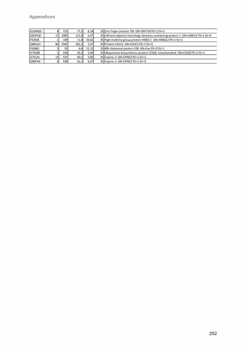
Appendices
252
Q32MQ0 8 723 77,3 8,18 35 Zinc finger protein 750 GN=ZNF750 PE=1 SV=1
Q9UPQ0 12 1083 121,8 6,47 35 LIM and calponin homology domains-containing protein 1 GN=LIMCH1 PE=1 SV=4
P52926 1 109 11,8 10,62 35 High mobility group protein HMGI-C GN=HMGA2 PE=1 SV=1
Q8NG31 84 2342 265,2 5,47 35 Protein CASC5 GN=CASC5 PE=1 SV=3
P62862 3 59 6,6 12,15 35 40S ribosomal protein S30 GN=Fau PE=3 SV=1
O75208 1 318 35,5 5,94 35 Ubiquinone biosynthesis protein COQ9, mitochondrial GN=COQ9 PE=1 SV=1
O75131 13 537 60,1 5,85 35 Copine-3 GN=CPNE3 PE=1 SV=1
Q96FN4 8 548 61,2 6,07 35 Copine-2 GN=CPNE2 PE=1 SV=3
Eidesstattliche Erklärung
253
14 Eidesstattliche Erklärung
„Hiermit versichere ich, dass ich die vorliegende Arbeit selbstständig verfasst habe. Zur
Anfertigung wurden keine anderen als die angegebenen Hilfsmittel verwendet. Die Stellen,
die aus anderen Werken wörtlich oder sinngemäß entnommen wurden, sind als solche
kenntlich gemacht. Ich versichere weiterhin, dass diese Arbeit in gleicher oder ähnlicher
Form noch keiner anderen Prüfungsbehörde vorgelegen hat.“
___________________
Mainz, den 15.09.2014

Eidesstattliche Erklärung
254

Eidesstattliche Erklärung
255
Mainz, September 15th 2014