Prostate carcinoma
21
Prostate Carcinoma
-
Upload
mohamed-shaaban -
Category
Health & Medicine
-
view
89 -
download
1
Transcript of Prostate carcinoma

Prostate Carcinoma

Prostate Carcinoma
• Many men are found to have had incidental microscopic
foci of prostate cancer at postmortem examination, and
most prostate cancers are slow growing and do not
manifest during the man's lifetime.
• Thus, many men die with prostate cancer rather than die
from prostate cancer.

Prostate Carcinoma
• 95% of prostate cancers are adenocarcinomas
developing in the acini of prostatic ducts.
Pathology

Prostate Carcinoma
• 70% of cancers occur in the peripheral zone
(PZ), and approximately 20% are found in the
transition zone (TZ).
Pathology

Prostate Carcinoma
• Local spread: capsular penetration, invasion of the seminal vesicles, or local
extension along the neurovascular bundles.
• Lymphatic spread: obturator LNs and from there to the common iliac and
para-aortic lymph nodes.
• Hematogenous spread: bones, and lungs. Skeletal metastases are common
in advanced prostate cancer, but hepatic metastases are uncommon.
Spread

Prostate Carcinoma
• Asymptomatic.
• Symptoms of advanced disease with weight loss,
lethargy, bladder outflow obstruction, or bone pain.
Clinical presentation

Prostate Carcinoma
• Serum PSA levels increase with age:
PSA
Patients aged 40-49 years, 0-2.5 ng/mL
Patients aged 50-59 years, 0-3.5 ng/mL
Patients aged 60-69 years, 0-4.5 ng/mL
Patients aged 70-79 years, 0-6.5 ng/mL

Prostate Carcinoma
• Elevated serum PSA levels may also be
associated with prostatitis, prostate infarction,
PIN, prostate biopsy, transurethral resection of the
prostate, and urethral catheterization.
PSA

Prostate Carcinoma
• Serum PSA levels are lowered with finasteride
treatment for benign prostatic hyperplasia
PSA

Prostate Carcinoma
Imaging

Prostate Carcinoma
• Sclerotic metastases or lytic
lesions with bone destruction.
Plain X-Ray

Prostate Carcinoma
• Sclerotic metastases or lytic
lesions with bone destruction.
Plain X-Ray
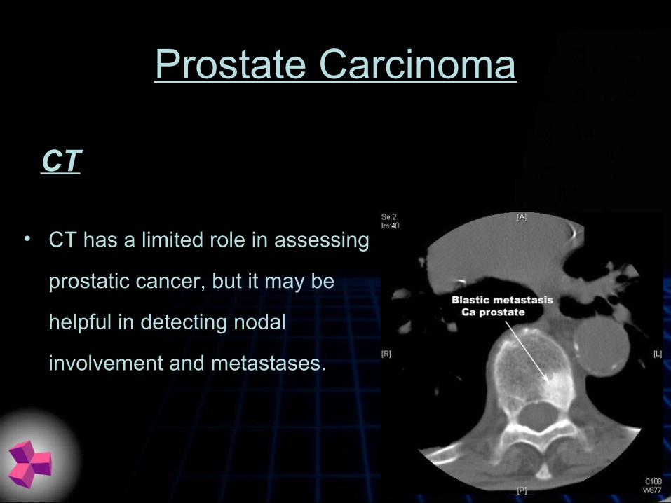
Prostate Carcinoma
• CT has a limited role in assessing
prostatic cancer, but it may be
helpful in detecting nodal
involvement and metastases.
CT

Prostate Carcinoma
• CT has a limited role in assessing
prostatic cancer, but it may be
helpful in detecting nodal
involvement and metastases.
CT

Prostate Carcinoma
• MRI can demonstrate the internal anatomy of the prostate, and it can
identify areas of altered signal intensity, which representing focal
pathology in the gland.
• MRI is used primarily for staging, but the availability of interventional
MRI units means that MRI is likely to have a future role in the
diagnosis of prostate cancer.
MRI

Prostate Carcinoma
MRI

Prostate Carcinoma
MRI

Prostate Carcinoma
MRI

Prostate Carcinoma
ULTRASOUND
• TRUS plays a central role in the contemporary
diagnosis of prostate cancer because it enables
accurate image-guided biopsy of the gland.

Prostate Carcinoma
ULTRASOUND
Hypoechoic area in the PZ.

Prostate Carcinoma
ULTRASOUND
Hypoechoic area in the PZ.