Prostate carcinoma raiology
66
Dr Mohit Goel JRIII 20/9/14 AND RECENT ADVANCEMENTS Imaging Prostate Cancer: A Multidisciplinary Perspective Advancements in MR Imaging of the Prostate: From Diagnosis to Interventions A clinically relevant approach to imaging prostate cancer: review.
-
Upload
dr-mohit-goel -
Category
Education
-
view
63 -
download
1
Transcript of Prostate carcinoma raiology

Dr Mohit GoelJRIII20/9/14
AND RECENT ADVANCEMENTS
Imaging Prostate Cancer: A Multidisciplinary PerspectiveAdvancements in MR Imaging of the Prostate: From Diagnosis to InterventionsA clinically relevant approach to imaging prostate cancer: review.

CT does not provide sufficient soft-tissue contrast beyond size assessment of the prostate. Although CT is valuable in the evaluation of pelvic lymphadenopathy and bone metastases, MR imaging and bone scanning have been found superior in their assessment.

Prostate is divided from superior to inferior into the base (just below the urinary bladder), the midgland, and the apex.
In the axial plane, the prostate is divided into four zones: (a)the anterior fibromuscular stroma, which contains no glandular tissue;
(b)the transition zone surrounding the urethra, which contains 5% of the glandular tissue;
(c)the central zone, which contains 20% of the glandular tissue; &
(d)the outer peripheral zone, which contains 70%–80% of the glandular tissue.
The volume of the peripheral zone increases from the base to the apex of the gland.

Ninety-five percent of prostate cancers are adenocarcinomas that develop from the acini of the prostatic ducts.
Thus, prostate cancers arise in the glandular tissue, with about 70% originating in the peripheral zone, 25% in the transition zone, and 5% in the central zone.
At imaging, the transition zone cannot be separated from the central zone; therefore, these two zones are often referred to together as the central gland.

Endorectal MR images obtained at 1.5 T

Axial T2WI at the level of base of prostate gland
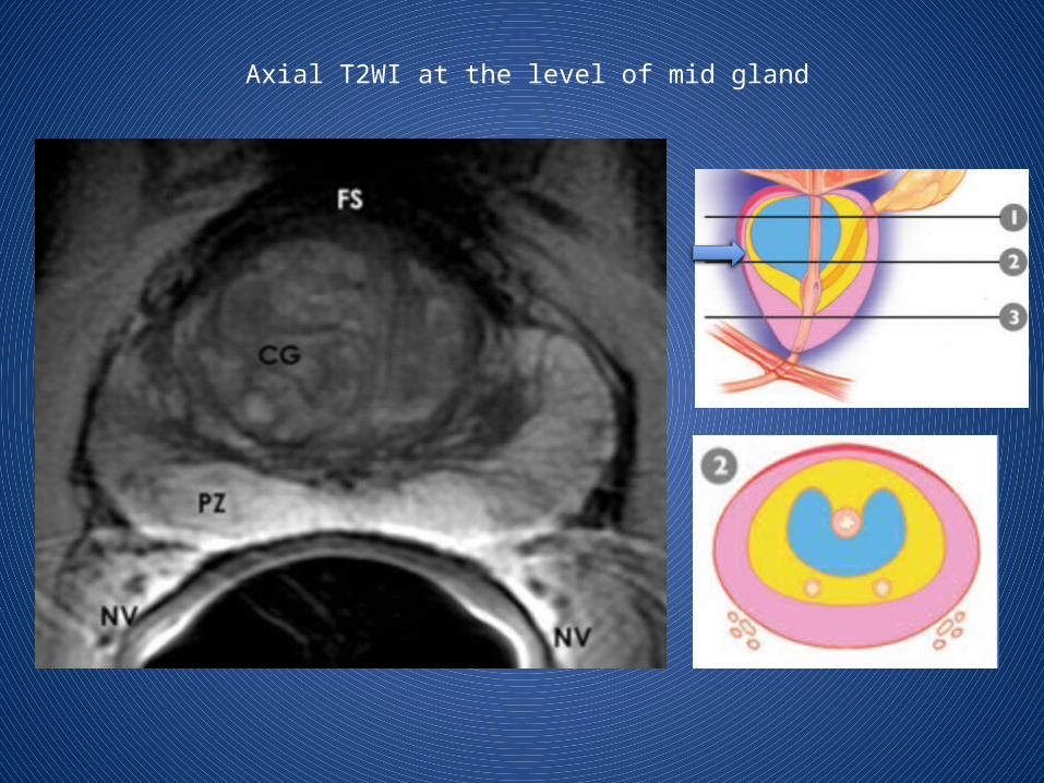
Axial T2WI at the level of mid gland


parasagittal T2-weighted image

coronal T2-weighted image
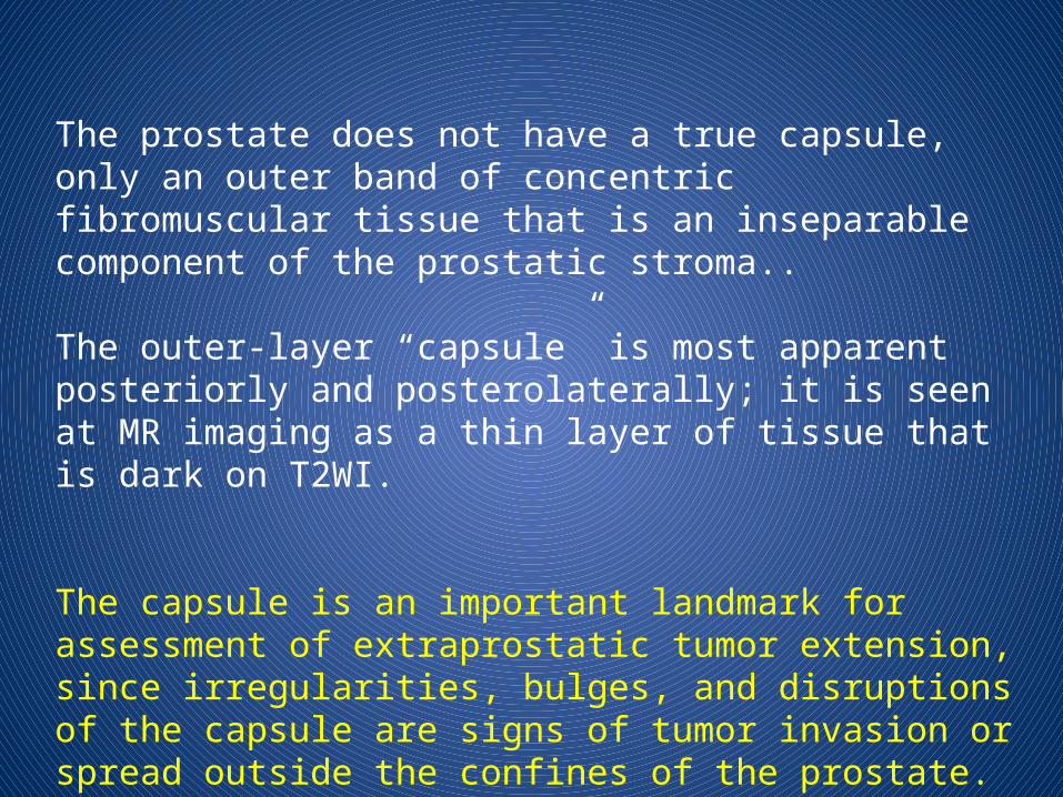
The prostate does not have a true capsule, only an outer band of concentric fibromuscular tissue that is an inseparable component of the prostatic stroma..
The outer-layer “capsule” is most apparent posteriorly and posterolaterally; it is seen at MR imaging as a thin layer of tissue that is dark on T2WI.
The capsule is an important landmark for assessment of extraprostatic tumor extension, since irregularities, bulges, and disruptions of the capsule are signs of tumor invasion or spread outside the confines of the prostate.

The periprostatic neurovascular bundles course posterolateral to the prostate bilaterally.
They are well seen at imaging at the 5-o'clock and 7-o'clock positions in reference to the prostate .
At the apex and base, the nerve bundles send penetrating branches through the capsule, which provide a route for extraprostatic tumor extension.
TRUS (Transrectal US )
CT
MR- 1. Conventional MR (T2WI) +/- endorectal coil2. DWI & ADC mapping3. MR spectroscopy4. DCE MR imaging
Bone ScanSPECT (Single photon emission CT)
PET
IMAGING MODALITIES

Transrectal US is primarily used to guide prostate biopsies, but new developments in microbubble contrast agents offer the possibility of improving prostate cancer detection by detecting tumor angiogenesis.
When prostate cancer is suspected, the diagnostic test of choice is a systematic needle biopsy with US guidance.
Cores are typically obtained from six areas, or sextants, of the peripheral zone: left and right apex, left and right midgland, and left and right base.
Cancer, depending on its size, grade, and location, usually appears hypoechoic relative to the normal peripheral zone of the prostate


Many cancers detected at biopsy are not visible at US (low sensitivity) and many hypoechoic areas do not prove to be malignant at biopsy (low specificity); therefore, transrectal US alone, without the addition of biopsy, has limited value in the detection of cancer.
The criteria for identifying extracapsular extension (ECE) on transrectal US scans are bulging or irregularity of the capsule adjacent to a hypoechoic lesion.
The length of the contact of a visible lesion with the capsule is also associated with the probability of ECE.


Seminal vesicle invasion (SVI) is heralded by a visible extension of a hypoechoic lesion at the base of the prostate into a seminal vesicle or by echogenic cancer within the normally fluid-filled seminal vesicle.
Asymmetry of the seminal vesicles or solid hypoechoic masses within the seminal vesicles are indirect indicators of disease extension.
TRUS continues to play an important role in therapy. Worldwide, TRUS is the modality of choice for directing brachytherapy seeds into the prostate.
Cryotherapy of the prostate also requires US guidance as does high-intensity focused ultrasound, which, coupled with US targeting, is used for focal ablation of prostate cancers.

New treatment approaches such as hyperthermia, photodynamic therapy, direct injection of oncolytic viruses, tumor vaccines, and gene therapy also depend on transrectal US for easy access to cancers of the prostate.
Future developments in transrectal US include the use of microbubble contrast agents and targeted imaging.

it has virtually no role in prostate cancer detection or primary tumor staging.
The major role of CT is in the nodal staging of prostate cancer.
CT has been used to monitor bone metastases, but bone scanning and MR imaging are superior to CT in the diagnosis of bone metastases.
Lytic and blastic bone metastases will commonly be visible at CT.
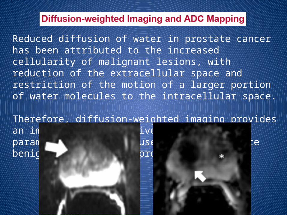
Reduced diffusion of water in prostate cancer has been attributed to the increased cellularity of malignant lesions, with reduction of the extracellular space and restriction of the motion of a larger portion of water molecules to the intracellular space.
Therefore, diffusion-weighted imaging provides an important quantitative biophysical parameter that can be used to differentiate benign from malignant prostate tissue.
DWI is effective in detecting recurrent disease after radiation therapy or surgery. Subtle changes in apparent diffusion coefficient detected on DWI images are indicative of recurrent disease in patients with increasing PSA levels after treatment.
DWI improves detection of nodal disease with the use of lymphotropic superparamagnetic nanoparticles as a contrast agent at MR imaging. The nanoparticles are taken up by circulating macrophages, which then traffic to the normal nodal tissue.
The inability of malignant nodes to take up the agent provides tissue contrast within the lymph node and allows detection of metastases, even in nodes that do not meet the standard size criteria for metastasis.

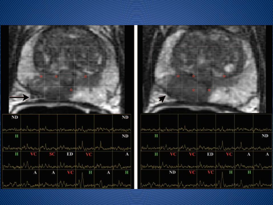
MR spectroscopy provides important information about the biochemical and metabolic environment of the tissue, it is increasingly being used as a biomarker for detection of cancers, including prostate cancer.
MR spectroscopy of the prostate requires use of multivoxel volume selection techniques
Citrate is found in fairly high concentrations in healthy prostate epithelium and prostatic fluid; it is found in low concentrations elsewhere in tissue.
The normal prostate has an MR spectrum with a prominent citrate peak at 2.6 ppm, which is usually seen as a doublet with occasional visualization of small additional side peaks.
A decreased citrate level is found in prostate cancer, as well as in prostatitis and hemorrhage.
Choline is represented at 3.2 ppm.
Owing to increased cell membrane turnover and increased cell surface compared with cell volume in cellular tumors, choline concentrations are increased in prostate cancer.
Increased choline signal or concentration is considered the spectroscopic hallmark of cancer; however, it has also been found in benign conditions of the prostate such as prostatitis.
An increase in the choline-to-citrate ratio or the (choline + creatine)/citrate ratio is often used as a marker of malignancy in prostate cancer and increases the specificity of diagnosis; however, it is most reliable in the peripheral zone.


BPH shows significant overlap with the findings of prostate cancer. In addition, prostatitis has previously been found to be able to mimic prostate cancer. This leads to possible significant difficulty in distinguishing prostate cancer from prostatitis and stromal BPH, especially in the transition zone.
When combined with anatomic imaging, MR spectroscopy has been found to increase the accuracy of tumor volume estimation in prostate cancer.

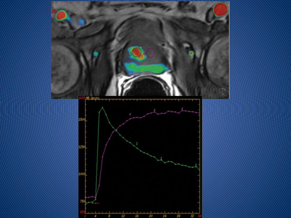
DCE MR imaging is an advanced prostate imaging modality that allows derivation of parameters that are closely related to microvascular properties and angiogenesis in tissues.
In prostate cancer, increased tumor vascularity leads to early hyperenhancement (higher and earlier peak enhancement than in normal tissue) and to rapid washout of contrast material from the tumor, in comparison with normal prostate tissue.
Microvascular alterations and neovascularity are in general most severe in prostate cancer, in comparison with other processes in the prostate such as BPH or prostatic intraepithelial neoplasia.

The normal prostate is a vascular organ, and so rapid injections with rapid scanning are needed to detect angiogenesis in a lesion.
Typically, a full dose (0.1 mmol/kg) of gadolinium chelate is injected at 3 mL/sec, and serial 3D acquisitions are obtained every 2–5 seconds through the prostate.
Cancers often demonstrate early nodular enhancement before the rest of the parenchyma and early washout of signal intensity. This pattern is highly predictive of prostate cancer but is not pathognomonic. Some prostate cancers are mildly or moderately hypervascular and thus are not detectable with this method.
Combining dynamic contrast-enhanced MR imaging and 3D proton spectroscopy may prove helpful in tumor localization when T2-weighted images are inconclusive for a tumor focus.

voxel-wise analysis of the prostate

In prostate cancer, there is early, rapid, and intense enhancement with quick washout of contrast material.
The presence of washout is highly indicative of prostate cancer, even in the absence of low T2 signal intensity.

Criteria for extracapsular extension or seminal vesicle invasion at DCE MR imaging include:
1. abnormally high or asymmetric peak enhancement, 2. contrast agent washout, and 3. short onset time and 4. Short time to peak enhancement.
In the case of extracapsular extension, these findings are detected near the neurovascular bundle or rectoprostatic angle, broadly abutting the capsule, or in an extracapsular location.

On T2WI, the normal peripheral zone has homogeneous high signal intensity and the central gland has variable amounts of intermediate signal intensity, which is often replaced by well-circumscribed hyperplastic nodules of BPH.


A series of studies in the late 1980s established that prostate cancer is characterized by low T2 signal intensity replacing the normally high T2 signal intensity in the peripheral zone.
However, the presence of decreased T2 signal intensity in the peripheral zone is of limited sensitivity because some prostate tumors are isointense.
This finding is also of limited specificity because there are other possible causes of low T2 signal intensity in the peripheral zone, including hemorrhage, prostatitis, scarring, atrophy, and effects of radiation therapy, cryosurgery, or hormonal therapy.
Postbiopsy hemorrhage may hamper tumor detection. The imaging appearance depends on the length of time between the biopsy and MR imaging. A delay of 6–8 weeks is recommended.


Prostate cancer arising in the transition zone poses additional imaging difficulties because of the heterogeneity of signal intensity in the central gland.
Findings supporting the diagnosis of a transition zone tumor include :
1. a homogeneous low-signal-intensity region in the transition zone,
2. poorly defined or spiculated lesion margins,3. lack of a low-signal-intensity rim (seen commonly in association
with benign adenomatous nodules), 4. interruption of the surgical pseudocapsule (transition zone–to–
peripheral zone boundary of low signal intensity),5. urethral or anterior fibromuscular stromal invasion, and 6. lenticular shape


The most important aspect of local staging is differentiation between organ-confined disease (stage T1 or T2) and early advanced disease in the form of extracapsular extension or seminal vesicle invasion (stage T3).


Criteria for extracapsular extension include:
1. Asymmetry of the neurovascular bundle,
2. Tumor encasement of the neurovascular bundle,
3. A bulging prostatic contour,
4. An irregular or spiculated margin,
5. Obliteration of the rectoprostatic angle,
6. Capsular retraction,
7. A tumor-capsule interface of greater than 1 cm, and
8. A breach of the capsule with evidence of direct tumor extension

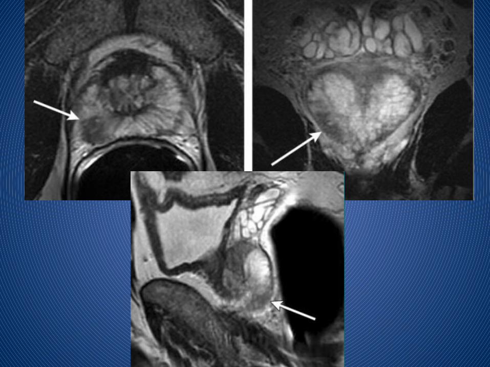
T1WIT2WIT2WI
Extraprostatic tumor extension
Involvement of left neurovascular bundle.

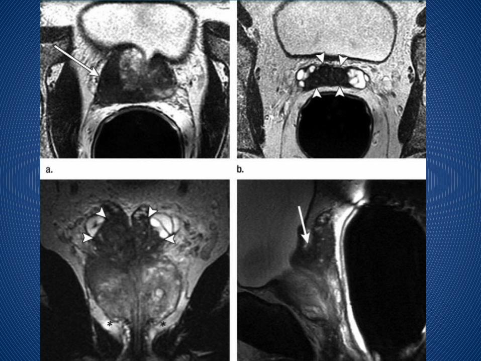
The features of seminal vesicle invasion at endorectal MR imaging include:
1. disruption or loss of the normal architecture of the seminal vesicle
2. focal low signal intensity within and along the seminal vesicle,
3. enlarged low-signal-intensity ejaculatory ducts & seminal vesicle,
4. obliteration of the angle between the prostate and the seminal vesicle, and
5. demonstration of direct tumor extension from the base of the prostate into and around the seminal vesicle

It has no role in prostate cancer detection or local staging; however, bone scanning continues to be the mainstay of diagnosis of initial spread of cancer to bone – BONE METS.
A focal area of increased tracer uptake, usually in the axial skeleton, related to host osteoblastic bone response to tumor invasion.
However, other causes of altered marrow signal intensity (eg, infection, infarction, trauma) can mimic metastatic disease at MR imaging.


Radionuclide bone scanning can also be used to assess the response to treatment, as uptake usually decreases after chemotherapy, hormone therapy, or radiation therapy if a response is obtained.
In patients with prostate and breast cancer, a “flare” phenomenon may be observed, where uptake initially increases after chemotherapy or hormone therapy, peaking at 6 weeks after treatment as bone turnover increases as part of the healing process. Care must be taken in this situation to avoid mistaking apparent new lesions for areas of new metastatic disease, when subtle changes were in fact present in prior studies.
Single photon emission computed tomographic (SPECT)
SPECT studies of the skeleton have been shown to be more sensitive in the detection of metastatic disease than planar images alone.
This technique has particular utility in the evaluation of the spine in patients with low back pain who present for evaluation of possible metastatic disease.
Sites of abnormal uptake may be localized to the vertebral body or posterior elements, leading to other radiologic techniques for confirmation.
Combined SPECT/CT, a recent development, adds anatomic information to SPECT.

SPECT

PET uses compounds labeled with positron-emitting radioisotopes to detect pathologic processes.
It is performed with the glucose analogue fluorine 18 (18F) fluorodeoxyglucose (FDG).
Cancers have increased metabolism and utilize the less-efficient glycolytic pathway, both of which lead to increased glucose analogue uptake.
PET/CT demonstrate tumor location in the prostate bed and to better assess pelvic lymph node disease.
FDG has limitations with regard to distinguishing tumors from inflammation. Future applications of PET will therefore involve new tracers, which are currently under clinical investigation


The fundamental impetus for the migration to higher static magnetic field strengths lies in an increased SNR and increased spectral resolution.
In comparison with 1.5-T imaging, improvements in spatial or temporal resolution and in patient comfort—when no endorectal coil is used—are possible.
Imaging without endorectal coils is feasible at 3 T and helps decrease patients’ reluctance to undergo an uncomfortable and invasive examination.
At 3 T, however, if endorectal coils are used, significant improvements in localization and staging of prostate cancer have been reported.

Voxel sizes for 3-T MR spectroscopy is smaller then 1.5-T MR spectroscopy.
Smaller voxel sizes are desirable to reduce volume averaging of tumors with periprostatic fat, the seminal vesicles, postbiopsy hemorrhage, or periurethral tissues.
DWI is another low-SNR technique that benefits from higher field strengths, allowing higher-quality imaging and higher spatial resolution.
In summary, detection, localization, and staging of prostate cancer benefit from the higher achievable spatial resolution and increased SNR at 3 T, as tissue interfaces are better visualized and lower-contrast structures are better identified.

Because MR imaging provides more detailed anatomic images of the prostate than does transrectal US and because it has been shown to be the most accurate imaging modality for localization of prostate cancer, MR imaging–based guidance offers the possibility of more precise targeting.
Real-time virtual sonography is a technique in which the MR imaging dataset is coregistered to landmarks during the US examination, so that a needle can be placed sonographically. However, experience with this technique is currently limited.
Transrectal MR imaging–guided prostate brachytherapy can also be performed in a closed-bore 1.5-T imaging unit.
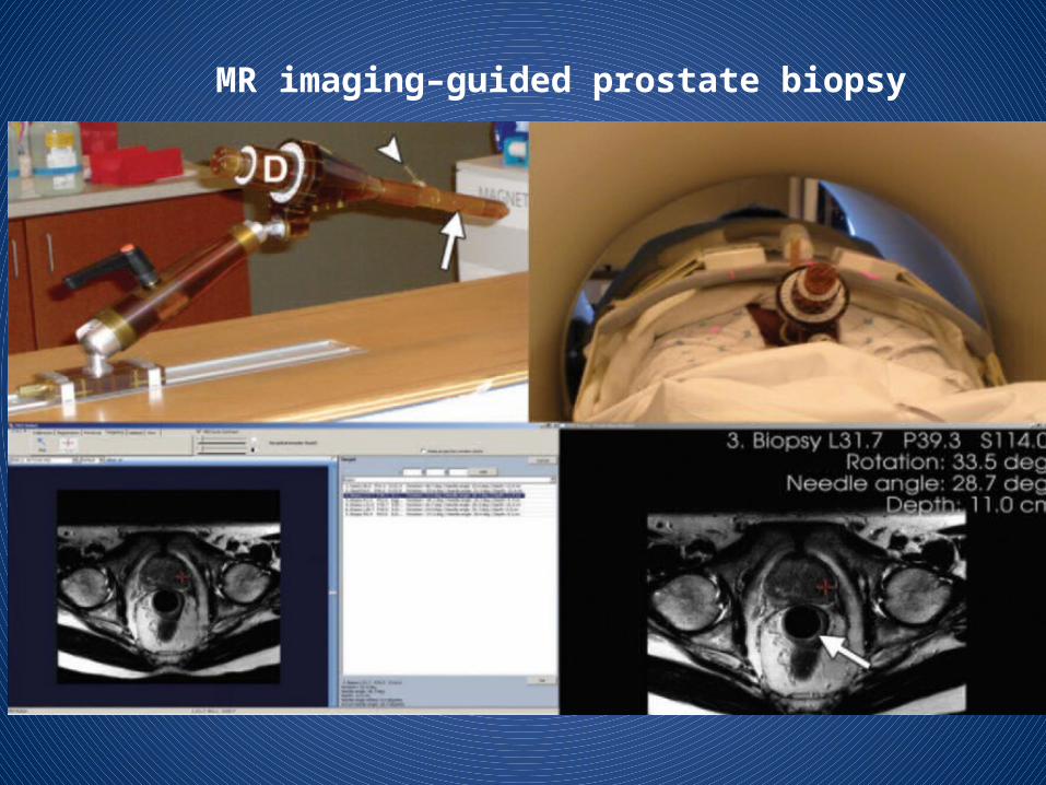
MR imaging–guided prostate biopsy

Robotic prostate biopsy promises high accuracy and is a topic of intense research. This robot can perform biopsies and automatically place brachytherapy seeds in the prostate.

THANK YOU