Hematochezia with Colonic Polypoid Angiodysplasia in a Young ...

5/21/2018
1
Common Differential
Diagnoses in
Urological Pathology
Jonathan I. Epstein
Prostate Adenocarcinoma vs.
Urothelial Carcinoma

5/21/2018
2

5/21/2018
3
NKX3.1
NKX3.1

5/21/2018
4

5/21/2018
5

5/21/2018
6

5/21/2018
7
Proposed ISUP Recommendations
• Option to use PSA as a first test to identify Pca. and GATA3 to
identify UC.
• If GATA3 not available, then HMWCK and p63.
• If the tumor shows strong PSA positivity and negative for HMWCK
and p63, the findings are diagnostic of Pca.
• If the tumor is negative for PSA and diffusely strongly positive for
p63 and HMWCK, the findings are diagnostic of UC.
Proposed ISUP Recommendations
• If the tumor is equivocal/weak/negative for PSA and negative/focal
for p63 and HMWCK, then need to do p501S, NKX3.1 and GATA3.

5/21/2018
8
Clear Cell Adenocarcinoma vs.
Nephrogenic Adenoma
Clear Cell Adenocarcinoma

5/21/2018
9

5/21/2018
10
5/21/2018
11
Nephrogenic Adenoma

5/21/2018
12

5/21/2018
13

5/21/2018
14

5/21/2018
15
Ki67 in Clear Cell Adenocarcinoma
Ki67 in NA
Nephrogenic Adenoma vs. Clear Cell
Adenocarcinoma
Nephrogenic Adenoma
• Usually < 1cm. Can be large.
• 20% multifocal
• M:F 2:1
• Prior injury
• Solid – rare, focal
• No mitoses
• No clear cells
• PAX8 positive
Clear Cell Adenocarcinoma
• Typically large
• Unifocal
• Rare in men
• No prior injury
• Solid areas common
• Mitotic figures common
• Typically clear cells
• PAX8 positive

5/21/2018
16
Polypoid Cystitis
• Reaction to injury
• Indwelling catheters, fistulae, abscesses, long-
standing urinary obstruction
• Often recognized as inflammatory by urologist at
cystoscopy
• Spectrum: bullous, polypoid, papillary
Mimic of Urothelial Carcinoma
• Can have isolated papillary fronds rarely branching papillae
• Base of the papillary stalks typically both broad and narrow yet uncommonly can be only narrow
• Urothelium diffusely and focally thickened in some cases
• Reactive urothelial atypia often present
• Rare mitotic figures not uncommon
• Fibrosis (not edema) within polypoid stalks in some cases

5/21/2018
17

5/21/2018
18

5/21/2018
19

5/21/2018
20
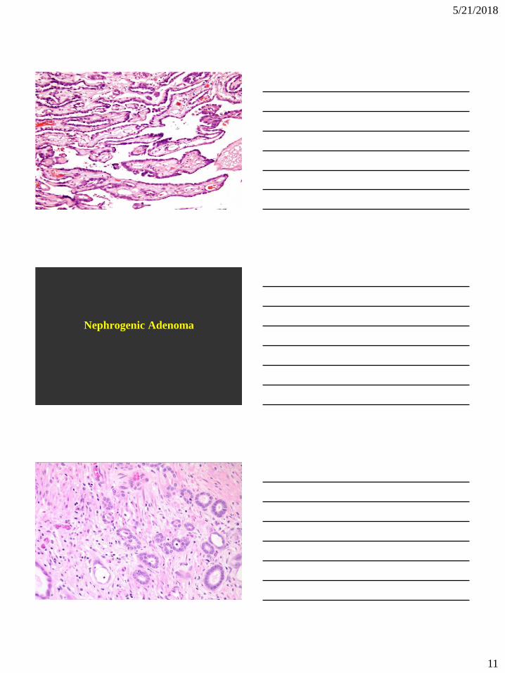
Nested Variant of Urothelial Cancer vs.
Von Brunn Nests

5/21/2018
21
Nested Variant
Urothelial Carcinoma
• Murphy WM, Deana DG. The nested variant of
transitional cell carcinoma: a neoplasm resembling
proliferation of Brunn’s nests. Mod Pathol
1992;5:240-3
Clinical Features
• Older male patients
• To date, nested variant of urothelial carcinoma has
been reported in only 4 women
• Typically present with hematuria
Location
• Anywhere in the bladder
• Only two cases of nested variant of urothelial
carcinoma in the ureter

5/21/2018
22
Histology – Difficult to Diagnose as Cancer
• Cytologically, may show very uniform, bland cells
with only focal moderate atypia
• Nucleoli may be prominent and mitoses may be seen,
but are usually not numerous
• Lymphatic invasion uncommon
• Overlying urothelium may be normal in appearance

5/21/2018
23

5/21/2018
24
5/21/2018
25
Von Brunn Nests

5/21/2018
26

5/21/2018
27
Prognosis (Mayo Clinic 2013)
• Clinical course generally aggressive
• 69% pT3-pT4 & 19% LN+
• 10 year cancer specific survival 41%
• Same prognosis vs. usual urothelial carcinoma stage for stage
• Nested variant of urothelial carcinoma is critical to
recognize given its aggressive biology despite its
deceptively bland cytology
• The diagnosis is based on the H&E appearance
• In cases of diagnostic uncertainty or a superficial
biopsy, convey your uncertainty to the urologist and
request additional tissue sampling

5/21/2018
28
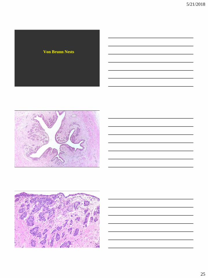
Radiation Change
Radiation Change in the Prostate
• RT affect in Benign Prostate - Differential Diagnosis
from Prostate Cancer
• RT results – Affect on Prognosis
– Positive for cancer (w/o treatment affect)
– Negative for cancer
– Positive for cancer (with treatment affect)
• More atypia in cases treated with IRT (seeds) than XRT
• No change in epithelial atypia over time in men treated with IRT. With XRT, less epithelial atypia in cases biopsies >48 months after treatment
• RT atypia may persist for a long time: Prominent RT atypia detected 72 months after IRT
• Some cases clinicians not aware of remote h/o of RT or do not relay this on to pathologists. Pathologists must be able to recognize RT atypia w/o relying on the clinician to provide this history

5/21/2018
29
5/21/2018
30

5/21/2018
31
Radiation Biopsy Results: Cancer with
Treatment Affect
Histologically cancer is seen, yet shows treatment effect
with degenerative features. Cancer is present, yet is it
viable?
Prognosis: Similar to cases with no cancer.
Do not grade radiated cancer with treatment effect.
5/21/2018
32
Radiation Biopsy Results: Positive
Histologically, ordinary prostate cancer is seen, which
resembles non-radiated cancer.
If biopsy is done >12 months following radiation,
indicates progression of cancer. Can assign a Gleason
grade.
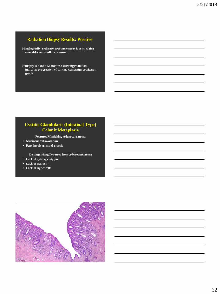
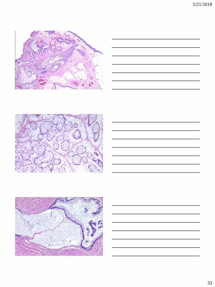
Cystitis Glandularis (Intestinal Type)
Colonic Metaplasia
Features Mimicking Adenocarcinoma
• Mucinous extravasation
• Rare involvement of muscle
Distinguishing Features from Adenocarcinoma
• Lack of cytologic atypia
• Lack of necrosis
• Lack of signet cells
5/21/2018
33

5/21/2018
34

5/21/2018
35
Endocervicosis
Endocervicosis
• Typically in women in their 30s and 40s
• Symptoms of pelvic pain, frequency, dysuria, hematuria,
dyspareunia, dysmenorrhea
• Most common in bladder. Also seen in uterine cervix, vagina
• Mass (up to 5 cm) seen in posterior bladder wall with
occasional extravesical involvement

5/21/2018
36

5/21/2018
37
Infiltrating Adenocarcinoma
of the Bladder
5/21/2018
38

5/21/2018
39

5/21/2018
40
5/21/2018
41
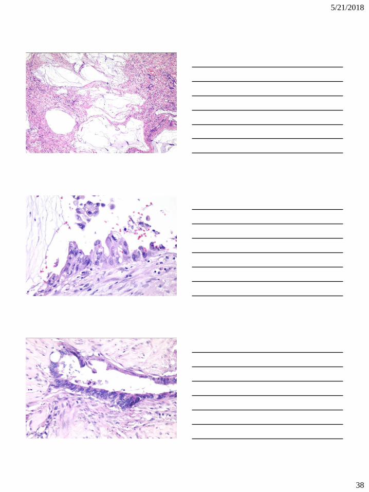
Radiotherapy of the Bladder:
Pseudocarcinomatous Hyperplasia
Less familiar to pathologists is radiation-induced
pseudocarcinomatous hyperplasia.
The first series to report this mimicker of invasive
urothelial carcinoma was by Baker and Young in
2000 (2). Four cases were described, with follow-up
available in one patient, which was benign.
• 70 cases: 60 males/10 females
• Mean age 67 (33-85)
• 76.5% prior pelvic RT
• Developed on average 4.5 years (9 mos.-13 yrs). after
prior RT
• 2 systemic chemo, 3 indwelling catheter, 2 intravesical
chemo, 1 prior RP, 4 severe peripheral vascular disease,
1 AV malformation in the bladder, 1 sickle cell trait, 2
(2.9%) no identifiable contributing factor.

5/21/2018
42
Follow-up
• Three of 40 patients with follow-up (mean 27 months)
had subsequently found to have UC
• 1 – prior positive cytology and FISH
• 1 – prior high grade papillary UC
• 1 – unknown history

5/21/2018
43

5/21/2018
44
Histology: Resemblance to Cancer
• Architectural pattern mimicking cancer:
44% with >50% involvement of LP
• Most cases (61%) had prominent nucleoli
• All had mild to moderate pleomorphism
• 28% with mitotic figures (1-8/10HPF)
Histologic Clues to Benign Nature
• Edema (94%), vascular congestion (78%), hemosiderin (56%)
• Nests do not extend irregularly down into the lamina propria or muscularis propria as is seen with the urothelial carcinoma.
• Ulceration (39%) and thickened vessels (72%), which are clues to the prior irradiation.
• Most importantly fibrin deposits with in many cases the urothelial nests encircling the fibrin.
5/21/2018
45
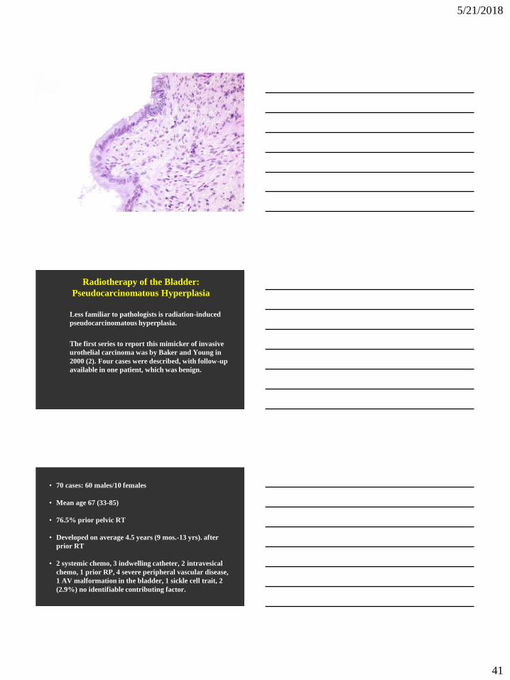
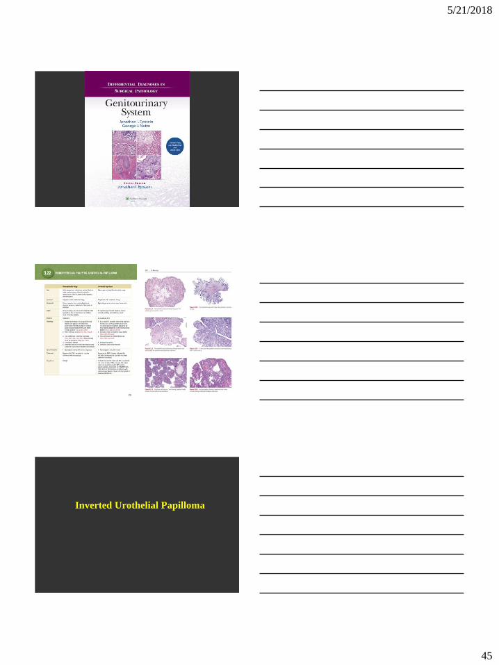
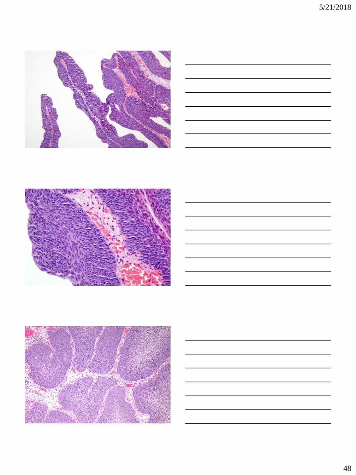
Inverted Urothelial Papilloma

5/21/2018
46
• Most commonly seen at trigone, yet may be anywhere
urothelium present.
• Usually solitary (3% multiple).
• Polypoid or sessile with smooth surface
• Wide size range
• Not related to increased risk of subsequent urothelial
carcinoma

5/21/2018
47
5/21/2018
48

5/21/2018
49

5/21/2018
50
Comparison to Urothelial Carcinoma
• Lacks cytological atypia
• Mitotic activity limited to basal cell layer
• Lacks inflammation and reactive stroma
• Squamous metaplasia lacks keratin formation
• Lacks muscularis propria invasion