Propionic acid induces dendritic spine loss by MAPK/ERK ...
11
RESEARCH Open Access Propionic acid induces dendritic spine loss by MAPK/ERK signaling and dysregulation of autophagic flux Hyosun Choi 1,2 , In Sik Kim 1,3 and Ji Young Mun 2* Abstract Propionic acid (PPA) is a short-chain fatty acid that is an important mediator of cellular metabolism. It is also a by- product of human gut enterobacteria and a common food preservative. A recent study found that rats administered with PPA showed autistic-like behaviors like restricted interest, impaired social behavior, and impaired reversal in a T-maze task. This study aimed to identify a link between PPA and autism phenotypes facilitated by signaling mechanisms in hippocampal neurons. Findings indicated autism-like pathogenesis associated with reduced dendritic spines in PPA-treated hippocampal neurons. To uncover the mechanisms underlying this loss, we evaluated autophagic flux, a functional readout of autophagy, using relevant biomedical markers. Results indicated that autophagic flux is impaired in PPA-treated hippocampal neurons. At a molecular level, the mitogen-activated protein kinase (MAPK)/extracellular signal-regulated kinase (ERK) pathway was activated and autophagic activity was impaired. We also observed that a MAPK inhibitor rescued dendritic spine loss in PPA-treated hippocampal neurons. Taken together, these results suggest a previously unknown link between PPA and autophagy in spine formation regulation in hippocampal neurons via MAPK/ERK signaling. Our results indicate that MAPK/ERK signaling participates in autism pathogenesis by autophagy disruption affecting dendritic spine density. This study may help to elucidate other mechanisms underlying autism and provide a potential strategy for treating ASD-associated pathology. Keywords: Propionic acid, Short-chain fatty acid, Autophagy, MAPK/ERK signaling, Spine density Introduction The human microbiome represents a diverse ecosys- tem of microbes. Reports on its influences on the im- mune system [1], metabolic processes [2], gene expression [3, 4], and nervous system [5] have led to increased recognition of the importance of the gut microbiome in human health and disease. The rela- tionship between the gut microbiome and nervous system as a significant component of the gut-brain axis has also attracted increasing interest in recent years [6]. The gut microbiome possesses significant meta- bolic capacity, and certain microbe-derived metabolites are released into circulation and can cross the blood– brain barrier [7, 8]. Fermentation of dietary fiber by the colonic microbiome is the primary source of short-chain fatty acids (SCFAs), such as acetic acid, propionic acid (PPA), and butyric acid [9, 10]. PPA is used as a preserva- tive in many processed foods, such as milk and cheese, and naturally occurs in yogurt [11, 12]. At appropriate levels, PPA improves immune function, facilitates cell sig- naling, and reduces food intake [9]. However, recent stud- ies have reported that PPA is also associated with negative effects on health and behavior. Rats treated with PPA demonstrate impaired social behavior and inflammatory © The Author(s). 2020 Open Access This article is licensed under a Creative Commons Attribution 4.0 International License, which permits use, sharing, adaptation, distribution and reproduction in any medium or format, as long as you give appropriate credit to the original author(s) and the source, provide a link to the Creative Commons licence, and indicate if changes were made. The images or other third party material in this article are included in the article's Creative Commons licence, unless indicated otherwise in a credit line to the material. If material is not included in the article's Creative Commons licence and your intended use is not permitted by statutory regulation or exceeds the permitted use, you will need to obtain permission directly from the copyright holder. To view a copy of this licence, visit http://creativecommons.org/licenses/by/4.0/. The Creative Commons Public Domain Dedication waiver (http://creativecommons.org/publicdomain/zero/1.0/) applies to the data made available in this article, unless otherwise stated in a credit line to the data. * Correspondence: [email protected] 2 Neural Circuit Research Group, Korea Brain Research Institute, Daegu 41068, Republic of Korea Full list of author information is available at the end of the article Choi et al. Molecular Brain (2020) 13:86 https://doi.org/10.1186/s13041-020-00626-0
Transcript of Propionic acid induces dendritic spine loss by MAPK/ERK ...

RESEARCH Open Access
Propionic acid induces dendritic spine lossby MAPK/ERK signaling and dysregulationof autophagic fluxHyosun Choi1,2, In Sik Kim1,3 and Ji Young Mun2*
Abstract
Propionic acid (PPA) is a short-chain fatty acid that is an important mediator of cellular metabolism. It is also a by-product of human gut enterobacteria and a common food preservative. A recent study found that ratsadministered with PPA showed autistic-like behaviors like restricted interest, impaired social behavior, and impairedreversal in a T-maze task. This study aimed to identify a link between PPA and autism phenotypes facilitated bysignaling mechanisms in hippocampal neurons. Findings indicated autism-like pathogenesis associated withreduced dendritic spines in PPA-treated hippocampal neurons. To uncover the mechanisms underlying this loss, weevaluated autophagic flux, a functional readout of autophagy, using relevant biomedical markers. Results indicatedthat autophagic flux is impaired in PPA-treated hippocampal neurons. At a molecular level, the mitogen-activatedprotein kinase (MAPK)/extracellular signal-regulated kinase (ERK) pathway was activated and autophagic activity wasimpaired. We also observed that a MAPK inhibitor rescued dendritic spine loss in PPA-treated hippocampal neurons.Taken together, these results suggest a previously unknown link between PPA and autophagy in spine formationregulation in hippocampal neurons via MAPK/ERK signaling. Our results indicate that MAPK/ERK signalingparticipates in autism pathogenesis by autophagy disruption affecting dendritic spine density. This study may helpto elucidate other mechanisms underlying autism and provide a potential strategy for treating ASD-associatedpathology.
Keywords: Propionic acid, Short-chain fatty acid, Autophagy, MAPK/ERK signaling, Spine density
IntroductionThe human microbiome represents a diverse ecosys-tem of microbes. Reports on its influences on the im-mune system [1], metabolic processes [2], geneexpression [3, 4], and nervous system [5] have led toincreased recognition of the importance of the gutmicrobiome in human health and disease. The rela-tionship between the gut microbiome and nervoussystem as a significant component of the gut-brainaxis has also attracted increasing interest in recent
years [6]. The gut microbiome possesses significant meta-bolic capacity, and certain microbe-derived metabolitesare released into circulation and can cross the blood–brain barrier [7, 8]. Fermentation of dietary fiber by thecolonic microbiome is the primary source of short-chainfatty acids (SCFAs), such as acetic acid, propionic acid(PPA), and butyric acid [9, 10]. PPA is used as a preserva-tive in many processed foods, such as milk and cheese,and naturally occurs in yogurt [11, 12]. At appropriatelevels, PPA improves immune function, facilitates cell sig-naling, and reduces food intake [9]. However, recent stud-ies have reported that PPA is also associated with negativeeffects on health and behavior. Rats treated with PPAdemonstrate impaired social behavior and inflammatory
© The Author(s). 2020 Open Access This article is licensed under a Creative Commons Attribution 4.0 International License,which permits use, sharing, adaptation, distribution and reproduction in any medium or format, as long as you giveappropriate credit to the original author(s) and the source, provide a link to the Creative Commons licence, and indicate ifchanges were made. The images or other third party material in this article are included in the article's Creative Commonslicence, unless indicated otherwise in a credit line to the material. If material is not included in the article's Creative Commonslicence and your intended use is not permitted by statutory regulation or exceeds the permitted use, you will need to obtainpermission directly from the copyright holder. To view a copy of this licence, visit http://creativecommons.org/licenses/by/4.0/.The Creative Commons Public Domain Dedication waiver (http://creativecommons.org/publicdomain/zero/1.0/) applies to thedata made available in this article, unless otherwise stated in a credit line to the data.
* Correspondence: [email protected] Circuit Research Group, Korea Brain Research Institute, Daegu 41068,Republic of KoreaFull list of author information is available at the end of the article
Choi et al. Molecular Brain (2020) 13:86 https://doi.org/10.1186/s13041-020-00626-0

responses in the brain [13, 14]. High levels of PPA, butnot other SCFAs, have been reported in the stools of aut-istic spectrum disorder (ASD) individuals [15]. Studieshave demonstrated that intraventricular infusions of PPAcaused abnormal behavioral patterns, such as abnormalsocial interaction and anxiety-like behavior in rats similarto those seen in humans with ASD [14, 16]. Althoughthere is growing evidence that PPA can affect behavioraldevelopment, the direct effects of PPA on neuronal cellsremain poorly understood.It was previously published that SCFAs may modulate
the levels of neurotransmitters [17]. For instance,neurogenesis-relevant signaling molecules such as brain-derived neurotrophic factor (BDNF) are shown to bemodulated by change actions of SCFAs in the brain [18–20]. These data could reveal some clues about howSCFAs could regulate neuronal development. In neur-onal circuits, dendrites from an individual neuron mayhave a thousand or more spines that participate in theestablishment of excitatory synapses. Spines and synap-ses are produced in excess numbers during developmentthat are later pruned via activity-dependent stabilizationor elimination [21]. Dendritic spines are highly dynamicand undergo constant turnover and morphological plas-ticity depending on developmental stage and activity,and defective synapse formation or development hasbeen implicated in many neurological diseases. However,whether PPA regulates the development of spine has notbeen experimentally tested. Changes in the number, for-mation, or elimination of dendritic spines depend on dif-ferent molecules including actin [22], the cytoskeleton[23], cell surface receptors [24], and signaling pathwayssuch as Ras and Rap kinase [25]. Moreover, activation ofautophagy has also been reported to be related to spinemorphology [26].Macroautophagy is a cellular pathway, wherein dam-
aged organelles are degraded, particularly those pro-duced during oxidative stress, and metabolic precursorsare regenerated [27]. In the initial step of autophagy, or-ganelles are engulfed inside a double-membraned vesiclecalled an autophagosome. The autophagosome thenfuses with an autolysosome to degrade organelles withlysosomal enzymes. Components are thereby recycled toproduce energy and maintain synthesis [28]. Disruptionof autophagy has been associated with several cellularpathologies, including tumors and neurological diseases[29, 30]. Impaired autophagy significantly reduces thesize of synapses and number of boutons in Drosophila[31]. A previous study found that deletion of the vitalautophagy gene Atg7 resulted in increased immaturedendritic filopodia and defects in synaptic refinement[32]. Evidence points to a relationship between autoph-agy and dendritic spine defects, but the mechanisticbasis for these defects remains elusive.
Autophagy is regulated by a range of signaling path-ways such as mammalian target of rapamycin (mTOR),ERK, and protein kinase B (AKT) [33]. ERK signaling iscentral to the MAPK pathway that regulates many cellu-lar processes such as proliferation, differentiation, devel-opment, learning, and apoptosis [34]. The MAPK/ERKpathway is also a key regulator of autophagy, and starva-tion, a stimulator of autophagy, transiently activatesMAPK/ERK to stimulate the maturation of the autopha-gosome [35]. Inhibition of MAPK/ERK activation byMAPK inhibitor pretreatment abolishes starvation-induced autophagy [36]. While it is difficult to draw afirm conclusion about the relationship of ERK to au-tophagy, it is clear that the MAPK/ERK pathway is animportant factor therein.We examined autophagic activity and the MAPK/ERK
pathway to characterize the biological effects of PPA onhippocampal neurons. Results suggest that spine defectsare associated with autophagy impairment and activationof the MAPK/ERK signaling pathway.
MethodsPrimary culturePrimary cultures of rat hippocampal neurons were pre-pared from the brains of day 18 embryonic rats. Briefly,the hippocampus was dissected in free HBSS and incu-bated with a 0.125% trypsin solution for 15 min at 37 °C.The resulting cell suspensions were diluted in neuroba-sal medium (#21103–049, Gibco), supplemented withSM1 components (#05711, Stemcell), and plated onto100 μg/mL poly-D-lysine (#P0899, Sigma-Aldrich) and2 μg/mL laminin (#11–243–217-001, Roche)-coatedplates or coverslips.
Pharmacological treatment of hippocampal neuronPropionic acid (#402907) and bafilomycin A1 (B1793)were purchased from Sigma-Aldrich, USA, and PD98059(#513000) was purchased from Calbiochem. PPA wasdissolved in phosphate-buffered saline (PBS) for treat-ment (100 μmole/mL). Bafilomycin A1 (2 nmole/mL)and PD98059 (10 μmole/mL) were dissolved in dimethylsulfoxide and stored in aliquots at − 20 °C until the ex-periments. Vehicle (PBS), bafilomycin A1 (2 nmole/mL),and PD98059 (10 μmole/mL) were simultaneouslytreated with PPA (100 μmole/mL). PPA treatment wasdenoted as DIV 18, and cells were harvested on DIV 21.
Western blotting analysisCultured neurons were harvested by scraping in ice-coldradio-immunoprecipitation assay buffer (#89900,Thermo Scientific) solution containing a protease inhibi-tor (A32963, Thermo Scientific) and phosphatase cock-tail inhibitors (#5970, Cell Signaling) to avoidphosphorylation and degradation of proteins. After
Choi et al. Molecular Brain (2020) 13:86 Page 2 of 11

incubation, all lysates were centrifuged at 15,000 g at4 °C for 30 min. The supernatant was then evaluated fortotal protein concentration using a BCA protein assaykit (#23225, ThermoFisher). Equal amounts of proteinsamples were incubated with 5X SDS sample loadingbuffer (CBSS-9005, CHEM-BIO) at 95 °C for 5 min. Thesamples (10 μg) were subjected to SDS-polyacrylamidegel electrophoresis on precast, 4–15% gradient mini-gels(#456–1085, Bio-rad). Following transfer to PVDF mem-branes (#1620177, Bio-rad), the membranes wereblocked in Tris-buffered saline (#CBTB-9110, CHEM-BIO) containing 3% BSA (#9048-4-8, GENEray Biotech-nology) and 0.1% Tween 20 (H5152, Promega) for 1 h.Membranes were then washed with TBST and incubatedovernight at 4 °C with primary antibodies against phos-phorylated ERK1/2 (#4370, Cell Signaling), phosphory-lated AKT (#4060, Cell Signaling), LC3A/B (#12741, CellSignaling), p62 (ab56416, Abcam), and beclin-1 (#3495,Cell Signaling). Membranes were then probed with horseradish peroxidase-conjugated secondary antibody (1:5000) for 1 h and developed using an enhanced chemilu-minescence immunoblot detection system (Fusion FX7,VILBER). Immunoblots for phosphorylated ERK1/2 andphosphorylated AKT were subsequently stripped and re-probed with anti-ERK1/2 (#4692, Cell Signaling) andanti-AKT (#4691, Cell Signaling) antibodies. Immuno-blots were analyzed by densitometry using ImageJ soft-ware (National Institutes of Health). Only film exposuresthat were in the linear range of the ECL reaction(#32106, Thermo Scientific) were used for quantificationanalysis.
Immunofluorescence stainingCultured neurons were fixed with 1% paraformaldehydein PBS containing 4% sucrose for 5 min at roomtemperature. Without washing, neurons were thenpermeabilized and blocked simultaneously in 100%methanol for 7 min at − 20 °C. Primary antibodiesagainst LC3A/B (#12741, Cell Signaling), p62 (ab56416,Abcam), lysosomal-associated membrane protein(LAMP1) (H4A3, Santa Cruz), and ubiquitin (P4D1,Santa Cruz) were added to blocking solution containing0.1% gelatin, 0.3% Triton X-100, 16 mM sodium phos-phate, and 450 mM NaCl and incubated overnight at4 °C. After washing with PBS, coverslips were incubatedwith AlexaFluor488 (#4412, Cell Signaling) or Alexa-Fluor594 (#8890, Cell Signaling)-conjugated secondaryantibodies for 1 h at room temperature and then washedextensively with PBS and distilled water. Subsequently,coverslips were mounted with mounting medium (H-1000, Vector Laboratories). The samples were imagedwith a confocal laser-scanning microscope (Nikon,Japan) using a 60X oil lens and 488 nm and 594 nmemission lasers.
Confocal microscopy for dendritic spinesCells were maintained in an incubator with 5% CO2 at37 °C. The neurons were transfected with 2 μg of GFPconstruct with 2 μL of lipofectamine 2000 (Invitrogen).Then, cells were stained by immunofluorescence labelingwith anti-GFP antibody (A11122, Invitrogen). The sam-ples were imaged with a confocal laser-scanning micro-scope (Nikon, Japan) using a 60X oil lens and 488 nmlaser. Our data indicate that all spine categories wererepresented in pyramidal neurons. Dendritic spine dens-ities were assessed by analyzing high-resolution digitalimages with ImageJ software (National Institutes ofHealth). Dendritic spines were counted manually usingthe point picker function in ImageJ particle analysis. Thedendritic field was estimated to be 10 μm in length.
Detection of autophagic fluxThe formation of autophagosomes and autolysosomes inhippocampal neurons of control and PPA-treated cellswas detected using a Premo™ Autophagy Tandem SensorRFP-GFP-LC3B Kit (P36239, Invitrogen), according tothe manufacturer’s instructions. The RFP-GFP-LC3Bsensor enables the detection of LC3B-positive, neutralpH autophagosomes in green fluorescence (GFP), andLC3B-positive acidic pH autolysosome in red fluores-cence (RFP). Cells were grown on coverslips and incu-bated with 10 μL of BacMam reagents containing RFP-GFP-LC3B for 16 h. Cells were then washed in PBS threetimes. Coverslips were mounted with mounting medium(H-1000, Vector Laboratories), and fluorescent imageswere taken using confocal microscopy (Nikon, Japan).LC3B-positive autophagosomes (GFP and YFP) andLC3B-positive autolysosomes (RFP only) were analyzedand quantified using ImageJ software (National Institutesof Health).
Transmission electron microscopy for autophagyNeuron cells were treated with 100 μM of PPA for 3 daysand then fixed at 4 °C in 2.5% glutaraldehyde (#16210,EMS) and 2% paraformaldehyde (#19210, EMS); cellswere then post-fixed with 2% osmium tetroxide (#19150,Sigma-Aldrich) for 30 min at 4 °C. Then, the cells werestained en bloc with 0.1 mg TCH (T1136, TCI) in 10mLdistilled water and 1% uranyl acetate (#22400, EMS) anddehydrated via a graded ethanol series. Samples werethen embedded with an EMBed-812 embedding kit(#14120, EMS). Embedded samples were sectioned (60nm) with an ultra-microtome (Leica, Germany), and thesections were then viewed on a Tecnai 20 transmissionelectron microscope (TEM) (ThermoFisher, USA) at120 kV. Numbers of autophagosomes and autolysosomesper 100 μm2 were measured, as well as the size of theultrastructure, using ImageJ software (National Institutesof Health).
Choi et al. Molecular Brain (2020) 13:86 Page 3 of 11
Statistical analysisAll data are represented as mean ± standard error of themean (SEM) of at least three individual experiments.Statistical analysis was performed using one-way analysisof variance, and statistical significance (p-value) was cal-culated with GraphPad Prism 5 (GraphPad Software).Results at the 95% confidence level were consideredsignificant.
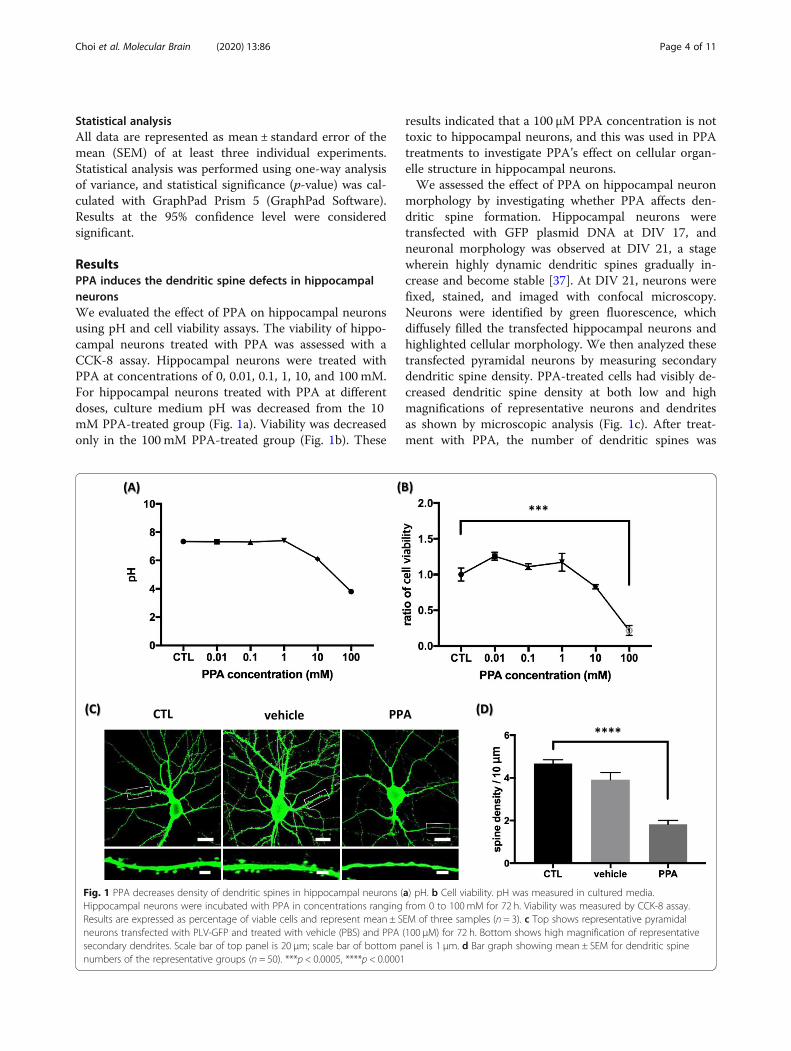
ResultsPPA induces the dendritic spine defects in hippocampalneuronsWe evaluated the effect of PPA on hippocampal neuronsusing pH and cell viability assays. The viability of hippo-campal neurons treated with PPA was assessed with aCCK-8 assay. Hippocampal neurons were treated withPPA at concentrations of 0, 0.01, 0.1, 1, 10, and 100 mM.For hippocampal neurons treated with PPA at differentdoses, culture medium pH was decreased from the 10mM PPA-treated group (Fig. 1a). Viability was decreasedonly in the 100 mM PPA-treated group (Fig. 1b). These
results indicated that a 100 μM PPA concentration is nottoxic to hippocampal neurons, and this was used in PPAtreatments to investigate PPA’s effect on cellular organ-elle structure in hippocampal neurons.We assessed the effect of PPA on hippocampal neuron
morphology by investigating whether PPA affects den-dritic spine formation. Hippocampal neurons weretransfected with GFP plasmid DNA at DIV 17, andneuronal morphology was observed at DIV 21, a stagewherein highly dynamic dendritic spines gradually in-crease and become stable [37]. At DIV 21, neurons werefixed, stained, and imaged with confocal microscopy.Neurons were identified by green fluorescence, whichdiffusely filled the transfected hippocampal neurons andhighlighted cellular morphology. We then analyzed thesetransfected pyramidal neurons by measuring secondarydendritic spine density. PPA-treated cells had visibly de-creased dendritic spine density at both low and highmagnifications of representative neurons and dendritesas shown by microscopic analysis (Fig. 1c). After treat-ment with PPA, the number of dendritic spines was
Fig. 1 PPA decreases density of dendritic spines in hippocampal neurons (a) pH. b Cell viability. pH was measured in cultured media.Hippocampal neurons were incubated with PPA in concentrations ranging from 0 to 100mM for 72 h. Viability was measured by CCK-8 assay.Results are expressed as percentage of viable cells and represent mean ± SEM of three samples (n = 3). c Top shows representative pyramidalneurons transfected with PLV-GFP and treated with vehicle (PBS) and PPA (100 μM) for 72 h. Bottom shows high magnification of representativesecondary dendrites. Scale bar of top panel is 20 μm; scale bar of bottom panel is 1 μm. d Bar graph showing mean ± SEM for dendritic spinenumbers of the representative groups (n = 50). ***p < 0.0005, ****p < 0.0001
Choi et al. Molecular Brain (2020) 13:86 Page 4 of 11

1.828 ± 0.1794, whereas that of untreated neurons was4.671 ± 0.1818 in 10 μm. Quantitative analyses showed39.13% lesser dendritic spine density in treated neuronsthan in controls (Fig. 1d). These findings indicate thatPPA induces dendritic spine loss in hippocampalneurons.
PPA upregulates biochemical markers of autophagyinitiation in hippocampal neuronsWe addressed the impact of PPA on induction of autophagyby assessing several autophagy markers, such as intracellularmicrotubule-associated protein 1 light chain 3 (LC3), beclin-1, and LAMP1. As lipidation of LC3 and its association withautophagosome membranes is an indicator of autophagy[38], we detected LC3 by immunoblot and fluorescence mi-croscopy. LC3-I was modified to the PE-conjugated form,LC3-II, because LC3-II is associated with mature autopha-gosomes upon autophagy induction. LC3 conversion from Ito II is a measurement of autophagic activity [39]. We de-tected a significant increase in LC3-II/LC3-I ratio in PPA-treated cells compared with control, as shown by westernblot (Fig. 2a, b). The number of LC3 puncta in PPA-treatedcells was visibly increased as shown by immunofluorescence
(Fig. 2c). After treatment with PPA, the number of punctawas 89.6 ± 6.765, whereas that of untreated neurons was43.42 ± 5.549 (Fig. 2d). We then measured protein levels ofbeclin-1 by western blot (Fig. 2e). The beclin-1 protein is atumor suppressor and a central regulator of autophagy crit-ical to the nucleation phase of autophagy [40]. Wong andcolleagues used beclin-1 antibody to visualize the formationof the autophagosome complex of protein aggregates to elu-cidate the autophagosome formation process [41]. AfterPPA treatment, beclin-1 was elevated by 27% comparedwith control neurons (Fig. 2f).We next assessed the impact of PPA on protein
aggregation-lysosome state levels of autophagic markers.PPA markedly increased the number of LAMP-1 andpoly-ubiquitin puncta (Fig. 2g, i). After treatment withPPA, the number of LAMP-1 puncta was 188 ± 22.65,whereas that of the untreated neurons was 100.8 ± 8.645(Fig. 2h). After treatment, the number of poly-ubiquitinpuncta was 90.8 ± 16.91, whereas that of untreated neu-rons was 2.667 ± 0.9482 (Fig. 2j). These findings stronglysuggest PPA as an inducer of autophagy throughout theprocess, from protein aggregation to autophagosomeinitiation.
Fig. 2 PPA upregulates biochemical markers of autophagy in hippocampal neurons (a) Representative western blot of LC3 after PPA treatment. bQuantification of LC3-II levels with respect to LC3-I levels (n = 4). c) Representative immunofluorescence images showing LC3 staining inhippocampal neurons and PPA-treated cells. d Quantification of number of LC3 puncta from images in (C) (n = 10). e Representative Western blotshowing increased expression of beclin-1 for 72 h after PPA treatment. f Quantification of beclin-1 levels with respect to GAPDH levels (n = 4). gRepresentative immunofluorescence images showing LAMP1 staining in hippocampal neurons and PPA-treated cells. h Quantification of thenumber of LAMP1 puncta from images in (G) (n = 12). i Representative Immunofluorescence images showing poly-ubiquitin staining inhippocampal neurons and PPA-treated cells. j Quantification of the number of poly-ubiquitin puncta from the images in (I) (n = 10). Bar graphshowing mean ± SEM of the representative groups. Scale bar is 10 μm. *p < 0.05, **p < 0.01, ****p < 0.0001
Choi et al. Molecular Brain (2020) 13:86 Page 5 of 11

Autophagic degradation is impaired by PPAThe interaction between LC3 and p62 was assessed bymeasuring protein levels of p62, a selective substrate ofautophagy; p62 binds directly to LC3 at a short LC3 inter-action region [42], and the induction of autophagic activityleads to p62 expression decline [43]. However, our immu-noblot (Fig. 3a) and immunofluorescence (Fig. 3c) resultsshowed a significant increase in p62 in PPA-treated cellscompared with control. Specific immunoblot resultsshowed a significant increase (30.8%) in p62 in PPA-treated cells over controls (Fig. 3b). After treatment withPPA, the number of p62 puncta was 158 ± 6.525, whereasthat of untreated neurons was 112.3 ± 9.66 (Fig. 3d). PPAtreatment was associated with elevated levels of p62, aprotein degraded by autophagy that accumulates when au-tophagy is impaired. This finding indicated that the in-crease in LC3-II levels was not caused by enhancedformation but rather impaired clearance of the autophago-some. An increased LC3-II/LC3-I ratio (Fig. 2b, d) and anaccumulation of p62 protein (Fig. 3b, d) were indicative ofimpaired autophagy in PPA-treated cells.To understand the autophagy defect better, we investi-
gated by transfecting hippocampal neurons with amarker gene, tandem fluorescent-tagged LC3 (GFP-RFP-LC3) in a baculoviral vector to monitor LC3 transloca-tion. Results in Fig. 4a show that there is no significantchange in autophagosome (GFP+ and YFP+ puncta)(Fig. 4b). After treatment with PPA, the number of RFP+
only puncta was 592 ± 104.4, whereas in the absence ofPPA treatment, it was 1097 ± 109.4. A quantification ana-lysis revealed a 50.2% decrease in red fluorescence expres-sion (Fig. 4c), suggesting that PPA dysregulates the fusionof autophagosome to autolysosome.We then further assessed fusion inhibition by analyzing
the number of autolysosomes using TEM in control andPPA-treated cells (Fig. 4d). The number of autolysosomeswas significantly decreased in PPA-treated cells, indicatinginefficient fusion associated with PPA (Fig. 4f). These re-sults strongly suggest that PPA dysregulates autolysosomefusion.Moreover, to investigate whether autophagy defects by
PPA are critical for spine loss, we investigated the effect ofbafilomycin A1 in hippocampal neurons. Bafilomycin A1is a well-known drug that disrupts autophagic flux by pre-venting cargo degradation [44]. Bafilomycin A1-treatedcells visibly decreased dendritic spine density at both lowand high magnifications of representative neurons anddendrites, as shown by microscopic analysis (Fig. 4g).After treatment with bafilomycin A1, the number of den-dritic spines was 2.161 ± 0.5523, whereas that of untreatedneurons was 4.726 ± 0.1715 in 10 μm. Quantitative ana-lyses showed 45.72% lesser dendritic spine density intreated neurons than in controls (Fig. 4h). The reductionof spine density in bafilomycin A1-treated hippocampalneurons is similar compared with PPA-treated cells. Theseresults show that PPA induces dendritic spine loss in
Fig. 3 Autophagic degradation impaired by PPA (a) Representative western blot of p62 after PPA treatment. b Quantification of p62 levels withrespect to GAPDH (n = 4). c Representative immunofluorescence images showing p62 staining in hippocampal neurons and PPA-treated cells. dQuantification of number of p62 puncta from images in (C) (n = 8). Bar graph showing mean ± SEM of representative groups. Scale bar is 10 μm.*p < 0.05, **p < 0.01
Choi et al. Molecular Brain (2020) 13:86 Page 6 of 11

hippocampal neurons by dysregulating autolysosome fu-sion, similar to bafilomycin A1.
ERK signaling pathway is involved in PPA-inducedautophagy defects in hippocampal neuronsMAPK/ERK signaling regulates the expression of au-tophagic and lysosomal genes and stimulates autophagy
by interacting with LC3. We investigated whether PPAexposure changes ERK1/2 signaling in hippocampal neu-rons by detecting phospho-ERK1/2 and total-ERK1/2using immunoblot assays (Fig. 5a). As shown in Fig. 5b,we observed a significant increase (30.2%) in the p-ERK/ERK ratio in PPA-treated cells compared with controls(Fig. 5b). We then assessed whether PPA changes AKT
Fig. 4 PPA dysregulates fusion from autophagosome to autolysosome (a) Representative fluorescent images after transfection with RFP-GFP-LC3Bin control hippocampal neurons and PPA-treated cells. Scale bar is 20 μm. b, c Quantification of number of GFP and YFP puncta(autophagosome) and RFP only puncta (autolysosome) from images in (A) (n = 5). d Black box shows representative TEM images in controlhippocampal neurons and PPA-treated cells. Red box shows representative TEM high magnification images of autolysosome and autophagosome(black arrow indicates autophagosome and red arrow indicates autolysosome). Black scale bar is 500 μm. e, f Quantification of autophagosomeand autolysosome number per area (n = 35). Bar graph showing mean ± SEM of the representative groups. g Top shows representative pyramidalneurons transfected with PLV-GFP and treated with bafilomycin A1 (2 nM) and PPA (100 μM) for 72 h. Bottom shows high magnification ofrepresentative secondary dendrites. Scale bar of top panel is 20 μm; scale bar of bottom panel is 1 μm. h Bar graph showing mean ± SEM fordendritic spine numbers of the representative groups (n = 50). *p < 0.05, ***p < 0.0005, ****p < 0.0001
Choi et al. Molecular Brain (2020) 13:86 Page 7 of 11

signaling in hippocampal neurons by detecting phospho-AKT and total AKT using immunoblot assays (Fig. 5c).Compared with the level of p-AKT/AKT, no significantchange in p-AKT/AKT ratio in PPA-treated hippocam-pal neurons was observed (Fig. 5d). These results suggestthat the MAPK/ERK pathway is more associated withautophagy impairment than the AKT pathway in thisstudy.
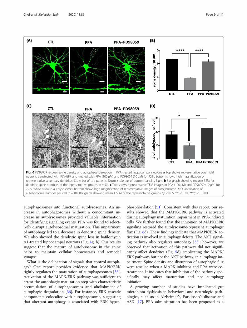
Inhibition of MAPK/ERK signaling rescues PPAphenotypes in vitroFinally, we analyzed the impact of PD98059, a MAPK in-hibitor, on dendritic spine loss and autophagic flux inhippocampal neurons. PD98059 treatment rescued den-dritic spine density in PPA-treated neurons, an effectthat was visible at both low and high magnification ofrepresentative neurons and dendrites (Fig. 6a). Quantita-tive analyses showed a 42.2% rescue of dendritic spinedensity in PD98059-treated neurons over PPA-treated-only neurons (Fig. 6b). In addition, to investigatewhether PPA-induced MAPK/ERK activation is involvedin autophagy regulation, we observed fusion activity byanalyzing the number of autolysosomes using TEM.PD98059 treatment rescued the number of autolysosomein PPA-treated neurons (Fig. 6c). The number of autoly-sosomes was significantly increased (40.9%) in PPA andPD98059-treated neurons than in only PPA-treated neu-rons (Fig. 6d). Taken together, our data showed that
inhibition of MAPK/ERK signaling rescued dendriticspine loss and autophagic disruption by PPA.
DiscussionRecent studies have demonstrated that autophagy is in-volved in synaptic remodeling in Drosophila melanoga-ster and mice [45]. Autophagy dysfunction is associatedwith reduced synaptic plasticity and dendritic spine defi-ciency [46]. Autophagic activity is required for BDNF-mediated synaptic plasticity in the hippocampus [47].Auerbach and colleagues demonstrated that TSC1/2 de-ficiency associated with decreased autophagy leads tosynaptic plasticity impairment in the hippocampus [48].Another study showed that cortical neuron dendriticspine density was reduced in TSC1 knockout mice [49].Goo et al. reported that lysosome inhibition alters lyso-some trafficking in dendrites; furthermore, it decreasesdendritic spine density [50]. In our study, PPA treatmentof hippocampal neurons resulted in increased autophagyinitiation but impaired autolysosome fusion. Electronand immunofluorescence microscopy analyses indicatedthat PPA-induced autophagosomes had not matured.They were double membrane-enclosed vesicles contain-ing organelles, but the subsequent fusion with the outermembrane of lysosomes indicating autophagosome mat-uration, wherein the inner contents of autolysosomes aredegraded for synthesis of new molecules and organelles,did not occur. Our findings suggest that PPA inducesspine loss by disrupting the maturation of
Fig. 5 PPA activates MAPK/ERK pathway associated with autophagy impairment a Representative western blot of p-ERK and ERK after PPAtreatment. b Quantification of p-ERK levels compared to ERK levels (n = 3). c Representative western blot of p-AKT and AKT after PPA treatment. dQuantification of p-AKT levels compared to AKT (n = 3). Bar graph showing mean ± SEM of the representative groups. *p < 0.05
Choi et al. Molecular Brain (2020) 13:86 Page 8 of 11
autophagosomes into functional autolysosomes. An in-crease in autophagosomes without a concomitant in-crease in autolysosomes provided valuable informationfor identifying signaling events. PPA was found to select-ively disrupt autolysosomal maturation. This impairmentof autophagy led to a decrease in dendritic spine density.We also showed the dendritic spine loss in bafilomycinA1-treated hippocampal neurons (Fig. 4g, h). Our resultssuggest that the mature of autolysosome in the spinehelps to maintain cellular homeostasis and remodelsynapse.What is the delineation of signals that control autoph-
agy? One report provides evidence that MAPK/ERKtightly regulates the maturation of autophagosomes [35].Activation of the MAPK/ERK pathway was sufficient toarrest the autophagic maturation step with characteristicaccumulation of autophagosomes and abolishment ofautophagic degradation [36]. For instance, ERK cascadecomponents colocalize with autophagosome, suggestingthat aberrant autophagy is associated with ERK hyper-
phosphorylation [51]. Consistent with this report, our re-sults showed that the MAPK/ERK pathway is activatedduring autophagy maturation impairment in PPA-inducedcells. We further found that the inhibition of MAPK/ERKsignaling restored the autolysosome-represent autophagicflux (Fig. 6d). These findings indicate that MAPK/ERK ac-tivation is involved in autophagy defects. The AKT signal-ing pathway also regulates autophagy [33]; however, weobserved that activation of this pathway did not signifi-cantly affect dendrites (Fig. 5d), implicating the MAPK/ERK pathway, but not the AKT pathway, in autophagy im-pairment. Spine density and disruption of autophagic fluxwere rescued when a MAPK inhibitor and PPA were co-treatment. It indicates that inhibition of the pathway spe-cifically may affect maturation and not autophagyinitiation.A growing number of studies have implicated gut
microbiota dysbiosis in behavioral and neurologic path-ologies, such as in Alzheimer’s, Parkinson’s disease andASD [17]. PPA administration has been proposed as a
Fig. 6 PD98059 rescues spine density and autophagy disruption in PPA-treated hippocampal neurons a Top shows representative pyramidalneurons transfected with PLV-GFP and treated with PPA (100 μM) and PD98059 (10 μM) for 72 h. Bottom shows high magnification ofrepresentative secondary dendrites. Scale bar of top panel is 20 μm; scale bar of bottom panel is 1 μm. b Bar graph showing mean ± SEM fordendritic spine numbers of the representative groups (n = 50). c Top shows representative TEM images in PPA (100 μM) and PD98059 (10 μM) for72 h (white arrow is autolysosome). Bottom shows high magnification of representative images of autolysosome. d Quantification ofautolysosome number per cell (n = 10). Bar graph showing mean ± SEM of the representative groups. *p < 0.05, **p < 0.01, ****p < 0.0001
Choi et al. Molecular Brain (2020) 13:86 Page 9 of 11

target for ASDs [52, 53]. Dendritic spines may serve as acommon substrate for many neuropsychiatric disorders,particularly those that involve cognitive deficits, and dys-regulation in spine morphology has been implicated inASD [54]. There are two types of dendritic spine modifi-cations in animal models of ASD-related genes. For ex-ample, SHANK3-mutated mice and PTEN transgenicmice models of ASD show a decrease in spine density.However, FMRP, MeCP2, neuroligin-3/4, and neurexin1ASD models show an increase in spine density [55]. Ourresults illustrated a possible link between PPA and re-duction of dendritic spine density in ASD animal model.Taken together, we speculate that autophagy degrad-
ation disruption and MAPK/ERK signaling pathway acti-vation induce dendritic spine defects and may affectsynapse function and plasticity, leading to autistic-likephenotypes in PPA-treated hippocampal neurons. It willbe of interest to determine if altered autophagy functionand MAPK/ERK inhibition is a contributing factor toASD. Our study represents the first empirical data sup-porting MAPK/ERK signaling pathway involvement inautophagic degradation disruption and the resultingpathologies associated with ASD.
AbbreviationsPPA: Propionic acid; MAPK: Mitogen-activated protein kinase;ERK: Extracellular signal-regulated kinase; mTOR: Mammalian target ofrapamycin; AKT: Protein kinase B; ASD: Autism spectrum disorder;BDNF: Brain-derived neurotrophic factor; PBS: Phosphate-buffered saline;DIV: Days in vitro; GFP: Green fluorescent protein; YFP: Yellow fluorescentprotein; RFP: Red fluorescent protein; LC3: Intracellular microtubule-associated protein 1 light chain 3; LAMP1: Lysosomal-associated membraneprotein 1; TEM: Transmission electron microscope; SEM: Standard error of themean
AcknowledgmentsTransmission electron microscopy and confocal microscopy data wereacquired at Brain Research Core Facilities in KBRI.
Authors’ contributionsJ.Y.M, I.S.K and H.C. designed research. H. C performed research and analyzeddata. J.Y.M and. H.C. wrote the paper. All authors read and approved the finalmanuscript.
FundingThis research was supported by the National Research Foundation of Korea(NRF) grant funded by the Korea government (MSIP) (No.2019R1A2C1010634) and KBRI basic research program through Korea BrainResearch Institute funded by Ministry of Science and ICT (20-BR-04-01).
Availability of data and materialsAll data generated or analyzed during this study are included in thispublished article.
Ethics approvalAll animal experiments were carried out in compliance with the Guide forCare and Use of Laboratory Animals of the National Institutes of Health andwere approved by the Institutional Animal Care and Use Committee of KoreaBrain Research Institute (IACUC-18-00013).
Consent for publicationNot applicable.
Competing interestsThe authors declare no competing financial interests.
Author details1BK21 Plus Program, Department of Senior Healthcare, Graduate School, EuljiUniversity, Daejeon, South Korea. 2Neural Circuit Research Group, Korea BrainResearch Institute, Daegu 41068, Republic of Korea. 3Department ofBiomedical Laboratory Science, Eulji University School of Medicine, Daejeon,South Korea.
Received: 24 February 2020 Accepted: 26 May 2020
References1. Johnson CC, Ownby DR. Allergies and asthma: do atopic disorders result
from inadequate immune homeostasis arising from infant gut dysbiosis?Expert Rev Clin Immunol. 2016;12(4):379–88.
2. Nicholson JK, Holmes E, Kinross J, Burcelin R, Gibson G, Jia W, et al. Host-gutmicrobiota metabolic interactions. Science. 2012;336(6086):1262–7.
3. Cureau N, AlJahdali N, Vo N, Carbonero F. Epigenetic mechanisms inmicrobial members of the human microbiota: current knowledge andperspectives. Epigenomics. 2016;8:1259–73.
4. Woo V, Alenghat T. Host–microbiota interactions: epigenomic regulation.Curr Opin Immunol. 2017;44:52–60.
5. Bourassa MW, Alim I, Bultman SJ, Ratan RR. Butyrate, neuroepigenetics andthe gut microbiome: can a high fiber diet improve brain health? NeurosciLett. 2016;625:56–63.
6. Mayer EA, Knight R, Mazmanian SK, Cryan JF, Tillisch K. Gut microbes andthe brain: paradigm shift in neuroscience. J Neurosci. 2014;34(46):15490–6.
7. Stilling RM, Dinan TG, Cryan JF. Microbial genes, brain & behaviour -epigenetic regulation of the gut-brain axis. Genes Brain Behav. 2014;13(1):69–86.
8. Karuri A, Dobrowsky E, Tannock I. Selective cellular acidification and toxicityof weak organic acids in an acidic microenvironment. Br J Cancer. 1993;68(6):1080.
9. Al-Lahham SH, Peppelenbosch MP, Roelofsen H, Vonk RJ, Venema K.Biological effects of propionic acid in humans; metabolism, potentialapplications and underlying mechanisms. Biochim Biophys Acta. 2010;1801(11):1175–83.
10. Hsu ST, Yang ST. Propionic acid fermentation of lactose byPropionibacterium acidipropionici: effects of pH. Biotechnol Bioeng. 1991;38(6):571–8.
11. Rook J, Balch C. The effects of intraruminal infusions of acetic, propionic andbutyric acids on the yield and composition of the milk of the cow. Br JNutr. 1961;15(3):361–9.
12. Dengate S, Ruben A. Controlled trial of cumulative behavioural effects of acommon bread preservative. J Paediatr Child Health. 2002;38(4):373–6.
13. MacFabe DF, Cain NE, Boon F, Ossenkopp KP, Cain DP. Effects of theenteric bacterial metabolic product propionic acid on object-directedbehavior, social behavior, cognition, and neuroinflammation inadolescent rats: relevance to autism spectrum disorder. Behav Brain Res.2011;217(1):47–54.
14. Choi J, Lee S, Won J, Jin Y, Hong Y, Hur T-Y, et al. Pathophysiological andneurobehavioral characteristics of a propionic acid-mediated autism-like ratmodel. PLoS One. 2018;13(2):e0192925.
15. Wang L, Christophersen CT, Sorich MJ, Gerber JP, Angley MT, Conlon MA.Elevated fecal short chain fatty acid and ammonia concentrations inchildren with autism spectrum disorder. Dig Dis Sci. 2012;57(8):2096–102.
16. MacFabe D. Autism: metabolism, mitochondria, and the microbiome. GlobAdv Health Med. 2013;2(6):52–66.
17. Silva YP, Bernardi A, Frozza RL. The role of short-chain fatty acids from gutmicrobiota in gut-brain communication. Front Endocrinol. 2020;11:25.
18. Maqsood R, Stone TW. The gut-brain axis, BDNF, NMDA and CNS disorders.Neurochem Res. 2016;41(11):2819–35.
19. Barichello T, Generoso JS, Simões LR, Faller CJ, Ceretta RA, Petronilho F,et al. Sodium butyrate prevents memory impairment by re-establishingBDNF and GDNF expression in experimental pneumococcal meningitis. MolNeurobiol. 2015;52(1):734–40.
20. Binder DK, Scharfman HE. Brain-derived neurotrophic factor. Growth factors(Chur, Switzerland). 2004;22(3):123.
Choi et al. Molecular Brain (2020) 13:86 Page 10 of 11

21. Changeux J-P, Danchin A. Selective stabilisation of developing synapses as amechanism for the specification of neuronal networks. Nature. 1976;264(5588):705.
22. Osterweil E, Wells DG, Mooseker MS. A role for myosin VI in postsynapticstructure and glutamate receptor endocytosis. J Cell Biol. 2005;168(2):329–38.
23. Tada T, Sheng M. Molecular mechanisms of dendritic spine morphogenesis.Curr Opin Neurobiol. 2006;16(1):95–101.
24. Washbourne P, Dityatev A, Scheiffele P, Biederer T, Weiner JA,Christopherson KS, et al. Cell adhesion molecules in synapse formation. JNeurosci. 2004;24(42):9244–9.
25. Vazquez LE, Chen H-J, Sokolova I, Knuesel I, Kennedy MB. SynGAP regulatesspine formation. J Neurosci. 2004;24(40):8862–72.
26. Shen D-N, Zhang L-H, Wei E-Q, Yang Y. Autophagy in synapticdevelopment, function, and pathology. Neurosci Bull. 2015;31(4):416–26.
27. Mizushima N, Levine B, Cuervo AM, Klionsky DJ. Autophagy fights diseasethrough cellular self-digestion. Nature. 2008;451(7182):1069–75.
28. Levine B, Klionsky DJ. Development by self-digestion: molecular mechanismsand biological functions of autophagy. Dev Cell. 2004;6(4):463–77.
29. Hara T, Nakamura K, Matsui M, Yamamoto A, Nakahara Y, Suzuki-MigishimaR, et al. Suppression of basal autophagy in neural cells causesneurodegenerative disease in mice. Nature. 2006;441(7095):885.
30. Rubinsztein DC, Gestwicki JE, Murphy LO, Klionsky DJ. Potential therapeuticapplications of autophagy. Nat Rev Drug Discov. 2007;6(4):304.
31. Shen W, Ganetzky B. Autophagy promotes synapse development inDrosophila. J Cell Biol. 2009;187(1):71–9.
32. Kim H, Cho M, Shim W, Kim J, Jeon E, Kim D, et al. Deficient autophagy inmicroglia impairs synaptic pruning and causes social behavioral defects. MolPsychiatry. 2017;22(11):1576–84.
33. Petiot A, Pattingre S, Arico S, Meley D, Codogno P. Diversity of signalingcontrols of macroautophagy in mammalian cells. Cell Struct Funct. 2002;27(6):431–41.
34. Shaul YD, Seger R. The MEK/ERK cascade: from signaling specificity todiverse functions. Biochimica et Biophysica Acta (BBA)-molecular. Cell Res.2007;1773(8):1213–26.
35. Corcelle E, Djerbi N, Mari M, Nebout M, Fiorini C, Fénichel P, et al. Control ofthe autophagy maturation step by the MAPK ERK and p38: lessons fromenvironmental carcinogens. Autophagy. 2007;3(1):57–9.
36. Corcelle E, Nebout M, Bekri S, Gauthier N, Hofman P, Poujeol P, et al.Disruption of autophagy at the maturation step by the carcinogen Lindane isassociated with the sustained mitogen-activated protein kinase/extracellularsignal–regulated kinase activity. Cancer Res. 2006;66(13):6861–70.
37. Nwabuisi-Heath E, LaDu MJ, Yu C. Simultaneous analysis of dendritic spinedensity, morphology and excitatory glutamate receptors during neuronmaturation in vitro by quantitative immunocytochemistry. J NeurosciMethods. 2012;207(2):137–47.
38. He C, Klionsky DJ. Regulation mechanisms and signaling pathways ofautophagy. Annu Rev Genet. 2009;43:67–93.
39. Kabeya Y, Mizushima N, Ueno T, Yamamoto A, Kirisako T, Noda T, et al. LC3,a mammalian homologue of yeast Apg8p, is localized in autophagosomemembranes after processing. EMBO J. 2000;19(21):5720–8.
40. Wirawan E, Lippens S, Vanden Berghe T, Romagnoli A, Fimia GM, PiacentiniM, et al. Beclin1: a role in membrane dynamics and beyond. Autophagy.2012;8(1):6–17.
41. Wong E, Bejarano E, Rakshit M, Lee K, Hanson HH, Zaarur N, et al. Moleculardeterminants of selective clearance of protein inclusions by autophagy. NatCommun. 2012;3:1240.
42. Komatsu M, Waguri S, Koike M, Sou YS, Ueno T, Hara T, et al. Homeostaticlevels of p62 control cytoplasmic inclusion body formation in autophagy-deficient mice. Cell. 2007;131(6):1149–63.
43. Jiang P, Mizushima N. LC3-and p62-based biochemical methods for theanalysis of autophagy progression in mammalian cells. Methods. 2015;75:13–8.
44. Klionsky DJ, Elazar Z, Seglen PO, Rubinsztein DC. Does bafilomycin A1 blockthe fusion of autophagosomes with lysosomes? Taylor & Francis; 2008; 849-50. .
45. Rowland AM, Richmond JE, Olsen JG, Hall DH, Bamber BA. Presynapticterminals independently regulate synaptic clustering and autophagy ofGABAA receptors in Caenorhabditis elegans. J Neurosci. 2006;26(6):1711–20.
46. Lieberman OJ, McGuirt AF, Tang G, Sulzer D. Roles for neuronal and glialautophagy in synaptic pruning during development. Neurobiol Dis. 2019;122:49–63.
47. Nikoletopoulou V, Sidiropoulou K, Kallergi E, Dalezios Y, Tavernarakis N.Modulation of autophagy by BDNF underlies synaptic plasticity. Cell Metab.2017;26(1):230–42 e5.
48. Auerbach BD, Osterweil EK, Bear MF. Mutations causing syndromic autismdefine an axis of synaptic pathophysiology. Nature. 2011;480(7375):63–8.
49. Meikle L, Pollizzi K, Egnor A, Kramvis I, Lane H, Sahin M, et al. Response of aneuronal model of tuberous sclerosis to mammalian target of rapamycin(mTOR) inhibitors: effects on mTORC1 and Akt signaling lead to improvedsurvival and function. J Neurosci. 2008;28(21):5422–32.
50. Goo MS, Sancho L, Slepak N, Boassa D, Deerinck TJ, Ellisman MH, et al.Activity-dependent trafficking of lysosomes in dendrites and dendriticspines. J Cell Biol. 2017;216(8):2499–513.
51. Martinez-Lopez N, Athonvarangkul D, Mishall P, Sahu S, Singh R. Autophagyproteins regulate ERK phosphorylation. Nat Commun. 2013;4:2799.
52. Dinan TG, Cryan JF. Microbes, immunity, and behavior:psychoneuroimmunology meets the microbiome.Neuropsychopharmacology. 2017;42(1):178–92.
53. Shultz SR, Aziz NA, Yang L, Sun M, MacFabe DF, O’Brien TJ.Intracerebroventricular injection of propionic acid, an enteric metaboliteimplicated in autism, induces social abnormalities that do not differbetween seizure-prone (FAST) and seizure-resistant (SLOW) rats. Behav BrainRes. 2015;278:542–8.
54. Fiala JC, Spacek J, Harris KM. Dendritic spine pathology: cause orconsequence of neurological disorders? Brain Res Rev. 2002;39(1):29–54.
55. Penzes P, Cahill ME, Jones KA, VanLeeuwen J-E, Woolfrey KM. Dendriticspine pathology in neuropsychiatric disorders. Nat Neurosci. 2011;14(3):285.
Publisher’s NoteSpringer Nature remains neutral with regard to jurisdictional claims inpublished maps and institutional affiliations.
Choi et al. Molecular Brain (2020) 13:86 Page 11 of 11

![Candida albicans -Glucan Exposure Is Controlled by the ... · mediated pathway by the deletion of the CEK1 (Candida albicans extracellular signal-regulated kinase [ERK]-like 1) MAPK](https://static.fdocuments.in/doc/165x107/5cc532cc88c9936d678d3eab/candida-albicans-glucan-exposure-is-controlled-by-the-mediated-pathway.jpg)