Synapsin I (Protein I), a Nerve Terminal-Specific Phosphoprotein. III ...
881Research Article
IntroductionFormation and plasticity of synapses are the result of complexmolecular interactions that regulate assembly of pre- and post-synaptic specializations and modulate their functions. Synapsinproteins are a family of vesicle-associated phosphoproteins that arelocalized at presynaptic terminals (De Camilli et al., 1983; Huttneret al., 1983; Hirokawa et al., 1989; Kao et al., 1999) and promoteneurite outgrowth and synaptogenesis, as well as regulating vesicledynamics and neurotransmitter release (Greengard et al., 1993;Pieribone et al., 1995; Rosahl et al., 1995; Hilfiker et al., 1999; Chiet al., 2001). Synapsin is a substrate for several protein kinases,including PKA, CaMKs and MAPK/Erk, which strongly modulateits biochemical properties. However, the specific effect ofMAPK/Erk-dependent synapsin phosphorylation in synaptogenesishas not been clarified, and its physiological role in neurotransmitterrelease and synaptic plasticity, mainly inferred from biochemicaland imaging studies (Jovanovic et al., 1996; Jovanovic et al., 2000;Chin et al., 2002; Chi et al., 2003), has not been directly exploredby electrophysiology.
The MAPK/Erk pathway has an important role in both synapseformation and plasticity. Evidence exists that this signalling cascademediates the synaptogenic action of neurotrophic factors (Huangand Reichardt, 2001; Alonso et al., 2004; Hans et al., 2004), althoughits molecular targets during synaptogenesis are still unknown.Moreover, it has been proposed that the MAPK/Erk pathway
participates in long-term synaptic plasticity by regulating theactivity of transcriptional factors upon nuclear translocation (Martinet al., 1997). Some studies show that MAPKs are also present andactive in synaptic terminals, suggesting that this pathway might haveseveral functions in distinct subcellular compartments during short-and long-term plasticity, acting through phosphorylation of synaptictargets, including synapsin (Sweatt, 2004; Boggio et al., 2007).Some studies have excluded the involvement of the MAPK/Erkpathway in short-term heterosynaptic plasticity (Martin et al., 1997;Purcell et al., 2003; Phares and Byrne, 2005). However, it has beenobserved that enhanced paired-pulse facilitation and long-termpotentiation in mice expressing an activated form of H-Ras areabolished upon synapsin knockout, indicating that synapsin is animportant target of the H-Ras-MAPK-Erk pathway in these formsof homosynaptic plasticity (Kushner et al., 2005).
In this work, we studied whether synapsin is a relevantMAPK/Erk target during synaptogenesis and short-termhomosynaptic plasticity by directly manipulating the levels ofsynapsin and its mutant phosphoforms in presynaptic terminals. Tothis aim, we developed an in vitro model of synaptogenesis betweenB2 Helix neurons (Altrup and Speckmann, 1994) formingbidirectional excitatory synapses in presence of extrinsic trophicfactors. Our experiments show that the formation and short-termplasticity of functional synapses are MAPK/Erk-dependent synapsinphosphorylation processes. In fact, using phosphorylation site
MAPK/Erk-dependent phosphorylation of synapsinmediates formation of functional synapses andshort-term homosynaptic plasticityCarlo Natale Giuseppe Giachello1,*, Ferdinando Fiumara1, Caterina Giacomini2, Anna Corradi2, Chiara Milanese1, Mirella Ghirardi1,3, Fabio Benfenati2,3,4 and Pier Giorgio Montarolo1,3
1Department of Neuroscience, Section of Physiology, University of Torino, Torino, Italy2Department of Experimental Medicine, Section of Physiology, University of Genova and Istituto Nazionale di Neuroscienze, Genova, Italy. 3Istituto Nazionale di Neuroscienze, Torino, Italy4Department of Neuroscience and Brain Technologies, The Italian Institute of Technology, Genova, Italy*Author for correspondence ([email protected])
Accepted 18 December 2009Journal of Cell Science 123, 881-893 © 2010. Published by The Company of Biologists Ltddoi:10.1242/jcs.056846
SummaryMAPK/Erk is a protein kinase activated by neurotrophic factors involved in synapse formation and plasticity, which acts at both the nuclearand cytoplasmic level. Synapsin proteins are synaptic-vesicle-associated proteins that are well known to be MAPK/Erk substrates atphylogenetically conserved sites. However, the physiological role of MAPK/Erk-dependent synapsin phosphorylation in regulating synapticformation and function is poorly understood. Here, we examined whether synapsin acts as a physiological effector of MAPK/Erk insynaptogenesis and plasticity. To this aim, we developed an in vitro model of soma-to-soma paired Helix B2 neurons, that establishbidirectional excitatory synapses. We found that the formation and activity-dependent short-term plasticity of these synapses is dependenton the MAPK/Erk pathway. To address the role of synapsin in this pathway, we generated non-phosphorylatable and pseudo-phosphorylatedHelix synapsin mutants at the MAPK/Erk sites. Overexpression experiments revealed that both mutants interfere with presynapticdifferentiation, synapsin clustering, and severely impair post-tetanic potentiation, a form of short-term homosynaptic plasticity. Our findingsshow that MAPK/Erk-dependent synapsin phosphorylation has a dual role both in the establishment of functional synaptic connectionsand their short-term plasticity, indicating that some of the multiple extranuclear functions of MAPK/Erk in neurons can be mediated bythe same multifunctional presynaptic target.
Key words: Synapsin, MAPK/Erk, Synaptogenesis, Post-tetanic potentiation, Helix, B2 neurons
Jour
nal o
f Cel
l Sci
ence
882
mutants of Helix synapsin that are either non-phosphorylatable, ormimick a persistent phosphorylation, we showed that presynapticoverexpression of non-phosphorylatable synapsin interferes withformation of active connections without affecting neurite outgrowth.Moreover, both phosphomutants virtually abolished post-tetanicpotentiation (PTP) by exerting a dominant-negative effect overendogenous synapsin. Some of these results have been previouslypresented in abstract form (Giachello et al., 2008).
ResultsPaired Helix B2 neurons form bidirectional soma-somasynapses in cultureIn-vitro-reconstructed synaptic connections between identifiedneurons of Helix, Aplysia and other gastropods offer anadvantageous model for studying synapse formation, as well assynaptic function. We previously observed that various types ofHelix neurons can form uni- and bi-directional synapses when pairedin culture in soma-to-soma configuration (Fiumara et al., 2005).
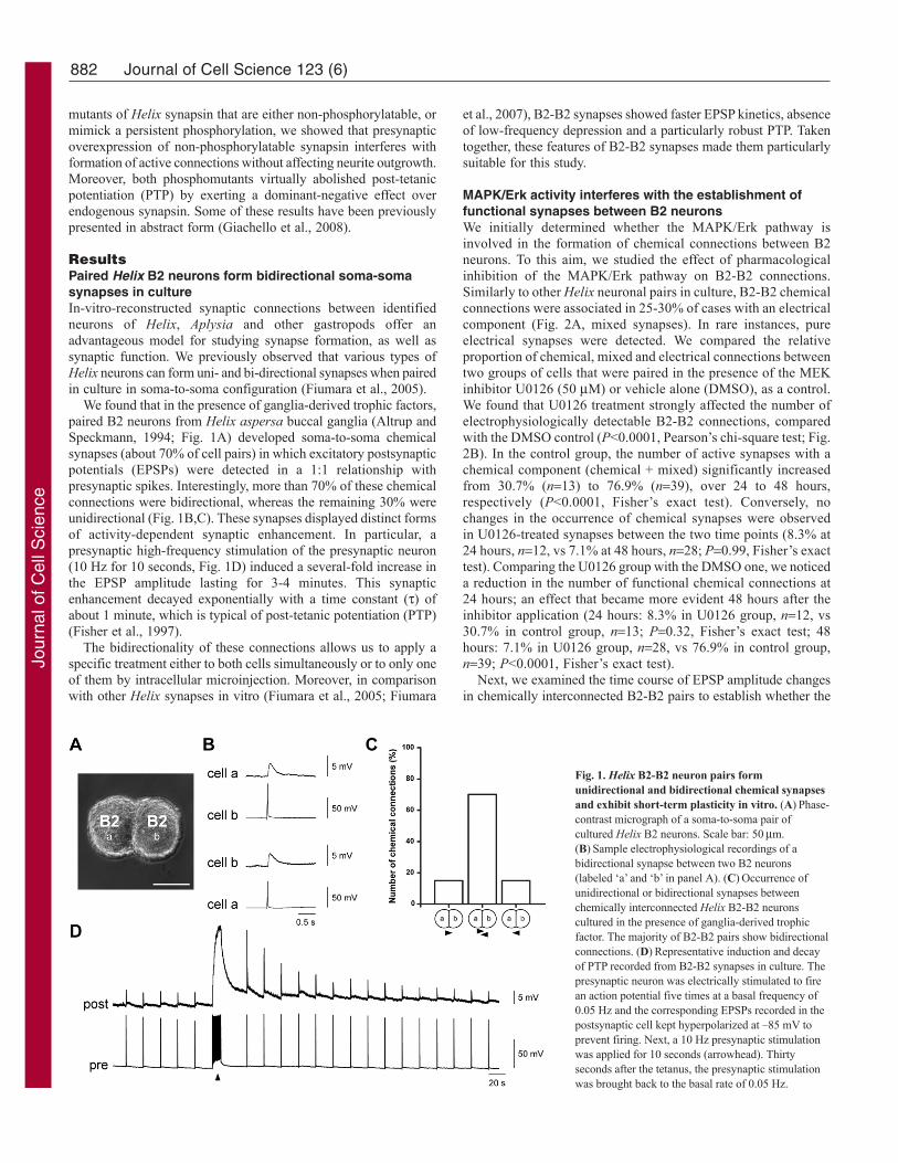
We found that in the presence of ganglia-derived trophic factors,paired B2 neurons from Helix aspersa buccal ganglia (Altrup andSpeckmann, 1994; Fig. 1A) developed soma-to-soma chemicalsynapses (about 70% of cell pairs) in which excitatory postsynapticpotentials (EPSPs) were detected in a 1:1 relationship withpresynaptic spikes. Interestingly, more than 70% of these chemicalconnections were bidirectional, whereas the remaining 30% wereunidirectional (Fig. 1B,C). These synapses displayed distinct formsof activity-dependent synaptic enhancement. In particular, apresynaptic high-frequency stimulation of the presynaptic neuron(10 Hz for 10 seconds, Fig. 1D) induced a several-fold increase inthe EPSP amplitude lasting for 3-4 minutes. This synapticenhancement decayed exponentially with a time constant () ofabout 1 minute, which is typical of post-tetanic potentiation (PTP)(Fisher et al., 1997).
The bidirectionality of these connections allows us to apply aspecific treatment either to both cells simultaneously or to only oneof them by intracellular microinjection. Moreover, in comparisonwith other Helix synapses in vitro (Fiumara et al., 2005; Fiumara
et al., 2007), B2-B2 synapses showed faster EPSP kinetics, absenceof low-frequency depression and a particularly robust PTP. Takentogether, these features of B2-B2 synapses made them particularlysuitable for this study.
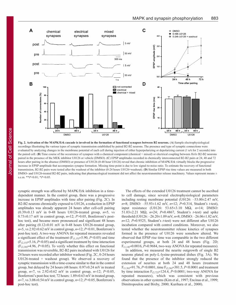
MAPK/Erk activity interferes with the establishment offunctional synapses between B2 neuronsWe initially determined whether the MAPK/Erk pathway isinvolved in the formation of chemical connections between B2neurons. To this aim, we studied the effect of pharmacologicalinhibition of the MAPK/Erk pathway on B2-B2 connections.Similarly to other Helix neuronal pairs in culture, B2-B2 chemicalconnections were associated in 25-30% of cases with an electricalcomponent (Fig. 2A, mixed synapses). In rare instances, pureelectrical synapses were detected. We compared the relativeproportion of chemical, mixed and electrical connections betweentwo groups of cells that were paired in the presence of the MEKinhibitor U0126 (50 M) or vehicle alone (DMSO), as a control.We found that U0126 treatment strongly affected the number ofelectrophysiologically detectable B2-B2 connections, comparedwith the DMSO control (P<0.0001, Pearson’s chi-square test; Fig.2B). In the control group, the number of active synapses with achemical component (chemical + mixed) significantly increasedfrom 30.7% (n13) to 76.9% (n39), over 24 to 48 hours,respectively (P<0.0001, Fisher’s exact test). Conversely, nochanges in the occurrence of chemical synapses were observedin U0126-treated synapses between the two time points (8.3% at24 hours, n12, vs 7.1% at 48 hours, n28; P0.99, Fisher’s exacttest). Comparing the U0126 group with the DMSO one, we noticeda reduction in the number of functional chemical connections at24 hours; an effect that became more evident 48 hours after theinhibitor application (24 hours: 8.3% in U0126 group, n12, vs30.7% in control group, n13; P0.32, Fisher’s exact test; 48hours: 7.1% in U0126 group, n28, vs 76.9% in control group,n39; P<0.0001, Fisher’s exact test).
Next, we examined the time course of EPSP amplitude changesin chemically interconnected B2-B2 pairs to establish whether the
Journal of Cell Science 123 (6)
Fig. 1. Helix B2-B2 neuron pairs formunidirectional and bidirectional chemical synapsesand exhibit short-term plasticity in vitro. (A)Phase-contrast micrograph of a soma-to-soma pair ofcultured Helix B2 neurons. Scale bar: 50m.(B)Sample electrophysiological recordings of abidirectional synapse between two B2 neurons(labeled ‘a’ and ‘b’ in panel A). (C)Occurrence ofunidirectional or bidirectional synapses betweenchemically interconnected Helix B2-B2 neuronscultured in the presence of ganglia-derived trophicfactor. The majority of B2-B2 pairs show bidirectionalconnections. (D)Representative induction and decayof PTP recorded from B2-B2 synapses in culture. Thepresynaptic neuron was electrically stimulated to firean action potential five times at a basal frequency of0.05 Hz and the corresponding EPSPs recorded in thepostsynaptic cell kept hyperpolarized at –85 mV toprevent firing. Next, a 10 Hz presynaptic stimulationwas applied for 10 seconds (arrowhead). Thirtyseconds after the tetanus, the presynaptic stimulationwas brought back to the basal rate of 0.05 Hz.
Jour
nal o
f Cel
l Sci
ence
883MAPK and synapsin phosphorylation
synaptic strength was affected by MAPK/Erk inhibition in a time-dependent manner. In the control group, there was a progressiveincrease in EPSP amplitudes with time after pairing (Fig. 2C). InB2-B2 neurons chronically exposed to U0126, a reduction in EPSPamplitudes was already apparent 24 hours after cell-cell pairing(0.39±0.13 mV in 0-48 hours U0126-treated group, n5, vs0.73±0.17 mV in control group, n12; P>0.05, Bonferroni’s post-hoc test), and became more pronounced and significant 48 hoursafter pairing (0.12±0.01 mV in 0-48 hours U0126-treated group,n5, vs 2.92±0.62 mV in control group, n12; P<0.01, Bonferroni’spost-hoc test). A two-way ANOVA for repeated measures revealeda significant effect of the treatment (F(1,15)5.90, P<0.05) and time(F(2,15)5.16, P<0.05) and a significant treatment by time interaction(F(2,30)4.96, P<0.05). To verify whether this effect on functionaltransmission was reversible, B2-B2 pairs incubated with U0126 for24 hours were recorded after inhibitor washout (Fig. 2C, 0-24 hoursU0126-treated + washout group). We observed a recovery ofsynaptic transmission with a time-course similar to that in the controlgroup, but delayed by 24 hours (48 hours: 0.97±0.24 mV in treatedgroup, n7, vs 2.92±0.62 mV in control group, n12; P<0.05,Bonferroni’s post-hoc test; 72 hours: 1.89±0.63 mV in treated group,n7, vs 3.08±0.54 mV in control group, n12; P>0.05, Bonferroni’spost-hoc test).
The effects of the extended U0126 treatment cannot be ascribedto cell damage, since several electrophysiological parametersincluding resting membrane potential (U0126: –53.00±2.47 mV,n8; DMSO: –55.93±1.62 mV, n12; P0.314, Student’s t-test),input resistance (U0126: 53.65±3.10 M, n14; DMSO:51.03±2.21 M, n24; P0.4867, Student’s t-test) and spikethreshold (U0126: –26.20±1.09 mV, n8; DMSO: –26.06±1.82 mV,n12; P0.9525, Student’s t-test) were not different after U0126incubation compared with control conditions. Moreover, we alsotested whether the neurotransmitter release kinetics of synapsesformed in the presence of U0126 were somehow altered. Weobserved that EPSP rise time was comparable in the two differentexperimental groups, at both 24 and 48 hours (Fig. 2D;F(1,14)0.0016, P0.9684, two-way ANOVA for repeated measures).
In addition, we measured the neurite outgrowth of single B2neurons plated on poly-L-lysine-pretreated dishes (Fig. 3A). Wefound that the presence of the inhibitor strongly reduced theextension of neurites at both 24 and 48 hours (treatmentF(1,36)91.76, P<0.0001, time F(2,36)381.5, P<0.0001 and treatmentby time interaction F(2,72)124.4, P<0.0001; two-way ANOVA forrepeated measures), which was consistent with previousobservations in other systems (Kim et al., 1997; Encinas et al., 1999;Dimitropoulou and Bixby, 2000; Kurihara et al., 2000).
Fig. 2. Activation of the MAPK/Erk cascade is involved in the formation of functional synapses between B2 neurons. (A)Sample electrophysiologicalrecordings illustrating the various types of synaptic transmission established by paired B2-B2 neurons. The presence and type of synaptic connections wereevaluated by analyzing changes in the membrane potential of each cell during injection of either hyperpolarizing or depolarizing current (1 nA for 2 seconds) intothe paired cell. (B)Time course of the occurrence of synapses with a chemical component (chemical + mixed) or electrical coupling between Helix B2-B2 neuronspaired in the presence of the MEK inhibitor U0126 or vehicle (DMSO). (C) EPSP amplitudes recorded in chemically interconnected B2-B2 pairs at 24, 48 and 72hours after pairing in the absence (DMSO) or presence of U0126 (0-48 hour U0126) reveal that chronic inhibition of MAPK/Erk virtually blocks the progressiveincrease in EPSP amplitude that accompanies synapse formation. Missing time-point is due to low signal-to-noise ratio. To estimate the recovery of functionaltransmission, B2-B2 pairs were tested after the washout of the inhibitor (0-24 hours U0126+washout). (D)Similar EPSP rise time values are measured in bothDMSO- and U0126-treated B2-B2 pairs, indicating that pharmacological treatment did not affect the neurotransmitter release machinery. Values represent means ±s.e.m. **P<0.01; *P<0.05.
Jour
nal o
f Cel
l Sci
ence

884
Taken together, our data indicate that the MAPK/Erk pathwayregulates both neurite outgrowth and the occurrence of functionalchemical connections between B2 neurons.
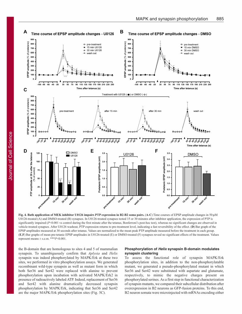
Post-tetanic potentiation at B2-B2 synapses depends onMAPK/Erk activationNext, we studied whether the MAPK/Erk pathway is also involved,in ways other than in synapse formation, in the functional plasticityof already established B2-B2 synapses. To this aim, we inducedPTP, a form of short-term homosynaptic plasticity, before and afterthe bath application of the inhibitor U0126 (Fig. 4A,C) or vehiclealone (DMSO) as a control (Fig. 4B,C). In the same synapses, weobserved that the presence of U0126 strongly reduced the expressionof PTP at both 15 and 30 minutes after bath application. We alsodetermined the reversibility of the U0126 effect by testing PTP 15minutes after wash out of the inhibitor. We found that the U0126effect was completely reversible and PTP was fully restored afterwashout. A two-way ANOVA for repeated measures revealed asignificant effect of the treatment (n7; F(3,24)4.83, P<0.01) anda significant treatment by time interaction (F(57,456)4.98, P<0.001).In particular, the peak amplitude of PTP measured 30 seconds aftertetanus was reduced to 38±8.0% of control amplitude at 15 minutesand to 32.92±7.1% at 30 minutes after the application (n7;F(3,18)12.79, P0.0001, one-way ANOVA for repeated measures;Fig. 4D). In the control group, we did not detect significant changesin PTP before or after addition of vehicle (n5; treatmentF(1,16)0.54, P0.6614 and treatment by time interactionF(57,304)0.30, P1.00, two-way ANOVA for repeated measures; Fig.4B).
To rule out the possibility that the U0126 effect might be relatedto general changes in efficiency of synaptic transmission and notto a specific effect on short-term plasticity, we verified that thepresence of U0126 or DMSO did not alter the basal EPSPamplitudes. To this aim, we compared the mean pre-tetanic basalEPSPs before and after application of U0126 and found no effectsof the inhibitor or of vehicle alone on basal synaptic transmission(U0126: F(3,18)15.23, P0.774, n7; DMSO: F(3,12)9.008,
P0.3232, n5; one-way ANOVA for repeated measures; Fig.4E,F).
Taken together, these data indicate that MAPK/Erk pathway hasan important role in expression of PTP at B2-B2 synapses. The fastand fully reversible effect of U0126 indicates that the role ofMAPK/Erk activation in this form of plasticity is likely to be relatedto phosphorylation of synaptic substrates rather than totranscriptional effects.
Helix synapsin is a MAPK/Erk substrate atphylogenetically conserved phosphorylation sitesSynapsin proteins are major presynaptic components and are knowntargets for MAPK/Erk in vertebrates. To determine whether synapsinis a mediator of the MAPK/Erk actions in B2-B2 synapticfunctionality and plasticity that we observed, we studied theprimary sequence of Helix synapsin looking for potentialMAPK/Erk consensus sites. MAPK/Erk is known to phosphorylatemammalian synapsin I (at sites 4/5 in domain B and site 6 in domainD) as well as Aplysia synapsin (Angers et al., 2002). Sincemolluscan synapsin lacks the D-domain, in search for conservedphosphorylation sites we focused our analysis on the N-terminalB-domain. A bioinformatic analysis with the GPS 2.1 softwarerevealed three putative sites in this region (Ser36, Ser42 and Ser48).Whereas Ser36 and Ser42 had high probability scores, the Ser48was only at the prediction threshold. Interestingly, Ser36 and Ser42were highly conserved among the known invertebrate synapsinproteins (Fig. 5A). MAPK/Erk phosphorylation of the closely relatedAplysia synapsin was previously reported, and the former two serineresidues were identified as putative MAPK sites (Angers et al.,2002). Moreover, these two sites seemingly corresponded to sites4 and 5 of mammalian synapsin. As illustrated in Fig. 5B, in amultiple alignment of mammalian synapsin I with the Helix andAplysia orthologues, Ser36 aligned with mammalian site 4 (Ser62).Within this alignment, Helix Ser42 and the corresponding Aplysiasite were shifted by one residue with respect to mammalian site 5.Based on this analysis, we concluded that Helix and Aplysia Ser36and Ser42 might represent the MAPK/Erk phosphorylation sites in
Journal of Cell Science 123 (6)
Fig. 3. Inhibition of the MAPK/Erk cascade affects neurite outgrowth through a synapsin phosphorylation-independent mechanism. (A)Graphicrepresentation of the neurite outgrowth of Helix B2 neurons plated on adhesive substrate in the presence of ganglia-derived trophic factors, upon application ofeither U0126 or DMSO as a control. Each time-point represents the percentage mean value of radius length of a circumferential line interconnecting the tips of thethree longest neurites, normalized to the control mean value measured at 24 hours. The presence of the inhibitor strongly reduces the extension of neurite outgrowthat both 24 and 48 hours (24 hours: 53.3±4.0% in U0126 group, n21, vs 100.0±7.3% in control group, n17; P<0.001, Bonferroni’s post-hoc test; 48 hours:60.6±4.6% in U0126 group, n21, vs 213.7±13.8% in control group, n17; P<0.001, Bonferroni’s post-hoc test). (B)Neurite growth profile of Helix B2 neuronsoverexpressing the different Helix synapsin (helSyn) mutants. No differences are observed among the experimental groups (n6 in each group) with respect tocontrol (uninjected cells, n20). Values represent means ± s.e.m. ***P<0.001.
Jour
nal o
f Cel
l Sci
ence
885MAPK and synapsin phosphorylation
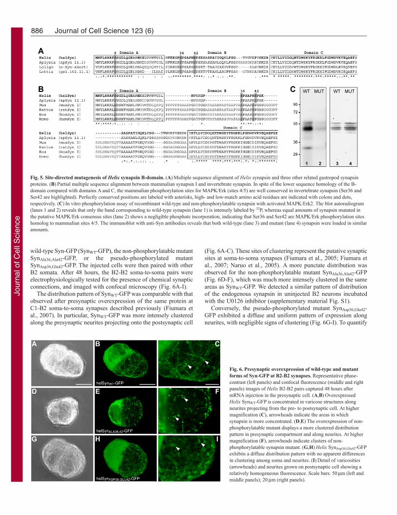
the B-domain that are homologous to sites 4 and 5 of mammaliansynapsin. To unambiguously confirm that Aplysia and Helixsynapsin was indeed phosphorylated by MAPK/Erk at these twosites, we performed in vitro phosphorylation assays. We generatedrecombinant wild-type synapsin as well as mutant form in whichboth Ser36 and Ser42 were replaced with alanine to preventphosphorylation upon incubation with activated MAPK/Erk2 inpresence of radioactively labeled ATP. Indeed, replacement of Ser36and Ser42 with alanine dramatically decreased synapsinphosphorylation by MAPK/Erk, indicating that Ser36 and Ser42are the major MAPK/Erk phosphorylation sites (Fig. 5C).
Phosphorylation of Helix synapsin B-domain modulatessynapsin clusteringTo assess the functional role of synapsin MAPK/Erkphosphorylation sites, in addition to the non-phosphorylatablemutant, we generated a pseudo-phosphorylated mutant in whichSer36 and Ser42 were substituted with aspartate and glutamate,respectively, to mimic the negative charges present onphosphorylated serines. As a first step in functional characterizationof synapsin mutants, we compared their subcellular distribution afteroverexpression in B2 neurons as GFP-fusion proteins. To this end,B2 neuron somata were microinjected with mRNAs encoding either
Fig. 4. Bath application of MEK inhibitor U0126 impairs PTP expression in B2-B2 soma pairs. (A-C)Time courses of EPSP amplitude changes in 50MU0126-treated (A) and DMSO-treated (B) synapses. In U0126-treated synapses tested 15 or 30 minutes after inhibitor application, the expression of PTP issignificantly impaired (P<0.001 vs control during the first minute after the tetanus, Bonferroni’s post-hoc test), whereas no significant changes are observed invehicle-treated synapses. After U0126 washout, PTP expression returns to pre-treatment level, indicating a fast reversibility of the effect. (D)Bar graph of theEPSP amplitudes measured at 30 seconds after tetanus. Values are normalized to the mean peak PTP amplitude measured before the treatment in each group.(E,F)Bar graphs of mean pre-tetanic EPSP amplitudes in U0126-treated (E) or DMSO-treated (F) synapses reveal no significant effects of the treatment. Valuesrepresent means ± s.e.m. ***P<0.001.
Jour
nal o
f Cel
l Sci
ence
886
wild-type Syn-GFP (SynWT-GFP), the non-phosphorylatable mutantSynAla36,Ala42-GFP, or the pseudo-phosphorylated mutantSynAsp36,Glu42-GFP. The injected cells were then paired with otherB2 somata. After 48 hours, the B2-B2 soma-to-soma pairs wereelectrophysiologically tested for the presence of chemical synapticconnections, and imaged with confocal microscopy (Fig. 6A-I).
The distribution pattern of SynWT-GFP was comparable with thatobserved after presynaptic overexpression of the same protein atC1-B2 soma-to-soma synapses described previously (Fiumara etal., 2007). In particular, SynWT-GFP was more intensely clusteredalong the presynaptic neurites projecting onto the postsynaptic cell
(Fig. 6A-C). These sites of clustering represent the putative synapticsites at soma-to-soma synapses (Fiumara et al., 2005; Fiumara etal., 2007; Naruo et al., 2005). A more punctate distribution wasobserved for the non-phosphorylatable mutant SynAla36,Ala42-GFP(Fig. 6D-F), which was much more intensely clustered in the sameareas as SynWT-GFP. We detected a similar pattern of distributionof the endogenous synapsin in uninjected B2 neurons incubatedwith the U0126 inhibitor (supplementary material Fig. S1).
Conversely, the pseudo-phosphorylated mutant SynAsp36,Glu42-GFP exhibited a diffuse and uniform pattern of expression alongneurites, with negligible signs of clustering (Fig. 6G-I). To quantify
Journal of Cell Science 123 (6)
Fig. 5. Site-directed mutagenesis of Helix synapsin B-domain. (A)Multiple sequence alignment of Helix synapsin and three other related gastropod synapsinproteins. (B)Partial multiple sequence alignment between mammalian synapsin I and invertebrate synapsin. In spite of the lower sequence homology of the B-domain compared with domains A and C, the mammalian phosphorylation sites for MAPK/Erk (sites 4/5) are well conserved in invertebrate synapsin (Ser36 andSer42 are highlighted). Perfectly conserved positions are labeled with asterisks, high- and low-match amino acid residues are indicated with colons and dots,respectively. (C)In vitro phosphorylation assay of recombinant wild-type and non-phosphorylatable synapsin with activated MAPK/Erk2. The blot autoradiogram(lanes 1 and 2) reveals that only the band corresponding to wild-type synapsin (lane 1) is intensely labeled by 32P, whereas equal amounts of synapsin mutated inthe putative MAPK/Erk consensus sites (lane 2) shows a negligible phosphate incorporation, indicating that Ser36 and Ser42 are MAPK/Erk phosphorylation siteshomolog to mammalian sites 4/5. The immunoblot with anti-Syn antibodies reveals that both wild-type (lane 3) and mutant (lane 4) synapsin were loaded in similaramounts.
Fig. 6. Presynaptic overexpression of wild-type and mutantforms of Syn-GFP at B2-B2 synapses. Representative phase-contrast (left panels) and confocal fluorescence (middle and rightpanels) images of Helix B2-B2 pairs captured 48 hours aftermRNA injection in the presynaptic cell. (A,B)OverexpressedHelix SynWT-GFP is concentrated in varicose structures alongneurites projecting from the pre- to postsynaptic cell. At highermagnification (C), arrowheads indicate the areas in whichsynapsin is more concentrated. (D,E)The overexpression of non-phosphorylatable mutant displays a more clustered distributionpattern in presynaptic compartment and along neurites. At highermagnification (F), arrowheads indicate clusters of non-phosphorylatable synapsin mutant. (G,H)Helix SynAsp36,Glu42-GFPexhibits a diffuse distribution pattern with no apparent differencesin clustering among soma and neurites. (I)Detail of varicosities(arrowheads) and neurites grown on postsynaptic cell showing arelatively homogeneous fluorescence. Scale bars: 50m (left andmiddle panels); 20m (right panels).
Jour
nal o
f Cel
l Sci
ence
887MAPK and synapsin phosphorylation
the fluorescence signal, 50�50 m areas were selected in five cellpairs for each experimental group, and computationally convertedinto a matrix of pixel values (supplementary material Fig. S2A).Statistical analysis revealed significant changes in pixel valuevariance, indicating a phosphorylation-dependent subcellulardistribution of synapsin. In fact, fluorescence intensities werecharacterized by an even distribution for the pseudo-phosphorylatedform and a markedly uneven distribution for the non-phosphorylatable form of Syn-GFP (F(2,12)23.77, P<0.0002, n5in each group, one-way ANOVA; supplementary material Fig. S2B).
To better evaluate the degree of clustering in discrete structuresof synapsin-GFP phosphoforms, we isolated areas encompassing asingle cluster and drew a plot of the corresponding intensity values(supplementary material Fig. S2C). From nonlinear fitting of thefluorescence intensity profiles, we measured the half-intensitywidth parameter (i.e. the distance between the peak of fluorescenceand half-intensity point). The subcellular distribution of non-phosphorylatable synapsin greatly differed from the wild-typeform, and was highly concentrated at putative presynaptic spots(Fiumara et al., 2005; Fiumara et al., 2007), suggesting a reduceddegree of dissociation from the presynaptic vesicle clusters(supplementary material Fig. S2D). However, the pseudo-phosphorylated mutant showed a broader shape curve that indicatesa more diffuse distribution of the fluorescence intensity, suggestinga reduced binding capacity to synaptic vesicles. These data indicatethat MAPK/Erk-dependent phosphorylation modulates the extentof clustering of Helix synapsin at presynaptic sites (F(3,36)28.68,P<0.0001, one-way ANOVA) and are consistent with previousstudies that show significant changes in clustering and dispersionof mammalian synapsin after mutagenesis of MAPK/Erkphosphorylation sites (Chi et al., 2003).
MAPK/Erk-dependent synapsin phosphorylation regulatesfunctional synaptic connectivity at presynaptic levelTo elucidate the potential role of synapsin as a downstream effectorof MAPK/Erk during synapse formation, we compared the effectof wild-type synapsin and of its phosphorylation site mutants onthe formation of functional synaptic contacts.
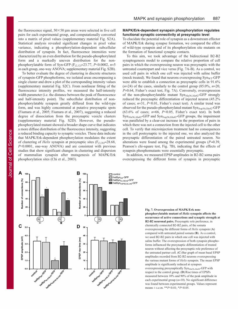
To this aim, we took advantage of the bidirectional B2-B2synaptogenesis model to compare the relative proportion of cellpairs in which the overexpressing neuron was presynaptic with theuntreated counterpart and vice versa (Fig. 7A-B). As a control, weused cell pairs in which one cell was injected with saline buffer(mock treated). We found that neurons overexpressing SynWT-GFPwere able to establish a connection as presynaptic cells in 91.6%(n24) of the cases, similarly to the control group (85.0%, n20,P0.64, Fisher’s exact test, Fig. 7A). Conversely, overexpressionof the non-phosphorylatable mutant SynAla36,Ala42-GFP stronglyreduced the presynaptic differentiation of injected neuron (45.2%of cases; n31, P<0.01, Fisher’s exact test). A similar trend wasobserved for the pseudo-phosphorylated mutant SynAsp36,Glu42-GFP(63.6% of cases; n66, P<0.05, Fisher’s exact test). In bothSynAla36,Ala42-GFP and SynAsp36,Glu42-GFP groups, the impairmentwas paralleled by a clear-cut increase in the proportion of pairs inwhich there was not a connection from the injected cell to the othercell. To verify that microinjection treatment had no consequencesin the cell postsynaptic to the injected one, we also analyzed thepresynaptic differentiation of the paired untreated neuron. Noalterations were found among the experimental groups (P0.39,Pearson’s chi-square test, Fig. 7B), indicating that the effects ofsynapsin phosphomutants were essentially presynaptic.
In addition, we measured EPSP amplitudes in B2-B2 soma pairsoverexpressing the different forms of synapsin in presynaptic
Fig. 7. Overexpression of MAPK/Erk non-phosphorylatable mutant of Helix synapsin affects theoccurrence of active connections and synaptic strength atB2-B2 neuronal pairs. Presynaptic role preference, inchemically connected B2-B2 pairs, of the somataoverexpressing the different forms of Helix synapsin (A)compared with untreated paired somata (B). As a control,we used B2-B2 pairs in which one cell was injected withsaline buffer. The overexpression of both synapsin phospho-forms influenced the presynaptic differentiation of treatedneuron without affecting the presynaptic role preference ofthe untreated partner cell. (C)Bar graph of mean basal EPSPamplitudes recorded from B2-B2 neurons overexpressingthe various mutant forms of Helix synapsin. The mean EPSPamplitude is significantly reduced at synapsesoverexpressing presynaptically SynAla36,Ala42-GFP withrespect to the control group. (D)Rise times of EPSPsmeasured between 10% and 90% of the peak amplitude ineach experimental group (n10). No significant differencewas found between experimental groups. Values representmeans ± s.e.m. **P<0.01; *P<0.05.
Jour
nal o
f Cel
l Sci
ence

888
compartment (Fig. 7C). We found that the mean EPSP amplitudewas not affected by overexpression of SynWT-GFP (3.22±0.60 mV,n15), and was only slightly reduced by the pseudo-phosphorylatedform of synapsin (2.79±0.51 mV, n14), compared with controlcells (3.36±0.50 mV, n33). Conversely, the overexpression of non-phosphorylatable mutant SynAla36,Ala42-GFP significantly reducedbasal EPSP amplitude to about 30% of control values (1.01±0.18mV, n11, F(3,69)2.821, P<0.05, one-way ANOVA followed byBonferroni’s post-hoc test).
To rule out a direct interference of Helix synapsin mutants withthe neurotransmitter release machinery, we measured the EPSP risetime in the four groups of injected neurons (34.68±3.6 ms in controlgroup, 33.84±3.4 ms in SynWT-GFP group, 31.63±3.5 ms inSynAla36,Ala42-GFP group and 33.81±2.2 ms in SynAsp36,Glu42-GFPgroup; Fig. 7D) and they were not statistically different (F(3,36)0.16,P0.92, one-way ANOVA).
Since we observed that the MAPK/Erk pathway affects neuriteoutgrowth, we tested the hypothesis that this action might bemediated by synapsin phosphorylation. To this aim, we measuredneurite outgrowth in single B2 overexpressing neurons. Under theseexperimental conditions, we did not observe any alterations with
respect to uninjected cells (treatment F(3,34)0.09, P0.96 andtreatment by time interaction F(6,68)0.26, P0.95, two-way ANOVAfor repeated measures; Fig. 3B), thus ruling out the possibility thatthe effects of the mutants on the formation of functional synapticcontacts that we observed are related to differences in neuritegrowth.
Taken together, these findings indicate that MAPK/Erk-dependentphosphorylation of Helix synapsin is important for the establishmentof functional synaptic connections through presynaptic mechanismsthat are independent of neurite outgrowth.
Presynaptic MAPK/Erk-dependent synapsinphosphorylation mediates PTP at B2-B2 synapsesTo evaluate the role of synapsin MAPK/Erk-dependentphosphorylation in short-term plasticity, we recorded the inductionand decay of PTP at B2-B2 synapses in which the presynapticcell overexpressed either wild-type or mutant synapsin (Fig. 8A-D). In each synaptic pair, we used as an internal control the B2-B2 synapse in which the uninjected neuron was the presynapticcell. The amplitude of PTP is known to be related to the basalstrength of synaptic connections (Fiumara et al., 2005). Therefore,
Journal of Cell Science 123 (6)
Fig. 8. Presynaptic overexpression of MAPK/Erk phosphorylation mutants of Helix synapsin impairs PTP. (A)Sample electrophysiological recordings ofEPSPs before and 30 seconds after the tetanic stimulation at B2-B2 synapses overexpressing presynaptically either wild-type Helix synapsin or its phosphorylationsite mutants. (B)Time course of EPSP amplitude changes in the four experimental groups. (C)Mean amplitudes of the peak PTP measured 30 seconds aftertetanus. EPSP amplitudes recorded 30 seconds after tetanization in control neurons (n21) and in neurons overexpressing SynWT-GFP (n12) are similar. In theSynAla36,Ala42-GFP group (n10), the peak PTP was reduced to 24.4±4.1% of the control. A less-pronounced decrease (60.2±7.0%; n13) was also observed inneurons overexpressing SynAsp36,Glu42-GFP. Values were normalized to the mean peak PTP amplitude measured in the control group. One-way ANOVA analysisconfirms the highly significant effect of treatment (F(3,52)15.10; P<0.0001). (D)Bar graph of the decay time constants of PTP measured in B2-B2 synapsesshowing that only overexpression of SynAla36,Ala42-GFP significantly reduces .Values represent means ± s.e.m. ***P<0.001; **P<0.01; *P<0.05.
Jour
nal o
f Cel
l Sci
ence
889MAPK and synapsin phosphorylation
for these experiments we selected synapses of comparable strengthin each group (basal EPSP 0.5-5 mV), and we verified that themean amplitudes of the last five pre-tetanic EPSPs werehomogenous among the experimental groups (F(3,52)1.954,P0.13, one-way ANOVA). A two-way ANOVA for repeatedmeasures revealed overall a significant effect of treatment, i.e. theoverexpression of synapsin phosphoforms (F(3,52)8.04, P<0.001),and a significant treatment by time interaction (F(48,832)8.14,P<0.0001; Fig. 8B). In particular, we found that PTP was notaffected by overexpression of SynWT-GFP (peak PTP amplitudeat 30 seconds after tetanus: 545.7±61.4% in SynWT-GFP group,n12, vs 591.2±48.5%, n21, in control synapses). This result isconsistent with our previous observations at Helix C1-B2 synapses(Fiumara et al., 2007). Conversely, overexpression of the non-phosphorylatable mutant SynAla36,Ala42-GFP determined apronounced impairment of PTP. In fact, the peak PTP amplitude(144.0±24.4%, n10) was decreased to 24.4±4.1% of the controlvalue (Fig. 8C) and the amplitude of post-tetanic EPSPs remainedsignificantly reduced for 2 minutes after the tetanus with respectto control (P<0.001, Bonferroni’s post-hoc test). In synapsesoverexpressing the pseudo-phosphorylated SynAsp36,Glu42-GFP, weobserved a less-dramatic reduction in PTP amplitude. The peakmeasured at 30 seconds was 355.8±41.5% (n13), correspondingto 60.2±7.0% of control amplitude (P<0.01, Bonferroni’s post-hoc test).
We also analyzed the time course of PTP decay fitted accordingto a mono-exponential function to estimate the decay constant ()in control and treated synapses. We found a value of 57.8±3.0seconds in the control group, 55.5±3.3 seconds in the SynWT-GFPgroup, 30.9±5.3 seconds in the SynAla36,Ala42-GFP group and64.7±3.3 seconds in the SynAsp36,Glu42-GFP group (Fig. 8D). On thewhole, only the non-phosphorylatable mutant form gave asignificant reduction of PTP decay time (F(3,36)14.62, P<0.0001,one-way ANOVA), whereas overexpression of the pseudo-phosphorylated form of synapsin affected the PTP amplitude butnot its decay. Taken together, these results show that MAPK/Erk-dependent synapsin phosphorylation, besides its role in thegeneration of functional B2-B2 synapses, is also an importantmediator of their short-term plasticity.
DiscussionIn this study, we found that MAPK/Erk-dependent phosphorylationof synapsin has a dual role in regulating both functional synapseformation and activity-dependent short-term plasticity. Thesefindings extend the established role of MAPK/Erk beyond theregulation of nuclear transcription to the modulation of synapticformation and function through a synapsin-dependent mechanism.
A well-characterized role of MAPK/Erk in neurons is theregulation of gene expression upon nuclear translocation (Martinet al., 1997). However, growing experimental evidence suggeststhat this pathway also exerts other fast actions mediated byphosphorylation of synaptic protein substrates (Sweatt, 2004;Boggio et al., 2007). Bioinformatic analysis of Helix, Aplysia, andother gastropod synapsin proteins that display a quite well-conservedprimary sequence, revealed the presence of two putative MAPK/Erkphosphorylation sites in the B-domain, consistent with thelocalization of mammalian synapsin phosphorylation sites 4 and 5.The same sites were predicted to be MAPK/Erk consensus sites inAplysia synapsin (Angers et al., 2002). Interestingly, in an alignmentof four molluscan synapsin proteins, the highest degree of aminoacid identity in B-domain is observed around these residues,
suggesting the high functional importance of this region. Moreover,the two molluscan MAPK/Erk sites align quite well withmammalian sites, despite the low degree of sequence conservationbetween vertebrate and invertebrate synapsin proteins in this region.An in vitro phosphorylation assay validated this prediction,confirming that molluscan synapsin is a MAPK/Erk substrate andthis phosphorylation is strongly reduced by Ser36 to Ala and Ser42to Ala mutations. In addition to the highly conserved PKA-CaMKI/IV phosphorylation site in the A-domain described in ourprevious study (Fiumara et al., 2004), the present findings showthat molluscan synapsin also possesses MAPK/Erk functionalphosphorylation sites.
The MAPK/Erk-synapsin pathway as a mediator offunctional synapse formationMAPK/Erk is one of the major intracellular effectors of neurotrophicfactors and has been implicated in both synaptogenesis and plasticity(Martinez et al., 2008; Vicario-Abejón et al., 1998; Vicario-Abejónet al., 2002; Collin et al., 2001; Tyler and Pozzo-Miller, 2001;Alonso et al., 2004). We have previously found that ganglia-derivedtrophic factors have an essential role in formation of excitatorysynapses between Helix neurons through a trk-dependentmechanism (Fiumara et al., 2005), although the existence ofcanonical neurotrophin-trk signalling in invertebrates is debated(McKay et al., 1999; Chao, 2000; Jaaro et al., 2001). However, therecent identification of neurotrophin and trk receptor orthologuesin Aplysia (Kassabov et al., 2007), indicates a phylogeneticconservation of neurotrophin signalling from gastropods tovertebrates. Here, we found that MAPK/Erk inhibition affectsneurite outgrowth, as well as the formation of functional connectionsinduced by ganglia-derived trophic factors (Fiumara et al., 2005),suggesting that this kinase is a mediator of neurotrophic factoractions in Helix nervous system. These findings indicate aphylogenetically conserved role, from molluscs to mammals, of theneurotrophin-MAPK/Erk pathway in regulating neuronal growthand synaptic connectivity.
Pharmacological inhibition of MAPK/Erk with U0126 caused areduction in the occurrence of functional chemical synapses.Although in principle this effect might also be partially attributedto aspecific U0126 actions on synaptic transmission per se and/oron neuritic growth – rather than on synaptogenesis – several linesof evidence point to a direct involvement of the MAPK/Erk-synapsinpathway in the formation of functional B2-B2 connections. In fact,we did not observe changes in basal transmission after 30 minutesof U0126 application on preformed B2-B2 synapses, despite thealmost complete impairment of PTP that was rapidly and completelyreverted after inhibitor washout. This indicates that U0126 does notaffect basal synaptic transmission in such a manner to prevent thedetection of already established synaptic connections. Thisconclusion is consistent with the results of experiments performedon Aplysia synapses under similar conditions (Khoutorsky and Spira,2009). Moreover, we found that washout of U0126 did not lead toa fast recovery of synaptic strength in B2-B2 neurons chronicallytreated with inhibitor for 24 hours starting at the time of cell-cellpairing. In this case, once the inhibitor was washed out, weobserved a very slow increase in synaptic strength that reachedcontrol levels over a time span of 2 days after washout of theinhibitor, with kinetics similar to those of the control group. Thisslow recovery indicates the formation of new synaptic contacts afterwashout rather than a re-establishment of synaptic transmission aftera transient inhibition. In addition, the measurement of other
Jour
nal o
f Cel
l Sci
ence
890
electrophysiological parameters – such as EPSP rise time – alsorules out the possibility that our experimental manipulations woulddisrupt the function of neurotransmitter release machinery to theextent of preventing the electrophysiological detection of functionalsynaptic contacts.
We found that the presence of the MEK inhibitor U0126 stronglyreduces the neurite growth of B2 neurons. This might indicate thatMAPK/Erk inhibition could affect synapse formation both throughthis growth impairment (leading to a reduction of the cell-cellsurface contact) and through a direct effect on the assembly ofsynaptic structures. However, we also demonstrated that presynapticoverexpression of MAPK/Erk phosphorylation mutants of synapsinregulates the occurrence and the strength of synaptic connectionsthrough a dominant-negative effect over endogenous synapsinwithout affecting neurite outgrowth. This indicates that MAPK-dependent synapsin phosphorylation regulates the occurrence ofchemical synapses through a growth-independent mechanism. Thefact that both non-phosphorylatable and pseudo-phosphorylatedsynapsin mutants had similar effects suggests that cycles ofMAPK/Erk phosphorylation are required for proper activity ofsynapsin during synapse formation. Repeated episodes ofphosphorylation and dephosphorylation might regulate cytoskeletalassembly and vesicle clustering at synaptic terminals, consistentwith the established role of MAPK/Erk phosphorylation inmodulating the synapsin affinity for actin (Jovanovic et al., 1996).
The MAPK/Erk-synapsin pathway as a mediator of short-term homosynaptic plasticityMAPK/Erk has a fundamental role in many forms of long-termsynaptic plasticity in vertebrates (Kawasaki et al., 1999; Robersonet al., 1999; Huang et al., 2000; Zhang et al., 2003) and invertebrates(Bailey et al., 1997; Martin et al., 1997; Sharma et al., 2003), byregulating gene expression upon nuclear translocation. In addition,this kinase has also been implicated in the short-term regulation ofneurotransmitter release and synaptic function through thephosphorylation of cytoplasmic substrates. For instance, MAPK/Erkactivation in presynaptic terminals regulates synaptic vesicledynamics upon AMPA receptor activation (Schenk et al., 2005) orBDNF-induced neurotransmitter release from mammaliansynaptosomes (Jovanovic et al., 2000). We previously found thattarget-dependent modulation of neurotransmitter release from Helixpresynaptic terminals crucially depends on MAPK/Erk activation(Ghirardi et al., 2004). Although MAPK/Erk activity does not appearto be required for short-term heterosynaptic facilitation induced byserotonin at Aplysia sensorimotor synapses (Martin et al., 1997;Purcell et al., 2003; Phares and Byrne, 2005), other studies ininvertebrates show that modulation of short- and long-term synapticplasticity paradigms is mediated by MAPK/Erk (Tancredi et al.,2000; Chin et al., 2002; Roberto et al., 2003; Giachello et al., 2008;Khoutorsky and Spira, 2009). An involvement of MAPK/Erk inshort-term plasticity is also supported by studies in transgenic micethat express a constitutively active form of H-Ras, which exhibitan enhancement of paired-pulse facilitation and long-termpotentiation that is dependent on MAPK/Erk activation (Kushneret al., 2005). Our findings are consistent with this view, showingdirectly that PTP, a specific form of short-term homosynapticenhancement, crucially depends on MAPK/Erk activation, whichmight occur upon intracellular calcium build-up during the tetanus(Impey et al., 1999; Agell et al., 2002) or via cross-talk with othercalcium-dependent pathways (Soderling, 1999; Ferguson and Storm,2004; Shaul and Seger, 2007; Gerits et al., 2008). Another possibility
is that the high-frequency presynaptic activity triggers pre- and/orpost-synaptic release of neurotrophins, which, after binding to pre-and/or post-synaptic trk receptors, ultimately leads to MAPK/Erkactivation (Lu et al., 2002).
PTP is a relatively long-lasting form of short-term homosynapticplasticity that is thought to rely on several concurring mechanismsmodulating several steps of synaptic vesicle cycle (Zucker andRegehr, 2002; Habets and Borst, 2005; Felmy and von Gersdorff,2006). The supply of synaptic vesicles from a reserve to a readilyreleasable pool is one of the key steps that sustain the enhancedneurotransmitter release during PTP (Kuromi and Kidokoro, 2003;Kidokoro et al., 2004). Synapsin proteins are strongly implicatedin maintenance of presynaptic vesicular pools and in the regulationof vesicle mobility among them during short-term plasticity(Humeau et al., 2001; Cousin et al., 2003; Giovedi et al., 2004).Our findings suggest that MAPK/Erk-dependent synapsinphosphorylation is a key regulator of this process. This conclusionis supported primarily by the fact that presynaptic overexpressionof non-phosphorylatable mutant impairs PTP, indicating thatMAPK/Erk-dependent synapsin phosphorylation has a permissiverole for mobilization of synaptic vesicles (Prekeris and Terrian,1997). Accordingly, we observed a higher degree of clustering ofoverexpressed GFP-tagged synapsin mutant compared with thewild-type protein, suggesting a stronger association with vesicles,similarly to what we reported for the PKA-CaMKI/IV site mutant(Fiumara et al., 2007). Both observations are consistent withprevious morphological studies showing that serotonin-induceddispersion of synapsin clusters in Aplysia neurons depends on bothPKA and MAPK/Erk activity (Angers et al., 2002), and that PKAand MAPK/Erk phosphorylation regulates the mobility of synapsinas well as the trafficking of synaptic vesicles in nerve terminalsupon stimulation (Chi et al., 2003). The lack of any obvious effectsof overexpression of synapsin mutants on EPSP kinetics suggeststhat this mechanism does not interfere with the function of thisprotein at post-docking stages of synaptic vesicle cycle (Hilfiker etal., 1998; Hilfiker et al., 2005; Humeau et al., 2001), as also shownfor the PKA-CaMKI/IV site in the A-domain (Fiumara et al., 2007).
The present findings, together with our previous study (Fiumaraet al., 2007), indicate that PTP is associated with the phosphorylationof several synapsin sites by distinct kinases that are concurrentlyactivated by tetanic stimulation. The functional meaning of thesemultiple regulatory events during plasticity might be to fine-tunetrafficking and availability of synaptic vesicles for release to thetype, intensity, and duration of presynaptic activation. Various kinasepathways can be differentially recruited and with specific timecourses, depending on the frequency, the number of presynapticspikes and the presence of heterosynaptic modulators. Interestingly,MAPK/Erk-dependent synapsin mobilization upon presynapticstimulation is frequency dependent (Chi et al., 2003). Therefore,the type and sequence of synapsin phosphorylation reactions mightspecify a change in the functional properties of these proteins thatis appropriate for each type of presynaptic activity pattern.
Materials and MethodsMaterialsAll reagents and materials were of analytical grade and purchased from Sigma (Milan,Italy).
Cell culture and electrophysiologyJuvenile Helix aspersa and Helix pomatia land snails were purchased from localbreeders. Cell cultures were performed as previously described (Ghirardi et al., 1996).Briefly, buccal B2 neurons were isolated and grown under non-adhesive conditions.Floating somata were then paired to form B2-B2 soma-to-soma synapses (Fiumara
Journal of Cell Science 123 (6)
Jour
nal o
f Cel
l Sci
ence

891MAPK and synapsin phosphorylation
et al., 2005). In growth experiments, single B2 somata were plated on poly-L-lysinepretreated dishes and analyzed at 24 and 48 hours as previously described (Milaneseet al., 2008). Conventional intracellular recordings of synaptic activity were performedas previously reported (Fiumara et al., 2005; Fiumara et al., 2007).
Pharmacological treatmentU0126 (Sigma, Milan, Italy), a selective inhibitor of MEK, was used to block theMAPK/Erk cascade pathway. In synaptogenesis experiments, prior to pairing, isolatedB2 neurons were incubated for 2 hours either in 50 M U0126 or in 0.25% DMSO.Cells were subsequently paired in ganglia-conditioned medium (Fiumara et al., 2005)in the presence of either U0126 or DMSO. Electrophysiological recordings wereperformed at 24, 48 and 72 hours after cell-cell pairing.
BioinformaticsPrediction of kinase-specific phosphorylation sites in the amino acid sequence ofHelix synapsin (GenBank AY533823) was performed using the Group-basedPhosphorylation Scoring method, GPS 2.1 (http://gps.biocuckoo.org/online.php)(Xue et al., 2008). A high threshold setting and a cut-off value of 3.757 were chosento minimize the false-positive rate. Phylogenetic analysis of putative phosphorylationsites in the synapsin B-domain was carried out with the multiple alignment softwareAlignX, part of Vector NTI Advance 9 suite (Invitrogen, Carlsbad, CA), based onthe ClustalW algorithm.
Site-directed mutagenesis of GFP-tagged Helix synapsinHelix synapsin (SynWT) sequence subcloned into the expression vector pCS2-mt-GFP(kindly provided by Mike Klymkowsky, University of Colorado, Boulder, CO) wasused as a template for site-directed mutagenesis by QuikChange Site-DirectedMutagenesis Kit (Stratagene, Milan, Italy) following the manufacturer’s procedurewith two antiparallel primers carrying codons for the required substitutions. To removeconsensus phosphorylation sites, Ser36 and Ser42 were replaced with alanine residuesgenerating a non-phosphorylatable mutant (SynAla36,Ala42-GFP). Primers used were:forward, TAAGAAGGGGCCGGCTCCCAGTGCCCCTAACGCACCATCCAAGA;reverse, TCTTGGATGGTGCGTTAGGGGCACTGGGAGCCGGCCCCTTCTTA.Moreover, substitutions of negatively charged aspartate and glutamate residues atpositions 36 and 42 were exploited to mimic the effect of a constitutive phosphory-lation (SynAsp36,Glu42-GFP). Primers used were: forward, TAAGAAGGGGCCG-GATCCCAGTGCCCCTAACGAACCATCCAAGA; reverse, TCTTGGATGGTT -CGTTAGGGGCACTGGGATCCGGCCCCTTCTTA. The presence of mutations wasverified by sequence analysis.
Expression and production of synapsin recombinant proteinsThe wild-type and non-phosphorylatable synapsin mutant were subcloned into pGEX-4T vector and transformed in BL21 cells. Large-scale cultures of Luria broth containingampicillin (100 g/ml) were inoculated with small overnight cultures, grown at 37°Cto log phase and induced with isopropyl -D-thiogalactopyranoside (100 M) for 3-5 hours. GST-synapsin fusion proteins were extracted from bacterial lysates, purifiedto homogeneity by affinity chromatography on glutathione-Sepharose and dialyzedagainst 25 mM Tris-HCl, 50 mM NaCl, pH 7.4. The purified synapsin-GST fusionproteins were then subjected to cleavage with thrombin (1 U/mg fusion protein) for3 hours at 22°C and repurified by a further passage through the glutathione-Sepharosecolumn following the manufacturer’s procedure.
Phosphorylation of wild-type and MAPK/Erk non-phosphorylatablesynapsin mutantFor the analysis of synapsin phosphorylation by activated MAPK/Erk2, equal amountsof either wild-type and non-phosphorylatable mutant (Ser36Ala and Ser42Ala) wereincubated in a buffer containing 20 mM Tris-HCl, pH 7.4, 20 mM MgCl2, 2 mMMnCl2, 0.5 mM dithiothreitol, 0.5 mM Na3VO4, 1.5 g/ml purified activeMAPK/Erk2. The incubation were started by the addition of 50 M [-32P]ATP (2Ci/sample), carried out at 30°C for 30 minutes, and stopped by the addition ofconcentrated sample buffer (Laemmli, 1970) and boiled for 2 minutes. Phosphorylatedsamples were resolved by SDS-PAGE on 10% polyacrylamide gels. Gels were thenfixed, stained with Coomassie brilliant blue and exposed to Kodak X-Omat films.Parallel samples were assessed by immunoblotting with an antibody (G143)recognizing the highly conserved domain A of synapsin.
In vitro transcription and intracellular injection of mRNAsIn vitro transcription was performed according to the protocol of Sahly and colleagues(Sahly et al., 2003) with minor modifications, using the RiboMAX Large Scale RNAProduction System SP6 (Promega, Milan, Italy). The quality of the synthesized mRNAwas checked by agarose gel electrophoresis and spectrophotometrically quantified.Generally, the final concentration of purified mRNAs ranged from 1 to 3 g/l andit was intracellularly injected in B2 somata as previously reported (Fiumara et al.,2007).
Cell imaging and fluorescence analysisCell images were captured with a Monochrome Evolution QE camera(Mediacybernetics, Bethesda, MD) using an Eclipse TE200 inverted microscope(Nikon Instruments, Tokyo, Japan) equipped with phase contrast and epifluorescence
optics. Confocal images were acquired with a Fluoview 300 confocal laser-scanningmicroscope (Olympus, Hamburg, Germany). Quantitative analysis of fluorescencedistribution was performed on selected areas (50�50 m, with an almost completecoverage of the post-synaptic cell) digitally converted into matrices of pixel valuesand plotted using the Bitmap Analysis and the Surface Plot tool of Image Pro Plusversion 6.3 (Mediacybernetics, Bethesda, MD). To analyze the dispersion of GFP-tagged proteins, the Line Profile tool of Image Pro Plus was applied to areas of 20�20pixels (25 m2) encompassing a single cluster, obtaining a plot of the pixel intensityvalues that was fitted by a bell-shaped curve. All figures were assembled usingPhotoshop CS2 version 9.0 software (Adobe Systems, San Jose, CA).
Statistical analysisData were expressed as means ± s.e.m. Statistical analysis was performed usingGraphPad Prism version 5 (GraphPad Software, San Diego, CA). Statisticalsignificance between group means was assessed using Student’s t-test or ANOVAanalysis (one or two-way and with or without repeated measures where appropriate)followed by the Bonferroni post-hoc test. Contingency tests, such as Pearson’s chisquare test or Fisher’s exact test, were performed as indicated. Significance levelswere set at P<0.05.
We are grateful to Claudio Franchino, Antonis Petalotis andDomenico Lombardini for technical assistance. The project wassupported by grants from the Ministry of the University and ResearchGrants (PRIN 2006 grants to P.G.M. and F.B.), Compagnia di San Paolo(to P.G.M. and F.B.), Regione Piemonte (to M.G.). The support ofTelethon-Italy and Fondazione Pierfranco and Luisa Mariani grants (toF.B.) is also acknowledged.
Supplementary material available online athttp://jcs.biologists.org/cgi/content/full/123/6/881/DC1
ReferencesAgell, N., Bachs, O., Rocamora, N. and Villalonga, P. (2002). Modulation of the
Ras/Raf/MEK/ERK pathway by Ca2+, and calmodulin. Cell. Sig. 14, 649-654.Alonso, M., Medina, J. H. and Pozzo-Miller, L. (2004). ERK1/2 activation is necessary
for BDNF to increase dendritic spine density in hippocampal CA1 pyramidal neurons.Learn. Mem. 11, 172-178.
Altrup, U. and Speckmann, E. J. (1994). Identified neuronal individuals in the buccalganglia of Helix pomatia. Neurosci. Behav. Physiol. 24, 23-32.
Angers, A., Fioravante, D., Chin, J., Cleary, L. J., Bean, A. J. and Byrne, J. H. (2002).Serotonin stimulates phosphorylation of Aplysia synapsin and alters its subcellulardistribution in sensory neurons. J. Neurosci. 22, 5412-5422.
Bailey, C. H., Kaang, B. K., Chen, M., Martin, K. C., Lim, C. S., Casadio, A. andKandel, E. R. (1997). Mutation in the phosphorylation sites of MAP kinase blockslearning-related internalization of apCAM in Aplysia sensory neurons. Neuron 18, 913-924.
Boggio, E. M., Putignano, M., Sassoe-Pognetto, M., Pizzorusso, T. and Giustetto, M.(2007). Visual stimulation activates ERK in synaptic and somatic compartments of ratcortical neurons with parallel kinetics. PLoS ONE 2, e604.
Chao, M. V. (2000). Trophic factors: An evolutionary cul-de-sac or door into higher neuronalfunction? J. Neurosci. Res. 59, 353-355.
Chi, P., Greengard, P. and Ryan, T. A. (2001). Synapsin dispersion and reclustering duringsynaptic activity. Nat. Neurosci. 4, 1187-1193.
Chi, P., Greengard, P. and Ryan, T. A. (2003). Synaptic vesicle mobilization is regulatedby distinct synapsin I phosphorylation pathways at different frequencies. Neuron 38, 69-78.
Chin, J., Angers, A., Cleary, L. J., Eskin, A. and Byrne, J. H. (2002). Transforminggrowth factor beta 1 alters synapsin distribution and modulates synaptic depression inAplysia. J. Neurosc. 22, RC220.
Collin, C., Vicario-Abejón, C., Rubio, M. E., Wenthold, R. J., McKay, R. D. G. andSegal, M. (2001). Neurotrophins act at presynaptic terminals to activate synapses amongcultured hippocampal neurons. Eur. J. Neurosci. 13, 1273-1282.
Cousin, M. A., Malladi, C. S., Tan, T. C., Raymond, C. R., Smillie, K. J. and Robinson,P. J. (2003). Synapsin I-associated phosphatidylinositol 3-kinase mediates synapticvesicle delivery to the readily releasable pool. J. Biol. Chem. 278, 29065-29071.
De Camilli, P., Harris, S. M., Huttner, W. B. and Greengard, P. (1983). Synapsin I(Protein I), a nerve terminal-specific phosphoprotein. II. Its specific association withsynaptic vesicles demonstrated by immunocytochemistry in agarose-embeddedsynaptosomes. J. Cell Biol. 96, 1355-1373.
Dimitropoulou, A. and Bixby, J. L. (2000). Regulation of retinal neurite growth byalterations in MAPK/ERK kinase (MEK) activity. Brain Res. 858, 205-214.
Encinas, M., Iglesias, M., Llecha, N. and Comella, J. X. (1999). Extracellular-regulatedkinases and phosphatidylinositol 3-kinase are involved in brain-derived neurotrophicfactor-mediated survival and neuritogenesis of the neuroblastoma cell line SH-SY5Y. J.Neurochem. 73, 1409-1421.
Felmy, F. and von Gersdorff, H. (2006). Late switch for post-tetanic potentiation: Onceagain it’s Ca2+. Focus on “An increase in calcium influx contributes to post-tetanicpotentiation at the rat calyx of held synapse”. J. Neurophysiol. 96, 2840-2841.
Ferguson, G. D. and Storm, D. R. (2004). Why calcium-stimulated adenylyl cyclases?Physiology 19, 271-276.
Jour
nal o
f Cel
l Sci
ence
892
Fisher, S. A., Fischer, T. M. and Carew, T. J. (1997). Multiple overlapping processesunderlying short-term synaptic enhancement. Trends Neurosci. 20, 170-177.
Fiumara, F., Giovedi, S., Menegon, A., Milanese, C., Merlo, D., Montarolo, P. G.,Valtorta, F., Benfenati, F. and Ghirardi, M. (2004). Phosphorylation by cAMP-dependent protein kinase is essential for synapsin-induced enhancement ofneurotransmitter release in invertebrate neurons. J. Cell Sci. 117, 5145-5154.
Fiumara, F., Leitinger, G., Milanese, C., Montarolo, P. G. and Ghirardi, M. (2005).In vitro formation and activity-dependent plasticity of synapses between Helix neuronsinvolved in the neural control of feeding and withdrawal behaviors. Neurosci. 134, 1133-1151.
Fiumara, F., Milanese, C., Corradi, A., Giovedi, S., Leitinger, G., Menegon, A.,Montarolo, P. G., Benfenati, F. and Ghirardi, M. (2007). Phosphorylation of synapsindomain A is required for post-tetanic potentiation. J. Cell Sci. 120, 3228-3237.
Gerits, N., Kostenko, S., Shiryaev, A., Johannessen, M. and Moens, U. (2008). Relationsbetween the mitogen-activated protein kinase and the cAMP-dependent protein kinasepathways: Comradeship and hostility. Cell. Signal. 20, 1592-1607.
Ghirardi, M., Casadio, A., Santarelli, L. and Montarolo, P. G. (1996). Aplysiahemolymph promotes neurite outgrowth and synaptogenesis of identified Helix neuronsin cell culture. Invert. Neurosci. 2, 41-49.
Ghirardi, M., Benfenati, F., Giovedi, S., Fiumara, F., Milanese, C. and Montarolo, P.G. (2004). Inhibition of neurotransmitter release by a nonphysiological target requiresprotein synthesis and involves cAMP-dependent and mitogen-activated protein kinases.J. Neurosci. 24, 5054-5062.
Giachello, C. N. G., Fiumara, F., Milanese, C., Corradi, A., Leitinger, G., Benfenati,F., Ghirardi, M. and Montarolo, P. G. (2008). The role of MAPKs synapsinphosphorylation in the expression of post-tetanic potentiation in Helix neurons. FENSAbstracts. 4, 045.6.
Giovedi, S., Vaccaro, P., Valtorta, F., Darchen, F., Greengard, P., Cesareni, G. andBenfenati, F. (2004). Synapsin is a novel Rab3 effector protein on small synaptic vesicles-I. Identification and characterization of the synapsin I-Rab3 interactions in vitro and inintact nerve terminals. J. Biol. Chem. 279, 43760-43768.
Greengard, P., Valtorta, F., Czernik, A. J. and Benfenati, F. (1993). Synaptic vesiclephosphoproteins and regulation of synaptic function. Science 259, 780-785.
Habets, R. L. P. and Borst, J. G. G. (2005). Post-tetanic potentiation in the rat calyx ofHeld synapse. J. Physiol. 564, 173-187.
Hans, A., Bajramovic, J. J., Syan, S., Perret, E., Dunia, I., Brahic, M. and Gonzalez-Dunia, D. (2004). Persistent, non-cytolytic infection of neurons by Borna disease virusinterferes with ERK 1/2 signaling and abrogates BDNF-induced synaptogenesis. FASEBJ. 18, 863-865.
Hilfiker, S., Schweizer, F. E., Kao, H. T., Czernik, A. J., Greengard, P. and Augustine,G. J. (1998). Two sites of action for synapsin domain E in regulating neurotransmitterrelease. Nat. Neurosci. 1, 29-35.
Hilfiker, S., Pieribone, V. A., Czernik, A. J., Kao, H. T., Augustine, G. J. and Greengard,P. (1999). Synapsins as regulators of neurotransmitter release. Phil. Trans. R. Soc. Lond.B Biol. Sci. 354, 269-279.
Hilfiker, S., Benfenati, F., Doussau, F. D. R., Nairn, A. C., Czernik, A. J., Augustine,G. J. and Greengard, P. (2005). Structural domains involved in the regulation oftransmitter release by synapsins. J. Neurosci. 25, 2658-2669.
Hirokawa, N., Sobue, K., Kanda, K., Harada, A. and Yorifuji, H. (1989). Thecytoskeletal architecture of the presynaptic terminal and molecular structure of synapsin1. J. Cell Biol. 108, 111-126.
Huang, E. J. and Reichardt, L. F. (2001). Neurotrophins: Roles in neuronal developmentand function. Ann. Rev. Neurosci. 24, 677-736.
Huang, Y. Y., Martin, K. C. and Kandel, E. R. (2000). Both protein kinase A and mitogen-activated protein kinase are required in the amygdala for the macromolecular synthesis-dependent late phase of long-term potentiation. J. Neurosci. 20, 6317-6325.
Humeau, Y., Doussau, F., Vitiello, F., Greengard, P., Benfenati, F. and Poulain, B. (2001).Synapsin controls both reserve and releasable synaptic vesicle pools during neuronalactivity and short-term plasticity in Aplysia. J. Neurosci. 21, 4195-4206.
Huttner, W. B., Schiebler, W., Greengard, P. and De Camilli, P. (1983). Synapsin I(protein I), a nerve terminal-specific phosphoprotein. III. Its association with synapticvesicles studied in a highly purified synaptic vesicle preparation. J. Cell Biol. 96, 1374-1388.
Impey, S., Obrietan, K. and Storm, D. R. (1999). Making new connections: Role of ERKMAP kinase signaling in neuronal plasticity. Neuron 23, 11-14.
Jaaro, H., Beck, G., Conticello, S. G. and Fainzilber, M. (2001). Evolving better brains:a need for neurotrophins? Trends Neurosci. 24, 79-85.
Jovanovic, J. N., Benfenati, F., Siow, Y. L., Sihra, T. S., Sanghera, J. S., Pelech, S. L.,Greengard, P. and Czernik, A. J. (1996). Neurotrophins stimulate phosphorylation ofsynapsin I by MAP kinase and regulate synapsin I-actin interactions. Proc. Natl. Acad.Sci. USA 93, 3679-3683.
Jovanovic, J. N., Czernik, A. J., Fienberg, A. A., Greengard, P. and Sihra, T. S. (2000).Synapsins as mediators of BDNF-enhanced neurotransmitter release. Nat. Neurosci. 3,323-329.
Kao, H. T., Porton, B., Hilfiker, S., Stefani, G., Pieribone, V. A., DeSalle, R. andGreengard, P. (1999). Molecular evolution of the synapsin gene family. J. Exp. Zool.285, 360-377.
Kassabov, S. R., Monje, F. J., Fiumara, F. and Kandel, E. R. (2007). Identification ofApTrk, a Trk receptor homolog expressed in Aplysia sensory and motor neurons criticalfor the induction and maintenance of secretion dependent long term facilitation. Soc.Neurosci. Abstract 473, 6.
Kawasaki, H., Fujii, H., Gotoh, Y., Morooka, T., Shimohama, S., Nishida, E. andHirano, T. (1999). Requirement for mitogen-activated protein kinase in cerebellar longterm depression. J. Biol. Chem. 274, 13498-13502.
Khoutorsky, A. and Spira, M. E. (2009). Activity-dependent calpain activation plays acritical role in synaptic facilitation and post-tetanic potentiation. Learn. Mem. 16, 129-141.
Kidokoro, Y., Kuromi, H., Delgado, R., Maureira, C., Oliva, C. and Labarca, P. (2004).Synaptic vesicle pools and plasticity of synaptic transmission at the Drosophila synapse.Brain Res. Brain Res. Rev. 47, 18-32.
Kim, B., Leventhal, P. S., Saltiel, A. R. and Feldman, E. L. (1997). Insulin-like growthfactor-I-mediated neurite outgrowth in vitro requires mitogen-activated protein kinaseactivation. J. Biol. Chem. 272, 21268-21273.
Kurihara, S., Hakuno, F. and Takahashi, S. (2000). Insulin-like growth factor-I-dependent signal transduction pathways leading to the induction of cell growth anddifferentiation of human neuroblastoma cell line SH-SY5Y: The roles of MAP kinasepathway and PI 3-kinase pathway. Endocrine J. 47, 739-751.
Kuromi, H. and Kidokoro, Y. (2003). Two synaptic vesicle pools, vesicle recruitmentand replenishment of pools at the Drosophila neuromuscular junction. J. Neurocytol.32, 551-565.
Kushner, S. A., Elgersma, Y., Murphy, G. G., Jaarsma, D., Hojjati, M. R., Cui, Y. J.,LeBoutillier, J. C., Marrone, D. F., Choi, E. S., De Zeeuw, C. I. et al. (2005).Modulation of presynaptic plasticity and learning by the H-ras/extracellular signal-regulated kinase/synapsin I signaling pathway. J. Neurosci. 25, 9721-9734.
Laemmli, U. K. (1970). Cleavage of structural proteins during the assembly of the headof bacteriophage T4. Nature 227, 680-685.
Lu, Q., Ebling, H., Mittler, J., Baur, W. E. and Karas, R. H. (2002). MAP kinase mediatesgrowth factor-induced nuclear translocation of estrogen receptor alpha. FEBS Lett. 516,1-8.
Martin, K. C., Michael, D., Rose, J. C., Barad, M., Casadio, A., Zhu, H. X. and Kandel,E. R. (1997). MAP kinase translocates into the nucleus of the presynaptic cell and isrequired for long-term facilitation in Aplysia. Neuron 18, 899-912.
Martinez, A., Alcantara, S., Borrell, V., Del Rio, J. A., Blasi, J., Otal, R., Campos, N.,Boronat, A., Barbacid, M., Silos-Santiago, I. et al. (1998). TrkB and TrkC signalingare required for maturation and synaptogenesis of hippocampal connections. J. Neurosci.18, 7336-7350.
McKay, S. E., Purcell, A. L. and Carew, T. J. (1999). Regulation of synaptic functionby neurotrophic factors in vertebrates and invertebrates: Implications for developmentand learning. Learn. Mem. 6, 193-215.
Milanese, C., Fiumara, F., Bizzoca, A., Giachello, C., Leitinger, G., Gennarini, G.,Montarolo, P. G. and Ghirardi, M. (2008). F3/contactin-related proteins in Helixpomatia nervous tissue (HCRPs): Distribution and function in neurite growth andneurotransmitter release. J. Neurosci. Res. 86, 821-831.
Naruo, H., Onizuka, S., Prince, D., Takasaki, M. and Syed, N. I. (2005). Sevofluraneblocks cholinergic synaptic transmission postsynaptically but does not affect sbort-termpotentiation. Anesthesiology 102, 920-928.
Phares, G. A. and Byrne, J. H. (2005). Analysis of 5-HT-induced short-term facilitationat Aplysia sensorimotor synapse during bursts: Increased synaptic gain that does notrequire ERK activation. J. Neurophysiol. 94, 871-877.
Pieribone, V. A., Shupliakov, O., Brodin, L., Hilfiker-Rothenfluh, S., Czernik, A. J.and Greengard, P. (1995). Distinct pools of synaptic vesicles in neurotransmitter release.Nature 375, 493-497.
Prekeris, R. and Terrian, D. M. (1997). Brain myosin V is a synaptic vesicle-associatedmotor protein: Evidence for a Ca2+-dependent interaction with the synaptobrevin-synaptophysin complex. J. Cell Biol. 137, 1589-1601.
Purcell, A. L., Sharma, S. K., Bagnall, M. W., Sutton, M. A. and Carew, T. J. (2003).Activation of a tyrosine kinase-MAPK cascade enhances the induction of long-termsynaptic facilitation and long-term memory in Aplysia. Neuron 37, 473-484.
Roberson, E. D., English, J. D., Adams, J. P., Selcher, J. C., Kondratick, C. and Sweatt,J. D. (1999). The mitogen-activated protein kinase cascade couples PKA and PKC tocAMP response element binding protein phosphorylation in area CA1 of hippocampus.J. Neurosci. 19, 4337-4348.
Roberto, M., Nelson, T. E., Ur, C. L., Brunelli, M., Sanna, P. P. and Gruol, D. L. (2003).The transient depression of hippocampal CA1 LTP induced by chronic intermittent ethanolexposure is associated with an inhibition of the MAP kinase pathway. Euro. J. Neurosci.17, 1646-1654.
Rosahl, T. W., Spillane, D., Missler, M., Herz, J., Selig, D. K., Wolff, J. R., Hammer,R. E., Malenka, R. C. and Sudhof, T. C. (1995). Essential functions of synapsin-I andsynapsin-II in synaptic vesicle regulation. Nature 375, 488-493.
Sahly, I., Erez, H., Khoutorsky, A., Shapira, E. and Spira, M. E. (2003). Effectiveexpression of the green fluorescent fusion proteins in cultured Aplysia neurons. J.Neurosci. Meth. 126, 111-117.
Schenk, U., Menna, E., Kim, T., Passafaro, M., Chang, S., De Camilli, P. and Matteoli,M. (2005). A novel pathway for presynaptic mitogen-activated kinase activation viaAMPA receptors. J. Neurosci. 25, 1654-1663.
Sharma, S. K., Sherff, C. M., Shobe, J., Bagnall, M. W., Sutton, M. A. and Carew, T.J. (2003). Differential role of mitogen-activated protein kinase in three distinct phasesof memory for sensitization in Aplysia. J. Neurosci. 23, 3899-3907.
Shaul, Y. D. and Seger, R. (2007). The MEK/ERK cascade: From signaling specificity todiverse functions. Biochimica Et Biophysica Acta-Molecular Cell Research 1773, 1213-1226.
Soderling, T. R. (1999). The Ca2+-calmodulin-dependent protein kinase cascade. Trendsin Biochem. Sci. 24, 232-236.
Sweatt, J. D. (2004). Mitogen-activated protein kinases in synaptic plasticity and memory.Curr. Opin. Neurobiol. 14, 311-317.
Tancredi, V., D’Antuono, M., Cafe, C., Giovedi, S., Bue, M. C., D’Arcangelo, G., Onofri,F. and Benfenati, F. (2000). The inhibitory effects of interleukin-6 on synaptic plasticity
Journal of Cell Science 123 (6)
Jour
nal o
f Cel
l Sci
ence

893MAPK and synapsin phosphorylation
in the rat hippocampus are associated with an inhibition of mitogen-activated proteinkinase ERK. J. Neurochem. 75, 634-643.
Tyler, W. J. and Pozzo-Miller, L. D. (2001). BDNF enhances quantal neurotransmitterrelease and increases the number of docked vesicles at the active zones of hippocampalexcitatory synapses. J. Neurosci. 21, 4249-4258.
Vicario-Abejón, C., Collin, C., McKay, R. D. G. and Segal, M. (1998). Neurotrophinsinduce formation of functional excitatory and inhibitory synapses between culturedhippocampal neurons. J. Neurosci. 18, 7256-7271.
Vicario-Abejón, C., Owens, D., McKay, R. and Segal, M. (2002). Role of neurotrophinsin central synapse formation and stabilization. Nat. Rev. Neurosci. 3, 965-974.
Xue, Y., Ren, J., Gao, X. J., Jin, C. J., Wen, L. P. and Yao, X. B. (2008). GPS 2.0, atool to predict kinase-specific phosphorylation sites in hierarchy. Mol. Cell. Proteomics7, 1598-1608.
Zhang, J. J., Okutani, F., Inoue, S. and Kaba, H. (2003). Activation of the mitogen-activated protein kinase/extracellular signal-regulated kinase signaling pathway leadingto cyclic AMP response element-binding protein phosphorylation is required for thelong-term facilitation process of aversive olfactory learning young rats. Neuroscience121, 9-16.
Zucker, R. S. and Regehr, W. G. (2002). Short-term synaptic plasticity. Ann. Rev. Physiol.64, 355-405.
Jour
nal o
f Cel
l Sci
ence