Prophylaxis of post-ERCP pancreatitis: European …hotmail.com Guideline 799 Abbreviations! CT...
17
Prophylaxis of post-ERCP pancreatitis: European Society of Gastrointestinal Endoscopy (ESGE) Guideline – Updated June 2014 Authors Jean-Marc Dumonceau 1 , Angelo Andriulli 2 , B. Joseph Elmunzer 3 , Alberto Mariani 4 , Tobias Meister 5 , Jacques Deviere 6 , Tomasz Marek 7 , Todd H. Baron 8 , Cesare Hassan 9 , Pier A. Testoni 4 , Christine Kapral 10 Institutions Institutions are listed at the end of article. Bibliography DOI http://dx.doi.org/ 10.1055/s-0034-1377875 Published online: 22.8.2014 Endoscopy 2014; 46: 799–815 © Georg Thieme Verlag KG Stuttgart · New York ISSN 0013-726X Corresponding author Jean-Marc Dumonceau, MD PhD Gedyt Endoscopy Center Beruti 2347 (C1117AAA) Buenos Aires Argentina Fax: +52886100 [email protected] Guideline 799 Abbreviations ! CT computed tomography DGW double guidewire EPBD endoscopic papillary balloon dilation ESGE European Society of Gastrointestinal Endoscopy ERCP endoscopic retrograde cholangio- pancreatography NSAID nonsteroidal anti-inflammatory drug PEP post-ERCP pancreatitis PGW pancreatic guidewire RCT randomized controlled trial SOD sphincter of Oddi dysfunction SOM sphincter of Oddi manometry ULN upper limit of normal 1. Introduction ! The Guideline on prophylaxis of post-ERCP pan- creatitis (PEP) issued by the European Society of Gastrointestinal Endoscopy (ESGE) in 2010 aimed to provide a qualified basis for gastrointestinal endoscopists to take measures to minimize the incidence and severity of PEP [1]. Shortly before the publication of the ESGE Guideline, nonsteroi- dal anti-inflammatory drugs (NSAIDs) were re- portedly rarely used in clinical practice for pre- vention of PEP (16% of respondents to a survey performed in June 2009), and this was attributed by survey participants to the lack of sufficient data [2]. Similarly, in an Austrian nationwide ERCP survey, PEP prophylaxis was administered Dumonceau Jean-Marc et al. Updated ESGE Guideline for PEP prophylaxis … Endoscopy 2014; 46: 799–815 This Guideline is an official statement of the European Society of Gastrointestinal Endoscopy (ESGE). It addresses the prophylaxis of post-endoscopic retrograde cholangiopancreatography (post-ERCP) pancreatitis. Main recommendations 1 ESGE recommends routine rectal administra- tion of 100 mg of diclofenac or indomethacin immediately before or after ERCP in all patients without contraindication. In addition to this, in the case of high risk for post-ERCP pancreatitis (PEP), the placement of a 5-Fr prophylactic pan- creatic stent should be strongly considered. Sub- lingually administered glyceryl trinitrate or 250 μg somatostatin given in bolus injection might be considered as an option in high risk cases if nonsteroidal anti-inflammatory drugs (NSAIDs) are contraindicated and if prophylactic pancre- atic stenting is not possible or successful. 2 ESGE recommends keeping the number of cannulation attempts as low as possible. 3 ESGE suggests restricting the use of a pancre- atic guidewire as a backup technique for biliary cannulation to cases with repeated inadvertent cannulation of the pancreatic duct; if this meth- od is used, deep biliary cannulation should be attempted using a guidewire rather than the contrast-assisted method and a prophylactic pancreatic stent should be placed. 4 ESGE suggests that needle-knife fistulotomy should be the preferred precut technique in pa- tients with a bile duct dilated down to the papil- la. Conventional precut and transpancreatic sphincterotomy present similar success and complication rates; if conventional precut is se- lected and pancreatic cannulation is easily ob- tained, ESGE suggests attempting to place a small-diameter (3-Fr or 5-Fr) pancreatic stent to guide the cut and leaving the pancreatic stent in place at the end of ERCP for a minimum of 12 – 24 hours. 4 ESGE does not recommend endoscopic papil- lary balloon dilation as an alternative to sphinc- terotomy in routine ERCP, but it may be advan- tageous in selected patients; if this technique is used, the duration of dilation should be longer than 1 minute.
Transcript of Prophylaxis of post-ERCP pancreatitis: European …hotmail.com Guideline 799 Abbreviations! CT...
Prophylaxis of post-ERCP pancreatitis:European Society of Gastrointestinal Endoscopy (ESGE)Guideline – Updated June 2014
Authors Jean-Marc Dumonceau1, Angelo Andriulli2, B. Joseph Elmunzer3, Alberto Mariani4, Tobias Meister5, Jacques Deviere6,Tomasz Marek7, Todd H. Baron8, Cesare Hassan9, Pier A. Testoni4, Christine Kapral10
Institutions Institutions are listed at the end of article.
BibliographyDOI http://dx.doi.org/10.1055/s-0034-1377875Published online: 22.8.2014Endoscopy 2014; 46: 799–815© Georg Thieme Verlag KGStuttgart · New YorkISSN 0013-726X
Corresponding authorJean-Marc Dumonceau,MD PhDGedyt Endoscopy CenterBeruti 2347 (C1117AAA)Buenos AiresArgentinaFax: [email protected]
Guideline 799
Abbreviations!
CT computed tomographyDGW double guidewireEPBD endoscopic papillary balloon dilationESGE European Society of Gastrointestinal
EndoscopyERCP endoscopic retrograde cholangio-
pancreatographyNSAID nonsteroidal anti-inflammatory drugPEP post-ERCP pancreatitisPGW pancreatic guidewireRCT randomized controlled trialSOD sphincter of Oddi dysfunctionSOM sphincter of Oddi manometryULN upper limit of normal
1. Introduction!
The Guideline on prophylaxis of post-ERCP pan-creatitis (PEP) issued by the European Society ofGastrointestinal Endoscopy (ESGE) in 2010 aimedto provide a qualified basis for gastrointestinalendoscopists to take measures to minimize theincidence and severity of PEP [1]. Shortly beforethe publication of the ESGE Guideline, nonsteroi-dal anti-inflammatory drugs (NSAIDs) were re-portedly rarely used in clinical practice for pre-vention of PEP (16% of respondents to a surveyperformed in June 2009), and this was attributedby survey participants to the lack of sufficientdata [2]. Similarly, in an Austrian nationwideERCP survey, PEP prophylaxis was administered
Dumonceau Jean-Marc et al. Updated ESGE Guideline for PEP prophylaxis… Endoscopy 2014; 46: 799–815
This Guideline is an official statement of the European Society of Gastrointestinal Endoscopy (ESGE).It addresses the prophylaxis of post-endoscopic retrograde cholangiopancreatography (post-ERCP)pancreatitis.
Main recommendations1 ESGE recommends routine rectal administra-tion of 100mg of diclofenac or indomethacinimmediately before or after ERCP in all patientswithout contraindication. In addition to this, inthe case of high risk for post-ERCP pancreatitis(PEP), the placement of a 5-Fr prophylactic pan-creatic stent should be strongly considered. Sub-lingually administered glyceryl trinitrate or 250µg somatostatin given in bolus injection mightbe considered as an option in high risk cases ifnonsteroidal anti-inflammatory drugs (NSAIDs)are contraindicated and if prophylactic pancre-atic stenting is not possible or successful.2 ESGE recommends keeping the number ofcannulation attempts as low as possible.3 ESGE suggests restricting the use of a pancre-atic guidewire as a backup technique for biliarycannulation to cases with repeated inadvertentcannulation of the pancreatic duct; if this meth-od is used, deep biliary cannulation should be
attempted using a guidewire rather than thecontrast-assisted method and a prophylacticpancreatic stent should be placed.4 ESGE suggests that needle-knife fistulotomyshould be the preferred precut technique in pa-tients with a bile duct dilated down to the papil-la. Conventional precut and transpancreaticsphincterotomy present similar success andcomplication rates; if conventional precut is se-lected and pancreatic cannulation is easily ob-tained, ESGE suggests attempting to place asmall-diameter (3-Fr or 5-Fr) pancreatic stentto guide the cut and leaving the pancreatic stentin place at the end of ERCP for a minimum of12–24 hours.4 ESGE does not recommend endoscopic papil-lary balloon dilation as an alternative to sphinc-terotomy in routine ERCP, but it may be advan-tageous in selected patients; if this technique isused, the duration of dilation should be longerthan 1 minute.

in only 4.0% of patients in 2010 and in 7.0% of patients in 2011[3]. A more recent survey from the UK found that the proportionof endoscopists using NSAIDs had increased to 34.6% in 2012 [4].Obviously, prophylactic measures against PEP are still greatly un-derused in daily clinical practice. At the same time, PEP is still themost frequent and severe complication encountered followingERCP.New evidence that has become available since the publication ofthe ESGE Guideline in 2010 is discussed in the present updateand new recommendations are issued.
2.Methods!
ESGE commissioned this update of the Guideline on preventionof PEP. Methods similar to those used in the previous Guidelinewere applied [1]. A literature search of PubMed/MEDLINE, asearch using the Cochrane Library, Embase, and the internet wasperformed to identify publications since 2009 on this topic. Thesearch focused on fully published prospective studies, particular-ly randomized controlled trials (RCTs) and meta-analyses. Retro-spective analyses and pilot studies were also included if they ad-dressed topics not covered in the prospective studies.Thereafter, the commissioned authors met once and subsequent-ly developed the updated Guideline. The Guideline committeechairs (C.K., J.M.D.) worked with the subgroup leaders (C.K., T.M.,A.A., T.M., P.T., T.B., J.M.D.) who developed draft proposals thatwere distributed and reviewed electronically. In May 2014, adraft prepared by C.K. and J.M.D. was sent to all group members.After agreement on a final version, the manuscript was sent to allindividual ESGEmembers and individual ESGEmember societies,and was reviewed by two experts selected by the ESGE Govern-ing Board. After incorporation of comments, the manuscript wasthen sent to the journal Endoscopy for publication. The finalwording of the Guideline document was agreed by all commis-sioned authors.
3.Definitions!
▶ Statement 2010:None.▶ Statement 2014:Two definitions of PEP may currently be used, neither of thesebeing ideal in the setting of PEP: the consensus definition andgrading of severity of PEP according to Cotton et al. and themore recent revised Atlanta international consensus definitionand classification of acute pancreatitis.Background:The consensus definition and grading of severity of PEP devel-oped by Cotton et al. has been used for>20 years [5]. It has al-lowed standardized reporting of the incidence and severity ofPEP. Post-ERCP pancreatitis was originally defined as “clinicalpancreatitis with amylase at least three times the upper limit ofnormal at more than 24 hours after the procedure, requiring hos-pital admission or a prolongation of planned admission.” Variousmodifications were introduced by Freeman et al. who proposedusing lipase as a possible alternative to amylase and defining clin-ical pancreatitis as “new or worsened abdominal pain,” hencetaking into account patients who undergo ERCP in the setting ofacute pancreatitis or of a flare of chronic pancreatitis [6]. Thegrading system for the severity of PEP by the consensus defini-
tion is not ideal as it is mainly based on the length of hospitaliza-tion.New information since 2009:The Atlanta classification of acute pancreatitis was updated in2012 [7]. Although this classification provides clear definitionsto classify acute pancreatitis, its limitations include the fact thatit was not primarily developed to define PEP. Also, the benefit ofa contrast-enhanced computed tomography (CT) scan has notbeen demonstrated in the setting of PEP (contrast-enhanced CTscan is required if abdominal pain suggests strongly that acutepancreatitis is present, but the serum amylase and/or lipase ac-tivity is less than three times the upper limit of normal [ULN]).According to this classification, the diagnosis of PEP requirestwo of the three following criteria: (i) abdominal pain consistentwith acute pancreatitis (acute onset of a persistent, severe, epi-gastric pain often radiating to the back); (ii) serum lipase or amy-lase activity at least three times greater than the ULN; and (iii)characteristic findings of acute pancreatitis on contrast-en-hanced CT and, less commonly, magnetic resonance imaging ortransabdominal ultrasonography. This classification definesthree degrees of severity based on the presence or absence of or-gan failure (plus its duration) and of local or systemic complica-tions.A prospective study has shown the two definitions presentedabove to be poorly correlated [8].The Pancreatitis Across Nations Clinical Research and EducationAlliance (PANCREA) has defined four degrees of severity for pan-creatitis, based on the presence or absence of complications, bothlocal (necrosis of the pancreas and/or peripancreatic tissue) andsystemic (cardiovascular, renal, or respiratory organ failure) [9].
4. Incidence, risk factors, and severity of PEP!
4.1. Incidence▶ Statement 2010:Pancreatitis is the most frequent complication after ERCP with anincidence of 3.5% in unselected patients; it is of mild or moderateseverity in approximately 90% of cases.▶ Statement 2014:No changes.Background:Data on incidence rate and severity of PEP were mainly based ona systematic review of 21 prospective studies involving morethan 16 000 patients [10]. Post-ERCP pancreatitis was found tobe the most frequent complication following ERCP, with an inci-dence of 3.47% (95% confidence interval [95%CI] 3.19%–3.75%).Based upon data from studies that have included unselected pa-tients, PEP is mild, moderate, and severe in 45%, 44%, and 11%of cases, respectively. Death occurs in 3% of cases of PEP (95%CI1.65%–4.51%) [10].New information since 2009:Few new data have become available and these have yielded sim-ilar results. In an ERCP benchmarking program, PEP was reportedto occur in 4.2% of 13 513 unselected procedures [3]. In a retro-spective study of 886 procedures, 39 patients (4.4%) were diag-nosed with pancreatitis, of mild moderate, and severe type in69%, 23%, and 8%, respectively [11].
Dumonceau Jean-Marc et al. Updated ESGE Guideline for PEP prophylaxis… Endoscopy 2014; 46: 799–815
Guideline800

4.2.Risk stratification4.2.1 Hospital and endoscopist volume for ERCP▶ Statement 2010:There is no evidence that hospital ERCP volume has an influenceon the incidence of PEP; data about a potential relationship be-tween PEP incidence and endoscopist case volume are conflict-ing. Low annual case volumes, of endoscopists and centers, areassociated with higher ERCP failure rates (Evidence level 2+).▶ Statement 2014:No changes.Background:Factors that may affect the outcome of ERCP that are specificallyrelated to hospital procedure volume include availability ofequipment and adequacy of anesthesia, endoscopic and radiolo-gic support, and nursing assistance. The number of ERCPs per-formed in many centers is not as high as commonly believed: inthree large (regional or national) studies, the median annualnumber of ERCPs was between 49 and 235 [12–14]. In one largestudy, the median annual number of ERCPs per endoscopist was111 and 40% of endoscopists performed fewer than 50 sphinc-terotomies/year [15].Multivariate analyses from two prospective audits performed inEngland and Italy (66 and 9 centers, respectively) found no signif-icant association between annual hospital volume of ERCPs andincidence of PEP [16,17].
New information since 2009:A prospective Swedish study of 12 718 procedures showed nosignificant difference among PEP rates in centers with low (<100ERCPs/year), medium, and high (>500 ERCPs/year) volumes [18].In a prospective multicenter study that included 3635 ERCPs in11 high volume (>200 ERCPs/year) and 10 low volume centers(median of 275 and 45 ERCPs/year, respectively), there was nosignificant difference in the incidence of PEP (3.9% vs. 3.1%)[19]. However, these results were confounded because a higherproportion of patients at high risk of PEP was treated in high vol-ume centers. In this study, the PEP rates did not differ significant-ly between expert and nonexpert operators (3.8% vs. 5.5%,respectively; P=0.34).
4.2.2.Patient- and procedure-related risk factors for PEP▶ Statement 2010:Independent patient-related and procedure-related risk factorsfor PEP are listed in●" Table1. Risk factors synergistically increasethe risk of PEP (Evidence level 1+).▶ Statement 2014:Risk factors for PEP, in particular those related to the procedure(cannulation attempts>10 minutes and pancreatic guidewirepassages>1) have been updated in●" Table1. Risk factors syner-gistically increase the risk of PEP.Background:Independent risk factors for PEP were presented in a table basedon data from a meta-analysis [20] plus those from five prospec-
Table 1 Independent risk factorsfor post-endoscopic retrogradecholangiopancreatography (post-ERCP) pancreatitis (PEP).
1
Adjusted odds ratios (95% confi-
dence intervals in parentheses
except where indicated other-
wise)
Pooled incidence of PEP in patients
with vs. those without risk factor
Patient-related risk factors
Definite risk factors
Suspected sphincter of Oddidysfunction (SOD)
1.91 (1.37–2.65) 8.6% vs. 2.5%
Female gender 3.5 (1.1–10.6) 4.0% vs. 2.1%*
Previous pancreatitis 2.46 (1.93–3.12) 6.7% vs. 3.8%
Likely risk factors
Previous PEP 8.7 (3.2–23.86) 30% vs. 3.5%
Younger age Range of odds ratios: 1.09 –2.87 6.2% vs. 2.6%
Nondilated extrahepatic bile ducts 3.8% vs. 2.3%
Absence of chronic pancreatitis 1.87 (1.00–3.48) 4.0% vs. 3.1%
Normal serum bilirubin 1.89 (1.22–2.93) 4.15% vs. 1.43%
Procedure-related risk factors
Definite risk factors
Cannulation attempts duration> 10 minutes2
1.76 (1.13–2.74) 3.8% vs. 10.8%
Pancreatic guidewire passages > 1 2.77 (1.79 –4.30) 2.9% vs. 9.5%
Pancreatic injection 2.2 (1.60 –3.01) 3.3% vs. 1.7%
Likely risk factors
Precut sphincterotomy3 2.3 (1.4–3.7) 5.3% vs. 3.1%
Pancreatic sphincterotomy 3.07 (1.64 –5.75) 2.6% vs. 2.3%
Biliary balloon sphincter dilation 4.51 (1.51 –13.46) 9.3% vs. 2.6%
Failure to clear bile duct stones 3.35 (1.33–9.10) 1.7% vs. 1.6%
Intraductal ultrasound (IDUS)4 2.41 (1.33–4.39) 8.37% vs. 2.76%
1 For definite or likely risk factors, adjusted odds ratios are reproduced either from Masci et al. [20] or from included studies that identifiedthe characteristic as an independent risk factor. Pooled incidences were calculated using figures available in all of the included studiesthat provided sufficient data for calculation [6,16,17,19,21,22,25–27,115,116,177]. (See text for details about included studies)
2 Cannulation attempts of duration>5 minutes may already increase the incidence of PEP as shown by Halttunen et al. (11.7% vs. 2.7%for cannulation attempts≥5 minutes vs.<5 minutes, respectively) [115].
3 Evidence is growing that precut sphincterotomy is not a definite risk factor for PEP by itself, the increased risk of PEP being related tocannulation efforts that preceded precut [28].
4 Based on one retrospective study [29].
Dumonceau Jean-Marc et al. Updated ESGE Guideline for PEP prophylaxis… Endoscopy 2014; 46: 799–815
Guideline 801

tive, multicenter studies that analyzed potential risk factors forPEP using multivariate analysis [6,16,17,21,22]. The list was notexhaustive because not all potential risk factors had been ana-lyzed. For example, ampullectomy is generally considered to bea definitive risk factor for PEP on the basis of several small pro-spective studies [23,24].As risk factors for PEP were shown to be independent by multi-variate analysis, they might have a cumulative effect. Freeman etal. calculated the adjusted odds ratio (OR) for various combina-tions of risk factors by using data prospectively collected fromabout 2000 ERCPs: the highest risk of PEP (42%) was found for fe-male patients with a normal serum bilirubin level, suspectedsphincter of Oddi dysfunction (SOD), and difficult biliary cannu-lation [21].New information since 2009:Suspected or known dysfunction of the sphincter of Oddi, femalegender, younger age, and previous history of pancreatitis arewell-known independent risk factors for PEP. A recent prospec-tive study confirmed these findings [25]. In a large ERCP seriesthat included 11 497 procedures, SOD was confirmed as an inde-pendent risk factor [26]. Recently, in a Swedish case–controlstudy of 12 718 ERCP procedures, independent risk factors wereyoung age, female gender, prolonged procedure time, and elec-tive ERCP; whereas rendezvous procedures reduced the risk ofPEP [18]. Another prospective multicenter study confirmed inde-pendent procedure- and patient-related risk factors for PEP (>10attempts to cannulate the papilla of Vater, OR 14.9; previous PEP,OR 8.7; precut, OR 3.1; pancreatic duct cannulation, OR 2.1) [19].Some of these predictors were confirmed in two other studies[25,27]. However, needle-knife sphincterotomy was found notto be an independent risk factor for PEP [3,25,28].The role of intraductal ultrasound as a risk factor for PEP remainsunclear but this factor was identified bymultivariate analysis (OR2.41, P=0.004) in a retrospective analysis of 2364 ERCP proce-dures [29].No new data have become available regarding the synergistic ef-fect of risk factors for PEP.
4.3.Prediction of PEP▶ Statement 2010:Serum amylase values less than 1.5 times the ULN, obtained at 2–4 hours post-ERCP, almost exclude PEP; values more than 3 or 5times the ULN at 4–6 hours post-ERCP have increasing positivepredictive values for PEP (Evidence level 2+). It is recommendedthat serum amylase be determined in patients to be dischargedon the day of ERCP; patients with amylase values less than 1.5times the ULN can be discharged without concern about risk ofPEP (Recommendation grade B).▶ Statement 2014:Serum amylase or lipase values less than 1.5 and 4 times the ULN,respectively, obtained at 2–4 hours post-ERCP have a very highnegative predictive value for PEP (Evidence level 2+). ESGE sug-gests testing serum amylase or lipase 2–6 hours after ERCP in pa-tients presenting with pain and who are to be discharged on theday of ERCP; patients with amylase or lipase values less than 1.5and 4 times the ULN, respectively, can be discharged withoutconcern about risk of PEP (Recommendation grade B).Background:The recommendations were based on five studies that reportedsimilar predictive values based on serum amylase levels obtained2 to 6 hours following ERCP. In one study of PEP [30], lipase values
at a cutoff of 4 times the ULN had a negative and positive predic-tive value for PEP of 99% and 15%, respectively.New information since 2009:Two studies confirmed previous findings. A prospective studyfrom Brazil that included 300 patients showed that serum hyper-amylasemia <1.5 times the ULN at 4 hours and <2 times the ULNat 12 hours had a negative predictive value of 94% for the devel-opment of PEP [8]. Serum hyperamylasemia following ERCP had apoor positive predictive value for PEP. A retrospective study in-vestigated, in addition to the 4-hour post-ERCP serum amylaselevel, the impact of having a pancreatogram in predicting PEPamong 886 ERCPs [11]; the negative predictive value of serumamylase <2.5 times ULN for moderate or severe PEP was 99.2%and 100%, in patients who, respectively, did and did not have apancreatogram.
5.Pharmacologic agents available for PEP prophylaxis!
5.1. IntroductionPost-ERCP pancreatitis appears unavoidable even in the hands ofexpert endoscopists. Consequently, attempts to reduce the rate ofthis complication by pharmacological intervention should bepursued. While a few medications have proven effective for pre-venting PEP, we acknowledge that multiple factors in addition toefficacy influence the decision to issue a clinical recommenda-tion. In particular, the magnitude of benefit, as expressed by thenumber needed to treat (NNT), the robustness and consistency ofsupporting RCTs, the safety profile of the medication, its ease ofadministration, availability, and cost were considered in issuingthese recommendations.
5.2.Drugs with proven efficacy5.2.1.Nonsteroidal anti-inflammatory drugs (NSAIDs)▶ Statement 2010:NSAIDs reduce the incidence of PEP; effective PEP prophylaxishas only been demonstrated using 100mg of diclofenac or indo-methacin administered rectally (Evidence level 1++). Routine rec-tal administration of 100mg of diclofenac or indomethacin im-mediately before or after ERCP is recommended (Recommenda-tion grade A).▶ Statement 2014:NSAIDs reduce the incidence of PEP in patients at high as well aslow risk for PEP; effective PEP prophylaxis has only been demon-strated using diclofenac or indomethacin administered rectally(Evidence level 1++). ESGE recommends routine rectal adminis-tration of 100mg of diclofenac or indomethacin immediately be-fore or after ERCP in all patients without contraindication (Re-commendation grade A).Background:Three different meta-analyses pooled data from four RCTs thatcompared rectally administered diclofenac or indomethacin at adose of 100mg vs. placebo [31–33]. Two RCTs evaluated the ef-fect of rectal administration of 100mg diclofenac immediatelyafter the procedure, while the other two evaluated rectal admin-istration of 100mg indomethacin immediately before the proce-dure. Both schedules showed similar results. Patients who wereconsidered to be at high risk for PEP were included in two stud-ies. Overall, PEP occurred in 4.4% patients in the treatmentgroups vs. 12.5% patients in the placebo groups with an estima-ted pooled relative risk (RR) of 0.36 (95%CI 0.22–0.60), and anNNT to prevent one episode of PEP of 15.The administration of
Dumonceau Jean-Marc et al. Updated ESGE Guideline for PEP prophylaxis… Endoscopy 2014; 46: 799–815
Guideline802
NSAIDs was associatedwith a similar decrease in the incidence ofPEP regardless of risk [33]. No adverse events attributable toNSAIDs were reported.New information since 2009:Despite previous meta-analytical results, NSAIDs were not com-monly used by endoscopists in clinical practice: only 16% of re-spondents to a survey performed in June 2009, shortly beforepublication of the ESGE Guideline, used NSAIDs for PEP prophy-laxis and this low figure was attributed to the lack of sufficientdata [2]. A more recent survey (June 2012) found that the propor-tion of endoscopists using NSAIDs has increased to 35% [4].In a multicenter RCT [34], patients at high risk for PEP receivedeither a single dose of rectal indomethacin or of placebo immedi-ately after ERCP. A total of 602 patients were enrolled, including82% with a clinical suspicion of SOD and 82% who received pro-phylactic pancreatic stenting based on the high risk of PEP. Post-ERCP pancreatitis developed in 9.2% vs. 16.9% of patients in theindomethacin vs. placebo group, respectively (P=0.005). Of note,moderate-to-severe PEP was less frequent in the indomethacinvs. placebo group (4.4% vs. 8.8%, respectively; P=0.03). The ben-efit of 100mg rectal indomethacin was also confirmed in an RCT
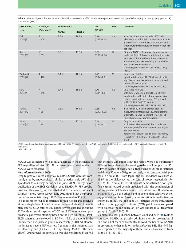
that included 228 patients but the results were not significantlydifferent from placebo, likely owing to the small sample size [35].A lower dosage of diclofenac, either 50mg or 25mg in patientsweighing≥50kg or<50kg, respectively, was compared with pla-cebo in a small RCT from Japan: the PEP incidence was 3.9% vs.18.9% in the diclofenac vs. the control group, respectively (P=0.017) [36]. A small RCT of 80 patients demonstrated a nonsigni-ficant trend toward benefit associated with the combination ofintramuscular diclofenac and intensive intravenous fluid admin-istration [37]. On the other hand, it seems that selective cyclo-oxygenase-2 inhibitor (coxib) administration is ineffective, asshown by an RCT that included 371 patients where intravenousvaldecoxib or glyceryl trinitrate (GTN) patch were comparedwith placebo: the incidence of PEP was similar in all treatmentgroups (10%, P=0.986) [38].Six meta-analyses published between 2009 and 2014 (●" Table2)compared NSAIDs vs. placebo administration for prevention ofPEP, and all of them concordantly showed the benefit of NSAIDsin preventing either mild or moderate/severe PEP. The NNT fig-ures, reported in the majority of these studies, have varied from11 to 34 [31,39–43].
Table 2 Meta-analyses published in 2009 or later that assessed the effect of NSAIDs on post-endoscopic retrograde cholangiopancreatography (post-ERCP)pancreatitis (PEP).1
First author,
year
Studies, n
(Patients, n)
PEP incidence OR
(95%CI)
NNT Comments
NSAIDs Placebo
Dai,2009 [31]
6(1300)
8.9% 16.8% 0.46(0.32–0.65)
n.a. Inclusion of placebo-controlled RCTs only(diclofenac or indomethacin administered oral-ly or rectally). Different ERCP techniques andcriteria for pancreatitis, low number of high riskpatients
Ding,2012 [39]
10(2269)
8.0% 13.9% 0.57(0.38–0.86)1
17 Different NSAIDs (diclofenac, indomethacin,valdecoxib) and different administration routes(oral, rectal, intraduodenal, intramuscular andintravenous) and ERCP techniques; moderateand severe PEP also reduced.Moderate/severe PEP: RR 0.46 (0.28–0.38);NNT, 34
Yaghoobi,2013 [40]
4(1470)
5.1% 10.3% 0.49(0.34–0.71)
20 Only rectal NSAIDs;significant decrease of PEP incidence in bothhigh risk and low risk patients, moderate andsevere PEP also reduced.Moderate/severe PEP: RR 0.45 (0.24–0.83)
Sun,2013 [41]
7(1846)
6.4% 16.0% 0.45(0.34–0.61)1
n.a. Only rectal NSAIDs;both diclofenac and indomethacin effective,significant in both high risk and average-riskcohorts, moderate and severe PEP reduced.Mild PEP: RR 0.54 (0.35–0.83)Moderate/severe PEP: RR 0.39 (0.22–0.70)
Yuhara,2014 [42]
9(1981)
7.8% 16.0% 0.55(0.43–0.72)*
n.a. Inclusion of RCTs testing rectal, oral, intra-muscular and intravenous NSAIDs (diclofenac,indomethacin). No significant effect on PEPwith intramuscular administration.
Sethi,2014 [43]
7(2133)
6.6% 15.1% 0.44(0.34–0.57)1
11 Only rectal NSAIDs;no difference between diclofenac and indo-methacin, no difference between timing (pre-and post-ERCP).Relative risk in low risk and high risk patients,respectively: 0.42 (0.26–0.66) and 0.45 (0.31–0.65).
NSAIDs, nonsteroidal anti-inflammatory drugs; OR, odds ratio; CI, confidence interval; NNT, number needed to treat; n.a., not available; RCT, randomized controlled trial; RR,relative risk.1 Risk reduction.
Dumonceau Jean-Marc et al. Updated ESGE Guideline for PEP prophylaxis… Endoscopy 2014; 46: 799–815
Guideline 803

In a post hoc analysis of an RCTof NSAIDs vs. placebo for PEP pro-phylaxis, administration of rectal NSAIDs alone was more effec-tive and less costly than prophylactic pancreatic stent placementalone or combinedwith rectal NSAIDs [44]. However, the authorscautioned that these findings should not change current clinicalpractice because their post hoc observational study did not pro-duce the same quality of evidence as their RCT, which supportedthe use of indomethacin in addition to prophylactic pancreaticstenting in high risk cases [34].
5.3.Possibly effective drugs5.3.1.Somatostatin and octreotide▶ Statement 2010:Based on an ad hoc meta-analysis of results from 10 high qualityRCTs, somatostatin proved to be ineffective in preventing PEP(Evidence level 1++). We do not recommend universal adminis-tration of prophylactic somatostatin in average-risk patients un-dergoing ERCP (Recommendation grade A). Administration of so-matostatin might be more efficacious using specific dose sche-dules, but caution is neededwhen interpreting the results of sub-group analyses as they often exaggerate differences betweentreatments in RCTs.Octreotide administration did not affect the overall incidence ofPEP when data from eight high quality trials were pooled (Evi-dence level 1++). Prophylaxis with octreotide is not recommen-ded (Recommendation grade A). In future studies the efficacy ofprophylactic administration of octreotide should be evaluatedusing a dose greater than or equal to 0.5mg.▶ Statement 2014:Some meta-analytical results seem to support the benefit of so-matostatin and octreotide for averting PEP but their clinical usecannot be recommended except in well selected cases, owing todiscordant data from different routes or dosages and the exces-sively high NNTvalues (Recommendation grade A).Background:Three meta-analyses assessed the effect of somatostatin/octreo-tide for PEP prophylaxis before the 2010 ESGE Guideline publica-tion [1]; an updated meta-analysis of high quality RCTs (Jadadscore>3) was performed for writing the ESGE Guideline. Respec-tively, 10 and 8 RCTs were included for assessing somatostatinand octreotide. Overall, the updated meta-analysis found no sig-nificant reduction of PEP incidence with somatostatin vs. placebo(OR 0.57; 95%CI 0.32–1.03) or with octreotide vs. placebo (OR0.73; 95% CI, 0.41–1.30). However, subgroup analyses suggestedthat some dosages or administration schedules might be asso-ciated with a protective effect against PEP.New information since 2009:A meta-analysis that compared octreotide vs. placebo (18 RCTs,3171 patients) found no significant difference in PEP incidence(OR 0.77; 95%CI 0.56–1.05) [45]. However, at post hoc subgroupanalysis, dosage seemed to have an impact: when the compoundwas given at a dose≥0.5mg (6 RCTs; 1470 patients) the OR for de-veloping PEP dropped to 0.45 (95%CI 0.28–0.73; NNT=25),whereas the agent proved ineffective at a dosage<0.5mg. Othersubgroup analyses of octreotide administration that assessedthe route (intravenous vs. subcutaneous) and the schedule (be-fore or after ERCP) were inconclusive.A second meta-analysis assessed the effect of somatostatin (10RCTs) and of octreotide (7 RCTs) in a total of 3818 patients [46].Overall, somatostatin reduced the risk of PEP (RR 0.52; 95%CI0.30–0.90), while octreotide was not effective (RR 0.86; 95%CI0.45–1.63). Subgroup analyses suggested that higher doses of so-
matostatin (3mg given as an infusion over 12 hours) or lower do-ses (250µg) given as bolus injection may be most efficacious,especially for the subgroup of patients at relatively higher riskfor PEP, i. e., those undergoing pancreatic duct injection (OR0.35; 95%CI 0.15–0.82) and biliary sphincterotomy (OR 0.33;95%CI 0.16–0.70). With regard to octreotide, subgroup analysisshowed a protective effect when administered at high dose (OR0.42; 95%CI 0.20–0.90).More recently, two RCTs compared the effect of somatostatin vs.placebo for PEP prophylaxis. In one RCT, the efficacy of somatos-tatin alone could not be assessed because the treatment group re-ceived somatostatin plus diclofenac [47]; the other RCT foundthat high dose somatostatin was associated with a reduction ofpost-ERCP hyperamylasemia, but not incidence of PEP [48].
5.3.2.Protease inhibitors▶ Statement 2010:Prophylaxis with gabexate or ulinastatin does not reduce the in-cidence of PEP (Evidence 1++). Neither drug is recommended forprophylaxis of PEP (Recommendation grade A).▶ Statement 2014:Some meta-analytical results seem to support the benefit of ga-bexate and ulinastatin at high doses for averting PEP but theirclinical use cannot be recommended owing to the discordanceof the data. A novel protease inhibitor, nafamostat, is likely effec-tive for preventing PEP in patients at low risk of PEP but not inhigh risk patients.Background:Gabexate for PEP prophylaxis has been evaluated in six high qual-ity RCTs [49–54]; when the results were pooled, no significantdifference was found between the control and treatment groups.Ulinastatin for PEP prophylaxis has been compared with placebo(two RCTs) and with gabexate (two RCTs), with contradictory re-sults [55–58]. Nafamostat is a novel protease inhibitor that inhi-bits trypsin, a proteolytic enzyme considered to play an initialrole in the pathogenesis of pancreatitis; compared with gabexateits half-life is 20 times longer and its potency 10 to 100 timesgreater.New information since 2009:Four meta-analyses assessed the efficacy of protease inhibitorsfor PEP prophylaxis [42,59–61]. Subgroup analysis of 8 highquality (Jadad score >3) RCTs on gabexate administrationshowed that the agent was not associated with a decreased riskof PEP (RR 0.64; 95%CI 0.36–1.13) and there was great heteroge-neity among the studies. Subgroup analysis of 6 ulinastatin RCTsshowed the agent was not associatedwith a decreased risk of PEPin either high quality studies (RR 0.65; 95%CI 0.33–1.30) or lowquality studies (RR 0.75; 95%CI 0.49–1.16).The question of drug dosagewas addressed in one meta-analysis:when ulinastatin was administered at sufficient doses (≥150 000units), it averted PEP (OR 0.39; 95%CI 0.19–0.81; NNT 6); in asimilar fashion, gabexate was effective at either slow infusion ofhigh dose (≥150 000 units) (OR 0.44; 95%CI 0.25–0.79; NNT 7) orrapid infusion of low dose (OR 0.37; 95%CI 0.20–0.69; NNT 6)[61].The benefit of nafamostat has been assessed in a meta-analysisthat pooled data from 5 RCTs and 2678 patients [42]. This agentlowered the incidence of PEP (RR 0.47; 95%CI 0.33–0.67). Allthree RCTs available as full text found nafamostat to be effectivein low risk patients but not in high risk patients [62–64]. Whenthe results observed for high risk patients in the three RCTs werepooled, PEP incidence was not statistically different in the treat-
Dumonceau Jean-Marc et al. Updated ESGE Guideline for PEP prophylaxis… Endoscopy 2014; 46: 799–815
Guideline804

ment vs. the placebo group (8.9% [32/358] vs. 13.1% [37/283],respectively; P=0.12).
5.3.3.Drugs influencing sphincter of Oddi pressure▶ Statement 2010:Nitroglycerin reduces the incidence of PEP; however, when ad-ministered transdermally, it is ineffective (Evidence grade 1++).Side effects such as transient hypotension and headache may oc-cur. We do not recommend the routine use of nitroglycerin forprophylaxis of PEP (Recommendation grade A).There is no evidence that … drugs reducing sphincter of Oddipressure (other than nitroglycerin) [namely, botulinum toxin,epinephrine, lidocaine, and nifedipine] … reduce the incidenceof PEP … [cf. section 5.4 below].▶ Statement 2014:Glyceryl trinitrate (GTN) may be effective in preventing PEPwhen administered sublingually. Topical epinephrine may be ef-fective to prevent PEP in purely diagnostic ERCP. ESGE does notrecommend the routine use of GTN or of epinephrine for PEP pro-phylaxis. No changes concerning botulinum toxin, lidocaine, andnifedipine.Background:The influence of GTN on the incidence of PEP was evaluated intwo meta-analyses that pooled data from 5 RCTs involving 1662patients [65, 66]. Both meta-analyses showed an overall signifi-cant reduction of PEP with an RR of 0.61 (95%CI 0.44–0.86) andan NNT of 26. In the majority of the patients, GTN was adminis-tered transdermally. In a subgroup analysis, transdermal GTNfailed to show a significant reduction of PEP (RR 0.66; 95%CI0.43–1.01).Botulinum toxin [67], epinephrine [68], lidocaine [69], and nife-dipine [70,71], were not found to prevent PEP in the correspond-ing RCTs.New information since 2009:A meta-analysis that pooled data from 8 RCTs (1920 patients)found that GTN decreases PEP incidence compared with placebo(5.9% vs. 9.8%, respectively; P=0.002) [72]. A more recent meta-analysis extended the observation to 12 RCTs (2649 patients) andfound again that GTN reduces the overall incidence of PEP (RR0.67; 95%CI 0.52–0.87) but was, however, ineffective in loweringthe incidence of moderate to severe PEP (RR 0.70; 95%CI 0.42–1.15) [73]. The route of GTN administration may influence its ef-fectiveness. Subgroup analyses revealed that sublingual adminis-tration of GTN was more effective than transdermal and topicaladministration (RR 0.47; 95%CI 0.28–0.78).The prophylactic merit of topical epinephrine (0.02%, 20mLsprayed on the papilla) was compared with placebo in diagnos-tic-only ERCP in two RCTs [68,74]. Matsushita et al. originallyevaluated spraying of epinephrine onto the papilla in 370 pa-tients undergoing diagnostic ERCP [68]; the incidence of PEPwas 0% in the epinephrine group vs. 2.2% in the control group(not significant). In the second study, Xu et al. randomized 941patients to topical epinephrine vs. placebo; the rates of PEPwere 1.95% vs. 6.45%, respectively (P=0.0086) [74]. When the re-sults of the two studies were pooled in a meta-analysis, topicalepinephrine proved efficacious in reducing PEP (OR 0.25; 95%CI0.06–0.65; NNT 15) [75]. However, in the two RCTs of epine-phrine, only patients with purely diagnostic ERCP were included,no guidewire was initially used, cannulation times were verylong, and the definition of PEP was not standard. Based on theseshortcomings, routine use of topical epinephrine cannot be re-commended for PEP prophylaxis.
5.3.4.Antibiotics▶ Statement 2010:Ceftazidime reduced the incidence of PEP in a single study (Evi-dence grade 1– ). Further data are needed before recommendingceftazidime for the prophylaxis of PEP (Recommendation gradeC).▶ Statement 2014:Antibiotics have not been proven effective in PEP prophylaxis;further data are needed (Recommendation grade C).Background:In an RCT that tested ceftazidime for prophylaxis of PEP, the inci-dence of PEP was lower in the treatment than in the controlgroup (2.6% vs. 9.4%, respectively; P=0.009) [76]. This study wasof low methodological quality owing to unclear allocation con-cealment.New information since 2009:A network meta-analysis ranked antibiotics in fourth positionamong 16 drugs for the efficacy of PEP prophylaxis (OR 0.46;95%CI 0.15–1.07; NNT 21) [75]. However, the difference ob-served between antibiotics and placebo was not statistically sig-nificant, only 254 patients were included in the treatment armsof four RCTs [76–79] included in the meta-analysis (not 1082 asstated in the meta-analysis), and two of these RCTs did not havePEP prophylaxis as their primary study endpoint [76,78].
5.3.5. Intensive hydration▶ Statement 2010:No statement.▶ Statement 2014:In a pilot study, intensive hydration seemed to effectively preventPEP. Large-scale RCTs to establish an evidence-based approach tointensive hydration are needed.New information since 2009:Based on a pilot study in 62 patients, intensive hydration in theperiprocedural period with intravenous lactated Ringer’s solu-tion appears to reduce PEP incidence [80]. None of the patientswho received aggressive hydration developed PEP, comparedwith 17% of patients who received standard hydration (P=0.016). No patients had evidence of volume overload. Two obser-vational studies support this strategy of hydration for attenuatingthe severity of PEP [81, 82].
5.4.Drugs proven ineffective▶ Statement 2010:There is no evidence that glucocorticoids, drugs reducing sphinc-ter of Oddi pressure (other than nitroglycerin), antioxidants, he-parin, interleukin-10, or some anti-inflammatory drugs (otherthan diclofenac and indomethacin), such as pentoxifylline, sema-pimod, and the recombinant platelet-activating factor acetylhy-drolase, reduce the incidence of PEP (Evidence levels from 1– to1++). None of these drugs is recommended for PEP prophylaxis(Recommendation grade A).▶ Statement 2014:No change except for GTN and epinephrine (cf. section 5.3.3above).Background:The efficacy of glucocorticoids for PEP prophylaxis was evaluatedin twometa-analyses that included 6 RCTs [83,84]; the incidenceof PEP was not significantly different in the glucocorticoids vs.the control group (11.8% vs. 10.6%, respectively). Three RCTsthat evaluated interleukin-10 have yielded contradictory results[85–87]. Subcutaneous heparin was not found to reduce PEP in-
Dumonceau Jean-Marc et al. Updated ESGE Guideline for PEP prophylaxis… Endoscopy 2014; 46: 799–815
Guideline 805

cidence in 2 RCTs [88, 89]. Drugs potentially reducing the sphinc-ter of Oddi pressure (other than GTN and topical epinephrine),including botulinum toxin, lidocaine, and nifedipine, have notbeen found to reduce PEP incidence in RCTs [67, 68,90–92].Three antioxidant agents, namely allopurinol, N-acetylcysteine,and natural beta-carotene were not found to be effective in pre-venting PEP in 5 RCTs and one meta-analysis [93–98].New information since 2009:Regarding heparin, a meta-analysis that analyzed four clinicaltrials (including 3 RCTs) with a total of 1438 patients showed nobeneficial effect of prophylactic heparin for the prevention of PEP[99]. Regarding antioxidant supplementation, a meta-analysis of11 RCTs (total, 3010 patients) showed no beneficial effect on theincidence and the severity of PEP [100]. Regarding allopurinol, anRCT found no effect for the prevention of PEP [101]. No new dataare available for glucocorticoids, interleukin-10, pentoxifylline,semapimod, and the recombinant platelet-activating factor acet-ylhydrolase.
6.Pancreatic stent placement for PEP prophylaxis!
▶ Statement 2010:Prophylactic pancreatic stent placement is recommended to pre-vent PEP in patients who are at high risk for development of PEP.Short, 5-Fr diameter, plastic pancreatic stents are currently re-commended. Passage of the stent from the pancreatic ductshould be evaluated within 5 to 10 days of placement and re-tained stents should be promptly removed endoscopically (Evi-dence level 1+; Recommendation grade A).▶ Statement 2014:Prophylactic pancreatic stenting decreases the risk of PEP in highrisk andmixed-case groups; it nearly eliminates the risk of severePEP. 5-Fr pancreatic stents are more efficacious than 3-Fr stentsin preventing PEP. ESGE recommends the placement of 5-Fr pan-creatic stents in cases at high risk of PEP. Passage of the stent fromthe pancreatic duct should be evaluated within 5 to 10 days ofplacement and retained stents should be promptly removedendoscopically (Evidence level 1+; Recommendation grade A).Background:Two meta-analyses have demonstrated that, in patients at highrisk of PEP, prophylactic pancreatic stent placement significantlyreduces the incidence of PEP [102, 103]. The OR was 0.44 (95%CI0.24–0.81), with an absolute risk reduction of 12.0% (95%CI 3.0–21.0). A multicenter RCT (201 patients) showed a decreased inci-dence of PEP when prophylactic pancreatic stent placement wasperformed, regardless of the concomitant occurrence of otherknown risk factors for PEP (PEP incidence in the stent vs. no-stentgroup, 3.2% vs. 13.6%, respectively; P=0.019) [104]. In thesestudies, the risk of severe PEP was nearly eliminated followingsuccessful placement of a prophylactic pancreatic stent.Pancreatic stent placement was shown to be cost-effective onlyin patients at high risk for PEP [105].New information since 2009:Ameta-analysis of 14 RCTs with a total of 1541 patients showed asignificant reduction in the incidence and the severity of PEPwhen prophylactic pancreatic stenting was used [106]. In addi-tion, subgroup analysis showed that pancreatic stenting reducedthe risk of PEP in high risk and mixed-case groups. In a networkmeta-analysis, prophylactic pancreatic stenting alone was shownto be less effective than NSAIDs alone, and the combination of
NSAIDs with prophylactic pancreatic stenting did not further re-duce the risk of PEP [107].The ideal stent characteristics for PEP prophylaxis and the opti-mal duration of stent placement are not definitively known.However, a network meta-analysis showed that the probabilitiesof 5-Fr and 3-Fr stents being ranked as the most efficacious forthe prevention of PEP were 96.8% vs. 3.1%, respectively, with 5-Fr single-pigtail, unflanged stents and 5-Fr straight, flangedstents producing similar results [108]. Furthermore, placementof 5-Fr stents requires fewer guidewires and is easier than thatof 3-Fr stents [109, 110]. It is believed that stents need to remainin place for a minimum of 12–24 hours to provide benefit, sinceremoval at the end of ERCP negates the protection from PEP [111]and early outward migration may also result in PEP [112]. Ad-verse events related to attempted prophylactic pancreatic stent-ing include PEP, stent-induced pancreatic ductal damage, and in-ward migration [113]. Removal of proximally migrated small-di-ameter stents can be technically challenging, if not impossible[114].
7.ERCP techniques!
7.1.General considerations7.1.1.Patient position▶ Statement 2010:There is no evidence that the incidence of PEP is influenced bypatient position during ERCP (Evidence level 2++). Therefore, norecommendation is made regarding patient position.▶ Statement 2014:No changes.Background:Comparisons in RCTs of different patient positions during ERCPshowed no significant difference in PEP incidence.New information since 2009:None.
7.1.2.Cannulation attempts▶ Statement 2010:Trauma resulting from repeated attempts at biliary cannulationhas been proven to be a risk factor for the development of PEP(Evidence level 2++). The number of cannulation attemptsshould be minimized (Recommendation grade B).▶ Statement 2014:ESGE recommends keeping the number of cannulation attemptsas low as possible (Recommendation grade B).Background:The risk of PEP is higher after multiple attempts at duct cannula-tion [6, 21,115,116]. The rendezvous technique allows cannula-tion trauma to be minimized, and has been used in most studiesthat evaluated intraoperative endoscopic sphincterotomy.New information since 2009:A meta-analysis assessed 5 RCTs that compared preoperative vs.intraoperative endoscopic sphincterotomy [117]; PEP was lessfrequent with the latter approach while the incidence of otherERCP-related complications was similar with both techniques. Anationwide case–control study from Sweden confirmed that therendezvous technique was associated with a reduction in therisk of PEP compared with standard cannulation techniques,from 3.6% to 2.2% (OR 0.5; 95%CI 0.2–0.9; P=0.02) [18].
Dumonceau Jean-Marc et al. Updated ESGE Guideline for PEP prophylaxis… Endoscopy 2014; 46: 799–815
Guideline806
7.1.3.Contrast medium▶ Statement 2010:Injection of contrast medium into the pancreatic duct is an inde-pendent predictor of PEP (Evidence level 1+). If pancreatic ductinjection occurs incidentally or is required, the number of injec-tions and volume of contrast medium injected into the pancreaticduct should be kept as low as possible (Recommendation gradeB). Compared with traditional, high-osmolality contrast agents,low-osmolality contrast agents are costlier but are not associatedwith reduction in the rates of PEP (Evidence level 1– ). The rou-tine use of these agents for ERCP is not recommended (Recom-mendation grade B).▶ Statement 2014:No changes.Background:In a large meta-analysis, pancreatic duct injection was found tobe an independent predictor of PEP (RR 2.2; 95%CI 1.60–3.01)[20]. In a retrospective study that included more than 14 000ERCPs, the extent of pancreatic duct injection (head-only vs.head and body vs. injection to the tail) was independently asso-ciated with PEP [118]. The hypothesis that low-osmolality con-trast agents would be less harmful than high-osmolality contrastagents because of less important fluid shifts in the pancreas wasinvalidated in a meta-analysis of 13 RCTs that involved 3381 pa-tients [119].New information since 2009:One article examined the role of low-osmolality contrast agentsbut it does not warrant any change in the Guideline [120].
7.1.4.Carbon dioxide▶ Statement 2010:Use of carbon dioxide (CO2) as a replacement for air for luminalinsufflation during ERCP does not influence the incidence of PEPbut decreases the incidence and severity of post-procedural ab-dominal pain (Evidence level 1+). Carbon dioxide is recommen-ded for insufflation, and might be particularly useful for outpati-ent ERCPs, to reduce post-procedural abdominal pain and toavoid confusion with PEP (Recommendation grade B).▶ Statement 2014:No changes.Background:Because of its higher solubility in water compared with nitrogenand oxygen, the main components of air, carbon dioxide iscleared from the bowel following endoscopy much faster thanair (through the bloodstream and respiration).New information since 2009:Three meta-analyses, which included between 5 and 7 RCTs (be-tween 446 and 818 patients), compared carbon dioxide vs. air forgut distension during ERCP exclusively [121–123]. All thesemeta-analyses found that the use of carbon dioxide reducespost-ERCP abdominal pain without change in other complicationrates or in procedure duration. A survey showed that the use ofcarbon dioxide during endoscopy is uncommon; this may berelated to implementation costs and unawareness by endosco-pists of the advantages of carbon dioxide [124].
7.1.5.Cannulation techniques▶ Statement 2010:For deep biliary cannulation, the wire-guided technique reducesthe risk of PEP and increases the success rate of primary cannula-tionwhen compared with the standard contrast-assisted method
(Evidence level 1++). The wire-guided technique is recommen-ded for deep biliary cannulation (Recommendation grade A).▶ Statement 2014:No changes.Background:The wire-guided biliary cannulation technique entails passage ofa guidewire inserted through a catheter (most often a hydrophilicguidewire inserted into a sphincterotome) for deeply cannulatingthe bile duct. Two meta-analyses published in 2009 showed that,in RCTs, the incidence of PEP was significantly lower with thewire-guided as compared with the standard contrast-assistedcannulation technique [125,126].New information since 2009:Five comparative studies and a meta-analysis comparing thewire-guided vs. the standard contrast-assisted method for selec-tive biliary cannulation were published between 2009 and 2013[127–132]. Four studies [128–131], two of which were RCTs[128,131], did not confirm the results of previous meta-analysesthat showed a lower risk of PEP with the wire-guided method. Inmost studies, the wire-guided method shortened cannulationand fluoroscopy times. However, in a recent meta-analysis thatextended the analysis to 12 RCTs (3450 patients), the wire-guid-ed method significantly lowered the incidence of PEP comparedwith the contrast-assisted method (RR 0.51; 95%CI, 0.32–0.82)[132]. In addition, the wire-guided cannulation technique wasassociated with greater primary cannulation success (RR 1.07;95%CI 1.00–1.15), fewer precut sphincterotomies (RR 0.75; 95%CI 0.60–0.95), and no increase in other ERCP-related complica-tions.
7.1.6.Electrosurgical current▶ Statement 2010:The incidence of post-sphincterotomy pancreatitis is not influ-enced by the type of electrosurgical current used (whetherpure-cut or blended) (Evidence level 1+). Blended current is re-commended for biliary sphincterotomy, particularly in patientsat high risk of bleeding (Recommendation grade A).▶ Statement 2014:No changes.Background:As pure-cut current produces less edema than blended current[133], it was hypothesized that it might reduce the incidence ofPEP after biliary sphincterotomy. A meta-analysis of four RCTsthat included 804 patients found no significant difference in theincidence of PEP following the use of pure vs. blended current[134]. However, the incidence of bleeding was significantly high-er when pure-cut current was used.New information since 2009:No new evidence has become available.
7.2.Effect of difficult biliary cannulation7.2.1 Definition▶ Statement 2010:None.▶ Statement 2014:ESGE recommends that future studies define difficult biliary can-nulation in an intact papilla as any of the following: cannulationattempts of duration>5 minutes,>5 attempts, or 2 pancreaticguidewire passages.
Dumonceau Jean-Marc et al. Updated ESGE Guideline for PEP prophylaxis… Endoscopy 2014; 46: 799–815
Guideline 807

Background:Many different definitions of “difficult” biliary cannulation havebeen used, which make comparisons between studies impracti-cal.New information since 2009:In an effort to standardize this definition, Halttunen et al. pro-spectively collected data on 907 biliary cannulations attemptedby experienced endoscopists at 10 centers [115]. The authorsfound the incidence of PEP progressively increased with variousfactors perceived as causing difficulty in cannulation. Any of thefollowing factors was associated with a PEP incidence of>10%during wire-guided cannulation of a native papilla: cannulationattempts of duration >5 minutes, >5 attempts, or 2 pancreaticguidewire passages. The latter was also noted in another prospec-tive study [116].For difficult cannulation, commonly used options include persist-ent attempts at cannulation using standard methods, pancreaticguidewire placement (with biliary cannulation attempted eitherusing a guidewire, the so-called “double guidewire” (DGW) tech-nique, or using contrast medium injection), precut of varioustypes, repeat attempts at ERCP 24–48 hours later, and patient re-ferral to another endoscopist.
7.2.2.Pancreatic guidewire-assisted technique▶ Statement 2010:Data about the usefulness and safety of pancreatic guide wireplacement to facilitate biliary cannulation in difficult cases areconflicting. Prophylactic pancreatic stent placement decreasesthe incidence of PEP with this technique (Evidence level 2+). Pan-creatic guide wire assistance may facilitate biliary cannulationmostly in the case of inadvertent but repeated cannulation ofthe pancreatic duct; if this method is used, a pancreatic stentshould be placed for PEP prophylaxis (Recommendation grade B).▶ Statement 2014:In cases of difficult biliary cannulation, pancreatic guidewire(PGW) placement allows biliary cannulation in a proportion ofcases similar to persistence in attempting cannulation withstandard cannulation techniques (or precut if it is used as a back-up technique), but the risk of PEP is likely higher. In such circum-stances, PEP is effectively prevented by prophylactic pancreaticstenting (Evidence level 1–). ESGE suggests restricting the use ofa PGW as a backup technique to cases with repeated inadvertentcannulation of the pancreatic duct; if this method is used, deepbiliary cannulation should be attempted using a guidewire ratherthan the contrast-assisted method and a prophylactic pancreaticstent should be placed (Recommendation grade B).Background:In the PGW-assisted technique, a guidewire is inserted in themain pancreatic duct to facilitate deep biliary cannulation bystraightening the papillary anatomy and to prevent repeated can-nulation of the pancreatic duct [135,136]. In two RCTs, comparedwith persistence in applying the standard cannulation technique,the PGW technique yielded overall similarly low success rates forbiliary cannulation (means, 57% vs. 56%, respectively) and a non-significantly higher incidence of PEP (means, 14% vs. 6%, respec-tively) [137,138]. Discordances between these RCTs in terms ofcannulation success and of PEP incidencemay be related to differ-ences in the inclusion criteria (i. e., difficult biliary cannulationalone or combined with repeated unintended pancreatic cannu-lation) and in the use of prophylactic pancreatic stenting.
New information since 2009:Seven new studies are summarized in●" Table3 [139–145]. Intwo RCTs that compared the PGW vs. the precut techniques[142,143], success rates of biliary cannulation were similar but,in one of the RCTs [143], the PGW technique was plagued by ahigher incidence of PEP (38% vs. 11%, respectively; P=0.01). Pro-phylactic pancreatic stenting was not used in any of the RCTsmentioned above. Another RCTshowed that prophylactic pancre-atic stenting significantly decreased the incidence of PEP afterthe PGW technique had been used [146]. In a retrospective studythat included 146 patients, prophylactic pancreatic stenting wasalways attempted after the PGW technique had been used, andfailed prophylactic pancreatic stenting was the only independentpredictor of PEP [144]. In another retrospective study that in-volved 142 patients, the incidence of PEP decreased after the au-thors changed their cannulation technique following PGW place-ment, from contrast-assisted to guidewire-assisted biliary can-nulation [147].
7.2.3.Precut biliary sphincterotomy▶ Statement 2010:Various techniques of precut biliary sphincterotomy have beendescribed; the fistulotomy technique may present a lower inci-dence of PEP than standard needle-knife sphincterotomy but fur-ther RCTs are required to determine which technique is safer andmore effective, based upon the papillary anatomy. There is noevidence that the success and complication rates of biliary precutare affected by the level of endoscopist experience in this tech-nique but published data only report on the experience of oneendoscopist (Evidence level 2– ). Prolonged cannulation attemptsusing standard techniques may impart a risk for PEP greater thanthe precut sphincterotomy itself (Evidence level 2+). Precutsphincterotomy should be performed by endoscopists with ex-pertise in standard cannulation techniques (Recommendationgrade D). The decision to perform precut biliary sphincterotomy,the timing, and the technique, are based on anatomic findings,endoscopist preference, and procedural indication (Recommen-dation grade C).▶ Statement 2014:In cases of difficult biliary cannulation, early precut is associatedwith a lower PEP incidence than persistent attempts using thestandard approach but the overall success and complication ratesare similar with both approaches. Needle-knife fistulotomyseems to be associated with fewer complications, including PEP,compared with other precut techniques.ESGE suggests that needle-knife fistulotomy should be the pre-ferred precut technique in patients with a bile duct dilateddown to the papilla. Conventional precut and transpancreaticsphincterotomy present similar success and complication rates;if conventional precut is elected and pancreatic cannulation is ea-sily obtained, ESGE suggests attempting to place a small-diame-ter (3-Fr or 5-Fr) pancreatic stent to guide the cut and leaving thepancreatic stent in place at the end of ERCP for a minimum of12–24 hours (Recommendation grade B).Background:Precut biliary sphincterotomy can be done by different tech-niques: needle-knife precut starting at the orifice of the papilla(conventional precut), needle-knife above the orifice (fistulot-omy), transpancreatic sphincterotomy (septotomy) and needle-knife over a pancreatic stent. Compared with prolonged attemptsat biliary cannulation using standard techniques, the use of pre-
Dumonceau Jean-Marc et al. Updated ESGE Guideline for PEP prophylaxis… Endoscopy 2014; 46: 799–815
Guideline808

cut sphincterotomy has long been thought to increase the successof biliary cannulation and the incidence of PEP [6,20].New information since 2009:Two meta-analyses investigated the issue of timing of the precutprocedure in 6 RCTs [148, 149]. Aside from the definition of earlyprecut, differences between studies included the technique ofprecut and the randomization ratio (from 1:1 to 1:3). Endosco-pists experienced in precut techniques performed all procedures.The overall incidence of PEP was lower in patients allocated toearly precut than to persistence in the use of standard techniques(2.5% vs. 5.3%; OR 0.47; 95%CI 0.24–0.91) but the overall cannu-lation and complication rateswere similar in patients allocated toearly precut or to persistence in the use of standard cannulationtechniques.In a retrospective study comparing different precut techniques,the risk of PEP was significantly lower after fistulotomy (2.6%)compared with conventional precut (20.9%) and pancreatic sep-totomy (22.4%); the overall complication rate was also signifi-cantly lower after fistulotomy vs. other precut techniques [150].In another retrospective study of needle-knife fistulotomy per-formed in 204 patients, complications (mostly PEP) progressivelyincreased with decreasing common bile duct (CBD) diameter, upto 14% in patients with a CBD diameter <4mm [151]. Two retro-spective studies compared transpancreatic septotomy and nee-dle-knife precut (with pancreatic stenting or not) for biliary ac-
cess [152,153]; they found no significant differences in terms ofPEP, overall complication and overall success rates.Needle-knife precut assisted by pancreatic stenting has been pro-posed for reducing PEP. One RCT found that, compared with stentremoval at the end of the procedure, leaving the stent for 7–10days reduces the incidence and severity of PEP (P<0.05 for bothcomparisons) [111].
7.3.Specific therapeutic techniques7.3.1.Balloon dilation as a substitute for endoscopicsphincterotomy▶ Statement 2010:Compared with endoscopic sphincterotomy, endoscopic papil-lary balloon dilation (EPBD) using small-caliber balloons (≤10mm) is associated with a significantly higher incidence of PEPand significantly less bleeding (Evidence level 1++). EPBD is notrecommended as an alternative to sphincterotomy in routineERCP but may be useful in patients with coagulopathy and al-tered anatomy (e.g. Billroth II) (Recommendation grade A). If bal-loon dilation is performed in young patients, the placement of aprophylactic pancreatic stent should be strongly considered (Evi-dence level 4; Recommendation grade D).▶ Statement 2014:Compared with endoscopic sphincterotomy, endoscopic papil-lary balloon dilation (EPBD) using small-caliber balloons (≤10mm) is associated with more PEP, less bleeding, and fewer late
Table 3 New studies evaluating pancreatic guidewire (PGW) placement for biliary cannulation in the case of difficult biliary cannulation.
First author,
year
Design
Patients, n
Additional
inclusion criteria
Pancreatic
stenting
Successful biliary
cannulation rate
PEP rate Note
Xinopoulos,2011 [139]
Retrospective112
Repeated uninten-tional pancreaticduct cannulation
No 44% (vs. 81% withprecut in patients whohad no repeated un-intentional pancreaticduct cannulation)
6.1% (similar togroup with easybiliary cannula-tion [5.3%] and togroup with precutin place of PGW[7.5%])
Comparative study
Grönroos,2011 [140]
Prospective50
Repeated uninten-tional pancreaticduct cannulation
No 66% 2% Authors advocateto shift to othertechnique if DGW iscumbersome
Belverde,2012 [141]
Retrospective121
Selective pancreat-ic duct cannulationachieved
No 97% 2.6%
Angsuwatcharakon,2012 [142]
RCT44
No No PGW vs. precut:74% vs. 81% (n.s.)
PGW vs. precut:17.4% vs. 9.5%
Shorter cannula-tion time with thePGW vs. precuttechnique(172 s vs. 394 s(P < 0.001)
Yoo,2013 [143]
RCT71
Selective pancrea-tic duct cannula-tion achieved
No DGW vs. precut:91.2% vs. 91.9% (n.s.)
DGW vs. precut:38% vs. 11%(P=0.01)
Ito,2013 [144]*
Retrospective146
n.a. Attempted in all PGW: 70%;if failure with PGW,DGW was successful in72%
PGW, 8%;DGW, 4%
Independentpredictor of PEP:failed pancreaticstenting (OR 8.3;95%CI 2.3–30)
Tanaka,2013 [145]1
Retrospective79
Opacification ofthe pancreatic duct
40 /79 82% (PGW) vs. 83%(DGW)
11% (PGW) vs. 7%(DGW)
PEP, post-endoscopic retrograde cholangiopancreatography (post-ERCP) pancreatitis; PGW, pancreatic guidewire; DGW, double guidewire; RCT, randomized controlled trial; n.s.,not significant; n.a., not available; OR, odds ratio; CI, confidence interval1 Biliary cannulation attempted by searching the bile duct through contrast medium injection with a guidewire in the main pancreatic duct (PGW technique), as opposed tosearching through manipulation of a guidewire inserted in a catheter (DGW technique).
Dumonceau Jean-Marc et al. Updated ESGE Guideline for PEP prophylaxis… Endoscopy 2014; 46: 799–815
Guideline 809

stone recurrences; the risk of PEP is inversely associated with theduration of EPBD (Evidence level 1++). ESGE does not recom-mend endoscopic papillary balloon dilation as an alternative tosphincterotomy in routine ERCP, but it may be advantageous inselected patients; if this technique is used, the duration of dila-tion should be longer than 1 minute (Recommendation grade A).Background:The use of EPBD may be advantageous compared with endo-scopic sphincterotomy, by decreasing clinically significant bleed-ing in patients with coagulopathy, for preserving sphincter ofOddi function in younger patients [154], and in patients with al-tered anatomy (Billroth II) where sphincterotomy is technicallydifficult. In two meta-analyses, the use of EPBD resulted in a low-er success rate than endoscopic sphincterotomy for the initial re-moval of biliary stones, with a significantly higher incidence ofPEP and significantly lower incidence of bleeding [155,156].New information since 2009:Ameta-analysis of 10 high quality RCTs (1451 patients) that com-pared EPBD vs. endoscopic sphincterotomy for biliary stone ex-traction found similar success rates of biliary stone extractionwith both methods but did not bring new findings with regardto complications [157]; indeed it included fewer RCTs than thepreviously published Cochrane meta-analysis [156].A meta-analysis that included 12 RCTs (1649 patients) indica-ted that duration of EPBD is inversely associated with the inci-dence of PEP: short (≤1 minute) EPBD was associated with ahigher risk of PEP (OR 3.87; 95%CI 1.08–13.84) comparedwith endoscopic sphincterotomy (4 RCTs), but long EPBD infla-tion time (>1 minute) was not (OR 1.14; 95%CI 0.56–2.35) (6RCTs) [158]. A lower risk of PEP after long EPBD was also re-ported in the single RCT that compared long (5-minute) vs.short (1-minute) EPBD (RR 0.32; 95%CI 0.11–0.93) [159]. Theauthors suggested that the increased risk of PEP after shortEPBD might be related to inadequate balloon expansion result-ing in a worsened compression of the pancreatic duct.A meta-analysis that included 3 RCTs (496 patients) with a fol-low-up longer than 1 year showed that stone recurrence wasless frequent after EPBD vs. endoscopic sphincterotomy (OR0.48; 95%CI 0.26–0.90) [160]. Another RCT that was not includedin this meta-analysis and included 474 patients also reportedthat, in patients with biliary stones≤8mm, overall late complica-tions and stone recurrence were less frequent after EPBD thanafter endoscopic sphincterotomy (5.3% vs. 17.3%, P=0.009; 4.4%vs. 12.7%; P=0.048, respectively) (mean follow-up, 55 months);the difference was not significant for patients with stones>8mm [161]. A retrospective cohort study with a median follow-up of 92 months also showed a lower incidence of common bileduct stone recurrence after EPBD vs. endoscopic sphincterotomy[162].
7.3.2.Large-balloon dilation for extraction ofdifficult biliary stones▶ Statement 2010:Potential advantages of performing large-balloon dilation in ad-dition to endoscopic sphincterotomy for extraction of difficultbiliary stones remain unclear (Evidence level 3). Endoscopicsphincterotomy plus large-balloon dilation does not seem to in-crease the risk of PEP and can avoid the need for mechanical li-thotripsy in selected patients, but not enough data are availableto recommend routine use over biliary sphincterotomy alone inconjunction to lithotripsy techniques (Recommendation gradeD).
▶ Statement 2014:For the extraction of difficult biliary stones, endoscopic sphinc-terotomy plus large-balloon dilation presents a risk of PEP similarto that of endoscopic sphincterotomy alone; it presents a lowerbleeding risk and, possibly, lower overall morbidity and requiresless use of mechanical lithotripsy. ESGE suggests performingendoscopic sphincterotomy plus large-balloon dilation in placeof endoscopic sphincterotomy alone for the extraction of selecteddifficult biliary stones (Recommendation grade B).Background:In patients with a tapered distal bile duct or large biliary stones,endoscopic sphincterotomy followed by biliary dilation using alarge-diameter (12–20mm) balloon has been proposed to facili-tate stone extraction [163]. This technique has been associatedwith high success rates for stone extraction without the need formechanical lithotripsy and with acceptable complication rates incase series [164–167]; the single RCT that had compared thismethod with endoscopic sphincterotomy at the time of publica-tion of the first Guideline had found no differences in rates of suc-cessful stone clearance, need for mechanical lithotripsy, andcomplications [168].New information since 2009:A meta-analysis of 7 RCTs that included 790 patients found that,compared with endoscopic sphincterotomy alone, endoscopicsphincterotomy followed by large balloon dilationwas associatedwith similar PEP rates but significantly fewer overall complica-tions and significantly less bleeding [169]. However in thismeta-analysis, studies which, according to their authors, wereretrospective, were considered to be RCTs [170]. Two othermeta-analyses that included 3 RCTs and 4–6 retrospective stud-ies (including between 90 and 1295 patients) found similar inci-dences of PEP but significantly lower bleeding risk with endo-scopic sphincterotomy plus large balloon dilation vs. endoscopicsphincterotomy [171,172]. Additional differences in favor ofendoscopic sphincterotomy plus large balloon dilation weremostly attributable to the results of retrospective studies; theyincluded a lower use of mechanical lithotripsy and a lower over-all complication rate [171,172].
7.3.3.Sphincter of Oddi manometry▶ Statement 2010:In patients undergoing pancreatic sphincter of Oddi manometry,use of the standard perfusion catheter without an aspiration porthas been shown to increase the risk of PEP compared with mod-ified water perfusion catheters (Evidence level 2++). Pancreaticsphincter of Oddi manometry should be done using a modifiedtriple-lumen perfusion catheter with simultaneous aspiration ora microtransducer catheter (non-water-perfused) (Recommen-dation grade B).▶ Statement 2014:ESGE recommends that all patients undergoing ERCP for knownor suspected sphincter of Oddi dysfunction (SOD) receive rectalNSAIDs combined with pancreatic stenting. Pancreatic sphincterof Oddi manometry should be done using a modified triple-lu-men perfusion catheter with simultaneous aspiration or a micro-transducer catheter (non-water-perfused) (Recommendationgrade B).Background:It is well documented that patients undergoing ERCP for knownor suspected SOD are at high risk of PEP, regardless of whethersphincter of Oddi manometry (SOM) is performed [173]. BiliarySOM does not appear to increase the risk of PEP [173]; pancreatic
Dumonceau Jean-Marc et al. Updated ESGE Guideline for PEP prophylaxis… Endoscopy 2014; 46: 799–815
Guideline810

SOM is associated with an increased risk of PEP depending on thetechnique used [174]. When water-perfused catheters are usedto measure pancreatic sphincter pressure, continuous aspirationof fluid from the main pancreatic duct through one of the threemanometry ports prevents overfilling of the duct [174], a knownrisk factor for PEP. A specialized aspirationmanometry catheter isavailable for this purpose, though one port of a standard triple lu-men catheter can be used for aspiration [174]. The use of solid-state manometry catheters decreases the risk of PEP as there isno perfusion or filling of the pancreatic duct [175].The results of SOM should not influence the decision of whetherto institute measures for prevention of PEP. As mentioned, all pa-tients with known or suspected SOD are at high risk for PEP andshould receive PEP prophylaxis [175]. Indeed one study showedthat pancreatic stent placement reduced PEP in patients with anintact papilla undergoing SOM and when the manometry resultswere normal [176].New information since 2009:Recent data suggest that rectal NSAIDs are at least as effective aspancreatic stents in patients with SOD [34,44,107]. However, themost evidence-based approach for preventing PEP in high riskcases remains the combination of rectal NSAIDs and prophylacticpancreatic stenting. In the RCT mentioned above, patients whoreceived rectal indomethacin and a pancreatic stent (n=247)had a PEP rate of 9.7% compared with 16.1% in those who receiv-ed a stent alone (n=249) (P=0.04) [34]. To date, there are no clin-ical trial data examining whether rectal NSAIDs are effectivewhen administered instead of prophylactic pancreatic stenting.The data apply mainly to the initial SOM or in patients with SODand intact papillae. One exception to routine PEP prophylaxismight be in patients with SOD who have had dual sphincterot-omy and continued pain who undergo repeat ERCP with re-eval-uation. If both sphincters are found to be patent and/or only thebiliary sphincter is studied, onemight consider not placing a pan-creatic stent. However, the risk of administration of one dose ofrectal NSAIDS is so small that its use outweighs the risk of avoid-ing them.
8.Selection of measures for PEP prophylaxis!
▶ Statement 2010:For low-risk ERCPs, periprocedural rectal administration of non-steroidal anti-inflammatory drugs (NSAIDs) is recommended.For high risk ERCPs, prophylactic pancreatic stent placementshould be strongly considered (Evidence level 1+; Recommenda-tion grade A).▶ Statement 2014:ESGE recommends routine rectal administration of 100mg of di-clofenac or indomethacin immediately before or after ERCP in allpatients without contraindication. In addition to this, in cases athigh risk for PEP, the placement of a 5-Fr prophylactic pancreaticstent should be strongly considered. Sublingually administeredGTN or 250µg somatostatin given in bolus injection might beconsidered as an option in high risk cases if NSAIDs are contra-indicated and if prophylactic pancreatic stenting is not possibleor successful (Recommendation grade A).The following conditions are considered to represent high risk forPEP: endoscopic ampullectomy, known or suspected SOD, pan-creatic sphincterotomy, precut biliary sphincterotomy, pancreat-ic guidewire-assisted biliary cannulation, endoscopic balloonsphincteroplasty, and presence of more than three of the risk fac-
tors listed in●" Table1 (definite or likely). Procedures and patientconditions that do not fulfill these criteria are considered to re-present low risk for PEP.
Competing interests: None of the authors reports financial com-peting interests.
Institutions1 Gedyt Endoscopy Center, Buenos Aires, Argentina2 Division of Gastroenterology, Casa Sollievo Sofferemza Hospital, IRCCS,San Giovanni Rotondo, Italy
3 Division of Gastroenterology, Medical University of South Carolina, Charles-ton, South Carolina, USA
4 Division of Gastroenterology and Gastrointestinal Endoscopy, Vita-SaluteSan Raffaele University, Scientific Institute San Raffaele, Milan, Italy
5 Department of Gastroenterology, HELIOS Albert-Schweitzer Klinik, GöttingenUniversity Teaching Hospital, Northeim, Germany
6 Departments of Gastroenterology and Hepato-Pancreatology, ErasmeUniversity Hospital, Université Libre de Bruxelles, Brussels, Belgium
7 Department of Gastroenterology, Silesian Academy of Medicine, Medyków,Katowice, Poland
8 Division of Gastroenterology and Hepatology, University of North Carolina,Chapel Hill, North Carolina, USA
9 Digestive Endoscopy Unit, Catholic University, Rome, Italy10 Department of Gastroenterology and Hepatology, Elisabethinen Hospital,Linz, Austria
References1 Dumonceau JM, Andriulli A, Devière J et al. European Society of Gastro-
intestinal Endoscopy (ESGE) Guideline: prophylaxis of post-ERCP pan-creatitis. Endoscopy 2010; 42: 503–515
2 Dumonceau J-M, Rigaux J, Kahaleh M et al. Prophylaxis of post-ERCPpancreatitis: a practice survey. Gastrointest Endosc 2010; 71: 934–939 (e931–e932)
3 Kapral C, Mühlberger A, Wewalka F et al. Quality assessment of endo-scopic retrograde cholangiopancreatography: results of a running na-tionwide Austrian benchmarking project after 5 years of implementa-tion. Eur J Gastroenterol Hepatol 2012; 24: 1447–1454
4 Hanna MS, Portal AJ, Dhanda AD et al. UK wide survey on the preven-tion of post-ERCP pancreatitis. Frontline Gastroenterol 2014; 5: 103–110
5 Cotton PB, Lehman G, Vennes J et al. Endoscopic sphincterotomy com-plications and their management: an attempt at consensus. Gastroin-test Endosc 1991; 37: 383–393
6 Freeman ML, Nelson DB, Sherman S et al. Complications of endoscopicbiliary sphincterotomy. N Engl J Med 1996; 335: 909–918
7 Banks PA, Bollen TL, Dervenis C et al. Classification of acute pancreati-tis–2012: revision of the Atlanta classification and definitions by inter-national consensus. Gut 2013; 62: 102–111
8 Artifon EL, Chu A, Freeman M et al. A comparison of the consensus andclinical definitions of pancreatitis with a proposal to redefine post-endoscopic retrograde cholangiopancreatography pancreatitis. Pan-creas 2010; 39: 530–535
9 Dellinger EP, Forsmark CE, Layer P et al. Determinant-based classifica-tion of acute pancreatitis severity: an international multidisciplinaryconsultation. Ann Surg 2012; 256: 875–880
10 Andriulli A, Loperfido S, Napolitano G et al. Incidence rates of post-ERCPcomplications: a systematic survey of prospective studies. Am J Gas-troenterol 2007; 102: 1781–1788
11 Sutton VR, Hong MK, Thomas PR. Using the 4-hour post-ERCP amylaselevel to predict post-ERCP pancreatitis. JOP 2011; 12: 372–376
12 Williams EJ, Taylor S, Fairclough P et al. Are we meeting the standardsset for endoscopy? Results of a large-scale prospective survey of endo-scopic retrograde cholangio-pancreatograph practice Gut 2007; 56:821–829
13 Varadarajulu S, Kilgore ML,Wilcox CM et al. Relationship among hospi-tal ERCP volume, length of stay, and technical outcomes. GastrointestEndosc 2006; 64: 338–347
14 AllisonMC, Ramanaden DN, FouweatherMG et al. Provision of ERCP ser-vices and training in the United Kingdom. Endoscopy 2000; 32: 693–699
15 Hilsden RJ, Romagnuolo J, May GR. Patterns of use of endoscopic retro-grade cholangiopancreatography in a Canadian province. Can J Gastro-enterol 2004; 18: 619–624
Dumonceau Jean-Marc et al. Updated ESGE Guideline for PEP prophylaxis… Endoscopy 2014; 46: 799–815
Guideline 811

16 Loperfido S, Angelini G, Benedetti G et al. Major early complicationsfrom diagnostic and therapeutic ERCP: a prospective multicenterstudy. Gastrointest Endosc 1998; 48: 1–10
17 Williams EJ, Taylor S, Fairclough P et al. Risk factors for complication fol-lowing ERCP; results of a large-scale, prospective multicenter study.Endoscopy 2007; 39: 793–801
18 Swahn F, Nilsson M, Arnelo U et al. Rendezvous cannulation techniquereduces post-ERCP pancreatitis: a prospective nationwide study of12,718 ERCP procedures. Am J Gastroenterol 2013; 108: 552–559
19 Testoni PA, Mariani A, Giussani A et al. Risk factors for post-ERCP pan-creatitis in high- and low-volume centers and among expert and non-expert operators: a prospective multicenter study. Am J Gastroenterol2010; 105: 1753–1761
20 Masci E, Mariani A, Curioni S et al. Risk factors for pancreatitis follow-ing endoscopic retrograde cholangiopancreatography: a meta-analy-sis. Endoscopy 2003; 35: 830–834
21 Freeman ML, DiSario JA, Nelson DB et al. Risk factors for post-ERCP pan-creatitis: a prospective, multicenter study. Gastrointest Endosc 2001;54: 425–434
22 Masci E, Toti G, Mariani A et al. Complications of diagnostic and thera-peutic ERCP: a prospective multicenter study. Am J Gastroenterol2001; 96: 417–423
23 Harewood GC, Pochron NL, Gostout CJ. Prospective, randomized, con-trolled trial of prophylactic pancreatic stent placement for endoscopicsnare excision of the duodenal ampulla. Gastrointest Endosc 2005; 62:367–370
24 Norton ID, Gostout CJ, Baron TH et al. Safety and outcome of endoscopicsnare excision of the major duodenal papilla. Gastrointest Endosc2002; 56: 239–243
25 Bailey AA, Bourke MJ, Kaffes AJ et al. Needle-knife sphincterotomy: fac-tors predicting its use and the relationshipwith post-ERCP pancreatitis(with video). Gastrointest Endosc 2010; 71: 266–271
26 Cotton P, Garrow D, Gallagher J et al. Risk factors for complications afterERCP: a multivariate analysis of 11,497 procedures over 12 years. Gas-trointest Endosc 2009; 70: 80–88
27 Wilcox CM, Phadnis M, Varadarajulu S. Biliary stent placement is asso-ciated with post-ERCP pancreatitis. Gastrointest Endosc 2010; 72:546–550
28 Swan MP, Alexander S, Moss A et al. Needle knife sphincterotomy doesnot increase the risk of pancreatitis in patients with difficult biliarycannulation. Clin Gastroenterol Hepatol 2013; 11: 430–436 .e431
29 Meister T, Heinzow H, Heinecke A et al. Post-ERCP pancreatitis in 2364ERCP procedures: is intraductal ultrasonography another risk factor?Endoscopy 2011; 43: 331–336
30 Gottlieb K, Sherman S, Pezzi J et al. Early recognition of post-ERCP pan-creatitis by clinical assessment and serum pancreatic enzymes. Am JGastroenterol 1996; 91: 1553–1557
31 Dai HF, Wang XW, Zhao K. Role of nonsteroidal anti-inflammatorydrugs in the prevention of post-ERCP pancreatitis: a meta-analysis.Hepatobiliary Pancreat Dis Int 2009; 8: 11–16
32 Elmunzer B, Waljee A, Elta G et al. A meta-analysis of rectal NSAIDs inthe prevention of post-ERCP pancreatitis. Gut 2008; 57: 1262
33 ZhengM-H, Xia H, Chen Y-P. Rectal administration of NSAIDs in the pre-vention of post-ERCP pancreatitis: a complementary meta-analysis.Gut 2008; 57: 1632
34 Elmunzer BJ, Scheiman JM, Lehman GA et al. A randomized trial of rectalindomethacin to prevent post-ERCP pancreatitis. N Engl J Med 2012;366: 1414–1422
35 Dobronte Z, Toldy E, Mark L et al. [Effects of rectal indomethacin in theprevention of post-ERCP acute pancreatitis]. Orv Hetil 2012; 153:990–996
36 Otsuka T, Kawazoe S, Nakashita S et al. Low-dose rectal diclofenac forprevention of post-endoscopic retrograde cholangiopancreatographypancreatitis: a randomized controlled trial. J Gastroenterol 2012; 47:912–917
37 Senol A, Saritas U, Demirkan H. Efficacy of intramuscular diclofenacand fluid replacement in prevention of post-ERCP pancreatitis. WorldJ Gastroenterol 2009; 15: 3999–4004
38 Bhatia V, Ahuja V, Acharya SK et al. A randomized controlled trial ofvaldecoxib and glyceryl trinitrate for the prevention of post-ERCP pan-creatitis. J Clin Gastroenterol 2011; 45: 170–176
39 Ding X, Chen M, Huang S et al. Nonsteroidal anti-inflammatory drugsfor prevention of post-ERCP pancreatitis: a meta-analysis. GastrointestEndosc 2012; 76: 1152–1159
40 Yaghoobi M, Rolland S,Waschke KA et al. Meta-analysis: rectal indome-thacin for the prevention of post-ERCP pancreatitis. Aliment Pharma-col Ther 2013; 38: 995–1001
41 Sun HL, Han B, Zhai HP et al. Rectal NSAIDs for the prevention of post-ERCP pancreatitis: A meta-analysis of randomized controlled trials.Surgeon 2014; 12: 141–147
42 Yuhara H, Ogawa M, Kawaguchi Y et al. Pharmacologic prophylaxis ofpost-endoscopic retrograde cholangiopancreatography pancreatitis:protease inhibitors and NSAIDs in a meta-analysis. J Gastroenterol2014; 49: 388–399
43 Sethi S, Sethi N, Wadhwa V et al. A meta-analysis on the role of rectaldiclofenac and indomethacin in the prevention of post-endoscopicretrograde cholangiopancreatography pancreatitis. Pancreas 2014;43: 190–197
44 Elmunzer BJ, Higgins PD, Saini SD et al. Does rectal indomethacin elim-inate the need for prophylactic pancreatic stent placement in patientsundergoing high-risk ERCP? Post hoc efficacy and cost-benefit analy-ses using prospective clinical trial data Am J Gastroenterol 2013; 108:410–415
45 Zhang Y, Chen QB, Gao ZY et al. Meta-analysis: octreotide preventspost-ERCP pancreatitis, but only at sufficient doses. Aliment Pharma-col Ther 2009; 29: 1155–1164
46 Omata F, Deshpande G, Tokuda Y et al. Meta-analysis: somatostatin orits long-acting analogue, octreotide, for prophylaxis against post-ERCP pancreatitis. J Gastroenterol 2010; 45: 885–895
47 Katsinelos P, Fasoulas K, Paroutoglou G et al. Combination of diclofenacplus somatostatin in the prevention of post-ERCP pancreatitis: a ran-domized, double-blind, placebo-controlled trial. Endoscopy 2012; 44:53–59
48 Wang ZK, Yang YS, Cai FC et al. Is prophylactic somatostatin effective toprevent post-endoscopic retrograde cholangiopancreatography pan-creatitis or hyperamylasemia? A randomized, placebo-controlled pilottrial Chin Med J (Engl) 2013; 126: 2403–2408
49 Andriulli A, Clemente R, Solmi L et al. Gabexate or somatostatin admin-istration before ERCP in patients at high risk for post-ERCP pancreati-tis: a multicenter, placebo-controlled, randomized clinical trial. Gas-trointest Endosc 2002; 56: 488–495
50 Andriulli A, Solmi L, Loperfido S et al. Prophylaxis of ERCP-related pan-creatitis: a randomized, controlled trial of somatostatin and gabexatemesylate. Clin Gastroenterol Hepatol 2004; 2: 713–718
51 Benvenutti S, Zancanella L, Piazzi L et al. Prevention of post-ERCP pan-creatitis with somatostatin versus gabexate mesylate: A randomizedplacebo controlled multicenter study. Dig Liv Dis 2006; 38: S15
52 Cavallini G, Tittobello A, Frulloni L et al. Gabexate for the prevention ofpancreatic damage related to endoscopic retrograde cholangiopan-creatography. Gabexate in digestive endoscopy - Italian Group. N EnglJ Med 1996; 335: 919–923
53 Manes G, Ardizzone S, Lombardi G et al. Efficacy of postprocedure ad-ministration of gabexate mesylate in the prevention of post-ERCP pan-creatitis: a randomized, controlled, multicenter study. GastrointestEndosc 2007; 65: 982–987
54 Xiong GS, Wu SM, Zhang XW et al. Clinical trial of gabexate in the pro-phylaxis of post-endoscopic retrograde cholangiopancreatographypancreatitis. Braz J Med Biol Res 2006; 39: 85–90
55 Fujishiro H, Adachi K, Imaoka T et al. Ulinastatin shows preventive ef-fect on post-endoscopic retrograde cholangiopancreatography pan-creatitis in a multicenter prospective randomized study. J Gastroenter-ol Hepatol 2006; 21: 1065–1069
56 Yoo JW, Ryu JK, Lee SH et al. Preventive effects of ulinastatin on post-endoscopic retrograde cholangiopancreatography pancreatitis inhigh-risk patients: a prospective, randomized, placebo-controlledtrial. Pancreas 2008; 37: 366–370
57 Tsujino T, Komatsu Y, Isayama H et al. Ulinastatin for pancreatitis afterendoscopic retrograde cholangiopancreatography: a randomized, con-trolled trial. Clin Gastroenterol Hepatol 2005; 3: 376–383
58 Ueki T, Otani K, Kawamoto K et al. Comparison between ulinastatin andgabexate mesylate for the prevention of post-endoscopic retrogradecholangiopancreatography pancreatitis: a prospective, randomizedtrial. J Gastroenterol 2007; 42: 161–167
59 Chen S, Shi H, Zou X et al. Role of ulinastatin in preventing post-endo-scopic retrograde cholangiopancreatography pancreatitis: the Emper-or’s New Clothes or Aladdin’s Magic Lamp? Pancreas 2010; 39: 1231–1237
Dumonceau Jean-Marc et al. Updated ESGE Guideline for PEP prophylaxis… Endoscopy 2014; 46: 799–815
Guideline812
60 Seta T, Noguchi Y. Protease inhibitors for preventing complicationsassociated with ERCP: an updated meta-analysis. Gastrointest Endosc2011; 73: 700–706 (e1–e2)
61 Zhang ZF, Yang N, Zhao G et al. Preventive effect of ulinastatin and ga-bexate mesylate on post-endoscopic retrograde cholangiopancreato-graphy pancreatitis. Chin Med J (Engl) 2010; 123: 2600–2606
62 Choi CW, Kang DH, Kim GH et al. Nafamostat mesylate in the preven-tion of post-ERCP pancreatitis and risk factors for post-ERCP pancrea-titis. Gastrointest Endosc 2009; 69: e11–e18
63 Park KT, Kang DH, Choi CW et al. Is high-dose nafamostat mesilate ef-fective for the prevention of post-ERCP pancreatitis, especially in high-risk patients? Pancreas 2011; 40: 1215–1219
64 Yoo KS, Huh KR, Kim YJ et al. Nafamostat mesilate for prevention ofpost-endoscopic retrograde cholangiopancreatography pancreatitis: aprospective, randomized, double-blind, controlled trial. Pancreas2011; 40: 181–186
65 Shao L, Chen Q, Chen M et al. Nitroglycerin in the prevention of post-ERCP pancreatitis: a meta-analysis. Dig Dis Sci 2010; 55: 1–7
66 Bang UC, Nøjgaard C, Andersen PK et al. Meta-analysis: nitroglycerin forprevention of post-ERCP pancreatitis. Aliment Pharmacol Ther 2009;29: 1078–1085
67 Gorelick A, Barnett J, Chey W et al. Botulinum toxin injection after bili-ary sphincterotomy. Endoscopy 2004; 36: 170–173
68 Matsushita M, Takakuwa H, Shimeno N et al. Epinephrine sprayed onthe papilla for prevention of post-ERCP pancreatitis. J Gastroenterol2009; 44: 71–75
69 Schwartz JJ, Lew RJ, Ahmad NA et al. The effect of lidocaine sprayed onthemajor duodenal papilla on the frequency of post-ERCP pancreatitis.Gastrointest Endosc 2004; 59: 179–184
70 Prat F, Amaris J, Ducot B et al. Nifedipine for prevention of post-ERCPpancreatitis: a prospective, double-blind randomized study. Gastroin-test Endosc 2002; 56: 202–208
71 Sand J, Nordback I. Prospective randomized trial of the effect of nifedi-pine on pancreatic irritation after endoscopic retrograde cholangio-pancreatography. Digestion 1993; 54: 105–111
72 Bai Y, Xu C, Yang X et al. Glyceryl trinitrate for prevention of pancreati-tis after endoscopic retrograde cholangiopancreatography: a meta-a-nalysis of randomized, double-blind, placebo-controlled trials. Endos-copy 2009; 41: 690–695
73 Ding J, Jin X, Pan Y et al. Glyceryl trinitrate for prevention of post-ERCPpancreatitis and improve the rate of cannulation: a meta-analysis ofprospective, randomized, controlled trials. PloS One 2013; 8: e75645
74 Xu LH, Qian JB, Gu LG et al. Prevention of post-endoscopic retrogradecholangiopancreatography pancreatitis by epinephrine sprayed onthe papilla. J Gastroenterol Hepatol 2011; 26: 1139–1144
75 Akshintala VS, Hutfless SM, Colantuoni E et al. Systematic review withnetwork meta-analysis: pharmacological prophylaxis against post-ERCP pancreatitis. Aliment Pharmacol Ther 2013; 38: 1325–1337
76 Räty S, Sand J, Pulkkinen M et al. Post-ERCP pancreatitis: reduction byroutine antibiotics. J Gastrointest Surg 2001; 5: 339–345 (discussion345)
77 Wollschläger S, Pätzold K, Bulang T et al. [Effect of preventive seleniumadministration on development of ERCP-induced acute pancreatitis].Med Klin (Munich) 1999; 94: 0381–83
78 Niederau C, Pohlmann U, Lübke H et al. Prophylactic antibiotic treat-ment in therapeutic or complicated diagnostic ERCP: results of a ran-domized controlled clinical study. Gastrointest Endosc 1994; 40: 533–537
79 Alveyn CG, Robertson DA, Wright R et al. Prevention of sepsis followingendoscopic retrograde cholangiopancreatography. J Hosp Infect 1991;19: Suppl C 65–70
80 Buxbaum J, Yan A, Yeh K et al. Aggressive hydration with lactated ring-er’s solution reduces pancreatitis after endoscopic retrograde cholan-giopancreatography. Clin Gastroenterol Hepatol 2014; 12: 303–307(e301)
81 Sagi SV, Schmidt S, Fogel E et al. Association of greater intravenous vol-ume infusion with shorter hospitalization for patients with post-ERCPpancreatitis. J Gastroenterol Hepatol 2014; 29: 1316–1320
82 DiMagno MJ, Wamsteker E-J, Maratt J et al. Do larger periproceduralfluid volumes reduce the severity of post-endoscopic retrograde cho-langiopancreatography pancreatitis? Pancreas 2014; 43: 642–647
83 Bai Y, Gao J, Shi X et al. Prophylactic corticosteroids do not preventpost-ERCP pancreatitis: a meta-analysis of randomized controlledtrials. Pancreatology 2008; 8: 504–509
84 Zheng M, Bai J, Yuan B et al. Meta-analysis of prophylactic corticoster-oid use in post-ERCP pancreatitis. BMC Gastroenterol 2008; 8: 6 DOI10.1186/1471-230X-8-6
85 Sherman S, Cheng C-L, Costamagna G et al. Efficacy of recombinant hu-man interleukin-10 in prevention of post-endoscopic retrograde cho-langiopancreatography pancreatitis in subjects with increased risk.Pancreas 2009; 38: 267–274
86 Dumot JA, Conwell DL, Zuccaro G et al. A randomized, double blindstudy of interleukin 10 for the prevention of ERCP-induced pancreati-tis. Am J Gastroenterol 2001; 96: 2098–2102
87 Devière J, Le Moine O, Van Laethem JL et al. Interleukin 10 reduces theincidence of pancreatitis after therapeutic endoscopic retrograde cho-langiopancreatography. Gastroenterology 2001; 120: 498–505
88 Barkay O, Niv E, Santo E et al. Low-dose heparin for the prevention ofpost-ERCP pancreatitis: a randomized placebo-controlled trial. SurgEndosc 2008; 22: 1971–1976
89 Rabenstein T, Fischer B,Wiessner Vet al. Low-molecular-weight heparindoes not prevent acute post-ERCP pancreatitis. Gastrointest Endosc2004; 59: 606–613
90 Schwartz JJ, Lew RJ, Ahmad NA et al. The effect of lidocaine sprayed onthemajor duodenal papilla on the frequency of post-ERCP pancreatitis.Gastrointest Endosc 2004; 59: 179–184
91 Prat F, Amaris J, Ducot B et al. Nifedipine for prevention of post-ERCPpancreatitis: a prospective, double-blind randomized study. Gastroin-test Endosc 2002; 56: 202–208
92 Sand J, Nordback I. Prospective randomized trial of the effect of nifedi-pine on pancreatic irritation after endoscopic retrograde cholangio-pancreatography. Digestion 1993; 54: 105–111
93 Andriulli A, Annese V. Risk of post-endoscopic retrograde cholangio-pancreatography pancreatitis and ways to prevent it: old myths, a cur-rent need? The case of allopurinol Clin Gastroenterol Hepatol 2008; 6:374–376
94 Bai Y, Gao J, ZhangW et al. Meta-analysis: allopurinol in the preventionof postendoscopic retrograde cholangiopancreatography pancreatitis.Aliment Pharmacol Ther 2008; 28: 557–564
95 Katsinelos P, Kountouras J, Paroutoglou G et al. Intravenous N-acetylcys-teine does not prevent post-ERCP pancreatitis. Gastrointest Endosc2005; 62: 105–111
96 Lavy A, Karban A, Suissa A et al. Natural beta-carotene for the preven-tion of post-ERCP pancreatitis. Pancreas 2004; 29: e45–e50
97 Milewski J, Rydzewska G, Degowska M et al. N-acetylcysteine does notprevent post-endoscopic retrograde cholangiopancreatography hy-peramylasemia and acute pancreatitis. World J Gastroenterol 2006;12: 3751–3755
98 Zheng M, Chen Y, Bai J et al. Meta-analysis of prophylactic allopurinoluse in post-endoscopic retrograde cholangiopancreatography pan-creatitis. Pancreas 2008; 37: 247–253
99 Li S, Cao G, Chen X et al. Low-dose heparin in the prevention of postendoscopic retrograde cholangiopancreatography pancreatitis: a sys-tematic review and meta-analysis. Eur J Gastroenterol Hepatol 2012;24: 477–481
100 Gu W-J, Wei C-Y, Yin R-X. Antioxidant supplementation for the pre-vention of post-endoscopic retrograde cholangiopancreatographypancreatitis: a meta-analysis of randomized controlled trials. Nutr J2013; 12: 23
101 Abbasinazari M, Mohammad Alizadeh AH, Moshiri K et al. Does allo-purinol prevent post endoscopic retrograde cholangio- pancreato-graphy pancreatitis? A randomized double blind trial Acta Med Iran2011; 49: 579–583
102 Andriulli A, Forlano R, Napolitano G et al. Pancreatic duct stents in theprophylaxis of pancreatic damage after endoscopic retrograde cho-langiopancreatography: a systematic analysis of benefits and asso-ciated risks. Digestion 2007; 75: 156–163
103 Singh P, Das A, Isenberg G et al. Does prophylactic pancreatic stentplacement reduce the risk of post-ERCP acute pancreatitis? A meta-a-nalysis of controlled trials Gastrointest Endosc 2004; 60: 544–550
104 Sofuni A,Maguchi H, Itoi T et al. Prophylaxis of post-endoscopic retro-grade cholangiopancreatography pancreatitis by an endoscopic pan-creatic spontaneous dislodgement stent. Clin Gastroenterol Hepatol2007; 5: 1339–1346
105 Das A, Singh P, Sivak MV et al. Pancreatic-stent placement for preven-tion of post-ERCP pancreatitis: a cost-effectiveness analysis. Gastroin-test Endosc 2007; 65: 960–968
Dumonceau Jean-Marc et al. Updated ESGE Guideline for PEP prophylaxis… Endoscopy 2014; 46: 799–815
Guideline 813

106 Mazaki T, Mado K, Masuda H et al. Prophylactic pancreatic stentplacement and post-ERCP pancreatitis: an updated meta-analysis. JGastroenterol 2014; 49: 343–355
107 Akbar A, Abu Dayyeh BK, Baron TH et al. Rectal nonsteroidal anti-in-flammatory drugs are superior to pancreatic duct stents in prevent-ing pancreatitis after endoscopic retrograde cholangiopancreatogra-phy: a network meta-analysis. Clin Gastroenterol Hepatol 2013; 11:778–783
108 Afghani E, Akshintala VS, Khashab MA et al. 5-Fr vs. 3-Fr pancreaticstents for the prevention of post-ERCP pancreatitis in high-risk pa-tients: a systematic review and network meta-analysis. Endoscopy2014; 46: 173–80
109 Chahal P, Tarnasky PR, Petersen BT et al. Short 5Fr vs long 3Fr pancre-atic stents in patients at risk for post-endoscopic retrograde cholan-giopancreatography pancreatitis. Clin Gastroenterol Hepatol 2009; 7:834–839
110 Zolotarevsky E, Fehmi SM, Anderson MA et al. Prophylactic 5-Fr pan-creatic duct stents are superior to 3-Fr stents: a randomized con-trolled trial. Endoscopy 2011; 43: 325–330
111 Cha SW, Leung WD, Lehman GA et al. Does leaving a main pancreaticduct stent in place reduce the incidence of precut biliary sphincterot-omy-associated pancreatitis? A randomized, prospective study Gas-trointest Endosc 2013; 77: 209–216
112 Moffatt DC, Pradermchai K, Avula H et al. Moderate and severe post-endoscopic retrograde cholangiopancreatography pancreatitis de-spite prophylactic pancreatic stent placement: the effect of early pro-phylactic pancreatic stent dislodgement. Can J Gastroenterol 2011;25: 215–219
113 Rashdan A, Fogel EL, McHenry L et al. Improved stent characteristicsfor prophylaxis of post-ERCP pancreatitis. Clin Gastroenterol Hepatol2004; 2: 322–329
114 Price LH, Brandabur JJ, Kozarek RA et al. Good stents gone bad: endo-scopic treatment of proximally migrated pancreatic duct stents. Gas-trointest Endosc 2009; 70: 174–179
115 Halttunen J, Meisner S, Aabakken L et al. Difficult cannulation as de-fined by a prospective study of the Scandinavian Association for Di-gestive Endoscopy (SADE) in 907 ERCPs. Scand J Gastroenterol 2014;49: 752–758
116 Wang P, Li Z-S, Liu F et al. Risk factors for ERCP-related complications:a prospective multicenter study. Am J Gastroenterol 2009; 104: 31–40
117 Wang B, Guo Z, Liu Z et al. Preoperative versus intraoperative endo-scopic sphincterotomy in patients with gallbladder and suspectedcommon bile duct stones: system review andmeta-analysis. Surg En-dosc 2013; 27: 2454–2465
118 Cheon YK, Cho KB, Watkins JL et al. Frequency and severity of post-ERCP pancreatitis correlated with extent of pancreatic ductal opacifi-cation. Gastrointest Endosc 2007; 65: 385–393
119 George S, Kulkarni AA, Stevens G et al. Role of osmolality of contrastmedia in the development of post-ERCP pancreatitis: a metanalysis.Dig Dis Sci 2004; 49: 503–508
120 Ogawa M, Kawaguchi Y, Kawashima Y et al. A comparison of ionic,monomer, high osmolar contrast media with non-ionic, dimer, iso-osmolar contrast media in ERCP. Tokai J Exp Clin Med 2013; 38:109–113
121 Cheng Y, Xiong X-Z,Wu S-J et al. Carbon dioxide insufflation for endo-scopic retrograde cholangiopancreatography: A meta-analysis andsystematic review. World J Gastroenterol 2012; 18: 5622–5631
122 Shi H, Chen S, Swar G et al. Carbon dioxide insufflation during endo-scopic retrograde cholangiopancreatography: a review and meta-a-nalysis. Pancreas 2013; 42: 1093–1100
123 Wu J, Hu B. Carbon dioxide insufflation versus air insufflation duringendoscopic retrograde cholangiopancreatography: a meta-analysis. JInterv Gastroenterol 2013; 3: 37–42
124 Janssens F, Deviere J, Eisendrath P et al. Carbon dioxide for gut disten-sion during digestive endoscopy: technique and practice survey.World J Gastroenterol 2009; 15: 1475–1479
125 Cennamo V, Fuccio L, Zagari RM et al. Can a wire-guided cannulationtechnique increase bile duct cannulation rate and prevent post-ERCPpancreatitis? A meta-analysis of randomized controlled trials Am JGastroenterol 2009; 104: 2343–2350
126 Cheung J, Tsoi KK, QuanW-L et al. Guidewire versus conventional con-trast cannulation of the common bile duct for the prevention of post-ERCP pancreatitis: a systematic review and meta-analysis. Gastroin-test Endosc 2009; 70: 1211–1219
127 Lee TH, Park DH, Park J-Y et al. Can wire-guided cannulation preventpost-ERCP pancreatitis? A prospective randomized trial GastrointestEndosc 2009; 69: 444–449
128 Kawakami H, Maguchi H, Mukai T et al. A multicenter, prospective,randomized study of selective bile duct cannulation performed bymultiple endoscopists: the BIDMEN study. Gastrointest Endosc2012; 75: 362–372 (e361)
129 Mariani A, Giussani A, Di Leo M et al. Guidewire biliary cannulationdoes not reduce post-ERCP pancreatitis compared with the contrastinjection technique in low-risk and high-risk patients. GastrointestEndosc 2012; 75: 339–346
130 Mohammad Alizadeh AH, Afzali ES, Zafar Doagoo S et al. Preventiverole of wire-guided cannulation to reduce hyperamylasemia andpancreatitis following endoscopic retrograde cholangiopancreato-graphy. Diagn Ther Endosc 2012; 2012: 821376
131 Kobayashi G, Fujita N, Imaizumi K et al. Wire-guided biliary cannula-tion technique does not reduce the risk of post-ERCP pancreatitis:multicenter randomized controlled trial. Dig Endosc 2013; 25: 295–302
132 Tse F, Yuan Y, Moayyedi P et al. Guide wire-assisted cannulation forthe prevention of post-ERCP pancreatitis: a systematic review andmeta-analysis. Endoscopy 2013; 45: 605–618
133 Rey JF, Beilenhoff U, Neumann CS et al. European Society of Gastroin-test Endosc (ESGE) guideline: the use of electrosurgical units. Endos-copy 2010; 42: 764–772
134 Verma D, Kapadia A, Adler DG. Pure versus mixed electrosurgical cur-rent for endoscopic biliary sphincterotomy: a meta-analysis of ad-verse outcomes. Gastrointest Endosc 2007; 66: 283–290
135 Dumonceau J-M, Deviere J, Cremer M. A new method of achievingdeep cannulation of the common bile duct during endoscopic retro-grade cholangiopancreatography. Endoscopy 1998; 30: S80
136 Gyökeres T, Duhl J, Varsányi M et al. Double guide wire placement forendoscopic pancreaticobiliary procedures. Endoscopy 2003; 35: 95–96
137 Maeda S, Hayashi H, Hosokawa O et al. Prospective randomized pilottrial of selective biliary cannulation using pancreatic guide-wireplacement. Endoscopy 2003; 35: 721–724
138 Herreros de Tejada A, Calleja JL, Diaz G et al. Double-guidewire tech-nique for difficult bile duct cannulation: a multicenter randomized,controlled trial. Gastrointest Endosc 2009; 70: 700–709
139 Xinopoulos D, Bassioukas SP, Kypreos D et al. Pancreatic duct guide-wire placement for biliary cannulation in a single-session therapeuticERCP. World J Gastroenterol 2011; 17: 1989–1995
140 Grönroos JM, Vihervaara H, Gullichsen R et al. Double-guidewire-as-sisted biliary cannulation: experiences from a single tertiary referralcenter. Surg Endosc 2011; 25: 1599–1602
141 Belverde B, Frattaroli S, Carbone A et al. Double guidewire techniquefor ERCP in difficult bile cannulation: experience with 121 cases.Ann Ital Chir 2012; 83: 391–393
142 Angsuwatcharakon P, Rerknimitr R, Ridtitid W et al. Success rate andcannulation time between precut sphincterotomy and double-guide-wire technique in truly difficult biliary cannulation. J GastroenterolHepatol 2012; 27: 356–361
143 Yoo YW, Cha S-W, Lee WC et al. Double guidewire technique vs trans-pancreatic precut sphincterotomy in difficult biliary cannulation.World J Gastroenterol 2013; 19: 108–114
144 Ito K, Horaguchi J, Fujita N et al. Clinical usefulness of double-guide-wire technique for difficult biliary cannulation in endoscopic retro-grade cholangiopancreatography. Dig Endosc 2014; 26: 442–449
145 Tanaka R, Itoi T, Sofuni A et al. Is the double-guidewire technique su-perior to the pancreatic duct guidewire technique in cases of pancre-atic duct opacification? J Gastroenterol Hepatol 2013; 28: 1787–1793
146 Ito K, Fujita N, Noda Y et al. Can pancreatic duct stenting prevent post-ERCP pancreatitis in patients who undergo pancreatic duct guidewireplacement for achieving selective biliary cannulation? A prospectiverandomized controlled trial J Gastroenterol 2010; 45: 1183–1191
147 Hisa T. Impact of changing our cannulation method on the incidenceof post-endoscopic retrograde cholangiopancreatography pancreati-tis after pancreatic guidewire placement. World J Gastroenterol2011; 17: 5289
148 Cennamo V, Fuccio L, Zagari RM et al. Can early precut implementa-tion reduce endoscopic retrograde cholangiopancreatography-relat-ed complication risk? Meta-analysis of randomized controlled trialsEndoscopy 2010; 42: 381–388
Dumonceau Jean-Marc et al. Updated ESGE Guideline for PEP prophylaxis… Endoscopy 2014; 46: 799–815
Guideline814

149 Gong B,Hao L, Bie L et al. Does precut technique improve selective bileduct cannulation or increase post-ERCP pancreatitis rate? A meta-a-nalysis of randomized controlled trials Surg Endosc 2010; 24: 2670–2680
150 Katsinelos P, Gkagkalis S, Chatzimavroudis G et al. Comparison of threetypes of precut technique to achieve common bile duct cannulation:a retrospective analysis of 274 cases. Dig Dis Sci 2012; 57: 3286–3292
151 Lopes L, Dinis-Ribeiro M, Rolanda C. Safety and efficacy of precut nee-dle-knife fistulotomy. Scand J Gastroenterol 2014; 49: 759–765
152 Chan CHY, Brennan FN, Zimmerman MJ et al. Wire assisted transpan-creatic septotomy, needle knife precut or both for difficult biliary ac-cess. J Gastroenterol Hepatol 2012; 27: 1293–1297
153 Wang P, Zhang W, Liu F et al. Success and complication rates of twoprecut techniques, transpancreatic sphincterotomy and needle-knifesphincterotomy for bile duct cannulation. J Gastrointest Surg 2010;14: 697–704
154 Yasuda I, Tomita E, Enya M et al. Can endoscopic papillary balloon di-lation really preserve sphincter of Oddi function? Gut 2001; 49:686–691
155 Baron TH, Harewood GC. Endoscopic balloon dilation of the biliarysphincter compared to endoscopic biliary sphincterotomy for remov-al of common bile duct stones during ERCP: a metaanalysis of ran-domized, controlled trials. Am J Gastroenterol 2004; 99: 1455–1460
156 Weinberg BM, Shindy W, Lo S. Endoscopic balloon sphincter dilation(sphincteroplasty) versus sphincterotomy for common bile ductstones. Cochrane Database Syst Rev 2006; 18: CD004890
157 Liu Y, Su P, Lin S et al. Endoscopic papillary balloon dilatation versusendoscopic sphincterotomy in the treatment for choledocholithiasis:a meta-analysis. J Gastroenterol Hepatol 2012; 27: 464–471
158 Liao W-C, Tu Y-K, Wu M-S et al. Balloon dilation with adequate dura-tion is safer than sphincterotomy for extracting bile duct stones: asystematic review and meta-analyses. Clin Gastroenterol Hepatol2012; 10: 1101–1109
159 LiaoW-C, Lee C-T, Chang C-Yet al. Randomized trial of 1-minute versus5-minute endoscopic balloon dilation for extraction of bile ductstones. Gastrointest Endosc 2010; 72: 1154–1162
160 Zhao H-C. Meta-analysis comparison of endoscopic papillary balloondilatation and endoscopic sphincteropapillotomy. World J Gastroen-terol 2013; 19: 3883–3891
161 Natsui M, Saito Y, Abe S et al. Long-term outcomes of endoscopic pap-illary balloon dilation and endoscopic sphincterotomy for bile ductstones. Dig Endosc 2013; 25: 313–321
162 Doi S, Yasuda I, Mukai T et al. Comparison of long-term outcomesafter endoscopic sphincterotomy versus endoscopic papillary balloondilation: a propensity score-based cohort analysis. J Gastroenterol2012; 48: 1090–1096
163 Ersoz G, Tekesin O, Ozutemiz AO et al. Biliary sphincterotomy plus di-lation with a large balloon for bile duct stones that are difficult to ex-tract. Gastrointest Endosc 2003; 57: 156–159
164 Attasaranya S, Cheon YK, Vittal H et al. Large-diameter biliary orificeballoon dilation to aid in endoscopic bile duct stone removal: a multi-center series. Gastrointest Endosc 2008; 67: 1046–1052
165 Maydeo A, Bhandari S. Balloon sphincteroplasty for removing difficultbile duct stones. Endoscopy 2007; 39: 958–961
166 Minami A, Hirose S, Nomoto T et al. Small sphincterotomy combinedwith papillary dilation with large balloon permits retrieval of largestones without mechanical lithotripsy. World J Gastroenterol 2007;13: 2179–2182
167 Misra SP, Dwivedi M. Large-diameter balloon dilation after endo-scopic sphincterotomy for removal of difficult bile duct stones.Endoscopy 2008; 40: 209–213
168 Heo JH, Kang DH, Jung HJ et al. Endoscopic sphincterotomy plus large-balloon dilation versus endoscopic sphincterotomy for removal ofbile-duct stones. Gastrointest Endosc 2007; 66: 720–726 ; quiz 768,771
169 Feng Y, Zhu H, Chen X et al. Comparison of endoscopic papillary largeballoon dilation and endoscopic sphincterotomy for retrieval of cho-ledocholithiasis: a meta-analysis of randomized controlled trials. JGastroenterol 2012; 47: 655–663
170 Kim TH, Oh HJ, Lee JY et al. Can a small endoscopic sphincterotomyplus a large-balloon dilation reduce the use of mechanical lithotripsyin patients with large bile duct stones? Surg Endosc 2011; 25: 3330–3337
171 Liu Y, Su P, Lin Y et al. Endoscopic sphincterotomy plus balloon dila-tion versus endoscopic sphincterotomy for choledocholithiasis: Ameta-analysis. J Gastroenterol Hepatol 2013; 28: 937–945
172 Madhoun MF,Wani S, Hong S et al. Endoscopic papillary large balloondilation reduces the need for mechanical lithotripsy in patients withlarge bile duct stones: a systematic review and meta-analysis. DiagnTher Endosc 2014; 2014: 1–8
173 Singh P, Gurudu SR, Davidoff S et al. Sphincter of Oddi manometrydoes not predispose to post-ERCP acute pancreatitis. Gastrointest En-dosc 2004; 59: 499–505
174 Sherman S,Hawes RH, Troiano FP et al. Pancreatitis following bile ductsphincter of Oddi manometry: utility of the aspirating catheter. Gas-trointest Endosc 1992; 38: 347–350
175 Pfau PR, Banerjee S, Barth BA et al. Sphincter of Oddi manometry. Gas-trointest Endosc 2011; 74: 1175–1180
176 Saad AM, Fogel EL, McHenry L et al. Pancreatic duct stent placementprevents post-ERCP pancreatitis in patients with suspected sphincterof Oddi dysfunction but normal manometry results. Gastrointest En-dosc 2008; 67: 255–261
177 Kaffes AJ, Sriram PVJ, Rao GV et al. Early institution of pre-cutting fordifficult biliary cannulation: a prospective study comparing conven-tional vs. a modified technique. Gastrointest Endosc 2005; 62: 669–674
Dumonceau Jean-Marc et al. Updated ESGE Guideline for PEP prophylaxis… Endoscopy 2014; 46: 799–815
Guideline 815