Inuence of Contractors Management Practices on Performance ...

Page 1/23
In�uence of B-Site Gd+3 Substitution on VariousProperties of Co-Ferrite NanoparticlesManoj Salgaonkar ( [email protected] )
St. Xavier’s College, MapusaR. S. Gad
Goa University
Research Article
Keywords: Ferrite nanoparticles, lattice constant, saturation magnetization, DC resistivity, dielectricconstant
Posted Date: June 24th, 2021
DOI: https://doi.org/10.21203/rs.3.rs-640949/v1
License: This work is licensed under a Creative Commons Attribution 4.0 International License. Read Full License
Version of Record: A version of this preprint was published at Applied Physics A on October 24th, 2021.See the published version at https://doi.org/10.1007/s00339-021-05026-2.

Page 2/23
AbstractThe effect of rare earth (Gd+ 3) substitution on various properties of nanocrystalline cobalt ferrite materialwith composition CoFe2 − xGdxO (x = 0, 0.02, 0.04, 0.06, 0,08, 0.1) prepared using combustion method wasinvestigated. The alterations produced in the structural parameters of the spinel lattice of cobalt ferritewere investigated using X-ray diffraction (XRD). The structural parameter such as Lattice constant, massdensity, strain, and crystallite size showed irregular variation triggered due to Rare earth inclusion. Thechemical composition analysis was done using energy-dispersive X-ray spectroscopy (EDS).Morphological investigations were done using a scanning electron microscope (SEM). The dependenceof A.C. susceptibility and other crucial magnetic properties on rare earth content in the ferrite matrix wasalso investigated. The temperature dependence of electrical properties such as DC resistivity, dielectricconstant, and dielectric loss was also investigated to study the alterations caused due to incorporation ofrare earth ions.
IntroductionThe remarkable advancement in the research based on magnetic nanoparticles such as Co ferrite, Niferrite, Mn Zn ferrite, and numerous combinations of transition and inner transition metal oxides in thelast two decades is mainly due to their promising application in ferro�uid technology, biomedical sensors,advanced magnetic storage technology, electromagnetic interference suppression technology, targeteddrug delivery systems and hyperthermia [1–3]. In recent times cobalt ferrite in its nano dimensional formhas been subjected to extensive investigations mainly because of properties structural, electrical, andmagnetic properties that are highly tuneable and has been done using various factors such as differentdopant ions, radiation exposure, pressure, and heat treatment, etc. It belongs to the cubic spinel (AB2O4)
family with Fe+ 3 ions occupying the octahedral site (B site) and Co+ 2 occupying the tetrahedral site [5–10].
The effects of substitution of rare earth elements such as La+ 3, Ce+ 3, Nd+ 3, Gd+ 3, Sm+ 3, Er+ 3, Ho+ 3, Dy+ 3,
etc. on various combinations of ferrites have been extensively studied. According to the reports, even aminute substitution affects structural, electrical, and magnetic properties signi�cantly. These propertyalterations have been attributed to larger ionic radii, magnetic ordering in rare earth metal ions [10–19].
However, it is di�cult to substitute a larger quantity of rare earth dopant ions due to the large size andtendency to occupy the octahedral site replacing the smaller Fe+ 3 ions which may lead to the formationof a secondary phase [20–27]. Numerous methods are being employed by researchers all over the globeto synthesize ferrite nanoparticles. Methods such as the co-precipitation method, Hydrothermal synthesis,Sol-gel method are very frequently used methods. The selection of a suitable method for materialsynthesis is done based upon various parameters such as yield percentage, purity of phase, energyconsumption, the time required for the synthesis, and repeatability [25–31]. Compared to these materialpreparation methods, the combustion method appears to be the most ensuring method because of itsutmost simplicity, high productivity, cost-effectiveness, and approximate control over the factors such as

Page 3/23
size, morphology, composition, and agglomeration degree by varying the experimental conditions such astemperature, time, reactants, and stirring rate [28–32].
In this study, we intend to report the effect of increasing rare earth (Gd+ 3) substitution on structural,morphological, elemental, and electrical properties of cobalt ferrites with composition CoFe2 − xGdxO4 (xrestricted to 5% to reduce the chances of secondary phase formation) nanoparticles prepared usingcombustion method.
1.2 Material PreparationThe nanoparticles of Gd+ 3 doped cobalt ferrite powders with chemical formula CoFe2 − xGdxO4 (x = 0.0,0.02, 0.04, 0.05, 0.06, 0.08 and, 0.1) were prepared using combustion method [30]. The metal salts suchas Cobaltous (II) nitrates (Co(NO3)26H2O), Ferric (III) nitrate nonahydrate (Fe(NO3)39H2O), andGadolinium acetate were dissolved in double distilled water along with nitrilotriacetic acid (N(CH2CO2H)3)as complexing agents and urea (CO(NH2)2) as fuel. The clear solution obtained from the mixture of the
above reagents heated at an elevated temperature of 90oC was heated further till the ignition temperatureis reached. The dry residue obtained as a result of the combustion process was �nely crushed andground for two hours to obtained ferrite nano-powders [21, 25, 27–28].
1.3 Material characterizationThe nanocrystalline samples with composition CoFe2 − xGdxO4 (x = 0.0, 0.02, 0.04, 0.06, 0.08, 0.1)prepared using combustion synthesis were analyzed using several experimental investigations. The X-raydiffraction (XRD) patterns were obtained on the Rigaku X-Ray diffractometer (Cu Kα, λ = 1.5418 Å) in 2θscanning range of 20o to 80o with a step size of 0.02o. Rietveld re�nement on the XRD patterns wascarried out using Full Prof software. The Fourier transforms infrared spectra of Gd+ 3 doped cobalt ferritenanoparticles were recorded on Shimadzu FTIR 8900 setup using KBr pellets (diameter 1.5mm, radius5mm) containing 2mg of the sample. The Scanning electron microscope (SEM) Carl Zeiss EVO18 wasused to obtain surface images of ferrite nanoparticles. The Hitachi transmission electron microscope wasemployed to obtain images of ferrite material. The variation of normalized AC susceptibility with thetemperature of rare earth doped cobalt ferrite samples was studied using the high temperature ACsusceptibility set up supplied by ADEC Embedded Technology & Solutions Pvt Ltd. Magnetic hysteresisdata was recorded on Quantum Design Versa Lab’s 3T vibrating sample magnetometer (VSM)Thesamples were pressed into pellets of diameter 5 mm and thickness 2.5 mm with an approximate weightof 0.75g and were used to investigate electrical properties. The dielectric constant ‘ε’ variation withfrequency of applied �eld (10Hz to 3MHz) for different concentrations of Gd+ 3 was investigated withWayne Kerr precision component analyzer 6440B. The variation of DC resistivity ‘ρ’ with temperature overa range of 30oC to 500oC for rare-earth-doped ferrite nano-powders was studied using a two probe setup.
Results And Discussion

Page 4/23
2.1 X-ray diffractionThe Rietveld re�nement of XRD patterns of nanocrystalline CoFe2 − xGdxO4 powders recorded at roomtemperature indicated the existence of a pure spinel phase belonging to the Fd3m space group (Fig. 1).The absence of any extra peak in the diffraction pattern indicated that the rare earth ions were perfectlydissolved into the spinel structure without any traces of hematite formation [21, 25, 30–33]. Thesubstitution of Gd+ 3 (ionic radii 1.02 Å) at the octahedral site replacing a Fe+ 3 without any secondaryphase formation can be considered as a prominent feature of the combustion method. The re�nementparameters obtained from the re�nement of XRD patterns of CoFe2 − xGdxO4 nanoparticles are presentedin Table 1.
Table 1
Rietveld re�nement parameters obtained for CoFe2
− xGdxO4 nanoparticles
Sample
CoFe2 − xGdxO4
χ2 RBragg factor RF−factor
x = 0.0 1.39 14.1 12.3
x = 0.02 1.3 15.3 13.41
x = 0.04 1.33 14.8 12.82
x = 0.05 1.29 14.5 11.0
x = 0.06 1.34 14.9 12.87
x = 0.08 1.36 11.5 8.5
x = 0.1 1.37 12.8 10.2
The samples with concentrations x = 0.04, 0.05, and 0.1showed a drastic increase in lattice parametersand unit cell volume indicating successful substitution of Gd+ 3 at the octahedral site. While the otherconcentrations (x = 0.06 and 0.08) showed a marginal increase in structural parameters indicatingoccupancy of Gd+ 3 ions at the interstitial sites. Reduction in intensity of (311) peak indicating anincrease in amorphous content in the material [34–41].
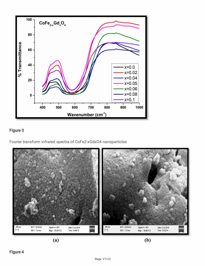
2.2 Fourier transforms infrared spectroscopy (FTIR)The FTIR spectra recorded for CoFe2 − xGdxO4 nano-crystalline powders are shown in Fig. 3. It is a crucialstudy that helps in understanding the changes that are caused by the different dopants in the ferritestructure. The FTIR spectra for all the samples showed two major absorption bands in the range 400-650cm− 1 which are the characteristic features of ferrite material. The absorption band with the lowerwavenumbers (400–425 cm− 1) is associated with the vibrations of the metal-oxygen bond at the

Page 5/23
octahedral site or B-site. The impact of rare earth substitution is predominantly felt on this band as itshows a marginal blue shift in frequency absorption with increasing rare earth concentration [35–43].
2.3 Scanning Electron MicroscopyThe morphological study of Gd+ 3 doped cobalt ferrite powders was done using the scanning electronmicroscope (SEM) instrument. The scanning electron micrographs of CoFe1.98Gd0.02O4 andCoFe1.96Gd0.04O4 are shown in Fig. 4. The SEM micrographs showed a highly agglomerated structureindicating strong magnetic behavior which is a characteristic of transition elements Fe and, Co, and alsoof inner transition element Gadolinium present in the ferrite samples under investigation [44–47].
Energy dispersive spectra (EDS)
The EDS spectra recorded for CoFe2O4 and CoFe0.6Gd0.04O4 are shown in Fig. 5 (a,b) and the elementalanalysis data of three samples is presented in Table 2. All the doped samples showed the presence of Co,Fe, Gd, and O. The weight percentage of Gd in the material was seen to increase with increasing rare earthsubstitution levels in the cobalt ferrite matrix [44, 45].
Table 2
EDS data of CoFe2 − xGdxO4 nanoparticles
Sample Weight % Atomic %
CoFe2O4 Co 21.04 15.28
Fe 41.29 31.64
O 18.09 48.39
CoFe1.98Gd0.02O4 Co 25.31 20.77
Fe 46.04 39.87
Gd 3.23 0.99
O 11.47 34.68
CoFe1.96Gd0.04O4 Co 29.09 23.25
Fe 54.79 46.20
Gd 6.40 1.92
O 9.73 28.64
2.4 Magnetic properties using vibrating samplemagnetometer

Page 6/23
The hysteresis loops obtained for Gd+ 3 doped cobalt ferrite samples are shown in Fig. 6. The variation ofsaturation magnetization (Ms), retentivity (MR), coercive �eld (HC), and anisotropy constant ‘K’ with Gd+ 3
concentration are shown in Fig. 7 (a,b,c). Although the magnetic parameters MS, MR, and HC showed a
random variation with increasing Gd+ 3 concentrations, the MS and MR were seen to vary inversely tolattice constant and while a direct analogy was observed between HC and (311) peak height depicting therelation between crystalline nature of the ferrite material. The magnetic environment in cobalt ferrite isadministered by Co+ 2, Fe+ 3 ions. With the spinel geometry, cobaltous ions occupy the tetrahedral site andferric ions occupy the octahedral site. Generally, the magnetic moment of the material,l in this case,e isexpressed in terms of Eq. 1 [45].
Where M is the magnetic moment of the spinel ferrite system, MB is the magnetic moment of theoctahedral site and MA is the net magnetic moment of the tetrahedral site. Howe, ver with the substitution
of a larger rare earth ion Gd+ 3 at the octahedral site, the system experiences an overall lattice expansionand cationic redistribution which changes the overall magnetic moment of the system. This may reducethe strength of super-exchange interaction between the Co+ 2 and Fe+ 3 ions at two cationic sublattice andincomplete ordering of Co+ 2 and Fe+ 3 at the tetrahedral sites and the octahedral sites in the spinelstructure
The variations in the magnetic moment can be also caused by changes in canting Yafet-Kittel angle (qYK)due to rare earth substitution. The magnetic moment of the rare earth doped system in this situation isexpressed in terms of Eq. 2 [46–50].
2.5 Magnetic susceptibilityThe AC susceptibility plots obtained for as prepared CoFe2 − xGdxO4 are shown in Fig. 8. Thismeasurement proves vital information about the Curie temperature and the type of magnetic domainspresent in the material. Depending upon the grain size, the domains present in the ferrite are categorizedinto three categories namely Single domain (SD), multi-domain (MD), and superparamagnetic domain(SP). The MD grains are characterized by the presence of multiple walls within the grain. If the grain sizeis comparable to that of the wall thickness then these grains are termed as single domains.Superparamagnetic behavior is predominantly seen in particles with dimensions < 10 nm. Here, themagnitude of thermal energy is comparable to that of magnetic anisotropy energy.
It can be seen from Fig. 8 that there is a gradual increase in normalized AC susceptibility followed by asteep rise creating a peak at high temperature before a sharp fall in the vicinity of the Curie temperature.This indicates that the as prepared samples contain a large section of single domain and a good numberof multi-domain grains as well. The values of curie temperature for all the samples were in the range of525oC to 575oC [51–53].
2.6 DC Resistivity

Page 7/23
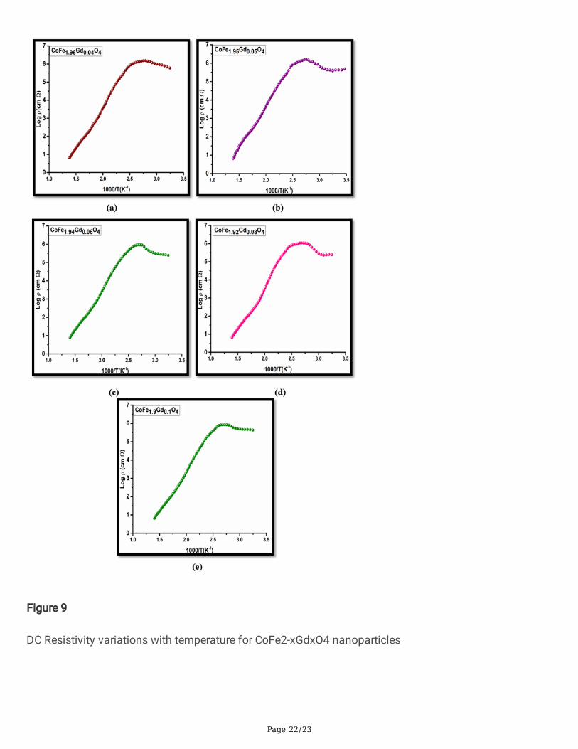
The plots of temperature-dependent variation of DC resistivity (ρ) over a range of 300K to 773K for rareearth doped cobalt ferrite samples are presented in Fig. 9. The rare earth doped ferrite powders showedan increase in resistivity with increasing temperature initially till a resistivity maxima of the order 107 Ω-cm in the temperature range of 473K to 523K. These high resistivity values are the attributes of theremoval of moisture present in the interstitial sites that facilitates the conduction mechanism. The highresistivity of the order 107 Ω-cm can be attributed to the smaller grain size and the large number of grainboundaries available in the nanomaterials. These grain boundaries are characterized by their insulatingnature and play a very important role in increasing the material resistivity [54–57]. The rare-earth ions(Gd+ 3) being a larger ion occupies the spacious octahedral site replacing Fe+ 3 ions which are generallyknown as a source of charge carriers. Hence this reduction in Fe+ 3 ions also causes the depletion ofcharge carriers [58–60]. The larger ionic radii of Gd+ 3 ions also alter the electron hopping mechanismadversely by causing lattice expansion. This increases the separation between Co+ 2 and Fe+ 3 ionsreducing the electronic exchange [61, 62].
2.7 Dielectric constant variation with frequencyThe plot of frequency dependence of dielectric constant is shown in Fig. 10 (a) and the values ofdielectric constant as at 20Hz for different Gd+ 3 concentrations are shown in Fig. 10 (b)
The Dielectric constant ‘ε’ was seen to decrease with the increasing frequency of the applied electric �eldand was seen to remain constant beyond a critical value of frequency. The increasing Gd3+ occupancy atthe B site replacing a smaller ion Fe3+ was also seen to affect the variation of the dielectric constant [63–66]. The Dielectric behavior of cobalt ferrite nanomaterials material is administered by the electronicexchange between metal ions and primarily dominated by the Fe3+/Co2+ ions. The doping of rare-earthions Gd3+ ions alters the structural parameters such as cell volume, density, lattice constant etc. and alsodecrease Fe+ 3 ion concentrations at the octahedral site. As a result, the electron hopping between Fe3+
and Co2+ ions gets affected adversely [67–70]. The dielectric constant pro�le showed a steep reduction inmagnitude to a lower value with an increase in frequency in the lower frequency region attain a constantlow value that remains constant over higher frequency values of the applied �eld. This variation indielectric properties can be explained using the space charge polarization phenomenon. An idealdielectric material is comprised of highly conducting grains. These grains are bounded by nonconductinggrain boundaries. When the electric �eld is applied, a potential drop occurs at the grain boundaries as aresult of the accumulation of space charge in this region. The in�uence of the grain boundaries isobserved distinctly over the lower frequency range. Beyond a certain value of applied �eld frequency, theelectronic transfer between the metal ions starts lagging behind the rate of variation in the frequency ofan applied electric �eld. This results in uniform lower magnitudes of dielectric constants[68–70].
2.8 Dielectric loss variation with frequencyThe variation of dielectric loss versus frequency for varying Gd3+ concentrations is presented in Fig. 11a.While the loss values for different Gd+ 3 concentrationS in cobalt ferrite samples at 20Hz are presented in

Page 8/23
Fig. 11b.
It can be seen that for nanoparticle samples dielectric loss decreases with increasing frequency ofapplied electric �eld and attains a low constant value beyond a critical frequency. At lower frequencies,the dielectric curve is seen to exhibit broad peaks in the neighborhood of 100Hz. The pro�le depicted inFig. 11b shows an approximately inverse behavior of dielectric loss to that of dielectric constant at 10Hz.The variation in the dielectric loss with increasing Gd3+ concentrations at low frequencies could be due tohindrance in the electronic exchange between Co+ 2/Fe+ 3 and Fe+ 2/Fe+ 3 [69–75]. At higher frequencies,the dielectric loss decreases drastically which may allow these materials to �nd their applications in high-frequency devices [76–79].
ConclusionThe ultra�ne powders of Gd+3doped cobalt ferrite with composition CoFe2-xGdxO4 (x=0.0, 0.02,0.04,0.05,0.06, 0.08, 0.1) with cubic spinel structure and crystallite size ranging between 21 nm to 41 nm wereprepared successfully using combustion route. The structural parameters such as lattice constant 'a’, X-density ‘DX', and unit cell volume were altered drastically due to Gd+3 substitution at the octahedral site.The particle agglomerates seen in the SEM images indicated a strong magnetic behavior. The EDSspectra showed the presence of Co, Fe, and Gd in desired quantities without any impurities. The A.C.susceptibility measurements indicated the presence of both single-domain and multidomain particles inall the samples. The changes observed in magnetic properties were attributed to cationic redistribution,lattice expansion, and alterations in magnetic ordering due to Gd+3 substitution.
The DC resistivity pro�le for all the samples showed high room-temperature resistivity of the order 107W-cm. The dielectric constant values were also seen to be administered by the concentrations of Gd+3 at theoctahedral site. The dielectric constant variation was explained using the space charge polarizationphenomenon. The variation in the dielectric loss with increasing Gd3+ concentrations at low frequencieswas attributed to hindrance in the electronic exchange between Fe+2/Fe+3 and Co+2/Fe+3. At higherfrequencies, all the samples were seen to exhibit low losses making them a potential candidate for highenergy applications.
DeclarationsWe, the authors of this manuscript entitled “In�uence of B-site Gd+3 substitution on various properties ofCo-ferrite nanoparticles” declare that, This article is original And has been written by the stated authorswho are all aware of its content and approve its submission. The article has not been publishedpreviously is not under consideration for publication elsewhere. We also declare that there exist nocon�ict of interest.

Page 9/23
References1. S. Thakur, S.C. Katyal, M. Singh, Structural and magnetic properties of nano nickel-zinc ferrite
synthesized by reverse micelle technique. J. Magn. Magn. Mater. 321, 1–7 (2009)
2. M. Sertkol, Y. Köseoğlu, A. Baykal, H. Kavas, M.S. Toprak, Syn- thesis and magnetic characterizationof Zn0.7Ni0.3Fe2O4 nanopar- ticles via microwave-assisted combustion route. J. Magn. Magn. Mater.322, 866–871 (2010)
3. M.H. Abdellatif, G.M.E. Komy, A.A. Azab, Magnetic characterization of rare earth doped spinel ferrite.J. Magn. Magn. Mater. 442, 445–452 (2017)
4. F. Nirav Joshi, Luís, H.S. da Silva, F.M. Jadhav, P.H. Shimizu, Jean-ClaudeM.’Peko Suman, MarceloOrnaghi Orlandi, Jeong Gil Seo, Valmor R Mastelaro, Osvaldo N Oliveira Jr, Yolk-shelled ZnCo2O4
microspheres: Surface properties and gas sensing application. Sens. Actuators B 257, 906–915(2018)
5. F. Nirav Joshi, Luis, H. Silva, M.'Peko Jadhav, Jean-Claude, B.B.M. Torres, K. Aguir, R. Valmor, N.Mastelaro, Osvaldo, Oliveira, One-step approach for preparing ozone gas sensors based onhierarchical NiCo2O4 structures. RSC Adv. 6, 92655–92662 (2016)
�. F. Nirav Joshi, Luís, H.S. da Silva, F.M. Jadhav, P.H. Shimizu, M.’Peko Suman, Jean-Claude, MarceloOrnaghi Orlandi, Jeong Gil Seo, R. Valmor, N. Mastelaro, Osvaldo, Oliveira Jr., Yolk-shelled ZnCo2O4microspheres: Surface properties and gas sensing application, Sensors and Actuators B: Chemical,257, 906–915, (2018)
7. N. Joshi, T. Hayasaka, Y. Liu, H. Liu, N. Osvaldo, L. Oliveira Jr., Lin, A review on chemiresistive roomtemperature gas sensors based on metal oxide nanostructures, graphene and 2D transition metaldichalcogenides. Microchim. Acta 185(4), 213 (2018)
�. V.K. Ritu Malik, N. Tomer, T. Joshi, L. Dankwort, L. Lin, Kienle, Au–TiO2-Loaded Cubic g-C3N4Nanohybrids for Photocatalytic and Volatile Organic Amine Sensing Applications, 10 (ACS appliedmaterials & interfaces, 2018), 40, 34087–34097
9. A. Singh, A. Kumar, A. Kumar, S. Samanta, N. Joshi, V. Balouria, A.K. Debnath, R. Prasad, Z. Salmi,M.M. Chehimi, D.K. Aswal, S.K. Gupta, Bending stress induced improved chemiresistive gas sensingcharacteristics of �exible cobalt-phthalocyanine thin �lms, Applied Physics Letters,102,132107,(2013)
10. B. Skolyszewska, W. Tokarz, K. Przybylski, Z. Kakol, Preparation and magnetic properties of Mg-Znand Mn-Zn ferrites. Physica C 38, 290 (2003)
11. H. Ibrahim Shari�, S. Shokrollahi, Amiri, Ferrite-based magnetic nano�uids used in hyperthermiaapplications. J. Magn. Magn. Mater. 324, 903–915 (2012)
12. Stephen, Blundell, Magnetism in Condensed Matter (Oxford University Press, Oxford, 2001)
13. S. Mahalakshmi, M. Srinivasa, S. Nithiyanantham, Magnetic studies of nickel ferrite doped with rareearth ions. Russ. J. Phys. Chem. 87, 1938 (2013)
14. A.A. Sattar, A.H. Wa�k, K.M. El-Shokroty, M.M. El-Tabby, Phys. State Solid A 171, 563 (1999)

Page 10/23
15. M.P. Ghosh, S. Mukherjee, J. Magn. Magn. Mater. 489, 165320 (2019)
1�. H. Yoon, J.S. Lee, J.H. Min, J.H. Wu, Y.K. Kim, Nanosci. Res. Lett. 8, 530 (2013)
17. Y. Shifeng, E. WeiLing, Zhou, Rapid synthesis of Mn0.65Zn0.35 Fe2O4/SiO2 homogeneous nano-composites by modi�ed sol-gel auto-combustion method,Journal Crystal Growth, 226–233 (2004)
1�. C. Scherer, A.M. Figueiredo Neto, Ferro�uids: properties and applications. Braz. J. Phys. 35(3), 718–727 (2005)
19. C.C. Berry, “Progress in functionalization of magnetic nanoparticles for applications in biomedicine,”Journal of Physics D, vol. 42, no. 22, Article ID 224003, 9 pages, 2009
20. H. Tan, J.M. Xue, B. Shuter, X. Li, J. Wang, Synthesis of PEOlated Fe3 O4 @SiO2 nanoparticles viabioinspired sili�cation for magnetic resonance imaging. Advanced Functional Materi- als 20(5), 722–731 (2010)
21. P. Pranav, R.B. Naik, Tangsali, Enduring effect of rare earth (Nd3+) doping, and γ- radiation onelectrical properties of nanoparticle manganese zinc ferrite. J. Alloy. Compd. 723, 266–275 (2017)
22. G. Baldi, D. Bonacchi, C. Innocenti, G. Lorenzi, C. Sangre- gorio, Cobalt ferrite nanoparticles: thecontrol of the particle size and surface state and their effects on magnetic properties. J. Magn.Magn. Mater. 311(1), 10–16 (2007)
23. G. Baldi, D. Bonacchi, M.C. Franchini et al., Synthesis and coating of cobalt ferrite nanoparticles: a�rst step toward the obtainment of new magnetic nanocarriers. Langmuir 23(7), 4026–4028 (2007)
24. A.R. Alam Abedini, Daud, Muhammad Azmi Abdul Hamid, Norinsan Kamil Othman, Radiolyticformation of Fe3O4 nanoparticles: in�uence of radiation dose on the structure and magneticproperties, (3) 1–8. (2014)
25. P. Pranav, R.B. Naik, S.S. Tangsali, S.M. Meena, Yusuf, In�uence of rare earth (Nd3+) doping onstructural and magnetic properties of nanocrystalline manganese-zinc ferrite. Mater. Chem. Phys.191, 215–224 (2017)
2�. P.S. Bharat Kataria, U. Solanki, M. Khachar, A. Vagadia, M.J. Ravalia, P. Keshvani, D. Trivedi, V.Venkateshwarlu, K. Ganesan, N.A. Asokan, D.G. Shah, Kuberkar, Role of strain and microstructure inchemical solution deposited La0.7Pb0.3MnO3 manganite �lms: thickness-dependent swift heavy ionsirradiation studies. Rad. Phys. Chem. 85, 173–178 (2013)
27. M. Deepak Kumar, A. D’souza, M. Chatim, V. Naik, P.P. Naik, R.B. Tangsali, Effect of Rare-earth dopingon Magnetic and Electrical Transport Properties of Nanoparticle Mn Zn Ferrite. Adv. Sci. Lett. 22,773–779 (2016)
2�. S. Nidhi Tendulkar, V. Patil, P.P. Kuncalienkar, M. Naik, S. Kundaikar, Keluskar, Study of Structural andMagnetic Properties of Mn0.8Zn0.2Fe2O4 Nanoparticles, Advanced Engineering Technology andApplication, 5, No. 1, 19–22 (2016)
29. N.Hanh, O.K.Quy, N.P.Thuy, L.D.Tung, L..Spinu, Synthesis of cobalt ferrite nanocrystallites by theforced hydrolysis method and investigation of their magnetic properties. Phys. B 327, 382–384(2003)

Page 11/23
30. L. Xuebo Caol, Gu, Spindly cobalt ferrite nanocrystals: preparation, characterization and magneticproperties, Nanotechnology16 180–185 (2005)
31. Z. Cvejic, B. Antic, A. Kremenovic, S. Rakic, G.F. Goya, H.R. Rechenberg, C. Jovalekic, V. Spasojevic,In�uence of heavy rare earth ions substitution on microstructure and magnetism of nanocrystallinemagnetite. J. Alloys Compd. 472, 571–575 (2009)
32. S.M. Patange, S.E. Shirsath, G.S. Jangam, K.S. Lohar, S.S. Jadhav, K.M. Jadhav, Rietveld structurere�nement, cation distribution and magnetic properties of Al3+ substituted NiFe2O4 nanoparticles. J.Appl. Phys. 109, 053909 (2011)
33. N. Menon, S.R. Nagel, Evidence for a divergent suscepti- bility at the glass transition. Phys. Rev. Lett.74(7), 1230–1233 (1995)
34. G.A. Sawatzky, F. van der Woude, A.H. Morrish, Mo ̈ssbauer study of several ferrimagnetic spinels.Phys. Rev. 187(2), 747–757 (1969)
35. M.A. Amer, Structural and magnetic studies of the Co1 + xTixFe2 (1– x) O4 ferrites. J. Magn. Magn.Mater. 426, 771–778 (2017)
3�. M.A. Amer, A. Matsuda, G. Kawamura, R. El-Shater, T. Meaz, F. Fakhry, Characterization and structuraland magnetic studies of as-synthesized Fe2+CrxFe(2–x)O4 nanoparticles. J. Magn. Magn. Mater. 439,373–383 (2017)
37. E.H. El-Ghazzawy, M.A. Amer, Structural, Elastic and magnetic studies of the as-synthesized Co1 –xSrxFe2O4 nanoparticles. J. Alloy. Compd. 690, 293–303 (2017)
3�. Y.G. Zhang, Magnetic Materials, Chengdu, Pekin, 1988 (Chap. 1)
39. S.R. Naik, A.V. Salker, Change in the magnetostructural properties of rare-earth-doped cobalt ferritesrelative to the magnetic anisotropy. Journal of Material Chemistry 22, 2740 (2012)
40. L. Zhao, H. Yang, X. Zhao, L. Yu, Y. Cui, S. Feng, Magnetic properties of CoFe2O4 ferrite doped withrare-earth ion, Material Letters 60 1–6, (2006)
41. B.V. Ch Srinivas, A. Tirupanyam, V. Satish, D.L. Seshubai, O.F. Sas- try, Caltun, Effect of Ni2 +substitution on structural and mag- netic properties of Ni–Zn ferrite nanoparticles. J. Magn. Magn.Mater. 382, 15–19 (2015)
42. 17. Ch Srinivas, B.V. Tirupanyam, S.S. Meena, S.M. Yusuf, C.S. Babu, K.S. Ramakrishna, D.M.Potukuchi, D.L. Sastry, Structural and magnetic characterization of co-precipitated NixZn1–xFe2O4ferrite nanoparticles. J. Magn. Magn. Mater. 407, 135–141 (2016)
43. F.-X. Cheng, J.-T. Jia, Z.-G. Xu, B. Zhou, C.-S. Liao et al., Microstructure, magnetic, and magneto-optical properties of chemical synthesized Co–RE (RE = Ho,†Er,†Tm,†Yb,†Lu) ferrite nanocrystalline�lms. J. Appl. Phys. 86, 2727–2732 (1999)
44. K.S. Rane, V.M.S. Verenkar, Synthesis of ferrite grade γ-Fe2O3. Bull. Mater. Sci. 24, 39–45 (2001)
45. 20R.A. Porob, S.Z. Khan, S.C. Mojumdar, V.M.S. Veren- kar, Synthesis, TG, DSC, and infrared spectralstudy of NiMn2(C4H4O4)3·6N2H4-a precursor for NiMn2O4 nanoparticles. J. Therm. Anal. Calorim.86, 605–608 (2006)

Page 12/23
4�. S. Anjali Bishnoi, Kumar, Nirav Joshi Wide-Angle X-ray Diffraction (WXRD): Technique forCharacterization of Nanomaterials and Polymer Nanocomposites. Microscopy Methods inNanomaterials Characterization 1, 313–337 (2017)
47. A. Baykal, F. Genç, A.Z. Elmal, S. Geokçe, M. Sertkol, H. Seozeri, MnCrxFe2 – xO4 Nanoparticles:Magnetic and Microwave Absorption Properties. J. Inorg. Organomet. Polym Mater. 26, 134–141(2016)
4�. H. Shenker, Magnetic anisotropy of cobalt ferrite (Co1.01 Fe2.00O3.62) and nickel cobalt ferrite(Ni0.72Fe0.20Co0.08Fe2O4). Phys. Rev. 107(5), 1246–1249 (1957)
49. M.B. Morales, M.H. Phan, S. Pal, N.A. Frey, H. Srikanth, “Particle blocking and carrier �uid freezingeffects on the magnetic properties of Fe3O4 -based ferro�uids,” Journal of Applied Physics, vol. 105,no. 7, Article ID 07B511, 3 pages, 2009
50. G. Baldi, D. Bonacchi, C. Innocenti, G. Lorenzi, C. Sangre- gorio, Cobalt ferrite nanoparticles: thecontrol of the particle size and surface state and their effects on magnetic properties. J. Magn.Magn. Mater. 311(1), 10–16 (2007)
51. N.D. Chaudhari, R.C. Kambale, D.N. Bhosale, S.S. Suryavanshi, S.R. Sawant, Thermal hysteresis anddomain states in Ni-Zn fer- rites synthesized by oxalate precursor method. J. Magn. Magn. Mater.322, 1999–2005 (2010)
52. O.A. Li, C.-R. Lin, H.-Y. Chen, H.-S. Hsu, K.-Y. Shih, I.S. Edelman, K.-W. Wu, Y.-T. Tseng, S.G. Ovchinnikov,J.-S. Lee, Size dependent magnetic and magneto-optical properties of Ni0.2Zn0.8Fe2O4nanoparticles. J. Magn. Magn. Mater. 408, 206–212 (2016)
53. V. Grimal, D. Autissier, L. Longuet, H. Pascard, M. Gervais, Iron, nickel and zinc stoichiometricin�uences on the dynamic magneto-elastic properties of spinel ferrites. J. Eur. Ceram. Soc. 26, 3687–3693 (2006)
54. P.P. Naik, R.B. Tangsali, B. Sonaye, S. Sugur, Radiation stimulated permanent alterations in structuraland electrical properties of core-shell Mn-Zn ferrite nanoparticles. J. Nano Res. Vol 24, 194–202,(2013) pp
55. P. Pranav, R.B. Naik, B. Tangsali, S. Sonaye, Sugur, Sustained augmentation in electrical properties ofMnxZn1-xFe2O4 nanoparticles provoked by high energy gamma radiation. Journal ofNanotechnology and Advanced Materials 3(1), 1–7 (2015)
5�. V. Manisha, D. Rane, A.K. Bahadur, Nigam, C.M. Srivastava, Mössbauer and FT-IR studies on non-stoichiometric barium hexaferrites. J. Magn. Magn. Mater. 192, 288–296 (1999)
57. P. Pranav, R.B. Naik, Tangsali, Enduring Effect of Rare Earth (Nd+ 3) Doping and, γ- Radiation onElectrical properties of Nanoparticle Manganese Zinc Ferrite. J. Alloy. Compd. 723, 266–275 (2017)
5�. M. Asif Iqbal, M.U. Islam, I. Ali, M.A. Khan, I. Sadiq, I. Ali, High-frequency dielectric properties of Eu +3-substituted Li–Mg ferrites synthesized by sol-gel auto-combustion method. J. Alloy. Compd. 586,404–410, (2014)
59. B. Chandra Babu, N.V. Jayaprakash., S. B, Buddhudu, Structural, thermal and dielectric properties oflithium zinc silicate ceramic powders by sol-gel method. Ferro Electrics letter 38, 114–127 (2011)

Page 13/23
�0. N. Rezlescu, E. Rezlescu, Dielectric properties of copper containing ferrites. Physics Status Solidi A.23, 575–582 (1974)
�1. I.H. Gul, A. Maqsood, M. Naeem, M. Naeem Ashiq, Optical, magnetic and electrical investigation ofcobalt ferrite nanoparticles synthesized by co-precipitation route. J. Alloys Compd. 507, 201–206(2010)
�2. M.T. Farid, I. Ahmad, S. Aman, Characterization of nickel-based spinel ferrites with small substitutionof praseodymium. Journal of Chemical Society of Pakistan 35, 793–799 (2013)
�3. V.D. Mote, Y. Purushottam, B.N. Dole, Williamson-Hall analysis in estimation of lattice strain innanometer-sized ZnO particles. J. Theor. Appl. Phys. 6, 2251–7235 (2012)
�4. C. Venkataraju, Effect of nickel on the structural properties of Mn Zn ferrite nanoparticles. Appl. Phys.Res. 1(1), 41–45 (2009)
�5. L. Zhao, H. Yang, X. Zhao, L. Yu, Y. Cui, S. Feng, Magnetic properties of CoFe2O4 ferrite doped withrare earth ion, Mater. Lett. 60,1–6, (2006)
��. M.M. Haque, M. Huq, M.A. Hakim, Effect of Cu for Mn on the magnetic properties of Mn-Zn ferrites.Indian J. Phys. 78A(3), 397–400 (2004)
�7. M.M. Emad, M.M. Ewais, A. Hessien, Abdel-Hady. El-Geassy, In-situ synthesis of magnetic Mn-Znferrite ceramic object by solid state reaction. J. Aust. Ceram. Soc. 44(1), 57–62 (2008)
��. K. Rama Krishna, K. Vijaya Kumar, D. Ravinder, Structural and electrical conductivity studies in nickel-zinc ferrite. Adv. Mater. Phys. Chem. 2, 185–191 (2012)
�9. M.F. Sarac, Magnetic, Structural, and Optical Properties of Gadolinium-Substituted Co0.5Ni0.5Fe2O4
Spinel Ferrite Nanostructures, Journal of Superconductivity and Novel Magnetism,doi.org/10.1007/s10948-019-05359-3
70. C. Murugesan, G. Chandrasekaran, Impact of Gd3+ substitution on the structural, magnetic andelectrical properties of cobalt ferrite nanoparticles. RSC Adv. 5, 73714–73725 (2015)
71. J.G. Kang, B.K. Min, Y. Sohn, Synthesis and characterization of Gd(OH)3 and Gd2O3 nanorods.Ceram. Int. 41, 1243–1248 (2015)
72. M.T. Farid, I. Ahmad, S. Aman, M. Kanwal, G. Murtaza, I. Ali, I. Ahmad, M. Iashfaq, Structural, electricaland dielectric behavior of NixCo1-xNdyFe2-yO4 nano ferrites synthesized by Sol-Gel Method. DigestJournal of Nanomaterials and Biostructures 10(1), 265–275 (2015)
73. G. Kumar, J. Shah, R.K. Kotnala, P. Dhiman, R. Rani, V.P. Singh, G. Garg, S.E. Shirsath, K.M. Batoo, M.Singh, Self-ignited synthesis of Mg–Gd–Mn nano ferrites and impact of cation distribution on thedielectric properties. Ceram. Int. 40(9), 14509–14516, (2014)
74. K.W. Wagner, Dissipation of energy under AC. American Physics 40, 317 (1973)
75. S. Snehal, P.P. Hasolkar, Naik, Effect of Gd + 3 doping on structural, Magnetic and electrical propertiesof Mn0.5Co0.5Fe2-xGdxO4nano-particles prepared using combustion synthesis. J. Alloy. Compd.823, 153605 (2020)

Page 14/23
7�. L. Ajroudi, N. Mliki, L. Bessais, V. Madigou, S. Villain, Ch. Leroux, Magnetic, electric and thermalproperties of cobalt ferrite nanoparticles. Mater. Res. Bull. 59, 49–58 (2014)
77. N.J. Joshi, G.S. Grewal, V. Shrinet, T.P. Govindan, A. Pratap, Synthesis and dielectric behavior of nano-scale barium titanate. IEEE dielectrics &Electrical Insulations 19, 83–90 (2012)
7�. N.J. Joshi, G.S. Grewal, V. Shrinet, A. Pratap, N.J. Buch, Synthesis and characterization of nano-barium titanate prepared by hydrothermal process. Integrated Ferroelectrics 115, 142–148, (2010)
79. A. Pratap, N.J. Joshi, P.B. Rakshit, G.S. Grewal, V. Shrinet, Dielectric behavior of nano barium titanate�lled polymeric composites, International Journal of Modern Physics: Conference Series,22,1–10,(2013)
Figures

Page 15/23
Figure 1
Rietveld re�nement of X-ray diffraction pattern CoFe2-xGdxO4 nanoparticles.

Page 16/23
Figure 2
Variation in structural parameters such as a) Lattice constant ‘a’, b) Lattice strain, c) X-ray density ‘DX’, d)Crystallite size ‘t’, and e) Cell volume f) (311) peak intensity with Gd+3 concentration
Page 17/23
Figure 3
Fourier transform infrared spectra of CoFe2-xGdxO4 nanoparticles
Figure 4

Page 18/23
Scanning Electron micrographs of CoFe1.98Gd0.02O4 and CoFe1.96Gd0.04O4
Figure 5
Energy dispersive spectrum of (a) CoFe2O4 (b) CoFe1.96Gd0.04O4

Page 19/23
Figure 6
Magnetic hysteresis loops of CoFe2-xGdxO4 nanoparticles

Page 20/23
Figure 7
Magnetic parameters of CoFe2-xGdxO4 nanoparticles

Page 21/23
Figure 8
The normalized AC susceptibility of CoFexGd1-xO4
Page 22/23
Figure 9
DC Resistivity variations with temperature for CoFe2-xGdxO4 nanoparticles

Page 23/23
Figure 10
Variation of dielectric constant ‘ε’ (a) with frequency (b) at 20Hz
Figure 11
Variation of dielectric loss (a) with frequency (b) at 20Hz