Prognostic value of cardiovascular parameters in CT ...
24
Early View Original article Prognostic value of cardiovascular parameters in CT Pulmonary Angiography in patients with acute pulmonary embolism Ludo F.M. Beenen, Patrick Bossuyt, Jaap Stoker, Saskia Middeldorp Please cite this article as: Beenen LFM, Bossuyt P, Stoker J, et al. Prognostic value of cardiovascular parameters in CT Pulmonary Angiography in patients with acute pulmonary embolism. Eur Respir J 2018; in press (https://doi.org/10.1183/13993003.02611-2017). This manuscript has recently been accepted for publication in the European Respiratory Journal. It is published here in its accepted form prior to copyediting and typesetting by our production team. After these production processes are complete and the authors have approved the resulting proofs, the article will move to the latest issue of the ERJ online. Copyright ©ERS 2018 . Published on May 17, 2018 as doi: 10.1183/13993003.02611-2017 ERJ Express Copyright 2018 by the European Respiratory Society.
Transcript of Prognostic value of cardiovascular parameters in CT ...
Early View
Original article
Prognostic value of cardiovascular parameters in
CT Pulmonary Angiography in patients with acute
pulmonary embolism
Ludo F.M. Beenen, Patrick Bossuyt, Jaap Stoker, Saskia Middeldorp
Please cite this article as: Beenen LFM, Bossuyt P, Stoker J, et al. Prognostic value of
cardiovascular parameters in CT Pulmonary Angiography in patients with acute pulmonary
embolism. Eur Respir J 2018; in press (https://doi.org/10.1183/13993003.02611-2017).
This manuscript has recently been accepted for publication in the European Respiratory Journal. It is
published here in its accepted form prior to copyediting and typesetting by our production team. After
these production processes are complete and the authors have approved the resulting proofs, the article
will move to the latest issue of the ERJ online.
Copyright ©ERS 2018
. Published on May 17, 2018 as doi: 10.1183/13993003.02611-2017ERJ Express
Copyright 2018 by the European Respiratory Society.

Title: Prognostic value of cardiovascular parameters in CT Pulmonary
Angiography in patients with acute pulmonary embolism.
Author contact details:
Ludo F.M. Beenen, MD (corresponding author) Department of Radiology and Nuclear Medicine, Academic Medical Center, University of Amsterdam Meibergdreef 9 1105 AZ Amsterdam The Netherlands Tel: 003120-5668698 Fax: 003120-5669119 E-mail: [email protected]
Prof. Patrick Bossuyt, PhD Department of Clinical Epidemiology, Academic Medical Center, University of Amsterdam Meibergdreef 9 1105 AZ Amsterdam The Netherlands E-mail: [email protected] Prof. Jaap Stoker, MD, PhD Department of Radiology and Nuclear Medicine, Academic Medical Center, University of Amsterdam Meibergdreef 9 1105 AZ Amsterdam The Netherlands E-mail:[email protected] Prof. Saskia Middeldorp, MD, PhD Department of Vascular Medicine, Academic Medical Center, University of Amsterdam Meibergdreef 9 1105 AZ Amsterdam The Netherlands E-mail: [email protected]

Introduction
Pulmonary embolism (PE) is the third cardiovascular disease worldwide with mortality
ranging up to 25%[1]. Calculating the risks of adverse outcome for a patient can guide
therapeutic decision (home therapy, hospitalization, or thrombolysis)[2-4]. This risk can be
based on clinical, biochemical and imaging parameters[5-7]. The detrimental consequences
from PE are thought to be mainly associated with the development of right ventricular
dysfunction (RVD), which could cause increase of cardiac biomarkers such as NT-proBNP[8].
The burden to the heart would lead to overall heart failure and subsequent death.
ESC guidelines categorize the risk of adverse outcome as high, intermediate or low. Risk
calculations are based on sPESI, and are suggested to guide treatment accordingly[2]. For
the large intermediate risk group, fine tuning can be done on the presence of RVD,
categorizing patients to intermediate-high or intermediate-low risk as assessed by
biomarkers or imaging[9]. In daily practice however, additional tests such as ultrasound or
NT-proBNP are frequently not performed [10]. It would be ideal, if CT Pulmonary
angiography (CTPA), the reference standard for the diagnosis of PE, could also be used to
assess the prognosis [11]. So far, heterogeneity in study groups, definitions, and outcomes
prohibits consensus on the prognostic performance of CTPA [12]. Two multicenter
prospective studies have suggested that the right-to-left ventricular ratio can be used as a
predictor for mortality. As these studies did not investigate other potential predictive
parameters, the unique position of the right-to-left ventricular ratio can be questioned [13,
14]. Other reported radiological findings such as cardiovascular diameters, backflow or clot
burden have been evaluated but findings on their value are inconsistent[15-20].
Consequently, it is unclear if one or more CTPA parameters can contribute to risk
stratification in patients with acute PE.
To add strong evidence to the debate on the value of CTPA parameters in risk stratification
we analysed imaging, clinical and follow-up data collected in a prospective multicenter trial
in patients with acute PE [21]. Our focus was on the evaluation of the predictive effects of
baseline CTPA parameters on short and long term clinical outcome.
Materials and Methods
Patients and study design
Patient data and images were collected in the context of a large international randomized
clinical trial comparing two anticoagulant regimens in patients with venous thrombo-
embolism (VTE). The results, design and methods of the Hokusai-VTE study have been
described in detail previously (ClinicalTrials.gov identifier: NCT00986154)[21]. In short,

eligible patients were patients aged 18 years or older with acute, symptomatic venous
thromboembolism (deep vein thrombosis and/or PE). Patients were excluded in case of
contraindications to heparin or warfarin, severely impaired renal function or pregnancy. The
institutional review board at each participating center approved the general study protocol,
and all patients provided written informed consent.
Patients were enrolled between January 2010 and October 2012 at 439 centers in 37
countries. All data for the present analysis had been collected and assessed prospectively
before the trial data lock. Follow-up was 12 months, covering both the in-hospital period as
well as regular outpatient clinic controls, and patients on and off anticoagulant treatment.
Adverse events were noted on separate forms, as well as whether this was PE related. An
independent committee adjudicated all predefined outcomes.
For this additional study all patients with PE, either with or without DVT, were selected.
Excluded were patients with DVT only, patients not evaluated by CTPA, or when images were
not available in DICOM format or inaccessible for reading in the used image viewer (e.g. hard
copy, corrupted discs).
Data collection
All clinical and radiological data were anonymized, and centrally registered with double data
entry by an independent trial data management agency. Clinical data were retrieved from
the original CRFs. In all patients NT-proBNP levels were measured at baseline.
CT-data were acquired from the local participating centers, using local settings and
protocols. This means that a wide variety of CT-scanners were used, from basic until high-
end CT. For quality evaluation a 5-point Likert scale was used, anchored at 1 (unacceptable),
2 (poor), 3 (satisfactory), 4 (good) and 5 (excellent quality). The enhancement of the
pulmonary trunk was assessed by measuring a 1 cm region of interest (ROI) and expressed in
Hounsfield Units (HU).
Anonymized patient images from the central database were evaluated by a radiologist (LB)
with 12 years of experience in chest imaging supported by a dedicated research assistant.
Both were unaware of patient details and clinical information. For image reading a
commercially available image viewer was used (eFilm Workstation for Windows Version
3.4.0, Build 10, Merge Technologies Inc., Milwaukee, USA). Images were primarily read in
axial sections with additional support of multiplanar reformatting (MPR). Standard
pulmonary angiography, mediastinal and lung parenchyma window settings were used, with
individual adaptation if deemed necessary. Data were registered on a specially designed CRF.
A random sample of 50 patients was used to evaluate the intra-observer variability for the
main study parameters, as assessed by Cohen’s k-statistic. Intra-observer agreement was
graded according to Landis and Koch, with 0-20 indicating poor correlation, 20-40 moderate,

40-60 fair, 60-80 good, and 80-100 excellent correlation. No additional readers were
engaged as intra-observer agreement for the selected parameters is reportedly high [22-24].
All continuous variables were noted in millimetres where applicable. The following
parameters were assessed: transverse diameter of right ventricle, left ventricle (both on
axial and reformatted short axis view), pulmonary trunk, ascending aorta, inferior and
superior caval vein, azygos vein, right atrium, and heart and intrathoracic diameters. For the
ventricular diameters, the largest cross-sectional distance between ventricular surfaces was
taken. The right atrium was measured at its largest transverse diameter. Pulmonary trunk
was measured at its largest transverse diameter, the ascending aorta at the level of the
carina, the caval veins were measured 2 cm from their entrance into the right atrium, and
the azygos vein at its most cranial part. For the heart volume and the intrathoracic distance,
the largest transverse diameters from pericardial contours and costal margins were taken.
The right-to-left ventricular (RV/LV), right-to-left ventricular short axis (RV/LVsa), and
pulmonary trunk-to-aorta (TP/Ao) ratios were calculated by dividing the values of respective
transverse diameters. All obtained values were then dichotomized at earlier reported
thresholds (RV/LV > 1.0; RV/LVsa > 0.9; TP/A0 > 1.0, TP > 29 mm; cardiothoracic ratio > 0.50).
Ordinal measures were: bowing of the interventricular septum (negative, neutral, positive);
reflux of contrast medium in the inferior caval vein (no, only into the IVC, intrahepatic < 3
cm, and intrahepatic veins > 3 cm) and in the azygos vein (yes or no). Interventricular septum
bowing was considered present when the septum was curved to the left ventricle, or
flattened if the septum was straightened or bowed. Backflow was considered positive if
reflux was into the intrahepatic veins; only into the inferior caval vein was considered
negative. Azygos vein reflux was considered present if it reached the crossing with the right
main stem bronchus.
Events were analysed focusing on 4 time points: early (1 week and 1 month) and late (on
treatment, mostly 3-6 months, and 12 month) period. For right ventricular dysfunction the
reference standard was an increased value of NT-proBNP ≥ 600 pg/ml at baseline[2].
Statistical analysis
Primary outcome for the study was mortality, secondary outcomes were recurrent VTE,
hospitalization, bleeding and all adverse events. We calculated odds ratios with 95%
confidence intervals (CI) to express the strength of the association between cardiovascular
CTPA parameters and mortality, as well as other clinical outcomes. We also calculated
estimates of sensitivity and specificity, PPV and NPV for mortality. Missing data were
excluded from the analysis. No correction for multiple testing was performed. Significance of
differences were evaluated with two-sided p-values; a p-value < 0.05 was considered to
imply statistical significance. All statistics were performed in SPSS version 23 (SPSS Inc,
Chicago Ill).

Results
In the RCT, 3,481 patients had PE, of which 3114 had been diagnosed using CTPA. After
screening, 1164 of these were excluded because because images were presented on hard
copies, jpeg or pdf only, no DICOM images were available, or because of a technically
inadequate study as e.g. insufficient coverage of heart and chest (Figure 1). To address
possible selection bias, we compared baseline characteristics of included and excluded
patients and found no relevant differences. One year outcomes also were not different, as
mortality and recurrent VTE was 3.0 and 2.6% for the included and 3.1% and 2.7% for the excluded
group, respectively. Hence, data of 1950 patients were included in this evaluation. Of these,
1049 (54%) were male. Mean age was 57 years. A summary of their characteristics is shown
in Table 1. PE was provoked in 1288 patients, 456 patients had PE with concomitant DVT. In
565 patients the NT-proBNP level was > 600 pg/ml.
Quality
Overall quality of the scans was good (3.7/5; SD=0.8). Mean Hounsfield Units in the
pulmonary trunk was 325 (SD=118). Intra-observer agreement on a random sample from the
complete database scored twice was excellent (kappa=0.9).
Frequencies
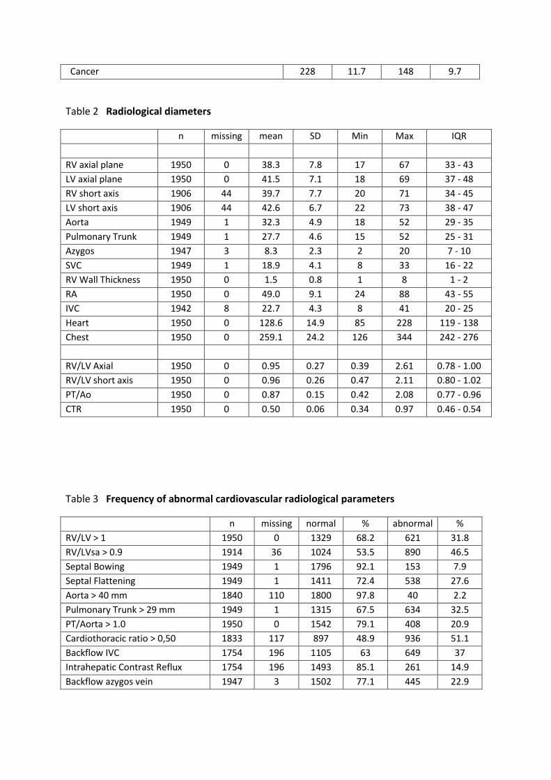
The median right-to-left-ventricle ratio on CTPA was 0.89 (SD=0.27); 621 patients (32%) had
a ratio >1 (Tables 2 and 3). Compared to those without RVD on CT, in patients with RV/LV>1
NT-proBNP more often was raised. The median short axis right-to-left-ventricle ratio was
0.88, of which 890 (47%) were > 0.90.
In 538 (28%) patients the septum was flattened, septal bowing occurred in 153 patients
(7.9%). The pulmonary trunk was enlarged in 634 patients (33%). A pulmonary trunk/aorta
ratio > 1 was present in 408 patients (20.9%). Backflow of contrast medium into the hepatic
veins occurred in 261 (15%), and into the azygos vein in 445 (23%) patients.
Short term outcomes
A summary of the investigated cardiovascular radiological parameters and their correlation
with short and long term adverse events are displayed in Tables 4a and 4b (mortality) and 5
(only online: recurrent VTE, hospitalization, major bleeding and all adverse events).
During the first month 29 adverse events occurred, including 18 deaths, 12 recurrent VTEs,
13 bleedings. There were 26 hospitalizations.
Of all the radiological parameters evaluated, only pulmonary trunk diameter > 29 mm was
significantly associated with mortality at 1 week (OR 4.18, CI=1.04-16.8; p=0.028, Table 2
and 3). The odds ratio at 1 month was lower and not statistically significant (OR 2.30,
CI=0.97-5.45; p=0.051). All other parameters (RV/LV ratio, RV/LV short axis, septal bowing,
pulmonary trunk/aorta ratio, cardiothoracic ratio and backflow to hepatic veins or azygos

vein) were not significantly associated with mortality. Of the 9 patients that died within the first
week, 6 (66.7%) had an enlarged pulmonary trunk. In total 18 patients died within one month, an
enlarged pulmonary trunk was present in half of these 18 patients. In patients who survived one
week or subsequently one month an enlarged pulmonary trunk was present in 628 and 625 patients
(32.4%, p = 0.028 respectively 32.4%, p=0.11).
An enlarged pulmonary trunk diameter was also associated with recurrent VTE (OR 5.22,
CI=1.01-26.7; p=0.028) at 1 week. Here also the odds ratio was lower and not significant at 1
month (1.8, CI=0.6-5.3; p=0.051). None of the evaluated radiological parameters, apart from
enlarged pulmonary trunk diameter was associated with hospitalization. Sensitivities were
low for all the researched parameters, as were the specificities and positive predictive
values; however, all parameters showed a high negative predictive value.
Long term outcomes
The median on treatment time was 215 days (IQR 178-358 days). During the complete 1
year period, 143 adverse events were registered in 131 patients. In total 58 patients died, 49
had recurrent VTE, 30 had a major bleeding and 90 were hospitalized.
An enlarged pulmonary trunk diameter was significantly associated with mortality during the
on-treatment time as well as for the complete 12 months (p=0.004 resp. 0.001). A TP/Aorta
ratio > 1.0 was also significantly associated with mortality during treatment (p=0.002; Table
4) but not for the complete period (p=0.055). Of the 11 patients with interstitial lung disease, 2
patients that had an enlarged pulmonary trunk died. In 43 patients with a history of pulmonary
hypertension, 21 had an enlarged pulmonary trunk of which 2 died. All other evaluated
cardiovascular parameters were not significantly associated with mortality or other adverse
events.
Discussion
Our study showed that most of the investigated cardiovascular radiological parameters -
including RV/LV ratio, septal bowing, cardiothoracic ratio and contrast medium backflow -
have no prognostic value for short or long term mortality. The exception was an enlarged
pulmonary trunk diameter, which on both short and long term was associated with
increased mortality and the risk of recurrent VTE and hospitalization.
A strength of our study is that data were prospectively collected in a large international trial,
and both imaging data and clinical outcomes were assessed blinded for treatment and
outcome.
Our study also has limitations. Although in literature many parameters have been evaluated,
we only analysed the most frequently used radiological parameters and cut off values as
these would be most easily implementable, had we found any of these to be of value. As

reconstructed views yield comparative values but are more time-consuming, plain axial
transverse images generally are preferred given the simplicity of analysis [25]. We evaluated
observer agreement only for the main continuous variables, and not for the ordinal
measurements. We also did not perform separate assessments for treatment allocation to
edoxaban or enoxaparin followed by warfarin, as this subgroup analysis was done in the
original dataset [21]. We did not perform a multivariable analysis, as we first aimed to assess
the prognostic value of each parameter separately. Also, echocardiography can be a useful
tool for short term mortality risk stratification [12]. As only 523 (26.8%) of the evaluated
patients received this test, this was not analysed in the present study. We are aware that
patients included in a randomized controlled trial do not necessarily reflect all those
presenting in regular practice, and our results cannot be unconditionally generalized to
those with exclusion criteria for the trial, such as hemodynamically unstable patients,
patients with a limited life expectancy and pregnant women.
How do our findings fit into the current assessment of prognosis in patients with acute PE?
We need better tools to identify high risk patients with a favourable risk-benefit ratio from
thrombolysis, or, alternatively, to identify those who would benefit from close clinical
monitoring in order to provide them with rescue thrombolysis. As the beneficial effect of
thrombolysis primarily reflects the first days, an easily applicable modifier like an enlarged
pulmonary trunk would probably facilitate such processes. In recent ESC guidelines primary
categorization into low, intermediate or high risk is based on sPESI. In second instance either
biomarkers, RV/LV ratio or echocardiography can be used for further stratification on RVD.
However, no consensus exists on its usefulness, as well as on the threshold, as RVD values
reported in the literatures are ranging from 0.9 until 1.8[26].
Several studies have reported that RVD on CTPA is an indicator of the risk of adverse events
[13, 27]. Many studies however had a single center, retrospective design, with short follow
up and surrogate outcomes. As such, they have intrinsic methodological limitations that
weaken their validity and generalizability. The larger series have shown conflicting results,
either confirming or denying that right-to-left ventricular ratio is associated with an
increased mortality [14],[28, 29],[30].
A recent systematic review stated that although RVD assessed by CT showed an association
with an increased risk of mortality in patients with hemodynamically stable PE, it resulted in
only small increases in the ability to classify risk[31]. Although additional publications
confirmed this finding [[31, 32], apparently, RV enlargement alone is not sufficient to
indicate a poor short-term prognosis, and other factors should also be taken into
consideration[33]. For the long-term persistent RV dysfunction seems common, reflecting on
diminished exercise capacity and reduced quality of life [34]. One of the differences with the
published cohorts is the fact that our study contains a population that was included in a
randomized clinical trial rather than a prospective cohort study of consecutive patients, and
thus could reflect different study populations. Our finding that right-to-left ventricular ratio

is not associated with an increased mortality could thus be an incentive to reconsider the
risk stratification algorithm.
Reports on the other investigated outcomes –recurrent VTE, hospitalization, bleeding and
adverse effects- are scarce, as most often they are used as a composite outcome, or focus on
differences between treatment regimens[35].
Although an enlarged pulmonary trunk diameter is an established feature in the work up of
chronic PE, for acute PE findings are contradictory, as an association with increased risk was
not always observed in previous studies[36-40]. However, most of these studies were
retrospective with limited number of patients. The assessment however is rather easy and
not as time consuming as e.g. clot obstruction scores, and thus could be used easily in daily
practice. Sensitivity for enlarged pulmonary trunk diameter may be low, but as specificity
was high, we may be able to better identify specific risk groups. Its high negative predictive
value indicates that it may be useful for identification of those patients that have a low risk
for adverse events who will not need for aggressive therapy, and can be discharged home
early. However, for prognostication towards high risk measures like admission to ICU or
thrombolysis a multifactorial risk-benefit analysis would be necessary.
One intriguing point is the apparent discrepancy between the relative high number of RVD
observed in the earlier published studies, and the fortunately relatively low mortality
percentages. In other words: although many patients are categorized as high risk, be it from
radiological, biochemical, or combined, this does not translate in the same manner in
mortality and adverse events. From this point it should be logical to better investigate the
role of radiological cardiovascular parameters in risk stratification, both separately, as well
as in combination with other biomarkers. At present, in patients with an intermediate risk
profile the ESC guidelines recommend to use an increased RV/LV-ratio either in CT or
echocardiographic evaluation, after patients have been stratified by clinical parameters
sPESI[2]. No statement has been made on the use of enlarged pulmonary trunk diameters.
Our results on the PA diameter should be considered explorative findings, done in a trial
population. The findings are promising with regard to predict poor prognosis/mortality but
should be confirmed in consecutive cohorts. Measurement of PA is quicker to perform than
a RV/LV ratio assessment, and hence easier to integrate/accept/adopt in daily practice.
Incorporation of enlarged pulmonary trunk diameters is an attractive radiological marker to
be further investigated in clinical management studies.
In conclusion, we found that several of the widely suggested radiological cardiovascular
parameters did not show an association with short or long term adverse events like
mortality, recurrent VTE, bleeding, hospitalization. Only an enlarged pulmonary trunk
diameter was associated with an increased risk of mortality, recurrent VTE both on short as
well as long term.

Acknowledgements
The Hokusai-VTE study was sponsored and funded by Daiichi Sankyo Pharma Development. We thank Paul Gerrits and Vidhi Dani from ITREAS, Academic Research Organization, Amsterdam, The Netherlands for their assistance in the data management and manuscript preparation.

References
1. Benjamin EJ, Blaha MJ, Chiuve SE, Cushman M, Das SR, Deo R, de Ferranti SD, Floyd J, Fornage M, Gillespie C, Isasi CR, Jimenez MC, Jordan LC, Judd SE, Lackland D, Lichtman JH, Lisabeth L, Liu S, Longenecker CT, Mackey RH, Matsushita K, Mozaffarian D, Mussolino ME, Nasir K, Neumar RW, Palaniappan L, Pandey DK, Thiagarajan RR, Reeves MJ, Ritchey M, Rodriguez CJ, Roth GA, Rosamond WD, Sasson C, Towfighi A, Tsao CW, Turner MB, Virani SS, Voeks JH, Willey JZ, Wilkins JT, Wu JH, Alger HM, Wong SS, Muntner P. Heart Disease and Stroke Statistics-2017 Update: A Report From the American Heart Association. Circulation 2017: 135(10): e146-e603. 2. Konstantinides S, Torbicki A. Management of venous thrombo-embolism: an update. European heart journal 2014: 35(41): 2855-2863. 3. Jaff MR, McMurtry MS, Archer SL, Cushman M, Goldenberg N, Goldhaber SZ, Jenkins JS, Kline JA, Michaels AD, Thistlethwaite P, Vedantham S, White RJ, Zierler BK. Management of massive and submassive pulmonary embolism, iliofemoral deep vein thrombosis, and chronic thromboembolic pulmonary hypertension: a scientific statement from the American Heart Association. Circulation 2011: 123(16): 1788-1830. 4. Meyer G, Vicaut E, Danays T, Agnelli G, Becattini C, Beyer-Westendorf J, Bluhmki E, Bouvaist H, Brenner B, Couturaud F, Dellas C, Empen K, Franca A, Galie N, Geibel A, Goldhaber SZ, Jimenez D, Kozak M, Kupatt C, Kucher N, Lang IM, Lankeit M, Meneveau N, Pacouret G, Palazzini M, Petris A, Pruszczyk P, Rugolotto M, Salvi A, Schellong S, Sebbane M, Sobkowicz B, Stefanovic BS, Thiele H, Torbicki A, Verschuren F, Konstantinides SV. Fibrinolysis for patients with intermediate-risk pulmonary embolism. The New England journal of medicine 2014: 370(15): 1402-1411. 5. Agrawal N, Ramegowda RT, Patra S, Hegde M, Agarwal A, Kolhari V, Gupta K, Nanjappa MC. Predictors of inhospital prognosis in acute pulmonary embolism: keeping it simple and effective! Blood coagulation & fibrinolysis : an international journal in haemostasis and thrombosis 2014: 25(5): 492-500. 6. Henzler T, Roeger S, Meyer M, Schoepf UJ, Nance JW, Jr., Haghi D, Kaminski WE, Neumaier M, Schoenberg SO, Fink C. Pulmonary embolism: CT signs and cardiac biomarkers for predicting right ventricular dysfunction. The European respiratory journal 2012: 39(4): 919-926. 7. Meyer M, Fink C, Roeger S, Apfaltrer P, Haghi D, Kaminski WE, Neumaier M, Schoenberg SO, Henzler T. Benefit of combining quantitative cardiac CT parameters with troponin I for predicting right ventricular dysfunction and adverse clinical events in patients with acute pulmonary embolism. European journal of radiology 2012: 81(11): 3294-3299. 8. Castillo C, Tapson VF. Right ventricular responses to massive and submassive pulmonary embolism. Cardiology clinics 2012: 30(2): 233-241.

9. Paiva L, Barra S, Providencia R. Pulmonary embolism risk stratification: the intermediate-risk group. Blood coagulation & fibrinolysis : an international journal in haemostasis and thrombosis 2013: 24(8): 896-898. 10. Spirk D, Willenberg T, Aujesky D, Husmann M, Hayoz D, Baldi T, Brugger A, Amann-Vesti B, Baumgartner I, Kucher N. Use of biomarkers or echocardiography in pulmonary embolism: the Swiss Venous Thromboembolism Registry. QJM : monthly journal of the Association of Physicians 2012: 105(12): 1163-1169. 11. Ghaye B, Ghuysen A, Bruyere PJ, D'Orio V, Dondelinger RF. Can CT pulmonary angiography allow assessment of severity and prognosis in patients presenting with pulmonary embolism? What the radiologist needs to know. Radiographics : a review publication of the Radiological Society of North America, Inc 2006: 26(1): 23-39; discussion 39-40. 12. Sanchez O, Trinquart L, Colombet I, Durieux P, Huisman MV, Chatellier G, Meyer G. Prognostic value of right ventricular dysfunction in patients with haemodynamically stable pulmonary embolism: a systematic review. European heart journal 2008: 29(12): 1569-1577. 13. van der Meer RW, Pattynama PM, van Strijen MJ, van den Berg-Huijsmans AA, Hartmann IJ, Putter H, de Roos A, Huisman MV. Right ventricular dysfunction and pulmonary obstruction index at helical CT: prediction of clinical outcome during 3-month follow-up in patients with acute pulmonary embolism. Radiology 2005: 235(3): 798-803. 14. Becattini C, Agnelli G, Vedovati MC, Pruszczyk P, Casazza F, Grifoni S, Salvi A, Bianchi M, Douma R, Konstantinides S, Lankeit M, Duranti M. Multidetector computed tomography for acute pulmonary embolism: diagnosis and risk stratification in a single test. European heart journal 2011: 32(13): 1657-1663. 15. Apfaltrer P, Walter T, Gruettner J, Weilbacher F, Meyer M, Henzler T, Neumaier M, Schoenberg SO, Fink C. Prediction of adverse clinical outcome in patients with acute pulmonary embolism: evaluation of high-sensitivity troponin I and quantitative CT parameters. European journal of radiology 2013: 82(3): 563-567. 16. Choi KJ, Cha SI, Shin KM, Lim J, Yoo SS, Lee J, Lee SY, Kim CH, Park JY, Lee WK. Prognostic implications of computed tomographic right ventricular dilation in patients with acute pulmonary embolism. Thrombosis research 2014: 133(2): 182-186. 17. Etesamifard N, Shirani S, Jenab Y, Lotfi-Tokaldany M, Pourjafari M, Jalali A. Role of clinical and pulmonary computed tomography angiographic parameters in the prediction of short- and long-term mortality in patients with pulmonary embolism. Internal and emergency medicine 2016: 11(3): 405-413. 18. Furlan A, Aghayev A, Chang CC, Patil A, Jeon KN, Park B, Fetzer DT, Saul M, Roberts MS, Bae KT. Short-term mortality in acute pulmonary embolism: clot burden and signs of right heart dysfunction at CT pulmonary angiography. Radiology 2012: 265(1): 283-293. 19. Heyer CM, Lemburg SP, Knoop H, Holland-Letz T, Nicolas V, Roggenland D. Multidetector-CT angiography in pulmonary embolism-can image parameters predict clinical outcome? European radiology 2011: 21(9): 1928-1937. 20. Jia D, Zhou XM, Hou G. Estimation of right ventricular dysfunction by computed tomography pulmonary angiography: a valuable adjunct for evaluating the severity of acute pulmonary embolism. Journal of thrombosis and thrombolysis 2016. 21. Buller HR, Decousus H, Grosso MA, Mercuri M, Middeldorp S, Prins MH, Raskob GE, Schellong SM, Schwocho L, Segers A, Shi M, Verhamme P, Wells P. Edoxaban versus warfarin for the treatment of symptomatic venous thromboembolism. The New England journal of medicine 2013: 369(15): 1406-1415. 22. Jimenez D, Lobo JL, Monreal M, Otero R, Yusen RD. Prognostic significance of multidetector computed tomography in normotensive patients with pulmonary embolism: rationale, methodology and reproducibility for the PROTECT study. Journal of thrombosis and thrombolysis 2012: 34(2): 187-192.

23. Kang DK, Ramos-Duran L, Schoepf UJ, Armstrong AM, Abro JA, Ravenel JG, Thilo C. Reproducibility of CT signs of right ventricular dysfunction in acute pulmonary embolism. AJR American journal of roentgenology 2010: 194(6): 1500-1506. 24. Kumamaru KK, Hunsaker AR, Wake N, Lu MT, Signorelli J, Bedayat A, Rybicki FJ. The variability in prognostic values of right ventricular-to-left ventricular diameter ratios derived from different measurement methods on computed tomography pulmonary angiography: a patient outcome study. Journal of thoracic imaging 2012: 27(5): 331-336. 25. Kamel EM, Schmidt S, Doenz F, Adler-Etechami G, Schnyder P, Qanadli SD. Computed tomographic angiography in acute pulmonary embolism: do we need multiplanar reconstructions to evaluate the right ventricular dysfunction? Journal of computer assisted tomography 2008: 32(3): 438-443. 26. Plasencia-Martinez JM, Carmona-Bayonas A, Calvo-Temprano D, Jimenez-Fonseca P. Prognostic value of computed tomography in acute pulmonary thromboembolism. Radiologia 2016: 58(5): 391-403. 27. Singanayagam A, Chalmers JD, Scally C, Akram AR, Al-Khairalla MZ, Leitch L, Hill LE, Hill AT. Right ventricular dilation on CT pulmonary angiogram independently predicts mortality in pulmonary embolism. Respiratory medicine 2010: 104(7): 1057-1062. 28. Jimenez D, Lobo JL, Monreal M, Moores L, Oribe M, Barron M, Otero R, Nauffal D, Rabunal R, Valle R, Navarro C, Rodriguez-Matute C, Alvarez C, Conget F, Uresandi F, Aujesky DA, Yusen RD. Prognostic significance of multidetector CT in normotensive patients with pulmonary embolism: results of the protect study. Thorax 2014: 69(2): 109-115. 29. Meinel FG, Nance JW, Jr., Schoepf UJ, Hoffmann VS, Thierfelder KM, Costello P, Goldhaber SZ, Bamberg F. Predictive Value of Computed Tomography in Acute Pulmonary Embolism: Systematic Review and Meta-analysis. The American journal of medicine 2015: 128(7): 747-759.e742. 30. Araoz PA, Gotway MB, Trowbridge RL, Bailey RA, Auerbach AD, Reddy GP, Dawn SK, Webb WR, Higgins CB. Helical CT pulmonary angiography predictors of in-hospital morbidity and mortality in patients with acute pulmonary embolism. Journal of thoracic imaging 2003: 18(4): 207-216. 31. Trujillo-Santos J, den Exter PL, Gomez V, Del Castillo H, Moreno C, van der Hulle T, Huisman MV, Monreal M, Yusen RD, Jimenez D. Computed tomography-assessed right ventricular dysfunction and risk stratification of patients with acute non-massive pulmonary embolism: systematic review and meta-analysis. Journal of thrombosis and haemostasis : JTH 2013: 11(10): 1823-1832. 32. Becattini C, Agnelli G, Germini F, Vedovati MC. Computed tomography to assess risk of death in acute pulmonary embolism: a meta-analysis. The European respiratory journal 2014: 43(6): 1678-1690. 33. Stein PD, Beemath A, Matta F, Goodman LR, Weg JG, Hales CA, Hull RD, Leeper KV, Jr., Sostman HD, Woodard PK. Enlarged right ventricle without shock in acute pulmonary embolism: prognosis. The American journal of medicine 2008: 121(1): 34-42. 34. Sista AK, Miller LE, Kahn SR, Kline JA. Persistent right ventricular dysfunction, functional capacity limitation, exercise intolerance, and quality of life impairment following pulmonary embolism: Systematic review with meta-analysis. Vascular medicine (London, England) 2017: 22(1): 37-43. 35. Brekelmans MP, Ageno W, Beenen LF, Brenner B, Buller HR, Chen CZ, Cohen AT, Grosso MA, Meyer G, Raskob G, Segers A, Vanassche T, Verhamme P, Wells PS, Zhang G, Weitz JI. Recurrent venous thromboembolism in patients with pulmonary embolism and right ventricular dysfunction: a post-hoc analysis of the Hokusai-VTE study. The Lancet Haematology 2016: 3(9): e437-445. 36. Aviram G, Rogowski O, Gotler Y, Bendler A, Steinvil A, Goldin Y, Graif M, Berliner S. Real-time risk stratification of patients with acute pulmonary embolism by grading the reflux of contrast into the inferior vena cava on computerized tomographic pulmonary angiography. Journal of thrombosis and haemostasis : JTH 2008: 6(9): 1488-1493. 37. Zhao DJ, Ma DQ, He W, Wang JJ, Xu Y, Guan CS. Cardiovascular parameters to assess the severity of acute pulmonary embolism with computed tomography. Acta radiologica (Stockholm, Sweden : 1987) 2010: 51(4): 413-419.

38. Seon HJ, Kim KH, Lee WS, Choi S, Yoon HJ, Ahn Y, Kim YH, Jeong MH, Cho JG, Park JC, Kang JC. Usefulness of computed tomographic pulmonary angiography in the risk stratification of acute pulmonary thromboembolism. Comparison with cardiac biomarkers. Circulation journal : official journal of the Japanese Circulation Society 2011: 75(2): 428-436. 39. Atasoy MM, Sariman N, Levent E, Cubuk R, Celik O, Saygi A, Atasoy I, Sahin S. Nonsevere acute pulmonary embolism: prognostic CT pulmonary angiography findings. Journal of computer assisted tomography 2015: 39(2): 166-170. 40. Bach AG, Nansalmaa B, Kranz J, Taute BM, Wienke A, Schramm D, Surov A. CT pulmonary angiography findings that predict 30-day mortality in patients with acute pulmonary embolism. European journal of radiology 2015: 84(2): 332-337.

Legend to Tables
Table 1
Baseline characteristics
Data are number (%) or median (IQR), unless otherwise specified. CHF – Chronic heart
failure; DVT –Deep Vein Thrombosis; PE – Pulmonary Embolus; sPESI – simplified pulmonary
embolism severity index; US – Ultrasound. . * - 523/1950 included and 496/1531 excluded
patients (mm: mean , SD); ** sPESI- item on O2 considered positive if patient needed oxygen
administration
Table 2
CT Pulmonary Angiography diameters
Table 3
Frequency of abnormal cardiovascular radiological parameters
Table 4
Short and long term mortality
A. Odds ratio
B. Sensitivity, specificity, PPV, NPV
Appendix online
Table 5 Adverse events during short term (1 month) and long term (12 months)
A Short and long term recurrent VTE
B. Short and long term hospitalization
C. Short and long term bleeding
D. Short and long term total adverse events
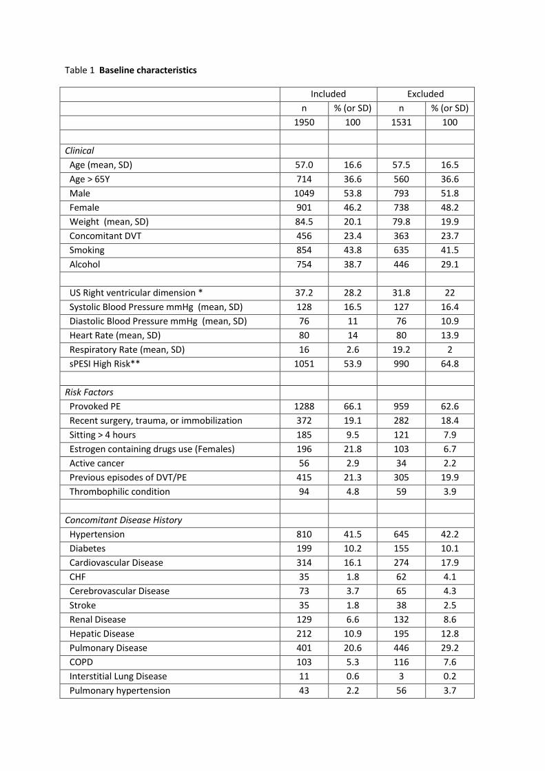
Table 1 Baseline characteristics
Included Excluded
n % (or SD) n % (or SD)
1950 100 1531 100
Clinical
Age (mean, SD) 57.0 16.6 57.5 16.5
Age > 65Y 714 36.6 560 36.6
Male 1049 53.8 793 51.8
Female 901 46.2 738 48.2
Weight (mean, SD) 84.5 20.1 79.8 19.9
Concomitant DVT 456 23.4 363 23.7
Smoking 854 43.8 635 41.5
Alcohol 754 38.7 446 29.1
US Right ventricular dimension * 37.2 28.2 31.8 22
Systolic Blood Pressure mmHg (mean, SD) 128 16.5 127 16.4
Diastolic Blood Pressure mmHg (mean, SD) 76 11 76 10.9
Heart Rate (mean, SD) 80 14 80 13.9
Respiratory Rate (mean, SD) 16 2.6 19.2 2
sPESI High Risk** 1051 53.9 990 64.8
Risk Factors
Provoked PE 1288 66.1 959 62.6
Recent surgery, trauma, or immobilization 372 19.1 282 18.4
Sitting > 4 hours 185 9.5 121 7.9
Estrogen containing drugs use (Females) 196 21.8 103 6.7
Active cancer 56 2.9 34 2.2
Previous episodes of DVT/PE 415 21.3 305 19.9
Thrombophilic condition 94 4.8 59 3.9
Concomitant Disease History
Hypertension 810 41.5 645 42.2
Diabetes 199 10.2 155 10.1
Cardiovascular Disease 314 16.1 274 17.9
CHF 35 1.8 62 4.1
Cerebrovascular Disease 73 3.7 65 4.3
Stroke 35 1.8 38 2.5
Renal Disease 129 6.6 132 8.6
Hepatic Disease 212 10.9 195 12.8
Pulmonary Disease 401 20.6 446 29.2
COPD 103 5.3 116 7.6
Interstitial Lung Disease 11 0.6 3 0.2
Pulmonary hypertension 43 2.2 56 3.7
Cancer 228 11.7 148 9.7
Table 2 Radiological diameters
n missing mean SD Min Max IQR
RV axial plane 1950 0 38.3 7.8 17 67 33 - 43
LV axial plane 1950 0 41.5 7.1 18 69 37 - 48
RV short axis 1906 44 39.7 7.7 20 71 34 - 45
LV short axis 1906 44 42.6 6.7 22 73 38 - 47
Aorta 1949 1 32.3 4.9 18 52 29 - 35
Pulmonary Trunk 1949 1 27.7 4.6 15 52 25 - 31
Azygos 1947 3 8.3 2.3 2 20 7 - 10
SVC 1949 1 18.9 4.1 8 33 16 - 22
RV Wall Thickness 1950 0 1.5 0.8 1 8 1 - 2
RA 1950 0 49.0 9.1 24 88 43 - 55
IVC 1942 8 22.7 4.3 8 41 20 - 25
Heart 1950 0 128.6 14.9 85 228 119 - 138
Chest 1950 0 259.1 24.2 126 344 242 - 276
RV/LV Axial 1950 0 0.95 0.27 0.39 2.61 0.78 - 1.00
RV/LV short axis 1950 0 0.96 0.26 0.47 2.11 0.80 - 1.02
PT/Ao 1950 0 0.87 0.15 0.42 2.08 0.77 - 0.96
CTR 1950 0 0.50 0.06 0.34 0.97 0.46 - 0.54
Table 3 Frequency of abnormal cardiovascular radiological parameters
n missing normal % abnormal %
RV/LV > 1 1950 0 1329 68.2 621 31.8
RV/LVsa > 0.9 1914 36 1024 53.5 890 46.5
Septal Bowing 1949 1 1796 92.1 153 7.9
Septal Flattening 1949 1 1411 72.4 538 27.6
Aorta > 40 mm 1840 110 1800 97.8 40 2.2
Pulmonary Trunk > 29 mm 1949 1 1315 67.5 634 32.5
PT/Aorta > 1.0 1950 0 1542 79.1 408 20.9
Cardiothoracic ratio > 0,50 1833 117 897 48.9 936 51.1
Backflow IVC 1754 196 1105 63 649 37
Intrahepatic Contrast Reflux 1754 196 1493 85.1 261 14.9
Backflow azygos vein 1947 3 1502 77.1 445 22.9
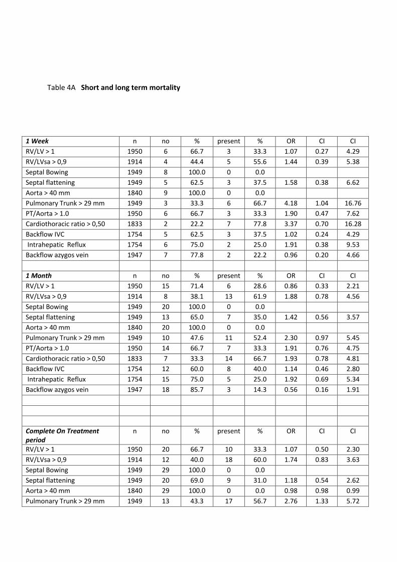
Table 4A Short and long term mortality
1 Week n no % present % OR CI CI
RV/LV > 1 1950 6 66.7 3 33.3 1.07 0.27 4.29
RV/LVsa > 0,9 1914 4 44.4 5 55.6 1.44 0.39 5.38
Septal Bowing 1949 8 100.0 0 0.0
Septal flattening 1949 5 62.5 3 37.5 1.58 0.38 6.62
Aorta > 40 mm 1840 9 100.0 0 0.0
Pulmonary Trunk > 29 mm 1949 3 33.3 6 66.7 4.18 1.04 16.76
PT/Aorta > 1.0 1950 6 66.7 3 33.3 1.90 0.47 7.62
Cardiothoracic ratio > 0,50 1833 2 22.2 7 77.8 3.37 0.70 16.28
Backflow IVC 1754 5 62.5 3 37.5 1.02 0.24 4.29
Intrahepatic Reflux 1754 6 75.0 2 25.0 1.91 0.38 9.53
Backflow azygos vein 1947 7 77.8 2 22.2 0.96 0.20 4.66
1 Month n no % present % OR CI CI
RV/LV > 1 1950 15 71.4 6 28.6 0.86 0.33 2.21
RV/LVsa > 0,9 1914 8 38.1 13 61.9 1.88 0.78 4.56
Septal Bowing 1949 20 100.0 0 0.0
Septal flattening 1949 13 65.0 7 35.0 1.42 0.56 3.57
Aorta > 40 mm 1840 20 100.0 0 0.0
Pulmonary Trunk > 29 mm 1949 10 47.6 11 52.4 2.30 0.97 5.45
PT/Aorta > 1.0 1950 14 66.7 7 33.3 1.91 0.76 4.75
Cardiothoracic ratio > 0,50 1833 7 33.3 14 66.7 1.93 0.78 4.81
Backflow IVC 1754 12 60.0 8 40.0 1.14 0.46 2.80
Intrahepatic Reflux 1754 15 75.0 5 25.0 1.92 0.69 5.34
Backflow azygos vein 1947 18 85.7 3 14.3 0.56 0.16 1.91
Complete On Treatment period
n no % present % OR CI CI
RV/LV > 1 1950 20 66.7 10 33.3 1.07 0.50 2.30
RV/LVsa > 0,9 1914 12 40.0 18 60.0 1.74 0.83 3.63
Septal Bowing 1949 29 100.0 0 0.0
Septal flattening 1949 20 69.0 9 31.0 1.18 0.54 2.62
Aorta > 40 mm 1840 29 100.0 0 0.0 0.98 0.98 0.99
Pulmonary Trunk > 29 mm 1949 13 43.3 17 56.7 2.76 1.33 5.72

PT/Aorta > 1.0 1950 17 56.7 13 43.3 2.95 1.42 6.13
Cardiothoracic ratio > 0,50 1833 10 33.3 20 66.6 1.94 0.90 4.16
Backflow IVC 1754 17 60.7 11 39.3 1.10 0.51 2.37
Intrahepatic Reflux 1754 21 75.0 7 25.0 1.93 0.81 4.59
Backflow azygos vein 1947 26 86.7 4 13.3 0.52 0.18 1.48
1 Year Study period n no % present % OR CI CI
RV/LV > 1 1950 42 72.4 16 27.6 0.81 0.45 1.45
RV/LVsa > 0,9 1914 28 48.3 30 51.7 1.24 0.74 2.09
Septal Bowing 1949 55 96.5 2 3.5 0.42 0.10 1.74
Septal flattening 1949 43 75.4 14 24.6 0.85 0.46 1.57
Aorta > 40 mm 1840 52 94.5 3 5.5 2.73 0.81 9.13
Pulmonary Trunk > 29 mm 1949 23 39.7 35 60.3 2.33 1.36 3.97
PT/Aorta > 1.0 1950 40 69.0 18 31.0 1.73 0.98 3.06
Cardiothoracic ratio > 0,50 1833 23 40.4 34 59.6 1.43 0.94 2.45
Backflow IVC 1754 28 54.9 23 45.1 1.41 0.81 2.48
Intrahepatic Reflux 1754 40 78.4 11 21.6 1.60 0.81 3.16
Backflow azygos vein 1947 51 87.9 7 12.1 0.46 0.21 1.01
Table 4B Short and long term mortality
1 Week Sens CI Spec CI PPV CI NPV CI
RV/LV > 1 0.33 0.03 0.64 0.68 0.66 0.70 0.00 0.00 0.01 1.00 0.99 1.00
RV/LVsa > 0,9 0.56 0.23 0.88 0.54 0.51 0.56 0.01 0.00 0.01 1.00 0.99 1.00
Septal Bowing 0.00 0.00 0.00 0.92 0.91 0.93 0.00 0.00 0.00 1.00 0.99 1.00
Septal flattening 0.38 0.04 0.71 0.72 0.70 0.74 0.01 0.00 0.01 1.00 0.99 1.00
Aorta > 40 mm 0.00 0.00 0.00 0.98 0.97 0.98 0.00 0.00 0.00 1.00 0.99 1.00
Pulm. Trunk > 29 mm 0.67 0.36 0.97 0.68 0.66 0.70 0.01 0.00 0.02 1.00 1.00 1.00
PT/Aorta > 1.0 0.33 0.03 0.64 0.79 0.77 0.81 0.01 0.00 0.02 1.00 0.99 1.00
Cardiothor. ratio > 0,50 0.78 0.51 1.05 0.49 0.47 0.51 0.01 0.00 0.01 1.00 0.99 1.00
Backflow IVC 0.38 0.04 0.71 0.63 0.61 0.65 0.00 0.00 0.01 1.00 0.99 1.00
Intrahepatic Reflux 0.25 0.00 0.55 0.85 0.83 0.87 0.01 0.00 0.02 1.00 0.99 1.00
Backflow azygos vein 0.22 0.00 0.49 0.77 0.75 0.79 0.00 0.00 0.01 1.00 0.99 1.00
1 Month Sens CI Spec CI PPV CI NPV CI
RV/LV > 1 0.29 0.09 0.48 0.68 0.66 0.70 0.01 0.00 0.02 0.99 0.98 0.99
RV/LVsa > 0,9 0.62 0.41 0.83 0.54 0.51 0.56 0.01 0.01 0.02 0.99 0.99 1.00
Septal Bowing 0.00 0.00 0.00 0.92 0.91 0.93 0.00 0.00 0.00 0.99 0.98 0.99
Septal flattening 0.35 0.14 0.56 0.72 0.70 0.74 0.01 0.00 0.02 0.99 0.99 1.00
Aorta > 40 mm 0.00 0.00 0.00 0.98 0.97 0.98 0.00 0.00 0.00 0.99 0.98 0.99

Pulm. Trunk > 29 mm 0.52 0.31 0.74 0.68 0.66 0.70 0.02 0.01 0.03 0.99 0.99 1.00
PT/Aorta > 1.0 0.33 0.13 0.53 0.79 0.77 0.81 0.02 0.00 0.03 0.99 0.99 1.00
Cardiothor. ratio > 0,50 0.67 0.47 0.87 0.49 0.47 0.51 0.01 0.01 0.02 0.99 0.99 1.00
Backflow IVC 0.40 0.19 0.61 0.63 0.61 0.65 0.01 0.00 0.02 0.99 0.98 1.00
Intrahepatic Reflux 0.25 0.06 0.44 0.85 0.84 0.87 0.02 0.00 0.04 0.99 0.98 1.00
Backflow azygos vein 0.14 0.00 0.29 0.77 0.75 0.79 0.01 0.00 0.01 0.99 0.98 0.99
On Treatment period Sens CI Spec CI PPV CI NPV CI
RV/LV > 1 0.33 0.16 0.50 0.68 0.66 0.70 0.02 0.01 0.03 0.98 0.98 0.99
RV/LVsa > 0,9 0.60 0.42 0.78 0.54 0.51 0.56 0.02 0.01 0.03 0.99 0.98 0.99
Septal Bowing 0.00 0.00 0.00 0.92 0.91 0.93 0.00 0.00 0.00 0.98 0.98 0.99
Septal flattening 0.31 0.14 0.48 0.72 0.70 0.74 0.02 0.01 0.03 0.99 0.98 0.99
Aorta > 40 mm 0.00 0.00 0.00 0.98 0.97 0.98 0.00 0.00 0.00 0.98 0.98 0.99
Pulm. Trunk > 29 mm 0.57 0.39 0.74 0.68 0.66 0.70 0.03 0.01 0.04 0.99 0.98 1.00
PT/Aorta > 1.0 0.43 0.26 0.61 0.79 0.78 0.81 0.03 0.01 0.05 0.99 0.98 0.99
Cardiothor. ratio > 0,50 0.67 0.50 0.84 0.49 0.47 0.52 0.02 0.01 0.03 0.99 0.98 1.00
Backflow IVC 0.39 0.21 0.57 0.63 0.61 0.65 0.02 0.01 0.03 0.98 0.98 0.99
Intrahepatic Reflux 0.25 0.09 0.41 0.85 0.84 0.87 0.03 0.01 0.05 0.99 0.98 0.99
Backflow azygos vein 0.14 0.01 0.26 0.77 0.75 0.79 0.01 0.00 0.02 0.98 0.98 0.99
1 Year Sens CI Spec CI PPV CI NPV CI
RV/LV > 1 0.28 0.16 0.39 0.68 0.66 0.70 0.03 0.01 0.04 0.97 0.96 0.98
RV/LVsa > 0,9 0.52 0.39 0.65 0.54 0.51 0.56 0.03 0.02 0.05 0.97 0.96 0.98
Septal Bowing 0.04 0.00 0.08 0.92 0.91 0.93 0.01 0.00 0.03 0.97 0.96 0.98
Septal flattening 0.25 0.13 0.36 0.72 0.70 0.74 0.03 0.01 0.04 0.97 0.96 0.98
Aorta > 40 mm 0.05 0.00 0.11 0.98 0.97 0.99 0.08 0.01 0.16 0.97 0.96 0.98
Pulm. Trunk > 29 mm 0.52 0.39 0.65 0.68 0.66 0.70 0.05 0.03 0.06 0.98 0.97 0.99
PT/Aorta > 1.0 0.31 0.19 0.43 0.79 0.78 0.81 0.04 0.02 0.06 0.97 0.97 0.98
Cardiothor. ratio > 0,50 0.60 0.47 0.72 0.49 0.47 0.52 0.04 0.02 0.05 0.97 0.96 0.98
Backflow IVC 0.45 0.31 0.59 0.63 0.61 0.66 0.04 0.02 0.05 0.97 0.97 0.98
Intrahepatic Reflux 0.22 0.10 0.33 0.85 0.84 0.87 0.04 0.02 0.07 0.97 0.97 0.98
Backflow azygos vein 0.12 0.04 0.20 0.78 0.76 0.80 0.02 0.00 0.03 0.97 0.96 0.98

8292 patients with
Venous Thrombo-embolism
3481 patients with
Pulmonary-embolism
3114 patients evaluated
with CTPA
1950 patients evaluable with
Pulmonary-embolism
4811 DVT only
52 excluded
367 no CTPA scan:
314 VQ-scan
34 pulmonary angiography
19 miscellaneous (e.g. US and
Perfusion scan, PET scan)
1164 not evaluable:
no DICOM images or only hard copy, PDF or JPEG,
technically inadequate study,
insufficient coverage

Appendix On line. 1 Month and 1 year recurrent VTE, hospitalization, major bleeding and
adverse events
Recurrent VTE
1 Month n no % present % OR CI CI
RV/LV > 1 1950 7 53.8 6 46.2 1.84 0.62 5.51
RV/LVsa > 0,9 1914 5 38.5 8 61.5 1.85 0.60 5.67
Septal Bowing 1949 11 91.7 1 8.3 1.07 0.14 8.32
Septal flattening 1949 6 50.0 6 50.0 2.64 0.85 8.23
Aorta > 40 mm 1840 11 91.7 1 8.3 4.17 0.53 33.10
Pulmonary Trunk > 29 mm 1949 7 53.8 6 32.4 1.79 0.60 5.33
PT/Aorta > 1.0 1950 8 61.5 5 38.5 2.38 0.77 7.31
Cardiothoracic ratio > 0,50 1833 6 50.0 6 50.0 0.96 0.31 2.98
Backflow IVC 1754 7 63.6 4 36.4 0.97 0.28 3.34
Intrahepatic Reflux 1754 9 81.8 2 18.2 1.27 0.27 5.93
Backflow azygos vein 1947 12 92.3 1 7.7 0.28 0.04 2.16
1 Year n no % present % OR CI CI
RV/LV > 1 1950 33 66.0 17 34.0 1.11 0.61 2.00
RV/LVsa > 0,9 1914 24 48.0 56 52.0 1.25 0.72 2.20
Septal Bowing 1949 45 91.8 4 8.2 1.05 0.37 2.94
Septal flattening 1949 35 71.4 14 28.6 1.05 0.56 1.97
Aorta > 40 mm 1840 45 95.7 2 4.3 2.05 0.48 8.77
Pulmonary Trunk > 29 mm 1949 35 70.0 15 30.0 0.89 0.48 1.64
PT/Aorta > 1.0 1950 41 82.0 9 18.0 0.83 0.40 1.71
Cardiothoracic ratio > 0,50 1833 25 51.0 24 49.0 0.92 0.52 1.62
Backflow IVC 1754 28 65.1 15 34.9 0.91 0.48 1.72
Intrahepatic Reflux 1754 34 79.1 9 20.9 1.53 0.73 3.23
Backflow azygos vein 1947 42 84.0 8 16.0 0.64 0.30 1.37
Hospitalisation
1 Month n no % present % OR CI CI
RV/LV > 1 1950 62 68.9 28 31.1 0.97 0.61 1.52

RV/LVsa > 0,9 1914 47 52.2 43 47.8 1.06 0.69 1.61
Septal Bowing 1949 81 91.0 8 9.0 1.17 0.55 2.46
Septal flattening 1949 65 73.0 24 27.0 0.97 0.60 1.56
Aorta > 40 mm 1840 82 96.5 3 3.5 1.70 0.51 5.63
Pulmonary Trunk > 29 mm 1949 54 60.0 36 40.0 1.41 0.91 2.17
PT/Aorta > 1.0 1950 67 74.4 23 25.6 1.32 0.81 2.14
Cardiothoracic ratio > 0,50 1833 41 46.1 48 53.9 1.13 0.74 1.73
Backflow IVC 1754 48 61.5 30 38.5 1.07 0.67 1.70
Intrahepatic Reflux 1754 66 84.6 12 15.4 1.04 0.56 1.96
Backflow azygos vein 1947 76 84.4 14 15.6 0.61 0.34 1.09
1 Year n no % present % OR CI CI
RV/LV > 1 1950 62 68.9 28 31.1 0.97 0.61 1.52
RV/LVsa > 0,9 1914 47 52.2 43 47.8 1.06 0.69 1.61
Septal Bowing 1949 81 91.0 8 9.0 1.17 0.55 2.46
Septal flattening 1949 65 73.0 24 27.0 0.97 0.60 1.56
Aorta > 40 mm 1840 82 96.5 3 3.5 1.70 0.51 5.63
Pulmonary Trunk > 29 mm 1949 54 60.0 36 40.0 1.41 0.91 2.17
PT/Aorta > 1.0 1950 67 74.4 23 25.6 1.32 0.81 2.14
Cardiothoracic ratio > 0,50 1833 41 46.1 48 53.9 1.13 0.74 1.73
Backflow IVC 1754 48 61.5 30 38.5 1.07 0.67 1.70
Intrahepatic Reflux 1754 66 84.6 12 15.4 1.04 0.56 1.96
Backflow azygos vein 1947 76 84.4 14 15.9 0.61 0.34 1.09
Major bleeding
1 Month n no % present % OR CI CI
RV/LV > 1 1950 7 53.8 6 46.2 1.84 0.62 5.51
RV/LVsa > 0,9 1914 7 53.8 6 46.2 0.99 0.33 2.95
Septal Bowing 1949 12 92.3 1 7.7 0.98 0.13 7.57
Septal flattening 1949 9 69.2 4 30.8 1.17 0.36 3.81
Aorta > 40 mm 1840 13 100.0 0 0.0 0.99 0.99 1.00
Pulmonary Trunk > 29 mm 1949 6 46.2 7 53.8 2.44 0.82 7.28
PT/Aorta > 1.0 1950 7 53.8 6 46.2 3.27 1.09 9.79
Cardiothoracic ratio > 0,50 1833 3 23.1 10 76.9 3.22 0.88 11.73
Backflow IVC 1754 7 53.8 6 46.2 1.46 0.49 4.37
Intrahepatic Reflux 1754 9 69.2 4 30.8 2.57 0.78 8.40
Backflow azygos vein 1947 9 69.2 4 30.8 1.51 0.46 4.91
1 Year n no % present % OR CI CI
RV/LV > 1 1950 18 60.0 12 40.0 1.43 0.69 3.00
RV/LVsa > 0,9 1914 15 51.7 14 48.3 1.08 0.52 2.24
Septal Bowing 1949 25 83.3 5 16.7 2.39 0.90 6.34
Septal flattening 1949 21 70.0 9 30.0 1.13 0.51 2.47

Aorta > 40 mm 1840 27 96.4 1 3.6 1.68 0.22 12.71
Pulmonary Trunk > 29 mm 1949 11 36.7 19 63.3 3.66 1.73 7.74
PT/Aorta > 1.0 1950 18 60.0 12 40.0 2.57 1.23 5.37
Cardiothoracic ratio > 0,50 1833 10 34.5 19 65.5 1.84 0.85 3.97
Backflow IVC 1754 16 57.1 12 42.9 1.28 0.60 2.73
Intrahepatic Reflux 1754 23 82.1 5 17.9 1.25 0.47 3.31
Backflow azygos vein 1947 24 80.0 6 20.0 0.84 0.34 2.07
All Adverse Events
1 Month n no % present % OR CI CI
RV/LV > 1 1950 92 69.7 40 30.3 0.93 0.63 1.36
RV/LVsa > 0,9 1914 66 50.4 65 49.6 1.14 0.80 1.63
Septal Bowing 1949 121 92.4 10 7.6 0.97 0.50 1.89
Septal flattening 1949 96 73.3 35 26.7 0.95 0.64 1.42
Aorta > 40 mm 1840 120 96.8 4 3.2 1.56 0.55 4.44
Pulmonary Trunk > 29 mm 1949 75 56.8 57 43.2 1.63 1.14 2.34
PT/Aorta > 1.0 1950 96 72.7 36 27.3 1.46 0.98 2.17
Cardiothoracic ratio > 0,50 1833 58 45.0 71 55.0 1.19 0.83 1.70
Backflow IVC 1754 71 61.2 45 38.8 1.09 0.74 1.60
Intrahepatic Reflux 1754 94 81.0 22 19.0 1.37 0.84 2.22
Backflow azygos vein 1947 110 83.3 22 16.7 0.66 0.41 1.05
1 Year n no % present % OR CI CI
RV/LV > 1 1950 92 69.7 40 30.3 0.93 0.63 1.36
RV/LVsa > 0,9 1914 66 50.4 65 19.6 1.14 0.80 1.63
Septal Bowing 1949 121 92.4 10 7.6 0.97 0.50 1.89
Septal flattening 1949 96 73.3 35 26.7 0.95 0.64 1.42
Aorta > 40 mm 1840 120 96.8 4 3.2 1.56 0.55 4.44
Pulmonary Trunk > 29 mm 1949 75 56.8 57 43.2 1.63 1.14 2.34
PT/Aorta > 1.0 1950 96 72.7 36 27.3 1.46 0.98 2.17
Cardiothoracic ratio > 0,50 1833 58 45.0 71 55.0 1.19 0.83 1.70
Backflow IVC 1754 71 61.2 45 38.8 1.09 0.74 1.60
Intrahepatic Reflux 1754 94 81.0 22 19.0 1.37 0.84 2.22
Backflow azygos vein 1947 110 83.3 22 16.7 0.66 0.41 1.05