Probing FinO–FinP RNA interactions by site-directed protein–RNA ...
8
Probing FinO–FinP RNA interactions by site-directed protein–RNA crosslinking and gelFRET ALEXANDRU F. GHETU, 1 DAVID C. ARTHUR, 1 TOM K. KERPPOLA, 2 and J.N. MARK GLOVER 1 1 Department of Biochemistry , University of Alberta, Edmonton, Alberta, T6G 2H7, Canada 2 Howard Hughes Medical Institute and Department of Biological Chemistry , University of Michigan Medical School, Ann Arbor , Michigan 48109-0650, USA ABSTRACT The conjugative transfer of F-plasmids is repressed by a two-component system, which consists of the antisense RNA FinP and the protein FinO. FinO binds FinP, protecting it from endonucleolytic degradation and facilitating duplex formation between FinP and its complementary RNA. Here we present the results of site-specific protein–RNA cross- linking and gel-based fluorescence resonance energy transfer (gelFRET) experiments used to probe the structure of a complex of FinO bound to an RNA target consisting of a duplex with 59 and 39 single-stranded tails. The crosslinking experiments reveal that an extensive, largely positively charged surface on FinO contacts RNA. The gelFRET mea- surements indicate that the 59 single-stranded tail of the RNA is in closer contact with much of the protein than the distal, blunt end of the RNA duplex. These data suggest that significant conformational adjustments in the protein and/or the RNA accompany complex formation. Keywords: conjugation; protein–RNA interactions; site-specific crosslinking INTRODUCTION Plasmid conjugation is a major mechanism for transfer of antibiotic resistance and virulence determinants be- tween bacteria (Mazodier & Davies, 1991)+ Perhaps the best studied group of conjugative plasmids is the F family + F plasmids contain a large, multicistronic trans- fer (tra ) operon, which encodes the majority of proteins required for conjugation (Frost et al+, 1994)+ Transcrip- tion of the tra operon is positively regulated by the plasmid-encoded product of the traJ gene (Mullineaux & Willetts, 1985)+ The production of TraJ is, itself, neg- atively regulated by a two-component repression sys- tem consisting of FinP , an RNA that is antisense to the 59 end of traJ mRNA, and FinO, a 21+5-kDa RNA bind- ing protein (Finnegan & Willetts, 1972)+ Duplex for- mation between FinP and traJ mRNA occludes the ribosomal binding site and prevents translation of traJ RNA (van Biesen & Frost, 1994)+ An RNase E recog- nition site located between the two stem-loops of FinP makes this RNA highly susceptible to degradation (Jer- ome et al+, 1999)+ FinO binds to both FinP and traJ mRNA, protecting FinP from degradation and enhanc- ing the rate of duplex formation between FinP and traJ mRNA (Lee et al+, 1992; van Biesen et al+, 1993; Ko- raimann et al+, 1996; Jerome et al+, 1999; Ghetu et al+, 2000)+ Recent results have begun to reveal the molecular mechanism underlying FinOP function+ Biochemical studies have shown that FinO binds as a monomer to stem-loop structures with short 59 and 39 single-stranded tails (Ghetu et al+, 1999; Jerome & Frost, 1999; Fig+ 1A)+ These interactions are not sequence specific, so that complementary stem-loops with single-stranded tails, such as stem-loop II (SLII) of FinP and the comple- mentary stem-loop in traJ RNA are bound with nearly identical affinities (Jerome & Frost, 1999)+ The crystal structure of FinO revealed an elongated, largely helical structure reminiscent of a right-handed fist with an ex- tended index finger and a thumb, touching the index finger near its base (Fig+ 2)+ The finger corresponds to a solvent-exposed N-terminal helix ( a1), and the thumb corresponds to the C-terminal-most helix ( a6)+ An ex- tended, positively charged surface composed of parts of a1, a6 and the fist was suggested to contact RNA targets (Ghetu et al+, 2000)+ The N-terminal 25 residues Reprint requests to: J+N+ Mark Glover , Department of Biochemis- try , University of Alberta, Edmonton, Alberta, T6G 2H7, Canada; e-mail: mark+glover@ualberta+ca+ RNA (2002), 8:816–823+ Cambridge University Press+ Printed in the USA+ Copyright © 2002 RNA Society + DOI: 10+1017+S1355838202026730 816
Transcript of Probing FinO–FinP RNA interactions by site-directed protein–RNA ...
Probing FinO–FinP RNA interactionsby site-directed protein–RNAcrosslinking and gelFRET
ALEXANDRU F. GHETU, 1 DAVID C. ARTHUR,1 TOM K. KERPPOLA, 2
and J.N. MARK GLOVER 1
1Department of Biochemistry, University of Alberta, Edmonton, Alberta, T6G 2H7, Canada2Howard Hughes Medical Institute and Department of Biological Chemistry, University of Michigan Medical School,Ann Arbor, Michigan 48109-0650, USA
ABSTRACT
The conjugative transfer of F-plasmids is repressed by a two-component system, which consists of the antisenseRNA FinP and the protein FinO. FinO binds FinP, protecting it from endonucleolytic degradation and facilitating duplexformation between FinP and its complementary RNA. Here we present the results of site-specific protein–RNA cross-linking and gel-based fluorescence resonance energy transfer (gelFRET) experiments used to probe the structure ofa complex of FinO bound to an RNA target consisting of a duplex with 5 9 and 39 single-stranded tails. The crosslinkingexperiments reveal that an extensive, largely positively charged surface on FinO contacts RNA. The gelFRET mea-surements indicate that the 5 9 single-stranded tail of the RNA is in closer contact with much of the protein than thedistal, blunt end of the RNA duplex. These data suggest that significant conformational adjustments in the proteinand/or the RNA accompany complex formation.
Keywords: conjugation; protein–RNA interactions; site-specific crosslinking
INTRODUCTION
Plasmid conjugation is a major mechanism for transferof antibiotic resistance and virulence determinants be-tween bacteria (Mazodier & Davies, 1991)+ Perhapsthe best studied group of conjugative plasmids is the Ffamily+ F plasmids contain a large, multicistronic trans-fer (tra) operon, which encodes the majority of proteinsrequired for conjugation (Frost et al+, 1994)+ Transcrip-tion of the tra operon is positively regulated by theplasmid-encoded product of the traJ gene (Mullineaux& Willetts, 1985)+ The production of TraJ is, itself, neg-atively regulated by a two-component repression sys-tem consisting of FinP, an RNA that is antisense to the59 end of traJ mRNA, and FinO, a 21+5-kDa RNA bind-ing protein (Finnegan & Willetts, 1972)+ Duplex for-mation between FinP and traJ mRNA occludes theribosomal binding site and prevents translation of traJRNA (van Biesen & Frost, 1994)+ An RNase E recog-nition site located between the two stem-loops of FinPmakes this RNA highly susceptible to degradation (Jer-
ome et al+, 1999)+ FinO binds to both FinP and traJmRNA, protecting FinP from degradation and enhanc-ing the rate of duplex formation between FinP and traJmRNA (Lee et al+, 1992; van Biesen et al+, 1993; Ko-raimann et al+, 1996; Jerome et al+, 1999; Ghetu et al+,2000)+
Recent results have begun to reveal the molecularmechanism underlying FinOP function+ Biochemicalstudies have shown that FinO binds as a monomer tostem-loop structures with short 59 and 39 single-strandedtails (Ghetu et al+, 1999; Jerome & Frost, 1999; Fig+ 1A)+These interactions are not sequence specific, so thatcomplementary stem-loops with single-stranded tails,such as stem-loop II (SLII) of FinP and the comple-mentary stem-loop in traJ RNA are bound with nearlyidentical affinities (Jerome & Frost, 1999)+ The crystalstructure of FinO revealed an elongated, largely helicalstructure reminiscent of a right-handed fist with an ex-tended index finger and a thumb, touching the indexfinger near its base (Fig+ 2)+ The finger corresponds toa solvent-exposed N-terminal helix (a1), and the thumbcorresponds to the C-terminal-most helix (a6)+ An ex-tended, positively charged surface composed of partsof a1, a6 and the fist was suggested to contact RNAtargets (Ghetu et al+, 2000)+ The N-terminal 25 residues
Reprint requests to: J+N+ Mark Glover, Department of Biochemis-try,University ofAlberta,Edmonton,Alberta,T6G 2H7,Canada; e-mail:mark+glover@ualberta+ca+
RNA (2002), 8:816–823+ Cambridge University Press+ Printed in the USA+Copyright © 2002 RNA Society+DOI: 10+1017+S1355838202026730
816

of FinO are not present in the crystal structure and arenot required for binding to individual RNA substrates;however, the N-terminus facilitates sense–antisenseRNA interactions between FinP and traJ RNAs (Ghetuet al+, 2000)+ We have suggested (Ghetu et al+, 2000)that the N-terminus of FinO may directly interact withan initial “kissing complex” formed between comple-mentary loops in FinP and traJ RNAs (Koraimann et al+,
1991), thereby facilitating FinP-traJ RNA recognition+This possibility, together with the structure of FinO, pro-vided the basis for a model of FinO bound to SLII fromFinP (Ghetu et al+, 2000)+ In this model, the stem-loopof SLII lies along the exposed, positively charged a1 ofFinO so that the N-terminus is positioned near the SLIIloop to participate in loop-loop recognition+ The single-stranded tails at the base of the stem interact with alarge positively charged patch on the globular body ofthe protein+
We have used site-specific protein–RNA crosslinkingand a gel-based fluorescence resonance energy trans-fer (gelFRET) assay to investigate the interactions be-tween FinO and SLII+ The results of the crosslinkingexperiments reveal an extensive surface on FinO thatcomes into contact with RNA+ The gelFRET experi-ments allowed us to map the relative proximity of specificsites on FinO and FinP and indicated that the single-stranded tails at the base of the duplex are in muchcloser proximity to FinO than is the opposite end of theduplex proximal to the loop+ These data suggest thatFinO binding to SLII RNA may involve conformationalchanges in FinO, SLII, or both+
RESULTS AND DISCUSSION
Site-specific FinO-SLII RNA crosslinking
To experimentally determine the regions of FinO thatare in close proximity to the target RNA, we used asite-specific crosslinking assay involving the photo-activated crosslinker, azidophenacyl bromide (APA-Br)+We first replaced the three native cysteine residuesin FinO with serines and used a gel electrophoreticmobility shift assay to show that these three substitu-tions do not alter the affinity of FinO binding to the RNA(data not shown)+ We next created a set of FinO mu-tants that contain single cysteine substitutions at vari-ous solvent-exposed positions, to which we could attachAPA via a thioester linkage (Fig+ 2)+ The sites of sub-stitution include the positively charged surfaces on thetip of the N-terminal helix, the body of the protein, theC-terminal helix and the negatively charged surface onthe bottom of the molecule+
APA-modified FinO mutants were incubated eitherwith SLII RNA, a minimal RNA target for FinO (Jerome& Frost, 1999) or tRNA, which does not bind FinO withhigh affinity (van Biesen & Frost, 1994; Sandercock &Frost, 1998; Ghetu et al+, 1999)+ Crosslinking was in-duced by irradiation of the APA-modified protein/RNAcomplexes with UV light,which activates the azido func-tional group of APA+ The resulting nitrene reacts in anonspecific manner with protein or RNA that is withinan ;10 Å radius of the modified cysteine (Pendergrastet al+, 1992; Chen & Ebright, 1993)+ The reaction prod-ucts were separated by a denaturing polyacrylamide
FIGURE 1. Schematic representation of SLII-based RNAs used inthis study+ A: The nucleotide sequence and predicted secondarystructure of SLII+ B: The nucleotide sequence and secondary struc-ture of the RNA duplex used in our experiments+ Sites where fluo-rescein has been attached to the duplex are indicated+ The duplexdiffers from SLII in that the loop is absent and the first three basepairs at the top of the stem are reordered+
FIGURE 2. The structure of FinO and the positions of single cys-teine substitutions+ Two ribbons diagrams of FinO, related by a 1808rotation about a vertical axis+ The positions of cysteine substitutionsthat serve as sites of attachment for APA or Texas Red are indicatedin red+ Also indicated in the figure are the N- and C-terminal ends ofFinO and a-helices 1 and 6+
Crosslinking and gelFRET of FinO-RNA complexes 817
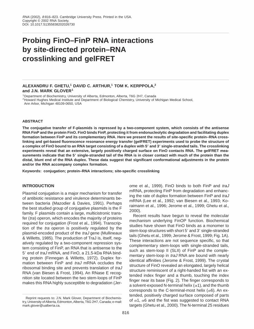
gel and the gel was stained with Coomassie blue andethidium bromide to visualize protein and RNA, respec-tively (Fig+ 3A)+ FinO-SLII crosslinking was detected bythe presence of high molecular weight species that con-tain both protein and RNA+ As a negative control, theAPA-treated cysteine-free FinO mutant protein does
not crosslink to SLII under these conditions+ The spec-ificity of the FinO-SLII crosslinking was demonstratedby the finding that tRNA is not efficiently crosslinked bythe same APA-modified FinO samples in parallel ex-periments carried out under identical solution condi-tions and protein/RNA concentrations+
FIGURE 3. Site specific crosslinking of FinO and SLII+ A: Cross-linked protein/RNA complexes were analyzed by SDS-PAGE in whichRNA-containing species were visualized by ethidium bromide stain-ing (left), and protein-containing species were detected by Coomas-sie blue staining (right)+ Indicated on the bottom of each gel pair arethe various FinO cysteine mutants used+ Indicated to the left of thetop ethidium bromide stained gel are the positions of free SLII, freetRNA, the FinO/SLII crosslinked product containing one protein boundto one SLII (resulting from specific interactions), and the FinO/tRNAcrosslinked product (resulting from nonspecific interactions)+ To theright of the top Coomassie stained gel are the positions of free FinO,the FinO/SLII crosslinked product, and nonspecific crosslinking prod-ucts between FinO molecules+ Indicated by arrows on the bottom twogel pairs is the position of the FinO/SLII crosslinked product+ C r Sindicates a protein in which all the native cysteine residues havebeen replaced with serine+ B: Two electrostatic surface representa-tions of FinO, in the same orientations as shown in Figure 2A+ Ma-genta circles indicate the sites of APA attachment that showedsignificant crosslinking to SLII RNA, and yellow circles indicate sitesthat do not crosslink to RNA+
818 A.F. Ghetu et al.

Significant levels of crosslinking to SLII RNA, but notto tRNA, were observed for many of the APA-modifiedresidues within positively regions of the FinO surface(Fig+ 3A)+ The most efficient crosslinking was observedfor residues 121 and 125, which are exposed on thesurface of a4, as well as residue 165, which togetherconstitute part of the large positively charged surfaceon the main body of the protein (Fig+ 3B)+ Weaker butsignificant levels of crosslinking were also observedbetween modified residues at the positively charged tipof the N-terminal FinO helix (residues 37, 40, 42, and46), as well as residue 176 near the C-terminus of a6+These results confirm that these two positively chargedregions are in close proximity to the RNA substrate andlikely play a significant role in recognizing specific tar-get stem-loop structures+ Weak crosslinking was alsoobserved between APA-modified residue 81 and SLIIRNA+ Residue 81 is on the body of FinO, but on theopposite face from the major positively charged sur-face+ As predicted (Ghetu et al+, 2000), crosslinkingwas not observed when APA was positioned on thenegatively charged region of FinO (residues 142 and147), nor were appreciable levels of RNA crosslinkingobserved for APA-modified residues 135 or 170, sug-gesting that these regions are not in direct contact withRNA+ These results are summarized in Figure 3B andindicate that the N-terminal helix and the positivelycharged surface on the body of the protein are in clos-est contact with RNA+ However, interactions also occuron the opposite face of the protein, possibly due to thewrapping of the SLII tails around FinO+
Probing FinO-RNA architectureusing gelFRET
We have previously suggested that FinO and SLII RNAinteract such that the long axis of FinO is parallel to thestem, the positively charged surface on the core ofFinO is in contact with the base of the stem, and theN-terminus of a1 lies near the SLII loop+ To test thismodel, we used a gelFRET analysis in which the RNAis labeled with the donor fluorophore fluorescein andthe protein is labeled at single, specific sites with thereceptor fluorophore Texas Red+ The fluorescent com-plexes are separated by nondenaturing gel electropho-resis and the FRET from the individual complexes isanalyzed by excitation of the separated complexeswithin the gel+ In this way, fluorescence from unboundprotein, RNA, and nonspecific protein–RNA complexescan be eliminated and the relative efficiencies of en-ergy transfer between donor and acceptor moleculesat different positions on the protein and nucleic acidcomponents can be assessed (Ramirez-Carrozzi & Kerp-pola, 2001a)+ This method has been used to determinethe orientation of Fos-Jun heterodimer binding at dif-ferent AP-1 binding sites (Diebold et al+, 1998; Leonard& Kerppola, 1998;Ramirez-Carrozzi & Kerppola, 2001b,
2001c, 2001d)+ Here we present the first use of gel-FRET to study protein–RNA complexes+
For our experiments, we have prepared two differentRNA duplexes that are labeled on their 59 ends with thedonor fluorophore fluorescein+ The 59 ends of the twostrands are located at opposite ends of the duplex RNA(Fig+ 1B)+ These RNAs are based on SLII, but lack thesingle-stranded loop that connects the two strands inSLII+ Using native gel electrophoresis, we determineddissociation constants of 4+8 6 0+3 nM and 4+1 6 0+5 nMfor FinO binding to SLII and the RNA duplex, respec-tively+ These results ensure that FinO binds the duplexsubstrate in a similar fashion to SLII and confirm pre-vious results that the loop has no significant effect onthe interaction between FinO and SLII RNA (Jerome &Frost, 1999)+Our binding affinities are ;20 times tighterthan those determined previously (Jerome & Frost,1999)+ These differences probably reflect changes inthe gel mobility shift assay and improvements in thepurification and quantitation of FinO+
FinO mutants labeled with Texas Red were mixedwith an equi-molar amount of RNA labeled with fluo-rescein+ The nucleoprotein complexes were separatedfrom unbound FinO, unbound duplex, and free TexasRed by gel electrophoresis+ The gel was scanned usinga 488-nm argon-ion laser that excites fluorescein+ Thefluorescence emissions of both the donor fluoresceinand acceptor Texas Red were measured at each po-sition in the gel in separate scans+ In Figure 4, thefluorescein and Texas Red fluorescent scans are over-laid so that the relative levels of fluorescein and TexasRed fluorescence in the different species in each lanecan be qualitatively assessed+ In these scans, greenband color indicates fluorescein fluorescence, red colorindicates Texas Red fluorescence, and yellow colorindicates a mix of both fluorescein and Texas Red flu-orescence+ Thus, the degree of redness in bands cor-responding to protein–RNA species gives a qualitativeindication of the efficiency of energy transfer betweenthe two fluorophores within that complex relative to theother complexes analyzed in parallel+
Labeling of FinO by the Texas Red fluorophore slightlyaltered the gel electrophoretic mobility for most of theFinO-RNA complexes (compare the mobilities of com-plexes labeled with Texas Red (1) to unlabeled com-plexes (2) in Fig+ 4B)+ In cases where the complexeswith and without acceptor could be separated, it waspossible to ascertain that the analysis of the nucleo-protein complexes would not be influenced by the pres-ence of unlabeled protein+ The percentage of proteinmodified with Texas Red depended on the location ofthe cysteine residue, but was calculated to be greaterthan 60% in all cases examined (see Materials andMethods)+ In complexes labeled at residues 142 and147, the Texas Red modification results in a more sig-nificant (;10%) reduction in the electrophoretic mobil-ity of the complex+ This may suggest that Texas Red
Crosslinking and gelFRET of FinO-RNA complexes 819

FIGURE 4. gelFRET analysis of the architecture of the FinO–RNA complex+ Samples containing various Texas red-labeledFinO molecules and duplex RNA substrates labeled with fluorescein were separated by native gel electrophoresis+ Fluo-rescence of the electrophoretically resolved species was excited directly in the gel+ In the bands corresponding to theFinO-SLII complex, the color reflects the efficiency of energy transfer, with a red color indicating higher energy transfer, andtherefore closer interfluorophore distance, than a green color+ A: gelFRET analysis for Texas Red-labeled cysteine mutantsbound to RNA duplex labeled with fluorescein either on the 59 tail (left lanes) or the top of the duplex stem (right lanes)+Indicated on the left of the figure are the positions of the free SLII RNA, FinO-SLII duplex, nonspecific complexes, andunincorporated Texas Red+ The cysteine substitutions used are indicated above the lanes+ B: Labeling of FinO with TexasRed leads to changes in the mobilities of several of the FinO-SLII complexes+ Shown in this panel are the band shifts ofTexas Red modified (lanes 1 and 2) or unmodified (lane 3) proteins in complex with duplex, labeled with fluorescein eitheron the 59 tail (lane 1) or at the top of the stem (lanes 2 and 3)+ Each row represents a different cysteine point mutant, asindicated in the middle of the figure+ Orange arrows indicate complexes with modified protein, and white arrows indicatecomplexes with unmodified proteins+ Changes in mobility varied from one mutant to another+ Although the addition of TRleads to changes in the mobility of most mutants, this was not the case for K125C+
820 A.F. Ghetu et al.

modification at these residues alters the structure ofFinO or the way in which the modified proteins interactwith RNA+
Strikingly, energy transfer between the RNA and eachof the labeled FinO proteins was higher when the flu-orescein was positioned on the 59 single-stranded tailof the RNA (left most lanes, Fig+ 4A) compared to theopposite end of the duplex stem, where the loop wouldbe found in SLII (right most lanes, Fig+ 4A)+ This dra-matic difference may suggest that all positions sam-pled on the surface of FinO are in closer proximity tothe 59 single-stranded tail of the RNA than the distalend of the duplex stem+Alternatively, these results couldbe explained by an inability of fluorescein to transferenergy to Texas Red when positioned at the blunt endof the duplex+ This latter possibility is highly unlikely,however, due to the fact that nonspecific FinO-RNAcomplexes that are blunt-end-labeled with fluoresceinshow efficient energy transfer,whereas the specific com-plexes in the same lanes do not (compare colors of thelow mobility, nonspecific, 2:1 FinO-RNA species withthose of the specific complexes for the proteins labeledat residues 125 and 176 in Fig+ 4A)+
We previously suggested that the tip of the N-terminalhelix of FinO contacts the RNA in or near the loop ofSLII+ However, our gelFRET results indicate that thispart of the N-terminal a helix is, instead,much closer tothe 59 single-stranded tail+ Moreover, our results indi-cate that not only are residues near the tip of a1 inclose proximity to the 59 tail, but residues within themain body of the protein are also close to the sameregion of the RNA+ In the structure of the free protein,these residues are separated by as much as 65 Å, andit is therefore difficult to imagine a way that this patternof energy transfer could occur without some reorgani-zation of the protein structure upon RNA binding+ It ispossible that the solvent-exposed N-terminal helix ofFinO might rearrange to allow its positively chargedN-terminus to come into closer proximity with the mainbody of the protein, when bound to RNA+ Indeed, theN-terminal helix is more flexible than the globular coreof the protein, as shown by its susceptibility to limitedproteolysis (Ghetu et al+, 1999) and its overall high crys-tallographic B factor, relative to the rest of the structure(Ghetu et al+, 2000)+
The gelFRET results indicate that FinO binds to thesingle-stranded regions of the RNA target+ However,single-stranded RNAs alone [such as the individualstrands used in the gelFRET studies (data not shown)or RNA homopolymers (Jerome & Frost, 1999)] are notbound by FinO with high affinity, and therefore it seemslikely that the region of the duplex proximal to the single-stranded tails is also contacted by FinO+ The distal,blunt end of the RNA duplex is, in contrast, not con-tacted by the protein in a specific complex+
Interestingly, we have recently discovered that FinOcan also alter the structure of its bound RNA substrate,
destabilizing the duplex region in a process that ulti-mately contributes to sense–antisense RNA recogni-tion and the repression of conjugation (A+F+Ghetu, D+C+Arthur, M+J+ Gubbins, R+A+ Edwards, L+S+ Frost, & J+N+Mark Glover, submitted)+ Because the observed en-ergy transfer is a weighted average of the energy trans-fer of the individual conformational states present duringthe measurement, such dynamic conformationalchanges in RNA structure could also have complexand profound effects on the observed energy transfer+For example, FinO might make intimate contact withthe end of the duplex closest to the unpaired loop, butif the lifetime of this conformational state is short, it willnot make a significant contribution to the observed FRETsignal+ Although we hope to probe the static structureof FinO-RNA complexes by X-ray crystallography, itseems likely that an understanding of the dynamic pro-cesses that may be critical to FinO function may re-quire other, solution-based approaches, such asquantitative gelFRET experiments and NMR+
MATERIALS AND METHODS
Production of FinO cysteine point mutants
FinO and variants containing single cysteines were expressedas GST fusions from the pGEX-KG vector (Ghetu et al+, 1999,2000)+ All substitutions in FinO were introduced using thePCR overlapping amplification protocol (Ho et al+, 1989)+ Ini-tially, we constructed a cysteine-free finO clone, in which thecodons encoding the three cysteines in the wild-type proteinwere mutated to serines+ Cysteine codons were then substi-tuted for the codons for residues Lys 37, Lys 40, Lys 42,Lys 46, Lys 81, Lys 118, Arg 121, Lys 125, Glu 147, Arg 165,Arg 170, or Lys 176+ Proteins containing native cysteine res-idues at either position 135 or 142 were also prepared+ DNAsequencing was used to confirm the presence of the cysteinesubstitutions+ Expression and purification of all the cysteinemutants was as described (Ghetu et al+, 1999)+ Protein con-centrations were determined using the BIORAD Bradford as-say, which was calibrated for true molar concentration byamino acid analysis+ We used a dithionitrobenzoate assay(DTNB; Sigma; Hall & Fox, 1999) to determine the reactivityof the cysteine residues at pH 7+0+ This assay revealed thatat least 90% of the thiol groups were accessible to DTNB foreach cysteine mutant+
Protein–RNA crosslinking
The RNA used in the crosslinking experiments, SLII, wasproduced by in vitro runoff transcription as described previ-ously (Ghetu et al+, 1999)+
The crosslinker APA (Sigma) was initially dissolved in meth-anol to a final concentration of 208 mM+ To attach APA to thecysteine mutants, 1 mL of the APA stock was added to 100 mLof an 80 mM protein solution containing the buffer 10 mM Tris,pH 7+0, 600 mM NaCl, and 1 mM EDTA+ The reaction mixturewas then incubated in the dark for 2 h at room temperature+Excess APA was subsequently removed using a BIORAD
Crosslinking and gelFRET of FinO-RNA complexes 821

P-30 spin column preequilibrated with 10 mM Tris, pH 7+0,600 mM NaCl, and 1 mM EDTA+
The crosslinking reactions were performed with 42 mMprotein and 81 mM SLII or yeast tRNA (Type X, Sigma) thathad been preincubated for 10 min at 4 8C in 10 mM Tris,pH 7+0, 600 mM NaCl, and 1 mM EDTA+ Reactions wereperformed in a 96-well plate that was placed on ice under a302-nm UV-light source (115 V, 60 Hz, and 160 mA) at adistance of approximately 4 cm+ Samples were exposed toUV light for 10 min, mixed with 33 load buffer (150 mM Tris,pH 6+8, 30% (v/v) glycerol, 6% (w/v) sodium dodecyl sulfate,0+3% (w/v) bromophenol blue) and electrophoresed for 70 minat 130 V on a 15% denaturing polyacrylamide gel+ Gels werethen stained with ethidium bromide to allow detection of theRNA, followed by Coomassie staining to visualize protein+
Duplex formation
59-fluorescein-labeled RNA was purchased from DharmaconResearch Inc+, and analysis of the RNAs by mass spectros-copy indicated that .90% of the RNAs contained fluorescein+RNA strands with no fluorescein were synthesized by in vitrorunoff transcription reactions using DNA templates that con-tained the T7 promoter element 59-TATAGTGAGTCGTATTA-39upstream of the coding sequence+ Prior to in vitro tran-scription reactions, DNA templates were annealed withan equi-molar amount of the complementary T7 primer(59-TAATACGACTCACTATAG-39)+
The nucleotide sequences of the two strands in the RNAduplexes used in the gelFRET assays are shown in Figure 1+The strands were annealed by slow cooling from an initialtemperature of 85 8C to room temperature over a 2-h period+Annealing was performed in the dark (to avoid bleaching offluorescein) with approximately 45 mM of the two comple-mentary strands in 1 mM EDTA, 10 mM Tris, pH 8+0, and100 mM KCl+ RNA duplexes were stored at 220 8C+ Individualaliquots of duplex were only used once to avoid freeze-thawand degradation+ The first three base pairs at the top of theduplex stem differ from those in SLII to maximize the tran-scriptional yield of each strand while maintaining the samebase-pair composition between the stems of the RNA duplexand SLII+
Affinity constants for binding of FinO to either SLII or RNAduplex were obtained by gel electrophoretic mobility shift as-says, essentially as described previously (Ghetu et al+, 1999)+
GelFRET assay
Texas Red C5 bromoacetamide (TR, Molecular Probes) wasdissolved in DMSO to a final concentration of 20 mM, dividedinto 10-mL aliquots, and stored at 220 8C in the dark+ Formodification of the cysteine point mutants, 100 mL of proteinat a concentration of 10 mM in 50 mM Tris, pH 8+0, 600 mMNaCl, and 1 mM EDTA was mixed with 1 mL of 20-mM TR andincubated at room temperature for 2 h+ The reaction mixturewas subsequently separated over a BioRad P30 spin-columnto remove unincorporated TR+ Some free TR passed throughthe spin column with the protein, with the amount gettingthrough varying from sample to sample+ This did not affectthe gel-FRET results, as free TR had a different electropho-retic mobility than the nucleoprotein complexes (Fig+ 4A)+ FinO-
RNA complexes were formed by the incubation of ;500 nMof protein with 370 nM of duplex, in 50 mM potassium phos-phate, pH 7, 450 mM NaCl, and 15% sucrose at 4 8C for atleast 10 min+ The complexes were then separated from freecomponents by native 10% PAGE at 4 8C and 250 V for 3 h+The gels were analyzed using a FluorImagerFSI fluores-cence scanner (Molecular Dynamics), with a 488-nm argonion laser to excite the fluorescein on the RNA, a 530 6 15-nmband pass filter to detect fluorescein fluorescence, and a610-nm long-pass filter to detect TR fluorescence+ Calibra-tion standards containing only donor or acceptor fluoro-phores were used to determine the ratio of fluorescenceemissions for each fluorophore through each filter+
To determine the percentage of Texas Red modified pro-tein, TR-reacted proteins were first separated from free TRby 15% SDS-PAGE, and visualized by UV excitation of TexasRed or Coomassie staining+ The intensity of the protein bands,upon UV excitation, were normalized to the intensity of Coo-massie stained bands, assuming that the R165C mutant,whichdisplayed the highest ratio of UV excitation intensity to Coo-massie intensity of all the mutants, was 100% modified+ Weestimate that all mutants were at least 60% modified+
ACKNOWLEDGMENTS
We thank Vladimir Ramirez-Carrozzi for assistance with gel-FRET experiments and Kevin Wilson and Ross Edwards forcritical reading of the manuscript+ This work was supportedby grants from the Canadian Institutes of Health Researchand the Alberta Heritage Foundation for Medical Research+
Received November 15, 2001; returned for revisionJanuary 3, 2002; revised manuscript receivedMarch 29, 2002
REFERENCES
Chen Y, Ebright RH+ 1993+ Phenyl-azide-mediated photocross-linkinganalysis of Cro-DNA interaction+ J Mol Biol 230:453–460+
Diebold RJ, Rajaram N, Leonard DA, Kerppola TK+ 1998+ Molecularbasis of cooperative DNA bending and oriented heterodimer bind-ing in the NFAT1-Fos-Jun-ARRE2 complex+ Proc Natl Acad SciUSA 95:7915–7920+
Finnegan D, Willetts N+ 1972+ The nature of the transfer inhibitor ofseveral F-like plasmids+ Mol Gen Genet 119:57–66+
Frost LS, Ippen-Ihler K, Skurray RA+ 1994+ Analysis of the sequenceand gene products of the transfer region of the F sex factor+Microbiol Rev 58:162–210+
Ghetu AF, Gubbins MJ, Frost LS, Glover JN+ 2000+ Crystal structureof the bacterial conjugation repressor FinO+ Nat Struct Biol 7:565–569+
Ghetu AF, Gubbins MJ, Oikawa K, Kay CM, Frost LS, Glover JN+1999+ The FinO repressor of bacterial conjugation contains twoRNA binding regions+ Biochemistry 38:14036–14044+
Hall KB, Fox RO+ 1999+ Directed cleavage of RNA with protein-tethered EDTA-Fe+ Methods 18:78–84+
Ho SN, Hunt HD, Horton RM, Pullen JK, Pease LR+ 1989+ Site-directed mutagenesis by overlap extension using the polymerasechain reaction+ Gene 77:51–59+
Jerome LJ, Frost LS+ 1999+ In vitro analysis of the interaction be-tween the FinO protein and FinP antisense RNA of F-like conju-gative plasmids+ J Biol Chem 274:10356–10362+
Jerome LJ, van Biesen T, Frost LS+ 1999+ Degradation of FinP anti-sense RNA from F-like plasmids: The RNA-binding protein, FinO,protects FinP from ribonuclease E+ J Mol Biol 285:1457–1473+
822 A.F. Ghetu et al.

Koraimann G, Koraimann C, Koronakis V, Schlager S, Hogenauer G+1991+ Repression and derepression of conjugation of plasmid R1by wild-type and mutated finP antisense RNA+ Mol Microbiol5:77–87+
Koraimann G, Teferle K, Markolin G, Woger W, Hogenauer G+ 1996+The FinOP repressor system of plasmid R1: Analysis of the anti-sense RNA control of traJ expression and conjugative DNA trans-fer+ Mol Microbiol 21:811–821+
Lee SH, Frost LS, Paranchych W+ 1992+ FinOP repression of the Fplasmid involves extension of the half-life of FinP antisense RNAby FinO+ Mol Gen Genet 235:131–139+
Leonard DA, Kerppola TK+ 1998+ DNA bending determines Fos-Junheterodimer orientation+ Nat Struct Biol 5:877–881+
Mazodier P, Davies J+ 1991+ Gene transfer between distantly relatedbacteria+ Annu Rev Genet 25:147–171+
Mullineaux P, Willetts N+ 1985+ Promoters in the transfer region ofplasmid F+ Basic Life Sci 30:605–614+
Pendergrast PS, Chen Y, Ebright YW, Ebright RH+ 1992+ Determina-tion of the orientation of a DNA binding motif in a protein-DNAcomplex by photocrosslinking+ Proc Natl Acad Sci USA 89:10287–10291+
Ramirez-Carrozzi V, Kerppola T+ 2001a+ Gel-based fluorescence res-onance energy transfer (gelFRET) analysis of nucleoprotein com-plex architecture Methods 25:31–43+
Ramirez-Carrozzi VR, Kerppola TK+ 2001b+ Long-range electrostaticinteractions influence the orientation of Fos-Jun binding at AP-1sites+ J Mol Biol 305:411–427+
Ramirez-Carrozzi VR, Kerppola TK+ 2001c+ Dynamics of Fos-Jun-NFAT1 complexes Proc Natl Acad Sci USA 98:4893–4898+
Ramirez-Carrozzi VR, Kerppola TK+ 2001d+ Control of the orientationof Fos-Jun binding and the transcriptional cooperativity of Fos-Jun-NFAT1 complexes+ J Biol Chem 276:21797–21808+
Sandercock JR, Frost LS+ 1998+ Analysis of the major domains ofthe F-fertility inhibition protein, FinO+ Mol Gen Genet 259:622–629+
van Biesen T, Frost LS+ 1994+ The FinO protein of IncF plasmidsbinds FinP antisense RNA and its target, traJ mRNA, and pro-motes duplex formation+ Mol Microbiol 14:427–436+
van Biesen T, Soderbom F, Wagner EG, Frost LS+ 1993+ Structuraland functional analyses of the FinP antisense RNA regulatorysystem of the F conjugative plasmid+ Mol Microbiol 10:35–43+
Crosslinking and gelFRET of FinO-RNA complexes 823