PrimaryMyeloidSarcomaMasqueradingasanObstructing...
8
Hindawi Publishing Corporation Case Reports in Hematology Volume 2012, Article ID 490438, 7 pages doi:10.1155/2012/490438 Case Report Primary Myeloid Sarcoma Masquerading as an Obstructing Duodenal Carcinoma Preeti Narayan, 1 Vijayashree Murthy, 2 Mu Su, 3 Rosemonde Woel, 4 I. Robert Grossman, 5 and Ronald S. Chamberlain 1, 2, 6 1 School of Medicine, St. George’s University, Grenada, West Indies 2 Department of Surgery, Saint Barnabas Medical Center, Livingston, NJ, USA 3 Department of Pathology, Saint Barnabas Medical Center, Livingston, NJ, USA 4 Department of Radiation Oncology, Saint Barnabas Medical Center, Livingston, NJ, USA 5 Department of Medical Oncology, Saint Barnabas Medical Center, Livingston, NJ, USA 6 Department of Surgery, University of Medicine and Dentistry of New Jersey (UMDNJ), Newark, NJ, USA Correspondence should be addressed to Ronald S. Chamberlain, [email protected] Received 2 October 2012; Accepted 18 October 2012 Academic Editors: G. Feher and Y. Shiozawa Copyright © 2012 Preeti Narayan et al. This is an open access article distributed under the Creative Commons Attribution License, which permits unrestricted use, distribution, and reproduction in any medium, provided the original work is properly cited. Myeloid Sarcoma (MS), a rare extra hematopoietic carcinoma composed of blast cells, is located primarily in extramedullary sites such as skin, soft tissue, lymph nodes, and bone. MS usually presents in the setting of coexisting acute myeloid leukemia (AML) and myeloproliferative disorders. Gastrointestinal involvement (GI) is extremely rare from nonspecific abdominal symptoms to obstruction. Eight cases of myeloid sarcoma involving the duodenum including the current case have been reported, overall mean age being 40 years (range 17–71) and M : F ratio 7 : 1. The prognosis of patients with de novo MS cases has been reported to be better than those who have a coexisting leukemia. MS is a rare extramedullary tumor, which should be considered in the differential diagnosis of a soft tissue mass involving the duodenum, especially if there is a coexisting hematological disorder. De novo cases often progress to AML, and current therapy involves Daunorubicin- and Cytarabine-based chemotherapy. The wide cytogenetic and molecular heterogeneity of MS implies a potential role for more targeted MS therapies, which may offer a curative strategy. 1. Introduction Myeloid sarcomas (MS) are rare and potentially destructive extramedullary tumors consisting of immature myeloid cells that most often present in the skin, soft tissues, bone, and lymph nodes [1, 2]. Although MS was first described in 1911 by Burns, it has come to be referred to by many names [3]. The name “chloroma” was termed by King (1953), when he described multiple tumors with green color secondary to the presence of myeloperoxidase [4]. MS was coined “gran- ulocytic sarcoma” by Rappaport, when he described tumors made up of granulocytes [5]. Today myeloid sarcoma is the preferred pathological term to describe tumors composed primarily of blast cells. These terms are also more reflective of the fact that many of the tumors are not green and have a white or pink color depending on their state of oxidation. Many MS patients (if not most) have either a coex- isting acute myeloid leukemia (AML), myeloproliferative, or myelodysplastic disorder at the time of diagnosis, or it appears at the first sign of relapse from one of these disorders. In rare cases, MS occurs de novo with no evidence of bone marrow involvement as seen in the current case. We report the case of a de novo MS presenting as a compressive mass involving the 3rd portion of the duodenum in a 48- year-old man who presented with nausea and vomiting. A comprehensive review of the literature with similar clinical presentation, diagnosis, management, and prognosis of patients with these rare GI de novo tumors are discussed. 2. Case Report A 48-year-old man presented to the emergency room at Saint Barnabas Medical Center in Livingston, NJ, USA, com- plaining of nausea and colicky nonradiating epigastric pain of a 2-weeks duration that was not associated with food ingestion. He also reported intermittent constipation over
Transcript of PrimaryMyeloidSarcomaMasqueradingasanObstructing...

Hindawi Publishing CorporationCase Reports in HematologyVolume 2012, Article ID 490438, 7 pagesdoi:10.1155/2012/490438
Case Report
Primary Myeloid Sarcoma Masquerading as an ObstructingDuodenal Carcinoma
Preeti Narayan,1 Vijayashree Murthy,2 Mu Su,3 Rosemonde Woel,4
I. Robert Grossman,5 and Ronald S. Chamberlain1, 2, 6
1 School of Medicine, St. George’s University, Grenada, West Indies2 Department of Surgery, Saint Barnabas Medical Center, Livingston, NJ, USA3 Department of Pathology, Saint Barnabas Medical Center, Livingston, NJ, USA4 Department of Radiation Oncology, Saint Barnabas Medical Center, Livingston, NJ, USA5 Department of Medical Oncology, Saint Barnabas Medical Center, Livingston, NJ, USA6 Department of Surgery, University of Medicine and Dentistry of New Jersey (UMDNJ), Newark, NJ, USA
Correspondence should be addressed to Ronald S. Chamberlain, [email protected]
Received 2 October 2012; Accepted 18 October 2012
Academic Editors: G. Feher and Y. Shiozawa
Copyright © 2012 Preeti Narayan et al. This is an open access article distributed under the Creative Commons Attribution License,which permits unrestricted use, distribution, and reproduction in any medium, provided the original work is properly cited.
Myeloid Sarcoma (MS), a rare extra hematopoietic carcinoma composed of blast cells, is located primarily in extramedullary sitessuch as skin, soft tissue, lymph nodes, and bone. MS usually presents in the setting of coexisting acute myeloid leukemia (AML)and myeloproliferative disorders. Gastrointestinal involvement (GI) is extremely rare from nonspecific abdominal symptoms toobstruction. Eight cases of myeloid sarcoma involving the duodenum including the current case have been reported, overall meanage being 40 years (range 17–71) and M : F ratio 7 : 1. The prognosis of patients with de novo MS cases has been reported to bebetter than those who have a coexisting leukemia. MS is a rare extramedullary tumor, which should be considered in the differentialdiagnosis of a soft tissue mass involving the duodenum, especially if there is a coexisting hematological disorder. De novo casesoften progress to AML, and current therapy involves Daunorubicin- and Cytarabine-based chemotherapy. The wide cytogeneticand molecular heterogeneity of MS implies a potential role for more targeted MS therapies, which may offer a curative strategy.
1. Introduction
Myeloid sarcomas (MS) are rare and potentially destructiveextramedullary tumors consisting of immature myeloid cellsthat most often present in the skin, soft tissues, bone, andlymph nodes [1, 2]. Although MS was first described in 1911by Burns, it has come to be referred to by many names [3].The name “chloroma” was termed by King (1953), whenhe described multiple tumors with green color secondary tothe presence of myeloperoxidase [4]. MS was coined “gran-ulocytic sarcoma” by Rappaport, when he described tumorsmade up of granulocytes [5]. Today myeloid sarcoma is thepreferred pathological term to describe tumors composedprimarily of blast cells. These terms are also more reflectiveof the fact that many of the tumors are not green and have awhite or pink color depending on their state of oxidation.
Many MS patients (if not most) have either a coex-isting acute myeloid leukemia (AML), myeloproliferative,
or myelodysplastic disorder at the time of diagnosis, orit appears at the first sign of relapse from one of thesedisorders. In rare cases, MS occurs de novo with no evidenceof bone marrow involvement as seen in the current case. Wereport the case of a de novo MS presenting as a compressivemass involving the 3rd portion of the duodenum in a 48-year-old man who presented with nausea and vomiting. Acomprehensive review of the literature with similar clinicalpresentation, diagnosis, management, and prognosis ofpatients with these rare GI de novo tumors are discussed.
2. Case Report
A 48-year-old man presented to the emergency room atSaint Barnabas Medical Center in Livingston, NJ, USA, com-plaining of nausea and colicky nonradiating epigastric painof a 2-weeks duration that was not associated with foodingestion. He also reported intermittent constipation over

2 Case Reports in Hematology
120 mm
Figure 1: Initial CT abdomen with contrast showing a diffuse6.1 cm × 5.9 cm × 8 cm thickening of the wall of duodenum withadjacent encasement of the superior mesenteric artery.
the past two weeks. Past medical history was significantfor bipolar disorder that was well controlled on Lithium.General physical examination revealed no abnormalities; thepatient was anicteric and had no palpable lymphadenopathy.The abdominal examination revealed a mildly distendedabdomen with moderate tenderness over the epigastrium.No rebound tenderness or guarding was present. Bowelsounds were normal and no organomegaly or masses werenoted. Laboratory evaluation, including complete bloodcount, liver enzymes, renal function, and electrolytes, was allwithin normal limits. A computed tomography (CT) scan ofthe abdomen showed concentric wall thickening of the 3rdand 4th portions of the duodenum with adjacent soft-tissueencasement of the superior mesenteric artery and prominentmesenteric lymph nodes. The mass measured 6.1 cm ×5.9 cm× 8.0 cm (Figure 1). At that time, the differential diag-nosis included lymphoma, carcinoma, or neuroendocrinetumor of the duodenum. An esophagogastroduodenoscopy(EGD) was performed, which revealed diffuse edematousand erythematous mucosa in the 3rd portion of the duode-num that extended to the 4th portion causing narrowing ofthe lumen. A small 5 mm area of ulceration in the 4th portionof the duodenum (Figure 2) was biopsied revealing diffuseinfiltration of uniformly blastoid appearing cells completelyoccupying the mucosal space on hematoxylin and eosin(H&E) staining. Immunohistochemistry was positive forCD45 (weakly +), CD34 (+) (Figure 3), CD117 (+), CD33(+), MPO (+), CD43 (bright +), and Bcl-2 (+). Ki-67 washighlighted in 70–80% of the cells. A peripheral blood smearrevealed no circulating blasts, and the bone marrow aspirateshowed no abnormal morphology in the cells present, andthe percentages of blasts, promyelocytes, and granulocyteswere within normal limits. Flow cytometry indicated noimmunophenotypic evidence of hematolymphoid malig-nancy. Karyotype analysis of the bone marrow revealeda normal appearing 46, XY male complement. Althoughduodenal biopsy was consistent with myeloid sarcoma,
additional tissue for cytogenetic studies was obtained viadiagnostic laparoscopy. At biopsy, the tumor mass was foundto encase the superior mesenteric artery and to be partiallycompressing both the 3rd and 4th portions of the duodenum.Chromosome testing from a laparoscopically obtained tissuefrom the base of the mesentery showed a chromosome com-plement with abnormal complex metaphase 47, XY male anda balanced translocation involving t(2,17) with breakpointsat 2q23 and 17q23. Numeric increases in chromosome 22were also seen. A diagnosis of primary duodenal myeloidsarcoma without a coexisting hematologic disorder wasconfirmed, and treatment was initiated.
The patient received 1 cycle of Cytarabine (1 g/m2)twice a day on days 1–3 and Idarubicin (12 mg/m2) ondays 4, 5, and 6. Treatment was tolerated well, with rapidresolution of abdominal pain and obstructive symptoms.A follow up abdominal CT scan done two months laterrevealed a residual 2.9 cm × 3.5 cm × 4.4 cm tissue massin the distal duodenum that was notably smaller than onoriginal CT. There was no evidence of bowel obstruction(Figure 4). Following chemotherapy, patient underwenta second-look laparoscopy, which revealed a large area oftreatment, related effect and multiple biopsies were obtained.Frozen-section analysis revealed no clear blast cells, andfinal histology demonstrated necrotic mesothelial liningfibroadipose tissue and single-fragment of fibrous tissuewith chronic inflammation. There was no morphologicevidence of residual MS or definitive leukemia cells. Giventhe improvement seen, the patient was placed on high-doseCytarabine (HiDAC) at 1.5 g/m2 as consolidation therapy.Patient also received tomotherapy 2400 cGy (12 fractions).At the time of report the patient remained symptom-freeand has shown no evidence of additional hematologicalinvolvement after 18 months.
3. Materials and Methods
A comprehensive English search for all articles pertinent tomyeloid sarcoma was conducted using PubMed, a searchengine provided by the U.S. National Library of Medicineand the National Institutes of Health. Key words searchedincluded myeloid sarcoma, duodenal myeloid sarcoma,gastrointestinal myeloid sarcoma, granulocytic sarcoma duo-denum, and duodenal AML. Cases identified were analyzedaccording to age, gender, symptoms, site of involvement,associated hematologic malignancy, karyotype and cytoge-netics, and treatment and patient prognosis.
4. Results
Eight cases of myeloid sarcoma with duodenal involvementincluding the current case have been reported since 1998.The clinical data, treatment, and patient prognosis in thesecases are detailed in Table 1. In this group, 7 patients weremale and 1 was female (M : F ratio 7 : 1). Overall meanage was 40 years (range 17–71). Three cases including thecurrent one presented as de novo MS (37.5%); however, 1patient in this group eventually developed AML at the time

Case Reports in Hematology 3
(a) (b)
Figure 2: Esophagogastroduodenoscopy image on the left shows edematous and eythematous mucosa of the 3rd portion of the duodenum.Image on the right shows an area of 5 mm of ulceration in the 4th portion of the duodenum.
(a) (b) (c)
Figure 3: (a) Duodenal biopsy showing infiltration of blastoid cells into the mucosa. (H&E original magnification, ×400). (b) Myelo-peroxidase (MPO) staining reveals same population of cells staining positive for MPO (original magnification, ×400). (c) Tumor cellsstaining bright yellow (positive) for CD34 (original magnification, ×400). (Image courtesy of Dr Mu Su).
120 mm
Figure 4: CT abdomen after 1 cycle of chemotherapy showing adecrease in size (2.9 cm × 3.5 cm × 4.4 cm tissue mass) in the distalduodenal mass.
of report (12.5%). Six cases presented with MS werelimited to the duodenum (75%), while two cases involvedadditional structures (25%). The prognosis of patients withde novo MS has been reported to be better than those whohave a coexisting leukemia, which was also seen in the
reported duodenal MS cases. Both patients with de novoMS who remained leukemia-free after treatment remainedin remission, while 50% of those with leukemia eventuallyexpired, 1 remained in remission (25%), and the outcome ofone patient was not reported.
5. Discussion
Myeloid sarcoma is a rare extramedullary malignant tumorcomposed of immature myeloid cells and can be identified bythe type of blast cells that compose the mass (granulocytic,monocytic, and erythroid). The World Health Organization(WHO) has further classified granulocytic sarcomas into3 main types based on the degree of maturation of thetumor: blastic (myeloblasts), immature (myeloblasts andpromyelocytes), and differentiated (promyelocytes and moremature myeloid cells) [17]. These tumors are often associatedwith AML, CML, myeloproliferative, and myelodysplasticdisorders either at the initial diagnosis, or at relapse of thosediseases [18].
MS has a predilection for males (2 : 1) but no apparentage preference as all age groups are represented in theliterature [1]. MS has been reported in 2.5%−9.1% of casesof patients with AML, and the prevalence is estimated at

4 Case Reports in Hematology
Table 1: Published reports of primary duodenal myeloid sarcomas (1998–2010).
Case report Sex, age SiteAssociatedmalignancy
Karyotype/cytogenetics
Treatment Prognosis
Kim 1998 [6] Male, 57 Duodenum De novo NRDaunorubicin + CytosineArabinoside
Remission 7 monthswith no leukemia
Goor et al.,2003 [7]
Male, 27 Duodenum NR, 30% blasts Inv (16)Induction (Cytarabine +Idarubicin), consolidation (highdose Cytarabine), allo BMT
Remission 2 years
Choi et al.,2007 [2]
Male, 71 Duodenum CML NR NR NR
Derenzini et al.,2008 [8]
Male, 40Stomach
(fundus, body),duodenum
De novo, AML20 days later
45, XY
Induction (Ara-C, Etoposide,Idarubicin, 2nd induction (Ara-C,Idarubicin) after clinical relapse,allo BMT
Expired 50 days postBMT
Ghafoor et al.,2010 [9]
Male, 17 Duodenum AML-M1 NR
2 courses induction (Ara-C,Daunorubicin, Etoposide),consolidation MACE (Amsacrine,Cytosine, Etoposide), + MIDAC(Mitoxantrone, high doseCytarabine)
In remission 25months posttreatment
Jeong et al.,2010 [10]
Male, 35
Duodenumjejunum, leftsternocleid-
omastoid
AML 46, XYInduction (Mitoxantrone,Etoposide, Cytarabine)
Expired due sepsisafter chemotherapy
Antic et al.,2010 [11]
Female, 28 DuodenumPrimary, AML 2
months later46, XX Patient refused treatment NR
NR: not reported; BMT: bone marrow transplant.
2/1,000,000 in adults and 0.7/1,000,000 in children, althoughthese sarcomas are likely under diagnosed [12, 19].
MS may occur in a variety of locations; however, the mostcommon sites are skin (13%–22%), bone/spine (9%–25%),and lymph nodes (15%–25%), and less often the centralnervous system and the orbits [12]. Gastrointestinal (GI)involvement has been infrequently reported. In a series of 62MS cases, Neiman et al. found that overall GI involvementoccurred only in 7% of the cases [19]. In a study of 72MS GI cases, Yamauchi et al. found the prevalence ofsmall bowel involvement to be 15% with no duodenal casesreported [20]. Other reported sites in the GI tract includethe stomach, liver, pancreas, ileum, jejunum, appendix, andrectum. These patients presented with variable symptomsincluding abdominal pain, anorexia, obstruction, and insome cases bleeding due to perforation depending on thelocation of the tumor [13]. Duodenal MS can present witha wide spectrum of symptoms as well, including generalizedabdominal pain, constipation, nausea, anorexia, jaundice,diarrhea, obstruction, or even perforation depending onthe portion of duodenum involved and whether associatedstructures such as the bile duct has been compromised [11].
46%–75% of patients with isolated MS are initiallydiagnosed with other conditions such as non-Hodgkin’slymphoma [13, 21]. Pileri et al. noted that 10 of 25 casesof de novo MS were initially diagnosed as diffuse large B-celllymphoma, small lymphocytic lymphoma, peripheral T-cell
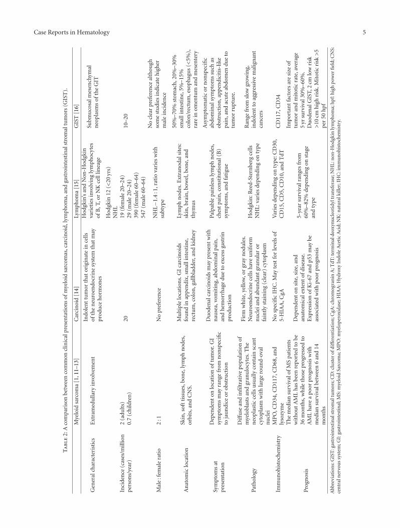
lymphoma, T-cell precursor lymphoma, and myeloid meta-plasia when adequate immunohistochemistry studies werenot performed [1]. Table 2 details the comparison betweenthe clinical features seen in myeloid sarcoma, carcinoidtumors, lymphoma, and gastrointestinal stromal tumors(GIST). Radiologically, MS may mimic lymphomas due tothe uniform contrast enhancement on CT; however, otherdiagnoses for a duodenal mass on CT also include neuroen-docrine tumors and adenocarcinoma [6]. On histologicalexamination, MS typically shows a diffuse and infiltrativepopulation of myeloblasts and granulocytes on H&E stain-ing. The neoplastic cells usually contain scant cytoplasm withlarge round-oval nuclei [22]. Immunohistochemistry offersone of the best methods in establishing the diagnosis of MS[18, 22]. Positive staining for markers MPO, CD34, CD117,and CD68 and lysozyme help identify the myeloid neoplasticcells [8, 22]. The chromosomal abnormalities that have beenreported in conjunction with these extramedullary sarcomasinclude t(8,21), inv(16), t(9,11), 11q23, del(16q), and tri-somy 8 [22, 23]. Interestingly, the patient presented in thispaper did not have any of these listed abnormalities; however,he did have a balanced translocation involving chromosomes2 and 17, which to our knowledge has not been reported inassociation with myeloid sarcomas. In a review of 20 GI MScases, Zhang et al. noted that the inv(16) was often associatedwith intestinal cases [13]. Our patient did not have thisabnormality. Standard investigation used in the workup of
Case Reports in Hematology 5
Ta
ble
2:A
com
pari
son
betw
een
com
mon
clin
ical
pres
enta
tion
sof
mye
loid
sarc
omas
,car
cin
oid,
lym
phom
a,an
dga
stro
inte
stin
alst
rom
altu
mor
s(G
IST
).
Mye
loid
sarc
oma
[1,1
1–13
]C
arci
noi
d[1
4]Ly
mph
oma
[15]
GIS
T[1
6]
Gen
eral
char
acte
rist
ics
Ext
ram
edu
llary
invo
lvem
ent
Indo
len
ttu
mor
that
orig
inat
ein
cells
ofth
en
euro
endo
crin
esy
stem
that
may
prod
uce
hor
mon
es
Hod
gkin
’san
dN
on-H
odgk
inva
riet
ies
invo
lvin
gly
mph
ocyt
esof
B,T
,or
NK
cell
linea
ge
Subm
uco
salm
esen
chym
aln
eopl
asm
sof
the
GIT
Inci
den
ce(c
ases
/mill
ion
per
son
s/ye
ar)
2(a
dult
s)0.
7(c
hild
ren
)20
Hod
gkin
12(<
20yr
s)N
HL
19(f
emal
e20
–24)
29(m
ale
20–2
4)39
0(f
emal
e60
–64)
547
(mal
e60
–64)
10–2
0
Mal
e:f
emal
era
tio
2:1
No
pref
eren
ceN
HL−1
.4:1
,rat
iova
ries
wit
hsu
btyp
e
No
clea
rpr
efer
ence
alth
ough
som
est
udi
esin
dica
teh
igh
erm
ale
inci
den
ce
An
atom
iclo
cati
onSk
in,s
oft
tiss
ues
,bon
e,ly
mph
nod
es,
orbi
ts,a
nd
CN
S.
Mu
ltip
lelo
cati
ons.
GI
carc
inoi
dsfo
un
din
appe
ndi
x,sm
alli
nte
stin
e,re
ctu
m,c
olon
,gal
lbla
dder
,an
dki
dney
Lym
phn
odes
.Ext
ran
odal
site
s:sk
in,b
rain
,bow
el,b
one,
and
thym
us
50%
–70%
stom
ach
,20%
–30%
smal
lin
test
ine,
5%–1
5%co
lon
/rec
tum
,eso
phag
us
(<5%
),ra
rein
omen
tum
and
mes
ente
ry
Sym
ptom
sat
pres
enta
tion
Dep
ende
nt
onlo
cati
onof
tum
or.G
Isy
mpt
oms
may
ran
gefr
omn
onsp
ecifi
cto
jau
ndi
ceor
obst
ruct
ion
Du
oden
alca
rcin
oids
may
pres
ent
wit
hn
ause
a,vo
mit
ing,
abdo
min
alpa
in,
and
hem
orrh
age
due
toex
cess
gast
rin
prod
uct
ion
Palp
able
pain
less
lym
phn
odes
,ch
est
pain
,con
stit
uti
onal
(B)
sym
ptom
s,an
dfa
tigu
e
Asy
mpt
omat
icor
non
spec
ific
abdo
min
alsy
mpt
oms
such
asob
stru
ctio
n,a
ppen
dici
tis-
like
pain
,an
dac
ute
abdo
men
due
totu
mor
rupt
ure
.
Path
olog
y
Diff
use
and
infi
ltra
tive
popu
lati
onof
mye
lobl
asts
and
gran
ulo
cyte
s.T
he
neo
plas
tic
cells
usu
ally
con
tain
scan
tcy
topl
asm
wit
hla
rge
rou
nd-
oval
nu
clei
Firm
wh
ite,
yello
w,o
rgr
ayn
odu
les.
Neu
roen
docr
ine
cells
hav
eu
nif
orm
nu
clei
and
abu
nda
nt
gran
ula
ror
fain
tly
stai
nin
g(c
lear
)cy
topl
asm
Hod
gkin
:Ree
d-St
ern
berg
cells
NH
L:va
ries
depe
ndi
ng
onty
pe
Ran
gefr
omsl
owgr
owin
g,in
dole
nt
toag
gres
sive
mal
ign
ant
can
cers
Imm
un
ohis
toch
emis
try
MP
O,C
D34
,CD
117,
CD
68,a
nd
lyso
zym
eN
osp
ecifi
cIH
C.M
ayte
stfo
rle
vels
of5-
HIA
A,C
gAV
arie
sde
pen
din
gon
type
:CD
30,
CD
15,C
D5,
CD
10,a
nd
TdT
CD
117,
CD
34
Pro
gnos
is
Th
em
edia
nsu
rviv
alof
MS
pati
ents
wit
hou
tA
ML
has
been
rep
orte
dto
be36
mon
ths,
wh
ileth
ose
prog
ress
edto
AM
Lh
ave
apo
orpr
ogn
osis
wit
hm
edia
nsu
rviv
albe
twee
n6
and
14m
onth
s
Dep
ende
nt
onsi
te,s
ize,
and
anat
omic
alex
ten
tof
dise
ase.
Exp
ress
ion
ofK
i-67
and
p53
may
beas
soci
ated
wit
hpo
orpr
ogn
osis
5-ye
arsu
rviv
alra
nge
sfr
om60
%–8
2%de
pen
din
gon
stag
ean
dty
pe
Impo
rtan
tfa
ctor
sar
esi
zeof
tum
oran
dm
itot
icra
te,a
vera
ge5
yrsu
rviv
al30
%–6
0%.
Du
oden
alG
IST,
2cm
low
risk
>10
cmh
igh
risk
.Mit
otic
risk
>5
per
50h
pf
Abb
revi
atio
ns:
GIS
T:g
astr
oin
test
inal
stro
mal
tum
ors;
CD
:clu
ster
ofdi
ffer
enti
atio
n;C
gA:c
hro
mog
ran
inA
;TdT
:ter
min
alde
oxyn
ucl
eoti
dylt
ran
sfer
ase;
NH
L:n
on-H
odgk
inly
mph
oma;
hpf
:hig
hpo
wer
fiel
d;C
NS:
cen
tral
ner
vou
ssy
stem
;GI:
gast
roin
test
inal
;MS:
mye
loid
Sarc
oma;
MP
O:m
yelo
pero
xida
se;H
IAA
:Hyd
roxy
Indo
leA
ceti
cA
cid;
NK
:nat
ura
lkill
er;I
HC
:im
mu
noh
isto
chem
istr
y.

6 Case Reports in Hematology
a duodenal mass was employed here, which included upperGI endoscopy, CT of the abdomen, and biopsy of the masswith H&E staining and immunohistochemistry.
Although there is no clear consensus or guidelines onhow to treat isolated MS, delays in treatment almost alwaysprogress to AML [21, 22]. The median time to the develop-ment of acute leukemia in the setting of isolated MS tumorshas ranged from 5 to 12 months [13, 19]. A result therapywhich should be instituted promptly and typically involvesstandard Daunorubicin- and Cytarabine-based chemother-apy for AML, which includes a 2-part-induction phase (toachieve complete remission) and a consolidation phase (tomaintain complete remission) [9, 22]. Low-dose radiationtherapy (24 Gy) has been suggested in cases that may requiredebulking due to compression of vital structures or in caseswhere intensive chemotherapy has failed [22]. Surgery is notthe primary modality of treatment for MS but is indicatedin cases of obstruction, perforation, or compression of vitalstructures [11]. The therapeutic response does not seem tobe influenced by whether the tumor presents de novo orin conjunction with AML or other hematologic entity evenwhen age, sex, and anatomical site are taken into consider-ation. Patients who undergo either allogenic or autologousbone marrow transplants seem to have a higher probabilityof survival and complete remission [1]. The median survivalof MS patients without AML has been reported to be36 months, while those progressing to AML have a poorprognosis with median survival between 6 and 14 months[20]. However given the rarity of MS, survival outcomeshave been reported in only a handful of reports, and nocontrol studies exist. The wide cytogenetic and molecularheterogeneity of the disease has prompted the use of targetedtherapies (TK inhibitors, C-KIT inhibitors Imatinib andDasatinib, and monoclonal antibodies) aimed at inhibitingthe pivotal pathways of leukemogenesis and restoring normalhemopoiesis. In the future, a combination of different newdrugs along with conventional chemotherapy may representa more effective and potentially curative strategy [24].
6. Conclusion
Diagnosing duodenal MS can be a diagnostic challengeespecially in patients with no previous leukemic disease.The symptoms related to the tumor mass are most oftennonspecific, with abdominal pain sometimes being the onlycomplaint, and as a result a wide variety of differentialdiagnoses are often entertained. In the evaluation of asolid GI mass, myeloid sarcoma should be considered inthe differential diagnosis with confirmation by biopsy andimmunohistochemical analysis of the tumor. Patients withMS should be thoroughly tested for the presence of con-current leukemias or other myelodysplastic disorders. Therapid initial diagnosis of MS and immediate treatment witheither chemotherapy, radiation, surgery, or a combinationof any of these modalities are associated with a betterprognosis, as misdiagnosis or treatment delays can resultin disease progression to AML. Further studies linkingcytogenic abnormalities with the location of the tumors maypermit more targeted therapies.
Consent
Written informed consent was obtained from the patient forthis paper.
Conflict of Interests
The authors declare that they have no conflict of interests.
References
[1] S. A. Pileri, S. Ascani, M. C. Cox et al., “Myeloid sarcoma:clinico-pathologic, phenotypic and cytogenetic analysis of 92adult patients,” Leukemia, vol. 21, no. 2, pp. 340–350, 2007.
[2] E. K. Choi, H. K. Ha, S. H. Park et al., “Granulocytic sarcomaof bowel: CT findings,” Radiology, vol. 243, no. 3, pp. 752–759,2007.
[3] A. Burns, Observation of Surgical Anatomy, Head and Neck,Thomas Royce, Edinburgh, Scotland, 1811.
[4] A. King, “A case of chloroma,” Monthly Journal of the MedicalSociety, vol. 17, p. 97, 1853.
[5] H. Rappaport, “Tumours of the hematopoietic system,” inAtlas of Tumor Pathology, Section III, Fascicle 8. Armed ForcesInstitute of Pathology, pp. 241–243, Washington, DC, USA,1966.
[6] H. Kim, “Primary granulocytic sarcoma of the duodenum:radiologic and endoscopic findings,” American Journal ofRoentgenology, vol. 170, no. 4, pp. 1115–1116, 1998.
[7] O. Goor, Y. Goor, F. Konikoff et al., “Epigastric distress causedby a duodenal polyp: a rare presentation of acute leukemia,”Journal of Clinical Oncology, vol. 21, no. 23, pp. 4455–4459,2003.
[8] E. Derenzini, S. Paolini, G. Martinelli et al., “Extramedullarymyeloid tumour of the stomach and duodenum presentingwithout acute myeloblastic leukemia: a diagnostic and thera-peutic challenge,” Leukemia and Lymphoma, vol. 49, no. 1, pp.159–162, 2008.
[9] T. Ghafoor, A. Zaidi, and I. Al Nassir, “Granulocytic sarcomaof the small intestine: an unusual presentation of acute myelo-genous leukaemia,” Journal of the Pakistan Medical Association,vol. 60, no. 2, pp. 133–135, 2010.
[10] S. H. Jeong, J. H. Han, S. Y. Jeong et al., “A case of donor-derived granulocytic sarcoma after allogeneic hematopoieticstem cell transplantation,” The Korean Journal of Hematology,vol. 45, no. 1, pp. 70–72, 2010.
[11] D. Antic, I. Elezovic, A. Bogdanovic et al., “Isolated myeloidsarcoma of the gastrointestinal tract,” Internal Medicine, vol.49, no. 9, pp. 853–856, 2010.
[12] M. Breccia, F. Mandelli, M. C. Petti et al., “Clinico-pathologi-cal characteristics of myeloid sarcoma at diagnosis and duringfollow-up: report of 12 cases from a single institution,” Leuke-mia Research, vol. 28, no. 11, pp. 1165–1169, 2004.
[13] X. H. Zhang, R. Zhang, and Y. Li, “Granulocytic sarcomaof abdomen in acute myeloid leukemia patient with inv(16)and t(6;17) abnormal chromosome: case report and reviewof literature,” Leukemia Research, vol. 34, no. 7, pp. 958–961,2010.
[14] General information about gastrointestinal carcinoid tumors,National Cancer Institute at the National Institutes of Health,2012, http://www.cancer.gov/cancertopics/pdq/treatment/ga-strointestinalcarcinoid/HealthProfessional.

Case Reports in Hematology 7
[15] Hodgkin and Non-Hodgkin lymphoma, Leukemia and Lym-phoma Society, 2012, http://www.lls.org/#/diseaseinforma-tion/getinformationsupport/factsstatistics/hodgkinlympho-ma/.
[16] General information about gastrointestinal stromal tumors,National Cancer Institute at the National Institutes of Health,2012, http://www.cancer.gov/cancertopics/pdq/treatment/gist/HealthProfessional.
[17] E. S. Jaffe, N. L. Harris, H. Stein et al., World Health Organi-zation Classification of Tumours—Tumours of Haematopoieticand Lymphoid Tissues, 2001.
[18] R. Sivan-Hoffmann, I. Waksman, H. I. Cohen et al., “Smallbowel obstruction as a presenting sign of granulocytic sar-coma,” The Israel Medical Association Journal, vol. 13, no. 8,pp. 507–509, 2011.
[19] R. S. Neiman, M. Barcos, and C. Berard, “Granulocytic sarco-ma: a clinicopathologic study of 61 biopsied cases,” Cancer,vol. 48, no. 6, pp. 1426–1437, 1981.
[20] K. Yamauchi and M. Yasuda, “Comparison in treatments ofnonleukemic granulocytic sarcoma: report of two cases and areview of 72 cases in the literature,” Cancer, vol. 94, no. 6, pp.1739–1746, 2002.
[21] J. M. Meis, J. J. Butler, B. M. Osborne, and J. T. Manning, “Gra-nulocytic sarcoma in nonleukemic patients,” Cancer, vol. 58,no. 12, pp. 2697–2709, 1986.
[22] R. L. Bakst, M. S. Tallman, D. Douer et al., “How I treat extra-medullary acute myeloid leukemia,” Blood, vol. 118, no. 14, pp.3785–3793, 2011.
[23] C. I. Jenkins and Y. Sorour, “Case report: a large extramedul-lary granulocytic sarcoma as the initial presenting feature ofchronic myeloid leukemia,” MedGenMed, vol. 7, no. 4, p. 23,2005.
[24] F. Ferrara, “New agents for acute myeloid leukemia: is it timefor targeted therapies?” Expert Opinion on InvestigationalDrugs, vol. 21, no. 2, pp. 179–189, 2012.

Submit your manuscripts athttp://www.hindawi.com
Stem CellsInternational
Hindawi Publishing Corporationhttp://www.hindawi.com Volume 2014
Hindawi Publishing Corporationhttp://www.hindawi.com Volume 2014
MEDIATORSINFLAMMATION
of
Hindawi Publishing Corporationhttp://www.hindawi.com Volume 2014
Behavioural Neurology
EndocrinologyInternational Journal of
Hindawi Publishing Corporationhttp://www.hindawi.com Volume 2014
Hindawi Publishing Corporationhttp://www.hindawi.com Volume 2014
Disease Markers
Hindawi Publishing Corporationhttp://www.hindawi.com Volume 2014
BioMed Research International
OncologyJournal of
Hindawi Publishing Corporationhttp://www.hindawi.com Volume 2014
Hindawi Publishing Corporationhttp://www.hindawi.com Volume 2014
Oxidative Medicine and Cellular Longevity
Hindawi Publishing Corporationhttp://www.hindawi.com Volume 2014
PPAR Research
The Scientific World JournalHindawi Publishing Corporation http://www.hindawi.com Volume 2014
Immunology ResearchHindawi Publishing Corporationhttp://www.hindawi.com Volume 2014
Journal of
ObesityJournal of
Hindawi Publishing Corporationhttp://www.hindawi.com Volume 2014
Hindawi Publishing Corporationhttp://www.hindawi.com Volume 2014
Computational and Mathematical Methods in Medicine
OphthalmologyJournal of
Hindawi Publishing Corporationhttp://www.hindawi.com Volume 2014
Diabetes ResearchJournal of
Hindawi Publishing Corporationhttp://www.hindawi.com Volume 2014
Hindawi Publishing Corporationhttp://www.hindawi.com Volume 2014
Research and TreatmentAIDS
Hindawi Publishing Corporationhttp://www.hindawi.com Volume 2014
Gastroenterology Research and Practice
Hindawi Publishing Corporationhttp://www.hindawi.com Volume 2014
Parkinson’s Disease
Evidence-Based Complementary and Alternative Medicine
Volume 2014Hindawi Publishing Corporationhttp://www.hindawi.com