Preparation and Preliminary Nonlinear Optical Properties ...
10
Research Article Preparation and Preliminary Nonlinear Optical Properties of BiFeO 3 Nanocrystal Suspensions from a Simple, Chelating Agent-Free Precipitation Route Théo Tytus, 1 Oisín Phelan, 1 Mathias Urbain, 1 Gareth Clarke, 1 Jérémy Riporto, 1 Ronan Le Dantec, 1 Gnon Djanta, 2 Sandrine Beauquis, 1 Virginie Monnier, 3 Yann Chevolot, 3 Christine Galez, 1 and Yannick Mugnier 1 1 Univ. Savoie Mont Blanc, SYMME, F-74000 Annecy, France 2 Faculté des Sciences, Université de LOME, Lomé, Togo 3 Université de Lyon, Institut des Nanotechnologies de Lyon UMR CNRS 5270, Ecole Centrale de Lyon, 36 Avenue Guy de Collongue, F-69134 Ecully, France Correspondence should be addressed to Yannick Mugnier; [email protected] Received 15 May 2018; Accepted 3 July 2018; Published 9 September 2018 Academic Editor: Jean M. Greneche Copyright © 2018 Théo Tytus et al. This is an open access article distributed under the Creative Commons Attribution License, which permits unrestricted use, distribution, and reproduction in any medium, provided the original work is properly cited. Preparation of stable BiFeO 3 nanocrystal suspensions through a simple, low-cost precipitation technique is described. Amorphous precursors are first precipitated from metal nitrate salts in highly basic KOH solutions, and a short high-temperature annealing step is then performed to induce crystallization. Nanoparticles are characterized by X-ray diffraction (XRD), TEM, DLS and ζ-potential measurements, and the synthesis conditions optimized after a systematic variation of the KOH concentration within the range of 1–12 M. The presence of residual impurities (mainly Bi 25 FeO 39 and Bi 2 Fe 4 O 9 ) quantified from XRD and mean nanocrystal size is found to be strongly influenced by the initial KOH solution content. A concentration at about 3–4 M is optimal in terms of BiFeO 3 phase-purity and nanocrystal size. Stability of aqueous dispersions of the amorphous precursors and of the purest crystallized nanoparticles is also characterized between pH = 2 and pH = 13. After preparation of stable, almost phase-pure BiFeO 3 nanocrystal suspensions, second and third harmonic scattering (SHS and THS) at excitation wavelengths of 1064 nm and 1250 nm are reported from nonlinear optical scattering measurements and compared with other recently published literature values. 1. Introduction Bismuth ferrite (BiFeO 3 ) is today a well-known single-phase magnetoelectric material that combines room temperature ferroelectricity and antiferromagnetic ordering and as such, has attracted a considerable amount of attention due to its multiferroic properties [1, 2]. BiFeO 3 exhibits a rhombohed- rally distorted perovskite structure (space group R3c) for which noncentrosymmetry arises from the rotation of the oxygen octahedrons around the pseudocubic [111] axis [3, 4]. Among potential applications based on bismuth ferrite, photocatalysis under ambient light [5], nonvolatile memory [6], spintronics [7, 8], and energy harvesting [9] can be mentioned as well as its increasing interest in bioimaging due to intrinsic, very high second [10, 11] and third harmonic [12] properties. Harmonic nanoparticles (HNPs) [13] based on BiFeO 3 not only display a low cytotoxicity [14] but also display several “optical” advantages in terms of excitation wavelength tunability for deep-imaging [15], of photostability for long-term observations [16, 17], and of improved sensitiv- ity due to specific harmonic signatures against endogenous signals [18–20]. Synthesis of BiFeO 3 nanoparticles has also been the subject of intense research, and several chemical routes that include, for instance, hydrothermal conditions [21–24], solvent evaporations [25], sol-gel polymerizations [26–29], sonochemical reactions, and microemulsion techniques [30] have already been studied. Hydrothermal syntheses with or Hindawi Journal of Nanomaterials Volume 2018, Article ID 3019586, 9 pages https://doi.org/10.1155/2018/3019586
Transcript of Preparation and Preliminary Nonlinear Optical Properties ...

Research ArticlePreparation and Preliminary Nonlinear OpticalProperties of BiFeO3 Nanocrystal Suspensions from a Simple,Chelating Agent-Free Precipitation Route
Théo Tytus,1 Oisín Phelan,1 Mathias Urbain,1 Gareth Clarke,1 Jérémy Riporto,1
Ronan Le Dantec,1 Gnon Djanta,2 Sandrine Beauquis,1 Virginie Monnier,3 Yann Chevolot,3
Christine Galez,1 and Yannick Mugnier 1
1Univ. Savoie Mont Blanc, SYMME, F-74000 Annecy, France2Faculté des Sciences, Université de LOME, Lomé, Togo3Université de Lyon, Institut des Nanotechnologies de Lyon UMR CNRS 5270, Ecole Centrale de Lyon, 36 Avenue Guy de Collongue,F-69134 Ecully, France
Correspondence should be addressed to Yannick Mugnier; [email protected]
Received 15 May 2018; Accepted 3 July 2018; Published 9 September 2018
Academic Editor: Jean M. Greneche
Copyright © 2018 Théo Tytus et al. This is an open access article distributed under the Creative Commons Attribution License,which permits unrestricted use, distribution, and reproduction in any medium, provided the original work is properly cited.
Preparation of stable BiFeO3 nanocrystal suspensions through a simple, low-cost precipitation technique is described. Amorphousprecursors are first precipitated frommetal nitrate salts in highly basic KOH solutions, and a short high-temperature annealing stepis then performed to induce crystallization. Nanoparticles are characterized by X-ray diffraction (XRD), TEM, DLS and ζ-potentialmeasurements, and the synthesis conditions optimized after a systematic variation of the KOH concentration within the range of1–12M. The presence of residual impurities (mainly Bi25FeO39 and Bi2Fe4O9) quantified from XRD and mean nanocrystal size isfound to be strongly influenced by the initial KOH solution content. A concentration at about 3–4M is optimal in terms of BiFeO3phase-purity and nanocrystal size. Stability of aqueous dispersions of the amorphous precursors and of the purest crystallizednanoparticles is also characterized between pH= 2 and pH= 13. After preparation of stable, almost phase-pure BiFeO3nanocrystal suspensions, second and third harmonic scattering (SHS and THS) at excitation wavelengths of 1064 nm and1250 nm are reported from nonlinear optical scatteringmeasurements and compared with other recently published literature values.
1. Introduction
Bismuth ferrite (BiFeO3) is today a well-known single-phasemagnetoelectric material that combines room temperatureferroelectricity and antiferromagnetic ordering and as such,has attracted a considerable amount of attention due to itsmultiferroic properties [1, 2]. BiFeO3 exhibits a rhombohed-rally distorted perovskite structure (space group R3c) forwhich noncentrosymmetry arises from the rotation of theoxygen octahedrons around the pseudocubic [111] axis[3, 4]. Among potential applications based on bismuth ferrite,photocatalysis under ambient light [5], nonvolatile memory[6], spintronics [7, 8], and energy harvesting [9] can bementioned as well as its increasing interest in bioimaging
due to intrinsic, very high second [10, 11] and third harmonic[12] properties. Harmonic nanoparticles (HNPs) [13] basedon BiFeO3 not only display a low cytotoxicity [14] but alsodisplay several “optical” advantages in terms of excitationwavelength tunability for deep-imaging [15], of photostabilityfor long-term observations [16, 17], and of improved sensitiv-ity due to specific harmonic signatures against endogenoussignals [18–20].
Synthesis of BiFeO3 nanoparticles has also been thesubject of intense research, and several chemical routes thatinclude, for instance, hydrothermal conditions [21–24],solvent evaporations [25], sol-gel polymerizations [26–29],sonochemical reactions, and microemulsion techniques [30]have already been studied. Hydrothermal syntheses with or
HindawiJournal of NanomaterialsVolume 2018, Article ID 3019586, 9 pageshttps://doi.org/10.1155/2018/3019586
without chelating agents and sol-gel processes are indeedwell-established approaches giving rise to phase-pure sam-ples even if size- and shape-control in the case of BiFeO3are clearly issues [31], especially without use of expensive,microwave-assisted solvothermal conditions. Ideally, a facile,green, and scalable synthesis route which yields monodis-perse and monocrystalline nanoparticles stabilized in aque-ous solutions would be very convenient for the above-citedbioapplications, as it then allows surface modification of barenanocrystals so as to improve their final biocompatibility andspecificity for cell-targeting. To this end, we further exploredthe simple and cost-effective coprecipitation route alreadyinvestigated [32–34] with the aim to prepare stable BiFeO3nanocrystal suspensions. Metal nitrate salts without anyother organic additive and KOH solutions at differentconcentrations have been employed for the precipitation ofthe primary amorphous precursors. After a short annealingstep at 720°C used here to promote crystallization and toinhibit as much as possible Ostwald ripening, it was foundthat an initial KOH concentration fixed at about 3–4M isthe best compromise in terms of size and residual impurities.The latter have been quantitatively assessed through Rietveldrefinements for all the samples prepared from a KOH con-centration varying between 1M and 12M. After optimizationof the chemical synthesis, phase-pure nanopowders preparedat 4M of KOH are dispersed in deionized water and stabilityof the resulting suspensions studied with DLS and ζ-potentialmeasurements. Finally, a quantitative assessment of the aver-aged <χ(2)> susceptibility at 1064 nm under femtosecondlaser excitation is compared with recently available literaturevalues and discussed according to the synthesis process heredeveloped. When the laser excitation wavelength is shifted to1250 nm, preliminary optical characterizations indicate thatthe second and third harmonic scattering signals are easilymonitored from ensemble measurements in the NIR biolog-ical transparency window.
2. Materials and Methods
For the preparation of BiFeO3 nanocrystal suspensions,1.58mmol of bismuth pentahydrate (Bi(NO3)·5H2O, 0.77 g,Sigma-Aldrich, >98%) and stoichiometric amounts of ironnitrate nonahydrate (Fe(NO3)·9H2O, 0.64 g, Sigma-Aldrich,>99.9%) are weighed and dissolved without further purifica-tion in 3mL of a 2M nitric acid (HNO3) solution. Afterdissolution of the precursors, the solution is slowly addedinto 17mL of freshly prepared potassium hydroxide (KOH)solution of varying concentrations (between 0.1M and13M) that is allowed to stir vigorously and continuouslyfor 5min. The as-obtained brownish precipitate is thentreated for 1 h with a standard sonic bath, and the primaryamorphous precipitate is finally collected by centrifugationat 15700 rpm. Five successive washings with deionized waterand three more with ethanol are applied before a 12h dryingstep at 85°C. The powder is then grounded with a mortar,carefully spread onto a thick alumina sample holder, andquickly annealed in air between 2min 30 and 4min, topromote crystallization at 720°C. Various amounts of crystal-lized powders can thus be obtained showing the scalability of
the chemical process. Stability studies and optical measure-ments were all performed from an initial mass concentrationfixed at 1mg/mL. Typically, 7mg of dried powders were son-icated for 30min in 7mL of aqueous solutions (or in ethanol)and then left to settle for several days to allow sedimentationof the remaining aggregates. Finally, DLS measurements andweighing of the residual fraction (assuming spherical nano-crystals) allow to estimate the number density of nanocrys-tals in the supernatant at the end of the sedimentation period.
Physicochemical characterizations were carried out byXRD (using Co Kα radiation from an INEL CPS 120 instru-ment and an acquisition time of 2 h per pattern and from aPANalytical X’Pert3 Powder diffractometer with a zero-background silicon sample holder), TEM (JEOL 2100 HToperating at 200 kV), and by ζ-potential and dynamic lightscattering (DLS) measurements (Malvern Zetasizer Nano).
Nonlinear optical properties were measured by probingthe second and third harmonic scattering signals as previ-ously reported [12, 35, 36] except that the excitation sourceis here provided by a tunable (700–1300 nm) femtosecondLaser (Insight X3 Spectra-Physics) vertically polarized. SHand TH signals are collected perpendicularly to the funda-mental beam and focused through two 5 cm focal lengthlenses onto the slit of a spectrometer (Andor Shamrock193) combined to a CCD camera (Andor iDus 401 BVF).No analyser is used in the detection arm but a short pass filterto remove the excitation wavelength and a Dove prism to ver-tically rotate the image focal point along the spectrometerentrance slit.
3. Results
3.1. XRD Characterizations. As detailed in Materials andMethods, bismuth and iron nitrate precursors are firstdissolved in 3mL of 2M nitric acid. Precipitation of theamorphous precursors is then obtained after mixing this acidprecursor solution with 17mL of a highly basic potassiumhydroxide (KOH) solution of varying concentrations. Toreduce as much as possible the amount of impurities afterthe calcination step, a homogeneous dispersion and concen-tration of the Fe3+ and Bi3+ ions is highly desirable and thelocal basic environment is to be stable enough during the pre-cipitation process. We observed that addition of the KOHsolution into the acid precursor one systematically resultsin very high impurity content (data not shown) whereaspouring the dissolved precursors into KOH solution undercontinuous stirring is much more favourable. In addition, aslow addition is vitally important as shown in SupplementaryMaterial (Figure S1). A higher amount of the Bi-richBi25FeO39 phase is indeed present when the acid precursorsolution is quickly poured whereas impurities are hardlyvisible for a drop-wise or very slow addition from a burette.
For a slow addition, amounts of impurities were alsofound to be very dependent on the initial KOH concentrationthat was allowed to vary between 0.1M and 13M. At 0.1M ofKOH, the XRD pattern indicates that α-Bi2O3 is the maincrystallized phase produced after the short annealing step(data not shown). At higher KOH concentration, the BiFeO3phase is systematically obtained as the major phase, but the
2 Journal of Nanomaterials
impurity content strongly varies as depicted in Figure 1.After the 2min 30 crystallization step performed with10mg of amorphous precursors, residual impurities identi-fied as Bi25FeO39 and Bi2Fe4O9 are particularly visible inthe low 2θ region, namely, between 30° and 40°, of the dif-fraction patterns. A small amorphous contribution is alsoobserved whatever the initial KOH concentration is. Azero-background sample holder has then been used in thefollowing series of experiments to better investigate theamount of residual amorphous precursors and to eventu-ally reduce their content.
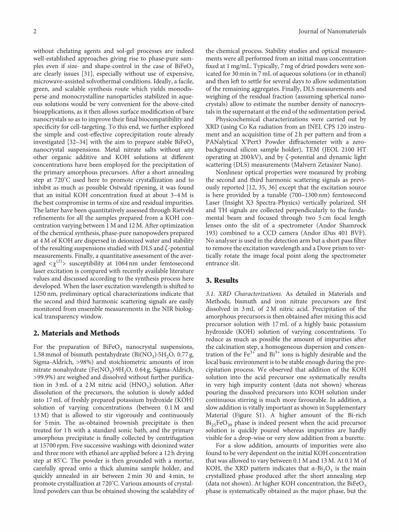
For each sample, the mean diameter of BiFeO3 nanocrys-tals is first derived after extraction of the integrated intensi-ties within FullProf according to the Le Bail global fittingprocedure [37]. Isotropic broadening of the diffraction peaks(without contribution of the Gaussian part in the FWHM)and the absence of residual strain are assumed. Quasi-spherical nanocrystals are thus considered for the final calcu-lation of the mean diameter which is estimated below 40nmin Figure 2(a) and with a trend to decrease when the KOHconcentration is raised from 1M to 12M (see Figure S2 forthe fitted profile of the sample precipitated at 3M of KOH).
30 40 50 60 70
0,0
0,2
0,4
0,6
0,8
1,0
1,2
1,4
24 28 32 36 40
0.0
0.5
⁎
ICSD 152991M3M4M5M8MIn
tens
ity (a
.u.)
2�휃 (º)
12M
⁎
Figure 1: XRD patterns of BiFeO3 samples depending on the initial amount of KOH and after 2min 30 of annealing at 720°C. Peaks denotedas ∗ belong to the aluminium sample holder. The BiFeO3 reference profile (ICSD#15299) is shown in red at the bottom of the graph. Inset:peaks belonging to Bi25FeO39 (light gray square) and Bi2Fe4O9 (light gray circle) are shown for the two most impure samples obtained atKOH=1M and 12M.
0 2 4 6 8 10 12
20
30
40
50
KOH concentration (mol/L)
XRD
dia
met
er (n
m)
(a)
Impu
ritie
s (%
)
0 2 4 6 8 10 12
0
4
8
12
16
20
24
KOH concentration (mol/L)
TotalBi2Fe4O9Bi25FeO39
(b)
Figure 2: (a) Mean BiFeO3 nanocrystal diameters obtained from the Le Bail fitting procedure versus initial KOH concentration used forprecipitation. (b) Weight fraction of residual impurities (Bi25FeO39 and Bi2Fe4O9) as derived from FullProf considering a structural modelfor each phase. Lines are a guide for the eye.
3Journal of Nanomaterials
The mean crystallite size being derived, quantitative assess-ment of the weight fraction of the two impurity phases is thenobtained from the scale factors after a full refinement (includ-ing structural models) of the XRD profiles within FullProf.The atomic positions from the ICSD files #15299, #68627,and #262861 corresponding to the BiFeO3, Bi25FeO39, andBi2Fe4O9 phases, respectively, are used as inputs and keptconstant. Only the scale factors, cell parameters, and meannanocrystal size of each phase are allowed to vary. In the caseof BiFeO3, we noticed that assuming an anisotropic broaden-ing improves to some extent the overall quality of the fits lead-ing to slightly elongated particles along the [101] direction.The as-obtained mean diameters are still very consistentthough with the ones plotted in Figure 2(a). Interestingly,the total amount of impurities in terms of weight fraction ofthe total mass shows in Figure 2(b) a well-defined minimumfor the initial KOH concentration fixed at about 4–5M. Forthe two corresponding samples, the mass fractions are foundbelow 0.5% and 3.5% for the (heavy) Bi-rich Bi25FeO39 phaseand the (light) Fe-rich Bi2Fe4O9 phase, respectively. At 1M ofKOH, the total weight fraction for the two impurities attains24% and reaches about 11% for the 12M sample but with onlythe Bi25FeO39 phase present (see also Figure S3 for the fittedprofiles of these two most impure samples).
3.2. Reproducibility and Scalability of the Chemical Process.Larger numbers of BiFeO3 amorphous precursors, namely,20mg, have also been annealed by using as before a smallround alumina crucible (see Supplementary Material,Figure S4). A longer annealing period of 3min, still at720°C, is however necessary to promote crystallizationwithout experimental evidence from the XRDprofiles of a sig-nificant amorphous background arising from an incompletecrystallization (a cleaved-silicon zero-background sampleholder is here used).When the initial KOH content is system-atically varied in the range of 1–8M, almost phase-pureBiFeO3 nanocrystals are again produced at 3–4M of KOHwhile the mean nanocrystal size obtained from Scherrer’sformulae and the (012) reflection at 2θ~26.3° again decreasesas long as the initial KOH content is raised till 4M (inset ofFigure S4). It can be noticed though the absence of a Fe-richphase in these syntheses since only the Bi25FeO39 and thetrigonal metal Bi phases have been detected as residual impu-rities at high KOH content despite initial stoichiometricamounts of Bi and Fe.
For larger numbers of amorphous precursors, a flatalumina crucible has also been tested for which 40mg ofalmost phase-pure ~30 nm nanocrystals could again be pro-duced at 4M of KOH and for a prolonged annealing timeof 4min at 720°C (see in Figure S5 the XRD patterns ofBiFeO3 samples depending on the initial amount of KOH).Comparatively to the thicker round crucible, the presenceof impurity phases are more clearly visible at low and highKOH concentrations, but scalability of the chemical processis not prevented at 4M of KOH. For an initial mass of precur-sors further increased at 200mg, phase-purity and meannanocrystal XRD size at ~30 nm have again been found ingood agreement with previous results (Figure S6).
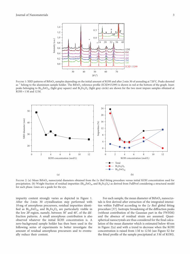
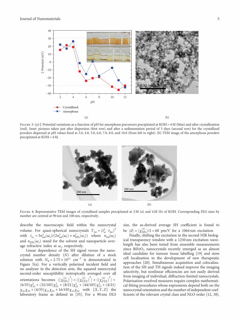
3.3. Suspension Stability. For the studies of the suspensionstability, powders were all issued from the 200mg precipitateobtained at 4M of KOH, and mass concentration for bothamorphous and crystallized particles was fixed at 1mg/mL.Desired amounts of HNO3 or KOH in deionized water wereused to adjust the pH in the range of 2–13. Note that amor-phous and crystallized particles dissolve very quickly atpH=1 whereas they are chemically stable above pH=2. Asseen in Figure 3(a), ζ-potential measurements versus pHindicate an isoelectric point at pH~5 for both amorphousand crystallized nanoparticles and that stability is better forthe three samples dispersed at pH above 10. A ζ-potentialvalue below −30mV is then observed whereas the DLS sizedistribution by number is found to be centred at 90–100nmfor each sample. After a sedimentation period of 5 days, pic-tures included in Figure 3(a) are consistent with ζ-potentialdata (taken 3 days after the sonication process), especiallythe transparent vial at pH=5 for which the absence of sus-pended particles can be noticed. Because of the size increaseinduced after calcination, nanocrystals are also found lessstable than their amorphous counterpart. Only aqueoussolutions with a pH fixed below 4 and above 9 are thus suit-able (transparency of the supernatant shows no colourchange after weeks) for any further surface chemical modifi-cation. Otherwise, nanocrystals are even more stable inethanol. Regarding TEM imaging in Figure 3(b), amorphouspowders precipitated at 4M of KOH are composed of poorlyshape-defined precursors of average size below ~10 nm.
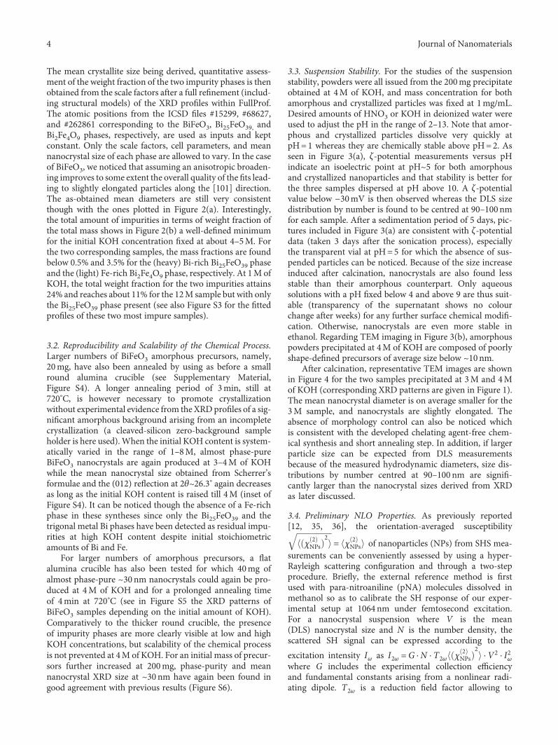
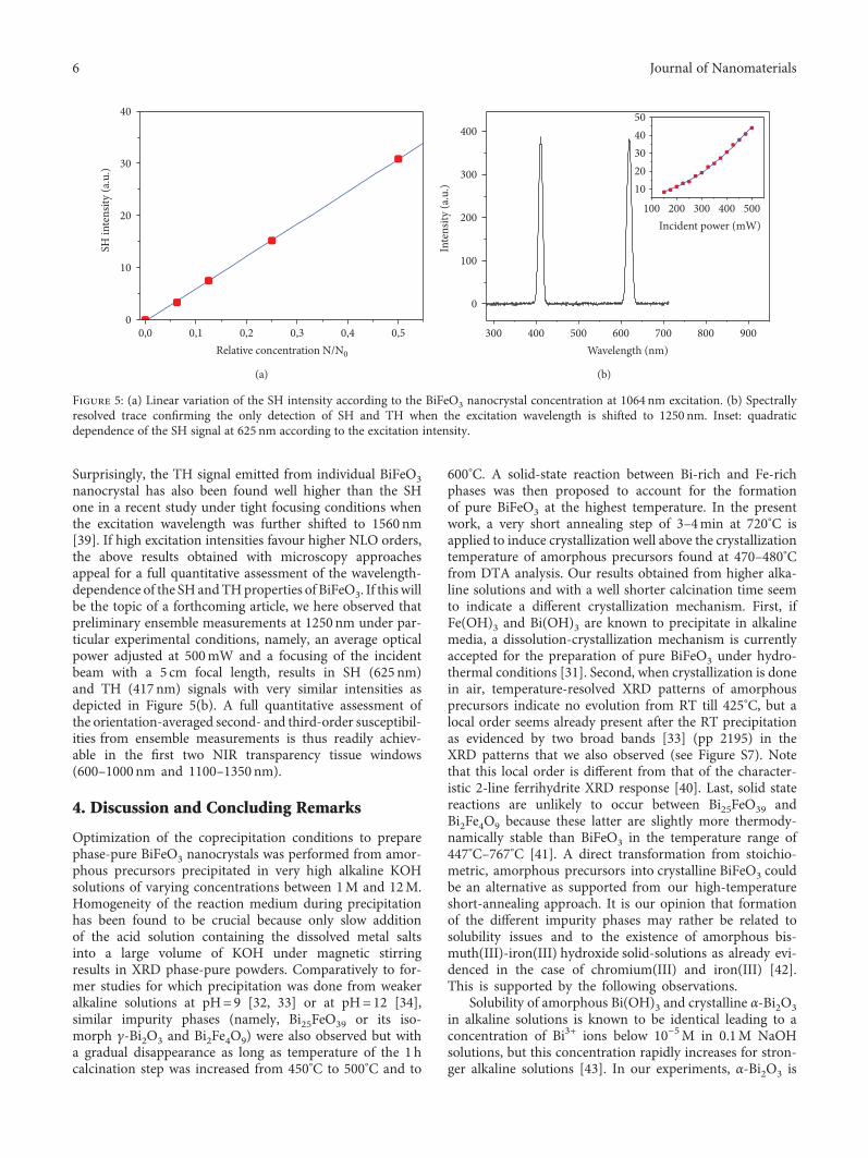
After calcination, representative TEM images are shownin Figure 4 for the two samples precipitated at 3M and 4Mof KOH (corresponding XRD patterns are given in Figure 1).The mean nanocrystal diameter is on average smaller for the3M sample, and nanocrystals are slightly elongated. Theabsence of morphology control can also be noticed whichis consistent with the developed chelating agent-free chem-ical synthesis and short annealing step. In addition, if largerparticle size can be expected from DLS measurementsbecause of the measured hydrodynamic diameters, size dis-tributions by number centred at 90–100 nm are signifi-cantly larger than the nanocrystal sizes derived from XRDas later discussed.
3.4. Preliminary NLO Properties. As previously reported[12, 35, 36], the orientation-averaged susceptibility
χ 2NPs
2 = χ 2NPs of nanoparticles (NPs) from SHS mea-
surements can be conveniently assessed by using a hyper-Rayleigh scattering configuration and through a two-stepprocedure. Briefly, the external reference method is firstused with para-nitroaniline (pNA) molecules dissolved inmethanol so as to calibrate the SH response of our exper-imental setup at 1064 nm under femtosecond excitation.For a nanocrystal suspension where V is the mean(DLS) nanocrystal size and N is the number density, thescattered SH signal can be expressed according to the
excitation intensity Iω as I2ω =G ⋅N ⋅ T2ω χ 2NPs
2⋅ V2 ⋅ I2ω
where G includes the experimental collection efficiencyand fundamental constants arising from a nonlinear radi-ating dipole. T2ω is a reduction field factor allowing to
4 Journal of Nanomaterials
describe the macroscopic field within the nanocrystal
volume. For quasi-spherical nanocrystals T2ω = t2ω ⋅ t2ω2
with tωi= 3n2sol ωi / 2n2sol ωi + n2NPs ωi where nsol ωi
and nNPs ωi stand for the solvent and nanoparticle aver-age refractive index at ωi, respectively.
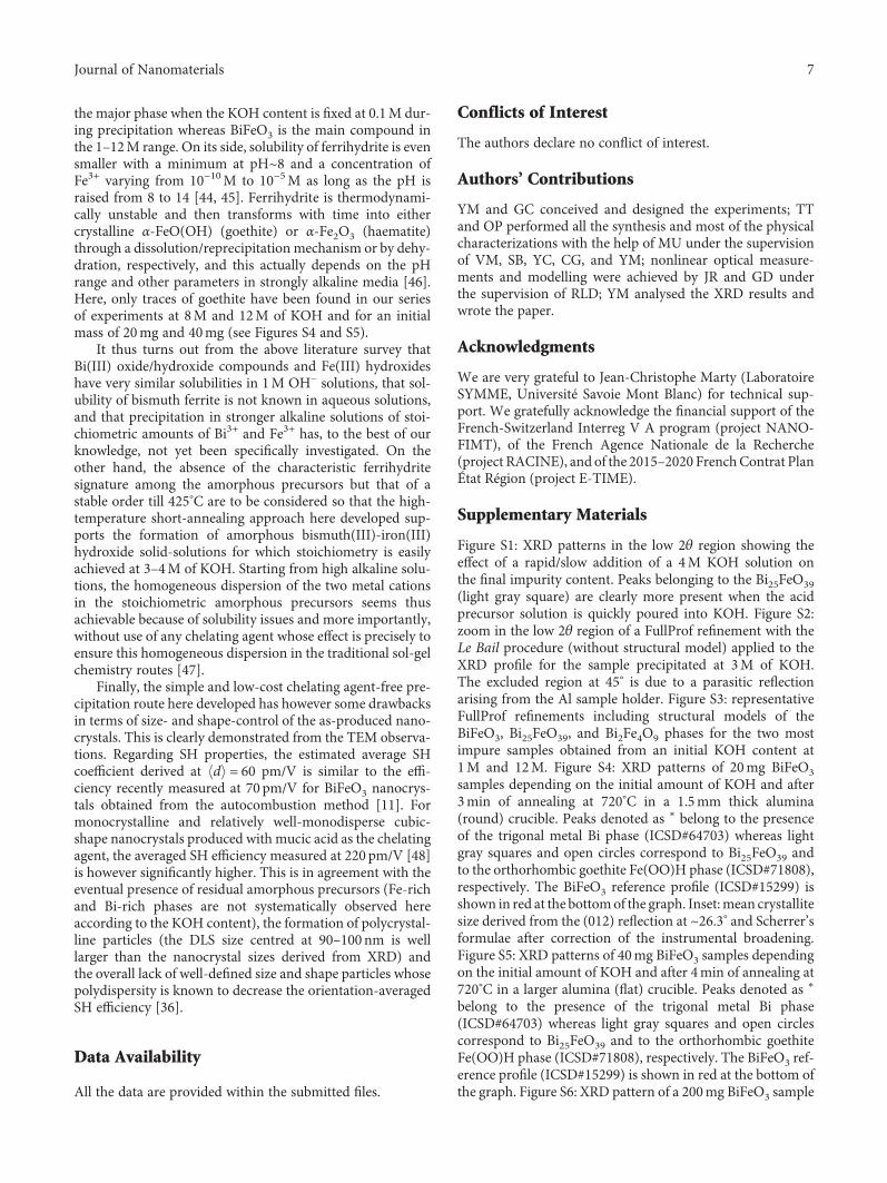
Linear dependence of the SH signal versus the nano-crystal number density (N) after dilution of a stocksolution with N0 = 2 75 × 1011 cm−3 is demonstrated inFigure 5(a). For a vertically polarized incident field andno analyser in the detection arm, the squared nanocrystalsecond-order susceptibility isotropically averaged over all
orientations becomes χ 2NPs
2= χ 2
XXX
2+ χ 2
ZXX
2=
6/35 χ233 + 32/105 χ2
31 + 8/21 χ222 + 44/105 χ2
15 + 4/21χ31χ33 + 4/35 χ15χ33 + 16/105χ15χ31 with X, Y , Z thelaboratory frame as defined in [35]. For a 90nm DLS
size, the as-derived average SH coefficient is found to
be d = χ 2NPs /2 = 60 pm/V for a 1064 nm excitation.
Finally, shifting the excitation in the second NIR biolog-ical transparency window with a 1250 nm excitation wave-length has also been tested from ensemble measurementssince BiFeO3 nanocrystals recently emerged as an almostideal candidate for tumour tissue labelling [19] and stemcell localization in the development of new therapeuticapproaches [20]. Simultaneous acquisition and colocaliza-tion of the SH and TH signals indeed improve the imagingselectivity, but nonlinear efficiencies are not easily derivedfrom imaging of individual, diffraction-limited nanocrystals.Polarization-resolved measures require complex mathemati-cal fitting procedures whose expressions depend both on thenanocrystal orientation and the number of independent coef-ficients of the relevant crystal class and NLO order [12, 38].
2 4 6 8 10 12−40
−30
−20
−10
0
10
20
30
40
pH
�휁-Po
tent
ial (
mV
)
CrystallizedAmorphous
(a) (b)
Figure 3: (a) ζ-Potential variations as a function of pH for amorphous precursors precipitated at KOH=4M (blue) and after crystallization(red). Inset: pictures taken just after dispersion (first row) and after a sedimentation period of 5 days (second row) for the crystallizedpowders dispersed at pH values fixed at 3.0, 4.0, 5.0, 6.0, 7.0, 8.0, and 10.0 (from left to right). (b) TEM image of the amorphous powdersprecipitated at KOH=4M.
200 nm
(a)
200 nm
(b)
Figure 4: Representative TEM images of crystallized samples precipitated at 3M (a) and 4M (b) of KOH. Corresponding DLS sizes bynumber are centred at 90 nm and 100 nm, respectively.
5Journal of Nanomaterials
Surprisingly, the TH signal emitted from individual BiFeO3nanocrystal has also been found well higher than the SHone in a recent study under tight focusing conditions whenthe excitation wavelength was further shifted to 1560 nm[39]. If high excitation intensities favour higher NLO orders,the above results obtained with microscopy approachesappeal for a full quantitative assessment of the wavelength-dependence of the SH andTHproperties of BiFeO3. If this willbe the topic of a forthcoming article, we here observed thatpreliminary ensemble measurements at 1250 nm under par-ticular experimental conditions, namely, an average opticalpower adjusted at 500mW and a focusing of the incidentbeam with a 5 cm focal length, results in SH (625 nm)and TH (417nm) signals with very similar intensities asdepicted in Figure 5(b). A full quantitative assessment ofthe orientation-averaged second- and third-order susceptibil-ities from ensemble measurements is thus readily achiev-able in the first two NIR transparency tissue windows(600–1000nm and 1100–1350 nm).
4. Discussion and Concluding Remarks
Optimization of the coprecipitation conditions to preparephase-pure BiFeO3 nanocrystals was performed from amor-phous precursors precipitated in very high alkaline KOHsolutions of varying concentrations between 1M and 12M.Homogeneity of the reaction medium during precipitationhas been found to be crucial because only slow additionof the acid solution containing the dissolved metal saltsinto a large volume of KOH under magnetic stirringresults in XRD phase-pure powders. Comparatively to for-mer studies for which precipitation was done from weakeralkaline solutions at pH=9 [32, 33] or at pH=12 [34],similar impurity phases (namely, Bi25FeO39 or its iso-morph γ-Bi2O3 and Bi2Fe4O9) were also observed but witha gradual disappearance as long as temperature of the 1 hcalcination step was increased from 450°C to 500°C and to
600°C. A solid-state reaction between Bi-rich and Fe-richphases was then proposed to account for the formationof pure BiFeO3 at the highest temperature. In the presentwork, a very short annealing step of 3–4min at 720°C isapplied to induce crystallization well above the crystallizationtemperature of amorphous precursors found at 470–480°Cfrom DTA analysis. Our results obtained from higher alka-line solutions and with a well shorter calcination time seemto indicate a different crystallization mechanism. First, ifFe(OH)3 and Bi(OH)3 are known to precipitate in alkalinemedia, a dissolution-crystallization mechanism is currentlyaccepted for the preparation of pure BiFeO3 under hydro-thermal conditions [31]. Second, when crystallization is donein air, temperature-resolved XRD patterns of amorphousprecursors indicate no evolution from RT till 425°C, but alocal order seems already present after the RT precipitationas evidenced by two broad bands [33] (pp 2195) in theXRD patterns that we also observed (see Figure S7). Notethat this local order is different from that of the character-istic 2-line ferrihydrite XRD response [40]. Last, solid statereactions are unlikely to occur between Bi25FeO39 andBi2Fe4O9 because these latter are slightly more thermody-namically stable than BiFeO3 in the temperature range of447°C–767°C [41]. A direct transformation from stoichio-metric, amorphous precursors into crystalline BiFeO3 couldbe an alternative as supported from our high-temperatureshort-annealing approach. It is our opinion that formationof the different impurity phases may rather be related tosolubility issues and to the existence of amorphous bis-muth(III)-iron(III) hydroxide solid-solutions as already evi-denced in the case of chromium(III) and iron(III) [42].This is supported by the following observations.
Solubility of amorphous Bi(OH)3 and crystalline α-Bi2O3in alkaline solutions is known to be identical leading to aconcentration of Bi3+ ions below 10−5M in 0.1M NaOHsolutions, but this concentration rapidly increases for stron-ger alkaline solutions [43]. In our experiments, α-Bi2O3 is
0,0 0,1 0,2 0,3 0,4 0,50
10
20
30
40
Relative concentration N/N0
SH in
tens
ity (a
.u.)
(a)
300 400 500 600 700 800 900
0
100
200
300
400
100 200 300 400 5001020304050
Incident power (mW)
Inte
nsity
(a.u
.)
Wavelength (nm)
(b)
Figure 5: (a) Linear variation of the SH intensity according to the BiFeO3 nanocrystal concentration at 1064 nm excitation. (b) Spectrallyresolved trace confirming the only detection of SH and TH when the excitation wavelength is shifted to 1250 nm. Inset: quadraticdependence of the SH signal at 625 nm according to the excitation intensity.
6 Journal of Nanomaterials
the major phase when the KOH content is fixed at 0.1M dur-ing precipitation whereas BiFeO3 is the main compound inthe 1–12M range. On its side, solubility of ferrihydrite is evensmaller with a minimum at pH~8 and a concentration ofFe3+ varying from 10−10M to 10−5M as long as the pH israised from 8 to 14 [44, 45]. Ferrihydrite is thermodynami-cally unstable and then transforms with time into eithercrystalline α-FeO(OH) (goethite) or α-Fe2O3 (haematite)through a dissolution/reprecipitation mechanism or by dehy-dration, respectively, and this actually depends on the pHrange and other parameters in strongly alkaline media [46].Here, only traces of goethite have been found in our seriesof experiments at 8M and 12M of KOH and for an initialmass of 20mg and 40mg (see Figures S4 and S5).
It thus turns out from the above literature survey thatBi(III) oxide/hydroxide compounds and Fe(III) hydroxideshave very similar solubilities in 1M OH− solutions, that sol-ubility of bismuth ferrite is not known in aqueous solutions,and that precipitation in stronger alkaline solutions of stoi-chiometric amounts of Bi3+ and Fe3+ has, to the best of ourknowledge, not yet been specifically investigated. On theother hand, the absence of the characteristic ferrihydritesignature among the amorphous precursors but that of astable order till 425°C are to be considered so that the high-temperature short-annealing approach here developed sup-ports the formation of amorphous bismuth(III)-iron(III)hydroxide solid-solutions for which stoichiometry is easilyachieved at 3–4M of KOH. Starting from high alkaline solu-tions, the homogeneous dispersion of the two metal cationsin the stoichiometric amorphous precursors seems thusachievable because of solubility issues and more importantly,without use of any chelating agent whose effect is precisely toensure this homogeneous dispersion in the traditional sol-gelchemistry routes [47].
Finally, the simple and low-cost chelating agent-free pre-cipitation route here developed has however some drawbacksin terms of size- and shape-control of the as-produced nano-crystals. This is clearly demonstrated from the TEM observa-tions. Regarding SH properties, the estimated average SHcoefficient derived at d = 60 pm/V is similar to the effi-ciency recently measured at 70 pm/V for BiFeO3 nanocrys-tals obtained from the autocombustion method [11]. Formonocrystalline and relatively well-monodisperse cubic-shape nanocrystals produced with mucic acid as the chelatingagent, the averaged SH efficiency measured at 220 pm/V [48]is however significantly higher. This is in agreement with theeventual presence of residual amorphous precursors (Fe-richand Bi-rich phases are not systematically observed hereaccording to the KOH content), the formation of polycrystal-line particles (the DLS size centred at 90–100nm is welllarger than the nanocrystal sizes derived from XRD) andthe overall lack of well-defined size and shape particles whosepolydispersity is known to decrease the orientation-averagedSH efficiency [36].
Data Availability
All the data are provided within the submitted files.
Conflicts of Interest
The authors declare no conflict of interest.
Authors’ Contributions
YM and GC conceived and designed the experiments; TTand OP performed all the synthesis and most of the physicalcharacterizations with the help of MU under the supervisionof VM, SB, YC, CG, and YM; nonlinear optical measure-ments and modelling were achieved by JR and GD underthe supervision of RLD; YM analysed the XRD results andwrote the paper.
Acknowledgments
We are very grateful to Jean-Christophe Marty (LaboratoireSYMME, Université Savoie Mont Blanc) for technical sup-port. We gratefully acknowledge the financial support of theFrench-Switzerland Interreg V A program (project NANO-FIMT), of the French Agence Nationale de la Recherche(project RACINE), and of the 2015–2020 FrenchContrat PlanÉtat Région (project E-TIME).
Supplementary Materials
Figure S1: XRD patterns in the low 2θ region showing theeffect of a rapid/slow addition of a 4M KOH solution onthe final impurity content. Peaks belonging to the Bi25FeO39(light gray square) are clearly more present when the acidprecursor solution is quickly poured into KOH. Figure S2:zoom in the low 2θ region of a FullProf refinement with theLe Bail procedure (without structural model) applied to theXRD profile for the sample precipitated at 3M of KOH.The excluded region at 45° is due to a parasitic reflectionarising from the Al sample holder. Figure S3: representativeFullProf refinements including structural models of theBiFeO3, Bi25FeO39, and Bi2Fe4O9 phases for the two mostimpure samples obtained from an initial KOH content at1M and 12M. Figure S4: XRD patterns of 20mg BiFeO3samples depending on the initial amount of KOH and after3min of annealing at 720°C in a 1.5mm thick alumina(round) crucible. Peaks denoted as ∗ belong to the presenceof the trigonal metal Bi phase (ICSD#64703) whereas lightgray squares and open circles correspond to Bi25FeO39 andto the orthorhombic goethite Fe(OO)H phase (ICSD#71808),respectively. The BiFeO3 reference profile (ICSD#15299) isshown in red at the bottomof the graph. Inset:mean crystallitesize derived from the (012) reflection at ~26.3° and Scherrer’sformulae after correction of the instrumental broadening.Figure S5: XRD patterns of 40mg BiFeO3 samples dependingon the initial amount of KOH and after 4min of annealing at720°C in a larger alumina (flat) crucible. Peaks denoted as ∗
belong to the presence of the trigonal metal Bi phase(ICSD#64703) whereas light gray squares and open circlescorrespond to Bi25FeO39 and to the orthorhombic goethiteFe(OO)H phase (ICSD#71808), respectively. The BiFeO3 ref-erence profile (ICSD#15299) is shown in red at the bottom ofthe graph. Figure S6: XRD pattern of a 200mg BiFeO3 sample
7Journal of Nanomaterials
at 4M of KOH after 4min of annealing at 720°C in the largealumina (flat) crucible. Figure S7: XRD pattern of amor-phous precursors precipitated at 4M of KOH only show-ing diffraction peaks arising from the Al sample holder.(Supplementary Materials)
References
[1] G. Catalan and J. F. Scott, “Physics and applications of bismuthferrite,” Advanced Materials, vol. 21, no. 24, pp. 2463–2485,2009.
[2] R. Safi and H. Shokrollahi, “Physics, chemistry and synthesismethods of nanostructured bismuth ferrite (BiFeO3) as aferroelectro-magnetic material,” Progress in Solid StateChemistry, vol. 40, no. 1-2, pp. 6–15, 2012.
[3] J. M. Moreau, C. Michel, R. Gerson, and W. J. James, “Ferro-electric BiFeO3 X-ray and neutron diffraction study,” Journalof Physics and Chemistry of Solids, vol. 32, no. 6, pp. 1315–1320, 1971.
[4] I. Sosnowska, T. P. Neumaier, and E. Steichele, “Spiral mag-netic ordering in bismuth ferrite,” Journal of Physics C: SolidState Physics, vol. 15, no. 23, pp. 4835–4846, 1982.
[5] F. Gao, X. Y. Chen, K. B. Yin et al., “Visible-light photocata-lytic properties of weak magnetic BiFeO3 nanoparticles,”Advanced Materials, vol. 19, no. 19, pp. 2889–2892, 2007.
[6] J. T. Heron, J. L. Bosse, Q. He et al., “Deterministic switching offerromagnetism at room temperature using an electric field,”Nature, vol. 516, no. 7531, pp. 370–373, 2014.
[7] N. Balke, S. Choudhury, S. Jesse et al., “Deterministic controlof ferroelastic switching in multiferroic materials,” NatureNanotechnology, vol. 4, no. 12, pp. 868–875, 2009.
[8] D. Sando, A. Agbelele, D. Rahmedov et al., “Crafting the mag-nonic and spintronic response of BiFeO3 films by epitaxialstrain,” Nature Materials, vol. 12, no. 7, pp. 641–646, 2013.
[9] R. Moubah, O. Rousseau, D. Colson, A. Artemenko,M. Maglione, and M. Viret, “Photoelectric effects in singledomain BiFeO3 crystals,” Advanced Functional Materials,vol. 22, no. 22, pp. 4814–4818, 2012.
[10] A. Kumar, R. C. Rai, N. J. Podraza et al., “Linear and nonlinearoptical properties of BiFeO3,” Applied Physics Letters, vol. 92,no. 12, article 121915, 2008.
[11] S. Schwung, A. Rogov, G. Clarke et al., “Nonlinear optical andmagnetic properties of BiFeO3 harmonic nanoparticles,” Jour-nal of Applied Physics, vol. 116, no. 11, article 114306, 2014.
[12] C. Schmidt, J. Riporto, A. Uldry et al., “Multi-order investiga-tion of the nonlinear susceptibility tensors of individual nano-particles,” Scientific Reports, vol. 6, no. 1, article 25415, 2016.
[13] A. Rogov, Y. Mugnier, and L. Bonacina, “Harmonic nanopar-ticles: noncentrosymmetric metal oxides for nonlinear optics,”Journal of Optics, vol. 17, no. 3, article 033001, 2015.
[14] D. Staedler, S. Passemard, T. Magouroux et al., “Cellularuptake and biocompatibility of bismuth ferrite harmonicadvanced nanoparticles,” Nanomedicine: Nanotechnology,Biology and Medicine, vol. 11, no. 4, pp. 815–824, 2014.
[15] J. Extermann, L. Bonacina, E. Cuña et al., “Nanodoublers asdeep imaging markers for multi-photon microscopy,” OpticsExpress, vol. 17, no. 17, pp. 15342–15349, 2009.
[16] D. Staedler, T. Magouroux, R. Hadji et al., “Harmonic nano-crystals for biolabeling: a survey of optical properties and bio-compatibility,” ACS Nano, vol. 6, no. 3, pp. 2542–2549, 2012.
[17] L. Le Xuan, C. Zhou, A. Slablab et al., “Photostable second-harmonic generation from a single KTiOPO4 nanocrystal fornonlinear microscopy,” Small, vol. 4, no. 9, pp. 1332–1336,2008.
[18] P. Pantazis, J. Maloney, D. Wu, and S. E. Fraser, “Second har-monic generating (SHG) nanoprobes for in vivo imaging,”Proceedings of the National Academy of Sciences of the UnitedStates of America, vol. 107, no. 33, pp. 14535–14540, 2010.
[19] A. Rogov, M. Irondelle, F. Ramos Gomes et al., “Simultaneousmultiharmonic imaging of nanoparticles in tissues forincreased selectivity,” ACS Photonics, vol. 2, no. 10, pp. 1416–1422, 2015.
[20] L. Dubreil, I. Leroux, M. Ledevin et al., “Multi-harmonicimaging in the second near-infrared window of nanoparticle-labeled stem cells as a monitoring tool in tissue depth,” ACSNano, vol. 11, no. 7, pp. 6672–6681, 2017.
[21] J.-T. Han, Y.-H. Huang, X.-J. Wu et al., “Tunable synthesis ofbismuth ferrites with various morphologies,” Advanced Mate-rials, vol. 18, no. 16, pp. 2145–2148, 2006.
[22] H. Zhang and K. Kajiyoshi, “Hydrothermal synthesis and size-dependent properties of multiferroic bismuth ferrite crystal-lites,” Journal of the American Ceramic Society, vol. 93,no. 11, pp. 3842–3849, 2010.
[23] S. H. Han, K. S. Kim, H. G. Kim et al., “Synthesis and charac-terization of multiferroic BiFeO3 powders fabricated by hydro-thermal method,” Ceramics International, vol. 36, no. 4,pp. 1365–1372, 2010.
[24] Y. Wang, G. Xu, Z. Ren et al., “Low temperature polymerassisted hydrothermal synthesis of bismuth ferrite nanoparti-cles,” Ceramics International, vol. 34, no. 6, pp. 1569–1571,2008.
[25] S. Ghosh, S. Dasgupta, A. Sen, and H. Sekhar Maiti, “Low-temperature synthesis of nanosized bismuth ferrite by softchemical route,” Journal of the American Ceramic Society,vol. 88, no. 5, pp. 1349–1352, 2005.
[26] M. Popa, D. Crespo, J. M. Calderon-Moreno, S. Preda, andV. Fruth, “Synthesis and structural characterization of single-phase BiFeO3 powders from a polymeric precursor,” Journalof the American Ceramic Society, vol. 90, no. 9, pp. 2723–2727, 2007.
[27] H. Yang, T. Xian, Z. Q. Wei, J. F. Dai, J. L. Jiang, and W. J.Feng, “Size-controlled synthesis of BiFeO3 nanoparticles by asoft-chemistry route,” Journal of Sol-Gel Science and Technol-ogy, vol. 58, no. 1, pp. 238–243, 2011.
[28] S. M. Selbach,M. Einarsrud, T. Tybell, and T. Grande, “Synthe-sis of BiFeO3 by wet chemical methods,” Journal of theAmerican Ceramic Society, vol. 90, no. 11, pp. 3430–3434, 2007.
[29] E. C. Aguiar, M. A. Ramirez, F. Moura, J. A. Varela, E. Longo,and A. Z. Simões, “Low-temperature synthesis of nanosizedbismuth ferrite by the soft chemical method,” Ceramics Inter-national, vol. 39, no. 1, pp. 13–20, 2013.
[30] N. Das, R. Majumdar, A. Sen, and H. S. Maiti, “Nanosizedbismuth ferrite powder prepared through sonochemical andmicroemulsion techniques,” Materials Letters, vol. 61, no. 10,pp. 2100–2104, 2007.
[31] Q. Zhang, D. Sando, and V. Nagarajan, “Chemical routederived bismuth ferrite thin films and nanomaterials,” Journalof Materials Chemistry C, vol. 4, no. 19, pp. 4092–4124, 2016.
[32] S. Shetty, V. R. Palkar, and R. Pinto, “Size effect study inmagnetoelectric BiFeO3 system,” Pramana, vol. 58, no. 5-6,pp. 1027–1030, 2002.
8 Journal of Nanomaterials
[33] H. Ke, W. Wang, Y. Wang et al., “Factors controlling pure-phase multiferroic BiFeO3 powders synthesized by chemicalco-precipitation,” Journal of Alloys and Compounds, vol. 509,no. 5, pp. 2192–2197, 2011.
[34] M. Y. Shami, M. S. Awan, and M. Anis-ur-Rehman, “Phasepure synthesis of BiFeO3 nanopowders using diverse precursorvia co-precipitation method,” Journal of Alloys and Com-pounds, vol. 509, no. 41, pp. 10139–10144, 2011.
[35] R. Le Dantec, Y. Mugnier, G. Djanta et al., “Ensemble andindividual characterization of the nonlinear optical propertiesof ZnO and BaTiO3 nanocrystals,” The Journal of PhysicalChemistry C, vol. 115, no. 31, pp. 15140–15146, 2011.
[36] C. Joulaud, Y. Mugnier, G. Djanta, M. Dubled, and C. Galez,“Characterization of the nonlinear optical properties of nano-crystals by hyper Rayleigh scattering,” Journal of Nanobiotech-nology, vol. 11, Supplement 1, p. S8, 2013.
[37] A. Le Bail, H. Duroy, and J. L. Fourquet, “Ab-initio structuredetermination of LiSbWO6 by X-ray powder diffraction,”Materials Research Bulletin, vol. 23, no. 3, pp. 447–452, 1988.
[38] L. Bonacina, Y. Mugnier, F. Courvoisier et al., “Polar Fe(IO3)3nanocrystals as local probes for nonlinear microscopy,”Applied Physics B: Lasers and Optics, vol. 87, no. 3, pp. 399–403, 2007.
[39] J. Riporto, A. Demierre, V. Kilin et al., “Bismuth ferrite dielec-tric nanoparticles excited at telecom wavelengths as multicolorsources by second, third, and fourth harmonic generation,”Nanoscale, vol. 10, no. 17, pp. 8146–8152, 2018.
[40] D. E. Janney, J. M. Cowley, and P. R. Buseck, “Transmissionelectron microscopy of synthetic 2-and 6-line ferrihydrite,”Clays and Clay Minerals, vol. 48, no. 1, pp. 111–119, 2000.
[41] T. Rojac, A. Bencan, B. Malic et al., “BiFeO3 ceramics: process-ing, electrical, and electromechanical properties,” Journal ofthe American Ceramic Society, vol. 97, no. 7, pp. 1993–2011,2014.
[42] B. M. Sass and D. Rai, “Solubility of amorphous chromium(III)-iron (III) hydroxide solid solutions,” Inorganic Chemis-try, vol. 26, no. 14, pp. 2228–2232, 1987.
[43] D. Rai, M. Yui, H. T. Schaef, and A. Kitamura, “Thermody-namic model for BiPO4(cr) and Bi(OH)3(am) solubility inthe aqueous Na+–H+–H2PO−
4–HPO2−4 –PO3−
4 –OH−–Cl−–H2Osystem,” Journal of Solution Chemistry, vol. 39, no. 7,pp. 999–1019, 2010.
[44] X. Liu and F. J. Millero, “The solubility of iron hydroxide insodium chloride solutions,” Geochimica et CosmochimicaActa, vol. 63, no. 19-20, pp. 3487–3497, 1999.
[45] A. Stefa and Ä. Nsson, “Iron (III) hydrolysis and solubility at25°C,” Environmental Science & Technology, vol. 41, no. 17,pp. 6117–6123, 2007.
[46] R. M. Cornell, R. Giovanoli, and W. Schneider, “Review of thehydrolysis of Iron(III) and the crystallization of amorphousiron(III) hydroxide hydrate,” Journal of Chemical Technology& Biotechnology, vol. 46, no. 2, pp. 115–134, 1989.
[47] A. E. Danks, S. R. Hall, and Z. Schnepp, “The evolution of‘sol–gel’ chemistry as a technique for materials synthesis,”Materials Horizons, vol. 3, no. 2, pp. 91–112, 2016.
[48] G. Clarke, A. Rogov, S. Mccarthy et al., “Preparation from arevisited wet chemical route of phase-pure, monocrystallineand SHG-efficient BiFeO3 nanoparticles for harmonic bio-imaging,” Scientific Reports, vol. 8, no. 1, article 10473, 2018.
9Journal of Nanomaterials

CorrosionInternational Journal of
Hindawiwww.hindawi.com Volume 2018
Advances in
Materials Science and EngineeringHindawiwww.hindawi.com Volume 2018
Hindawiwww.hindawi.com Volume 2018
Journal of
Chemistry
Analytical ChemistryInternational Journal of
Hindawiwww.hindawi.com Volume 2018
Scienti�caHindawiwww.hindawi.com Volume 2018
Polymer ScienceInternational Journal of
Hindawiwww.hindawi.com Volume 2018
Hindawiwww.hindawi.com Volume 2018
Advances in Condensed Matter Physics
Hindawiwww.hindawi.com Volume 2018
International Journal of
BiomaterialsHindawiwww.hindawi.com
Journal ofEngineeringVolume 2018
Applied ChemistryJournal of
Hindawiwww.hindawi.com Volume 2018
NanotechnologyHindawiwww.hindawi.com Volume 2018
Journal of
Hindawiwww.hindawi.com Volume 2018
High Energy PhysicsAdvances in
Hindawi Publishing Corporation http://www.hindawi.com Volume 2013Hindawiwww.hindawi.com
The Scientific World Journal
Volume 2018
TribologyAdvances in
Hindawiwww.hindawi.com Volume 2018
Hindawiwww.hindawi.com Volume 2018
ChemistryAdvances in
Hindawiwww.hindawi.com Volume 2018
Advances inPhysical Chemistry
Hindawiwww.hindawi.com Volume 2018
BioMed Research InternationalMaterials
Journal of
Hindawiwww.hindawi.com Volume 2018
Na
nom
ate
ria
ls
Hindawiwww.hindawi.com Volume 2018
Journal ofNanomaterials
Submit your manuscripts atwww.hindawi.com