Preparation and characterization of 2,3-dialdehyde...
9
Preparation and characterization of 2,3-dialdehyde bacterial cellulose for potential biodegradable tissue engineering scaffolds Jian Li a , Yizao Wan a , Lianfeng Li b , Hui Liang a , Jiehua Wang c, ⁎ a School of Materials Science and Engineering, Tianjin University, Tianjin 300072, PR China b Technical Institute of Physics and Chemistry of CAS, Beijing 100080, PR China c School of Agriculture and Bioengineering, Tianjin University, Tianjin 300072, PR China abstract article info Article history: Received 18 August 2008 Received in revised form 28 December 2008 Accepted 6 January 2009 Available online 20 January 2009 Keywords: Tissue engineering Nano-network 2,3-Dialdehyde bacterial cellulose Biodegradable Bacterial cellulose (BC) is suitable for applications as scaffolds in tissue engineering due to its unique properties. However, BC is not enzymatically degradable in vivo and this has become an essential limiting factor in its potential applications. In this work, BC was modified by periodate oxidation to give rise to a biodegradable 2,3-dialdehyde bacterial cellulose (DABC). After characterization by scanning electron microscopy (SEM), energy dispersive spectroscopy (EDS), attenuated total reflectance–Fourier transform infrared (ATR–FTIR) spectroscopy, thin-film X-ray diffractometry (XRD) and X-ray photoelectron spectro- scopy (XPS), we demonstrated that the modified DABC nano-network was able to degrade into porous scaffold with micro-sized pores in water, phosphate buffered saline (PBS) and the simulated body fluid (SBF). The degradation process began from the oxidized amorphous part of the network and concurrently hydroxyapatite formed on the scaffold surface during the process in SBF. Our data also demonstrated that the tensile mechanical properties of the DABC nano-network were suitable for its use in tissue engineering scaffolds. © 2009 Elsevier B.V. All rights reserved. 1. Introduction In the process of designing and fabricating a scaffold suitable for tissue-engineering applications, scientists are always searching for the biocompatible and biodegradable polymeric materials with appro- priate mechanical properties that can be modified to incorporate biological activities [1]. The three-dimensional (3D) scaffolds are required to not only resemble the porous nano- and macro- architecture of the natural extracellular matrix (ECM) but also to perform its biological functions such as guiding the cellular migration, providing mechanical support, and regulating cellular activities [2,3]. Polymeric scaffolds with nano- and macro-architecture for tissue engineering can be prepared with a multitude of different techniques including electrospinning [4,5], induced phase separation of polymer solutions [6,7], and novel fabrication techniques such as biosynthesis methods [8,9]. Several scaffold materials including hydroxyapatite (HAp), poly(-hydroxyesters) and natural polymers such as collagen and chitin have been investigated for tissue engineering bone and cartilage [10–14], especially the polymer/HAp composites have been emphasized as potential bone tissue engineering for bionics [15–17]. Furthermore, whatever biomaterials chosen for tissue engineering scaffold, how to achieve the suitable porous architecture is still the essential problem at recent biomaterial research stage [8,18–20]. Bacterial cellulose (BC) is a very useful natural material with nanofibrous structure and it is biosynthesized by Acetobacter xylinum [21–23]. In addition to its common use in medical applications as wound dressings [24], artificial skin [25] and artificial blood vessels Materials Science and Engineering C 29 (2009) 1635–1642 ⁎ Corresponding author. Tel./fax: +86 2287402171. E-mail address: [email protected] (J. Wang). Fig. 1. BC and DABC mass loss curves in water and in PBS. 0928-4931/$ – see front matter © 2009 Elsevier B.V. All rights reserved. doi:10.1016/j.msec.2009.01.006 Contents lists available at ScienceDirect Materials Science and Engineering C journal homepage: www.elsevier.com/locate/msec
Transcript of Preparation and characterization of 2,3-dialdehyde...

Materials Science and Engineering C 29 (2009) 1635–1642
Contents lists available at ScienceDirect
Materials Science and Engineering C
j ourna l homepage: www.e lsev ie r.com/ locate /msec
Preparation and characterization of 2,3-dialdehyde bacterial cellulose for potentialbiodegradable tissue engineering scaffolds
Jian Li a, Yizao Wan a, Lianfeng Li b, Hui Liang a, Jiehua Wang c,⁎a School of Materials Science and Engineering, Tianjin University, Tianjin 300072, PR Chinab Technical Institute of Physics and Chemistry of CAS, Beijing 100080, PR Chinac School of Agriculture and Bioengineering, Tianjin University, Tianjin 300072, PR China
⁎ Corresponding author. Tel./fax: +86 22 87402171.E-mail address: [email protected] (J. Wang).
0928-4931/$ – see front matter © 2009 Elsevier B.V. Adoi:10.1016/j.msec.2009.01.006
a b s t r a c t
a r t i c l e i n f oArticle history:
Bacterial cellulose (BC) is Received 18 August 2008Received in revised form 28 December 2008Accepted 6 January 2009Available online 20 January 2009Keywords:Tissue engineeringNano-network2,3-Dialdehyde bacterial celluloseBiodegradable
suitable for applications as scaffolds in tissue engineering due to its uniqueproperties. However, BC is not enzymatically degradable in vivo and this has become an essential limitingfactor in its potential applications. In this work, BC was modified by periodate oxidation to give rise to abiodegradable 2,3-dialdehyde bacterial cellulose (DABC). After characterization by scanning electronmicroscopy (SEM), energy dispersive spectroscopy (EDS), attenuated total reflectance–Fourier transforminfrared (ATR–FTIR) spectroscopy, thin-film X-ray diffractometry (XRD) and X-ray photoelectron spectro-scopy (XPS), we demonstrated that the modified DABC nano-network was able to degrade into porousscaffold with micro-sized pores in water, phosphate buffered saline (PBS) and the simulated body fluid (SBF).The degradation process began from the oxidized amorphous part of the network and concurrentlyhydroxyapatite formed on the scaffold surface during the process in SBF. Our data also demonstrated that thetensile mechanical properties of the DABC nano-network were suitable for its use in tissue engineeringscaffolds.
© 2009 Elsevier B.V. All rights reserved.
1. Introduction
In the process of designing and fabricating a scaffold suitable fortissue-engineering applications, scientists are always searching for thebiocompatible and biodegradable polymeric materials with appro-priate mechanical properties that can be modified to incorporatebiological activities [1]. The three-dimensional (3D) scaffolds arerequired to not only resemble the porous nano- and macro-architecture of the natural extracellular matrix (ECM) but also toperform its biological functions such as guiding the cellular migration,providing mechanical support, and regulating cellular activities [2,3].Polymeric scaffolds with nano- and macro-architecture for tissueengineering can be prepared with a multitude of different techniquesincluding electrospinning [4,5], induced phase separation of polymersolutions [6,7], and novel fabrication techniques such as biosynthesismethods [8,9]. Several scaffold materials including hydroxyapatite(HAp), poly(-hydroxyesters) and natural polymers such as collagenand chitin have been investigated for tissue engineering bone andcartilage [10–14], especially the polymer/HAp composites have beenemphasized as potential bone tissue engineering for bionics [15–17].Furthermore, whatever biomaterials chosen for tissue engineeringscaffold, how to achieve the suitable porous architecture is still theessential problem at recent biomaterial research stage [8,18–20].
ll rights reserved.
Bacterial cellulose (BC) is a very useful natural material withnanofibrous structure and it is biosynthesized by Acetobacter xylinum[21–23]. In addition to its common use in medical applications aswound dressings [24], artificial skin [25] and artificial blood vessels
Fig. 1. BC and DABC mass loss curves in water and in PBS.
1636 J. Li et al. / Materials Science and Engineering C 29 (2009) 1635–1642
[26], BC is currently expanding its use as an excellent biomaterial with3D nano-network for scaffold preparation in tissue engineering field[27–29] and modified BC and HAp composites have been prepared bybiomimetic route for potential bone tissue engineering [30,31].However, BC has a similar chemical structure to other celluloses andis not enzymatically degradable in the human body. So far no detailedreport on its hydrolytic cleavage exists [32,33] and as a consequence,
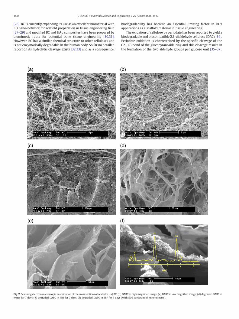
Fig. 2. Scanning electronmicroscopic examination of the cross sections of scaffolds. (a) BC, (bwater for 7 days (e) degraded DABC in PBS for 7 days, (f) degraded DABC in SBF for 7 days
biodegradability has become an essential limiting factor in BC'sapplications as a scaffold material in tissue engineering.
The oxidation of cellulose by periodate has been reported to yield abiodegradable and biocompatible 2,3-dialdehyde cellulose (DAC) [34].Periodate oxidation is characterized by the specific cleavage of theC2\C3 bond of the glucopyranoside ring and this cleavage results inthe formation of the two aldehyde groups per glucose unit [35–37].
) DABC in highmagnified image, (c) DABC in lowmagnified image, (d) degraded DABC in(with EDS spectrum of mineral parts).

Fig. 3. FTIR spectra of scaffolds. (a) BC, (b) DABC, (c) degraded DABC in water,(d) degraded DABC in PBS, (e) degraded and mineralized DABC in SBF.
Fig. 4. X-ray diffraction patterns of scaffolds. (a) BC, (b) DABC, (c) degraded DABC inwater, (d) degraded DABC in PBS, (e) degraded and mineralized DABC in SBF.
1637J. Li et al. / Materials Science and Engineering C 29 (2009) 1635–1642
DAC degrades at physiological pH in vivo and in vitro it could bedegraded into glycolic acid and 2,4-dihydroxybutyric acid [35]. Inmammals, glycolic acid is excreted in the urine or enters thetricarboxylic acid cycle, and 2,4-dihydroxybutyric acid is known toparticipate in the metabolism of L-homoserine in liver [38].Recently, the micron porous DAC membrane has been preparedby phase separation and was shown to be able to support celladhesion and proliferation and hence exhibited its potential to beused as a tissue engineering scaffold [38]. In this study, we pre-pared a degradable 3D nano-network from BC by periodatingoxidation procedure in order to make it a better biomaterial to beused as bioabsorbable tissue engineering scaffold. The microstruc-tural, crystallographic characters, functional groups, elementalcompositions, chemical states and wet tensile mechanical proper-ties of the resulted 2,3-dialdehyde bacterial cellulose (DABC) andits degradation and mineralizing in vitro were also presented inthis study.
2. Materials and methods
2.1. Preparation of 2,3-dialdehyde bacterial cellulose
BC pellicles were prepared by Acetobacter xylinum in static cultureand purified following the methods described previously [31]. BCpellicles were then immersed for 24 h in KCl/HCl (pH=1) buffersolution and added with 1.3 times the weight sodium metaperiodate.The mixture was gently stirred at 40 °C in the dark for 6 h. After theexcess periodate was decomposed with ethylene glycol, the DABCpellicles were washed by de-ionized water. The aldehyde content(60.3±0.5%) of the DABC samples was then analyzed by Schiff basereaction with hydroxylamine [39].
2.2. Degradation of DABC in vitro
Pieces of DABC were immersed in daily-renewed de-ionizedwater, phosphate buffered saline (PBS, pH=7.4) or simulated bodyfluid (SBF) [40] at 37 °C. By the end of 7 days, the obtained DABC wasrinsed by de-ionized water and freeze dried for further analysis.
Samples cut in square shape (10mm×10mm)were prepared andimmersed in daily-renewed de-ionized water, PBS and SBF at 37 °C.After 0 h (original sample), 4 h, 8 h, 12 h, 24 h, 72 h, 168 h, 336 h,504 h, 720 h and 1440 h, the obtained DABCwas rinsed by de-ionized
water and freeze dried and weighed. At least five specimens weretested for each sample and the averages together with standarddeviations were recorded. The percentage of mass loss wascalculated.
Pieces of DABC were immersed in de-ionized water at 37 °C for168 h. The supernatant was filtered and examined by Gel permeationchromatography (Waters 600, USA, Ultrahydrogel™ Linear300 mm×7.8 mmid×2 chromatography column).
2.3. Static tensile tests for BC and DABC hydrogel films
Static tensile testing was conducted for hydrogel film specimensin accordance with ASTM D 638-98 Type IV specimens by universalmaterial testing instrument (Testometric M350, UK) at roomtemperature and humidity (20 °C, 65% RH) at a constant speed of5 mm/min. The samples were tested in original hydrogel state.Values for wet tensile strength (σt), strain at break (εb) and Young'sModulus (Et) were obtained. At least five specimens were tested foreach sample and the averages together with standard deviationswere reported.
2.4. Characterizations of DABC
Morphology and microstructure of samples were characterizedby scanning electron microscopy (SEM, Philips XL-30, NL). Priorto SEM observation, all samples were sputter coated with a thinlayer of gold to avoid electrical charging. Energy dispersivespectroscopy (EDS, Oxford ISIS 300, UK) was performed inconjunction with SEM to identify the elements of the mineraldeposit on degraded DABC surface. The surface properties of theDABC were spectroscopically investigated by attenuated totalreflectance–Fourier transform infrared (ATR–FTIR, Nicolet Magna-560, USA), and also crystallographically characterized by thin-filmX-ray diffractometry (XRD, Rigaku D/Max 2500, JP). The collecteddata were processed by MDI/JADE6 software package attached tothe Rigaku XRD instrument. The elemental composition andchemical state of the samples were determined by an X-rayphotoelectron spectroscope (XPS, Perkin-Elmer PHI 1600, USA)and the curve fitting of the C 1s spectra were conducted by theuse of XPS Peak 4.1 software. The pore size of degraded samplewas determined by mercury intrusion porosimeter (Mic-AutoporeVI 9500, USA).

1638 J. Li et al. / Materials Science and Engineering C 29 (2009) 1635–1642
3. Results and discussions
3.1. Mass loss curve
Tissue engineering scaffolds should degrade naturally over time asnew tissue grows, so that it eliminates the need for further surgery. Todetermine the feasibility of BC as tissue engineering scaffoldmaterials,we firstly modified the BC pellicles by periodate oxidation and thenstudied its degradation properties in vitro. The mass loss rate in SBFwas ignored due to its invalidity as minerals were deposited onsamples. The mass loss curves of BC and DABC in water and in PBSwere shown in Fig. 1. As expected, the unmodified BC could hardlydegrade without cellulase. Note that the degradation rate in PBS is
Fig. 5. Schematic diagram of ion process with DABC. (a) DABC formation an
faster than in water because the breakage of DABC chain waspromoted in alkalescent surroundings. Similar findings of DAC werealso reported by Kim et al. [39] and Singh et al. [35,37]. Gel permeationchromatography analysis shows that the mean molecular weight oforganic matter in supernatant is about 380 g/mol. According to thedegradation chemistry reported by Singh et al. [34], the macromole-cules degraded into organic micro-molecules such as glycolic acid and2,4-dihydroxybutyric acid, etc [35].
3.2. SEM analysis
SEM photos in Fig. 2 compares the typical cross sections of freezedried BC and DABC samples. Note that the 3D nano-network structure
d degradation (b) HAp formation on DABC together with degradation.

Fig. 5 (continued).
1639J. Li et al. / Materials Science and Engineering C 29 (2009) 1635–1642
presented by BC (Fig. 2a) was still preserved in DABC (Fig. 2b). Thisnano-structure could help to support and promote cell adhesion andproliferation [3,41]. One obvious change in morphology after period-ate oxidation was that BC pellicles shrank distinctly in macroscopy(Fig. 2b and c) and the diameter of each fiber in nanonetworkincreased inmicroscopy (Fig. 2a vs. b). Themorphology of undegradedDABC and degraded DABC in water and PBS was shown in Fig. 2c–e,respectively. After 7 days in water, the freeze dried DABC scaffoldunderwent an obvious degradation, which was marked by formationof microporous structure and partly disappearance of the 3D nano-network (Fig. 2d). The freeze dried DABC scaffold underwent a furtherdegradation after 7 days immersion in PBS, which was signatured bythe disappearance of the 3D nano-network and the followingformation of microporous structure (Fig. 2e). The pores were thescale of about 50–100 µm, which fell into the proper size range for cellseeding and growth [42]. The pore size was determined by mercuryintrusion porosimeter. The diameter of pores in degraded DABC variedfrom 100 nanometers to 100 µm. The majority of the pores were 50–100 µm in diameter and themedian pore diameter was 59.1 µm.WhenDABC was immersed in SBF for 7 days, its degradationwas revealed bythe disappearance of the 3D nano-structure and the emergence ofmicroporous pieces. However, different from the degradation in PBS,minerals were observed to deposit on the surfaces of degraded DABCscaffold pieces (Fig. 2f). EDS analysis indicated that the depositedminerals consisted of calcium, phosphorous and oxygen, and the Ca/Pratio was 1.75.
3.3. FTIR analysis
Fig. 3 compares the FTIR spectra of BC, DABC, degraded DABC inPBS and degraded DABC in SBF. The spectral curve of BC in Fig. 3a isremarkably similar to the one generated for BC by Kim [43]. Thespectrum in Fig. 3b is a typical curve of DABC possessing the same setof subbands as DAC spectrum reported by Meng [44]. It is well knownthat oxidized cellulose by periodate should exhibit two characteristicFTIR bands at near 1740 and 880 cm−1 and their intensities increasewith increasing degree of oxidation [36,39]. In Fig. 3b, the FTIRspectrum of DABC exhibited a strong and sharp absorption peak at1740 cm−1 due to γ(CfO) stretching and an apparent absorption peakat 880 cm−1 caused by the formation of hemiacetal bonds betweennewly achieved aldehyde groups and their neighboring hydroxylgroups. The intensities of absorption bands near 1050 cm−1 ofγ(C\O) and 3400 cm−1 of γ(OH) in 2,3-dialdehyde cellulosedecreased after oxidation. All these characters described aboveindicated the formation of 2,3-dialdehyde in DABC.
During the degradation process inwater and PBS, the characteristicpeaks of DABC disappeared and the BC characteristic bands reap-peared in the FTIR spectrum (Fig. 3c and d). The degradation couldfirstly be detected in the part of the 2,3-dialdehyde chain element,same as reported for DAC by Kim [39]. During the degradation processin SBF, the characteristic bands of BC FTIR spectrum were masked bythe phosphate and carbonate deposits formed on the remaining BCscaffold (Fig. 3e).

1640 J. Li et al. / Materials Science and Engineering C 29 (2009) 1635–1642
3.4. XRD analysis
Fig. 4 shows the thin-film XRD patterns of BC, DABC and DABCdegraded in PBS and SBF. The diffraction peaks observed at 14.4°, 16.9°and 22.5° were attributed to well-defined cellulose I pattern [45], andpeaks at near 18.5° were attributed to the amorphous cellulose [33].Compared with the high crystallinity of original BC cultured in staticcircumstance (Fig. 4a), Fig. 4b shows the loss of crystallinity of DABCafter periodate oxidation. The loss of crystallinity is considered toresult from the opening of glucopyranose rings and destruction oftheir ordered packing [39,46]. As seen from Fig. 4c and d, the XRDpatterns maintained the BC characteristic fingerprints though themorphology of DABC changed remarkably after its degradation inwater and PBS, which indicated that the degradation of DABC nano-network began from the amorphous part, same as in the case ofoxycellulose reported by Kim [39] and Calvini [47]. XRD patterns ofdegraded DABC in SBF are shown in Fig. 4e and the diffraction peaks at25.9°, 31.9°, 39.5°, 46.7° and 49.6° were recognized for hydroxyapatite(HAp) [48], which was also revealed in the case of HAp formed on BCin biomimetic mineralization [29]. The current XRD results indicatedthat HAp crystals could form on the degraded DABC scaffold and thedegradation itself did not affect the deposition process. The XRD dataof HAp were processed by MDI/JADE6 software and the averagecrystallite size of HA along (002) was about 20 nm calculated by theScherrer equation and had a about 0.224% crystallinity, close to thereported value for the HAp crystal formed on BC [29].
Specific cleavage of the C2\C3 bond of the glucopyranoside ringresults in the formation of the two aldehyde groups per glucose unit.In our work, BC was partly modified into DABC and the aldehydecontent of DABC is about 60%, which means there are some BC chains
Fig. 6. XPS survey scan spectra of scaffolds. (a) BC, (b) DABC, (c) deg
and DABC chains coexisting in the DABC samples. The schematicdiagram is shown in Fig. 5a. As mentioned previously, sincedegradation firstly happened in the oxidized parts, so the primaryalcohol group in BC parts still remained intact. The Ca2+ and PO4
3−
could deposited to the degrading samples in the same way as it did onpure BC reported by Wan [31] and deposition itself wasn't affected bydegradation as shown in Fig. 5b.
3.5. XPS analysis
An XPS survey scan was utilized to determine the elementalconcentrations presented at the BC and DABC surfaces (Fig. 6). Asexpected, the main elements detected are carbon and oxygen. Usingarea sensitivity factors, the oxygen-to-carbon (O/C) atomic ratio wascalculated as an initial indication of surface oxidation. The O/C atomicratio for the BC was found to be 0.6 (Fig. 6a) which is close to theexperimental value of the cellulose (0.55–0.62) reported by Topalovic[49], although the theoretical value of pure cellulose is 0.83 [49]. TheO/C atomic ratio of DABC increased to 0.73 (Fig. 6b) after treating withperiodate [50]. After 7 days degradation in PBS or SBF, the O/C ratio ofDABC declined back to 0.61 and 0.64, respectively, which was similarto the original BC (Fig. 6a vs. Fig. 6c and d). This phenomenon alsoverified that the degradation happened at the highly oxidized part ofthe DABC scaffold. Meanwhile, the signals of calcium atom (Ca, 1.2 at.%) and phosphorous atom (P, 1.2 at.%) were found to be thecharacteristic elements of HAp [51] in the spectra of degraded DABCin SBF (Fig. 6d), The amount of HAp could be estimated as 5 wt.%according to the XPS signal of calcium and phosphorous elements.
The high-resolution C 1s peak in the XPS spectrum of the samplesgave information on carbon chemistry of BC and DABC samples
raded DABC in PBS, (d) degraded and mineralized DABC in SBF.

1641J. Li et al. / Materials Science and Engineering C 29 (2009) 1635–1642
(Fig. 7). The chemical shifts of carbon (C 1s) can usually be easilyclassified into four categories [52]: C1, unoxidised carbon (C\C), C2,carbon with one oxygen bond (C\O), C3, carbon with two oxygenbonds (O\C\O or CfO) and C4, carbon with three oxygen bonds(O\CfO). These four categories reveal C1s peak at 285.0, 286.6, 288.3and 289.5 eV, respectively. In this study, when BC was oxidized toDABC, two carbons corresponding to C2 transferred to C3 andconsequently the intensity of the C3 peaks increased and the relativeintensity of the C2 peaks decreased (Fig. 7a vs. b). In same way, whendegradation of DABC happened, the C3 peaks declined evidently(Fig. 7b vs. c and d). Although in theory, pure cellulose does notcontain any carbon that is not bound to oxygen and only negligibleamount of carboxylic groups exists, C1 peaks still can be found in itsC1s peaks. The non-oxidized alkane-type carbon atoms couldoriginate from the impurities in BC, and remained in DABC. WhenFig. 7c and d were compared with Fig. 7b, it showed that the intensityof C1 peaks increased after DABC degradation and the non-oxidizedalkane-type carbon atoms could be attributed to the degradationprocess.
3.6. Tensile properties in hydrogel condition
In order to evaluate the mechanical performance of BC and DABC,tensile strength, Young's modulus and strain at break in hydrogelconditionwere tested fordifferent samples and the results are presentedin Fig. 8. BC shows a tensile strengthof 0.60±0.02MPa, strain at break of11.8±0.6% and Young's Modulus of 6.3±0.3 MPa. The tensile strengthandmodulus are comparable to those of porcine carotid artery (tensile
Fig. 7. C 1s XPS high-resolution spectra of scaffolds. (a) BC, (b) DABC, (c)
strength 1.0±0.2 MPa, Young's modulus 2.3±0.1 MPa) [53]. Aftermodification into DABC, the wet tensile strength dropped to 0.27±0.03 MPa, which was still strong enough to be used as a nanofibrous biopolymer scaffold for tissue engineering when comparedto the tensile strength of cell-sponge (0.016±0.004 MPa) and cell-gel (0.03±0.014 MPa) in mesenchymal stem cell (MSC)–collagentissue engineering system [54,55]. After 7 days degradation in PBS,the mass loss of DABC is about 50±4.3% and its wet strengthdeclined to 0.09±0.03 MPa accordingly. No obvious difference wasfound between PBS and SBF treatments, even though minerals weredeposited to DABC scaffold in SBF. The strain at break of BC, DABCand DABC after 7 days degradation in PBS and SBF were 11.8±0.6%,9.3±0.3%, 5.8±0.6% and 5.1±0.7%, respectively. The strain atbreak of BC and DABC was not as high as other polymer filmsbecause its network structure limits the polymer molecules tostretch or extend. In Fig. 8c, the Young's modulus of BC, DABC andDABC degraded in PBS and SBF was shown to decline in a similarway as described above after periodate oxidization anddegradation.
4. Conclusion
Biodegradable DABC could be prepared from BC by periodateoxidization. While this chemical treatment maintained the original3D nano-network structure of BC, the DABC scaffold could degraderapidly in water, PBS and SBF solutions. The degradation rate wasfaster in PBS than in water. The degradation of DABC began from theamorphous part of the polymer and after 7 days, the 3D nano-
degraded DABC in PBS, (d) degraded and mineralized DABC in SBF.

Fig. 8. Tensile strength (a), break elongation rate (b) and Young's Modulus (c) ofsamples in hydrogel condition of scaffolds.
1642 J. Li et al. / Materials Science and Engineering C 29 (2009) 1635–1642
network structure collapsed and the remaining part of DABC scaffoldconverted into the porous structure with about 50–100 µm pores,which could provide the proper space for cell growth. Moreover, theHAp could deposit onto the DABC scaffold during its degradation inSBF. The strength of DABC hydrogel was 0.27±0.03MPa and declinedto 0.09±0.03MPa after 7 days degradation in vitro. Correspondingly,the Young's modulus of DABCwas 3.6±0.4MPa and declined to 1.5±0.3 MPa after 7 days degradation in vitro. We believe that theimproved biodegradability provided by periodate oxidization of BCcan increase its potential applications in tissue engineering.
Acknowledgements
The authors acknowledge the support from the National NaturalScience Foundation of China (Grants 50872088, 50673076 and50539060) and the Tianjin Municipal Science and TechnologyCommittee (Grants 07ZCKFSF01100 and 07JCZDJC07200). Financialsupport was also from the State Key Basic Research (973) Program(Grant 2007CB936100).
References
[1] S.Y. Chew, J. Wen, E.K.F. Yim, K.W. Leong, Biomacromolecules 6 (2005) 2017.[2] M.M. Stevens, J.H. George, Science 310 (2005) 1135.[3] S. Srouji, T. Kizhner, E. Suss-Tobi, E. Livne, E. Zussman, J. Mater. Sci.—Mater. M 19
(2008) 1249.[4] I.S. Yeo, J.E. Oh, L. Jeong, T.S. Lee, S.J. Lee, W.H. Park, B.M. Min, Biomacromolecules 9
(2008) 1106.[5] A. Matsuda, G. Kagata, R. Kino, J. Tanaka, J. Nanosci. Nanotechnol. 7 (2007) 852.[6] Y.Q. Goh, C.P. Ooi, J. of Mate. Sci.—Mater. M 19 (2008) 2445.[7] Z. Hong, R.L. Reis, J.F. Mano, Acta Biomater. 4 (2008) 1297.[8] C.R. Rambo, D.O.S. Recouvreux, C.A. Carminatti, A.K. Pitlovanciv, R.V. Antönio,
L.M. Porto, Mater. Sci. Eng. C 28 (2008) 549.[9] H. Luo, G. Xiong, Y. Huang, F. He, Y.Wang, Y.Wan, Mater. Chem. Phys.110 (2008) 193.[10] M.E.Gomes,A.S. Ribeiro, P.B.Malafaya, R.L. Reis, A.M.Cunha, Biomaterials 22 (2001)883.[11] M.J. Yaszemski, R.G. Payne, W.C. Hayes, R. Langer, A.G. Mikos, Biomaterials 17
(1996) 2127.[12] M. Borden, S.F. El-Amin, M. Attawia, C.T. Laurencin, Biomaterials 24 (2003) 597.[13] C.H. Wu, Y.J. Yin, R. Yang, K.D. Yao, Chin. J. Clin. Rehabil. 5 (2004) 929.[14] J.H. Ge, Y.J. Wang, Y.D. Zheng, New Chem. Mater. 2 (2003) 34.[15] L. Jiang, Y. Li, X. Wang, L. Zhang, J. Wen, M. Gong, Carbohyd. Polym. 74 (2008) 680.[16] C. Sairam Sundaram, N. Viswanathan, S. Meenakshi, Bioresour. Technol. 99 (2008)
8226.[17] Y. Zhang, J.R. Venugopal, A. El-Turki, S. Ramakrishna, B. Su, C.T. Lim, Biomaterials 29
(2008) 4314.[18] V. Karageorgiou, D. Kaplan, Biomaterials 26 (2005) 5474.[19] D. Gallego, N. Ferrell, Y. Sun, D.J. Hansford, Mater. Sci. Eng. C 28 (2008) 353.[20] C. Tsioptsias, I. Tsivintzelis, L. Papadopoulou, C. Panayiotou, Mater. Sci. Eng. C
(2008), doi:10.1016/j.msec.2008.06.003.[21] A. Svensson, E. Nicklasson, T. Harrah, B. Panilaitis, D.L. Kaplan, M. Brittberg,
P. Gatenholm, Biomaterials 26 (2005) 419.[22] A. Putra, A. Kakugo, H. Furukawa, J.P. Gong, Y. Osada, Polymer 49 (2008) 1885.[23] P. Ross, H. Weinhouse, Y. Aloni, Nature 325 (1987) 279.[24] A. Slezak, M. Kucharzewski, J. Jasik-Slezak, Polimery w medycynie 35 (2005) 23.[25] N. Sanchavanakit,W. Sangrungraungroj, R. Kaomongkolgit, T. Banaprasert, P. Pavasant,
M. Phisalaphong, Biotechnol. Prog. 22 (2006) 1194.[26] D. Klemm, D. Schumann, U. Udhardt, S. Marsch, Prog. Polym. Sci. 26 (2001) 1561.[27] A.N. Nakagaito, S. Iwamoto, H. Yano, Appl. Phys. A—Mater. 80 (2005) 93.[28] Q.P. Luo, C.H. Pei, F.D. Nie, Z.Q. Li, G.C. Yang, H. Huang, Curr. Nanosci. 3 (2007) 255.[29] L. Hong, Y.L. Wang, S.R. Jia, Y. Huang, C. Gao, Y.Z. Wan, Mater. Lett. 60 (2006) 1710.[30] Y.Z. Wan, L. Hong, S.R. Jia, Y. Huang, Y. Zhu, Y.L. Wang, H.J. Jiang, Compos. Sci.
Technol. 66 (2006) 1825.[31] Y.Z. Wan, Y. Huang, C.D. Yuan, S. Raman, Y. Zhu, H.J. Jiang, F. He, C. Gao, Mater. Sci.
Eng. C 27 (2007) 855.[32] A.S. Hoffman, Appl. Polym. Symp. (1977) 313.[33] R.L. Kronenthal, Polym. Sci. Technol. 8 (1975) 119.[34] M. Singh, A.R. Ray, P. Vasudevan, Biomaterials 3 (1982) 16.[35] M. Singh, A.R. Ray, P. Vasudevan, Biomater. Med. Dev. Artif. Org. 7 (1979) 495.[36] Q.G. Fan, D.M. Lewis, K.N. Tapley, J. Appl. Polym. Sci. 82 (2001) 1195.[37] M. Singh, P. Vasudevan, T.J.M. Sinha, J. Biomed. Mater. Res. 15 (1981) 655.[38] P. RoyChowdhury, V. Kumar, J. Biomed. Mater. Res. — A 76 (2006) 300.[39] U.J. Kim, S. Kuga, M. Wada, T. Okano, T. Kondo, Biomacromolecules 1 (2000) 488.[40] A. Oyane, H.M. Kim, T. Furuya, T. Kokubo, T. Miyazaki, T. Nakamura, J. Biomed.
Mater. Res. — A 65 (2003) 188.[41] H.S. Koh, T. Yong, C.K. Chan, S. Ramakrishna, Biomaterials 29 (2008) 3574.[42] M. Jäger, C. Zilkens, K. Zanger, R. Krauspe, J. Biomed. Biotechnol. 2007 (2007) 1.[43] D.Y. Kim, Y. Nishiyama, S. Kuga, Cellulose 9 (2002) 361.[44] S. Meng, Y. Feng, Z. Liang, Q. Fu, E. Zhang, Trans. Tianjin Uni. 11 (2005) 250.[45] S. Keshk, K. Sameshima, Enzyme Microb. Technol. 40 (2006) 4.[46] A.J. Varma, V.B. Chavan, Polymer. Degrad. Stabil. 49 (1995) 245.[47] P. Calvini, A. Gorassini, G. Luciano, E. Franceschi, Vib. Spectrosc. 40 (2006) 177.[48] S.C. Liou, S.Y. Chen, H.Y. Lee, J.S. Bow, Biomaterials 25 (2004) 189.[49] T. Topalovic, V.A. Nierstrasz, L. Bautista, D. Jocic, A. Navarro, M.M.C.G. Warmoes-
kerken, Coll. Surf. A 296 (2007) 76.[50] P. Stenstad, M. Andresen, B.S. Tanem, P. Stenius, Cellulose 15 (2008) 35.[51] J. Li, X. Yuan, F. He, A.F.T. Mak, J. Biomed. Mater. Res. Part B: App. Biomater. 86B
(2008) 381.[52] R. Mitchell, C.M. Carr, M. Parfitt, J.C. Vickerman, C. Jones, Cellulose 12 (2005) 629.[53] H. Bäckdahl, G. Helenius, A. Bodin, U. Nannmark, B.R. Johansson, B. Risberg, P.
Gatenholm, Biomaterials 27 (2006) 2141.[54] V.S. Nirmalanandhan, M.R. Dressler, J.T. Shearn, N. Juncosa-Melvin, M. Rao, C. Gooch,
G. Bradica, D.L. Butler, J. Biomech. Eng. 129 (2007) 919.[55] J.T. Shearn, N. Juncosa-Melvin, G.P. Boivin, M.T. Galloway, W. Goodwin, C. Gooch,
M.G. Dunn, D.L. Butler, J. Biomech. Eng. 129 (2007) 848.

本文献由“学霸图书馆-文献云下载”收集自网络,仅供学习交流使用。
学霸图书馆(www.xuebalib.com)是一个“整合众多图书馆数据库资源,
提供一站式文献检索和下载服务”的24 小时在线不限IP
图书馆。
图书馆致力于便利、促进学习与科研,提供最强文献下载服务。
图书馆导航:
图书馆首页 文献云下载 图书馆入口 外文数据库大全 疑难文献辅助工具