Prep for Practical II
40
Prep for Practical II BSC 2085L Tallahassee Community College Division of Science & Math
Transcript of Prep for Practical II

Prep for Practical II
BSC 2085L Tallahassee Community College
Division of Science & Math

BONES…

"Haversian Bone Models"
This image shows a model of compact bone, which contains functional units called osteons (Haversian systems).

1 OSTEON (HAVERSIAN SYSTEM) 2 CENTRAL CANAL 3 CIRCUMFERENTIAL LAMELLA 4 LAMELLA 5 SHARPEY’S FIBERS 6 PERIOSTEUM 7 OSTEOCYTES IN LACUNA
"Structure of Compact Bone"

1.Lamella 2.Central Canal 3.Osteocyte 4.Canaliculus 5.Lacuna
"The Osteon Model"

1.Epiphysis 2.Diaphysis 3.Spongy Bone 4.Compact Bone 5.Medullary Canal
"Split Femur"
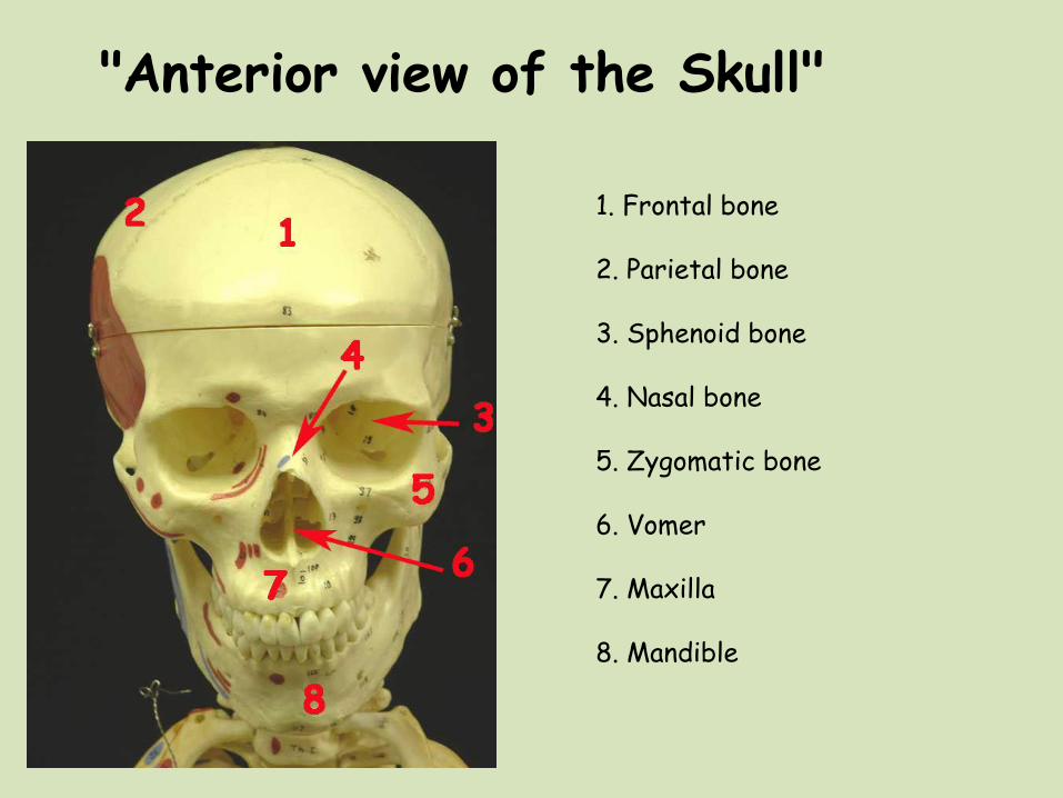
"Anterior view of the Skull"
1. Frontal bone 2. Parietal bone 3. Sphenoid bone 4. Nasal bone 5. Zygomatic bone 6. Vomer 7. Maxilla 8. Mandible
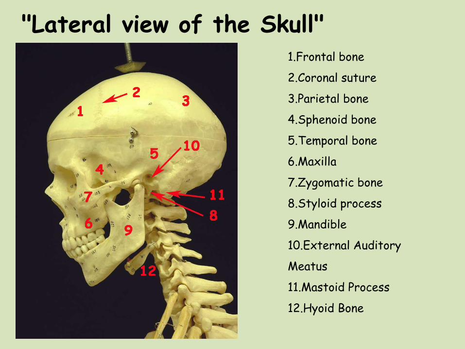
"Lateral view of the Skull" 1.Frontal bone
2.Coronal suture
3.Parietal bone
4.Sphenoid bone
5.Temporal bone
6.Maxilla
7.Zygomatic bone
8.Styloid process
9.Mandible
10.External Auditory
Meatus
11.Mastoid Process
12.Hyoid Bone

"Inferior view of the Skull" 1.Palantine Process 2.Zygomatic bone 3.Sphenoid bone 4.Vomer bone 5.Mandibular fossa 6.Mastoid process 7.Occipital condyle 8.Foramen magnum 9.Occipital bone 10.Palatine bone

"Interior view of the Skull"
1.Foramen Magnum 2.Crista Galli 3.Cribriform Plate 4.Greater Wing of Sphenoid 5.Lesser Wing of Sphenoid 6.Sella turcica 7.Occipital Bone

1. Spinous process 2. Superior articular facets 3. Vertebral foramen 4. Transverse foramina 5. Transverse processes 6. Body
Cervical vertebra (Superior view)
These vertebrae are distinguished from those of other regions of the spinal column by having a transverse foramen in each transverse process. The first two cervical vertebrae are specialized for articulating with the skull. The atlas (C1) lacks a body and spinous process. The axis (C2) has a spinous process and a body called the odontoid process (dens) that protrudes into a groove on the atlas.

1. Spinous process 2. Vertebral foramen 3. Inferior articular facets 4. Transverse foramina 5. Body
Cervical vertebra (Inferior view)

The Axis (superior view)
1. Spinous process 2. Vertebral foramen 3. Transverse process 4. Superior articular surface 5. Odontoid process (dens)
The first two cervical vertebrae are specialized for articulating with the skull. The axis (C2) has a spinous process and a body called the odontoid process (dens) that protrudes into a groove on the atlas.

The Atlas (Inferior view)
The first two cervical vertebrae are specialized for articulating with the skull. The atlas (C1) lacks a body and spinous process.
1. Posterior arch 2. Transverse foramen 3. Transverse process 4. Inferior articular facet 5. Anterior arch
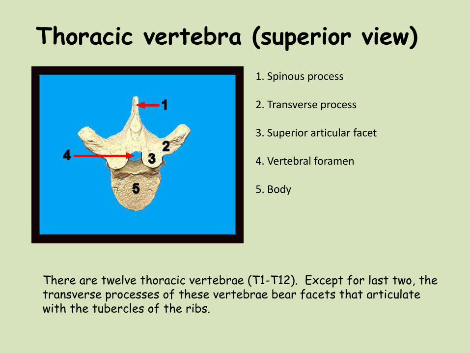
There are twelve thoracic vertebrae (T1-T12). Except for last two, the transverse processes of these vertebrae bear facets that articulate with the tubercles of the ribs.
1. Spinous process 2. Transverse process 3. Superior articular facet 4. Vertebral foramen 5. Body
Thoracic vertebra (superior view)

Thoracic vertebra (inferior view)
1. Spinous process 2. Transverse process 3. Vertebral foramen 4. Body

Thoracic Vertebrae (Right Lateral View)
1. Spinous process 2. Body 3. Transverse Process 4. Superior Articular Process
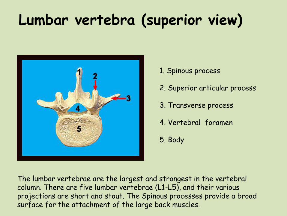
Lumbar vertebra (superior view)
1. Spinous process 2. Superior articular process 3. Transverse process 4. Vertebral foramen 5. Body
The lumbar vertebrae are the largest and strongest in the vertebral column. There are five lumbar vertebrae (L1-L5), and their various projections are short and stout. The Spinous processes provide a broad surface for the attachment of the large back muscles.

1. Spinous process 2. Inferior articular process 3. Transverse process 4. Vertebral foramen 5. Body
Lumbar vertebra (Inferior view)

Lumbar vertebra (lateral view)
1. Superior articular process 2. Transverse process 3. Body 4. Inferior articular process 5. Spinous process

"The Sacrum and Coccyx"
The human sacrum, is composed of five fused sacral vertebrae (S1-S5). The sacrum shapes, strengthens and stabilizes the pelvis. It articulates superiorly with the last lumbar vertebra (L5) and inferiorly with the coccyx .
1. Sacrum 2. Coccyx

The 12 Pairs of Ribs form The Walls of The Bony Thorax. The first seven are "True ribs", the next three are "False", and the last two "Floating".
1.Tubercle 2.Head of Rib 3.Shaft 4.Sternal End
"The Ribs"

"The Sternum"
1.Manubrium 2.Body 3.Xiphoid Process 4.Costal Cartilage
The sternum located in the anterior midline of the body is a flat bone approximately 15 cm long. It consists of three regions, the manubrium, the body and, at the inferior end the small xiphoid process.

The Appendicular Skeleton

"The Pectoral Girdle"
The pectoral girdle is composed of the clavicle and scapula, it functions to attach the upper limbs to the body and as a point of origin for many muscles that move the upper limb (Humerus), neck and trunk.
1.Scapula 2.Clavicle 3.Humerus 4.Acromial end of Clavicle 5.Sternal end of Clavicle 6.Spine of the Scapula 7.Acromion of the Scapula 8.Glenoid Cavity of the Scapula

Pelvic Girdle (Anterior view)
The pelvic girdle functions to attach the lower limbs to the axial skeleton. The pelvic girdle is comprised of the right and left coxal bones. The coxal bones articulate with the sacrum posteriorly and with each other anteriorly. Each coxal bone is formed by the fusion of three bones, the ilium, ischium and pubis.
1. Sacrum 2. Ilium 3. Ischium 4. Iliac crest 5. Obturator foramen 6. Pubis 7. Head of Femur 8. Neck of Femur 9. Pubic symphysis 10. Sacroiliac joint 11. Right Coxal bone

Pelvic Girdle (Posterior view)
1. Ilium 2. Ischium 3. Obturator foramen 4. Acetabulum 5. Ischial spine 6. Greater Trochanter of femur 7. Lesser Trochanter of femur 8. Sacrum 9. Coccyx

Anterior view of the male female pelvis. Several significant differences exist between the female and male pelvis (most of which are related to the requirements of child bearing). Specifically, in females the pelvic inlet and pelvic outlet are wider than in males, the pubic angle is greater in females, and the ischial spines are shorter and more everted than those of males.
1. Male Pelvic Girdle 2. Female Pelvic Girdle
Male and Female Pelvic Girdles

The Humerus, Radius and Ulna
1. Humerus 2. Radius 3. Ulna

The Left Humerus (Anterior view)
1. Head of Humerus 2. Coronoid fossa 3. Medial epicondyle 4. Traochlea 5. Capitulum 1.Radial fossa
2.Capitulum 3.Trochlea 4.Coronoid fossa 5.Medial epicondyle

The Left Humerus (Posterior view)
1. Head of Humerus 2. Lateral Epicondyle 3. Olecranon Fossa 4. Medial Epicondyle 5. Ulna

Left Radius and Ulna (Anterior view)
The radius is attached at its proximal end to the humerus. The other bone of the forearm is the ulna. The head of the radius articulates with the capitulum of the humerus and Medially, it articulates with the radial fossa of the ulna. Distally, the radius articulates with the carpal bones of the wrist. When the radius moves, the hand moves with it!
1. Radius 2. Ulna 3. Radial head 4. Radial tuberosity 5. Styloid process of Radius 6. Ulnar notch 7. Styloid process of Ulna 8. Coronoid process

Left Radius and Ulna (Posterior view)
The ulna is attached at its proximal end to the humerus. The olecranon and coronoid processes and the trochlear notch of the ulna articulate with the trochlea of the humerus. The head of the radius articulates on a lateral articulating surface of the ulna termed the radial notch.
1. Radius 2. Ulna 3. Olecranon process 4. Radial notch 5. Styloid process of Ulna 6. Styloid process of Radius
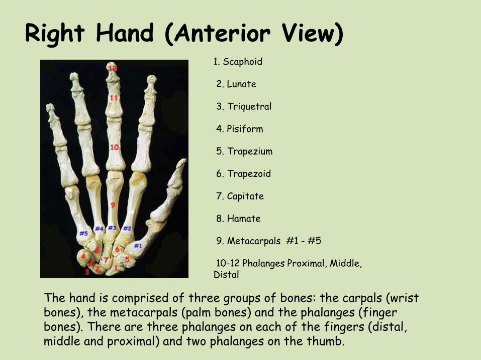
Right Hand (Anterior View)
The hand is comprised of three groups of bones: the carpals (wrist bones), the metacarpals (palm bones) and the phalanges (finger bones). There are three phalanges on each of the fingers (distal, middle and proximal) and two phalanges on the thumb.
1. Scaphoid 2. Lunate 3. Triquetral 4. Pisiform 5. Trapezium 6. Trapezoid 7. Capitate 8. Hamate 9. Metacarpals #1 - #5 10-12 Phalanges Proximal, Middle, Distal

The Femur, Tibia and Fibula
1. Tibia 2. Fibula 3. Femur 4. Patella

Left Femur (Anterior view)
1. Head 2. Neck 3. Greater trochanter 4. Lesser trochanter 5. Medial epicondyle 6. Patellar surface 7. Lateral epicondyle
The femur is the only bone of the thigh. The neck of the femur separates the head from the large, rough greater trochanter. Medial and inferior to the greater trochanter is the lesser trochanter. The trochanters serve as attachment sites for muscles of the thighs and buttocks. The long, narrow diaphysis of the femur separates the proximal from the distal end. At the distal end of the femur the most conspicuous feature is the lateral and medial condyles. The lateral and medial epicondyles are superior to the condyles and serve as attachment sites for muscles

Left Femur (Proximal End Posterior view)
1. Head 2. Neck 3. Greater Trochanter 4. Lesser Trochanter

Left Femur (Posterior view)
1. Greater Trochanter 2. Lesser Trochanter 3. Lateral Condyle 4. Medial Condyle
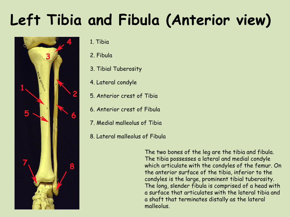
Left Tibia and Fibula (Anterior view) 1. Tibia 2. Fibula 3. Tibial Tuberosity 4. Lateral condyle 5. Anterior crest of Tibia 6. Anterior crest of Fibula 7. Medial malleolus of Tibia 8. Lateral malleolus of Fibula
The two bones of the leg are the tibia and fibula. The tibia possesses a lateral and medial condyle which articulate with the condyles of the femur. On the anterior surface of the tibia, inferior to the condyles is the large, prominent tibial tuberosity. The long, slender fibula is comprised of a head with a surface that articulates with the lateral tibia and a shaft that terminates distally as the lateral malleolus.

The Right Foot (Superior View)
1. Calcaneus 2. Talus 3. Cuboid 4. Lateral Cuneiform 5. Navicular 6. Intermediate Cuneiform 7. Medial Cuneiform 8. Metatarsals 9. Phalanges
The foot bones are divided into the 7 tarsals, 5 metatarsals and 14 phalanges (Proximal, Middle and Distal). The talus articulates with the fibula and tibia. The calcaneus or "heel bone" is the largest of the tarsal bones and is situated below the talus. The calcaneus helps support the weight of the body and serves for attachment of the large calf muscles.