Preliminary Observations on the Fine Structure of … · Preliminary Observations on the Fine...
4
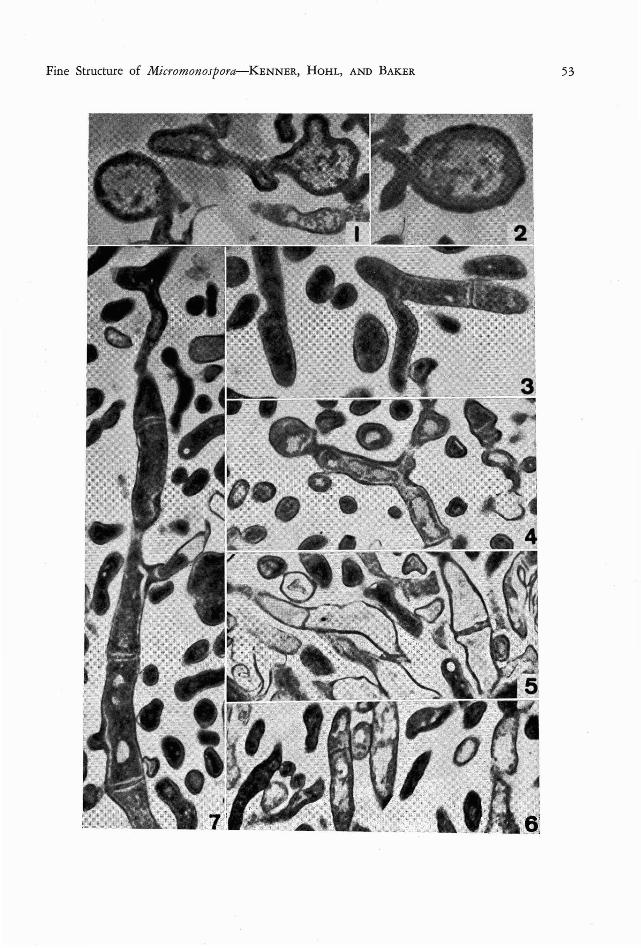
Preliminary Observations on the Fine Structure of Species of Micromonospora (Actinomycetales) DI ANA D. KENN ER!, HANS-RuDOLF HOHL 2 , AND GLADYS E. BAKER3 THE GE NUS M ieromonospora was first described by 0rskov in 1923 as a member of the Acti- nomycetales characterized by unicellar mycelium and the production of single, terminal conidia. The genus was little known for some time (Jensen, 1930), but subsequently the frequent occurrence of micromonosporae in soil (Jensen, 1932) , lake bottoms (Erikson , 1941; Potter and Baker, 1956) and lake water (Potter and Baker, 1956) , became well recognized. In spite of this wide acquaintance with the genus, the general morphology and cell structure is still not well understood and descriptions are some- times at variance. The chemical composition of the actinomycete wall, including micromono- sporae, (Erikson , 1947; Avery and Blank, 1951; Yamaguchi, 1965) is better documented than is the general morphology of the cell structure. Ultrastructure studies are few to date, and provide little detail (Agre, 1962 ; Leude- mann and Brodsky, 1964) . Both of these studies were concerned primarily with spores. A paper by Arai, Koyama, Kuroda, and Honda (1964) is more definitive for both mycelium and spores. It also includes some electron micrographs. 1193 4 N. E. Ballinger Way, Seattle, Washington 98155. 2 Department of Microbiology, Un iversity of Hawaii, Honolulu, Hawaii 96822. 3 Department of Botany, University of H awaii, Honolulu, Hawaii 96822. Manuscript received Janu- ary 24, 1967. The small size of the mycelium and spores in the micromonosporae has made study with the light microscope difficult and, in part, ac- counts for the lack of critical morphological and cytological information. Waksman (1961) characterized the substrate or vegetative hypae as straight or curved, branching, and without cross-walls. The lack of cross-walls has been accepted widely (Jensen, 1930 ; Erikson, 1941; Krasil'nikov and Agre, 1965), although in 1964 Arai et al. illustrated septa. There is increasing recognition of the eco- nomic importance of the micromonosporae as sources of antibiotics (Leudemann and Brodsky, 1964 ; Weinstein, Leudemann, Oden, and Wag- man, 1965) and as etiologic agents of disease in man (Castellani, De Brito, and Pinto, 1959). In view of this and of the discrepancies among reports on their structure, there is need for a consistent comparative study of the general cell structure and the process of spore production in the group. Consequently, studies at the ultra- structural level were undertaken for three spe- cies: M ierom onosp ora fusea Jensen (isolated from Flathead Lake water, Potter, No. M- 1012) ; M . purpurea Leudemann and Brodsky (NRRL No. 2953); and M. sp., isolated from a foot lesion at the University of Miami. All cultures came from the collection of Dr. Louise F. Potter, Biology Department, Elmira College, Elmira, New York. FIG. 1. Micromonospora fusca, showing the electron-dense cytoplasm and fibrillar nucleoid . The two round ish masses probably represent prespores. X 32,000. FIG. 2. Spore of Micromonospora [usca, showing the sculptured spore wall. X 26,000. FIG. 3. Micromonospora purpurea, with septum and V-shaped branching. X 25,000. FIG. 4. M icromonospor« purp urea, with prespore in form of a constricted and bulbous hyphal tip; V-shaped branching as in Figure 3. X 22,500. FIG. 5. M icromonospora purpure«, showing general cell morphology and septa of various thicknesses. X 22,500. FIG. 6. Example of septate hypha in pathogen ic Micromonospora spp . X 22,500. FIG. 7. Longitudinal section through hypha of M icromonospora purpurea, demonstr ating the frequent oc- currence of septa. X 25,000. 52
Transcript of Preliminary Observations on the Fine Structure of … · Preliminary Observations on the Fine...

Preliminary Observations on the Fine Structure of Species ofMicromonospora (Actinomycetales)
DIANA D. KENNER!, HANS-RuDOLF HOHL2, AND GLADYS E. BAKER3
THE GENUS M ieromonospora was first describedby 0rskov in 1923 as a member of the Actinomycetales characterized by unicellar myceliumand the production of single, terminal conidia.The genus was little known for some time(Jensen, 1930), but subsequently the frequentoccurrence of micromonosporae in soil (Jensen,1932) , lake bottoms (Erikson, 1941; Potterand Baker, 1956) and lake water (Potter andBaker, 1956) , became well recognized. In spiteof this wide acquaintance with the genus, thegeneral morphology and cell structure is stillnot well understood and descriptions are sometimes at variance. The chemical composition ofthe actinomycete wall, including micromonosporae, (Erikson, 1947; Avery and Blank,1951; Yamaguchi, 1965) is better documentedthan is the general morphology of the cellstructure. Ultrastructure studies are few to date,and provide little detail (Agre, 1962 ; Leudemann and Brodsky, 1964) . Both of these studieswere concerned primarily with spores. A paperby Arai, Koyama, Kuroda, and Honda (1964)is more definitive for both mycelium and spores.It also includes some electron micrographs.
11934 N. E. Ballinger Way, Seattle, Washington98155.
2 Department of Microbiology, University ofHawaii, Honolulu, Haw aii 96822.
3 Department of Botany, University of Hawaii,Honolulu, Hawaii 96822. Manuscript received January 24, 1967.
The small size of the mycelium and sporesin the micromonosporae has made study withthe light microscope difficult and, in part, accounts for the lack of critical morphologicaland cytological information. Waksman (1961)characterized the substrate or vegetative hypaeas straight or curved, branching, and withoutcross-walls. The lack of cross-walls has beenaccepted widely (Jensen, 1930 ; Erikson, 1941;Krasil'nikov and Agre, 1965), although in1964 Arai et al. illustrated septa.
There is increasing recognition of the economic importance of the micromonosporae assources of antibiotics (Leudemann and Brodsky,1964 ; Weinstein, Leudemann, Oden, and Wagman, 1965) and as etiologic agents of diseasein man (Castellani, De Brito, and Pinto, 1959).In view of this and of the discrepancies amongreports on their structure, there is need for aconsistent comparative study of the general cellstructure and the process of spore production inthe group. Consequently, studies at the ultrastructural level were undertaken for three species: M ierom onospora fusea Jensen (isolatedfrom Flathead Lake water, Potter, No . M1012) ; M . purpurea Leudemann and Brodsky(NRRL No. 2953); and M. sp., isolated froma foot lesion at the University of Miami. Allcultures came from the collection of Dr. LouiseF. Potter, Biology Department, Elmira College,Elmira, New York.
FIG. 1. Micromonospora fusca, showing the electron-dense cytoplasm and fibrillar nucleoid . The tworoundish masses probably represent prespores. X 32,000.
FIG. 2. Spore of M icromonospora [usca, showing the sculptured spore wall. X 26,000.FIG. 3. Micromonospora purpurea, with septum and V-shaped branching. X 25,000.FIG. 4. M icromonospor« purp urea, with prespore in form of a constricted and bulbous hyphal tip ; V-shaped
branching as in Figure 3. X 22,500.FIG. 5. M icromonospora purpure«, showing general cell morphology and septa of various thicknesses. X
22,500.FIG. 6. Example of septate hypha in pathogen ic Microm onospora spp . X 22,500.FIG. 7. Longitudinal section through hypha of M icromonospora purpurea, demonstrating the frequent oc
currence of septa. X 25,000.
52
Fine Structure of Mi cromonospora-KENNER, H OH L, AND B AKER 53
54
MATERIALS AND METHODS
Cultu res were grown in broth on a Gyrotaryshaker (N ew Brunswick Scienti fic Co.) at 200rpm for three to four days. Sodium caseinatebroth (Fred and W aksman, 1928) as modifiedby L. F. Potter ( BBL N o. 01-549) was the medium of choice. Fixation and embeddin g wereperformed according to the standard proceduresof Kellenberger and Ryter ( 1958) . Sectionswere cut on a Porter-Blum ultr amicrotome andpoststained with lead citrate. Electron micrographs were obtained on a N orelco EM 75microscope.
RESULTS
The preliminary findings reported here demonstrate that the techniq ue employed offerspromise for its use in future investigations.All pictur es taken clearly show the procaryoticnature of the three M icrom onospora species:the nuclear areas are fibrillar and not surroundedby a membrane; the cytoplasm is densely granular and essentially devoid of lamellar organelles.
The hyphae of all three species sectioned(Figs. 1, 6, 7) show cross-walls or septa. Theseare part icularly numerous in M . purpurea ( Figs.3, 4, 5, 7) . Apparently there are no septal pores.Some branching was observed (Figs. 3, 4, 7).Spore formation seems to be init iated by thedevelopment of a heavy septum or plug withinthe hypha which isolates the apical end of thehypha as a round ish, somewhat enlarged structure filled with both cytoplasmic and nuclearmaterial ( Figs. 1, 4 ) . The final spore (Fig. 2)has a sculptured wall. W all sculpturing wasshown by Leudemann and Brodsky (1964) onthe spores of M . ecbinospora, but their picturesare completely devoid of inner detail.
DISCUSSION
Despite the preliminary nature of this reportseveral points are worth emph asizing. The procaryotic nature of the cells has been establisheddefinitely for all three Micromonospora studied.Septa, which are particularly abundant in M.purpurea, are demonstrated clearly in all thestrains considered : there should be no questionof the occurrence of cross-walls in these three
PACIFIC SCIENCE, Vol. XXII, January 1968
micromonospo rae. The interpretation of thebranched cells is less clear. This might be interpreted as true branching or as the result ofanastomosis of two hyphae. Leudemann andBrodsky (1965) reported sectoring in coloniesof M. carbonacea which represented sporulatingand non-sporulating areas. They compared thisto the well-known heterocaryotic behavior infungi. Both anastomosis and sectoring are suggestive evidence which calls for confirmationin the entire group of micromonosporae.
SUMMARY
Electron microscope stud ies of three speciesof the genus M icrom onospora were made toclarify the scanty and conflicting informationabout cellular structure in this group. All materials revealed clearly the procaryotic natureof the cells and the presence of definite crosswalls in the hyphae.
REFERENCES
AGRE, N. S. 1962. Electron-microscopic investigation of spores of Microm onospora vulgaris. Microbiology USSR ( English trans.)31:226-227.
ARAI, T ., Y. KOYAMA, S. KURODA, and H .H ONDA. 1964. Biosynthesis of antibiotics byM icrom onospora. Kogyo Kagaku Zasshi 67:728-733.
AVERY, R. J., and F. BLANK. 1951. On thechemical composition of the cell walls of theActinomycetales and its relation to theirsystematic position . Canadian J. Microbiol.1 :140-143.
CASTELLANI, A., M. M. X . DE BRITO, andM. R. PINTO. 1959. An actinomycete isolatedfrom an autochthonous case of mycetoma inPortugal. J. Trop. Med. Hyg. 62 :27-36.
ERIKSON, D. 1941. Studies on some lake-mudstrains of M icromonospora. J. Bacterial. 41:277-300.
- - - 1947. Differentiation of the vegetativeand sporoge nous phases of the Actinomycetes.1. The lipid nature of the outer wall of theaerial mycelium. J. Gen. Microbial. 1 :39- 44.
FRED, E. B., and S. A. W AKSMAN. 1928. Lab-
Fine Structure of M icromonospora-KENNER, H OHL, AND BAKER 55
oratory Manual of General Microbiology.McGraw-Hill Book Co., Inc., New York.145 pp.
JENSEN, H. L. 1930. The genus Mic romonospora 0rskov, a little kn own group of soilmicro-organisms. Proc. Linnean Soc. N. S. W.55 :231-248.
- - - 1932. Contributions to our knowledgeof the Actinomycetales. III. Further observations on the genus Micromono spora. Ib id.57:1 73-180.
KELLENBERGER, E., and A. RYTER. 1958. Cellwall and cytoplasmic membrane of Escherichia coli . J. Biophys. and Biochem. Cytol.4:323- 325. ,
KRASIL'NIKOV, N . A., and N. S. AGRE. 1965.The brown gro up of Actinobifida chromogena n. sp. Microbiology USSR (Englishtrans.) 34:239-244.
LEUDEMANN, G . M. , and B. C. BRODSKY. 1964.Taxonomy of gentamicin-producing Micro monospora. Antimicrobial Agents and Chemotherapy, 1963: 116-1 24. ASM, Ann Arbor.
--- --- 1965. Micromonospora carbonacea sp. n., an everninomycin-producingorganism. Antimicrobial Agents and Chemotherapy, 1964: 47-52. ASM, Ann Arbor.
0RSKOV, J. 1923 . Inves tiga tions into the Morph ology of the Ray Fungi. Levin and Munksgaard, Copenhagen.
POTTER, L. F., and G. E. BAKER. 1956. Themicrobiology of Flathead and Rogers lakes,Mont ana. I. Preliminary survey of microb ialpopulations . Ecology 37 :35 1- 355.
W AKSMAN, S. A. 1961. The Actinomycetes.Vol. II. The W illiams and Wilkins Co.,Baltim ore. 363 pp.
W EINSTEIN, M. J., G. M. LEUDEMANN, E. M .ODEN, and G. H. W AGMAN. 1965. Everninomycin, a new antibiotic complex fromMicrom onospora carbonacea. An timicrobialAgents and Chemotherapy, 1964 :24--32.ASM, Ann Arbor.
YAMAGUCHI, T. 1965. Comparison of the cellwall composi tion of morphologically distinctactinomycetes. J. Bacteriol. 89: 444-453.