PREEMER TRIAL - PRINCIPAL INVESTIGATOR: Elisa …
66
PREEMER TRIAL - Prophylactic mesh versus no mesh in midline emergency laparotomy closure for prevention of incisional hernia: a multicenter, double-blind, randomized controlled trial 19.1.21 Version 3.0 PRINCIPAL INVESTIGATOR: Elisa Mäkäräinen-Uhlbäck M.D. ([email protected]) Oulu University Hospital STUDY GROUP: Tero Rautio M.D., Ph.D. Mirella Ahonen-Siirtola M.D. Oulu University Hospital Ville Sallinen M.D., Ph.D. Panu Mentula M.D., Ph.D. Matti Tolonen M.D., Ph.D. Ari Leppäniemi M.D., Ph.D. Helsinki University Hospital Filip Muysoms M.D., Ph.D. Hospital AZ Maria Middelares, Ghent, Belgium Juha Saarnio M.D., Ph.D. STATISTICIAN: Pasi Ohtonen MSc ([email protected]) Oulu University Hospital PARTICIPANTS: Oulu University Hospital Helsinki University Hospital, Meilahti Helsinki University Hospital, Jorvi Turku University Hospital Tampere University hospital Lahti Central Hospital FINANCE: Government funding PREEMER Trial 2019
Transcript of PREEMER TRIAL - PRINCIPAL INVESTIGATOR: Elisa …
PREEMER TRIAL -
Prophylactic mesh versus no mesh in midline emergency laparotomy closure for prevention of incisional hernia: a multicenter, double-blind, randomized controlled trial 19.1.21 Version 3.0
PRINCIPAL INVESTIGATOR: Elisa Mäkäräinen-Uhlbäck M.D. ([email protected]) Oulu University Hospital STUDY GROUP: Tero Rautio M.D., Ph.D. Mirella Ahonen-Siirtola M.D. Oulu University Hospital Ville Sallinen M.D., Ph.D. Panu Mentula M.D., Ph.D. Matti Tolonen M.D., Ph.D. Ari Leppäniemi M.D., Ph.D. Helsinki University Hospital Filip Muysoms M.D., Ph.D. Hospital AZ Maria Middelares, Ghent, Belgium Juha Saarnio M.D., Ph.D. STATISTICIAN: Pasi Ohtonen MSc ([email protected]) Oulu University Hospital PARTICIPANTS: Oulu University Hospital Helsinki University Hospital, Meilahti Helsinki University Hospital, Jorvi Turku University Hospital Tampere University hospital Lahti Central Hospital FINANCE: Government funding PREEMER Trial 2019
PREEMER TRIAL - ............................................................................................................................. ..................... 1 1. PROTOCOL SYNOPSIS ..................................................................................... .............................................. 4 2. INTRODUCTION................................................................................................................. ............................ 8
2.1 INCISIONAL HERNIA INCIDENCE ............................................................................................................................. ..... 8 2.2. INCISIONAL HERNIA ETIOLOGY ......................................................................................... ......................................... 8 2.3 INCISIONAL HERNIA DEFINITION AND EVALUATION .................................................................................................... 9 2.4 INCISIONAL HERNIA PREVENTION ............................................................................................................................. 10 3. STUDY OBJECTIVE ............................................................................................................................. .......... 12 4. STUDY DESIGN ............................................................................................................................. ................ 12 5. COST ANALYSIS ............................................................................................................................. .............. 13 6. STUDY ENDPOINTS.............................................................................................. ........................................ 13
6.1 PRIMARY ENDPOINTS ............................................................................................................................. .................. 13 6.2 SECONDARY ENDPOINTS ............................................................................................................................. ................... 14 7. STUDY POPULATION....................................................................................... ............................................ 14 7.1. INCLUSION CRITERIA ............................................................................................................................. .................. 15 7.2 EXCLUSION CRITERIA ............................................................................................................................. .................. 15 8. CENTER AND SURGEON SELECTION ........................................................................... ............................... 16 9. RISK ANALYSIS ............................................................................................................................. ............... 16
9.1. POTENTIAL RISKS ............................................................................................................................. ....................... 16 9.2. POTENTIAL BENEFITS ............................................................................................................................. ..................

16 10. STUDY METHODS ......................................................................................... ............................................. 17 10.1. ETHICS COMMITTEE APPROVAL ............................................................................................................................ 17 10.2. STUDY DURATION AND ENROLLMENT ................................................................................................................... 17 10.3. STUDY PLAN ............................................................................................................................. ............................. 17 10. DATA COLLECTION ............................................................................ ....................................................... 17 10.1 PATIENT INFORMED CONSENT ............................................................................................................................. ... 18 10.2 DATA COLLECTION ....................................................................................................................................... .......... 18 Baseline ............................................................................................................................. ........................................ 18 Operative Procedure ................................................................. .............................................................................. 19 Primary hospital stay and Discharge ...................................................................................................................... 19 30 days visit ............................................................................................................................. ................................. 20 2 years Visit ............................................................................ .................................................................................. 20 5 years visit........................................................................................................................ ....................................... 21 10.3 BLINDING............................................................................................................................. ................................... 21 11. DEFINITIONS ............................................................................................................................. ................. 21 11.1 DEFINITION OF INCISIONAL HERNIA ........................................................................ ............................................... 21 11.2 COMPREHENSIVE COMPLICATION INDEX ................................................................................................................ 23 11.3 DEFINITION OF INFECTION ............................................................................................................................. ........ 23 11.4 ACTIVITIES ASSESSMENT SCALE .................................................................................... .......................................... 23 11.5 PROMIS QUESTIONNAIRE ............................................................................................................................. ........... 24 12. SCHEDULE OF EVENTS .............................................................................................................................. 25 13. SUBJECT WITHDRAWAL AND DISCONTINUATION

................................................................................ 26 13.1 SUBJECT LOST TO FOLLOW-UP ............................................................................................................................. ... 26 PREEMER TRIAL 2
14 PROCEDURES ............................................................................................................................. ................. 26 14.1. PERIOPERATIVE CARE ................................................................................... ......................................................... 27 14.2 WOUND CLOSURE TECHNIQUE ............................................................................................................................. .. 27 14.3 POSTOPERATIVE TREATMENT ............................................................................................................................... .. 28 14.4. MESH (PROGRIPTM, MEDTRONIC) .......................................................................................................................... 28 15.GENERAL REPORTING REQUIREMENTS .............................................................................................. ..... 28 16. INVESTIGATOR’S
RESPONSIBILITIES AND QUALIFICATIONS ................................................................ 28 16.1 GENERAL RESPONSIBILITIES
............................................................................................................................. ....... 28 17 DATA HANDLING AND RECORD KEEPING
.................................................................................. ............. 29 17.1 CONFIDENTIALITY ............................................................................................................................. .....................
29 17.2 DATA MANAGEMENT
............................................................................................................................................. 29 18. CASE REPORT FORMS
............................................................................................................................. ... 29 19. SAMPLE SIZE AND STATISTICAL ANALYSIS
............................................................................................ 30 19.1 SAMPLE SIZE ............................................................................................................................. .....................
......... 30 19.2 ALLOCATION
............................................................................... ........................................................................... 30
19.3 ENDPOINT ANALYSIS ............................................................................................................................. ..................
30 20. PUBLICATION POLICY
............................................................................................................................. . 31 20.1. PUBLICATION PLAN
............................................................................................................................. ....................... 32
21. REFERENCES ....................................................................................... .......................................................
32 (

PREEMER TRIAL 3

1. Protocol Synopsis
PREEMER TRIAL 4
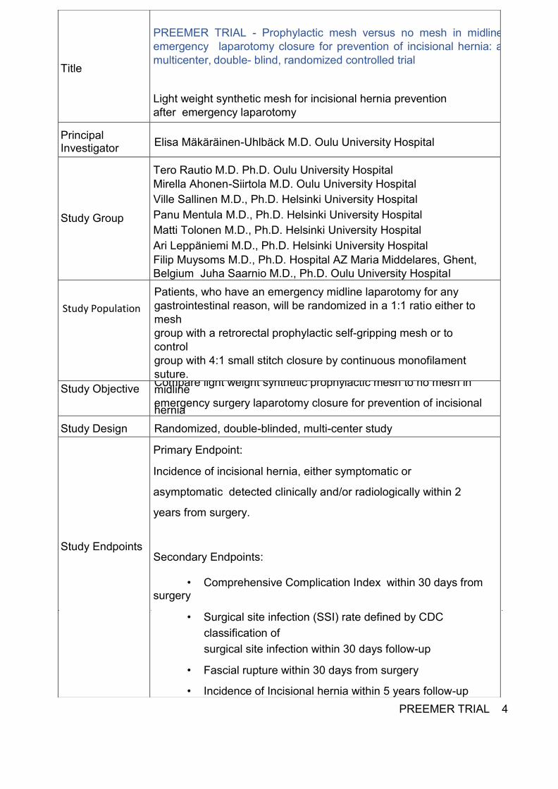
Title PREEMER TRIAL - Prophylactic mesh versus no mesh in midline emergency laparotomy closure for prevention of incisional hernia: a multicenter, double- blind, randomized controlled trial Light weight synthetic mesh for incisional hernia prevention after emergency laparotomy
Principal Investigator Elisa Mäkäräinen-Uhlbäck M.D. Oulu University Hospital
Study Group
Tero Rautio M.D. Ph.D. Oulu University Hospital Mirella Ahonen-Siirtola M.D. Oulu University Hospital Ville Sallinen M.D., Ph.D. Helsinki University Hospital Panu Mentula M.D., Ph.D. Helsinki University Hospital Matti Tolonen M.D., Ph.D. Helsinki University Hospital Ari Leppäniemi M.D., Ph.D. Helsinki University Hospital Filip Muysoms M.D., Ph.D. Hospital AZ Maria Middelares, Ghent, Belgium Juha Saarnio M.D., Ph.D. Oulu University Hospital Patients, who have an emergency midline laparotomy for any gastrointestinal reason, will be randomized in a 1:1 ratio either to mesh group with a retrorectal prophylactic self-gripping mesh or to control group with 4:1 small stitch closure by continuous monofilament suture. Study Objective Compare light weight synthetic prophylactic mesh to no mesh in midline emergency surgery laparotomy closure for prevention of incisional hernia Study Design Randomized, double-blinded, multi-center study
Study Endpoints
Primary Endpoint: Incidence of incisional hernia, either symptomatic or asymptomatic detected clinically and/or radiologically within 2 years from surgery.
Secondary Endpoints: • Comprehensive Complication Index within 30 days from surgery • Surgical site infection (SSI) rate defined by CDC
classification of surgical site infection within 30 days follow-up
• Fascial rupture within 30 days from surgery • Incidence of Incisional hernia within 5 years follow-up
Study Population

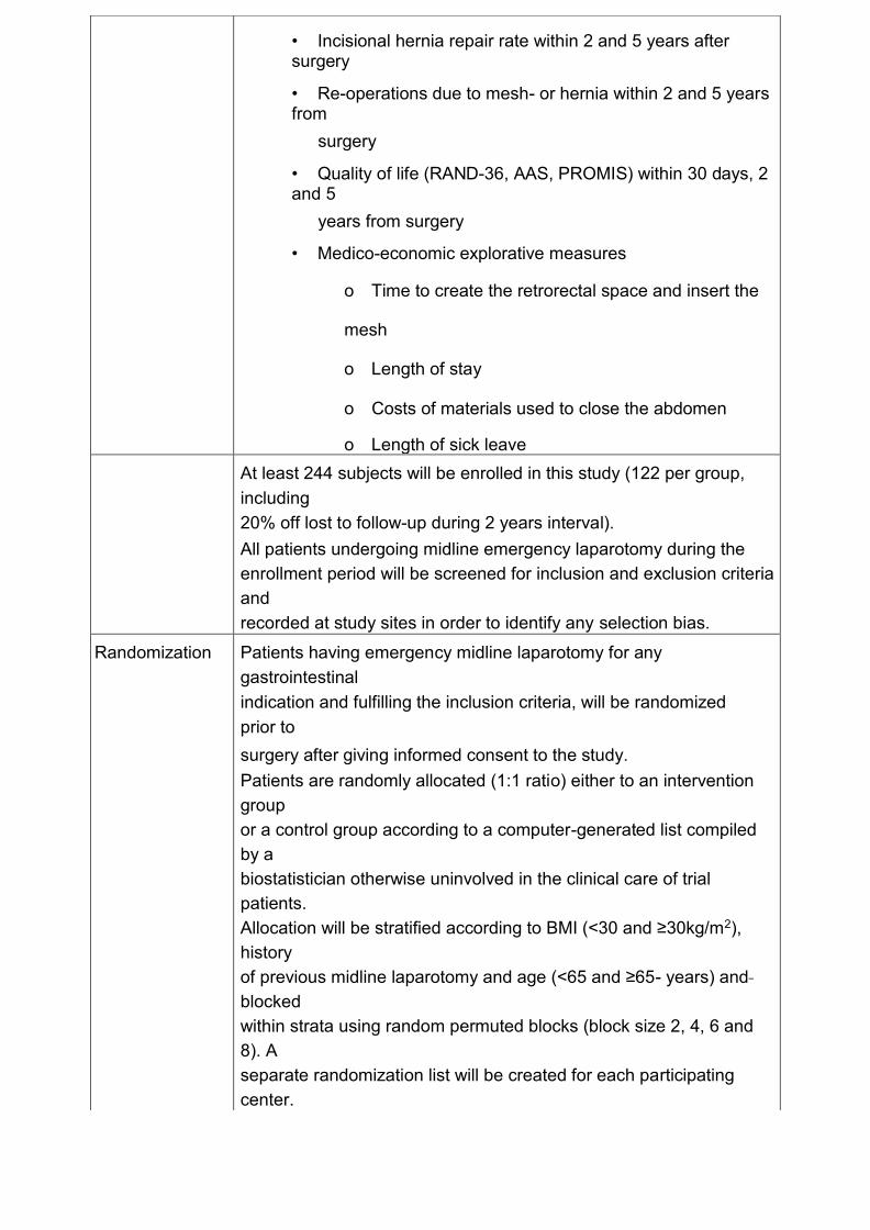
• Incisional hernia repair rate within 2 and 5 years after surgery • Re-operations due to mesh- or hernia within 2 and 5 years from
surgery • Quality of life (RAND-36, AAS, PROMIS) within 30 days, 2 and 5
years from surgery • Medico-economic explorative measures
o Time to create the retrorectal space and insert the mesh o Length of stay o Costs of materials used to close the abdomen o Length of sick leave At least 244 subjects will be enrolled in this study (122 per group,
including 20% off lost to follow-up during 2 years interval). All patients undergoing midline emergency laparotomy during the enrollment period will be screened for inclusion and exclusion criteria and recorded at study sites in order to identify any selection bias.
Randomization Patients having emergency midline laparotomy for any gastrointestinal indication and fulfilling the inclusion criteria, will be randomized prior to surgery after giving informed consent to the study. Patients are randomly allocated (1:1 ratio) either to an intervention group or a control group according to a computer-generated list compiled by a biostatistician otherwise uninvolved in the clinical care of trial patients. Allocation will be stratified according to BMI (<30 and ≥30kg/m2), history of previous midline laparotomy and age (<65 and ≥65- years) and blocked within strata using random permuted blocks (block size 2, 4, 6 and 8). A separate randomization list will be created for each participating center.
PREEMER TRIAL 5
Total Study Duration
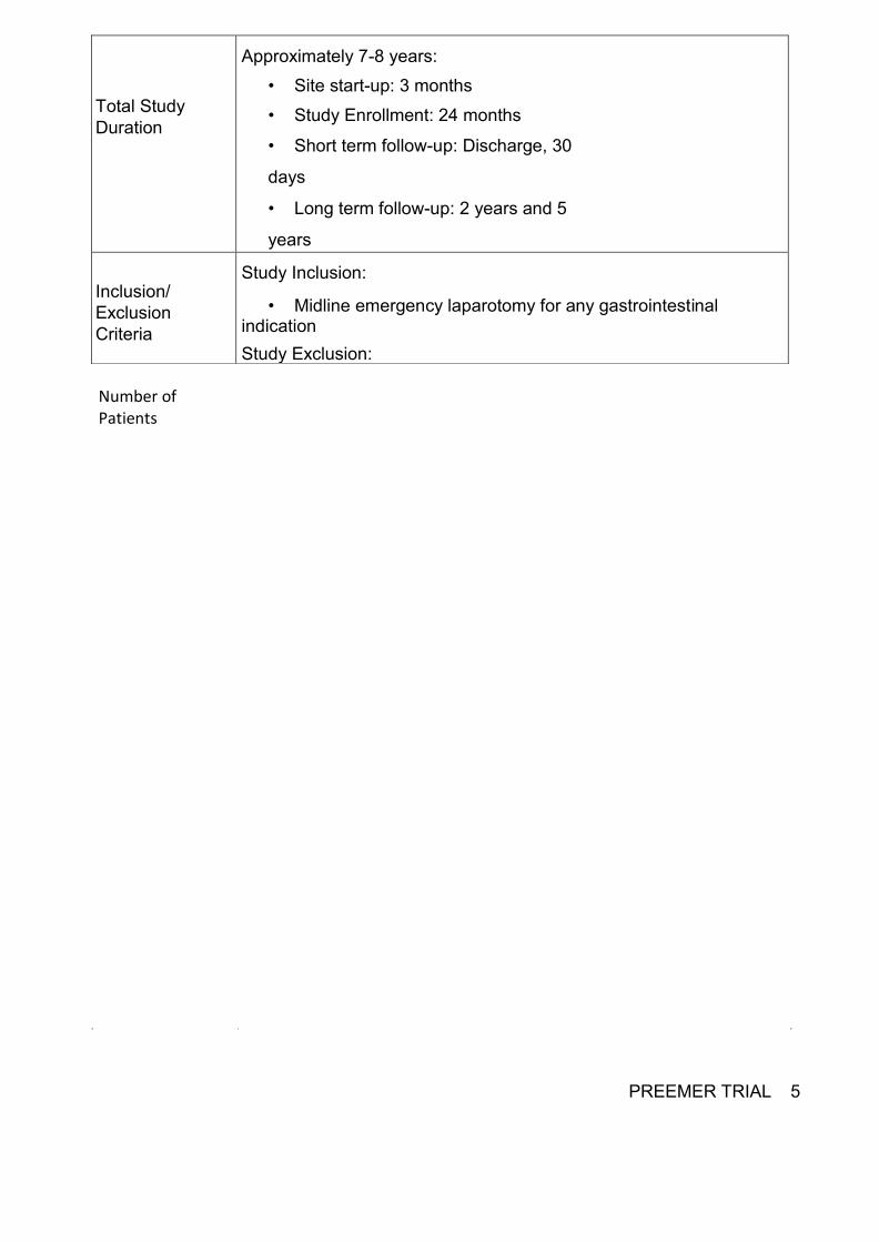
Approximately 7-8 years: • Site start-up: 3 months • Study Enrollment: 24 months • Short term follow-up: Discharge, 30 days • Long term follow-up: 2 years and 5 years
Inclusion/ Exclusion Criteria Study Inclusion:
• Midline emergency laparotomy for any gastrointestinal indication Study Exclusion:
Number of Patients
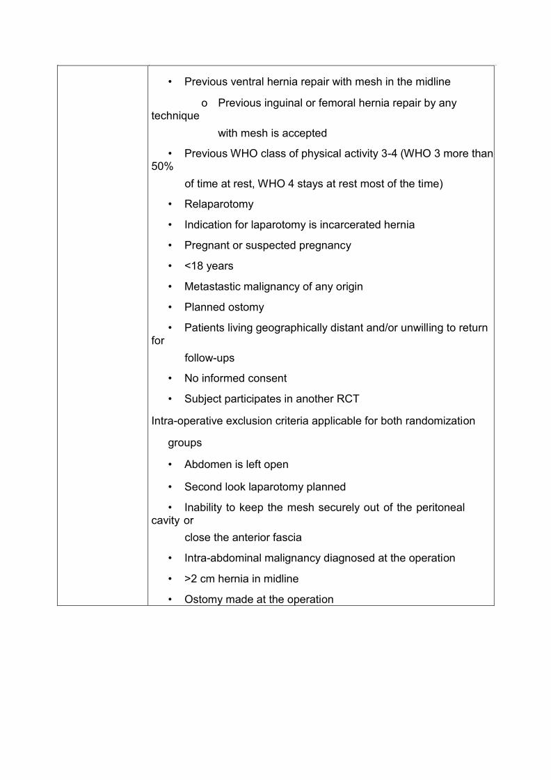
• Previous ventral hernia repair with mesh in the midline o Previous inguinal or femoral hernia repair by any technique
with mesh is accepted • Previous WHO class of physical activity 3-4 (WHO 3 more than 50%
of time at rest, WHO 4 stays at rest most of the time) • Relaparotomy • Indication for laparotomy is incarcerated hernia • Pregnant or suspected pregnancy • <18 years • Metastastic malignancy of any origin • Planned ostomy • Patients living geographically distant and/or unwilling to return for
follow-ups • No informed consent • Subject participates in another RCT
Intra-operative exclusion criteria applicable for both randomization groups • Abdomen is left open • Second look laparotomy planned • Inability to keep the mesh securely out of the peritoneal cavity or
close the anterior fascia • Intra-abdominal malignancy diagnosed at the operation • >2 cm hernia in midline • Ostomy made at the operation
PREEMER TRIAL 6
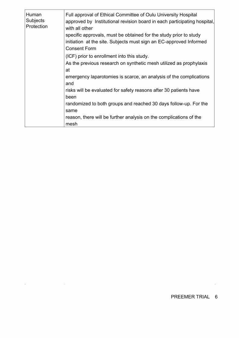
Human Subjects Protection Full approval of Ethical Committee of Oulu University Hospital approved by Institutional revision board in each participating hospital, with all other specific approvals, must be obtained for the study prior to study initiation at the site. Subjects must sign an EC-approved Informed Consent Form (ICF) prior to enrollment into this study. As the previous research on synthetic mesh utilized as prophylaxis at emergency laparotomies is scarce, an analysis of the complications and risks will be evaluated for safety reasons after 30 patients have been randomized to both groups and reached 30 days follow-up. For the same reason, there will be further analysis on the complications of the mesh
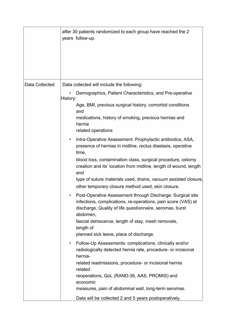
after 30 patients randomized to each group have reached the 2
years follow-up.
Data Collected Data collected will include the following: • Demographics, Patient Characteristics, and Pre-operative History:
Age, BMI, previous surgical history, comorbid conditions and medications, history of smoking, previous hernias and hernia related operations
• Intra-Operative Assessment: Prophylactic antibiotics, ASA, presence of hernias in midline, rectus diastasis, operative time, blood loss, contamination class, surgical procedure, ostomy creation and its’ location from midline, length of wound, length and type of suture materials used, drains, vacuum assisted closure, other temporary closure method used, skin closure.
• Post-Operative Assessment through Discharge: Surgical site infections, complications, re-operations, pain score (VAS) at discharge, Quality of life questionnaire, seromas, burst abdomen, fascial dehiscence, length of stay, mesh removals, length of planned sick leave, place of discharge.
• Follow-Up Assessments: complications, clinically and/or radiologically detected hernia rate, procedure- or incisional hernia- related readmissions, procedure- or incisional hernia related reoperations, QoL (RAND-36, AAS, PROMIS) and economic measures, pain of abdominal wall, long-term seromas. Data will be collected 2 and 5 years postoperatively.
PREEMER TRIAL 7
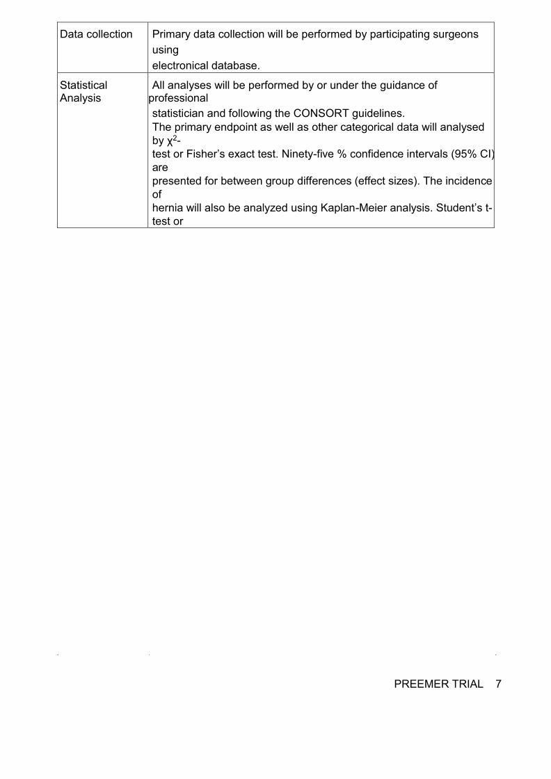
Data collection Primary data collection will be performed by participating surgeons using electronical database.
Statistical Analysis All analyses will be performed by or under the guidance of professional statistician and following the CONSORT guidelines. The primary endpoint as well as other categorical data will analysed by χ2- test or Fisher’s exact test. Ninety-five % confidence intervals (95% CI) are presented for between group differences (effect sizes). The incidence of hernia will also be analyzed using Kaplan-Meier analysis. Student’s t-test or
2. Introduction
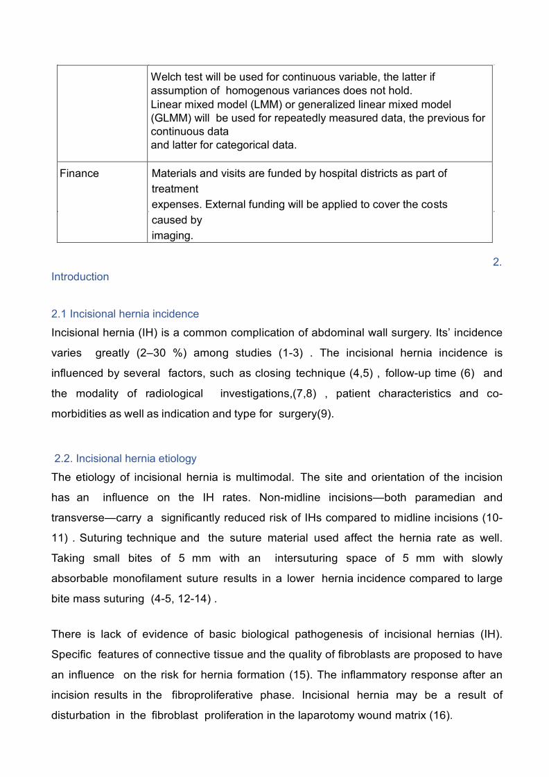
2.1 Incisional hernia incidence Incisional hernia (IH) is a common complication of abdominal wall surgery. Its’ incidence
varies greatly (2–30 %) among studies (1-3) . The incisional hernia incidence is influenced by several factors, such as closing technique (4,5) , follow-up time (6) and the modality of radiological investigations,(7,8) , patient characteristics and co-morbidities as well as indication and type for surgery(9). 2.2. Incisional hernia etiology The etiology of incisional hernia is multimodal. The site and orientation of the incision has an influence on the IH rates. Non-midline incisions—both paramedian and transverse—carry a significantly reduced risk of IHs compared to midline incisions (10-11) . Suturing technique and the suture material used affect the hernia rate as well. Taking small bites of 5 mm with an intersuturing space of 5 mm with slowly absorbable monofilament suture results in a lower hernia incidence compared to large bite mass suturing (4-5, 12-14) . There is lack of evidence of basic biological pathogenesis of incisional hernias (IH). Specific features of connective tissue and the quality of fibroblasts are proposed to have an influence on the risk for hernia formation (15). The inflammatory response after an incision results in the fibroproliferative phase. Incisional hernia may be a result of disturbation in the fibroblast proliferation in the laparotomy wound matrix (16).
Welch test will be used for continuous variable, the latter if assumption of homogenous variances does not hold. Linear mixed model (LMM) or generalized linear mixed model (GLMM) will be used for repeatedly measured data, the previous for continuous data and latter for categorical data. Finance Materials and visits are funded by hospital districts as part of
treatment expenses. External funding will be applied to cover the costs caused by imaging.

PREEMER TRIAL 8
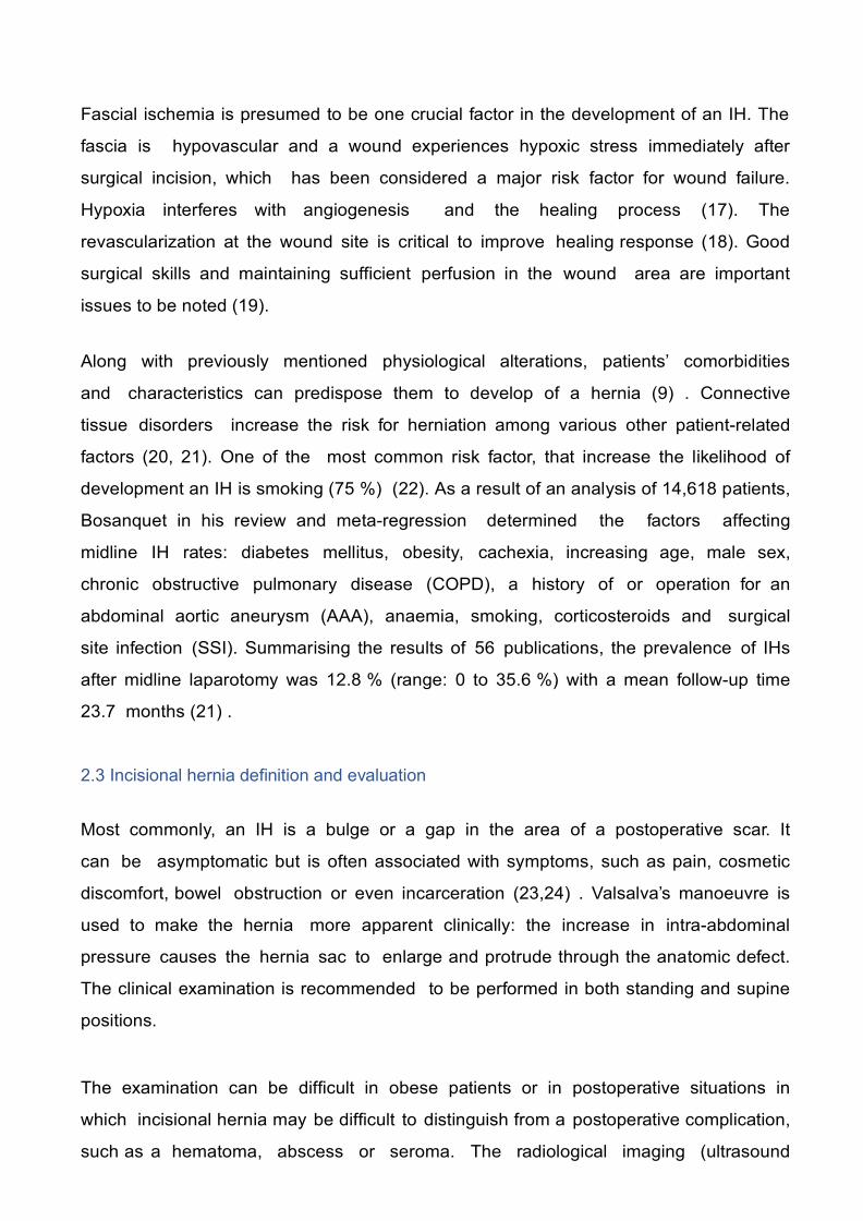
Fascial ischemia is presumed to be one crucial factor in the development of an IH. The fascia is hypovascular and a wound experiences hypoxic stress immediately after surgical incision, which has been considered a major risk factor for wound failure. Hypoxia interferes with angiogenesis and the healing process (17). The revascularization at the wound site is critical to improve healing response (18). Good surgical skills and maintaining sufficient perfusion in the wound area are important issues to be noted (19). Along with previously mentioned physiological alterations, patients’ comorbidities and characteristics can predispose them to develop of a hernia (9) . Connective tissue disorders increase the risk for herniation among various other patient-related factors (20, 21). One of the most common risk factor, that increase the likelihood of development an IH is smoking (75 %) (22). As a result of an analysis of 14,618 patients, Bosanquet in his review and meta-regression determined the factors affecting midline IH rates: diabetes mellitus, obesity, cachexia, increasing age, male sex, chronic obstructive pulmonary disease (COPD), a history of or operation for an abdominal aortic aneurysm (AAA), anaemia, smoking, corticosteroids and surgical site infection (SSI). Summarising the results of 56 publications, the prevalence of IHs after midline laparotomy was 12.8 % (range: 0 to 35.6 %) with a mean follow-up time 23.7 months (21) . 2.3 Incisional hernia definition and evaluation Most commonly, an IH is a bulge or a gap in the area of a postoperative scar. It can be asymptomatic but is often associated with symptoms, such as pain, cosmetic discomfort, bowel obstruction or even incarceration (23,24) . Valsalva’s manoeuvre is used to make the hernia more apparent clinically: the increase in intra-abdominal pressure causes the hernia sac to enlarge and protrude through the anatomic defect. The clinical examination is recommended to be performed in both standing and supine positions. The examination can be difficult in obese patients or in postoperative situations in which incisional hernia may be difficult to distinguish from a postoperative complication, such as a hematoma, abscess or seroma. The radiological imaging (ultrasound

(US), computed tomography (CT) or magnetic resonance (MRI)) is useful in specifying the diagnosis (8,25-27) . PREEMER TRIAL 9
A standardised dynamic abdominal sonography for hernias offers a safe and a low-cost diagnostic tool with great specificity and positive predictive value (28) . A CT scan can also be an option in some cases like when planning operative treatment, although it induces a radiation load for patients. When evaluating the IH rate, the difference between physical examination and imaging modalities (ultrasound or computer tomography) is important in terms of accuracy (29,30) . A standardised examination and dynamic evaluation by ultrasound of the abdominal wall is recommended in evaluating a possible hernia (14,31). Incidence of IHs increases during a follow-up study time from 12.6 % at 12 months to 22.4 % at 36 months(6,32,33). There is a great diversity of abdominal wall IHs. During the early years of 2000 the first proposals for the classification of incisional ventral hernias were published. Through 2009, there were several proposals for classifying IHs according to defect size, recurrence and topography to some extent, but none of these achieved wide recognition or routine use(34-36). In 2009, the European Hernia Society (EHS) published a formula to classify primary and incisional abdominal wall hernias (36). 2.4 Incisional hernia prevention European Hernia Society (EHS) guideline strongly recommends to utilise a non-midline approach to a laparotomy whenever possible to decrease the incidence of incisional hernia (14). However, this is clearly not an option in an emergency laparotomy, as midline incision is the fastest and the best visualizing opening to explore the whole abdominal cavity in an emergency setting. For elective midline incisions, evidence-based recommendation is to perform a continuous suturing technique with slowly absorbable monofilament suture when closing the incision (14) Suturation should be done performing a single layer aponeurotic closure technique without separate closure of the peritoneum. A small bites technique with a suture to wound length (SL/WL) ratio at least 4:1 is the current recommended method of fascial closure (12-14, 37,38). Prophylactic mesh augmentation in a non-emergency setting appears effective and safe and

can be suggested for high-risk patients (39-40). However, no recommendations can be given on the optimal PREEMER TRIAL 10
technique to close emergency laparotomy incisions because of lack of evidence (14) . This problem should be emphasized on due to high rates of IH after emergency laparotomy (41,42). All this makes the use of prophylactic mesh in the emergency setting an interesting proposition, as it may decrease the rate of IHs. However, there are concerns over potential mesh related complications including infection, chronic pain, seromas and bowel fistulas especially in emergency situations like peritonitis and intestinal obstruction. There is preliminary evidence published about the safety and efficiency of the prevention of IHs using meshes in the emergency laparotomy closure even in contaminated conditions (43,44). In the resent systematic review and meta-analysis, only results of 2 studies and altogether 299 patients were eligible for the analysis (42) . Swiss case-control study reported an IH rate of 3,2% (2/63) in the mesh group and 28,6% (20/70) in the control group (43) . Spanish study group had the same kind of results in their retrospective cohort; IH rate of 5,9% (3/50) in the mesh group and 33,3% (33/100) in the control group (44) . There was no statistically significant difference in the incidence of surgical site infection or other complications when prophylactic mesh group was compared to standard closure group. SSI rate in Swiss study was 60% and respectively only 17% in the Spanish study. This may reflect differences in the patient selection, therefore the safety profile of the prophylactic mesh in the emergency setting has not been adequately described. Neither of the studies included in meta-analysis were not randomized controlled trials. There were also many methodological differences including patient selection, used mesh, and mesh placement. Thus, the conclusion of the systematic review paper was that there are limited data to assess the effect or safety of the use of prophylactic mesh in the emergency laparotomy setting (42) . Randomized control trials are required to address this important clinical question. EHS guideline group resulted the same conclusion in their recommendation report (14). There are about 1650 patients are operated in Finland because of IH every year. According to the European study, the estimated cost for IH surgery is 6450 euros (45). The corresponding costs in Sweden were even higher reaching 9060 euros per treatment (12). Extrapolated to Finland, this means that operative treatment of IHs cause more than 10 million expenses to the Finnish health care sector in a year. Some of these costs may be avoidable by using the prophylactic mesh during the

closure of midline emergency laparotomies in the patients with IH risk factors. PREEMER TRIAL 11
Therefore, our study group stands on the idea to design and carry out the PREEMER randomized controlled trial (RCT) comparing prophylactic mesh to best standard suturing technique in this challenging setting. 3. Study Objective The objective of this study is to compare prospectively the feasibility and the potential benefits of retrorectus self-gripping mesh (ProgripTM, Medtronic) to controls operated with no mesh by using the best standard 4:1 small stitch suturing technique. 4. Study Design This study is a multicenter, double-blinded, randomized controlled trial. Parameters will be collected prospectively after randomization. All enrolled subjects will undergo assessments at the following intervals: pre-operative, operative, discharge, 30 days, 2- and 5-years post–surgery. A description of the study visits and required study procedures is summarized in Section 12, Schedule of events. All patients are evaluated both clinically and radiologically at 2 years after index procedure in order to diagnose clinical and/or radiological incisional hernia. The follow up will continue until 5 years after the surgery to assess long-term results and safety. Ultrasound with and without Valsalva maneuver will be performed to all patients 2 years after surgery. The extent of the fascial defect and hernia sac volume (ie. the volume of incisional hernia) are measured and graded according to the European hernia society criteria. (46) If there is a suspicion of symptomatic or asymptomatic incisional hernia according to clinical assessment and ultrasound findings are inconclusive, CT scan is required to verify the diagnosis of IH. If a patient have had no imaging done for any reason, the result of clinical

evaluation is recorded. In case of several imaging modalities accomplished, all results are recorded. All study
PREEMER TRIAL 12
patients are guided to contact their study site in case of any suspicion of incisional hernia at any point during follow up. Quality of life will be measured using RAND-36, Activities Assessment Scale (AAS) and PROMIS questionnaire at all follow-up visits at 1 months, 2 years and 5 years as well as when a hernia is diagnosed. 5. Cost analysis Costs of the treatment
• Mesh and other materials used to close the abdomen • Need for futher surgery and medical treatment
o All complications of primary surgery o Mesh-related need for surgery or other treatment o Hernia-related need for surgery or any help from medical system o Length of sick leave o Need for rehabilitation before returning to previous place of home o Length of stay in the hospital
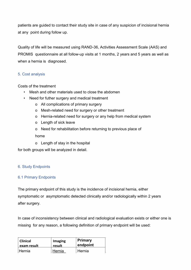
for both groups will be analyzed in detail. 6. Study Endpoints 6.1 Primary Endpoints The primary endpoint of this study is the incidence of incisional hernia, either symptomatic or asymptomatic detected clinically and/or radiologically within 2 years after surgery. In case of inconsistency between clinical and radiological evaluation exists or either one is missing for any reason, a following definition of primary endpoint will be used:
Primary endpoint Hernia Hernia Hernia Clinical exam result Imaging
result

PREEMER TRIAL 13
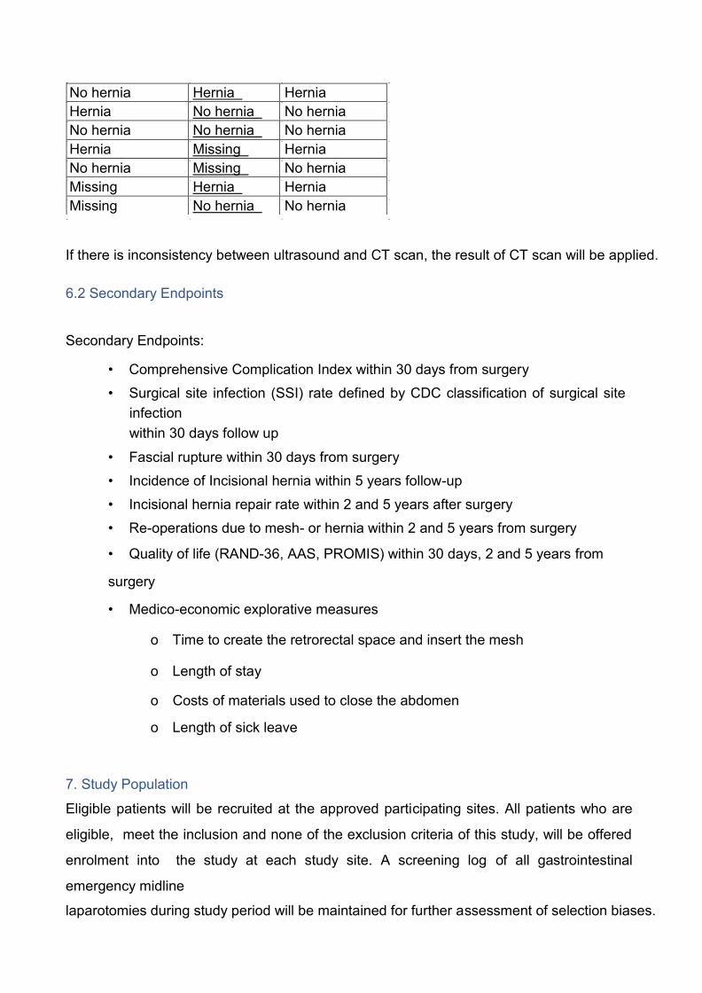
If there is inconsistency between ultrasound and CT scan, the result of CT scan will be applied. 6.2 Secondary Endpoints Secondary Endpoints:
• Comprehensive Complication Index within 30 days from surgery • Surgical site infection (SSI) rate defined by CDC classification of surgical site
infection within 30 days follow up
• Fascial rupture within 30 days from surgery • Incidence of Incisional hernia within 5 years follow-up • Incisional hernia repair rate within 2 and 5 years after surgery • Re-operations due to mesh- or hernia within 2 and 5 years from surgery • Quality of life (RAND-36, AAS, PROMIS) within 30 days, 2 and 5 years from surgery • Medico-economic explorative measures
o Time to create the retrorectal space and insert the mesh o Length of stay o Costs of materials used to close the abdomen o Length of sick leave
7. Study Population Eligible patients will be recruited at the approved participating sites. All patients who are eligible, meet the inclusion and none of the exclusion criteria of this study, will be offered enrolment into the study at each study site. A screening log of all gastrointestinal emergency midline laparotomies during study period will be maintained for further assessment of selection biases.
No hernia Hernia Hernia Hernia No hernia No hernia No hernia No hernia No hernia Hernia Missing Hernia No hernia Missing No hernia Missing Hernia Hernia Missing No hernia No hernia

PREEMER TRIAL 14
The following patient inclusion/exclusion criteria will be required for the study. 7.1. Inclusion Criteria
• Midline emergency laparotomy for any gastrointestinal indication o Conversion from laparoscopy to laparotomy is considered as inclusion criteria
from Study protocol version 3.0 onwards.
7.2 Exclusion Criteria • Previous ventral hernia repair with mesh in the midline
o Previous inguinal or femoral hernia repair by any technique with mesh is accepted • Previous WHO class of physical activity 3-4 (WHO 3 more than 50% of time at rest, WHO 4 stays at rest most of the time) • Relaparotomy • Indication for laparotomy is incarcerated hernia • Pregnant or suspected pregnancy • <18 years • Metastastic malignancy of any origin • Planned Ostomy • Patients living geographically distant and/or unwilling to return for follow-ups • No informed consent • Subject participates in another RCT
Intra-operative exclusion criteria applicable for both randomization groups • Abdomen is left open • Second look laparotomy planned • Inability to keep the mesh securely out of the peritoneal cavity or close the anterior fascia • Intra-abdominal malignancy diagnosed at the operation • >2 cm hernia in midline • Ostomy made at the operation
PREEMER TRIAL 15
8. Center and Surgeon Selection Participating investigators are qualified surgeons experienced with surgical emergency management of patients with emergency midline laparotomy and centers have a patient population large enough fitting the study requirements. All surgeons considered for participation must be experienced in closing the abdomen by 4:1 small stitch technique and prophylactic self- gripping polyester mesh (Progrip TM) placement. A detailed brochure with step-by-step pictures of midline laparotomy closure and mesh application will be delivered to each participating hospital. Principal investigator may advice with mesh application technique if desired. Hospitals located in Finland are considered for participation. 9. Risk Analysis 9.1. Potential Risks Surgeons performing emergency laparotomies will be trained and guided for the mesh placement. Based on previous studies, the use of the mesh is both safe and effective. Study patients will be followed very closely postoperatively. As the previous research on synthetic mesh utilized as prophylaxis at emergency midline laparotomy is scarce, an analysis of the complications and risks is done and evaluated for safety reasons after 30 patients have been randomized to each group and reached 30 days follow-up. For the same reason, there will be further analysis on the complications of the mesh after 30 patients randomized to each group have reached the 2 years follow-up. If there are significantly more serious complications in either group compared to other at 30 days or 2 years control, the trial will be discontinued. 9.2. Potential Benefits There may be some benefit due to trial-related closer follow-up of patients. Patients with a prophylactic mesh might have a lower incisional hernia rate.

PREEMER TRIAL 16
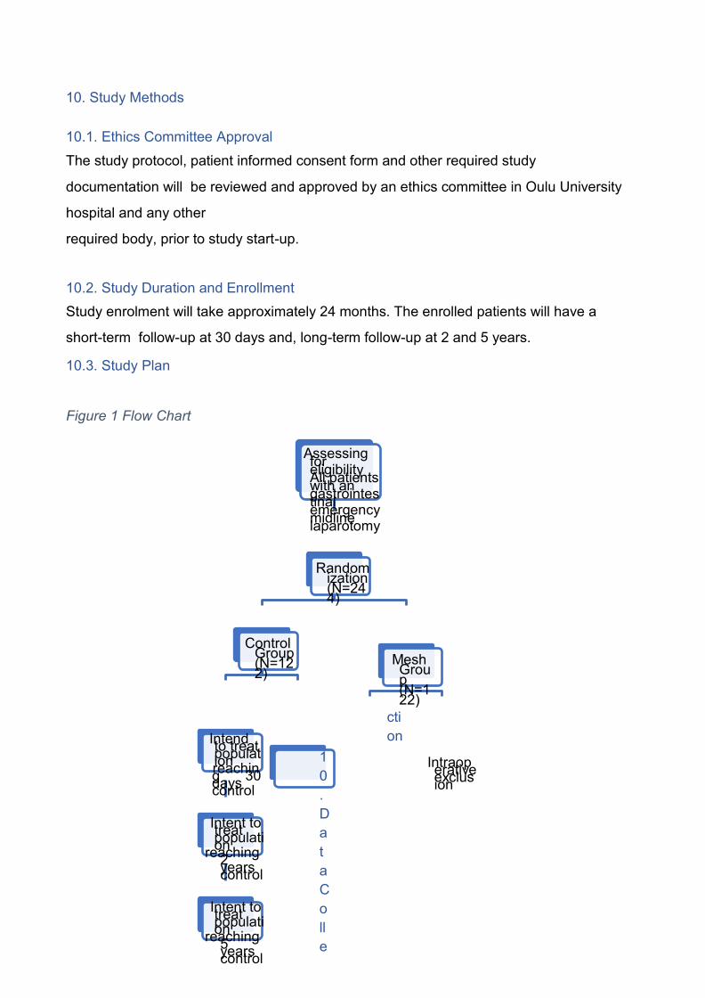
10. Study Methods 10.1. Ethics Committee Approval The study protocol, patient informed consent form and other required study documentation will be reviewed and approved by an ethics committee in Oulu University hospital and any other required body, prior to study start-up. 10.2. Study Duration and Enrollment Study enrolment will take approximately 24 months. The enrolled patients will have a short-term follow-up at 30 days and, long-term follow-up at 2 and 5 years. 10.3. Study Plan Figure 1 Flow Chart
Assessing for eligibility All patients with an gastrointestinal emergency midline laparotomy Randomization (N=244) Control Group (N=122)
Mesh Group (N=122) Intend to treat population reaching 30 days control Intent to treat population reaching 2 years control Intent to treat population reaching 5 years control
10. Data Colle
ction Intraoperative exclusion
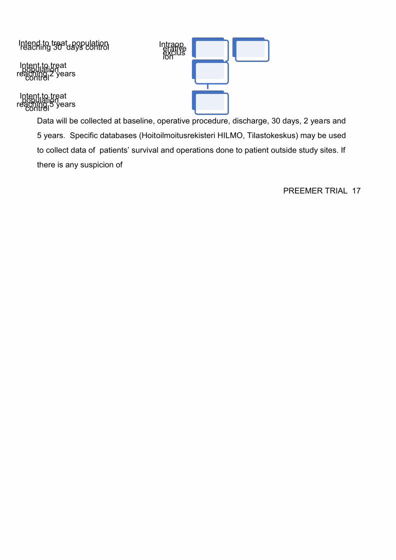
Intend to treat population reaching 30 days control Intent to treat population reaching 2 years control Intent to treat population reaching 5 years control
Intraoperative exclusion
Data will be collected at baseline, operative procedure, discharge, 30 days, 2 years and 5 years. Specific databases (Hoitoilmoitusrekisteri HILMO, Tilastokeskus) may be used to collect data of patients’ survival and operations done to patient outside study sites. If there is any suspicion of
PREEMER TRIAL 17
incisional hernia raised at any point of the follow-up, the patient is advised to contact the study site and additional clinical evaluation and ultrasonography if needed is arranged. The data will be collected using electronical CRFs and software designed for this study. 10.1 Patient Informed Consent Using the study-specific, ethics committee approved, informed consent form, information pertinent to this study will be provided to the subjects and/or representative in writing and using non-technical language. The consent form will include a description of the study and its purpose, potential benefits, potential risks, site contact information, and all other elements required of an informed consent. Subjects are required to voluntarily sign the informed consent form before any study-specific procedure is performed. The Investigator will conduct the informed consent process and will answer questions the subjects may have. If the subject agrees to participate, the informed- consent form must be signed and dated by the subject prior to enrolment in the study and separately signed and dated by the investigator taking consent. Only subjects who have signed the study informed consent will be included in the study. 10.2 Data Collection Following information of patients will be collected using electronic database. The patient is pseudonymized for data collection and all data will be handled using study-ID. Baseline
• Age • BMI • Charlson Comorbidity Index • Previous surgical history of abdomen • History of smoking • Previous hernias • Previous hernia-related operations • Previous WHO scale

• Medications affecting healing PREEMER TRIAL 18
o Corticosteroids o Immunosupressive medications o Biologics
• Creatinine • INR
Operative Procedure • Prophylactic antibiotics • ASA • Presence of hernias in midline • Presence and width of rectus diastasis • Contamination class • Surgical procedure • ICD-10 • Loss of blood • Time to create the retrorectal space and insert the mesh • Length of wound • Suture material and needle used • Drains left • Vacuum assisted closure/other temporary closure/skin left open • Skin closure
Primary hospital stay and Discharge • Surgical site infection (SSI) rate • All complications during hospital stay Comprehensive Complication Index • Re-operations • Burst abdomen • Fascial dehiscence • Length of stay (LoS) • Mesh removals
PREEMER TRIAL 19
• Place of discharge
Patients are guided to contact their study site in case of any problems with their recovery, any suspicion of hernia occurrence or wound complications. 30 days visit The recovery of all patients is assessed at 30 days after the operation. All the patients are called to. If there are any deviations in recovery, patient is invited to outpatient clinic for follow-up visit.
• Date of return to previous home unit • Return to previous level of activity • Return to work, length of sick leave • Bulging • Wound status • Any complications of recovery • Re-admissions • Re-operations • Removal of mesh • Quality of life (RAND-36, AAS, PROMIS) • Protocol deviations
2 years Visit Patient related recovery outcomes and QoL questionnaires will be completed and any complications, clinical signs and/or abdominal ultrasound findings of incisional hernia or protocol deviations will be reported. Both the patient and the surgeon assessing the recovery and well- being are blinded of the randomization group. Ultrasound findings will all be analyzed by single independent radiologist in each study site, blinded of the randomization group. Possible hernia opening, its size, location and

incisional hernia sack volume will be defined both at rest and with Valsalva maneuver. If the findings are PREEMER TRIAL 20
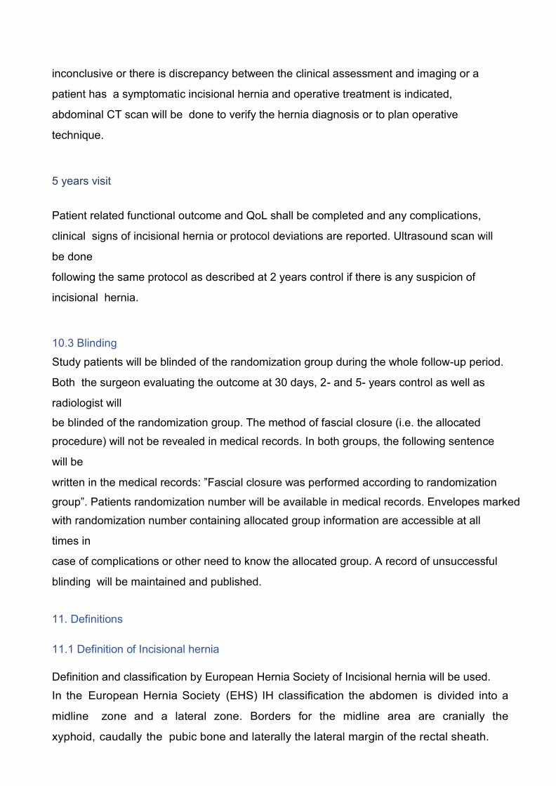
inconclusive or there is discrepancy between the clinical assessment and imaging or a patient has a symptomatic incisional hernia and operative treatment is indicated, abdominal CT scan will be done to verify the hernia diagnosis or to plan operative technique. 5 years visit Patient related functional outcome and QoL shall be completed and any complications, clinical signs of incisional hernia or protocol deviations are reported. Ultrasound scan will be done following the same protocol as described at 2 years control if there is any suspicion of incisional hernia. 10.3 Blinding Study patients will be blinded of the randomization group during the whole follow-up period. Both the surgeon evaluating the outcome at 30 days, 2- and 5- years control as well as radiologist will be blinded of the randomization group. The method of fascial closure (i.e. the allocated procedure) will not be revealed in medical records. In both groups, the following sentence will be written in the medical records: ”Fascial closure was performed according to randomization group”. Patients randomization number will be available in medical records. Envelopes marked with randomization number containing allocated group information are accessible at all times in case of complications or other need to know the allocated group. A record of unsuccessful blinding will be maintained and published. 11. Definitions 11.1 Definition of Incisional hernia Definition and classification by European Hernia Society of Incisional hernia will be used. In the European Hernia Society (EHS) IH classification the abdomen is divided into a midline zone and a lateral zone. Borders for the midline area are cranially the xyphoid, caudally the pubic bone and laterally the lateral margin of the rectal sheath.

PREEMER TRIAL 21
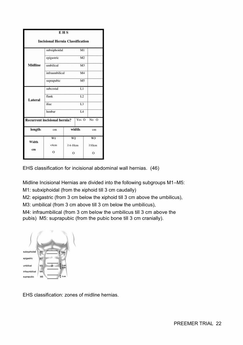
EHS classification for incisional abdominal wall hernias. (46) Midline Incisional Hernias are divided into the following subgroups M1–M5: M1: subxiphoidal (from the xiphoid till 3 cm caudally) M2: epigastric (from 3 cm below the xiphoid till 3 cm above the umbilicus), M3: umbilical (from 3 cm above till 3 cm below the umbilicus), M4: infraumbilical (from 3 cm below the umbilicus till 3 cm above the pubis) M5: suprapubic (from the pubic bone till 3 cm cranially). EHS classification: zones of midline hernias. PREEMER TRIAL 22
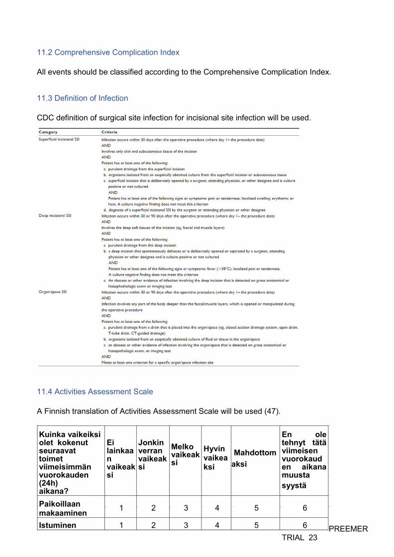
11.2 Comprehensive Complication Index All events should be classified according to the Comprehensive Complication Index. 11.3 Definition of Infection CDC definition of surgical site infection for incisional site infection will be used. 11.4 Activities Assessment Scale A Finnish translation of Activities Assessment Scale will be used (47).
PREEMER TRIAL 23
Kuinka vaikeiksi olet kokenut seuraavat toimet viimeisimmän vuorokauden (24h) aikana?
Ei lainkaan vaikeaksi
Jonkin verran vaikeaksi Melko vaikeaksi
Hyvin vaikeaksi Mahdottomaksi
En ole tehnyt tätä viimeisen vuorokauden aikana muusta syystä Paikoillaan makaaminen 1 2 3 4 5 6 Istuminen 1 2 3 4 5 6
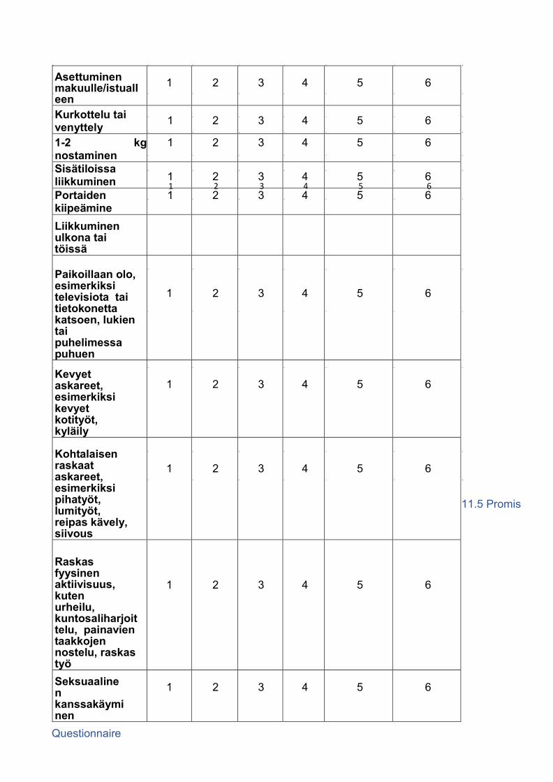
11.5 Promis
Questionnaire
Asettuminen makuulle/istualleen 1 2 3 4 5 6
Kurkottelu tai venyttely 1 2 3 4 5 6 1-2 kg nostaminen 1 2 3 4 5 6 Sisätiloissa liikkuminen 1 2 3 4 5 6 Portaiden kiipeämine 1 2 3 4 5 6 Liikkuminen ulkona tai töissä
Paikoillaan olo, esimerkiksi televisiota tai tietokonetta katsoen, lukien tai puhelimessa puhuen
1 2 3 4 5 6
Kevyet askareet, esimerkiksi kevyet kotityöt, kyläily
1 2 3 4 5 6
Kohtalaisen raskaat askareet, esimerkiksi pihatyöt, lumityöt, reipas kävely, siivous
1 2 3 4 5 6
Raskas fyysinen aktiivisuus, kuten urheilu, kuntosaliharjoittelu, painavien taakkojen nostelu, raskas työ
1 2 3 4 5 6
Seksuaalinen kanssakäyminen 1 2 3 4 5 6
1 2 3 4 5 6

A Finnish translation of Promis-questionnaire will be used to evaluate the likelihood of incisional hernia(48). PROMIS – kysely arpityräriskin arvioimiseksi
1. Uskotko, että Sinulla voi olla arpityrä?
Kyllä Ei
2. Tunnetko kipua arvessa? PREEMER TRIAL 24
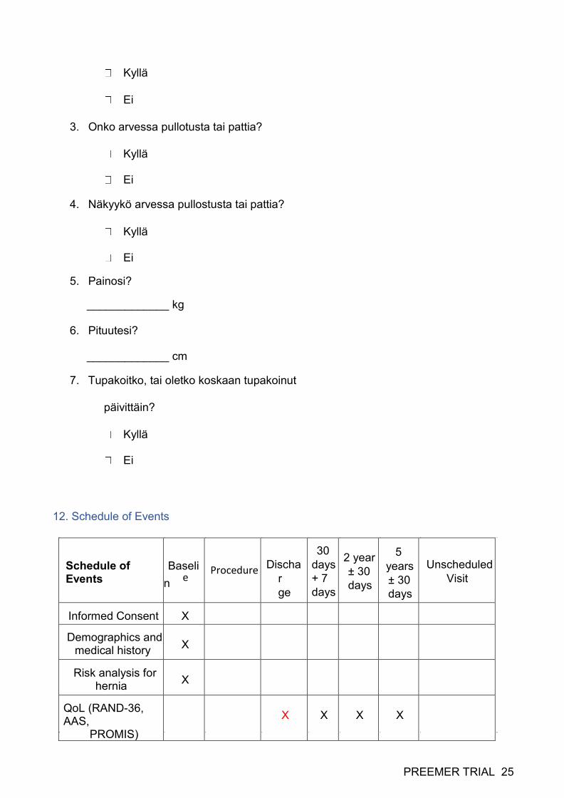
Kyllä Ei
3. Onko arvessa pullotusta tai pattia? Kyllä Ei
4. Näkyykö arvessa pullostusta tai pattia? Kyllä Ei
5. Painosi? _____________ kg
6. Pituutesi? _____________ cm
7. Tupakoitko, tai oletko koskaan tupakoinut päivittäin?
Kyllä Ei
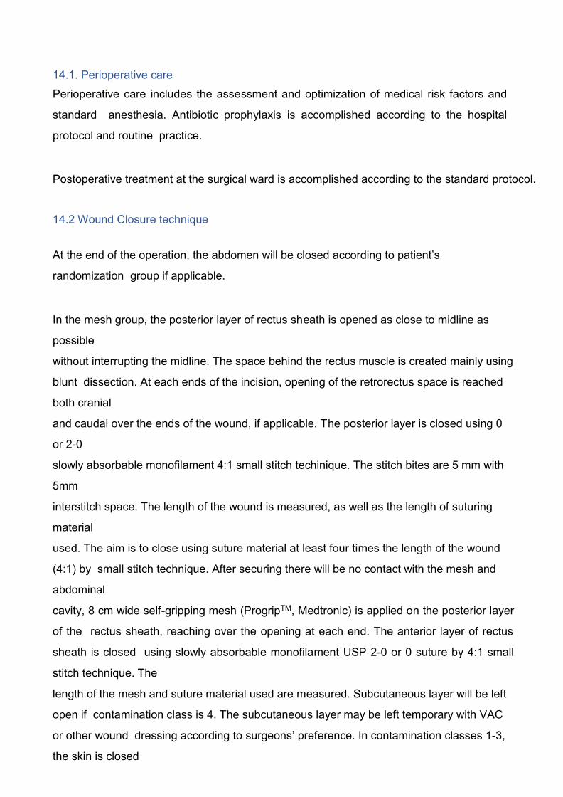
12. Schedule of Events
PREEMER TRIAL 25
Schedule of Events Baselin
Dischar ge
30 days + 7 days 2 year ± 30 days
5 years ± 30 days Unscheduled Visit
Informed Consent X Demographics and medical history X
Risk analysis for hernia X QoL (RAND-36, AAS, PROMIS)
X X X X
e Procedure
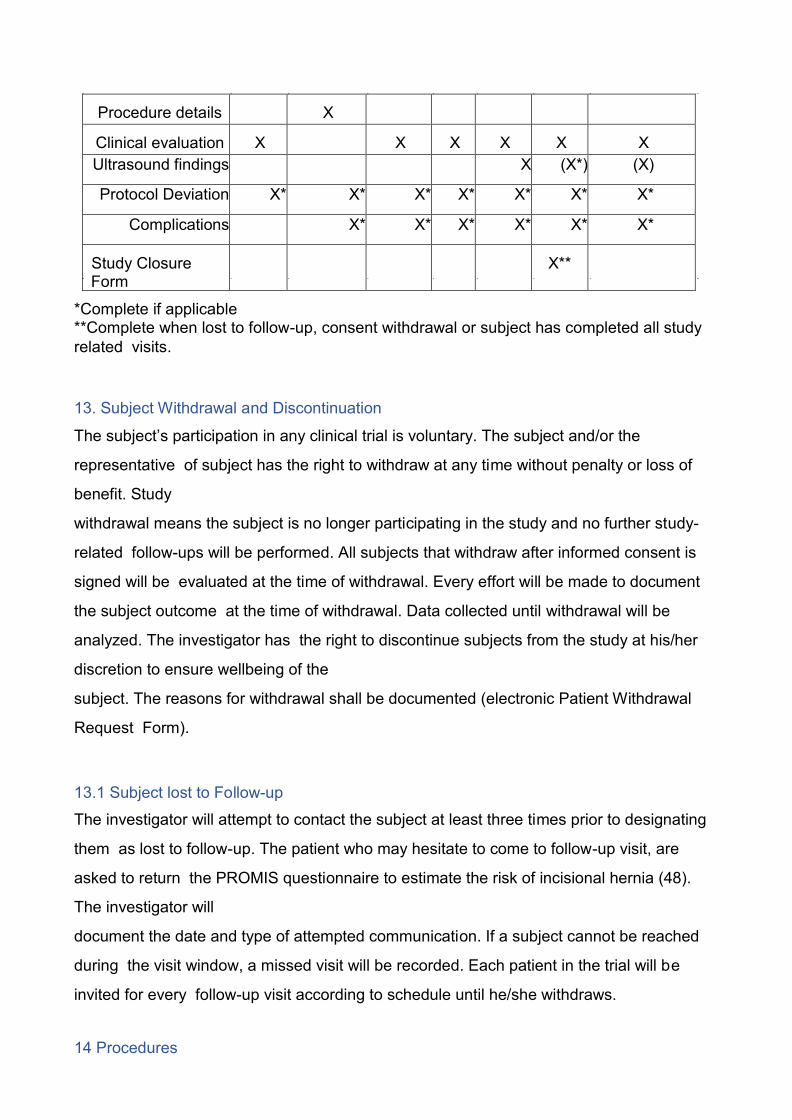
*Complete if applicable **Complete when lost to follow-up, consent withdrawal or subject has completed all study related visits.
13. Subject Withdrawal and Discontinuation The subject’s participation in any clinical trial is voluntary. The subject and/or the representative of subject has the right to withdraw at any time without penalty or loss of benefit. Study withdrawal means the subject is no longer participating in the study and no further study-related follow-ups will be performed. All subjects that withdraw after informed consent is signed will be evaluated at the time of withdrawal. Every effort will be made to document the subject outcome at the time of withdrawal. Data collected until withdrawal will be analyzed. The investigator has the right to discontinue subjects from the study at his/her discretion to ensure wellbeing of the subject. The reasons for withdrawal shall be documented (electronic Patient Withdrawal Request Form). 13.1 Subject lost to Follow-up The investigator will attempt to contact the subject at least three times prior to designating them as lost to follow-up. The patient who may hesitate to come to follow-up visit, are asked to return the PROMIS questionnaire to estimate the risk of incisional hernia (48). The investigator will document the date and type of attempted communication. If a subject cannot be reached during the visit window, a missed visit will be recorded. Each patient in the trial will be invited for every follow-up visit according to schedule until he/she withdraws. 14 Procedures
Procedure details X Clinical evaluation X X X X X X Ultrasound findings X (X*) (X) Protocol Deviation X* X* X* X* X* X* X*
Complications X* X* X* X* X* X* Study Closure Form X**

PREEMER TRIAL 26
14.1. Perioperative care Perioperative care includes the assessment and optimization of medical risk factors and standard anesthesia. Antibiotic prophylaxis is accomplished according to the hospital protocol and routine practice. Postoperative treatment at the surgical ward is accomplished according to the standard protocol. 14.2 Wound Closure technique At the end of the operation, the abdomen will be closed according to patient’s
randomization group if applicable. In the mesh group, the posterior layer of rectus sheath is opened as close to midline as possible without interrupting the midline. The space behind the rectus muscle is created mainly using blunt dissection. At each ends of the incision, opening of the retrorectus space is reached both cranial and caudal over the ends of the wound, if applicable. The posterior layer is closed using 0 or 2-0 slowly absorbable monofilament 4:1 small stitch techinique. The stitch bites are 5 mm with 5mm interstitch space. The length of the wound is measured, as well as the length of suturing material used. The aim is to close using suture material at least four times the length of the wound (4:1) by small stitch technique. After securing there will be no contact with the mesh and abdominal cavity, 8 cm wide self-gripping mesh (ProgripTM, Medtronic) is applied on the posterior layer of the rectus sheath, reaching over the opening at each end. The anterior layer of rectus sheath is closed using slowly absorbable monofilament USP 2-0 or 0 suture by 4:1 small stitch technique. The length of the mesh and suture material used are measured. Subcutaneous layer will be left open if contamination class is 4. The subcutaneous layer may be left temporary with VAC or other wound dressing according to surgeons’ preference. In contamination classes 1-3, the skin is closed

according to surgeons’ preference. In case the mesh cannot be safely kept outside the abdominal cavity, patient is intra-operatively excluded.
PREEMER TRIAL 27
In the no mesh group, the rectus aponeurosis is closed in a single aponeurotic layer by using slowly absorbable monofilament USP 2-0 or 0 suture by 4:1 small stitch technique. Both the length of the wound and length of the suture material used is measured. Catalogue of the operative technique will be sent to all participating surgeons to standardize the procedure. 14.3 Postoperative treatment Postoperative treatment will be accomplished according to standard protocol of each participating hospital. 14.4. Mesh (ProgripTM, Medtronic) A standard 8 cm self-gripping prophylactic mesh will be used in mesh group. The width of the mesh is standard. The length of the mesh is measured and reached over both ends of the laparotomy opening. 15.General Reporting Requirements Complications (Adverse events) reporting are an investigator’s responsibility to assess and report. Adverse Events (AE) will be identified and captured on the electronic Complications eCRF throughout the duration of the study as they occur and will be followed until they are adequately resolved or explained. Any Serious Mesh-Related Adverse Events with Clavien-Dindo Classification 3b or more should be reported to primary investigation site without delay after the site first learns of the event. 16. Investigator’s Responsibilities and Qualifications 16.1 General Responsibilities The role of the investigator is to implement and manage the day-to-day conduct of the clinical investigation and to ensure data integrity and the rights, safety and well-being of the subjects

PREEMER TRIAL 28
involved in the clinical investigation. The participating institution shall appoint an appropriately qualified person to be the site principal investigator. Prior to subject enrolment the investigational center must have Institutional review board approval for the study. Investigators shall be qualified by education, training and experience to assume responsibility for the proper conduct of the clinical investigation. Investigators shall disclose potential conflicts of interest, including financial, that interfere with the conduct of the clinical investigation or interpretation of the results. Investigators shall be knowledgeable with the method of obtaining informed consent. The Investigator shall ensure compliance with the applicable regulatory requirements and ethical principles for the process of obtaining informed consent. All protocol deviations should be recorded on the Protocol deviation form. 17 Data handling and record keeping 17.1 Confidentiality Patient confidentiality will be strictly maintained. Patients will be assigned a Study ID. Access to patient records will be limited to the study group and the Investigator-delegated study coordinator. 17.2 Data Management Dedicated software and electronic database and the case report forms (eCRF) will be used to host the Clinical Trial data for this study. The database is developed and utilized in accordance with international requirements and standards applicable to clinical investigations i.e. Good Clinical Practice (GCP) and is a GCP compliant environment meeting applicable 21 CFR Part 11 requirements. 18. Case Report Forms The electronical Case Report Forms (eCRF) and software are the primary data collection instruments for the study.

PREEMER TRIAL 29
All data requested on the eCRFs will be recorded. All missing data will be explained. 19. Sample Size and Statistical Analysis 19.1 Sample size To calculate a sample size needed to compare these two groups we estimated a 10 % rate of IH in mesh group and 25 % IH in control group on clinical assessment and ultrasound examination. Assuming α = 0.05 and power = 80%, we would need 97 patients per group. Further, assuming a 2- year dropout rate of 20%, 122 patients per group are needed (totally 244 patients). The sample size is calculated only for the primary outcome, the secondary outcomes will be interpreted as hypothesis generating only. If the estimated 20% dropout rate exceeds, the sample size may be recalculated. All analyses will be performed by or under the guidance of professional statistician and following the CONSORT guidelines. 19.2 Allocation Patients having emergency midline laparotomy for any indication and fulfilling the inclusion criteria will be randomized prior to surgery after informed consent is signed. Patients are randomly allocated (1:1 ratio) either to an intervention group or a control group according to a computer- generated list compiled by a biostatistician otherwise uninvolved in the clinical care of trial patients. Allocation will be stratified according to BMI (<30 and ≥30kg/m2), history of previous laparotomy and age (<65 and ≥65- years) and blocked within strata using random permuted blocks (block size 2, 4, 6 and 8). A separate randomization list will be created for each participating center. All patients who have an emergency midline laparotomy during randomization period are assessed for eligibility.

19.3 Endpoint analysis All analyses will be performed primarily according to modified intention to treat (ITT) principle. Patients who fulfill exclusion criteria intraoperatively (after randomization) will not be included in the analyses. Per protocol analyses will be used as safeguard against the risk of falsely claiming
PREEMER TRIAL 30
equality/superiority. The primary endpoint will be the incidence difference of IHs with 95% confidence interval between the study groups during 2-years follow up. Secondary outcomes are listed previously. The primary endpoint as well as other categorical data will be analyzed by the χ2- test or Fisher’s exact test. Student’s t-test or Welch test will be used for continuous variable, the latter if assumption of homogenous variances does not hold. The incidence of hernia will also be analyzed using Kaplan-Meier analysis. The linear mixed model (LMM) or generalized linear mixed model (GLMM) will be used for repeatedly measured data, the previous for continuous data and latter for categorical data. Multiple imputations of missing outcome data will be used for sensitivity analyses. Prospectively planned subgroup analyses are as follows: BMI>30, previous hernia and contamination class 4. However, sample size calculation is done only for the primary end point and subgroup analyses are hypothesis generating only. The statistical programs SPSS (IBM Corp. Released 2016. IBM SPSS Statistics for Windows, Version 24.0. Armonk, NY: IBM Corp) and SAS (version 9.4, SAS Institute Inc., Cary, NC, USA) will be used for the analyses. 20. Publication Policy The trial will be registered with an authorized registry, according to the International Committee of Medical Journal Editors (ICMJE) Guidelines, prior to the start of recruitment. The success of the trial depends upon the collaboration of all participants. For this reason, credit for the main results will be given to all those who have collaborated in the trial, through authorship and contributor-ship. Authorship decisions will be guided by standard requirements for authorship relating to submission of manuscripts to medical journals. These state that authorship credit should be based only on the following conditions being met (http://www.icmje.org): • Substantial contribution to conception and design, or acquisition of data, or

analysis and interpretation of data • Substantial contribution to drafting the article or revising it critically for important intellectual content • Substantial contribution to final approval of the version to be published. In light of this, the Principal Investigator and the main study group from Oulu and Helsinki University Hospitals and Dr Filip Muysoms will be named as authors in any publication, subject to
PREEMER TRIAL 31
journal authorship restrictions. In addition, all collaborators (surgeons as well as biostatistician) will be listed as contributors for the main trial publication, giving details of roles in planning, conducting and reporting the trial. It is planned that the recruiting surgeons will also be named as authors, if the set target of the number of randomized patients is achieved. To maintain the scientific integrity of the trial, data will not be released prior to the first publication of the analysis of the primary endpoint, either for trial publication or oral presentation purposes, without the permission of the whole study group. In addition, individual collaborators must not publish data concerning their patients, which is directly relevant to the questions posed in the trial until the first publication of the analysis of the primary endpoint. 20.1. Publication Plan The protocol of the trial will be published at the beginning of the trial. The results concerning the primary end point and results of secondary endpoints within 2 years follow up will be published once included patients have reached 2 years follow-up. The results of 5 years follow up will be published. 21. References (1) Höer J, Lawong G, Klinge U, Schumpelick V. Einflussfaktoren der Narbenhernienentstehung Retrospektive Untersuchung an 2.983 laparotomierten Patienten über einen Zeitraum von 10 Jahren. Der Chirurg 2002;73:474-80. (2) Mudge M, Hughes LE. Incisional hernia: A 10 year prospective study of incidence and attitudes. Br J Surg 1985;72:70-71. (3) Sajid MS, Bokhari SA, Mallick AS, Cheek E, Baig MK. Laparoscopic versus open repair of incisional/ventral hernia: a meta-analysis. The American Journal of Surgery 2009;197:64-72. (4) Harlaar JJ, Deerenberg EB, Dwarkasing RS, Kamperman AM, Kleinrensink GJ, Jeekel J, et al. Development of incisional herniation after midline laparotomy. BJS Open 2017;1:18-23. (5) Patel SV, Paskar DD, Nelson RL, Vedula SS, Steele SR. Closure methods for laparotomy incisions for preventing incisional hernias and other wound complications. The Cochrane Database of

Systematic Reviews 2017;11. (6) Fink C, Baumann P, Wente MN, Knebel P, Bruckner T, Ulrich A, et al. Incisional hernia rate 3 years after midline laparotomy. Br J Surg 2014;101:51-4. (7) Kroese LF, Gillion J, Jeekel J, Kleinrensink G, Lange JF. Primary and incisional ventral hernias are different in terms of patient characteristics and postoperative complications - A prospective cohort study of 4,565 patients. International Journal of Surgery 2018;51:114-9.
PREEMER TRIAL 32
(8) Bjork D, Cengiz Y, Weisby L, Israelsson LA. Detecting Incisional Hernia at Clinical and Radiological Examination. Surg Technol Int 2015;26:128-31. (9) Fischer JP, Basta MN, Mirzabeigi MN, Bauder AR, Fox JP, Drebin JA, et al. A Risk Model and Cost Analysis of Incisional Hernia After Elective, Abdominal Surgery Based Upon 12,373 Cases: The Case for Targeted Prophylactic Intervention. Annals of Surgery 2016;263:1010-1017. (10) Bickenbach KA, M.D, Karanicolas, Paul J., M.D., Ph.D, Ammori JB, M.D, Jayaraman, Shiva, M.D., M.E.S.C, Winter JM, M.D, Fields RC, M.D, et al. Up and down or side to side? A systematic review and meta-analysis examining the impact of incision on outcomes after abdominal surgery. Am J of Surgery 2013;206:400-9. (11) Brown SR, Goodfellow PB. Transverse verses midline incisions for abdominal surgery. Cochrane Database Syst Rev 2005;4. doi(4):CD005199. (12) Millbourn D, Wimo A, Israelsson L. Cost analysis of the use of small stitches when closing midline abdominal incisions. Hernia 2014;18:775-80. (13) Deerenberg EB, Harlaar JJ, Steyerberg EW, Lont HE, van Doorn HC, Heisterkamp J, et al. Small bites versus large bites for closure of abdominal midline incisions (STITCH): a double-blind, multicentre, randomised controlled trial. The Lancet 2015;386:1254-1260. (14) Muysoms F, Antoniou S, Bury K, Campanelli G, Conze J, Cuccurullo D, et al. European Hernia Society guidelines on the closure of abdominal wall incisions. Hernia 2015;19:1-24. (15) Thankam FG, Palanikumar G, Fitzgibbons RJ, Agrawal DK. Molecular Mechanisms and Potential Therapeutic Targets in Incisional Hernia. J Surg Res 2019;236:134-43. (16) Xing L, Culbertson EJ, Wen Y, Franz MG. Early laparotomy wound failure as the mechanism for incisional hernia formation. J Surg Res 2013;1;182:35. (17) Chang N, Goodson WH,3rd, Gottrup F, Hunt TK. Direct measurement of wound and tissue oxygen tension in postoperative patients. Ann Surg 1983;197:470-478. (18) Knighton DR, Silver IA, Hunt TK. Regulation of wound-healing angiogenesis-effect of oxygen gradients and inspired oxygen concentration. Surgery 1981;90:262-270. (19) Aicher BO, Woodall J, Tolaymat B, Calvert C, Monahan TS, Toursavadkohi S. Does perfusion matter? Preoperative prediction of incisional hernia development. Hernia 2019;2. doi: 10.1007/s10029-019-02018-3. (20) Harrison B, Sanniec K, Janis JE. Collagenopathies-Implications for Abdominal Wall Reconstruction: A Systematic Review. Plast Reconstr Surg Glob Open 201624;4:e1036. (21) Bosanquet DC, Ansell J, Abdelrahman T, Cornish J, Harries R, Stimpson A, et al. Systematic Review and Meta-Regression of Factors Affecting Midline Incisional Hernia Rates: Analysis of 14 618 Patients. PLoS ONE 2015;10(9). (22) Basta MN, Kozak GM, Broach RB, Messa CA,4th, Rhemtulla I, DeMatteo RP, et al. Can We Predict Incisional Hernia?: Development of a Surgery-specific Decision-Support Interface. Ann Surg 2019;270:544-553.

(23) Ah-Kee EY, Kallachil T, O'Dwyer PJ. Patient awareness and symptoms from an incisional hernia. Int Surg 2014;99:241-6. (24) Cassar K, Munro A. Surgical treatment of incisional hernia. Br J Surg 2000;89:534-45. (25) Emby DJ, Aoun G. CT technique for suspected anterior abdominal wall hernia. AJR Am J Roentgenol 2003;181:431-3.
PREEMER TRIAL 33
(26) Bloemen A, van Dooren P, Huizinga BF, Hoofwijk AG. Comparison of ultrasonography and physical examination in the diagnosis of incisional hernia in a prospective study. Hernia 2012;16:53-57. (27) Hojer AM, Rygaard H, Jess P. CT in the diagnosis of abdominal wall hernias: a preliminary study. Eur Radiol 1997;7:1416-8. (28) den Hartog D, Dur AH, Kamphuis AG, Tuinebreijer WE, Kreis RW. Comparison of ultrasonography with computed tomography in the diagnosis of incisional hernias. Hernia 2009;13:45-8. (29) Aguirre DA, Casola G, Sirlin C. Abdominal wall hernias: MDCT findings. AJR Am J Roentgenol 2004;183:681-90. (30) Lassandro F, Iasiello F, Pizza NL, Valente T, Stefano ML, Grassi R, et al. Abdominal hernias: Radiological features. World J Gastrointest Endosc 2011;16:110-17. (31) Bittner R, Bingener-Casey J, Dietz U, Fabian M, Ferzli G, Fortelny R, et al. Guidelines for laparoscopic treatment of ventral and incisional abdominal wall hernias. International Endohernia Society (IEHS)—Part 1. Surg Endosc 2014;28:2-29. (32) Albertsmeier M, Seiler CM, Fischer L, Baumann P, Husing J, Seidlmayer C, et al. Evaluation of the safety and efficacy of MonoMax(R) suture material for abdominal wall closure after primary midline laparotomy-a controlled prospective multicentre trial: ISSAAC [NCT005725079. Langenbecks Arch Surg 2012;397:363-71. (33) Seiler C, Baumann P, Kienle P, Kuthe A, Kuhlgatz J, Engemann R, et al. A randomised, multi- centre, prospective, double blind pilot-study to evaluate safety and efficacy of the non-absorbable Optilene Mesh Elastic versus the partly absorbable Ultrapro Mesh for incisional hernia repair. BMC Surg 2010;10:21-21. (34) Dietz UA, Hamelmann W, Winkler MS, Debus ES, Malafaia O, Czeczko NG, et al. An alternative classification of incisional hernias enlisting morphology, body type and risk factors in the assessment of prognosis and tailoring of surgical technique. J Plast Reconstr Aesthet Surg 2007;60:383-8. (35) Korenkov M, Paul A, Sauerland S, Neugebauer E, Arndt M, Chevrel JP, et al. Classification and surgical treatment of incisional hernia. Results of an experts' meeting. Langenbecks Arch Surg 2001;38:65-73. (36) Muysoms FE, Miserez M, Berrevoet F, Campanelli G, Champault GG, Chelala E, et al. Classification of primary and incisional abdominal wall hernias. Hernia 2009;13:407-414. (37) Diener M, Voss S, Jensen K, Büchler M, Seiler C. Elective Midline Laparotomy Closure: The INLINE Systematic Review and Meta-Analysis. Annals of Surgery 2010;251:843-856. (38) van’t Riet M, Steyerberg EW, Nellensteyn J, ym. Meta-analysis of techniques for closure of midline abdominal incisions.BJS 2002;89:1350-6.

(39) Jairam A, Timmermans L, Eker H, ym. Prevention of incisional hernia with prophylactic onlay and sublay mesh reinforcement versus primary suture only in midline laparotomies (PRIMA): 2- year follow-up of a multicentre, double-blind, randomised controlled trial. Lancet 2017;390:567- 76. PREEMER TRIAL 34
(40) Borab Z, Shakir S, Lanni, M, et al. Does prophylactic mesh placement in elective, midline laparotomy reduce the incidence of incisional hernia? A systematic review and meta-analysis. Surgery 2016;161:1149-63. (41) Mingoli A1, Puggioni A, Sgarzini G, Luciani G, Corzani F, Ciccarone F, Baldassarre E, Modini C. Incidence of incisional hernia following emergency abdominal surgery. Ital J Gastroenterol Hepatol. 1999;31:449-53. (42) Burns FA, Heywood EG, Challand CP, Lee MJ. Is there a role for prophylactic mesh in abdominal wall closure after emergency laparotomy? A systematic review and meta-analysis. Hernia 2019. doi: 10.1007/s10029-019-02060-1 (43) Kurmann A, Barnetta C, Candinas D, Beldi G. Implantation of Prophylactic Nonabsorbable Intraperitoneal Mesh in Patients With Peritonitis Is Safe and Feasible. World J Surg 2013;37:1656- 60. (44) Argudo N, Pereira J, Sancho J, et al. Prophylactic synthetic mesh can be safely used to close emergency laparotomies, even in peritonitis. Surgery 2014;156:1238-44. (45) Gillion J, Sanders D, Miserez M, Muysoms F. The economic burden of incisional ventral hernia repair: a multicentric cost analysis. Hernia 2016;20:819-30. (46) Muysoms F, Mizered M, Berrevoet F, et al. Classification of primary and incisional abdominal wall hernias. Hernia 2009;13:407-14. (47) McCarthy M, Jr, Jonasson O, Chang CH, et al. Assessment of patient functional status after surgery. J Am Coll Surg. 2005;201:171–8. (48) Jairam AP, Wilson M, Steyerberg EW, Jeekel J, Lange JF. Patient reported outcome measurements in the diagnosis of incisional hernia: PROMIS questionnaire, a pilot study. J Surg Res. 2016;203:378–82.

PREEMER TRIAL 35