Practical Aspects of Single Crystal X-ray Crystallography · Introduction to Single Crystal X-ray...
26
1 W I S S E N T E C H N I K L E I D E N S C H A F T www.tugraz.at www.ac.tugraz.at Practical Aspects of Single Crystal X-ray Crystallography
Transcript of Practical Aspects of Single Crystal X-ray Crystallography · Introduction to Single Crystal X-ray...

1
W I S S E N T E C H N I K L E I D E N S C H A F T
www.tugraz.at
www.ac.tugraz.at
Practical Aspects of Single Crystal X-ray Crystallography

2
2 X-ray Diffraction
Introduction to Single Crystal X-ray Crystallography Practical Aspects of Single Crystal X-ray Crystallography
X-ray crystallography is a method of determining the arrangement of atoms within a crystal.
• Structure of a compound on a molecular level • Connectivity • Chirality • Framework and Pore size
• Bond length and angle parameters
• Interatomic forces that aid in crystallization
• Solvent inclusion • Hydrogen bonding networks • π-interactions • Agostic interactions

3
3 X-ray Diffraction
Introduction to Single Crystal X-ray Crystallography Practical Aspects of Single Crystal X-ray Crystallography
X-ray is a form of electromagnetic radiation
4
4 X-ray Diffraction
Introduction to Single Crystal X-ray Crystallography Practical Aspects of Single Crystal X-ray Crystallography
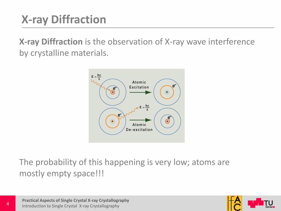
X-ray Diffraction is the observation of X-ray wave interference by crystalline materials.
The probability of this happening is very low; atoms are mostly empty space!!!

5
5 The Crystal
Introduction to Single Crystal X-ray Crystallography Practical Aspects of Single Crystal X-ray Crystallography
A crystal is a solid material whose atoms are arranged in an orderly repeating pattern extending in all three spatial dimensions
Smallest component of a crystal is the unit cell
• Smallest possible volume that when repeated, is representative of the entire crystal. • The unit cell contains the maximum symmetry that uniquely defines the crystal structure.
More than one molecule can be found inside the unit cell!!!

6
6 Unit Cell
Introduction to Single Crystal X-ray Crystallography Practical Aspects of Single Crystal X-ray Crystallography
x
z
y a
b
c
α β
γ
Angle between b & c is α Angle between a & c is β Angle between a & b is γ
Every crystal structure has a unique unit cell!!

7
7 Describing order in Space
Introduction to Single Crystal X-ray Crystallography Practical Aspects of Single Crystal X-ray Crystallography
7 crystal systems and 14 Bravais lattices (centering lattice points)

8
8 Symmetry Elements
Introduction to Single Crystal X-ray Crystallography Practical Aspects of Single Crystal X-ray Crystallography
• Identity, 1 • Inversion center, 1 • Rotation axis, 2,3,4,5,6 • Mirror plane, m • Screw axis, 21, 31, 32, 41 (rotation with a translation)
• Improper rotation, 3,4,5 • Glide planes, a, b, c, n, d (mirror reflection with a translation)
Zbigniew Dauter and Mariusz Jaskolski. J. Appl. Cryst. 2010, 43, 1150–1171
-
- - -

9
9 Describing order in Space
Introduction to Single Crystal X-ray Crystallography Practical Aspects of Single Crystal X-ray Crystallography
System Unit cell dimension
Unit cell angles (°)
Characteristic symmetry
International notation
Bravais Lattice (14)
Space Groups (230)
Triclinic a ≠ b ≠ c α ≠ β ≠ γ ≠ 90 Only inversion center
1, 1 P (Primitive) P1, P1
Monoclinic a ≠ b ≠ c α = γ = 90, β ≠ 90
Twofold axis or/and mirror plane
2, m, 2/m P, C (centered, A, B, C)
P21, P21/c, C2, C2/c
Orthorhombic a ≠ b ≠ c α = β = γ = 90
Three perpendicular twofold axesor/and mirrors
222, mm2, mmm
P, C, I (Body Centered), F (Face centered)
P2221, Pmma
Tetragonal a = b ≠ c α = β = γ = 90 Fourfold axis 4, 4, 4/m, 422 P, I P43, I4
Trigonal a = b ≠ c α = β = 90, γ = 120
Threefold axis 3, 3, 32, 3m, 3m
P3212
Rhombohedral a = b = c α = β = γ ≠ 90 Threefold axis R (Rhombohedral) R3, R32
Hexagonal a = b ≠ c α = β = 90, γ = 120
Sixfold axis 6, 6, 6/m
P P62, P6/mcc
Cubic a = b = c α = β = γ = 90 Four threefold axes
23, m3 P, I, F F432
_
_
_
_
_
_
_
Only 230 possible ways of arranging identical objects in an infinite three dimensional lattice!!!
10
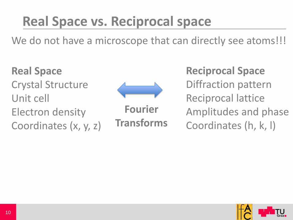
10 Real Space vs. Reciprocal space We do not have a microscope that can directly see atoms!!!
Real Space Crystal Structure Unit cell Electron density Coordinates (x, y, z)
Reciprocal Space Diffraction pattern Reciprocal lattice Amplitudes and phase Coordinates (h, k, l)
Fourier Transforms

11
11 Describing order in space
Introduction to Single Crystal X-ray Crystallography Practical Aspects of Single Crystal X-ray Crystallography
Parallel planes of atoms intersecting the unit cell define directions and distances in the crystal.
The (200) planes of atoms in NaCl
Planes are separated by a distance, dhkl. This distance is crucial to determine where diffraction peaks will be observed!!!!!
The planes and the distance between them is what we measure!!
x
z
y
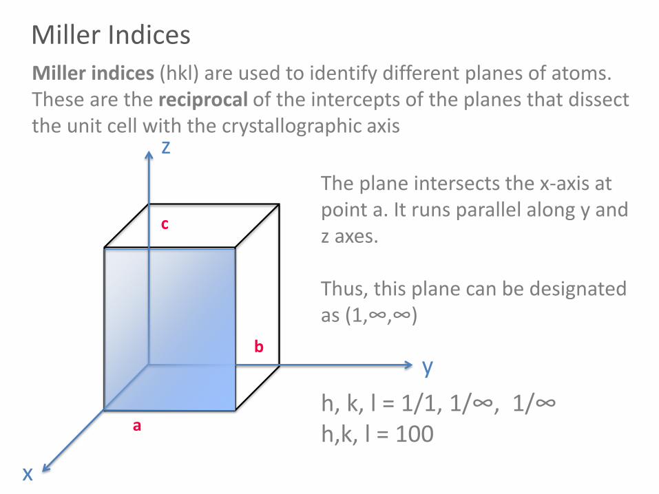
Miller Indices Miller indices (hkl) are used to identify different planes of atoms. These are the reciprocal of the intercepts of the planes that dissect the unit cell with the crystallographic axis
The plane intersects the x-axis at point a. It runs parallel along y and z axes. Thus, this plane can be designated as (1,∞,∞)
h, k, l = 1/1, 1/∞, 1/∞ h,k, l = 100 a
b
c

13
13 Bragg’s Law
Introduction to Single Crystal X-ray Crystallography Practical Aspects of Single Crystal X-ray Crystallography
(h, k, l)
Incident Wave
Scattered Wave
Diffraction; produces a reflection
θ θ
Only when the incident angle (θ) and the scattered angle (θ) are the same will constructive interference will occur
The scattering of an atom is the value for a free electron multiplied by the atomic number – Scattering factor

14
14 Bragg’s Law
Introduction to Single Crystal X-ray Crystallography Practical Aspects of Single Crystal X-ray Crystallography
(h, k, l)
d
Incident Wave
Scattered Wave
θ θ

15
15 Bragg’s Law
Introduction to Single Crystal X-ray Crystallography Practical Aspects of Single Crystal X-ray Crystallography
(h, k, l)
d
Incident Wave
Scattered Wave
θ θ
θ θ
For constructive interference to occur the waves must be in phase
Wave 1
Wave 2
Wave 2
Wave 1
|---ℓ---|
|λ |
ℓ = n λ

16
16 Bragg’s Law
Introduction to Single Crystal X-ray Crystallography Practical Aspects of Single Crystal X-ray Crystallography
(h, k, l)
d
θ
Incident Wave
Scattered Wave
θ
2dsinθ = nλ
ℓ = dsinθ
According to Bragg, when ℓ + ℓ = λ, the waves are in phase, resulting in constructive interference or Diffraction!!!
ℓ ℓ θ θ
ℓ = n λ

17
17 Applying Bragg’s Law – Reciprocal Space
Introduction to Single Crystal X-ray Crystallography Practical Aspects of Single Crystal X-ray Crystallography
2dhkl sinθhkl = nλ
“Real Space”
Reciprocal Space
d*hkl = 2 sinθhkl
λ
d*1
d*2
d*3

18
18 Diffraction Pattern – Unit Cell (real space)
Introduction to Single Crystal X-ray Crystallography Practical Aspects of Single Crystal X-ray Crystallography
Reciprocal Space
d*hkl = 2 sinθhkl
λ
Real Space
d*100 = a* = bcsinα V
d*010 = b* = acsinβ V
d*001 = c* = absinγ V
We can define our unit cell parameters

19
19 Diffraction Pattern
What does each spot tell us? • hkl position of atoms (x, y, z)
• Where the atoms are • Miller Indices and dhkl
• Intensity ∼ number of scattering electrons in the atom • What the atom is
• Scattering Factor
Each spot is called a reflection
Each frame has several reflections
Reciprocal Space!!!

20
20 Electron Density
ρxyz= (1/V) ∑hkl| Fhkl |observed cos2π (hx+ky+lz-αhkl )]
Intensities – Structure factor
? Phasing

21
21 Phasing Problem
ρxyz= (1/V) ∑hkl| Fhkl |observed cos2π (hx+ky+lz-αhkl )] ?
Direct Methods • Test random phases for a solution (1000-3000 cycles)
Patterson Methods
• Heavy atom method
Phase information is more important than the intensity We cannot focus an X-ray like a microscope!!

22
22 Refinement - Does the model fit the data?
R1 values (below 10 %) • Measure of how well the refined structure predicts the observed data.
wR2 values (below 20 %) • Least‐squares residual; measure of how intensities calculated for the X‐ray reflections match those measured experimentally.
Refinement Residuals
Data Collection
Close to 100% completeness • Most of the reflections in the asymmetric unit

23
23 Crystallographic Programs
Introduction to Single Crystal X-ray Crystallography Practical Aspects of Single Crystal X-ray Crystallography
Apex 2 • Collecting data • Solving and Refining the structure • Preparing structure for publication
• Picture (black and white) • Crystallographic tables • CIF (crystallographic information file)
• CIF must be checked online and issues fixed • PLATON
Endnote or PublCIF • Preparing CIF file for publication
24
24 Crystallographic Programs
Introduction to Single Crystal X-ray Crystallography Practical Aspects of Single Crystal X-ray Crystallography
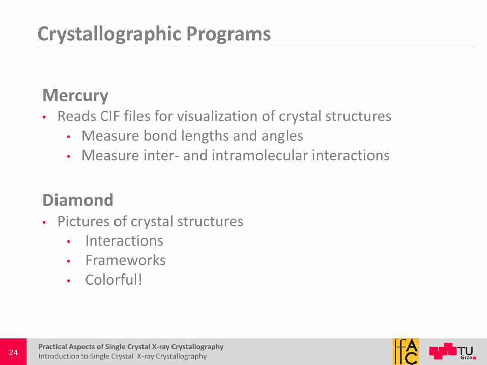
Mercury • Reads CIF files for visualization of crystal structures
• Measure bond lengths and angles • Measure inter- and intramolecular interactions
Diamond • Pictures of crystal structures
• Interactions • Frameworks • Colorful!

25
25 Further Information
Introduction to Single Crystal X-ray Crystallography Practical Aspects of Single Crystal X-ray Crystallography
Reference Materials • “Crystal Structure Determination”
• Werner Mass • “Crystal Structure Analysis: Principles and Practice”
• William Clegg • “Crystal Structure Refinement: a crystallographer’s guide to SHELXL
• Peter Müller • “How to read (and understand) Volume A of International Tables for Crystallography: an introduction for nonspecialists”
• Zbigniew Dauter and Mariusz Jaskolski. J. Appl. Cryst. 2010, 43, 1150–1171

26
26 Next Week
Introduction to Single Crystal X-ray Crystallography Practical Aspects of Single Crystal X-ray Crystallography
Mounting Samples • Individual basis
Refining
• File types (res, ins, cif) • Issues with completeness • Issues with refinement
SADABS
• Absorption methods