Immunohistochemistry Antibody Validation Report for Anti-CD20 Antibody (STJ96950)


2019

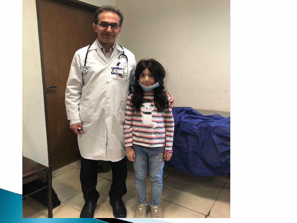
Our patiant was a ten-year-old girl who had a right
periorbital swelling
and
fever
for about one week before referral,

who was initially treated with an antibiotic(VAN+CEF) for the diagnosis of pre-septal cellulitis, but gradually fever and left periorbital swelling were added to symptoms.

painless swelling in the cheek area also spreads and the lesions become ulcerative.
The patient is the first child of the relative parents.


There was a history of systemic lupus erythematous ( SLE) in the patient's grandmother and aunt, and grandmother died from a pulmonary complication.

Sonographic evaluation showed extensive soft tissue edema in the face and upper neck particularly in superficial subcutaneous tissue
The study of MIBG scintigraphy was negative.


MRI images showed inflammatory signal changes in subcutaneous area of bilateral cheek and submandibular region in favor of cellulitis without evidence of collection or lymphadenitis.


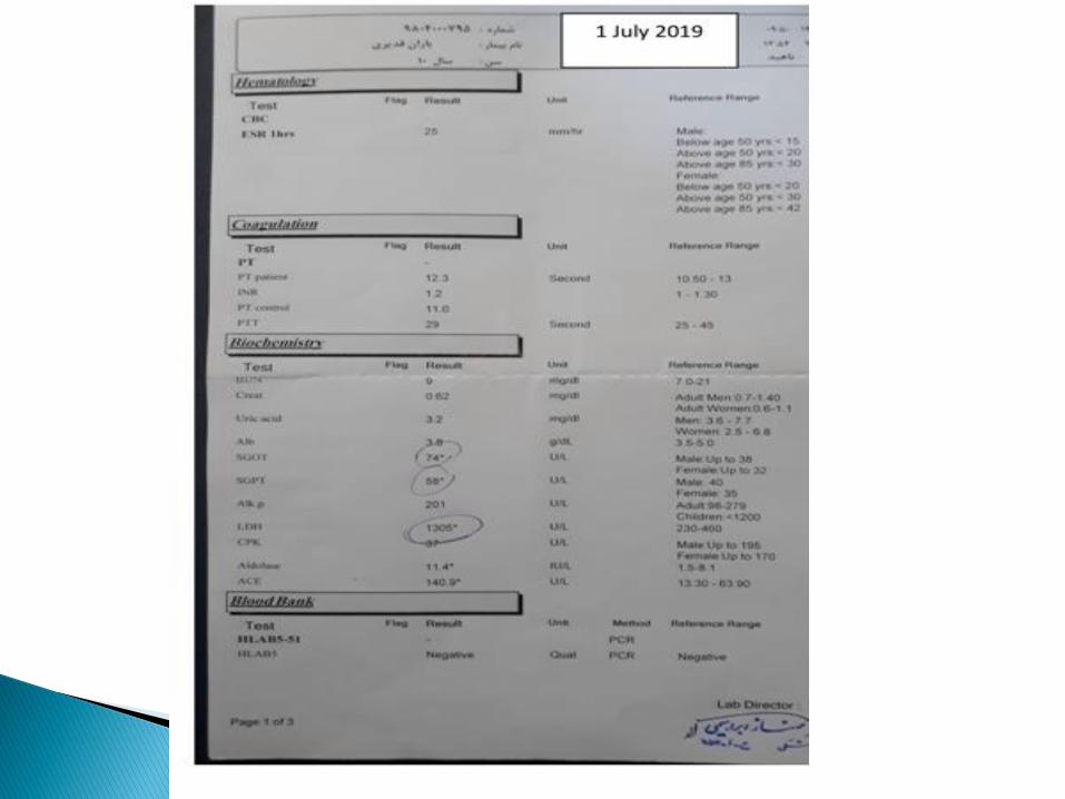
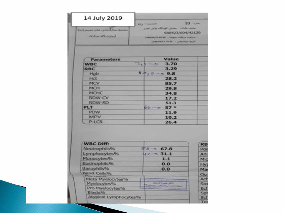
In laboratory studies only mild Leukopenia (3800 ) and lymphopeniaoccurred and other inflammatory markers and markers such as ESR and CRP were mildly elevated.

Bone marrow examinations were also performed for the patient with no evidence of abnormal cell involvement


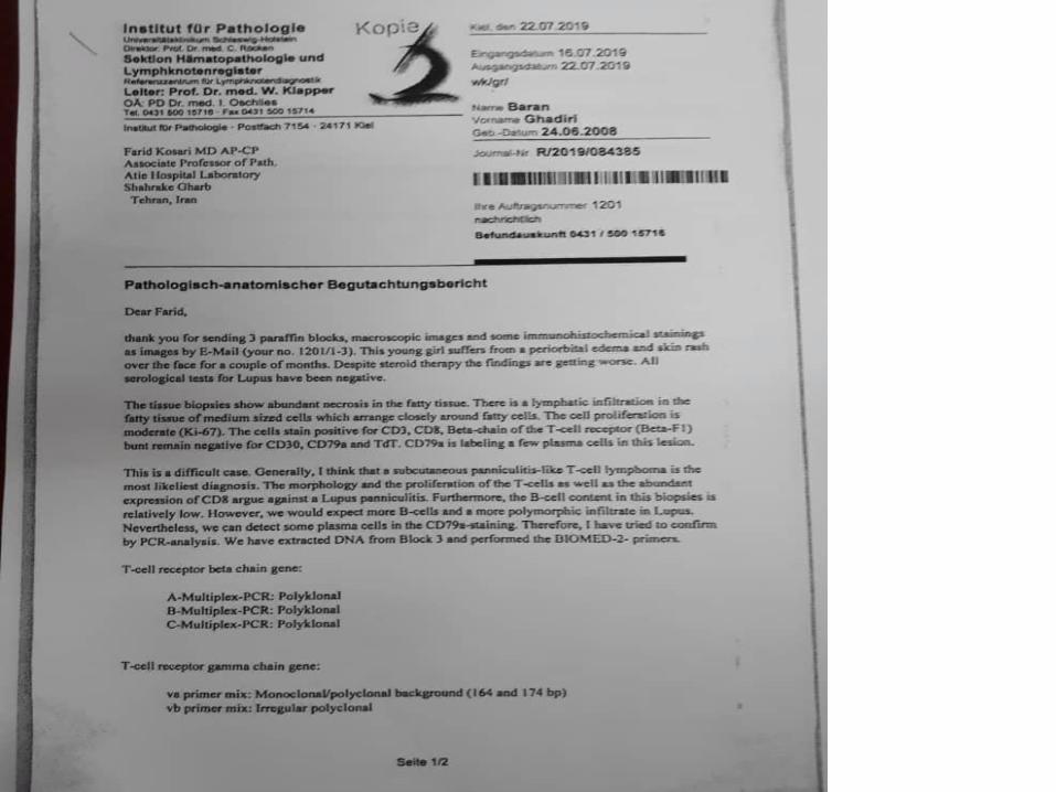
Finally, the patient underwent a biopsy


The general illness and the persistence of fever and the lack of response to broad-spectrum antibiotics and the negative result of cultures and infectious studies,


.
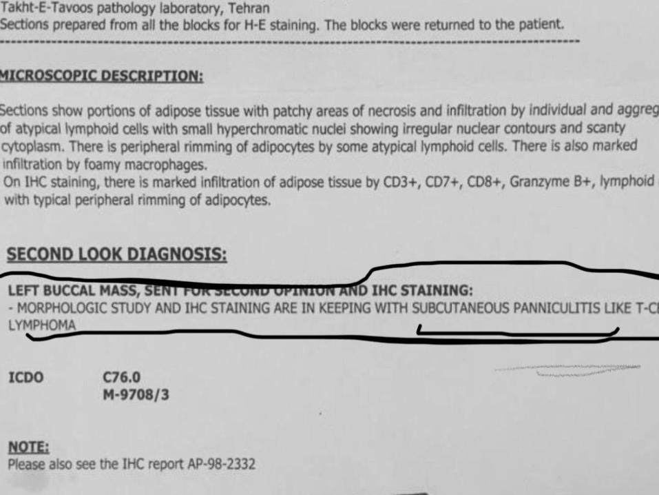
Biopsy showed lobules of fatty tissue with fibrinoid necrosis, small vessels were thrombosed and large vessels were surrounded by a number of lymphocytes, histiocytes and cell debris
and in IHC slides showed the most lymphoid cells were positive for CD3,CD7,CD19,CD20 and strongly positive for CD8 and Granzyme B and negative for CD4,CD34,CD56,c-kit, TdT and MPO , suggesting
panniculitis and necrotizing leukocytoclastic vasculitis


According to the positive family history of SLE in the family, sex of the child and lymphopenia, the study was performed for lupus and other rheumatologic diseases

In rheumatologic studies, the positive results included FANA with 1/320 titer and ACE 110 , but other autoantibodies and complement were normal.


she was treated empirically with intravenous pulse methylprednisolone at a dose of 30mg / kg for three consecutive days, which was temporarily discontinued for several days, but again with recurrent fever and exacerbation of swelling and ulcerative lesions.


according to all the studies and the approximate rejection of possible SLE , with the diagnosis of
Subcutanous Like T cell lymphoma,

At the start of chemotherapy, the patient developed
High grade fever,
pancytopenia,
high serum ferritin,
High triglycerides level,
Low serum fibrinogen


and again bone marrow examinations was performed, Hemophagocytosis images were observed, and hemophagocyticsyndrome (MAS)


Treatment with
pulse-methylprednisolone
IVIG

the chemotherapy according to BFM-NHL protocol for the patient began.
and fortunately improved after two weeks of clinical and laboratory symptoms and continued chemotherapy regimen. Following treatment, fever was discontinued and facial swelling declined markedly and continued with outpatient chemotherapy was discharged.

And in the fallow up, the patient was still afebrile and the swelling and ulcerative facial lesions improved completely.


In patients with suspected panniculitis, a deep biopsy to include the subcutaneous fat is fundamental.
Histopathologically, panniculitis can predominantly affect lobules or septa, with/without
vascular involvement.

the panniculitis can be classified into following main classes:
A . predominantly septal panniculitis with or without vasculitis,
B . predominantly lobular panniculitis with or without vasculitis and others

. A patchy lymphoplasmacytic infiltrate in the lobules of the subcutaneous fat along with lymphocytic nuclear dust is a clue for the histopathological diagnosis of LE panniculitis (LEP) .

Clinically, LEP and subcutaneous panniculitis-like T-cell lymphoma (SPTCL) are indistinguishable
in SPTCL, the presence of atypical CD8-positive T-lymphocytes and the absence of septal fibrosis, B-cell follicles, and plasma cells clinch the diagnosis.

SPTCL is an uncommon peripheral T-cell lymphoma accounting for less than 1 percent of all non-Hodgkin lymphoma subtypes. The exact incidence and variation in incidence with geography have not been well characterized.

Patients present at a median age of 36 years (range 9 to 79 years) with approximately 20 percent of patients presenting under the age of 20 . There appears to be a female predominance with a male:female ratio of 0.5.

Up to 20 percent of patients have at presentation an associated autoimmune disease such as systemic lupus erythematosus, juvenile idiopathic arthritis, Sjögren'ssyndrome, or type 1 diabetes mellitus.